Note: Descriptions are shown in the official language in which they were submitted.
CA 02620158 2008-02-22
WO 2007/024315 PCT/US2006/021361
LOW PROFILE ADAPTER FOR TRACHEAL TUBES
BACKGROUND
The present invention relates to medico-surgical tubes and assemblies. The
invention is
more particularly concerned with cuffed medico-surgical tubes, such as, for
example,
cuffed tracheal tubes in which an inflatable cuff seals the tube with a
patient's trachea.
For example, the conventional method of endotracheal intubation involves the
insertion of
a tubular device (an endotracheal tube) into the trachea. The endotracheal
tube continues
through the trachea and stops above the carina anterior to a position between
the 2nd and
4th thoracic vertebrate, allowing the gases to be directed through the tube
and into the
lungs.
A primary objective of the treatment is the mechanical ventilation of the
patient's lungs.
This function normally induced by the patient's breathing, is typically
impaired by the
disease or injury being treated. In order to create the air pressure necessary
to artificially
ventilate the lungs, the passageway around the tube must be sealed. This is
accomplished
by a balloon or inflatable cuff provided around the tube. With the tube in
place, the cuff is
located about 3 to 5 centimeters above the carina and within the tube-like
trachea.
The cuff is inflated to expand against the wall of the trachea and thereby to
prevent gases
that are being pumped into the lungs from simply backing up and escaping
around the
tube. This method of treatment has been quite successful for patients with
chronic and
acute respiratory disease. However, the method is not without complications.
Many intubated patients receiving the artificially induced ventilation may
develop
pneumonia. This pneumonia, known as Ventilator-Associated Pneumonia or VAP
results
from an infection of the lungs when pooled secretions, which have become
infectious, are
allowed, as a result of by-passing the upper airway, to enter the lungs.
Unfortunately, this
has been almost impossible to avoid with the prior endotracheal tubes.
The epigiottis is a form of valve that normally functions to selectively close
the entry to the
trachea and protect the airways (trachea and lungs) from secretions and
particulate
matter. The insertion of the endotracheal tube by-passes the protective system
of the
tracheo bronchial tree. Secretions that would normally be directed harmlessly
through the
digestive system follow the path of the tube, into the airway. The cuff above
the carina
blocks the downward flow of these secretions thus preventing them from
entering the
lungs. The secretions become pooled above the cuff and if unattended, rapidiy
grow
infectious bacteria that pose a serious risk to the patient.
I
CA 02620158 2008-02-22
WO 2007/024315 PCT/US2006/021361
The problem of these prior endotracheal tubes arises primarily upon cessation
of the
mechanical ventilation. The cuff is deflated in order to withdraw the
endotracheal tube
from the trachea. The infected fluid is now released to continue passage down
the trachea
and into the highly susceptible lungs where bronchitis or pneumonia develops
rapidly.
Whereas deflation of the cuff in the presence of the pooled secretions poses
the greatest
threat of lung infection, there is substantial risk also during treatment,
i.e. with the cuff
inflated. An incomplete seal caused by folds or creases in the cuff is not
unusual and fluids
can and do migrate along them into the lungs. Though the se folds or creases
are not
typically a problem to the ventilation treatment, because they do allow
microaspiration of
the pooled secretions past the cuff it is desirable to remove the secretions
and not allow
them to accumulate for very long.
Various proposals have been made previously for removing such secretions by
providing
a suction aperture above the cuff. In Heyden U.S. Pat. No. 4,607,635 there is
described a
tracheal tube having a channel which opens at various locations along its
length and
through which a suction catheter can be inserted to remove secretions at any
desired
location above the cuff. In Chester U.S. Pat. No. 4,305,392 there is described
a tracheal
tube with a bulbous chamber above the cuff in which secretions are collected
for removal
through a suction lumen extending through the wall of the tube. The problem
with both of
these tubes is that it is not possible to remove secretions that collect
immediately above
the cuff. This is because the cuff is conventionally attached to the wall of
the tube by
means of short collars at opposite ends of the cuff, which are adhered to the
tube and
extend above and below the cuff. The length of the collar above the cuff
defines the
closest distance by which the suction aperture can be spaced from the cuff,
because any
attempt to form a suction aperture through the collar would weaken the joint
between the
cuff and the tube which might possibly lead to leakage from the cuff. In
Porter U.S.
4,840,173 there is suggested a way in which secretions close to the cuff could
be
removed, by providing a suction tube which projects over the proximal collar
of the cuff.
This, however, would have the disadvantage of being relatively complex and
expensive to
make and may provide an undesirable projection from the side of the tube which
could
irritate the delicate surface of the trachea. There is also the risk that the
end of the suction
tube may damage the cuff or become blocked by the cuff. This risk can be
reduced by
making the upper end of the cuff more rigid, but this is a further
complication in the
construction of the tube.
2
CA 02620158 2008-02-22
WO 2007/024315 PCT/US2006/021361
What is needed is a means for removing secretions that collect immediately
above the cuff
that is simple, works with any cuff and does not negatively impact the trachea
or the
integrity of the cuff.
SUMMARY OF THE INVENTION
The present invention improves upon an apparatus for removing secretions by
providing a
tracheal tube with a suction lumen and an adapter to extend the suction
capabilities to a
point distal to a suction port into the catheter. The apparatus in one
embodiment is a
cannula having a length defined by a proximal end and a distal end. The
cannula has a
respiratory lumen extending along the length and at least one suction lumen
extending
1o along the length. The suction lumen is disposed adjacent to an exterior
wall of the
cannula. A suction source is connected to the proximal end. The adapter is at
least
partially inserted into the exterior wall of the cannula at a position along
the length of the
cannula. The adapter contains a plug and a duct. The duct has an inlet and an
outlet. The
outlet interconnects the duct with the suction source through the suction
lumen, whereas
the inlet extends the reach of the suction lumen through the duct. The plug is
adapted to
seai the suction lumen between the adapter itself and the distal end. An
inflatable cuff may
be provided on the cannula. The inflatable cuff would be shaped to block the
trachea
beneath a glottis of the patient. The inlet of the adapter would be oriented
such that
suction occurs proximal to the inflatable cuff and may be made to overlap the
collar portion
of the cuff itself.
In certain embodiments, the adapter is permanently affixed to the exterior
wall of the
cannula. A piercing member may be provided. The piercing member is for
piercing the
exterior wall of the cannula thereby connecting the duct with the suction
lumen. A plurality
of barbs may be provided on the adapter and/or plug portion of the adapter to
engage
walls internal to the cannula.
In many embodiments, the adapter may be configured into the shape of a partial
annu4us
or a ring. The adapter may be made of a flexible or inflexible material.
Moreover, a
plurality of inlets may be provided for accessing the duct.
Additional features may include providing the adapter with a first wall for
seating against
the exterior wall of the cannula and a second wall disposed a spaced distance
from the
first wall. The spaced distance would be the duct.
In another embodiment, the invention may simply be an adapter for insertion
into a
catheter. Such an adapter may have a first surface for seating upon an
exterior wall of the
catheter, a flange for contacting the exterior wall, a plug for engaging a
lumen within the
3
CA 02620158 2008-02-22
WO 2007/024315 PCT/US2006/021361
catheter, and a duct extending through the adapter. The duct would have an
inlet spaced
a distance from an outlet. The outlet wouid be adapted to communicate with the
lumen
through the insertion point, and the inlet would be adapted to effectively
extend the reach
of the lumen to a region external to the lumen. The plug would also serve to
seal the
lumen in one direction from an insertion point of the adapter into the
catheter. As such, it
may have a surface disposed opposite the flange. The plug working in
conjunction with the
surface of the flange would capture the exterior wall of the catheter and
secure the
adapter to the catheter.
Other objects, advantages and applications of the present invention will be
made clear by
1o the following detailed description of a preferred embodiment of the
invention and the
accompanying drawings wherein reference numerals refer to like or equivalent
structures.
BRIEF DESCRIPTION OF THE DRAWINGS
FIG. 1 is a front orthogonal view of one embodiment of a low profile adapter
in use on a
catheter in accordance with the present invention;
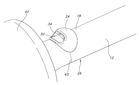
FIG. 1A is a magnification of the adapter portion of the FIG. 1 view;
FIG. 2 is a sectional view of the adapter portion of FIG. 1 shown in relation
to the lumens
within the catheter;
FIG. 3 is a front orthogonal view of the FIG. 1 low profile adapter;
FIG. 4 is a sectional view of the FIG. 1 low profile adapter; and
FIG. 5 is a sectional view of an alternative low profile adapter for use on a
catheter.
DETAILED DESCRIPTION
Reference will now be made to the drawings in which the various elements of
the present
invention will be given numeral designations and in which the invention will
be discussed
so as to enable one skilled in the art to make and use the invention. It is to
be understood
that the following description is only exemplary of the principles of the
present invention,
and should not be viewed as narrowing the pending claims. Those skilled in the
art will
appreciate that aspects of the various embodiments discussed may be
interchanged and
modified without departing from the scope and spirit of the invention.
Referring to FIGs. 1 and 1 a, a tracheal tube 10 in accordance with one
embodiment of the
present invention, is depicted. The tracheal tube 10 in the depicted
embodiment is a
4
CA 02620158 2008-02-22
WO 2007/024315 PCT/US2006/021361
multilumen cannula 12 having a length defined by a proximal end 20 and a
distal end 22.
An adapter 24 is partially inserted into an exterior wall 18 of the cannula 12
at a position
26 along the length. The tracheal tube 10 has an inflatable cuff 42 which is
shaped to seat
against and block a patient's trachea beneath the glottis. The cuff 42 is
attached to the
tube 10 in a conventional manner at cuff collars 43.
Referring now to the FIG. 2 cross-sectional view, it can be seen that the
cannula 12
contains at least one respiratory lumen 14 and at least one suction lumen 16
disposed
adjacent to an exterior wall 18 of the cannula 12. The respiratory lumen 14
and the
suction lumen 16 extend along the length of the cannula 12. A suction source
(not shown)
is connected to the proximal end 20 and used to create a vacuum or suction
within the
suction lumen 16. In this view, an inflation lumen 44 for inflating and
deflating the cuff 42 is
also shown.
Looking now to the adapter 24 on FIG. 2, it may be seen that it consists of
two major
portions, a plug 28 and a duct 30. The plug 28 is adapted to seai the suction
lumen 16
between the position 26 of the adapter 24 and the distal end 22, as seen on
FIGs. 1 and
1 a. The plug 28 may also assist in securing the adapter 24 to the cannula 12.
As may be
seen a flange 50 in conjunction with the plug 28 serves to capture the
exterior wall 18 of
the cannula 12 between surfaces 102 and 104. Moreover surface 106 of the plug
28 seats
against an interior wall 38 assisting this result. Barbs, prongs, or ridges
may be provided
upon any of these surfaces in order to better seat the adapter with the
cannula 12.
Additionally or alternatively, the adapter 24 may also be permanently affixed
to the exterior
wall 18 of the cannula 12 at surface 102 by solvent bonding, ultrasonic
welding, adhesive,
or other suitable methods intended to permanently affix the components to one
another.
Depending upon the needs of the apparatus, the adapter 24 may be flexible or
rigid and
may be made of a biocompatible polymer. For example, flexible materials could
be various
blends of PVC, Polyurethane, or Silicone whereas rigid materials could be
various blends
of Polycarbonate, ABS, Polyether Imide or a higher durometer PVC than the
flexible PVC.
Still looking at FIG. 2, it may be seen that the duct 30 of the adapter 24 has
an inlet 32 and
an outlet 34. The outlet 34 interconnects the duct 30 with the suction source
through the
suction lumen 16. The inlet 32 effectively extends the reach of the suction
lumen 16
through the duct 3o a distance from the cannula exterior wall 18. As may be
seen in FIGs.
1 and 1 a, the inlet 32 may be oriented such that suction is directed toward a
region
proximal to the inflatable cuff 42. In this instance, the overall design
intent is to overlap the
5
CA 02620158 2008-02-22
WO 2007/024315 PCT/US2006/021361
cuff collar 43 thereby extending the suction closer to the cuff 42 while
keeping the adapter
inlet 32 close to the tube thereby minimizing trauma to the trachea and vocal
cord during
intubation.
Looking at the adapter embodiment depicted in FIG. 3 and the cross-sectional
view
depicted in FIG. 4, it is evident that the adapter 24 may be configured as a
partial annulus
or partial ring. In this embodiment, the adapter 24 has an exterior wall 40
disposed a
spaced distance from the flange 50. The spaced distance forms the volume of
the duct
30. Due to the curvature and shape of the adapter 24, the volume of the duct
30 may be
maximized maintaining a cross sectional area greater than or equal to the
suction
1o lumen16 while also maintaining a low profile. For example, in one
embodiment, the
exterior wal{ 40 of the adapter 24 is envisioned to extend outward from the
exterior wall 18
from about 0.5 mm to about 4.0 mm, for example in one embodiment it is
envisioned that
the adapter will extend about 2.5 mm from the exterior wall 18 of the adapter
24 and
should not negatively affect the trachea.
Looking now at an alternative embodiment of the adapter 24 as depicted in the
cross-
sectional view of FIG. 5, it may be seen that the leading edge 52 of the plug
28 as
depicted in FIGs. 3 and 4 has been substituted with a piercing member 52. The
piercing
member 52 may be made so that it is capable of piercing the exterior wall 18
of the
cannula 12 at the position 26 thereby connecting the duct 30 to the suction
lumen 16
without requiring a preformed hole in the exterior wall 18 of the cannula 12.
This step may
be performed during the manufacturing process, that is, at the assembly of the
tracheal
tube 10 prior to affixing the adapter 24 to the cannula 12. In other
embodiments, not
shown, the inlet 32 may be oriented on the adapter 24 in any manner desired so
that
suction is directed in accordance with the needs of the surgical team.
Although the above embodiments have been described primarily as being
incorporated
onto endotracheal tubes, it may be incorporated into other tracheal devices
such as
tracheostomy tubes, as well as any other application wherein it is desirable
to extend the
reach of suctioning away from the passage within the tube itself. As used
herein and in the
claims, the term "comprising" is inclusive or open-ended and does not exclude
additional
unrecited elements, compositional components, or method steps.
While various patents have been incorporated herein by reference, to the
extent there is
any inconsistency between incorporated material and that of the written
specification, the
written specification shall control. In addition, while the invention has been
described in
detail with respect to specific embodiments thereof, it will be apparent to
those skilled in
6
CA 02620158 2008-02-22
WO 2007/024315 PCT/US2006/021361
the art that various alterations, modifications and other changes may be made
to the
invention without departing from the spirit and scope of the present
invention. It is
therefore intended that the claims cover all such modifications, alterations
and other
changes encompassed by the appended claims.
7