Note: Descriptions are shown in the official language in which they were submitted.
CA 02621153 2014-06-13
,
METHOD OF REPAIRING MENISCAL TEARS USING A COLLAGEN
MEMBRANE MATERIAL
BACKGROUND OF THE INVENTION
Field of the Invention
[002] The present invention relates to the field of repairing meniscal
tears.
Description of the Background Art
[003] Meniscal tears in a joint of a subject, e.g., in a knee, are frequent
injuries. In the past, a torn meniscus often was partially or completely
removed. In recent years, techniques have been developed for repairing
meniscal tears, including the use of arthroscopically placed tacks or suturing
the torn edges.
[004] There remains a need in the art for new methods of repairing meniscal
tears.
SUMMARY OF THE INVENTION
[005] In accordance with the present invention, a method of repairing a
meniscal tear of a subject comprises providing a sheet of collagen membrane
material, the sheet having on one side thereof a smooth barrier face which
inhibits cell adhesion thereon and inhibits passage of cells therethrough, the
sheet having a fibrous face opposite the smooth barrier face, the fibrous face
allowing cell growth thereon, the collagen of said sheet being predominantly
collagen I. The sheet of collagen membrane material is fixed over the meniscal
tear so that the fibrous face is oriented toward the meniscal tear.
- 1 -
CA 02621153 2013-06-12
A =
[005a] According to one aspect of the present invention, there is provided
the use of a first and a second sheet of collagen membrane material as a
scaffold for repairing a meniscal tear of a meniscus in a subject, said
scaffold arranged such that said first sheet of collagen material is for
fixation over one side of said meniscal tear and said second sheet of
collagen is for fixation over an opposite side of said meniscal tear, thereby
forming a sandwich to provide a barrier layer against ingrowth of
connective tissue into said meniscus, wherein:
= each of said sheets of collagen has on one side thereof a smooth
barrier face which inhibits cell adhesion thereon and inhibits passage of
cells therethrough
= each of said sheets of collagen has a fibrous face opposite to said
smooth barrier surface, said fibrous face allowing cell growth thereon,
= said collagen is greater than 60% by weight collagen I/membrane,
and
= said sheets of collagen are for fixation on opposite sides of said
meniscal tear with said sheets in opposed orientation, with said fibrous
face of each of said sheet of collagen oriented toward said meniscal tear.
BRIEF DESCRIPTION OF THE DRAWINGS
[006] Fig. 1 is a side elevation schematic view showing covering of
one side
of a meniscus tear in accordance with one embodiment of the invention.
- la -
CA 02621153 2008-02-29
WO 2007/028078 PCT/US2006/034329
[007] Fig. 2 is a schematic sectional view showing both sides of a meniscal
tear treated according to another embodiment.
[008] Fig. 3 is a side elevation schematic view showing a collagen
membrane for use in accordance with the present invention with adjacent
synovial cells.
DETAILED DESCRIPTION OF THE INVENTION
[009] Cells that contribute to the reparative process of a torn meniscus
are
from adjacent synovial tissue. In addition to contributing to the reparative
process in a torn meniscus, synovial cells have the capability to degrade
connective tissues and to contract. For example, synovial tissue can break
down typical collagen I scaffolds.
[0010] It has surprisingly been discovered that the predominantly collagen I
membrane of the present invention is able to maintain its integrity when in
contact with synovial tissue, and also serve as a scaffold into which synovial
cells can migrate to facilitate healing of a meniscal tear.
[0011] A sheet of collagen membrane material utilized in accordance with the
present invention has on one side thereof a smooth barrier face which inhibits
cell adhesion thereon and inhibits passage of cells therethrough. The collagen
sheet has a fibrous face opposite the barrier face, the fibrous face allowing
cell
growth thereon.
[0012] As noted above, the collagen of a membrane utilized in accordance
with the present invention is predominantly collagen I, i.e., greater than 50%
collagen I by weight. In preferred embodiments, the collagen I content of a
membrane sheet utilized in accordance with the present invention may be
greater than 60% by weight, greater than 70% by weight, greater than 80% by
weight or greater than 90% by weight. In accordance with one embodiment, the
collagen of a membrane sheet utilized in accordance with the present invention
is approximately 95% by weight collagen I. The collagen of such a membrane
may comprise approximately 5% by weight collagen III.
[0013] In preferred embodiments, the collagen utilized in the present
invention is of porcine or bovine origin. In particularly preferred
embodiments,
- 2 -
CA 02621153 2013-06-12
A
the sheet of collagen material utilized in the present invention is formed
from a
naturally occurring membrane of porcine or bovine origin, preferably from
calves or piglets. A preferred source is naturally occurring single-layered
sheets
of peritoneum membrane, most preferably from piglets. Peritoneum membranes
from young pigs aged 6-7 weeks old (weighing 60-80 kg) are especially
preferred. One such material is described in U.S. Patent No. 5,837,278.
[0014] The dry thickness of a membrane for use in the present invention may
be between about 0.1-5.0 mm, preferably between about 0.1-1.0 mm, or about
0.5 mm, but can be influenced by swelling of the material when exposed to
moisture.
[0015] A sheet of collagen membrane material utilized in accordance with the
present invention is fixed over a meniscal tear so that the fibrous face of
the
membrane is oriented toward the meniscal tear. The sheet may be fixed by any
suitable means, including sutures, a physiologically acceptable adhesive
(e.g.,
fibrin glue), or a combination thereof. The membrane preferably completely
covers at least one side of the tear, and the fibrous face preferably contacts
the
tear.
[0016] A sheet of collagen membrane material utilized in accordance with the
present invention may be fixed over one side of a meniscal tear, or
additionally
a second sheet of collagen membrane material may be fixed on an opposite side
of a meniscal tear, also with the fibrous face oriented toward the tear, so
that the
meniscal tear is sandwiched between two sheets of collagen membrane
material.
[0017] In accordance with one embodiment, when a single sheet of collagen
membrane material is fixed over one side of a meniscal tear, the fibrous face
of
the membrane is contact with synovial fluid in the subject which migrates
through the tear into the fibrous face.
[0018] One suitable membrane for use in accordance with the present
invention is ChondroGide , manufactured by Ed. Geistlich Soehne AG fur
Chemische Industrie, the assignee of the present invention.
- 3 -
CA 02621153 2008-02-29
WO 2007/028078 PCT/US2006/034329
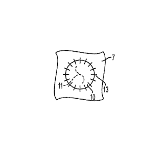
[0019] As shown in Fig. 1, the membrane material 10 may be fixed over a
meniscus tear 11 in meniscus 7 by adhesive or sutures 13 attached to meniscus
7. In accordance with one embodiment of the invention as shown in Fig. 2,
during surgery in which a meniscus tear 11 in meniscus 7 is treated, separate
sheets of collagen membrane material 10 are fixed over the meniscus tear 11 so
as to cover the tear on opposite sides thereof, with the tear being sandwiched
between the membrane material 10, to thereby provide a barrier against
ingrowth of connective tissue into meniscus tissue 7 following the surgery.
The
sheet of collagen membrane material preferably is fixed over the area to be
treated, for example, by adhesive bonding of the sheet, utilizing an organic
glue,
such as fibrin glue, or by sutures 13, or a combination thereof, or any other
suitable method.
[0020] As noted above, the collagen membrane material 10 is comprised of at
least one barrier layer having at least one smooth face 16 so as to inhibit
cell
adhesion thereon and act as a barrier to prevent passage of cells
therethrough.
See Fig. 3. The membrane 10 further has a fibrous face 18 opposite the smooth
face 16, the fibrous face allowing cell growth thereon. Synovial cells 20 may
contact the fibrous face 18 and migrate into the membrane to assist in healing
of
the tear.
[0021] In one embodiment, a collagen membrane material is utilized,
wherein the membrane and/or the fibrous face are impregnated with
chondrocytes, synovial fibroblast-like cells, mesenchymal stem cells, one or
more glycosaminoglycans, and/or one or more growth factors. Examples of
suitable glycosaminoglycans include hyaluronic acid, chondroitin 6-sulphate,
keratin sulphate, dermatan sulphate or the like. Suitable growth factors
include, but are not limited to, those which are described as follows.
Transforming growth factor-beta (TGF-beta) increases the proteoglycan
synthesis of fibrochondrocytes isolated from different sections of the menisci
in
a dose dependent manner. Human platelet-derived growth factor (PDGF-AB),
hepatocyte growth factor (HGF) and bone morphogenic protein-2 (BMP-2)
increase DNA synthesis in meniscal cells. In addition, BMP-2, insulin-like
growth factor-1 (IGF-1), and epidermal growth factor (EGF) stimulate migration
- 4 -
CA 02621153 2008-02-29
WO 2007/028078 PCT/US2006/034329
of bovine fibrochondrocytes from the different parts of the menisci. Also
suitable is osteogenic protein-1 (0P-1).
[0022] The present invention provides a smooth barrier face 16 in membrane
which protects the surgical site from ingrowth of unwanted cells during the
healing process, and a fibrous face 18 for promoting growth of reparative
cells
adjacent the tear. The collagen membrane material 10 is gradually resorbed
into the patient's body, avoiding any necessity of having to surgically remove
the membrane after healing.
[0023] While the invention has been described in detail, it is not intended
that the description and accompanying drawings be interpreted in a limiting
sense.
[0024] The invention is further illustrated by the following example, which is
not intended to be limiting.
Example 1
APPLICATION OF CHONDROGIDE MEMBRANE FOR THE TREATMENT OF
MENIS CAL TEARS:
IN VITRO EXPERIMENTS
A ChondroGide membrane may be applied to a torn meniscus to
facilitate its repair. The cells that contribute to the reparative process are
from
the adjacent synovial tissue. In addition to contributing to the reparative
process in a torn meniscus, these synovial cells have the capability to
degrade
connective tissues and to contract. The ChondroGide membrane will: (1)
guide synovial cells to the tear in the meniscus by serving as a scaffold on
which the cells can migrate, and (2) contain the cells in the defect during
the
reparative process. The ChondroGide membrane is able to maintain its
integrity when in contact with synovial tissue, and also serve as a scaffold
into
which the synovial cells can migrate. In vitro data demonstrates that, while
synovial tissue can break down bovine type I collagen scaffolds, it does not
degrade ChondroGide , and the ChondroGide membrane retains its size and
shape despite the contraction of synovium. Moreover, cells from synovium can
migrate into ChondroGide .
In the experimental work, samples of caprine synovium, 8 mm in
diameter, were placed on ChondroGide membranes and on a typical bovine
type I collagen scaffold. After 7 days in vitro, the synovium specimens
cultured
on the ChondroGide and directly on the tissue culture dish contracted to
about
- 5 -
CA 02621153 2008-02-29
WO 2007/028078 PCT/US2006/034329
1/2 the original size. Of importance was the finding that the ChondroGide
retained its original size and shape, and was not degraded by the synovium. As
a control, similar synovial tissue samples were cultured on bovine type I
collagen scaffolds. The synovial samples in these cultures also contracted.
The
data show that after only 24 hours the synovial cells digested the prior art
collagen I scaffold, and as a result some of the synovium samples were
displaced from the scaffold. A similar degradation of the bovine type I
collagen
scaffold was seen after 48 hours in culture.
Histology demonstrated that cells from synovium can migrate into
ChondroGide membranes. After 21 days in culture, cells from the synovial
tissue samples migrated from the synovium into the ChondroGide . Synovial
cells could be found throughout the ChondroGide membrane.
- 6 -