Note: Descriptions are shown in the official language in which they were submitted.
CA 02621386 2008-03-05
WO 2007/028243 PCT/CA2006/001467
- '~ -
Title: AN EMBEDDING METHOD AND APPARATUS FOR THE
PREPARATION OF FROZEN SECTION TISSUE
Field
[0001] Various aspects of the embodiments described herein relate to
the field of tissue sample preparation. More particularly, the embodiments
described herein relate to a method and apparatus for the preparation of
frozen section tissue samples.
Backaround
[0002] In one well-known frozen section procedure, a cold chuck is
retrieved from a cryostat (at approximately -20 degrees Celsius). A small
amount of a viscous embedding material, which is also known as a tissue
freezing compound, is placed on the generally planar surface of the chuck,
which may be textured, and the tissue sample is then placed into the
embedding material. The embedding material may be OCT (Optimum Cutting
Temperature); e.g. Tissue-TekTM provided by Sakura Finetek. The
combination of OCT and the tissue sample is referred to herein as a tissue
specimen. It is generally understood that the tissue specimen is supported on
a platform (such as a chuck) and/or contained in a receptacle (such as a
mold). The chuck and tissue specimen are then placed back into the cryostat
chamber and cooled until the tissue specimen is frozen. During this freezing
process, a heat sink, also known as a weighted heat extractor, may be placed
onto the tissue specimen to flatten the tissue sample and accelerate the
freezing process. The frozen tissue sample is then sectioned using a
microtome/cryostat. A section is typically several micrometers thick. The
sections are then processed by methods that are well known to those skilled
in the art. A medical practitioner then evaluates the processed sections.
[0003] The ability to produce full face microscopic sections of the true
deep margin of the excised tissue relies on three important steps in the
frozen
section process. First of all, the tissue must be laid down so that the deep
margin of the tissue lies in the same plane. Secondly, this planar orientation
must be maintained during freezing. Finally, the frozen tissue sample needs to
CA 02621386 2008-03-05
WO 2007/028243 PCT/CA2006/001467
-2-
be oriented parallel to the sectioning plane of the microtome, A breach of any
of these steps can result in excessive microtome "trimming in" before a full
face section is obtained, potentially exposing a portion of a tumor which did
not extend to the true deep margin.
Summary
[0004] In one aspect, at least one embodiment described herein
provides an apparatus for preparing a frozen tissue specimen from an excised
tissue sample. The apparatus comprises a freezing box including a first base,
a lid that covers the first base, and a freezing chamber defined by the first
base and the lid, the freezing chamber being adapted for receiving a freezing
agent and the freezing box being made from a thermal insulating material for
maintaining a reduced temperature environment in the freezing chamber for
freezing the excised tissue sample. The apparatus further comprises a
freezing platform having a flat freezing surface. The apparatus also comprises
at least one sample container configured for receiving, in use, the excised
tissue sample and the embedding material, the at least one sample container
including a well having a flat second base, walls and flat flanges connected
at
the upper portion of the walls to define a flat surface; and at least one
chuck,
the at least one chuck having a generally planar surface for placement on the
flat flanges of the sample container.
[0005] The sample container can be made from plastic material that is
at least semi-transparent to enable visual confirmation of the flattening of
the
excised tissue sample and the freezing of the excised tissue sample and the
embedding material.
[0006] At least one of the flanges of the at least one sample container
can be longer than the other flanges of the at least one sample container.
[0007] At least one of the walls of the well of the at least one sample
container can be beveled.
[0008] The freezing agent can comprise dry ice and the reduced
temperature environment is at less than -70 degrees Celsius.
CA 02621386 2008-03-05
WO 2007/028243 PCT/CA2006/001467
-3-
[0009] The freezing platform and the freezing chamber can be sized to
provide gaps between the freezing chamber and the freezing platform for
receiving at least one of: one or more pieces of dry ice and additional
insulating material.
5[0010] The freezing agent can comprise one of compressed carbon
dioxide gas, compressed liquid nitrogen, and a mechanical refrigeration
compressor.
[0011] The thermal insulating material can comprise polystyrene.
[0012] The freezing platform can comprise anodized aluminum.
[0013] The freezing platform can comprise one of copper, stainless
steel, aluminum and alloys thereof.
[0014] The flat freezing surface can comprise at least one of a bare
metal, anodized, glazed, painted, and ceramic surface.
[0015] The at least one chuck can comprise a post mounted opposite
the generally planar surface and the freezing platform comprises at least one
hole sized to receive the post.
[0016] The holes In the freezing platform can extend from the top of the
freezing platform to the bottom of the freezing platform.
[0017] The first base of the freezing box can comprise shoulders for
receiving and providing support for the freezing platform, the shoulders
having
a height for placing the upper surface of the freezing platform approximately
level with the upper surface of the first base.
[0018] The first base of the freezing box can comprise a first securing
member and the lid comprises a complementary second securing member
sized for releasably engaging the first securing member when the lid is placed
on the first base.
[0019] In another aspect, at least one embodiment described herein
provides a method for preparing a frozen tissue specimen from an excised
tissue sample. The method comprises:
CA 02621386 2008-03-05
WO 2007/028243 PCT/CA2006/001467
-4-
a) placing the excised tissue sample into a sample container
having a well with a lower flat base, and upper flat flanges;
b) flattening the excised tissue sample along the flat base of
the well of the sample container;
c) adding a sufficient amount of embedding material to the
well of the sample container after the excised tissue sample has been
flattened, wherein a top portion of the embedding material is above the
flanges of the sample container;
d) placing the flat base of the sample container on a flat
freezing surface of a freezing platform, the freezing platform being
maintained
at a reduced temperature for freezing the excised tissue sample and the
embedding material; and
e) placing a chuck on the sample container for a first time
period, the chuck having a generally planar surface that is placed on the flat
flanges of the sample container to maintain a flat contact surface therewith.
[0020] The method may further comprise:
f) inverting the chuck and placing the inverted chuck, tissue
specimen and sample container on the freezing platform for a second time
period; and
g) removing the sample container from the chuck and tissue
specimen after the excised tissue sample and the embedding material has
frozen.
[0021] The chuck can comprise a post and the freezing platform can
comprise a hole sized to receive the post, and the inverting step can further
comprise placing the post into the hole.
[0022] The method can further comprise maintaining the sample
container and the chuck in the range of 10 to 25 degrees Celsius prior to
freezing.
CA 02621386 2008-03-05
WO 2007/028243 PCT/CA2006/001467
-5-
[0023] The method can comprise placing the freezing platform in a
freezing chamber of a freezing box, and using a freezing agent in the freezing
chamber for maintaining the freezing platform at a reduced temperature.
[0024] The method can further comprise using dry ice as the freezing
agent to maintain the reduced temperature environment at less than -70
degrees Celsius.
[0025] The method can further comprise using one of compressed
carbon dioxide gas, compressed liquid nitrogen, and a mechanical
refrigeration compressor as the freezing agent.
[0026] The sample container can be at least semi-transparent and the
method can further comprise visually confirming the flattening of the excised
tissue sample and the freezing of the excised tissue sample and the
embedding material.
[0027] In another aspect, at least one embodiment described herein
provides a sample container for use in preparing a frozen tissue specimen
from an excised tissue sample. The sample container comprises a flat base
and walls extending upwardly from the flat base thereby defining a well for
receiving, in use, the excised tissue sample and an embedding material; and,
flat flanges connected at upper portions of the walls to define an upper flat
surface for the sample container and accommodate any overflow of the
embedding material.
[0028] The sample container can be made from material that is at least
semi-transparent to enable visual confirmation of flattening of the excised
tissue sample and freezing of the tissue sample and the embedding material.
[0029] The material for the sample container can be a plastic that is
suitable for withstanding temperatures in the range of about room temperature
to -80 degrees Celsius.
[0030] At least one of the flanges of the sample container can be longer
than the other flanges.
CA 02621386 2008-03-05
WO 2007/028243 PCT/CA2006/001467
-6-
[0031] At least one of the walls of the sample container can be beveled.
Brief description of the drawinas
[0032] For a better understanding of various embodiments described
herein and to show more clearly how they may be carried into effect,
reference will now be made, by way of example only, to the accompanying
drawings which show at least one exemplary embodiment and in which:
FIG. 1 is a diagram of an exemplary embodiment of a freezing
apparatus that can be used to prepare frozen tissue specimens;
FIG. 2A is a cross-sectional side view of a freezing box and a
freezing platform that can be used as part of the freezing apparatus;
FIG. 2B is a top view of the freezing box and freezing platform of
FIG. 2A;
FIG. 3A is a top view of a freezing platform;
FIG. 3B is a side view of the freezing platform of FIG. 3A;
FIG. 4A is a top view of an exemplary embodiment of a sample
container of the freezing apparatus of FIG. 1;
FIG. 4B is a cross-sectional side view of the sample container of
FIG. 4A;
FIG. 4C is a cross-sectional front view of the sample container
of FIG. 4A;
FIG. 5 is an exemplary embodiment of a flowchart of a frozen
tissue specimen preparation method; and,
FIGS. 6A-6D are illustrations of various steps of the frozen
tissue specimen preparation method.
Detailed description
[0033] It will be appreciated that for simplicity and clarity of illustration,
where considered appropriate, reference numerals may be repeated among
the figures to indicate corresponding or analogous elements or steps. In
CA 02621386 2008-03-05
WO 2007/028243 PCT/CA2006/001467
-7-
addition, numerous specific details are set forth in order to provide a
thorough
understanding of the various embodiments described herein. Some features
in the figures have not been drawn to scale. Further, it should be understood
by those of ordinary skill in the art that the embodiments described herein
may be practiced without these specific details. In other instances, well-
known
methods, procedures and components have not been described in detail so
as not to obscure the embodiments described herein. Furthermore, this
description should not be considered as limiting the scope of the
embodiments described herein, but rather as merely describing the
implementation of the various embodiments described herein.
[00341 An apparatus and method for preparing a frozen tissue
specimen that can be sectioned using a suitable cutting device, such as the
microtome in a cryostat, is described herein. A portion of the method involves
inserting a tissue sample into a sample container, also known as a platform
mold, adding an embedding medium and then joining a chuck to the sample
container and cooling the contents of the sample container to produce the
frozen tissue specimen. It should be understood that the-embedding-medium
is analogous to a tissue freezing compound. The cooling step can be
performed using a cooling box that has a cooling chamber and a freezing
platform. The cooling box is kept outside of the cryostat chamber. In some
embodiments, the cooling chamber may include dry ice to cool the freezing
chamber, which allows the cooling box to be self-contained, portable and
have small dimensions. The frozen tissue specimen preparation method is
rapid, simple to perform and highly reliable for processing various tissue
samples including multiple small tissue fragments, needle biopsies, irregular
surfaces and Mohs samples.
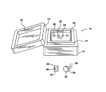
[00351 Referring now to FIG. 1, shown therein is a diagram of an
exemplary embodiment of a freezing apparatus that can be used to prepare
frozen tissue specimens. The freezing apparatus includes a freezing box 10
having a base 12 and a lid 14. The base 12 includes a first securing member
16 that is sized for releasably engaging a complementary second securing
CA 02621386 2008-03-05
= WO 2007/028243 PCT/CA2006/001467
-8-
member 18 that is located on the underside of the lid 14. The freezing box 10
further includes a freezing chamber 20. A freezing agent (not shown) is
placed within the freezing chamber 20 to maintain the freezing chamber 20 at
a temperature that is suitable for preparing frozen tissue specimens. The base
12 and the lid 14 are made from a suitable insulating material to maintain the
interior of the freezing box 10 at a suitable cold temperature, which is
described in more detail below.
[0036] The freezing apparatus further includes a freezing platform 22
that is placed within the freezing chamber 20. Once the freezing platform 22
has been cooled, it acts as a heat sink to cool any object that is placed in
contact with it. The freezing platform 22 includes a flat freezing surface 24,
and several holes 26 (only one of which is labeled for simplicity). In some
embodiments, the freezing platform 22 is smaller than the freezing chamber
so that there are gaps when the freezing platform 22 is placed within the
15 freezing chamber 20. In some embodiments, the freezing platform 22 is sized
so that the gaps are large enough to accommodate the freezing agent or
additional insulating material. The freezing platform 20 can also be sized so
that some edges of the freezing platform 22 contact corresponding sides of
the freezing chamber 20. This anchors the freezing platform 22 and also
20 ensures that the insulating walls of the freezing chamber 20 are in close
contact with at least some sides of the freezing platform 22 so that the
freezing plafform 22 is maintained at a cold temperature.
[0037] The freezing apparatus further includes a sample container 28
for holding a tissue sample and embedding material. It should be understood
that the sample container 28 can also be considered to be a platform mold.
The sample container 28 includes a well 30 with a flat base for receiving the
tissue sample and the embedding material, and flanges 32. The flat bottom of
the well 30 acts as a conforming plane for tissue flattening. The flanges 32
accommodate any overflow of the embedding material. A sufficient amount of
the embedding material is added to the well 30 so that a portion of the
embedding material lies above the plane defined by the flanges 32; this
CA 02621386 2008-03-05
WO 2007/028243 PCT/CA2006/001467
-9-
portion of the embedding material engages chuck grooves (described below)
for forming a better bond during freezing. The flanges 32 also provide ease of
handling during the preparation of the frozen tissue specimen. The flanges 32
also ensure that the conforming plane and a chuck surface are parallel to
each other. The flanges 32 do not have to be of equal size.
[0038] The freezing apparatus further includes a chuck 34 having a
disc with a generally planar surface 36 and a post 38 mounted opposite the
textured surface 36. The chuck 34 does not have to have a disc shape and
can generally have any shaped planar configuration (i.e. disc, square,
rectangle, elliptical, and the like) at its working end (i.e. the portion of
the
chuck 34 that makes contact with the embedding material and the sample
container). Furthermore, it may be possible to use a chuck that does not have
a post. The generafiy planar surface 36 includes a pattern of small grooves or
cross-hatches that provide a "gripping surface" for the embedding material
when it freezes thereby facilitating a bond between the embedding medium in
the sample container 28 and the chuck 34. The textured surface of the chuck
34 can be considered to be generally oriented in a plane; hence surface 36 is
referred to as being generally planar. The chuck 34 is used with the sample
container 28 for preparing the frozen tissue specimen. The generally planar
surface 36 of the chuck 34, when parallel to the flanges 32, ensures that the
sample tissue and the surface 36 are generally parallel to each other. This is
accomplished by placing the chuck 34 on top of the sample container 28, after
the sample tissue and embedding material have been added to the sample
container 28, so that the flanges 32 lie flat against the generally planar
surface 36 of the chuck 34. Further, the holes 26 on the freezing platform 22
are sized slightly larger than the post 38 of the chuck 34 so that the chuck
34
can be inverted and inserted at the top of the freezing platform 22 to
increase
the contact area between the chuck 34 and the freezing platform to further
accelerate the freezing process.
[0039] Although only one sample container 28 and one chuck 34 are
shown in FIG. 1, it should be understood that there can be several sets of
CA 02621386 2008-03-05
WO 2007/028243 PCT/CA2006/001467
-10-
sample containers 28 and chucks 34 so that more than one frozen tissue
specimen can be prepared at the same time. For example, with the freezing
platform 22, up to four frozen tissue specimens may be prepared at the same
time. However, there may be other embodiments with a larger or smaller
number of holes 26 in the freezing platform 24 and hence more or fewer
frozen tissue specimens can be prepared simultaneously. Accordingly, the
size of the freezing platform 22 and the freezing box 10 can be changed to
accommodate the preparation of a greater or fewer number of frozen tissue
specimens.
[0040] Referring now to FIGS. 2A and 2B, shown therein are a cross-
sectional side view and a top view, respectively, of the freezing platform 22
and an alternative embodiment of the freezing box 10'. The base 12 of the
freezing box 10' includes shoulders 12s to support the freezing platform 22
and provide a gap 20b underneath the freezing platform 22. The height of the
shoulders 12s can be selected so that the top surface of the freezing platform
22 is approximately level with the top of the base 12 when the lid 14' is off.
This makes it easier to work with the freezing platform 22 while preparing the
frozen tissue specimen. The width of the shoulders 12s is chosen to provide
enough support for the freezing platform 22. In some embodiments with larger
freezing platforms, there may be additional support in the base 12 of the
freezing box 10'. The additional support may be located centrally.
[00411 In one implementation, the base 12' and lid 14 may be made
from polystyrene, such as StyrofoamTM. In other implementations, a hard shell
insulated material may be used rather than polystyrene. In other
embodiments, other materials that can provide suitable insulation can also be
used.
[0042] Once the freezing platform 22 is placed within the freezing box
12' and the lid 14' is placed on the base 12', there are gaps 20a, 20b, 20c
and
20d that can be sized to accommodate the freezing agent. In some
embodiments, the freezing agent can be dry ice. In this case, the freezing
platform 22 and the freezing box 12' can be sized so that the gaps 20a, 20b,
CA 02621386 2008-03-05
WO 2007/028243 PCT/CA2006/001467
-11-
20c and 20d can accommodate one or more slabs of dry ice, which may be in
the order of 4 cm thick. Ideally, the gaps are sized just slightly larger than
the
slabs of dry ice to allow the slabs of dry ice to be slid into and out of
position
as needed and to more efficiently conduct heat away from the freezing
platform 22. In some embodiments, the gaps 20c and 20d may be about 3 cm
wide. Prior to placing the freezing platform 22 into the freezing chamber 20,
an appropriate number of slabs of dry ice are placed in the gap 20b. In other
instances, the slabs of dry ice can be broken into smaller chunks, which are
then placed within one or more of the gaps 20a-20d. Alternatively, chips of
dry
ice can also be used. In either case, the size of the gaps can be reduced. The
freezing platform 22 is then placed within the freezing chamber 20. Other
pieces of dry ice can then be placed in the gaps 20c and 20d. The gap 20a
provides space for pieces of dry ice and/or sample containers and inverted
chucks (an inverted chuck has its post engaging one of the holes 26). For
instance, pieces of dry ice may be placed in the gap 20a when the freezing
platform 22 is being cooled in preparation for freezing a tissue sample. Once
the freezing platform 22 has been cooled, then the pieces of dry ice in the
gap
20a can be removed and the sample container 28 can be placed on the
freezing platform 22. In one implementation, the gap 20a can have a height of
about 3 cm.
[0043] When dry ice is used as the freezing agent, the interior of the
freezing box 10 is maintained at a temperature of about -78 degrees Celsius.
This is in contrast to the interior of the cryostat, which is typically used
for
freezing the tissue specimen in many conventionally used methods. The
interior of the cryostat is maintained at about -22 degrees Celsius. The lower
temperature of the freezing box 10 accelerates the freezing process and
allows frozen tissue specimens to be prepared much faster than if the cryostat
or some other device was used that does not operate at such a low
temperature. The amount of dry ice that is required depends on several
variables including the degree of insulation provided by the freezing box 10,
the amount of time that the freezing box 10 remains open (i.e. the lid 14 is
removed), the amount of time spent pre-cooling the freezing plate 22, etc. In
CA 02621386 2008-03-05
WO 2007/028243 PCT/CA2006/001467
-12-
some embodiments, 2 kg of dry ice is sufficient for an entire day of operation
of the freezing box 10.
[0044] Freezing is complete when the OCT reaches a core temperature
of about -18 degrees Celsius, at which point the chuck and frozen tissue
specimen can be transferred to the cryostat for sectioning. If the chuck and
frozen tissue specimen are too cold, then they can be warmed up in the
cryostat to the proper temperature required for sectioning. Using a lower
temperature in the freezing box 10 virtually eliminates the visible ice
crystal
artifact. However, the sample container 28 needs to be made of a suitable
material that can withstand the low temperature.
[0045) Cooling with dry ice, rather than other freezing agents, can be
done for safety reasons, the low temperature provided by dry ice and the easy
availability of dry ice. Housing the freezing platform 22 and dry ice in a
fitted
insulating container renders the freezing box 10 completely transportable,
obviating the need for a bulky compressor or electrical connection. Frozen
tissue specimen preparation with the freezing box 10 is as fast as or faster
than traditional preparation techniques. The freezing box 10 has sufficient
thermal inertia to allow the preparation of multiple blocks over several
hours,
which can allow the cryostat to be dedicated to cutting frozen tissue
specimens.
[0046] However, in alternative embodiments, the freezing agent may be
provided by an alternate means. For example, compressed carbon dioxide
gas, compressed liquid nitrogen, a mechanical refrigeration compressor, or
other suitable means may be used to cool the interior of the cooling box 10.
The mechanical refrigeration compressor may or may not include a
thermoelectric device (such as a Peltier device). In another embodiment, the
freezing platform 22 can be cooled in a low temperature refrigeration unit and
then transferred to the freezing box 10 for short-term use. For instance, the
freezing platform 22 can be stored in a freezer that is at about -70 degrees
Celsius, and then taken from there to the operating room for use. In this
CA 02621386 2008-03-05
WO 2007/028243 PCT/CA2006/001467
-13-
instance, the space (i.e. gaps) designed to hold dry ice can be occupied by
one or more insulating inserts.
[0047] In general, if a cooling strategy other than dry ice is used, then
design changes can be made to the freezing platform 22 and/or the cooling
box 10, such as a change in box dimensions, to accommodate the different
cooling strategy. Further, if a cooling strategy other than dry ice is used,
then
changes can be made to the sample container 28 such as the type of material
that is used. This may have to be done due to the different temperatures that
will be encountered in the freezing box 10 due to the use of a different
freezing agent. For instance, if liquid nitrogen is used as the freezing
agent,
the temperature within the freezing box 10 will be about -195 degrees Celsius.
[0048] Referring now to FIGS. 3A and 3B, shown therein are top and
side views of a different embodiment of the freezing platform 22'. The holes
26' travel from the top to the bottom of the freezing platform 22'. This
allows
any debris to fall out of the bottom of the freezing platform 22' rather than
plugging any of the holes 26'. This also allows the holes 26' to accommodate
various lengths of the chuck post 38 as well as allowing any debris to fall
through. Holes made in this fashion also allow either side of the freezing
platform 22 to be used as the cooling platform (i.e. work surface).
[0049] The freezing platform 22' may be made from anodized
aluminum. Aluminum has low cost, is light-weight and has good thermal
conductive properties. The highly polished and anodized surfaces of the
freezing platform 22' facilitate optimum contact with the sample container 28
and/or chuck 34 and therefore maximum heat transfer. The anodized surfaces
are also durable. In other implementations, the freezing platform 22' may be
made from a different metal such as copper, stainless steel, an aluminum
alloy and alloys thereof. For example, alloys of stainless steel may include
chromium, nickel, manganese, molybedium and titanium. Furthermore, the
surface of the freezing platform may generally be anodized, glazed, ceramic
or painted. Using a different metal may require design changes in the
dimensions of the freezing platform 22' due to the different weight of the
CA 02621386 2008-03-05
WO 2007/028243 PCT/CA2006/001467
-14-
different materials. Various exemplary dimensions for different designs of the
freezing platform 22 are shown in Tabie 1. For at least some of these designs,
the holes 26 can be located about 2 cm from the rear edge of the freezing
platform 22' and 2 cm, 6 cm, 10 cm, and 14 cm from the left edge of the
freezing platform 22'.
TABLE 1. Exemplary sizes for the Freezing Platform
Design Number of Hole Length B Height C Width A
Holes diameter (cm) (cm) (cm)
(mm)
1 2 10 8 5 6
2 4 10 16 6 10
3 6 10 25 6 10
4 8 10 32 6 10
5 4 10 16 8 5
6 2 10 16 8 4
7 6 10 24 10 8
8 8 10 32 12 10
[0050] Referring now to FIGS. 4A-4C, shown therein respectively is a
top view, a cross-sectional side view, and a cross-sectional front view of an
exemplary embodiment of a sample container 28' that can be used in the
freezing apparatus. In this exemplary implementation, the sample container
28' can include a longer bottom flange (or tab) 32' in comparison with the
side
and top flanges 40 (only one of which has been labeled for simplicity). This
makes it easier to separate the sample container 28' from the chuck and
tissue specimen once freezing is complete. This larger tab may also serve as
both a writing surface (for tissue sample identification) and a pull-tab for
CA 02621386 2008-03-05
WO 2007/028243 PCT/CA2006/001467
- 15-
separating the sample container 28' from the chuck and tissue specimen. In
other embodiments, more than one side of the sample container may have a
larger tab (see FIG. 6A for example).
[0051] As shown, the flanges 40, 32' have flat surfaces so that a flat
contact is made with the generally planar surface 36 of the chuck 34. The
sample container 28' also has a well 30' that may have beveled edges 42. In
other implementations, only one wall of the well 30' may have a beveled edge.
Beveled edges make it easier to separate the chuck and frozen tissue
specimen from the sample container 28'. The beveled edges also provide
lateral clearance for scalpel and forceps while flattening/relaxing the
excised
tissue onto the conforming plane of the base of the sample container 28. The
well 30 can be square, rectangular, oval, or round. Straight edges can also be
used for the walls of the well 30'.
[0052] The sample container 28' can be made having a variety of
different sizes for the dimensions D, E, F, G and H. Exemplary sizes are
shown in Table 2. Different depths can also be used for different size sample
containers.
TABLE 2. Exemplary sizes for the Sample container
Design D(mm) E (mm) F (mm) G (mm) H (mm) d (mm)
1 5 15 15 15 1 4
2 5 15 25 20 1 5
3 5 15 28 24 2 6
4 5 20 15 15 1 5
5 5 15 24 24 2 5
6 5 15 34 24 2 5
CA 02621386 2008-03-05
WO 2007/028243 PCT/CA2006/001467
-16-
[0053] In some embodiments, the sample container 28' can be made
from a plastic material that remains pliable enough at the low temperatures
encountered in the freezing box 10 so that the sample container 28' can be
peeled from the chuck and frozen tissue specimen. Some plastics can only be
used for temperatures as low as -20 degrees Celsius and these plastics
become very brittle at -78 degrees Celsius and are not suitable for use in the
freezing box 10. The plastic material that is used for the sample container
28'
is also rigid enough at or near room temperature to maintain its shape under
moderate compression (i.e. when receiving the chuck 34), yet remains flexible
enough at very low temperatures (-78 degrees Celsius) so that it does not
split or crack when flexed (to facilitate separation from the chuck and tissue
specimen after freezing). Furthermore, when rapidly cooled, the plastic
material that is used for the sample container 28' will not distort or buckle
but
maintain its shape. An example of one such plastic material that may be used
is the plastic material that is similar to that used in the Tissue Prep
Disposable
Base Molds made by Fisher Scientific.
[0054] The plastic that is used for the sample container 30' can be
transparent or at least semi-transparent to faciiitate viewing of the tissue
sample from below for "flatness". This enables visual confirmation of tissue
flattening by inspecting the tissue sample through the transparent bottom of
the sample container 28', so that the tissue orientation can be manipulated,
if
need be, before freezing commences. At this point, the tissue sample can be
easily re-positioned since freezing has not yet been applied to the tissue
sample and the embedding material. For example, the transparent nature of
the sample container 28' allows for the deep margins to be pressed into the
conforming plane on the bottom of the well 30 of the sample container 28 and
visual confirmation that the tissue margin is completely flat.
[0055] Referring now to FIG. 5, shown therein is an exemplary
embodiment of a flowchart for a frozen tissue specimen preparation method
50. The method 50 begins at step 52 in which the excised tissue sample is
placed on the dry surface of the bottom of the sample container 28. The deep
CA 02621386 2008-03-05
WO 2007/028243 PCT/CA2006/001467
-17-
margin of the tissue sample is oriented along a single plane at the bottom of
the sample container 28 (see FIG. 6A), which maintains tissue "flatness". The
tissue sample is then allowed to "relax" so that it is lying flat; this may
require
further manipulation and can be confirmed visually. Once the tissue sample is
oriented, it will stick to the bottom of the sample container 28 through a
combination of capillary action and protein adhesion. This adhesion occurs to
the extent that the sample container 28 can be inverted without dislodging the
tissue sample. The ability to orient tissue samples and have them stay
stationary allows for the possibility of orienting sample tissue within an
operating room or clinic, and then transporting the sample container 28 and
tissue sample to a frozen section room for cutting. Due to the direct contact
of
the tissue sample and the sample container 28, the embedding medium does
not obscure the view of the tissue sample after freezing, which allows for a
minimization of "trimming in". The temperature of the sample container 28 can
be in the range of 10 to 25 degrees Celsius so that the tissue sample can be
manipulated so that it is flat. The tissue sample will become rigid for lower
temperatures which can affect its ability to be flattened.
[0056] In step 54, a sufficient amount of the embedding material is
added to the sample container 28 such that the sample container 28 is slightly
overfilled; this ensures that the generally planar surface 36 contacts the
embedding material during freezing. This allows some of the embedding
material, via capillary action, to flow up into the grooves that are located
on
the generally planar surface 36 of the chuck 34 when the chuck 34 is placed
on the sample container 28. This results in a stronger bond when the tissue
sample and the embedding material are frozen to the chuck 34. If the
embedding material is added to the sample container 28 before the tissue
sample, then the embedding material may interfere with establishing optimal
tissue "flatness".
[0057] . In step 56, the sample container 28 is then placed on the
freezing surface 24 of the freezing platform 22. If the sample container 28
and
the tissue sample are transferred to the freezing surface 24 of the freezing
CA 02621386 2008-03-05
WO 2007/028243 PCT/CA2006/001467
-18-
platform 22 before the embedding material is added the tissue sample, then
the tissue sample may freeze before a suitable bond is formed between the
tissue sample and the embedding material. The sample container 28 is placed
on the freezing platform 22 such that the bottom of the sample container 24 is
flat on the freezing surface 24, rather than machining a cutout on the
freezing
platform and placing the sample container 24 within the cutout. This is
because with the cutout, rapid cooling around the edges/flanges of the sample
container 28 results which prevents the surface 36 of the chuck 34 from
making a flat contact with the flanges 32 of the sample container 28. This in
turn impairs the establishment of generally parallel planes between the
surface 36 of the chuck 34 and the conforming plane of the bottom of the well
30 of the sample container 28.
[0058] In step 58, the generally planar surface 36 of the chuck 34 is
applied to the flanges of the sample container 28 before the embedding
material and the sample tissue freeze thereby joining the tissue, embedding
material, sample container and the chuck in a single step during freezing (see
FIGS. 6C and 6D). This step renders the deep tissue margin generally parallel
to the surface 36 of the chuck 34. This step is conducted for a first time
period, which is on the order of several seconds.
[0059] Step 58 of the method 50 involves applying a room-temperature
or near room-temperature chuck 34 to the sample container 28. The chuck 34
can be at a temperature in the range of 10 to 25 degrees Celsius. If a pre-
cooled chuck is applied to the sample container 28, the embedding material
freezes before the chuck 34 can properly sit flat on the flanges 32 of the
sample container 28, which in tum impairs the ability to produce parallel
planes for the generally planar surface 36 of the chuck 34 and the flanges 32
of the sample container 28 (and hence the frozen tissue specimen).
[0060] Once the tissue sample and embedding material have started to
freeze, the true margin is anchored in place. The chuck 34 is then inverted
and the chuck post 38 is placed into one of the holes 26 on the freezing
platform 22 in step 60 for a second time period, of approximately 1-2 minutes,
CA 02621386 2008-03-05
WO 2007/028243 PCT/CA2006/001467
- 19-
to accelerate the freezing process. The inverted chuck 34 allows for visual
confirmation of the end point of freezing. As mentioned previously, there can
also be some embodiments in which the chuck 34 does not have a post.
[0061] Once the freezing is complete, the sample container 28 can be
removed from the chuck 34 and tissue specimen in step 62. This is done by
peeling the sample container 28 from the chuck 34 and tissue specimen by
gripping the bottom of the sample container 28, grabbing one edge/flange of
the sample container 28 and then pulling. The frozen tissue specimen is then
ready for sectioning. Due to the flat tissue samples that can be obtained with
the method 50 and freezing apparatus, the sectioning plane of the microtome
can be set approximately parallel to the full face, deep margin of the frozen
tissue sample. With these planes aligned, true deep margin sections can be
obtained with no block alignment required and minimal tissue loss due to
trimming in. In accordance with standard techniques, the first full face
section
can be mounted on slides and stained with hematoxylin and eosin. The slides
can then be examined for any signs of tumor.
[0062] In alternative embodiments, the freezing platform 22 may not
have any holes 26. In these cases, once the chuck 34 is placed on the sample
container 28, the chuck 34 and sample container 28 are left on the freezing
platform 22 until the tissue sample and the embedding material have frozen.
[0063] The various embodiments of the freezing apparatus described
herein are elegant, have a minimal design with a minimal number of
components and require relatively low cost to manufacture and operate. The
freezing apparatus and technique provide a consistent and accurate way for
obtaining true, full-face deep margins from frozen sections for multiple
tissue
fragments, needle core biopsies, and irregular margins. Further, the use of a
semi-transparent or transparent sample container with a flat base provides an
ideal conforming plane to ensure complete flattening of the tissue sample
prior to freezing. The sample container also provides a single-stage freezing
technique (i.e. joining the chuck to the sample container), which used in
conjunction with a room temperature, or near room temperature chuck,
CA 02621386 2008-03-05
WO 2007/028243 PCT/CA2006/001467
-20-
ensures that the conforming plane of the sample container is generally
parallel to the generally planar surface of the chuck. Also, by processing in
a
freezing box, there is reduced clutter in the cryostat chamber, which results
in
a more organized workspace and a potentially reduced risk of technical error,
In addition, the various embodiments of the apparatus and freezing technique
can be applied to diagnostic tests having different frozen section
requirements
such as needle biopsies, for example, in which the tissue sample is a core
sample that is difficult to process using conventional techniques.
[0064] It should be understood that various modifications can be made
to the embodiments described and illustrated herein, without departing from
the embodiments, the general scope of which is defined in the appended
claims.