Note: Descriptions are shown in the official language in which they were submitted.
DEMANDES OU BREVETS VOLUMINEUX
LA PRESENTE PARTIE DE CETTE DEMANDE OU CE BREVETS
COMPREND PLUS D'UN TOME.
CECI EST LE TOME 1 DE 2
NOTE: Pour les tomes additionels, veillez contacter le Bureau Canadien des
Brevets.
JUMBO APPLICATIONS / PATENTS
THIS SECTION OF THE APPLICATION / PATENT CONTAINS MORE
THAN ONE VOLUME.
THIS IS VOLUME 1 OF 2
NOTE: For additional volumes please contact the Canadian Patent Office.
CA 02621482 2008-03-06
WO 2007/028223
PCT/BR2006/000180
LOPAP-BASED PHARMACEUTICAL COMPOSITIONS AND
USES THEREOF
Field of the Invention
The invention refers to pharmaceutical compositions
based on a prothrombin activating protease (Lopap),
including its recombinant form, and its use as a cell death
modulator, and as an anti-aging agent.
Background of the Invention
Genus Lonomia is known for causing systemic poisoning
from the inoculation of its poison through the skin, with
consequent hemorrhagic manifestations with variable
intensity, bringing the risk of death in some cases
(Lorini, L. M., Passo Fundo, Brazil: EDIUPF, 1999, pages
25-35). The species Lonomia obliqua (Lemaire, C., Ann. Soc.
Entomol. Fr. 8: 767-861, 1972) has caused epidemic
accidents in areas of Southern Brazil (Ministerio da Sande,
Fundagdo Nacional de Sande, Acidentes por Lepidopteros in:
Manual de diagnostico e tratamento de acidentes por animais
pegonhentos, Brasilia, 1998, page 131).
Patients who suffered accidents bear, among other
symptoms, mainly after a period of 1 to 48 hours, blood
dyscrasia (change in the proportion of blood elements),
followed or not by hemorrhagical occurrences, which may
cause death (Kelen, E. M. A. et al., J. Toxicol-Toxin Rev.,
1995; 14: 283-308; Brazil, 1998).
CA 02621482 2008-03-06
WO 2007/028223
PCT/BR2006/000180
2
Zannin established coagulation and fibrinolysis
standards in plasma of 105 patients, and confirmed that
poisoning affects coagulation and fibrinolysis. Their
results showed intense coagulopathy consumption, which may
be related to poison components in the bristles of
caterpillars Lonomia obliqua, which have powerful
procoagulant action, causing secondary activation of
fibrinolysis (Zannin M. et al., Thromb. Haemost., 89: 355-
364, 2003).
The extract of bristles of L. obliqua is effective for
the experimental prevention of vein thrombosis in mice
(Prezoto, B. C. et al., Braz. J. Med. Biol. Res. 2002; 35
(6): 703-12).
The poison of the caterpillar L. obliqua has some
components which interfere in the coagulation system. The
presence of prothrombin and Factor X activators in the
extract of bristles of L. obliqua has been detected
(Donato, J. L. et al., Thromb. Haemost. 1998; 79: 539-42;
Kelen et al., 1995).
The authors of the invention have previously isolated
and characterized a 69 kDa prothrombin activator protease
called Lopap (Lonomia obliqua prothrombin activator
protease), which has serinoprotease characteristics and
procoagulant activity, exhausting blood of fibrinogen
(Brazilian patent document PI 0200269). Lopap is
structurally different from other prothrombin activators:
the N-terminal portion bears 45.6% identity with the N-
terminal portion of insecticianine of hemolymph of Manduca
sexta; and Fragments I, II, III and IV show identity of
CA 02621482 2008-03-06
WO 2007/028223
PCT/BR2006/000180
3
36.4%, 37.5%, 42.9% and 55.5%, respectively, with the
corresponding internal fragments of insecticianine.
When intraperitoneally injected in mice in high
concentrations (>100 g/kg), Lopap develops thrombi in small
veins and arteries, and the migration of polymorphonuclei
to lungs and kidneys (Reis, C. V. et al., Lancet 1999, 353:
1942; Reis, C. V. et al., Thromb. Res. 2001, 102: 437-43;
Reis, C. V. et al., Thromb. Res., 2001, 102: 427-436).
Lopap also acts on endothelial cells (HUVECs), as an
expression inducer for adhesion molecules such as ICAM-1
and E-selectin, but not VCAM. The non-expression of VCAM
suggests that the action of Lopap is not comparable to TNF-
a or thrombin on endothelial cells.
The thrombin produced by Lopap is functional and
inhibited by Antithrombin III (AT), being able to add
platelets, coagulate plasma and fibrinogen, suggesting that
this protease is similar to a-thrombin (Chudzinski-Tavassi,
A. M. et al., Raemostasis 2001; 31: 257-265).
The recombinant form of Lopap is known. The Brazilian
patent document PI 0403882 discloses a process to obtain
recombinant Lopap (rLopap) in its monomeric form, its amino
acid sequence, and its use as a defibrinogenating agent.
The sequence of the recombinant protein, on average,
presents 35% identity with lipocalin family proteins.
Lipocalins are a family of proteins that store and
transport hydrophobic and/or chemically sensitive organic
compounds.
Summary of the Invention
CA 02621482 2008-03-06
WO 2007/028223
PCT/BR2006/000180
4
The present invention relates to the Lopap protein and
nucleic acids, pharmaceutical and cosmetic compositions
containing them, and methods of prophylactic and
therapeutic treatment. These treatments can be used to
modify, ameliorate, reduce, or prevent disorders. More
specifically, Lopap can be used to reduce cell death or
degeneration, to reduce or repair tissue degeneration, and
is useful for the treatment of cell or tissue disorders
caused by wounds, disease, aging, and external agents.
The invention is based, in part, on the discovery that
Lopap prevents apoptosis and increases cell viability.
Accordingly, the invention pertains to a method of treating
a disorder associated with loss of cell viability by
administering a pharmaceutically effective amount of a
composition comprising Lopap. The Lopap protein can be
combined with other agents that increase cell viability.
The invention is also based, in part, on the discovery
that Lopap increases cell expression of extracellular
matrix proteins, which are important for preserving the
integrity of tissues, and the cells within the tissues.
Accordingly, the invention pertains to a method for
treating a disorder associated with the loss of tissue
integrity, by administering a pharmaceutically effective
amount of Lopap. Lopap can be combined with other agents
that preserve integrity of tissues.
Lopap and Lopap compositions can be use to treat
disorders associated with cell death, such as bacterial and
viral infection (e.g., human immunodeficiency virus);
CA 02621482 2008-03-06
WO 2007/028223
PCT/BR2006/000180
neurological diseases (e.g., Alzheimer's
disease,
Parkinson's disease, amyotrophic lateral sclerosis (ALS)
retinitis pigmentosa, spinal muscular atrophy, and various
forms of cerebellar degeneration); hematologic diseases
5 (e.g., anemia associated with chronic disease, aplastic
anemia, chronic neutropenia, and myelodysplastic
syndromes); inflammatory disorders; myocardial infarctions;
stroke; and other disorders associated with cell death or
degeneration.
Lopap and Lopap compositions can be use to treat
disorders associated with loss of tissue integrity, such as
ulcers, asthma, acute respiratory distress syndrome, skin
aging, keratoconus, restenosis, osteo- and rheumatoid
arthritis, degenerative joint disease, bone disease,
wounds, hypovolemic shock, periodontal disease,
epidennolysis bullosa, scleritis, atherosclerosis, multiple
sclerosis, inflammatory diseases, vascular leakage
syndrome, and collagenase induced disease.
In another aspect, Lopap can be used in vivo or in
vitro to improve the viability of cells. In vitro uses
include cell culture methods to propagate or manipulate
cells. Contemplated methods include cell culture for tissue
engineering, stem cell work, and industrial work. In a
further aspect, Lopap can be used to improve the viability
of cells in biotechnology processes, for example, cell
methods for the production of molecules (e.g. organic,
inorganic, and macromolecule), and cell methods to alter or
degrade molecules. In another aspect, Lopap can be used in
vivo or in vitro to reduce or repair degeneration of
CA 02621482 2013-08-12
6
'
'
tissues. In vitro uses include culture methods to
propagate or manipulate tissues.
In another aspect, the invention refers to methods of
treatment of disorders with cell death or degeneration, or
tissue degeneration, including administering to a patient
a pharmaceutically effective amount of Lopap. In certain
embodiments, the disorders are caused by wound, disease,
aging, or an external agent. Lopap can be used alone or in
a composition, and administered topically, orally,
parenterally, nasally, or pulmonary, or by implant that
may use a slow-release formulation. Lopap can be
administered at about 1 Dg/kg/day to 500 mg/kg/day
relative to the patient's weight.
In another aspect, the invention includes kits
comprising a pharmaceutically effective amount of Lopap or
Lopap composition including instructions for use. In
another aspect, the invention includes Lopap or Lopap
composition for use as medicaments. And in another aspect,
the invention encompasses uses of Lopap or Lopap
compositions for the manufacture of a medicament.
Unless defined, all technical and scientific terms
used herein have the meaning as commonly understood by one
of ordinary skill in the art to which this invention
belongs. Although methods and materials similar or
equivalent to those described herein can be used in the
practice or testing of the present invention, suitable
methods and materials are described below. Unless defined,
all technical and scientific terms used herein have the
meaning as commonly understood by one of ordinary skill in
the art to which this invention belongs. Although methods
and materials similar or equivalent to those described
herein can be used in the practice or testing of the
1440123
CA 02621482 2013-08-12
7
present invention, suitable methods and materials are
described below. In case of conflict, the present
specification, including definitions, will control. In
addition, the materials, methods, and examples are
illustrative only and not intended to be limiting.
Brief Description of Drawings
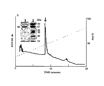
Figure 1 shows the profile of the protein Lopap,
purified by means of a process comprising a step of
chromatography by gel filtration and two steps of reverse
phase chromatography, presenting one single band of 69 kDa
molecular weight, determined by means of SDS-PAGE
analysis.
Figure 2 shows the activity of the protein Lopap on
the substrate Abz-YQTFFNPRTFGSQ-EDDnp (deduced from the
prothrombin molecule) .
Figure 3 shows cell viability. HUVECs (1 x 10⁴) were
incubated with RPMI medium supplemented with 1% FBS
without or with Lopap. An MTT assay was affected after 48
hours. The percentage of viable cells is expressed in
relation to non-treated cells.
Figure 4 shows the release of Prostaglandin 12.
HUVECs were incubated for one hour in RPMI 1640 culture
medium in the absence or presence of metalloproteases. PGI2
concentration was determined in the supernatant by the
accumulation of the metabolite 6-keto-PGF1c, in the culture
medium as measured by a competitive immunoenzimatic assay.
Figure 5 shows the release of nitric oxide. HUVECs
were incubated for 1 hour in HAM F12 culture medium in the
absence or presence of Lopap or r-Lopap. NO concentration
144mn
CA 02621482 2008-03-06
WO 2007/028223
PCT/BR2006/000180
8
was determined in the supernatant after reduction of
nitrate and nitrite to NO, as detected by chemiluminescence
in gas phase after reaction with ozone.
Figure 6 presents the expression of GAPDH detected by
RT-PCR in HUVECs (10% FBS), 1- control, non-stimulated
cells, 2- cells stimulated with 5 U/ml Thrombin, 3- cells
stimulated with 5 ng/ml TNFa, 4- cells stimulated with 5
g/ml LPS, 5- cells stimulated with 10 g/ml Lopap and 6-
cells stimulated with 10 g/ml rLopap.
Figure 7 presents the expression of Bc1-2 detected by
RT-PCR in HUVECs (10% FBS), 1- control, non-stimulated
cells, 2- cells stimulated with 5 U/ml Thrombin, 3- cells
stimulated with 5 ng/ml TNFa, 4- cells stimulated with 5
g/ml LPS, 5- cells stimulated with 10 g/ml Lopap, and 6-
cells stimulated with 10 g/ml rLopap. Expression is
calculated relative to expression of the control gene
GAPDH.
Figure 8 presents the expression of Bax in HUVECs (10%
FBS), 1- control, non-stimulated cells, 2- cells stimulated
with 5 U/ml Thrombin, 3- cells stimulated with 5 ng/ml
TNFa, 4- cells stimulated with 5 g/ml LPS, 5- cells
stimulated with 10 g/ml Lopap, and 6- cells stimulated
with 10 g/ml rLopap. Expression is calculated relative to
expression the control gene GAPDH.
Figure 9 shows fibronectin expression in fibroblasts
untreated and treated with rLopap (1 and 5 g).
Figure 10 shows tenascin expression in fibroblasts
untreated and treated with rLopap (1 and 5 g).
CA 02621482 2008-03-06
WO 2007/028223
PCT/BR2006/000180
9
Figure 11 shows indirect immunofluorescence for
fibronectin in: A) normal human fibroblasts (control
group); B) normal human fibroblasts grown in the presence
of 1 gg rLopap; C) normal human fibroblasts grown in the
presence of 5 gg rLopap.
Figure 12 shows indirect immunofluorescence for
tenascin in: A) normal human fibroblasts (control group);
B) normal human fibroblasts grown in the presence of 1 gg
rLopap; C) normal human fibroblasts grown in the presence
of 5 gg rLopap.
Figure 13 shows a photomicrograph of epidermis of
animals treated with rLopap, (A) treated skin and (B)
control skin. The presence of similar quantity of
fibroblast nuclei on the dermis is noticed.
Figure 14 shows expression by skin sample of Type III
- Group 1 collagen.
Figure 15 shows expression by skin sample of Type III
- Group 2 collagen.
Figure 16 shows expression by skin sample of Type III
- Group 3 collagen.
Figure 17 shows expression by skin sample of Type III
- Group 4 collagen.
Figure 18 shows photomicrographs of the dermis of
animals treated with rLopap (340X). Dense collagen fibers
(arrows) are observed.
Detailed Description of the Invention
The practice of the present invention employs, unless
CA 02621482 2008-03-06
WO 2007/028223
PCT/BR2006/000180
indicated, conventional methods of virology, microbiology,
molecular biology, and recombinant DNA techniques within
the skill of the art. Such techniques are explained fully
in the literature (see, e.g. Sambrook, et al. Molecular
5 Cloning: A Laboratory Manual (Current Edition); DNA
Cloning: A Practical Approach, Vol. I & II (D. Glover,
ed.), Oligonucleotide Synthesis (N. Gait, ea., Current
Edition); Nucleic Acid Hybridization (B. Hames & S.
Higgins, eds., Current Edition); Transcription and
10 Translation (B. Hames & S. Higgins, eds., Current Edition);
CRC Handbooks).
The invention is based, in part, on the discovery that
Lopap prevents apoptosis and increases cell viability. The
invention is also based, in part, on the discovery that
Lopap increases cell expression of extracellular matrix
proteins, which are important for preserving the integrity
of tissues, and the cells within the tissues. And the
invention is also based, in part, on the discovery that
Lopap increases cell expression of factors that regulate
muscle relaxation, which is important for preserving the
integrity of tissues, and the cells within the tissues.
The methods and compositions of the invention can be
used to treat disorders that involve cell death or
degeneration, or disorders that involve tissue
degeneration. The methods and compositions of the invention
can also be used to treat similar disorders occurring in
methods to culture or manipulate cells, methods to culture
or manipulate tissues, and methods that employ cells to
produce, alter, or degrade molecules.
CA 02621482 2008-03-06
WO 2007/028223
PCT/BR2006/000180
11
The invention is described in more detail in the
following subsections.
1. Cell Death and Degeneration
The poison from the caterpillar Lonomia oblique has
components with procoagulant action, which causes intense
coagulopathy consumption, and hemorrhagic manifestations.
Recently, the hemolymph of Lonomia oblique, which shares
many components with the poison, was also found to be able
to promote growth and longevity of Sf-9 cells (Souza, A.
P., et al., Biotechnol. Prog.; 21: 99-105, 2005). The
authors found hemolymph fractions with longevity activity.
There was no identification to the reactive agents involved
in the longevity activity.
It was determined in this invention that Lopap alone
increases the growth and longevity of cells (see Examples).
The data demonstrates that Lopap decreases apoptosis and
increases viability in HUVECs deprived of serum. The data
also demonstrates that Lopap increases expression of the
gene Bc1-2 (an anti-apoptotic protein), decreases
expression of the gene Bax (a pro-apoptotic protein).
Bc1-2 has been reported to be associated with
pathologic cell survival, is expressed at high levels in
several leukemias, and leads to tumor progression and
resistance to chemotherapy-induced and apoptosis. Bax has
been reported to countering these effects: Bax activation
leads to cytochrome c release and initation of the
mitochrondrial apoptosis program.
CA 02621482 2008-03-06
WO 2007/028223 PCT/BR2006/000180
12
It was also determined in this invention that Lopap
alone increases the expression of nitric oxide. Nitric
oxide has been reported to be an important signaling
molecule in mammals and humans. Among many functions,
nitric oxide has anti-oxidant activity, which can
contribute to preventing cell death and degeneration, and
tissue degeneration.
Methods of reducing cell death or degeneration are
also important as methods for reducing or repairing tissue
degeneration.
2. Tissue Degeneration
It was determined in this invention that Lopap alone
increases expression of proteins important in reducing and
repairing tissue degeneration (see Examples). Lopap is able
to stimulate expression of at least two groups of molecules
important for tissue structure: (1) proteins found in the
extracellular matrix; and (2) molecules that regulate
muscle relaxation.
The extracellular matrix (ECM) has been reported to be
the largest component of the dermal skin layer, and the
synthesis of ECM is a key feature of tissue growth and
wound healing, especially when there has been a significant
loss of tissue. The ECM is composed of three main classes
of molecules: (1) fibrous structural proteins (e.g.
collagens, fibronectin, and tenascin); (2) elastic fiber
proteins (e.g. elastin), and (3) proteoglycans. In addition
to serving as scaffold and structural support for cells,
CA 02621482 2008-03-06
WO 2007/028223
PCT/BR2006/000180
13
the ECM regulates cell adhesion, lubricates cells, and
provides a transport system for nutrients and waste.
Collagen contributes 80% of the weight of skin and is
responsible for tensile strength and protection against
external trauma. Elastic fibers contribute 2% to 4% of the
ECM, and provide elasticity to the skin (Uitto, J., J.
Invest. Dermatol. 72: 1-10, 1979); and (3) proteoglycans,
contributing 0.1% to 0.3% of the weight of tissue, support
(skin) hydration due to the water-retaining capacity of
hyaluronic acid (Davidson, E. A., Polysaccharide structure
and metabolism, in: Montagna W. (ed), Aging: Biology of
skin, Oxford, Pergamon Press, 1965, pp. 255-270). Processes
that alter or degrade these components can result in
detrimental clinical manifestations for tissue - especially
in tissues that are stretched or compressed (e.g. aorta,
lungs, skin, cartilage, and tendons) - such as lack of new
tissue growth, atrophy, loss of resilience, and ageing.
New tissue growth and fibrinogenesis is controlled by
the balanced synthesis and interaction of the ECM proteins.
For example, microfibrils - a component of elastic fibers -
are introduced into the extracellular medium by
fibroblasts, mesenchymal and other cells, which, with
aggregation, form a support structure for the elastic
fiber, where elastin is deposited. This basic structure
indicates the form and direction of the future elastic
fiber; Ross, R., J. Histochem. Cytochem., v. 21, p. 199-
208, 1973; Ross, R. et al, Adv. Exp. Med. Biol., v. 79, p.
7-17, 1977).
CA 02621482 2008-03-06
WO 2007/028223
PCT/BR2006/000180
14
In acute wounds, the provisional wound matrix,
containing fibrin and fibronectin, provides a scaffolding
to direct cells into the injury, as well as stimulating
them to proliferate, differentiate and synthesise new ECM.
Chronic wounds contain increased levels of inflammatory
cells, giving rise to proteases that degrade the ECM
components, growth factors and receptors essential for
healing.
Current approaches in wound healing focus on re-
establishing a functional ECM, including methods or
products that reduce excessive protease levels or
contribute functional ECM proteins, thereby facilitating
the healing process. Some of these approaches provide a
competitive substrate (collagen) for the proteases and
thereby reducing proteolytic destruction of essential ECM
components (fibronectin) and platelet-derived growth
factors (PDGFs). Other approaches provide unique proteins
(amelogenin) to replace corrupted ECM (Schultz, GS, World
Wide Wounds, Extracellular matrix: review of its roles in
acute aand chronic wounds, August 2005).
It was determined in this invention that Lopap
increases expression of extracellular proteins (ECM). More
specifically, Lopap increases expression of fibronectin,
tenascin, collagen, and elastin. The data demonstrates that
Lopap increases expression of Type III-Group 1 collagen,
Type III-Group 2 collagen, Type III-Group 3 collagen, and
Type III-Group 4 collagen.
Muscle is contractile tissue of the body whose
function is to produce force and cause (1) locomotion or
CA 02621482 2008-03-06
WO 2007/028223
PCT/BR2006/000180
(2) movement within internal organs. Muscle movement within
organs, disregulated or extensive over time, causes organs
to malfunction (e.g. eye myopia, spasms) or age (e.g. skin
structure, heart failure). Current approaches to treat
5 organ disorders related to disregulated or extensive muscle
use, includes the use of muscle relaxants (for examples,
see Scrips Reports, PJB Publications Ltd. Surrey, UK, 2000)
Nitric oxide and prostaglandin 12 (prostacycline) are
important signaling molecules in mammals, and they possess
10 several biological activities, including causing relaxation
of muscle tissue. It was determined in this invention that
Lopap increases expression of nitric oxide and
prostaglandin 12 in HUVECs (see Examples) and hence, can be
used to treat disorders caused by disregulated or extensive
15 muscle use. In one embodiment, nitric oxide and
prostaglandin 12 can be used to treat disorders associated
with aging, e.g. heart failure or skin structure such as
wrinkles. The popular cosmetic drug Botox is a muscle
relaxant, and it is contemplated that Lopap has the same
applications and uses. In another embodiment, nitric oxide
and prostaglandin 12 can be used to treat eye myopia or
muscle spasms.
Nitric oxide is being used in the treatment of other
disorders, including hypertension, sexual dysfunction (e.g
mechanism of alkyl nitrite) and erectile dysfunction.
Prostaglandin 12 is being used in the treatment of other
disorders, including hypertension and ulcers. In another
embodiment, Lopap can be used to treat diseases treatable
by nitric oxide or prostaglandin 12, including
CA 02621482 2008-03-06
WO 2007/028223
PCT/BR2006/000180
16
hypertension, sexual dysfunction, erectile dysfunction, and
ulcers.
Methods of reducing or repairing tissue degeneration
are also important methods for reducing cell death or
degeneration.
3. Lopap and rLopap
The naturally-ocurring Lopap protein is a prothrombin
activating protease from Lonomia obliqua, which can be
purified as presented in Figure 1, comprising a step of gel
filtration chromatography and two steps of reverse phase
chromatography, resulting in a major band of about 69 kDa
molecular weight, and may have at least one activity
corresponding to Figure 2, obtained by analysis of the
protein on the substrate Abz-YQTFFNPRTFGSQ-EDDnp (deduced
from prothrombin).
The term "functional form" of the Lopap protein refers
to any form of Lopap protein that retains at least one
therapeutic use of the naturally-occurring protein.
Examples of desired therapeuric uses include reduction of
cell death, reduction of cell or tissue degeneration,
repair of tissue, expression of extracellular matrix
proteins, expression of nitric oxide, or expression of
prostaglandin 12. For the purposes of this invention, a
functional form of Lopap includes, but is not limited to,
the naturally-occurring Lopap, rLopap, and/or a functional
derivative of any of these forms of the Lopap protein.
CA 02621482 2008-03-06
WO 2007/028223
PCT/BR2006/000180
17
The term "rLopap" refers to a Lopap protein (SEQ ID
NO: 1) derived from a recombinant DNA sequence encoding the
Lopap protein (Brazilian patent document PI 0403882).
Unless otherwise explicitly specified in this
application, any reference to "Lopap" should be construed
as a reference to the naturally-occurring Lopap, the
functional form of Lopap, rLopap (SEQ ID NO: 1), and/or a
functional derivative of any of these forms of Lopap.
The term "derivative" refers to a protein derived or
obtained from Lopap that retains at least one therapeutic
use of Lopap. Examples of therapeutic uses include
reduction of cell death, reduction of cell or tissue
degeneration, repair of tissue, expression of extracellular
matrix proteins, expression of nitric oxide, or expression
of prostaglandin 12. Derivatives may be produced by
techniques known in the art, including deletions or
additions or substitutions of amino acids, or other
chemical modifications that will not affect the ability of
the derivative to provide a therapeutically beneficial
effect to the treated cell or tissue.
A derivative may also result from the cleavage of the
parent molecule, cyclisation and/or coupling with one or
more additional moieties that improve Volubility, altering
the lipophilic characteristics to enhance uptake by cells,
altering stability or biological half-life, decreasing
cellular toxicity, or, in particular in vitro or ex vivo
applications, acting as a label for subsequent detection,
or the like. Moreover, a derivative may result from post-
translational or post-synthesis modification such as the
CA 02621482 2008-03-06
WO 2007/028223
PCT/BR2006/000180
18
attachment of carbohydrate moieties or chemical reaction(s)
resulting in chemical modification(s) such as alkylation or
acetylation of amino acid residues or other changes
involving the formation of chemical bonds.
Derivatives may also result from chemical
modifications such as coupling, acetylation, acylation,
ADP-ribosylation, amidation, covalent attachment of other
functional moiety, covalent attachment of lipid or lipid
derivative, covalent attachment of phosphotidylinositol,
cross-linking, icyclization, disulfide bond formation,
demethylation, formation of covalent cross-links,
formulation, gamma-carboxylation,
glycosylation,
glycophosphatidylinositol (GPI) anchor
formation,
hydroxylation, iodination, methylation, myristoylation,
oxidation, pegylation, proteolytic
processing,
phosphorylation, prenylation, racemization, selenoylation,
sulfation, transfer RNA mediated addition of amino acids to
proteins such as arginylation, and ubiguitination. For
instance, Creighton, Proteins-Structure and Molecular
Properties, 2nd Ed., W. H. Freeman and Company, New York
(1993); Johnson, Post Translational Covalent Modification
of Proteins, Academic Press, New York, (1983); Seifter et
al., Meth. Enzymol. 182:626-646 (1990); Rattan et al., Ann.
N.Y. Acad. Sci. 663:48-62 (1992); US 5876969; EP 0413622;
and US 5766883).
In another embodiment, the invention pertains to using
nucleic acids encoding Lopap. The nucleic acids can be RNA
or DNA. In a preferred embodiment, the nucleic acid encodes
the naturally-occurring Lopap or rLopap or the functional
CA 02621482 2008-03-06
WO 2007/028223
PCT/BR2006/000180
19
form of Lopap. In a more preferred embodiment, the nucleic
acid encodes a cDNA encoding Lopap (SEQ ID NO: 2) or a
functional derivative.
In one embodiment, the Lopap nucleic acid is contained
or associated with an expression vector (e.g. recombinant
rekoviruses, adenovirus, adeno-associated virus, herpes
simplex virus-1, or recombinant bacterial or eukaryotic
plasmids, or cosmid). Viral vectors can be transfect cells
directly, and plasmids can transfect cells with use of
cationic liposomes (lipofectin), polylysine conjugates,
gramacidin S. artificial viral envelopes, direct injection,
electroporation, or CaPO4. In another embodiment, the Lopap
nucleic acid is contained or associated with a cell (e.g.
transfected) or tissue or animal or plant (e.g.
transgenic). The cell or tissue or animal or plant that
contains and expresses the Lopap nucleic acid - contained
or not in an expression vector - can be used as a source
for Lopap protein, and the expressed protein can be
isolated using standard techniques.
The term "substantially identical", in the context of
two or more peptides, or two or more nucleic acids, refers
to two or more sequences or subsequences having at least
60%, preferably at least 80%, more preferably at least 85%
90%, 95% or higher identity between amino acid or
nucleotide residues, in comparison or aligned for maximum
correspondence, as measured by using a sequence comparison
algorithm, such as BLAST algorithm (Altschul et al, J. Mol.
Biol. 215: 403-410 (1990)), the local homology algorithm by
Smith & Waterman, Adv. App./. Math. 2: 482 (1981), the
CA 02621482 2008-03-06
WO 2007/028223
PCT/BR2006/000180
homology alignment algorithm by Needleman & Wunsch, J. Mol.
Biol. 48: 443 (1970), by similarity search by the Pearson &
Lipman method, Proc. Natl. Acad. Sci. U. S. A. 85: 2444
(1988), by computerized implementation of said algorithms
5 (GAP,
BESTFIT, FASTA and TFASTA in the Wisconsing Genetics
Software Package, Genetics Computer Group, 575 Science Dr.,
Madison, WI).
In another embodiment, the invention pertains to
polypeptides that are substantially identical to Lopap. In
10 a preferred embodiment, the polypeptides are substantially
identical to the naturally-occurring Lopap or the
functional form of Lopap or the functional derivative of
Lopap. In a more preferred embodiment, the polypeptides are
substantially identical to rLopap (SEQ ID NO: 1).
15 In
another embodiment, the invention pertains to
nucleic acids that are substantially identical to the
nucleic acids that encode Lopap. In a preferred embodiment,
the nucleic acids are substantially identical to the
nucleic acids that encode the naturally-occurring Lopap or
20 the functional form of Lopap or rLopap or the functional
derivative of Lopap. In a more preferred embodiment, the
nucleic acids are substantially identical to SEQ ID NO: 2.
Lopap can be tested for biological activity (e.g.,
cell viability, stimulation of cellular matrix proteins)
both in vitro or in vivo. Testing can be performed as
described in the examples section, or according to methods
well known in the art, such as DNA Fragmentation.
Lopap and rLopap can be administered alone, or in
addition with an agent to obtain a synergistic effect, e.g,
CA 02621482 2008-03-06
WO 2007/028223
PCT/BR2006/000180
21
a combination therapy. Examples of agents that can improve
cell viability include growth factors (e.g. epidermal
growth factor (EGF) and basic fibroblast growth factor
(bFGF)) and anti-oxidants (e.g. sodium metabisulfite,
sodium thiosulfate, -acetylcysteine,
butylated
hydroxyanisole, and butylated hydroxytoluene). Examples of
agents that can repair tissue include protease inhibitors
(e.g. inhibitors to matrix metalloproteases, e.g.
hydroxamic acid), and agents to stimulate or replace ECM
(e.g. amelogenin and ECM proteins and precursors). Other
examples include agents to suppress the specific disorder
being treated (e.g. immunosupressants for inflammatory
disorders).
4. Compositions and Formulations
In one aspect, this invention provides methods and
compositions that include a prophylactically and
therapeutically effective amount of at least one
polypeptide or nucleic acid which is preferably at least
60%, preferably at least 80%, more preferably at least 85%,
90%, even more preferably at least 95% identical to Lopap
or rLopap.
In another aspect, this invention provides methods and
compositions to reduce cell death or degeneration, or to
reduce or repair tissue degeneration. In general, the
methods involve providing an effective amount of Lopap
sufficient to modulate cell death or degeneration, or
tissue degeneration.
CA 02621482 2008-03-06
WO 2007/028223
PCT/BR2006/000180
22
In one embodiment of the invention, the cells or
tissues to be treated are undergoing death or degeneration
caused by natural or non-natural disorders, such as wound,
disease, aging, or caused by external agents.
Compositions - including pharmaceutical compositions
and cosmetic compositions - containing Lopap - may be
prepared by conventional techniques (e.g. Remington: The
Science and Practice of Pharmacy, 19th Ed., 1995) or the
techniques described below. The compositions may appear in
conventional forms, e.g., capsules, tablets, aerosols,
solutions, suspensions or topical applications.
Typical compositions include Lopap associated with a
pharmaceutically or cosmetically acceptable excipient,
which may be a carrier or diluent or combination thereof,
or enclosed within a carrier in the form of an ampule,
capsule, sachet, paper or other container. Conventional
techniques for the preparation of compositions may be used.
When the carrier serves as a diluent, it may be solid,
semi-solid, or liquid, which acts as a vehicle, excipient,
or medium for the polypeptide. Lopap can be adsorbed on a
granular solid container, e.g., in a sachet. Examples of
carriers are water, salt solutions, alcohols, polyethylene
glycols, polyhydroxyethoxylated castor oil, peanut oil,
olive oil, lactose, terra alba, sucrose, cyclodextrin,
microspheres, amylose, magnesium stearate, talc, gelatin,
agar, pectin, acacia, stearic acid or lower alkyl ethers of
cellulose, silicic acid, fatty acids, fatty acid arnines,
fatty acid monoglycerides and diglycerides, pentaerythritol
fatty acid esters, polyoxyethylene, hydroxymethylcellulose,
CA 02621482 2008-03-06
WO 2007/028223
PCT/BR2006/000180
23
and polyvinylpyrrolidone. Other examples are buffers (e.g.
phosphate, succinate, citrate, acetate, organic substances
or their salts); antioxidants (e.g. ascorbic acid); low
molecular weight peptides (<10 amino acids) (e.g.
polyarginine or tripeptides); proteins (e.g. serum albumin
or immunoglobulins); hydrophilic polymers
(e.g.
polyvinylpyrrolidone); amino acids (e.g. glycine, glutamic
acid, aspartic acid, arginine);
monosaccharides,
disaccharides; carbohydrates including cellulose and its
derivatives; glucose, mannose, or dextrins; chelating
agents (e.g. EDTA); sugar alcohols (e.g. mannitol or
sorbitol); counterions (e.g. sodium); surfactants (e.g.
polysorbates); poloxamers; and polyethyelene glycols.
The compositions may include additives, adjuvants,
auxiliary agents, emulsifying agents, suspending agents,
buffers, salt for osmotic pressure, preserving agents,
stabilizers, thickeners, wetting agents, coloring or
sweetening or flavoring agents. The compositions may be
formulated to provide quick, sustained, or delayed release
of Lopap by employing procedures known in the art.
The route of administration may be any route which
transports Lopap to the desired site, such as oral, nasal,
pulmonary, transdermal or parenteral, e.g., rectal, depot,
subcutaneous, intravenous, intraurethral, intramuscular,
intranasal, ophthalmic solution or an ointment - the
topical route being preferred.
To prepare topical formulations, Lopap is placed in a
dermatological vehicle as is known in the art. The amount
of Lopap to be administered and Lopap's concentration in
CA 02621482 2008-03-06
WO 2007/028223
PCT/BR2006/000180
24
the formulation depend upon the vehicle, delivery system or
device selected, the clinical condition of the patient, the
side effects and the stability of Lopap in the formulation.
The physician selects the appropriate (1) preparation, (2)
concentration of Lopap, and (3) amount of formulation to be
administered, depending upon clinical experience with the
patient or similar patients. The topical formulations may
be in the form of powders, ointments, gels, creams,
adhesives and the like.
For nasal administration, Lopap may be dissolved or
suspended in a liquid carrier for aerosol application. The
carrier may contain additives such as solubilizing agents,
e.g., propylene glycol, surfactants, absorption enhancers
such as lecithin (phosphatidylcholine) or cyclodextrin, or
preservatives such as parabens.
For ophthalmic formulations, see Mitra, Ophthalmic
Drug Delivery Systems, Marcel Delker, New York, NY (1993);
and Iavener, Ocular Pharmacology, C.V. Mosby Co., St. Louis
(1983).
For oral administration, solid or fluid doses can be
prepared. Solid doses of Lopap (e.g. tablets) can include
conventional ingredients such as talc, magnesium stearate,
dicalcium phosphate, magnesium aluminum silicate, calcium
sulfate, starch, lactose, acacia, methylcellulose, and
functionally similar materials as diluents or carriers.
Capsules can include Lopap plus an inert diluent in a hard
gelatin capsule. Soft gelatin capsules can include a slurry
of Lopap with vegetable oil, light liquid petrolatum or
other inert oil. Fluid doses in the form of syrups, elixirs
CA 02621482 2008-03-06
WO 2007/028223
PCT/BR2006/000180
and suspensions can be prepared. Lopap can be dissolved in
an aqueous vehicle with flavoring agents and preservatives
to form a syrup. An elixir can be prepared using a
hydroalcoholic vehicle (e.g. ethanol) with flavoring
5 agents. Suspensions can be prepared with aid of a
suspending agent (e.g. acacia, tragacanth, methylcellulose
and the like).
Appropriate formulations for parenteral use are known
to the practitioner of ordinary skill, such as injectable
10 solutions or suspensions. Lopap is prepared in an aqueous
solution in a concentration from about 1 to about 500
mg/ml. More typically, the concentration is from about 10
to 60 mg/ml or about 20 mg/ml. Concentrations below 1 mg/ml
may be necessary depending on solubility and potency. The
15 formulation - sterile - is suitable for various topical or
parenteral routes, including intravenous, intradermal,
intramuscular, intravascular, and subcutaneous (including
continous and semi-continous infusion). Examples for
injectable formulations are water, various salines, organic
20 or inorganic salt solutions, Ringer's solution, dextrose
solution, and Hank's solution.
Excipients can be included in the composition.
Examples include cosolvents, surfactants, oils, humectants,
emollients, preservatives, stabilizers and antioxidants.
25 Pharmacologically acceptable buffers may be used, e.g.,
tris or phosphate. Effective amounts of diluents,
additives, and excipients are those effective to obtain a
pharmaceutically acceptable formulation in terms of
solubility, biological activity, etc.
CA 02621482 2008-03-06
WO 2007/028223
PCT/BR2006/000180
26
Lopap may be incorporated into a microsphere.
Materials suitable for the preparation of microspheres
include albumin, agar, alginate, chitosan, starch,
hydroxyethyl starch, ovalbumin, agarose,
dextran,
hyaluronic acid, gelatin, collagen, and casein. The
microspheres can be produced by various processes known to
the person skilled in the art, such as a spray drying
process or an emulsification process. The microspheres can
be hardened by well-known cross-linking procedures such as
heat treatment or using chemical cross-linking agents.
Slow or extended-release delivery systems, including
biopolymers (biological-based systems),
liposomes,
colloids, resins, glyceryl monostearate, glyceryl
distearate, wax, and other polymeric delivery systems or
compartmentalized reservoirs, can be used with Lopap to
provide a continuous or long term source of the protein.
The systems also include semipermeable polymer matrices in
the form of molded articles, such as films or
microcapsules. Examples of polymer matrixes include
polylactides (U53773919 and EP58481), copolymers of L-
glutamic acid, and gamma-ethyl-L-glutamate (EP 133,988).
The matrix material is chosen based on biocompatibility,
biodegradability, mechanical properties, aesthetics, and
interphase properties. A sequestering agent may be useful,
as carboxymethyl cellulose, to prevent dissociation of
matrix polypeptide compositions.
Slow or extended release systems are useful for
compositions for delivery via topical, intraocular, oral,
and parenteral routes.
CA 02621482 2008-03-06
WO 2007/028223
PCT/BR2006/000180
27
Lopap can also be administered in combination with an
intervention procedure, such as placement of a shunt,
stent, synthetic or natural excision grafts, catheter,
valve, or other implantable devices.
Lopap can also be administered using a variety of
articles in shape of a medical device. Examples of medical
devices include wound closure devices (e.g. sutures,
staples, adhesives) tissue repair devices (e.g. meshes such
as o meshes for hernia repair), prosthetic devices (e.g.,
internal bone fixation devices, physical barriers for
guided bone registeration, stems, valves, electrodes),
tissue emgineering devices (e.g. for use with a blood
vessel, skin, a bone, cartilage, a liver), controlled drug
devivery systems (e.g. microcapsules, ion-exchange resins),
wound coverings, or wound fillers (e.g., alginate
dressings, chitosan powders). In some embodiments, the
device is a transcutaneous medical device (e.g. a catheter,
a pin, an implant) which can include coated or embedded
with Lopap. In some embodiments, the device is inform of a
patch (e.g. a patch with an adhesive layer adhering to the
skin, such as a transdermal patch).
The dose or "effective amount" of Lopap depends on the
protein(s) or nucleic acid(s) used, the subject being
treated (e.g. cells or tissue or patient), the condition
being treated, the method of administration, the site of
release, the side effects of the treatment, the scheme of
administration, and other factors known by experts in the
art. (For treatment of patients, the patient and his
medical history should be considered.) The dose or
CA 02621482 2008-03-06
WO 2007/028223
PCT/BR2006/000180
28
effective amount should prevent or ameliorate symptoms of
the disorder without producing unacceptable toxicity. A
parenteral, oral, and topical dose can contain from 0.1 gg
to 500 mg (preferably about 0.1 jig to 10 mg and more
preferably 0.1 jig to 1 mg) of the polypeptide(s) per kg of
body weight per day. An intranasal dose can contain 1-400
mg, e.g., 10 to 200 mg per person.
The method of use (treatment) can involve daily
administration of Lopap (e.g., once to twice to continous)
for a specified number of days (e.g., 2 days, 3 days, 4
days, 7 days, 14 days, 21 days, one month, three months,
six months or longer).
Lopap can be tested for biological activity (e.g.,
cell viability, stimulation of cellular matrix proteins)
both in vitro or in vivo. Testing can be performed as
described in the examples section, or according to methods
well known in the art, such as DNA Fragmentation.
Compositions, e.g. cosmetic compositions, may include
humectants (e.g. glycerol); glycols (e.g. ethylene glycol,
propylene glycol); emulsifiers, such as C1-05 alcohols,
optionally partially esterified poly-hydric alcohols with
fatty acids with C12-C24 long chains, such as glycerol
monostearate, isopropyl myristate, fatty acid ester of
sugar alcohols, e.g. monoester of fatty acid of sorbitan,
polyoxyalkylated derivatives, ester of fatty acid of
polyethoxyethylene, cholesterol, estearyl cetyl alcohol,
fatty alcohols of cotton and synthetic surfactants with low
HLB value; rheology modifiers (e.g., carbopol or natural or
synthetic polymers), low viscosity paraffins, emollient
CA 02621482 2008-03-06
WO 2007/028223
PCT/BR2006/000180
29
alcohol esters, triglicerides, lypophilic substances (e.g.
isopropyl miristate); pH regulators (e.g. TEA), carbonates
or phosphates; chelating agents, (e.g. EDTA and its salts),
and/or preserving agents. Compositions, e.g. cosmetic
compositions, may contain substances with UV filter
properties, pigments or coloring agents, vitamins,
essences, perfumes, cosmetic bases, and other formulations
and adjuvants used in compositions for topical application.
The nucleic acids encoding Lopap can be incorporated
in expression vectors and used in a cell therapy or gene
therapy protocols to treat disorders. The nucleic acid can
be contained or associated with an expression vector (viral
or bacterial or derivative thereof). Viral vectors can
transfect cells directly, and plasmids can transfect cells
with use of cationic liposomes, polylysine conjugates,
gramacidin S. artificial viral envelopes, direct injection,
or Ca204. The expression vectors or transfected cells can
be introduced into a tissue or patient by methods known in
the art, including intravenous injection, or by catheter
(US 5328470) or by stereotactic injection (e.g. Chen, et
al., (1994) PNAS 91: 3054-3057). Cells can also be
introduced by implantation.
5. Uses of Lopap and Compositions
The invention relates to polypeptides or nucleic acids
substantially identical to Lopap, pharmaceutical and
cosmetic compositions containing them, methods for
preparing the compositions, and their use as medicaments.
CA 02621482 2008-03-06
WO 2007/028223
PCT/BR2006/000180
In one aspect, the invention pertains to using Lopap for
treatment of disorders associated with cell death or
degeneration, or tissue degeneration. More specifically,
the Lopap and Lopap compositions can be used in treatment
5 of wounds, disease, aging, or disorder causing external
agents.
Lopap can be use to treat disorders associated with
cell death, such as bacterial and viral infection, e.g.,
human immunodeficiency virus. Several neurological diseases
10 are characterized by loss of neurons, and Lopap can be used
in the treatment of these disorders, such as Alzheimer's
disease, Parkinson's disease, amyotrophic lateral sclerosis
(ALS) retinitis pigmentosa, spinal muscular atrophy, and
various forms of cerebellar degeneration. Several
15 hematologic diseases are associated with a decreased
production of blood cells, and Lopap can be used in the
treatment of these disorders, such as anemia associated
with chronic disease, aplastic anemia, chronic neutropenia,
and the myelodysplastic syndromes. Some of these disorders,
20 e.g. myelodysplastic syndrome and some aplastic anemia, are
associated with cell death in the bone marrow, which may
result from programmed cell death, deficiencies in stromal
cells or hematopoietic survival factors, or mediators from
immune responses. Inflammatory disorders, such as graft
25 rejection, autoimmunity, and severe immune responses (e.g.
sepsis), are associated with organ-specific and/or systemic
cell death. Myocardial infarctions and stroke are
associated with cell death within the central area of
ischemia, resulting from the acute loss of blood flow. Both
CA 02621482 2008-03-06
WO 2007/028223 PCT/BR2006/000180
31
disorders are also associated with cell death in the area
outside the central ischemic zone, which occurs more slowly
and often involves apoptosis. Lopap can be used to treat
myocardial infarction, stroke, and other disorders
associated with cell death or degeneration.
In one embodiment, the patient has been diagnosed with
a disorder of cell death or degeneration, or tissue
degeneration. The method further includes administering to
the patient a pharmaceutically effective amount of Lopap
that reduces the symptoms of the disorder. In some
embodiments, Lopap is contacted directly with the cell or
tissue with the disorder. The method further includes a
test for the disorder after administering the Lopap to the
patient. In some embodiments, the test comprises evaluating
the viability or growth of cells or tissues affected by the
disorder. In other embodiments, the test comprises
evaluating secondary effects associated with the disorder,
such as measures known in the art for neurological,
hematologic, or cardiovascular function.
The term "patient" refers to any living organism in
which an immune response is elicited. The term subject
includes, but is not limited to, humans; non-human primates
such as chimpanzees and other apes and monkey species; farm
animals such as cattle, sheep, pigs, goats and horses;
domestic mammals such as dogs and cats; laboratory animals
including rodents such as mice, rats and guinea pigs, and
the like. The term does not denote a particular age or sex.
Thus, adult and newborn subjects, as well as fetuses,
whether male or female, are intended to be covered.
CA 02621482 2008-03-06
WO 2007/028223
PCT/BR2006/000180
32
In another aspect, Lopap can be used in vivo or in
vitro to improve viability of cells. In vitro use includes
cell culture methods to propagate cells and methods to
manipulate cells. Various cell types - prokayotes and
eukaryotes - can be treated with Lopap, such as animal,
plant, yeast and mold, insect, and bacterial cells.
Contemplated applications include cell culture for tissue
engineering and stem cell work, and industrial
applications. In a further aspect, Lopap can be used to
improve the viability of cells in biotechnology processes,
for example, cell methods for the production of molecules
(e.g. organic, inorganic, and macromolecule), and cell
methods to alter or degrade molecules. In one embodiment,
Lopap can be added to culture medium or bodily fluids that
will transport Lopap to the disordered cells.
In another aspect, Lopap can be used in vivo or in
vitro to reduce or repair degeneration of tissues. The use
includes methods of tissue culture, manipulation, and
growth (including use of tissue-forming molds). Various
cells types - prokayotes and eukaryotes - can be treated
with Lopap, such as animal, plant, yeast and mold, insect,
and bacterial cells. Contemplated applications include
tissues for industrial applications, and tissue
enginnering. In one embodiment, Lopap can be added to
culture medium or bodily fluids that will transport Lopap
to the disordered tissue; or Lopap can be applied directly
to the disordered tissue.
Lopap can be used to repair or grow the extracellular
matrix. In one embodiment, Lopap can be used to repair or
CA 02621482 2008-03-06
WO 2007/028223
PCT/BR2006/000180
33
grow fibronectin. In a second embodiment, Lopap can be used
to repair or grow tenascin. In a third embodiment, Lopap
can be used to repair or grow collagen. And in a fourth
embodiment, Lopap can be used to repair or grow elastin.
Lopap can be use to treat disorders associated with
the extracellular matrix, for example, disorders associated
with matrix metalloprotease (MMP) activity, such as ulcers,
asthma, acute respiratory distress syndrome, skin disease
and wounds, skin aging and wrinkles, keratoconus,
restenosis, osteo- and rheumatoid arthritis, joint disease
and wounds, bone disease and wounds, invasiveness,
hypovolemic shock, periodontal disease, epidennolysis
bullosa, scleritis, atherosclerosis, multiple sclerosis,
inflammatory diseases, vascular leakage syndrome, and
collagenase induced disease.
In another aspect, Lopap can be used to stimulate
nitric oxide and prostaglandin 12, which possess several
biological activities, including causing relaxation of
muscle tissue. In one embodiment, Lopap can be used to
treat disorders that benefit from muscle relaxation,
including hypertension and erectile dysfunction. In another
embodiment, Lopap can be used to treat disorders caused by
dysregulated or extensive muscle use, such as myopia,
spasms, aging, skin aging, and heart failure). In another
another embodiment, Lopap can be used to treat disorders
treatable by nitric oxide, such as sexual dysfunction (e.g
mechanism of alkyl nitrite). In another embodiment, Lopap
can be used to treat disorders treatable by prostaglandin
12, such as ulcers.
CA 02621482 2008-03-06
WO 2007/028223
PCT/BR2006/000180
34
In another embodiment, based on homology with
lipocalin proteins, Lopap is a transporter of hydrophobic
or organic compounds important in regulating cell death or
degeneration, or tissue degeneration (US20040084867;
US20050069877; Flower, et al., Biochim. Biophys. Acta 1482:
9-24, 2000). In one embodiment, Lopap is a transporter of
retinol, which is important in cell death or degeneration,
or tissue degeneration. In another embodiment, Lopap is a
transporter of lipids, which are important in cell death or
degeneration, or tissue degeneration.
The invention is also illustrated by way of the
examples below, which are provided as examples of the
invention, with no intention to limit the scope of the
invention.
Example 1
Anti-apoptotic effect of Lopap
Culture of endothelial cells
HUVECs were obtained by means of digestion with
collagenase, following the method by Jaffe and contributors
(Jaffe, E. A. et al., J. Clin. Invest., 1973; 52: 2745-
2756). Cells of 1-3 passages were used in confluent and
subconfluent monolayers according to the type of experiment
affected. Initially, cells were cultivated in 25 or 75 Cre
bottles with RPMI containing 10% fetal bovine serum (FBS).
When confluent, the monolayer was detached with
trypsin/EDTA and the cells were seeded over 24 well plates,
as required for the experiment.
CA 02621482 2008-03-06
WO 2007/028223 PCT/BR2006/000180
Anti-apoptotic effect
To evaluate the anti-apoptotic activity of rLopap and
the native Lopap (5 and 25 g/ml), programmed cell death
was induced by incubation of HUVECs for 48 hours in RPMI
5 medium containing 1% FBS without (control) or in the
presence of rLopap or native Lopap.
Morphologic changes and cell viability were analysed
by HUVEC coloring with a mixture of fluorescent colorants
linking to DNA, acridine orange (100 g/ml) to determine
10 the percentage of cells suffering apoptosis, and ethidium
bromide to differentiate between viable and non viable
cells.
The presence of apoptotic cells was evaluated by
fluorescence microscopy, by using non-adherent cells and
15 adherent cells detached with trypsin/EDTA. At least 200
cells were analysed in the experiment.
Lopap (5 g/ml and 25 g/ml) proved to be able to
inhibit the apoptosis of endothelial cells derived from
human umbilical cord (HUVECs) (Table 1) when the apoptosis
20 was induced by reduction of fetal bovine serum (1%). Higher
Lopap concentrations are able to produce more effective
consequences over the anti-apoptotic activity.
Table 1. Anti-apoptotic action of Lopap and rLopap on
25 HUVECs. Apoptosis was induced by reduction of FBS (1% FBS)
% OF APOPTOTIC CELLS
Control Lopap Lopap rLopap rLopap
CA 02621482 2008-03-06
WO 2007/028223 PCT/BR2006/000180
36
pig/m1 25 pig/m1 5 g/ml 25 ptg/m1
5 2 2 3 6 4 2 4 2 3 9 3 3 2 2
Example 2
Viability
5 Viability analysis was made by using the MTT method.
The reduction of 3-(4,5-dimethyltiazol-2-methyl)-2,5-
diphenyltetrazolium bromide (MTT) by intact cells was
evaluated on 96 well microplates. HUVECs were cultivated in
RPMI medium supplemented with 1% fetal bovine serum and,
after 48 hours of incubation with Lopap (0.15 to 20 g/ml),
the culture was washed with phosphate-buffered saline
(PBS). 10 l/well of 2.5 mg/ml MTT were added and the cells
were incubated for three hours at 37 C. The reaction was
interrupted by the addition of 150 1 of SDS. Absorbance
values at 540 nm were determined by using an automatic
microplate reader.
Lopap significantly improved cell viability in a
concentration-dependent way (Figure 3).
Example 3
Production of prostacycline
Prostacycline (PGI2) production was measured by the
accumulation of 6-keto-PGF1a (a metabolite from PGI2
hydrolysis) in the culture medium by ELISA after HUVEC
treatment with rLopap and native Lopap. The supernatant was
centrifuged at 400 xg for ten minutes at 4 C. HUVEC
CA 02621482 2008-03-06
WO 2007/028223
PCT/BR2006/000180
37
treatment with native Lopap for one hour (final 5 and 25
g/ml) produced a statistically significant improvement in
PGI2 release compared to control. rLopap (25 g/ml)
stimulated PGI2 release, similar to that induced by LPS (2
g/ml) (Figure 4).
Example 4
Production of nitric oxide
Nitric oxide (NO) production was measured by combining
the accumulation of nitrite and nitrate in the culture
medium HAM-F12. After the treatment of HUVECs with rLopap
or native Lopap (final 5 and 25 g/ml), the supernatant was
centrifuged at 400 xg for ten minutes at 4 C. Nitric oxide
concentration in the supernatant was determined by
chemioluminescence in gaseous phase, by using a nitric
oxide analyzer through the reaction between NO and ozone,
after the reduction of nitrate and nitrite with VC13
saturated solution in 1 M HC1 at 90 C. Nitrate
concentration was calculated from a standard curve of
sodium nitrate. Stimulation with native Lopap and rLopap
(25 g/ml) produced a statistically significant increase in
NO release in comparison with the control, being said
increase similar to the one induced by thrombin (5 UI/ml)
(Figure 5).
Example 5
RNA preparation (HUVECs)
CA 02621482 2008-03-06
WO 2007/028223
PCT/BR2006/000180
38
RNAs as obtained presented good quality (260/280:-.,..' 1.7
ratio) and 2% agarose gel analysis disclosed the presence
of bands 18S and 28S, confirming the integrity of RNA as
obtained.
Gene expression in endothelial cells
(HUVECs)
The expression of different target genes was measured
by RT-PCR from confluent cultures (500,000 cells/well) in
6-well plates, where HUVECs were incubated for eight hours
in RPMI medium containing 10% FBS in the absence (control)
or in the presence of LOPAP 10 g/ml, Thrombin 5U/ml, TNF-a
5 g/ml, or LPS 5 g/ml, to evaluate its direct action.
PRIMERS
Primers were designed based on a sequence of human
genes (already published) to amplify proteins of interest.
Table 2. Sequence of primers and size of PCR products
PRIMER Sense Anti-Sense
BCL-2
5'GAGGAAGTAGACTG 5'CCTTCCCAGAGGAA
(279 bp) ATATTA3' AAGCAA3'
BAX
5'GATGGACGGGTCCG 5'CTCAGCCCATCTTC
(542 bp) GAGA3' TTCCAG3'
GAPDH
5'GGTGAAGGTCGGAG 5'TCCTTGGAGGCCAT
(996 bp) TCAACG3' GTGGG000T3'
M Expression of the constitutive gene of
GAPDH cells (control)
CA 02621482 2008-03-06
WO 2007/028223
PCT/BR2006/000180
39
Using RT-PCR, densitometric analysis was performed on
test genes and the constitutively expressed gene,
glyceraldehyde-3-phosphate dehydrogenase (GAPDH) . In
HUVECs, GAPDH presented homogeneous expression for all
stimuli made. Electrophorectic migration of the applied
fragment was 996 bp, as expected (Figure 6) .
Expression of the gene B c 1 - 2
(anti-
apoptotic)
Electrophorectic migration in a 2% agarose gel from
the PCR reaction with primer to Bc1-2 showed the generation
of 279 bp fragments as expected. HUVECs stimulated with
Lopap and rLopap presented remarkable improvement in the
expression of the gene Bc1-2 over the negative control and
other stimuli made (Figure 7) .
Expression of the gene Bax ( p r o -
apoptotic)
Electrophorectic migration under 2% agarose gel from
the PCR reaction with primer to Bax showed the generation
of 542 bp fragments as expected. HUVECs stimulated with
Lopap and rLopap presented remarkable reduction in the
expression of the gene Bax; especially cells stimulated
with native Lopap, over the negative control and other
stimuli made (Figure 8) .
Example 6
Action of rLopap on
human skin
fibroblasts. Evaluation of components in the
CA 02621482 2008-03-06
WO 2007/028223
PCT/BR2006/000180
extracellular matrix: fibronectin
and
tenascin
Human skin fibroblasts in culture were plated over
platelets in a 24-well plate and incubated with rLopap
5 under different concentrations for seven days to evaluate
the action of said protein for the production of components
of the extracellular matrix ( fibronectin and tenascin) .
Biological material
The assay system comprised human skin fibroblasts in
10 culture, obtained from the fragment of normal skin from the
ear lobule of five female Negro individuals, aged 15 to 40
years old.
Collection of the biological material
Patients were submitted to excision biopsy, after
15 informed consentment, in a surgical room with usual asepsis
and antisepsis criteria. A fragment of normal skin
measuring 0.5 cm x 0.5 cm was collected and immediately
immersed in a sterile culture medium of Ham-F-12 with 15%
FBS ( fetal bovine serum) and antibiotics.
20 Test Plan:
Control and Test Groups
The control group was constituted by fibroblasts
cultured in culture medium of Ham-F-12 supplemented with
10% FBS .
Two assay groups were defined: 1) fibroblasts
25 cultivated in culture medium plus 1 lag rLopap, and 2)
fibroblasts cultivated in culture medium plus 5 lag rLopap.
Preparation of the plate
All fibroblast samples used in the experiment
consisted of the sixth subcultivation . Sterile culture
CA 02621482 2008-03-06
WO 2007/028223
PCT/BR2006/000180
41
plates with 24 wells were used. Fibroblasts were cultivated
in sterile round plates with 13 mm diameter, and in each
plate, 1 x 10⁵ fibroblasts were plated in 1 ml culture
added with FBS. After 24 hours for cell adherence, the
medium was changed with new culture medium and rLopap at
the final volume of 300 1/well.
rLopap used was stored at room temperature, filtered
through a 0.22 gm membrane under 1 g/ 1 and 5 g/ 1
concentrations.
Plates were kept at 37 C in a CO2 oven for seven days
and observed at an inverted microscope.
Fixation of plates
Plates were carefully washed 1X with 1 ml PBS. Plates
were fixed for 15 minutes with 400 1 fixing agent
comprising 3% paraformaldehyde and 0.2% glutaraldehyde in
0.1 M phosphate buffer pH 7.4, and washed 1X with 1 ml PBS.
Indirect immunofluorescence
Plates were incubated for 45 minutes with 50 1 of
primary monoclonal antibody - anti-cellular fibronectin or
anti-human tenascin - diluted 1:100 in PBS.
Subsequently, plates were incubated for 45 minutes
with 50 1 of the secondary antibody Alexa Fluor 488,
diluted 1:100 in PBS. Plates were assembled in a microscopy
slide with one drop of the Vectashield assembly medium with
DAPI (Vector Laboratories - U. S. A.).
Evaluation of fibronectin and tenascin production by
fibroblasts by histomorphometric analysis.
Slides, when submitted to immunofluorescence, were
analysed under light and fluorescence microscope (Zeiss)
CA 02621482 2008-03-06
WO 2007/028223
PCT/BR2006/000180
42
with 20X objective lens and 10X eye lens and the
quantitative evaluation was made with the support of an
Image Analysing System.
Images obtained in 10 microscopic fields were
digitalized with the support of the software, providing the
possibility to share data with the text editor (Microsoft
Word ) and spreadsheets (Microsoft Exce1C)). For a
quantitative evaluation of the production of fibronectin
and tenascin, fluorescent structures were marked, so to
differentiate them from other structures by color contrast.
The area of fibronectin and tenascin was obtained by
digital densitometry, which was transformed to square
micrometers oin2) Results obtained in each field
correspond to the percentual area of positive structures,
i. e. fraction of area.
Statistical Analysis
Data of area fraction for studied samples were
submitted to descriptive statistics and the comparison
between the groups was made by a non-parametric Kruskal-
Wallis test or by ANOVA parametric test. p < 0.05 was
considered as significant.
Values were obtained from the area fraction of
fibronectin and tenascin from the five samples of
fibroblasts cultured over the action of rLopap (1 Rg and 5
g) and control group. These values were submitted to
descriptive statistic analysis, whose results are specified
on Table 3.
CA 02621482 2008-03-06
WO 2007/028223
PCT/BR2006/000180
43
Table 3. Average and standard error for the expression
(%) fibronectin and tenascin in normal human fibroblast
cultures treated with 1 and 5 jig of rLopap and control
group
Treatment n Average Standard Max Min
Error _
% Fibronectin 50 46,1 15,1 72,8
16,8
rLopap (1 gg)
% Fibronectin 50 24,2 18,4 61,4 0,1
rLopap (5 gg)
% Fibronectin 50 19,2 7,4 38,4 5,1
Control
% Tenascin rLopap (1 50 2,3 1,5 5,8 0,4
jig)
% Tenascin rLopap (5 50 1,7 1,1 4,8 0,4
jig)
% Tenascin control 50 0,8 0,4 2,1 0,1
The comparison between fraction values in the area of
fibronectin as produced by fibroblasts grown with protein
under 1 jig and 5 jig concentrations, and the control group,
disclosed a statistically significant difference (p <
0.001), showing higher production of fibronectin by
fibroblasts cultivated with rLopap over the control group
(Figure 9).
The comparison between fraction values in the area of
tenascin as produced by fibroblasts grown with protein (1
jig and 5 jig) and the control group disclosed a
statistically significant difference (p < 0.001), showing
higher production of tenascin by fibroblasts cultivated
with rLopap over the control group (Figure 10).
CA 02621482 2008-03-06
WO 2007/028223
PCT/BR2006/000180
44
Figure 11 shows the distribution of fibronectin on
cultivated human skin fibroblasts: control group, (A) 1 gg
rLopap (B) and 5 gg Lopap (C).
Figure 12 shows the distribution of tenascin on
cultivated human skin fibroblasts: control group, (A) 1 gg
rLopap (B) and 5gg Lopap(C).
Example 7
The dorsal region of mice was hair-cutted and
submitted to topical treatment with rLopap jointly with
adjuvants on the right side (experimental) and just with
adjuvants on the left side (control).
Experimental groups were divided into:
Group 1: rLopap (0.33 mg) added to papain ointment
(final proportion 5%), topical administration once per week
for three times.
Group 2: rLopap (0.33 mg) diluted in 0.03 M NaC1,
topical administration once per week for three times.
Group 3: rLopap (0.33 mg) diluted in 0.03 M NaCl,
intradermal administration in a single dose.
Group 4: commercial ointment Vitanol-A, gel 0.025
tretinoin, topical administration once per week for three
times.
Hystopathological study
The treated region was taken off after animals were
sacrificed and fixed in a buffered solution of 10% formalin
with phosphate buffer. Skin samples were submitted to
crosswise sections and submitted to histological routine.
CA 02621482 2008-03-06
WO 2007/028223
PCT/BR2006/000180
Samples were dehydrated in a growing series of alcohols,
defatted in xylol and embedded in paraffin. Samples were
then inserted in paraffin and submitted to 3 m thick cuts
in histological microtome, which were collected on glass
5 blades. Said blades were submitted to Hematoxillin-Eosine
coloring, for analysis of histological characteristics and
Picrosirius coloring (Junqueira, L. C. V., et al., Anal.
Biochem. 94: 9609-13, 1979), for collagen quantitative
evaluation. Slides were examined under light microscopy, in
10 a microscope coupled to an Image Analyser System.
Histological characteristics
Skin of animals of all experimental groups as studied
showed architecture preserved with epidermis and dermis.
Collagen III Quantification
15 Dermis collagen was quantified with the support of an
Image Analyser Kontron 300 in skin cuts colored by
Picrosirius. Five microscopic fields were evaluated with
20X objective lens and 10X eye lens. Fields were randomly
chosen on the various skin fragments represented on the
20 slide of each animal.
We noticed that the skin treated with rLopap in the
different experimental groups significantly differs (p <
0.001) on the ability to synthesize collagen, in comparison
with non-treated skin (control) (Figures 13 to 18).
DEMANDES OU BREVETS VOLUMINEUX
LA PRESENTE PARTIE DE CETTE DEMANDE OU CE BREVETS
COMPREND PLUS D'UN TOME.
CECI EST LE TOME 1 DE 2
NOTE: Pour les tomes additionels, veillez contacter le Bureau Canadien des
Brevets.
JUMBO APPLICATIONS / PATENTS
THIS SECTION OF THE APPLICATION / PATENT CONTAINS MORE
THAN ONE VOLUME.
THIS IS VOLUME 1 OF 2
NOTE: For additional volumes please contact the Canadian Patent Office.