Note: Descriptions are shown in the official language in which they were submitted.
CA 02621597 2013-06-17
=
- 1 -
IN VIVO FORMED MATRICES INCLUDING NATURAL BIODEGRADABLE
POLYSACCHARIDES AND OPHTHALMIC USES THEREOF
Technical Field
The present invention relates to in vivo formed matrices comprising a natural
biodegradable polymeric material. Bioactive agents can be included in the in
vivo formed
matrices to provide a therapeutic effect to a patient. The formed matrices can
be particularly
useful in providing medical articles for implantation in the eye.
Background
In recent years, much attention has been given to site-specific delivery of
drugs
within a patient. Although various drugs have been developed for treatment of
a wide
variety of ailments and diseases of the body, in many instances, such drugs
cannot be
effectively administered systemically without risk of detrimental side
effects. Site-specific
drug delivery focuses on delivering the drugs locally, i.e., to the area of
the body requiring
treatment. One benefit of the local release of bioactive agents is the
avoidance of toxic
concentrations of drugs that are at times necessary, when given systemically,
to achieve
therapeutic concentrations at the site where they are required.
Site-specific drug delivery can be accomplished by injection and/or
implantation of
an article or device that releases the drug to the treatment site. Injection
of drugs can have
CA 02621597 2008-03-07
WO 2007/038126
PCT/US2006/036632
- 2 -
limitations, for example, by requiring multiple administrations, increasing
risk of
complications (such as infection), and patient discomfort. Implantation of an
article or
device that delivers drug to the treatment site has therefore gained much
interest in recent
years.
Further, site-specific drug delivery has been enhanced by technologies that
allow
controlled release of one or more drugs from an implanted device or article.
Controlled
release can relate to the duration of time drug is released from the device or
article, and/or
the rate at which the drug is released.
Several challenges confront the use of medical devices or articles that
release
bioactive agents into a patient's body. For example, treatment may require
release of the
bioactive agent(s) over an extended period of time (for example, weeks,
months, or even
years), and it can be difficult to sustain the desired release rate of the
bioactive agent(s) over
such long periods of time. Further, the device or article surface is
preferably biocompatible
and non-inflammatory, as well as durable, to allow for extended residence
within the body.
Generally speaking, a bioactive agent can be associated with the surface of a
medical device or article by surface modification, embedded and released from
within
polymeric materials (matrix-type), or surrounded by and released through a
carrier
(reservoir-type). The polymeric materials in such applications should
optimally act as a
biologically inert barrier and not induce further inflammation within the
body. However,
the molecular weight, porosity of the polymer, a greater percentage of coating
exposed on
the medical device or article, and the thickness of the polymer coating can
contribute to
adverse reactions to the medical device or article.
Another way to deliver bioactive agents from the surface of a medical device
or
article is by using a coating that has a biodegradable polymer, such as
polylactic acid. As
the coating degrades, the bioactive agent is released from the surface of the
device or article.
Some concerns exist that regard the use of biodegradable materials that
degrade into
CA 02621597 2008-03-07
WO 2007/038126
PCT/US2006/036632
- 3 -
materials that are not typically found in the body, or that are found at
particularly low levels
in the body. These types of biodegradable materials have the potential to
degrade into
products that cause unwanted side effects in the body by virtue of their
presence or
concentration in vivo. These unwanted side effects can include immune
reactions, toxic
adverse effects on cells or tissue in the body.
Another problem is that preparations of some biodegradable materials may not
be
obtained at consistent purity due to variations inherent in natural materials.
This is relevant
at least with regard to biodegradable materials derived from animal sources.
Inconsistencies
Additional concerns are that preparations from animal sources may provide
other
unwanted contaminants, such as antigenic factors. These antigenic factors may
promote a
localized immune response in the vicinity of the implanted article and foul
its function.
These factors may also cause infection as well as local inflammation.
15 In
particular, placement of implantable devices or articles in limited access
regions
of the body can present additional challenges. Limited access regions of the
body can be
characterized in terms of physical accessibility as well as therapeutic
accessibility. For
example, the relatively small size and sensitive tissues surrounding the eye
can contribute to
physical accessibility difficulties. In addition, ocular absorption of
systemically
junctions of the retinal pigment epithelium and vascular endothelial cells.
These can make
accessing the eye with therapeutics difficult. High systemic doses of
bioactive agents can
penetrate this blood ocular barrier in relatively small amounts, but expose
the patient to the
risk of systemic toxicity. Intravitreal injection of bioactive agents (such as
drugs) is an
concentrations. However, these repeated injections carry the risk of such
complications as
CA 02621597 2008-03-07
WO 2007/038126
PCT/US2006/036632
- 4 -
infection, hemorrhage, and retinal detachment. Patients also often find this
procedure
somewhat difficult to endure.
Because description of the invention will involve treatment of the eye as an
illustrative embodiment, basic anatomy of the eye will now be described in
some detail with
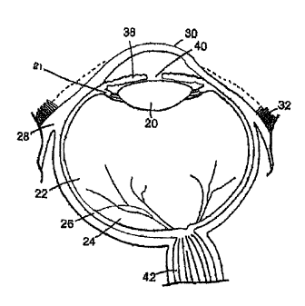
reference to Figure 1, which illustrates a cross-sectional view of the eye.
Beginning from
the exterior of the eye, the structure of the eye includes the iris 38 that
surrounds the pupil
40. The iris 38 is a circular muscle that controls the size of the pupil 40 to
control the
amount of light allowed to enter the eye. A transparent external surface, the
cornea 30,
covers both the pupil 40 and the iris 38. Continuous with the cornea 30, and
forming part of
the supporting wall of the eyeball, is the sclera 28 (the white of the eye).
The pars plana is a
region of the eye approximately 4 mm posterior to the point on the globe where
the colored
iris 38 meets the white sclera 28. The pars plana encircles the iris and is
not constant in
width, but rather typically varies between 2-3 mm in width around the iris
(with the largest
width of the pars plana typically lying on the temporal side and measuring
about 3 mm in
width).
The conjunctiva 32 is a clear mucous membrane covering the sclera 28. Within
the
eye is the lens 20, which is a transparent body located behind the iris 38.
The lens 20 is
suspended by ligaments attached to the anterior portion of the ciliary body
21. Light rays
are focused through the transparent cornea 30 and lens 20 upon the retina 24.
The central
point for image focus (the visual axis) in the human retina is the fovea (not
shown in the
figures). The optic nerve 42 is located opposite the lens.
There are three different layers of the eye, the external layer, formed by the
sclera
28 and cornea 30; the intermediate layer, which is divided into two parts,
namely the
anterior (iris 38 and ciliary body 21) and posterior (the choroid 26); and the
internal layer, or
the sensory part of the eye, formed by the retina 24. The sclera 28 is
composed of dense,
fibrous tissue and is composed of collagen fiber. Scleral thickness is
approximately 1 mm
=
CA 02621597 2008-03-07
WO 2007/038126
PCT/US2006/036632
- 5 -
posteriorly near the optic nerve and approximately 0.3 mm anteriorly. At the
pars plana, the
eye tissues are composed of sclera only; there is no choroidal or retinal
tissue layer within
this region. For this reason, the avascular pars plana is typically selected
for implantation
and/or injection of materials into the interior (vitreous) of the eye.
The lens 20 divides the eye into the anterior segment (in front of the lens)
and the
posterior segment (behind the lens). More specifically, the eye is composed of
two
chambers of fluid: the anterior chamber 34 (between the cornea 30 and the iris
38), and the
vitreous chamber 22 (between the lens 20 and the retina 24). The anterior
chamber 34 is
filled with aqueous humor whereas the vitreous chamber 22 is filled with a
more viscous
fluid, the vitreous humor.
The vitreous chamber 22 is the largest chamber of the eye, consisting of
approximately 4.5 ml of fluid. The vitreous chamber is filled with a
transparent gel
composed of a random network of thin collagen fibers in a highly dilute
solution of salts,
proteins and hyaluronic acid (the vitreous humor comprises approximately 98%
water).
Summary of the Invention
In one aspect, the present invention provides compositions and methods for
preparing biodegradable compositions that are particularly useful for forming
medical
articles within a patient's body, such as within a patient's eye. These
medical articles can be
useful for delivering bioactive agents to a treatment site within a body, such
as the eye.
These bioactive agent delivery compositions include a natural biodegradable
polysaccharide
as a component that can be crosslinked in situ to form a matrix from which a
therapeutic
material such as a drug, a biomolecule, or cells (referred to herein as a
"bioactive agents")
can be released or retained. In some embodiments of the invention, a bioactive
agent is
present in and can be released from the biodegradable matrix; in other
embodiments a
bioactive agent is present in a biodegradable microparticle, the microparticle
being
immobilized within the matrix.
CA 02621597 2008-03-07
WO 2007/038126
PCT/US2006/036632
- 6 -
In some aspects of the invention, the natural biodegradable polysaccharide is
used
to prepare an article that can be formed within the body (for example, by in
situ formation).
In some aspects, the article can be amorphous, such as a polymerized mass of
natural
biodegradable polysaccharides that is formed within or on a portion of the
body, by using an
in vivo matrix-forming composition.
In some aspects, the article, such as an in vivo formed matrix, is used in
methods for
the treatment of any one or more of a variety of medical conditions or
indications, including
retinal detachment; occlusions; proliferative retinopathy; proliferative
vitreoretinopathy;
diabetic retinopathy; inflammations such as uveitis, choroiditis, and
retinitis; degenerative
disease (such as age-related macular degeneration, also referred to as AMD);
vascular
diseases; and various tumors including neoplasms. In yet further embodiments,
the
biodegradable medical article can be used post-operatively, for example, as a
treatment to
reduce or avoid potential complications that can arise from ocular surgery. In
one such
embodiment, the medical article can be provided to a patient after cataract
surgical
procedures, to assist in managing (for example, reducing or avoiding) post-
operative
inflammation.
Illustrative bioactive agents include antiproliferative agents, anti-
inflammatory
agents, anti-angiogenic agents, hormonal agents, antibiotics, neurotrophic
factors, or
combinations thereof. Exemplary antiproliferative agents include 13-cis
retinoic acid,
retinoic acid derivatives, taxol, sirolimus (rapamycin), analogues of
rapamycin, tacrolimus,
ABT-578, everolimus, paclitaxel, taxane, and vinorelbine. Exemplary anti-
inflammatory
agents include hydrocortisone, hydrocortisone acetate, dexamethasone 21-
phosphate,
fluocinolone;medrysone, methylprednisolone, prednisolone 21-phosphate,
prednisolone
acetate, fluoromethalone, betamethasone, triamcinolone, and triamcinolone
acetonide.
Exemplary inhibitors of angiogensis include angiostatin, anecortave acetate,
thrombospondin, anti-VEGF antibody, and anti-VEGF fragment. Exemplary hormonal
CA 02621597 2008-03-07
WO 2007/038126
PCT/US2006/036632
- 7 -
agents include estrogens, estradiol, progesterol, progesterone, insulin,
calcitonin,
parathyroid hormone, peptide, and vasopressin hypothalamus releasing factor.
In some aspects, the biodegradable medical article can include a
radiopacifying
agent.
In alternative aspects of the invention, the natural biodegradable
polysaccharide is
used to prepare a medical device that can be formed within the body. In
accordance with
these aspects, the medical article is a medical device that performs a
function within the eye
(other than delivery of bioactive agent) and can be formed in vivo. In these
aspects,
inclusion of bioactive agent is optional. One illustrative example of a
medical device in
accordance with these aspects is a viscoelastic tamponade that can be utilized
in
combination with retinal reattachment.
In preparing the biodegradable compositions, a plurality of natural
biodegradable
polysaccharides are crosslinked to each other via coupling groups that are
pendent from the
natural biodegradable polysaccharide (i.e., one or more coupling groups are
chemically
bonded to the polysaccharide). In some aspects, the coupling group on the
natural
biodegradable polysaccharide is a polymerizable group. In a free radical
polymerization
reaction the polymerizable group can crosslink natural biodegradable
polysaccharides
together in the composition, thereby forming a natural biodegradable
polysaccharide matrix,
which can be an in-vivo formed matrix.
The natural biodegradable polysaccharides described herein are non-synthetic
polysaccharides that can be associated with each other to form a matrix, which
can be used
as an in-vivo formed matrix. The natural biodegradable polysaccharides can
also be
enzymatically degraded, but offer the advantage of being generally non-
enzymatically
hydrolytically stable. This is particularly advantageous for bioactive agent
delivery, as in
some aspects the invention provides articles capable of releasing the
bioactive agent under
conditions of enzyme-mediated degradation, but not by diffusion. Therefore,
the kinetics of
CA 02621597 2008-03-07
WO 2007/038126
PCT/US2006/036632
- 8 -
bioactive agent release from the articles of the invention are fundamentally
different than
those of coatings or medical implants prepared from synthetic biodegradable
materials, such
as poly(lactides).
Natural biodegradable polysaccharides include polysaccharide and/or
polysaccharide derivatives that are obtained from natural sources, such as
plants or animals.
Exemplary natural biodegradable polysaccharides include amylose, maltodextrin,
amylopectin, starch, dextran, hyaluronic acid, heparin, chondroitin sulfate,
dermatan sulfate,
heparan sulfate, keratan sulfate, dextran sulfate, pentosan polysulfate, and
chitosan.
Preferred polysaccharides are low molecular weight polymers that have little
or no
branching, such as those that are derived from and/or found in starch
preparations, for
example, amylose and maltodextrin.
Because of the particular utility of the amylose and maltodextrin polymers, in
some
aspects natural biodegradable polysaccharides are used that have an average
molecular
weight of 500,000 Da or less, 250,000 Da or less, 100,000 Da or less, or
50,000 Da or less.
In some aspects the natural biodegradable polysaccharides have an average
molecular
weight of 500 Da or greater. In some aspects the natural biodegradable
polysaccharides
have an average molecular weight in the range of about 1000 Da to about 10,000
Da.
Natural biodegradable polysaccharides of particular molecular weights can be
obtained
commercially or can be prepared, for example, by acid hydrolysis and/or
enzymatic
degradation of a natural biodegradable polysaccharide preparation, such as
starch. The
decision of using natural biodegradable polysaccharides of a particular size
range may
depend on factors such as the physical characteristics of the biodegradable
composition
(e.g., viscosity), the desired rate of degradation of the composition, the
presence of other
optional moieties in the composition (for example, bioactive agents, etc.),
and the like.
The natural biodegradable polysaccharides that are used in accordance with the
methods and compositions of the invention are readily available at a low cost
and/or can be
CA 02621597 2008-03-07
WO 2007/038126
PCT/US2006/036632
- 9 -
prepared easily using established techniques. This allows for a cost effective
method of
fabricating medical articles.
The use of natural biodegradable polysaccharides, such as maltodextrin or
amylose,
provides many advantages when used for the formation of an article, such as
one that can be
formed and used in vivo. Degradation of a natural biodegradable polysaccharide-
containing
article can result in the release of, for example, naturally occurring mono-
or disaccharides,
such as glucose, which are common components of bodily fluids, such as the
vitreous
humor. Furthermore, the use of natural biodegradable polysaccharides that
degrade into
common components found in bodily fluids, such as glucose, can be viewed as
more
acceptable than the use of synthetic biodegradable polysaccharides that
degrade into non-
natural compounds, or compounds that are found at very low concentrations in
the body.
In some aspects of the invention, this advantageous feature is reflected in
the use of
natural biodegradable polysaccharides which are non-animal derived, such as
amylose and
maltodextrin, and that degrade into products that present little or no
immunogenic or toxic
risk to the individual. The invention provides improved, cost-efficient,
natural
biodegradable polysaccharide compositions for articles that can be used in a
variety of
medical treatments.
Another advantage of the invention is that the natural biodegradable
polysaccharide-
based compositions are more resistant to hydrolytic degradation than other
biodegradable
polymers, such as poly(lactides). Degradation of the natural biodegradable
polysaccharides
of the invention are primarily enzyme-mediated, with minimal or no hydrolysis
of the
natural biodegradable polysaccharide occurring when a natural biodegradable
polysaccharide-containing composition is prepared under ambient conditions.
This allows
the natural biodegradable polysaccharide-based compositions to remain
substantially stable
(for example, resistant to degradation) prior to forming the medical article
in vivo. For
example, a natural biodegradable polysaccharide composition can be manipulated
in a non-
CA 02621597 2008-03-07
WO 2007/038126
PCT/US2006/036632
- 10 -
biological, aqueous-based-medium without risk that the composition will
prematurely
degrade due to non-enzyme-meditated hydrolysis. Other compositions that are
based on
biodegradable polymers such as poly(lactide) or poly(lactide-co-glycolide) are
subject to
hydrolysis even at relatively neutral pH ranges (e.g., pH 6.5 to 7.5) and
therefore do not
offer this advantage.
Therefore, the invention includes natural biodegradable polysaccharide-
containing
compositions, articles, and methods of preparing such that have the advantage
of providing
stability in the presence of an aqueous environment.
In one aspect, the invention provides a shelf-stable composition for preparing
a
biodegradable article, the shelf stable composition comprising a natural
biodegradable
polysaccharide comprising coupling groups. These compositions could be
obtained or
prepared, according to the details provided herein, and then stored for a
period of time
before the composition is used to form a biodegradable article, without
significant
degradation of the natural biodegradable polysaccharide occurring during
storage.
Accordingly, the invention also provides methods for preparing a biodegradable
medical
article comprising preparing a biodegradable composition comprising a natural
biodegradable polysaccharide comprising coupling group; storing the
biodegradable
composition for an amount of time; and then using the biodegradable
composition to
prepare a biodegradable article. In some aspects, the biodegradable article is
formed in situ,
for example, by promoting the polymerization of the natural biodegradable
polysaccharide
within the body. Optionally, one or more bioactive agents and/or
microparticles can be
added before or after storage of the biodegradable composition.
In a related aspect, the invention also provides the advantage of being able
to
perform methods wherein the natural biodegradable polysaccharide is subject to
exposure to
an aqueous solution without risking significant degradation of the natural
biodegradable
polysaccharide. For example, the natural biodegradable polysaccharide may be
contacted
CA 02621597 2008-03-07
WO 2007/038126
PCT/US2006/036632
- 11 -
with an aqueous solution in a synthetic or post-synthetic step, including
addition synthesis
reactions and purification steps, or a composition that includes the natural
biodegradable
polysaccharide can be contacted with an aqueous solution in, for example, a
sterilization
step or a step that involves incorporation of a bioactive agent into the
biodegradable
composition.
The invention also provides a useful way to deliver larger hydrophilic
bioactive
agents, such as polypeptides, nucleic acids, and polysaccharides, as well as
viral particles
and cells from the biodegradable article. Comparatively, the use of non-
degrading drug
delivery matrices may not be useful for the delivery of these larger bioactive
agents if they
are too large to diffuse out of the matrix. However, according to some aspects
of the
invention, an article that includes a matrix of the natural biodegradable
polysaccharide
having a bioactive agent can be formed in the body, and as the matrix degrades
the bioactive
agent is gradually released from the matrix. In one aspect of the invention,
the bioactive
agent has a molecular weight of about 10,000 Da or greater.
In some aspects, the invention provides a bioactive agent-releasing
biodegradable
ophthalmic article or composition comprising (i) a natural biodegradable
polysaccharide,
preferably selected from amylose and maltodextrin, comprising an ethylenically
unsaturated
group, (ii) an initiator, and (iii) a bioactive agent selected from the group
of polypeptides,
polynucleotides, and polysaccharides.
Therefore, in some aspects, the invention provides a method for delivery of a
bioactive agent, or more than one bioactive agent, to a subject. The method
comprises the
steps of forming a biodegradable article in vivo, the biodegradable article
comprising a
plurality of natural biodegradable polysaccharides associated via coupling
groups, and
bioactive agent. The biodegradable article is then exposed to a carbohydrase
to promote the
degradation of the article and release of the bioactive agent. For example, a
biodegradable
article including amylose and/or maltodextrin polymers can be exposed to an a-
amylase to
CA 02621597 2008-03-07
WO 2007/038126
PCT/US2006/036632
- 12 -
promote degradation of the article and release of the bioactive agent. The
step of exposing
can be performed by forming the biodegradable article in a patient. In the
absence of the =
carbohydrase there is substantially no release of the bioactive agent.
In other aspects, the bioactive agent is delivered from a medical implant
having a
biodegradable body member which comprises a plurality of natural biodegradable
polysaccharides associated via pendent coupling groups, the body member also
including a
bioactive agent. The medical implant is then exposed to a carbohydrase to
promote the
=
degradation of the implant and release of the bioactive agent.
In some aspects, the methods of the invention can be used to prepare medical
implants wherein an amount of bioactive agent in the range of 1% to 17% of the
total
amount of bioactive agent present in the medical implant is released within a
period of 8
days, medical implants wherein an amount of bioactive agent in the range of 1%
to 41% of
the total amount of bioactive agent present in the medical implant is released
within a period
of 14 days, and medical implants wherein an amount of bioactive agent in the
range of 1%
to 60% of the total amount of bioactive agent present in the medical implant
is released
within a period of 21 days.
In some aspects, a carbohydrase can be administered to a subject, or the
carbohydrase can be provided to a portion of the article, wherein the
carbohydrase is
released from the portion and locally causes the degradation of the implant.
Articles fabricated from the biodegradable polysaccharides can have favorable
bioactive agent-releasing properties when the article is formed within the
body. In this
regard, the present invention provides an overall improvement in terms of
providing
implantable medical articles having bioactive agent delivery capabilities.
In another aspect of the invention, the natural biodegradable polysaccharide
is
modified with a hydrophobic moiety in order to provide a biodegradable matrix
having
hydrophobic properties. Therefore, a biodegradable article can be formed from
natural
CA 02621597 2008-03-07
WO 2007/038126
PCT/US2006/036632
- 13 -
biodegradable polysaccharide comprising one or more pendent coupling groups
and one or
more pendent hydrophobic moieties. Exemplary hydrophobic moieties include
fatty acids
and derivatives thereof, and C2-C18 alkyl chains.
Therefore, in some aspects of the invention, modification of the natural
biodegradable polysaccharide allows for preparation of articles that are
biodegradable and
that can release a hydrophobic bioactive agent.
In other aspects, the hydrophobic moiety pendent from the natural
biodegradable
has properties of a bioactive agent. Upon degradation of the matrix, the
hydrophobic moiety
can be hydrolyzed from the natural biodegradable polymer and released to
provide a
therapeutic effect. Illustrative therapeutically useful hydrophobic moieties
include butyric
acid, valproic acid, retinoic acid, and the like.
In yet another aspect, the invention provides methods and articles for
improving the
stability of a bioactive agent that is delivered from an article by utilizing
a natural
biodegradable non-reducing polysaccharide. The non-reducing polysaccharide can
provide
an inert matrix and thereby improve the stability of sensitive bioactive
agents, such as
proteins and enzymes. The article can include a matrix having a plurality of
natural
biodegradable non-reducing polysaccharides along with a bioactive agent, such
as a
polypeptide. An exemplary non-reducing polysaccharide comprises polyalditol.
Biodegradable non-reducing polysaccharides can be useful for formulating
articles that
release the bioactive agent over a prolonged period of time.
While it is desirable to make articles that provide desired properties (for
example,
bioactive agent release, wettability, and the like), their actual preparation
can be
challenging. In particular, the use of some polysaccharides for preparing
coatings or articles
may result in products that are unsuitable for use. For example, some
polysaccharide-based
compositions, including those made from starch-based materials, have the
potential to be
overly brittle and inflexible. While these properties may be suitable for
pharmaceutical
CA 02621597 2008-03-07
WO 2007/038126
PCT/US2006/036632
- 14 -
capsules or tablets, they are generally undesirable as properties for medical
articles, such as
bioactive agent releasing medical implants.
Despite this, the present invention demonstrates the preparation of articles
that '
include natural biodegradable polysaccharides that are suitable for in vivo
formation and
use. These products display excellent physical characteristics and are
suitable for use in
applications wherein a particular function, such as bioactive agent delivery
is desired. For
example, articles can be prepared having viscoelastic properties. In one
aspect of the
invention, the article has an elastic modulus value in the range of 27 kPa to
30 kPa.
In some embodiments of the invention, the methods of preparing the
compositions
for fabrication of articles do not require the use of organic solvents. The
use of organic
solvents can be physically hazardous. Use of organic solvents can potentially
destroy the
activity of a bioactive agent that can be optionally included in the natural
biodegradable
polysaccharide-based composition.
Many of the advantageous features of the present natural biodegradable
polysaccharide-containing articles are thought to be provided by the starting
materials, in
particular the natural biodegradable polysaccharides having pendent coupling
groups. In
some aspects the natural biodegradable polysaccharides have pendent
polymerizable groups,
such as ethylenically unsaturated groups. In a preferred aspect, the
degradable
polymerizable polymers (macromers) are formed by reacting a natural
biodegradable
polysaccharide with a compound comprising an ethylenically unsaturated group.
For
example, in some cases, a natural biodegradable polysaccharide is reacted with
a compound
including an ethylenically unsaturated group and an isocyanate group. In
another example
of synthesis, a natural biodegradable polysaccharide is treated with an
oxidizing agent to
form a reactive aldehyde species on the polysaccharide and then reacted with a
compound
comprising an ethylenically unsaturated group and an amine group.
Polysaccharide
macromers were shown to have excellent matrix forming capabilities.
CA 02621597 2008-03-07
WO 2007/038126
PCT/US2006/036632
- 15 -
Synthesis can be carried out to provide the natural biodegradable
polysaccharide
with a desired quantity of pendent coupling groups. It has been found that use
of a natural
biodegradable polysaccharide having a predetermined amount of the coupling
groups allows
for the formation of an article having desirable physical characteristics (for
example, the
articles are not brittle). Therefore, in some aspects, the invention provides
natural ,
biodegradable polysaccharides having an amount of pendent coupling groups of
about 0.7
moles of coupling group per milligram of natural biodegradable polysaccharide.
Preferably the amount of coupling group per natural biodegradable
polysaccharide is in the
range of about 0.3 moles/mg to about 0.7 moles/mg. For example, amylose or
maltodextrin can be subject to a synthesis reaction with a compound having an
ethylenically
unsaturated group to provide an amylose or maltodextrin macromer having a
ethylenically
unsaturated group load level in the range of about 0.3 moles/mg to about 0.7
moles/mg.
In some aspects of the invention an initiator is used to promote the formation
of the
natural biodegradable polysaccharide matrix for article formation. The
initiator can be an
independent compound or a pendent chemical group used to activate the coupling
group
pendent from the natural biodegradable polymer and promote coupling of a
plurality of
natural biodegradable polymers. When the coupling group pendent from the
natural
biodegradable polysaccharide is a polymerizable group, the initiator can be
used in a free
radical polymerization reaction to promote crosslinking of the natural
biodegradable
polysaccharides together in the composition.
Therefore, in one aspect, the invention provides a biodegradable composition
for
forming an ophthalmic article comprising (i) a natural biodegradable
polysaccharide,
preferably selected from amylose and maltodextrin, comprising a coupling
group, (ii) an
initiator, and (iii) a bioactive agent, wherein the coupling group is able to
be activated by the
initiator and promote crosslinking of a plurality of natural biodegradable
polysaccharides.
In some aspects of the invention the initiator is independent of the natural
biodegradable
CA 02621597 2008-03-07
WO 2007/038126
PCT/US2006/036632
- 16 -*
polysaccharide and in other aspects the initiator is pendent from the natural
biodegradable
polysaccharide. Preferably, the natural biodegradable polysaccharide comprises
an
ethylenically unsaturated group. In some aspects a photoinitiator is used,
such as a
photoinitiator that is activated by light wavelengths having no or a minimal
effect on the
bioactive agent present in the composition and/or tissues of the eye.
In some aspects, the invention provides methods for forming a biodegradable
implant in situ, in an eye of a patient, the method comprising steps of:
(a) administering a composition to a patient, the composition comprising
(i) a natural biodegradable polysaccharide comprising a coupling
group,
(ii) an initiator, and
(iii) a bioactive agent;
(b) activating the initiator to couple the natural biodegradable
polysaccharides
present in the composition, thereby forming a solid implant within the eye of
the patient.
In another aspect, the initiator includes an oxidizing agent/reducing agent
pair, a
"redox pair," to drive polymerization of the biodegradable polysaccharide. In
preparing the
biodegradable article the oxidizing agent and reducing agent are combined in
the presence
of the biodegradable polysaccharide. One benefit of using a redox pair is
that, when
combined, the oxidizing agent and reducing agent can provide a particularly
robust initiation
system. This is advantageous as it can promote the formation of a matrix, for
example,
useful for medical article preparation, from biodegradable polysaccharide
compositions
having a relatively low viscosity. This can be particularly useful in many
applications,
especially when the biodegradable polysaccharide composition is used for the
formation of
an in situ polymerized article. For example, a low viscosity composition can
be passed
through a delivery conduit having a small inner diameter with relative ease to
provide the
composition that can polymerize in situ.
CA 02621597 2008-03-07
WO 2007/038126
PCT/US2006/036632
- 17 -
In some aspects of the invention, the viscosity of the composition is above
about 5
centi Poise (cP), or about 10 cP or greater. In other aspects of the invention
the viscosity of
the composition is between about 5 cP or 10 cP and about 700 cP, and in some
aspects
between about 5 cP or 10 cP and about 250 cP. In some aspects the viscosity of
the
composition is above about 5 cP or 10 cP and the biodegradable polysaccharides
in the
composition have an average molecular weight of 500,000 Da or less, 250,000 Da
or less,
100,000 Da or less, or 50,000 Da or less.
In some aspects of the invention, the composition is injectable through a
cannula
having an outer diameter of about 0.5 mm or less. This can be particularly
beneficial when
it is desirable to minimize the size of any incision in the body, thereby
reducing or avoiding
the use of sutures or other closure devices.
Polymerization of the composition can be induced by a variety of means such as
irradiation with light of suitable wavelength or by contacting members of a
reactive pair
(e.g., a redox pair). When irradiation is employed, UV irradiation is
preferred. UV
irradiation can be accomplished in the visible or long ultraviolet (LWUV)
wavelength range
using standard ophthalmic light sources. With standard ophthalmic light
sources having
wavelengths in the visible or long ultraviolet wavelength range,
polymerization generally
occurs in about two (2) seconds to about three (3) minutes, usually in about
five (5) seconds
to about thirty (30) seconds, typically at an exposure distance of about 2 cm
or less.
In some aspects, the power and wavelength of light are selected to provide a
suitable curing time for the biodegradable polysaccharide composition.
Suitable curing time
is generally a time sufficient so that matrix is cured into a stable polymeric
network within a
suitable working time for a surgeon.
When polymerization is initiated by a reactive pair (such as a redox pair),
typical
curing times can be in the range of about one (1) second to about ten (10)
minutes.
CA 02621597 2008-03-07
WO 2007/038126
PCT/US2006/036632
- 18 -
Depending upon the particular redox pair selected, polymerization can be
initiated almost
instantaneously upon contact of the members of the redox pair.
A method for preparing a medical article in situ in an eye of a patient can
include
the steps of (a) providing a first composition that includes a natural
biodegradable
polysaccharide comprising a polymerizable group and a first member of a redox
pair (for
example, the oxidizing agent); (b) providing a second composition comprising a
natural
biodegradable polysaccharide comprising a polymerizable group, and a second
member of a
redox pair; (c) administering the first composition, the second composition,
or a mixture of
the first and second composition in liquid form into the eye of a patient; and
(d) contacting
the first composition with the second composition where, in the step of
contacting, the redox
pair initiates polymerization of the natural biodegradable polysaccharides,
thereby forming a
solid implant within the eye. For example, the first composition can include
(a) a natural
biodegradable polysaccharide having a coupling group and an oxidizing agent
and the
second composition can include a (b) natural biodegradable polysaccharide
having a
coupling group and a reducing agent. In some aspects, when the first
composition is
combined with the second composition, the viscosity of the final composition
can be about 5
cP or greater.
The oxidizing agent can be selected from inorganic or organic oxidizing
agents,
including enzymes; the reducing agent can be selected from inorganic or
organic reducing
agents, including enzymes. Exemplary oxidizing agents include peroxides,
including
hydrogen peroxide, metal oxides, and oxidases, such as glucose oxidase.
Exemplary
reducing agents include salts and derivatives of electropositive elemental
metals such as Li,
Na, Mg, Fe, Zn, Al, and reductases. In one aspect, the reducing agent is
present in the
composition at a concentration of 2.5 mM or greater when mixed with the
oxidizing agent.
Other reagents, such as metal or ammonium salts of persulfate, can be present
in the
composition to promote polymerization of the biodegradable polysaccharide.
CA 02621597 2008-03-07
WO 2007/038126
PCT/US2006/036632
- 19 -
An article formed using redox polymerization can therefore comprise a
plurality of
natural biodegradable polysaccharides associated via polymerized groups, a
reduced
oxidizing agent, and an oxidized reducing agent.
The invention also provides alternative methods for preparing an article that
is
biodegradable and that can release a bioactive agent. For example, an
alternative method
for forming an article can include combining (a) a natural biodegradable
polysaccharide
comprising a first coupling group with (b) a natural biodegradable
polysaccharide
comprising a second coupling group that is reactive with the first coupling
group, and (c) a
bioactive agent. The article can be partially or fully formed when reagent (a)
reacts with (b)
to link the natural biodegradable polysaccharides together to form the
article, which
includes reagent (c), the bioactive agent.
In some aspects, the present invention employs the use of biodegradable
microparticles that include a bioactive agent and a natural biodegradable
polysaccharide,
such as amylose and maltodextrin that have pendent coupling groups. The
microparticles
are used in association with the natural biodegradable polysaccharides to
prepare a
biodegradable, bioactive agent-releasing medical article.
According to this aspect of the invention, a medical article that includes a
crosslinked matrix of natural biodegradable polysaccharides and biodegradable
microparticles having a bioactive agent can be formed in the body, and as the
biodegradable
microparticles degrade the bioactive agent is gradually released from the
medical article.
The natural biodegradable polysaccharide matrix provides the ability to
associate
the biodegradable microparticles with the medical article. For example,
microparticles can
be included in an implantable medical article that is formed in situ. In some
arrangements,
the biodegradable microparticles are dispersed in the natural biodegradable
polysaccharide
matrix. Such coatings can be formed by forming a mixture of (a) biodegradable
microparticles having a bioactive agent and (b) natural biodegradable
polysaccharides
CA 02621597 2008-03-07
WO 2007/038126
PCT/US2006/036632
-20 -
having pendent coupling groups, and then treating the composition to form a
biodegradable
matrix wherein the biodegradable microparticles are dispersed within the
matrix.
By including microparticles having a bioactive agent in the natural
biodegradable
polysaccharide-containing matrix, the invention also provides a way to
effectively and
efficiently prepare a variety of drug-delivery medical articles. The use of
microparticles
offers the ability to easily prepare medical articles having one or more
bioactive agents
present in desired amounts in the article: Such medical articles can be
prepared by obtaining
biodegradable microparticles that have a bioactive agent and then forming a
medical article
that includes the microspheres associated with the natural biodegradable
polysaccharide
matrix. In some aspects, different microparticles having different bioactive
agents can be
included in the medical article in desired amounts to provide a bioactive
agent-releasing
medical article that is able to release a desired combination of bioactive
agents in desired
amounts. This is a particular advantage when using bioactive agents that are
typically not
compatible in the same composition (for example, bioactive agents that have
different
physical properties).
These and other aspects and features of the invention will now be described in
more
detail.
Brief Description of the Drawings
Figure 1 is an illustration of a cross-sectional view of the eye.
Figure 2 is a graph of cumulative BSA release from maltodextrin-acrylate
filaments
treated with amylase, over a period of time.
Figure 3 is a graph of cumulative absorbance values of active and total IgG
Fab
fragment release from maltodextrin-acrylate filaments treated with amylase,
over a period of
time.
CA 02621597 2013-06-17
=
- 21 -
Figure 4 is a graph of cumulative absorbance values of active and total IgG
release
from a maltodextrin-acrylate filament treated with amylase and percent
degradation of the
filament, over a period of time.
Figure 5 is a graph of modulus of a maltodextrin-acrylate matrix formed via
redox
polymerization, over a period of time.
Detailed Description
The embodiments of the present invention described herein are not intended to
be
exhaustive or to limit the invention to the precise forms disclosed in the
following detailed
description. Rather, the embodiments are chosen and described so that others
skilled in the
art can appreciate and understand the principles and practices of the present
invention.
The publications and patents disclosed herein are provided solely for their
disclosure.
Nothing herein is to be construed as an admission that the inventors are not
entitled to
antedate any publication and/or patent, including any publication and/or
patent cited herein.
In one aspect, the invention provides methods of preparing biodegradable
articles,
such as in vivo formed medical articles. In some embodiments, the medical
article can
comprise a medical device that performs a function (i.e., other than delivery
of bioactive
agent) within the implantation site. One illustrative medical device is a
mechanical
tamponade. The biodegradable articles can also be used for the release of
bioactive agents,
and in this manner can function as bioactive agent-releasing implants or
depots. In some
aspects, the biodegradable articles of the invention biodegrade within a
period that is
acceptable for the desired application. In some aspects, the biodegradable
article is a
medical implant that is suitable for delivery of bioactive agent to an eye.
In some aspects, the invention provides methods for forming a biodegradable
implant in situ, in an eye of a patient, the methods comprising steps of: (a)
administering a
composition to a patient, the composition comprising a natural biodegradable
CA 02621597 2008-03-07
WO 2007/038126
PCT/US2006/036632
-22 -
polysaccharide comprising a coupling group, and an initiator; and (b)
activating the initiator
to couple the natural biodegradable polysaccharides present in the
composition, thereby
forming a solid implant within the eye of the patient.
The invention thus contemplates, as an initial step, administering a
composition to a
patient, the composition being capable of forming a biodegradable implant in
situ within the
patient's body. The composition is thus sufficiently flowable to be
administered (e.g., by
injection) to a targeted site within a patient, where it is subsequently
treated to form a solid
implant at the targeted site. The composition includes a natural biodegradable
polysaccharide having a coupling group. Exemplary natural biodegradable
polysaccharides
include amylose and maltodextrin. In some aspects, the present invention
provides
biodegradable medical articles having excellent physical characteristics (such
as optical
transparency, elasticity, and the like) and that can provide a suitable
vehicle for the delivery
of bioactive agents. Components of the composition will now be described.
As referred to herein, a "natural biodegradable polysaccharide" refers to a
non-
synthetic polysaccharide that is capable of being enzymatically degraded but
that is
generally non-enzymatically hydrolytically stable. Natural biodegradable
polysaccharides
include polysaccharide and/or polysaccharide derivatives that are obtained
from natural
sources, such as plants or animals. Natural biodegradable polysaccharides
include any
polysaccharide that has been processed or modified from a natural
biodegradable
polysaccharide (for example, maltodextrin is a natural biodegradable
polysaccharide that is
processed from starch). Exemplary natural biodegradable polysaccharides
include
hyaluronic acid, starch, dextran, heparin, chondroitin sulfate, dermatan
sulfate, heparan
sulfate, keratan sulfate, dextran sulfate, pentosan polysulfate, and chitosan.
Preferred
polysaccharides are low molecular weight polymers that have little or no
branching, such as
those that are derived from and/or found in starch preparations, for example,
amylose and
CA 02621597 2008-03-07
WO 2007/038126
PCT/US2006/036632
-23 -
maltodextrin. Therefore, the natural biodegradable polysaccharide can be a
substantially
non-branched or non-branched poly(glucopyranose) polymer.
Because of the particular utility of the amylose and maltodextrin polymers, it
is
preferred that natural biodegradable polysaccharides having an average
molecular weight of
500,000 Da or less, 250,000 Da or less, 100,000 Da or less, or 50,000 Da or
less. It is also
preferred that the natural biodegradable polysaccharides have an average
molecular weight
of 500 Da or greater. A particularly preferred size range for the natural
biodegradable
polysaccharides is in the range of about 1000 Da to about 10,000 Da. Natural
biodegradable
polysaccharides of particular molecular weights can be obtained commercially
or can be
prepared. The decision of using natural biodegradable polysaccharides of a
particular size
range may depend on factors such as the physical characteristics of the
biodegradable
composition (e.g., viscosity), the desired rate of degradation of the medical
article, the
presence of other optional moieties in the biodegradable composition, for
example,
bioactive agents, etc.
As used herein, "amylose" or "amylose polymer" refers to a linear polymer
having
repeating glucopyranose units that are joined by a-1,4 linkages. Some amylose
polymers
can have a very small amount of branching via a-1,6 linkages (about less than
0.5% of the
linkages) but still demonstrate the same physical properties as linear
(unbranched) amylose
polymers do. Generally amylose polymers derived from plant sources have
molecular
weights of about 1 X 10⁶ Da or less. Amylopectin, comparatively, is a branched
polymer
having repeating glucopyranose units that are joined by a-1,4 linkages to form
linear
portions and the linear portions are linked together via a-1,6 linkages. The
branch point
linkages are generally greater than 1% of the total linkages and typically 4% -
5% of the
total linkages. Generally amylopectin derived from plant sources have
molecular weights of
1 X 10⁷ Da or greater.
CA 02621597 2008-03-07
WO 2007/038126
PCT/US2006/036632
-24 -
Amylose can be obtained from, or is present in, a variety of sources.
Typically,
amylose is obtained from non-animal sources, such as plant sources. In some
aspects, a
purified preparation of amylose is used as starting material for the
preparation of the
amylose polymer having coupling groups. In other aspects, as starting
material, amylose
can be used in a mixture that includes other polysaccharides.
For example, in some aspects, starch preparations having a high amylose
content,
purified amylose, synthetically prepared amylose, or enriched amylose
preparations can be
used in the preparation of amylose having the coupling groups. In starch
sources, amylose
is typically present along with amylopectin, which is a branched
polysaccharide. According
to the invention, it is preferred to use coating compositions that include
amylose, wherein
the amylose is present in the composition in an amount greater than
amylopectin, if present
in the composition. For example, in some aspects, starch preparations having
high amylose
content, purified amylose, synthetically prepared amylose, or enriched amylose
preparations
can be used in the preparation of amylose polymer having the coupling groups.
In some
embodiments the composition includes a mixture of polysaccharides including
amylose
wherein the amylose content in the mixture of polysaccharides is 50% or
greater, 60% or
greater, 70% or greater, 80% or greater, or 85% or greater by weight. In other
embodiments
the composition includes a mixture of polysaccharides including amylose and
amylopectin
and wherein the amylopectin content in the mixture of polysaccharides is 30%
or less, or
15% or less.
In some cases it may be desirable to use non-retrograding starches, such as
waxy
starch, in the current invention. The amount of amylopectin present in a
starch may also be
reduced by treating the starch with amylopectinase, which cleaves a-1,6
linkages resulting
in the debranching of amylopectin into amylose.
In some cases a synthesis reaction can be carried out to prepare an amylose
polymer
having pendent coupling groups (for example, amylose with pendent
ethylenically
CA 02621597 2008-03-07
WO 2007/038126
PCT/US2006/036632
-25 -
unsaturated groups) and steps may be performed before, during, and/or after
the synthesis to
enrich the amount of amylose, or purify the amylose.
Amylose of a particular size, or a combination of particular sizes can be
used. The
choice of amylose in a particular size range may depend on the application,
for example, the
type of surface coated or the porosity of the surface. In some embodiments
amylose having
an average molecular weight of 500,000 Da or less, 250,000 Da or less, 100,000
Da or less,
50,000 Da or less, preferably greater than 500 Da, or preferably in the range
of about 1000
Da to about 10,000 Da is used. Amylose of particular molecular weights can be
obtained
commercially or can be prepared. For example, synthetic amyloses with average
molecular
masses of 70, 110, 320, and 1,0001d)a can be obtained from Nakano Vinegar Co.,
Ltd.
(Aichi, Japan). The decision of using amylose of a particular size range may
depend on
factors such as the physical characteristics of the biodegradable composition
(e.g.,
viscosity), the desired rate of degradation of the medical article, the
presence of other
optional moieties in the biodegradable composition (for example, bioactive
agents, etc.), etc.
Maltodextrin is typically generated by hydrolyzing a starch slurry with heat-
stable
a-amylase at temperatures at 85 - 90 C until the desired degree of hydrolysis
is reached and
then inactivating the a-amylase by a second heat treatment. The maltodextrin
can be
purified by filtration and then spray dried to a final product. Maltodextrins
are typically
characterized by their dextrose equivalent (DE) value, which is related to the
degree of
hydrolysis defined as: DE = MW dextrose/number-averaged MW starch hydrolysate
x 100.
A starch preparation that has been totally hydrolyzed to dextrose (glucose)
has a DE
of 100, where as starch has a DE of about zero. A DE of greater than 0 but
less than 100
characterizes the mean-average molecular weight of a starch hydrolysate, and
maltodextrins
are considered to have a DE of less than 20. Maltodextrins of various
molecular weights,
for example, in the range of about 500 ¨ 5000 Da are commercially available
(for example,
from CarboMer, San Diego, CA).
CA 02621597 2008-03-07
WO 2007/038126
PCT/US2006/036632
-26 -
Another contemplated class of natural biodegradable polysaccharides is natural
biodegradable non-reducing polysaccharides. A non-reducing polysaccharide can
provide
an inert matrix thereby improving the stability of sensitive bioactive agents,
such as proteins
and enzymes. A non-reducing polysaccharide refers to a polymer of non-reducing
disaccharides (two monosaccharides linked through their anomeric centers) such
as
trehalose (a-D-glucopyranosyl a-D-glucopyranoside) and sucrose (P-D-
fructofuranosyl a-
D-glucopyranoside). An exemplary non-reducing polysaccharide comprises
polyalditol
which is available from GPC (Muscatine, Iowa). In another aspect, the
polysaccharide is a
glucopyranosyl polymer, such as a polymer that includes repeating (1---*3)0-13-
D-
glucopyranosyl units.
In some aspects, the biodegradable compositions can include natural
biodegradable
polysaccharides that include chemical modifications other than the pendent
coupling group.
To exemplify this aspect, modified amylose having esterified hydroxyl groups
can be
prepared and used in biodegradable compositions in association with the
methods of the
invention. Other natural biodegradable polysaccharides having hydroxyl groups
may be
modified in the same manner. These types of modifications can change or
improve the
properties of the natural biodegradable polysaccharide making for a
biodegradable
composition that is particularly suitable for a desired application. Many
chemically
modified amylose polymers, such as chemically modified starch, have at least
been
considered acceptable food additives.
As used herein, "modified natural biodegradable polysaccharides" refers to
chemical modifications to the natural biodegradable polysaccharide that are
different than
those provided by the coupling group or the initiator group. Modified amylose
polymers
having a coupling group (and/or initiator group) can be used in the
compositions and
methods of the invention.
CA 02621597 2008-03-07
WO 2007/038126
PCT/US2006/036632
-27 -
To exemplify this aspect, modified amylose is described. By chemically
modifying
the hydroxyl groups of the amylose, the physical properties of the amylose can
be altered.
The hydroxyl groups of amylose allow for extensive hydrogen bonding between
amylose
polymers in solution and can result in viscous solutions that are observed
upon heating and
then cooling amylose-containing compositions such as starch in solution
(retrograding).
The hydroxyl groups of amylose can be modified to reduce or eliminate hydrogen
bonding
between molecules thereby changing the physical properties of amylose in
solution.
Therefore, in some embodiments the natural biodegradable polysaccharides, such
as
amylose, can include one or more modifications to the hydroxyl groups wherein
the
modifications are different than those provided by coupling group.
Modifications include
esterification with acetic anhydride (and adipic acid), succinic anhydride, 1-
octenylsuccinic
anhydride, phosphoryl chloride, sodium trimetaphosphate, sodium
tripolyphosphate, and
sodium monophosphate; etherification with propylene oxide, acid modification
with
hydrochloric acid and sulfuric acids; and bleaching or oxidation with hydrogen
peroxide,
peracetic acid, potassium permanganate, and sodium hypochlorite.
Examples of modified amylose polymers include carboxymethyl amylose,
carboxyethyl amylose, ethyl amylose, methyl amylose, hydroxyethyl amylose,
hydroxypropyl amylose, acetyl amylose, amino alkyl amylose, allyl amylose, and
oxidized
amylose. Other modified amylose polymers include succinate amylose and oxtenyl
succinate amylose.
In another aspect of the invention, the natural biodegradable polysaccharide
is
modified with a hydrophobic moiety in order to provide a biodegradable matrix
having
hydrophobic properties. Exemplary hydrophobic moieties include those
previously listed,
fatty acids and derivatives thereof, and C2-C18 alkyl chains. A
polysaccharide, such as
amylose or maltodextrin, can be modified with a compound having a hydrophobic
moiety,
CA 02621597 2008-03-07
WO 2007/038126
PCT/US2006/036632
- 28 -
such as a fatty acid anhydride. The hydroxyl group of a polysaccharide can
also cause the
ring opening of lactones to provide pendent open-chain hydroxy esters.
In some aspects, the hydrophobic moiety pendent from the natural biodegradable
has properties of a bioactive agent. The hydrophobic moiety can be hydrolyzed
from the
natural biodegradable polymer and released from the matrix to provide a
therapeutic effect.
One example of a therapeutically useful hydrophobic moiety is butyric acid,
which has been
shown to elicit tumor cell differentiation and apoptosis, and is thought to be
useful for the
treatment of cancer and other blood diseases. Other illustrative hydrophobic
moieties
include valproic acid and retinoic acid. Retinoic acid is known to possess
antiproliferative
effects and is thought to be useful for treatment of proliferative
vitreoretinopathy (PVR).
The hydrophobic moiety that provides a therapeutic effect can also be a
natural compound
(such as butyric acid, valproic acid, and retinoic acid). Therefore,
degradation of the matrix
having a coupled therapeutic agent can result in all natural degradation
products.
In further aspects, the natural biodegradable polysaccharide can be modified
with a
corticosteroid. In these aspects, a corticosteroid, such as triamcinolone, can
be coupled to
the natural biodegradable polymer. One method of coupling triamcinolone to a
natural
biodegradable polymer is by employing a modification of the method described
in Cayanis,
E. et al., Generation of an Auto-anti-idiotypic Antibody that Binds to
Glucocorticoid
Receptor, The Journal of Biol. Chem., 261(11): 5094-5103 (1986). Triamcinolone
hexanoic
acid is prepared by reaction of triamcinolone with ketohexanoic acid; an acid
chloride of the
resulting triamcinolone hexanoic acid can be formed and then reacted with the
natural
biodegradable polymer, such as maltodextrin or polyalditol, resulting in
pendent
triamcinolone groups coupled via ester bonds to the natural biodegradable
polymer.
Optionally, when the natural biodegradable polymer includes a pendent
hydrophobic moiety and/or corticosteroid, the inventive compositions can
further include an
CA 02621597 2008-03-07
WO 2007/038126
PCT/US2006/036632
- 29 -
enzyme, such as lipase, to accelerate degradation of the bond between the
hydrophobic
moiety and the polysaccharide (e.g., ester bond).
According to the invention, a natural biodegradable polysaccharide that
includes a
coupling group is used to form a medical article in vivo. Other
polysaccharides can also be
present in the biodegradable composition. For example, the two or more natural
biodegradable polysaccharides are used to form a medical article. Examples
include
amylose and one or more other natural biodegradable polysaccharide(s), and
maltodextrin
and one or more other natural biodegradable polysaccharide(s); in one aspect
the
composition includes a mixture of amylose and maltodextrin, optionally with
another
natural biodegradable polysaccharide.
In one preferred embodiment, amylose or maltodextrin is the primary
polysaccharide. In some embodiments, the composition includes a mixture of
polysaccharides including amylose or maltodextrin and the amylose or
maltodextrin content
in the mixture of polysaccharides is 50% or greater, 60% or greater, 70% or
greater, 80% or
greater, or 85% or greater by weight.
Purified or enriched amylose preparations can be obtained commercially or can
be
prepared using standard biochemical techniques such as chromatography. In some
aspects,
high-amylose cornstarch can be used.
In accordance with the invention, the natural biodegradable polysaccharide
comprises a coupling group. As used herein, "coupling group" can include (1) a
chemical
group that is able to form a reactive species that can react with the same or
similar chemical
group to form a bond that is able to couple the natural biodegradable
polysaccharides
together (for example, wherein the formation of a reactive species can be
promoted by an
initiator); or (2) a pair of two different chemical groups that are able to
specifically react to
form a bond that is able to couple the natural biodegradable polysaccharides
together. The
coupling group can be attached to any suitable natural biodegradable
polysaccharide,
CA 02621597 2008-03-07
WO 2007/038126
PCT/US2006/036632
- 30 -
including the amylose and maltodextrin polymers as exemplified herein. The
natural
biodegradable polysaccharide, once coupled, forms a natural biodegradable
polysaccharide
matrix.
Contemplated reactive pairs include Reactive Group A and corresponding
Reactive
Group B as shown in the Table 1 below. For the preparation of a biodegradable
composition, a reactive group from Group A can be selected and coupled to a
first set of
natural biodegradable polysaccharides and a corresponding reactive Group B can
be
selected and coupled to a second set of natural biodegradable polysaccharides.
Reactive
Groups A and B can represent first and second coupling groups, respectively.
At least one
and preferably two, or more than two reactive groups are coupled to an
individual natural
biodegradable polysaccharide polymer. The first and second sets of natural
biodegradable
polysaccharides can be combined and reacted, for example, thennochemically, if
necessary,
to promote the coupling of natural biodegradable polysaccharides and the
formation of a
natural biodegradable polysaccharide matrix.
Table 1
Reactive group A Reactive group B
amine, hydroxyl, sulfhydryl ... N-oxysuccinimide ("NOS")
amine ........................ .Aldehyde
amine ......................... .Isothiocyanate
........................ amine, sulfhydryl Bromoacetyl
amine, sulfhydryl ............. Chloroacetyl
amine, sulfhydryl ............. Iodoacetyl
amine, hydroxyl ............... .Anhydride
aldehyde ...................... .Hydrazide
........................ amine, hydroxyl, carboxylic acid Isocyanate
amine, sulfhydryl ............. Maleimide
CA 02621597 2008-03-07
WO 2007/038126
PCT/US2006/036632
- 31 -
sulfhydryl .................... Vinylsulfone
Amine also includes hydrazide (R-NH-NH2)
For example, a suitable coupling pair would be a natural biodegradable
polysaccharide having an electrophilic group and a natural biodegradable
polysaccharide
having a nucleophilic group. An example of a suitable electrophilic-
nucleophilic pair is N-
hydroxysuccinimide-amine pair, respectively. Another suitable pair would be an
oxirane-
amine pair.
In some aspects, the natural biodegradable polysaccharides of the invention
include
at least one, and more typically more than one, coupling group per natural
biodegradable
polysaccharide, allowing for a plurality of natural biodegradable
polysaccharides to be
coupled in linear and/or branched manner. In some preferred embodiments, the
natural
biodegradable polysaccharide includes two or more pendent coupling groups.
In some aspects, the coupling group on the natural biodegradable
polysaccharide is
a polymerizable group. In a free radical polymerization reaction the
polymerizable group
can couple natural biodegradable polysaccharides together in the composition,
thereby
forming a natural biodegradable polysaccharide matrix.
A preferred polymerizable group is an ethylenically unsaturated group.
Suitable
ethylenically unsaturated groups include vinyl groups, acrylate groups,
methacrylate groups,
ethacrylate groups, 2-phenyl acrylate groups, acrylamide groups,
methacrylamide groups,
itaconate groups, and styrene groups. Combinations of different ethylenically
unsaturated
groups can be present on a natural biodegradable polysaccharide, such as
amylose or
maltodextrin.
In preparing the natural biodegradable polysaccharide having pendent coupling
groups any suitable synthesis procedure can be used. Suitable synthetic
schemes typically
involve reaction of, for example, hydroxyl groups on the natural biodegradable
CA 02621597 2008-03-07
WO 2007/038126
PCT/US2006/036632
- 32 -
polysaccharide, such as amylose or maltodextrin. Synthetic procedures can be
modified to
produce a desired number of coupling groups pendent from the natural
biodegradable
polysaccharide backbone. For example, the hydroxyl groups can be reacted with
a coupling
group-containing compound or can be modified to be reactive with a coupling
group-
containing compound. The number and/or density of coupling groups (such as
acrylate
groups) can be controlled using the present method, for example, by
controlling the relative
concentration of reactive moiety to saccharide group content.
In some modes of practice, the biodegradable polysaccharides have an amount of
pendent coupling groups of about 0.7 moles of coupling group per milligram of
natural
biodegradable polysaccharide. In a preferred aspect, the amount of coupling
group per
natural biodegradable polysaccharide is in the range of about 0.3 gmoles/mg to
about 0.7
gmoles/mg. For example, amylose or maltodextrin can be reacted with an
acrylate groups-
containing compound to provide an amylose or maltodextrin macromer having a
acrylate
group load level in the range of about 0.3 moles/mg to about 0.7 moles/mg.
In accordance with the invention, the composition administered to a patient
includes
a natural biodegradable polysaccharide comprising a coupling group, and an
initiator. As
used herein, an "initiator" refers to a compound, or more than one compound,
that is capable
of promoting the formation of a reactive species from the coupling group. For
example, the
initiator can promote a free radical reaction of natural biodegradable
polysaccharide having
a coupling group. In some embodiments, the initiator can be an "initiator
polymer" that
includes a polymer having a backbone and one or more initiator groups pendent
from the
backbone of the polymer.
Generally speaking, the initiator can be provided as a photoreactive group
(photoinitiator) that is activated by radiation, or a redox initiator that is
activated when
members of a redox pair contact each other. Each of these aspects will now be
described.
CA 02621597 2008-03-07
WO 2007/038126
PCT/US2006/036632
- 33 -
In some aspects the initiator is a compound that is light sensitive and that
can be
activated to promote the coupling of the polysaccharide polymer via a free
radical
polymerization reaction. These types of initiators are referred to herein as
"photoinitiators."
In some aspects it is preferred to use photoinitiators that are activated by
light wavelengths
that have no or a minimal effect on a bioactive agent if present in the
composition. A
photoinitiator can be present in a biodegradable polysaccharide composition
independent of
the polysaccharide polymer or pendent from the polysaccharide polymer.
While the compositions of the invention can be used for a wide variety of
medical
procedures, some more specific applications involve use in ophthalmic
procedures. In
ophthalmology, many diagnostic and therapeutic devices are equipped with a
bright light
source to illuminate the fundus of the eye. Thus, the compositions of the
invention are
particularly suitable in connection with ophthalmic procedures because they
can be used
along with equipment that is commonly available in ophthalmology offices where
procedures utilizing light sources (e.g., PDT lasers) are performed. Such
equipment
includes light sources that can be used to initiate the photopolymerization of
the inventive
compositions. In this regard, the compositions of the invention are
advantageously used
because the activation systems such as metal halide, halogen and zenon
ophthalmic light
sources are typically in possession of the user. In some aspects,
photoinitiators that have
activation wavelengths in the visible light range or long wavelength UV (LWUV)
range can
be used in the compositions and methods of the invention.
In some embodiments, photoinitiation occurs using groups that promote an intra-
or
intermolecular hydrogen abstraction reaction. This initiation system can be
used without
additional energy transfer acceptor molecules and utilizing nonspecific
hydrogen
abstraction, but is more commonly used with an energy transfer acceptor,
typically a tertiary
amine, which results in the formation of both aminoalkyl radicals and ketyl
radicals.
CA 02621597 2008-03-07
WO 2007/038126
PCT/US2006/036632
- 34 -
Examples of molecules exhibiting hydrogen abstraction reactivity and useful in
a polymeric
initiating system, include analogs of benzophenone, thioxanthone, and
camphorquinone.
In some preferred embodiments the photoinitiator includes one or more charged
groups. The presence of charged groups can increase the solubility of the
photoinitiator
(which can contain photoreactive groups such as aryl ketones) in an aqueous
system and
therefore provide for an improved biodegradable composition. Suitable charged
groups
include, for example, salts of organic acids, such as sulfonate, phosphonate,
carboxylate,
and the like, and onium groups, such as quaternary ammonium, sulfonium,
phosphonium,
protonated amine, and the like. According to this embodiment, a suitable
photoinitiator can
include, for example, one or more aryl ketone photogroups selected from
acetophenone,
benzophenone, anthraquinone, anthrone, anthrone-like heterocycles, and
derivatives thereof;
and one or more charged groups, for example, as described herein. Examples of
these types
of water-Soluble photoinitiators have been described in U.S. Patent No.
6,077,698.
In some aspects the photoinitiator is a compound that is activated by long-
wavelength ultraviolet (LWUV) and visible light wavelengths. For example, in
some
aspects, the initiator includes a photoreducible or photo-oxidizable dye.
Photoreducible
dyes can also be used in conjunction with a compound such as a tertiary amine.
The tertiary
amine intercepts the induced triplet producing the radical anion of the dye
and the radical
cation of the tertiary amine. Examples of molecules exhibiting
photosensitization reactivity
and useful as an initiator include acridine orange, camphorquinone, ethyl
eosin, eosin Y,
erythrosine, fluorescein, methylene green, methylene blue, phloxime,
riboflavin, rose
bengal, thionine, and xanthine dyes. Use of these types of photoinitiators can
be particularly
advantageous when a light-sensitive bioactive agent is included in the
biodegradable
composition.
Therefore, in yet another aspect, the invention involves administration of a
composition comprising a biodegradable composition comprising (i) a natural
biodegradable
CA 02621597 2008-03-07
WO 2007/038126
PCT/US2006/036632
- 35 -
polysaccharide comprising an ethylenically unsaturated group and (ii) a
photoinitiator
selected from the group consisting of acridine orange, camphorquinone, ethyl
eosin, eosin
Y, erythrosine, fluorescein, methylene green, methylene blue, phloxime,
riboflavin, rose
bengal, thionine, and xanthine dyes.
In some aspects, the photoinitiator is a water soluble photoinitiator. A
"water
soluble" photoinitiator has a solubility in the composition of about 0.5% or
greater.
In some embodiments, a water-soluble derivative of camphorquinone is utilized.
Camphor or camphorquinone can be derivatized by techniques known in the art to
add, for
example, charged groups. See, for example, G. Ullrich et al. (2003) Synthesis
and
photoactivity of new camphorquinone derivatives"; Austrian Polymer Meeting 21,
International H. F. Mark-Symposium, 131.
In some aspects of the invention, the water soluble photoinitiator is a
diketone,
which can be selected from water-soluble derivatives of camphoroquinone, 9,10-
phenanthrenequinone, and naphthoquinone having an absorbance of 400 nm and
greater. In
some aspects of the invention, for example, the photoinitiator is a water-
soluble non-
aromatic alpha diketones, selected from water-soluble derivatives of
camphorquinone.
Other suitable long-wave ultra violet (LWUV) or light-activatable molecules
include, but are not limited to, [(9-oxo-2-thioxanthany1)-oxy]acetic acid, 2-
hydroxythioxanthone, and vinyloxymethylbenzoin methyl ether. Suitable visible
light
activatable molecules include, but are not limited to water soluble forms of
initiators
comprising acridine orange, ethyl eosin, eosin Y, Eosin B, erythrosine,
fluorescein,
methylene green, methylene blue, phloxime, riboflavin, rose bengal, thionine,
xanthine
dyes, and the like.
As mentioned above, the initiator can comprise a photoinitiator or a redox
initiator.
Thus, in some aspects, the initiator includes an oxidizing agent/reducing
agent pair, a "redox
pair," to drive polymerization of the biodegradable polysaccharide. In this
case,
CA 02621597 2008-03-07
WO 2007/038126
PCT/US2006/036632
- 36 -
polymerization of the biodegradable polysaccharide is carried out upon
combining one or
more oxidizing agents with one or more reducing agents. In general,
combinations of
organic and inorganic oxidizers, and organic and inorganic reducing agents are
used to
generate radicals for polymerization. A description of redox initiation can be
found in
Principles of Polymerization, 2nd Edition, Odian G., John Wiley and Sons, pgs
201-204,
(1981). Other compounds can be included in the composition to promote
polymerization of
the biodegradable polysaccharides.
When combined, the oxidizing agent and reducing agent can provide a
particularly
robust initiation system and can drive the formation of a polymerized matrix
of
polysaccharides from a composition having a low viscosity. A polysaccharide
composition
with a low viscosity may be due to a low concentration of polysaccharide in
the
composition, a polysaccharide having a low average molecular weight, or
combinations
thereof. Matrix formation from a polysaccharide composition having a low
viscosity is
particularly advantageous in many applications, especially for in situ
polymerization. In
some aspects of the invention, a low viscosity polysaccharide composition is
passed through
a small delivery conduit (e.g., having a small inner diameter), such as a
needle, wherein the
redox pair causes the polymerization of the polysaccharides in situ.
In some aspects of the invention, the viscosity of the composition is above
about 5
cP, or about 10 cP or greater. In other aspects of the invention the viscosity
of the
composition is between about 5 cP or 10 cP and about 700 cP, or between about
5 cP or 10
cP and about 250 cP.
In order to promote polymerization of the biodegradable polysaccharides in a
composition to form a matrix, the oxidizing agent is added to the reducing
agent in the
presence of the one or more biodegradable polysaccharides. For example, a
composition
including a biodegradable polysaccharide and a reducing agent is added to a
composition
including an oxidizing agent, or a composition including a biodegradable
polysaccharide
CA 02621597 2008-03-07
WO 2007/038126
PCT/US2006/036632
- 37 -
and an oxidizing agent is added to a composition containing a reducing agent.
One
desirable method of preparing a matrix is to combine a composition including a
biodegradable polysaccharide and an oxidizing agent with a composition
including a
biodegradable polysaccharide and a reducing agent. For purposes of describing
this method,
the terms "first composition" and "second composition" can be used.
The viscosities of biodegradable polysaccharide in the first and second
compositions can be the same or can be different. Generally, though, it has
been observed
that good mixing and subsequent matrix formation is obtained when the
compositions have
the same or similar viscosities. In this regard, if the same biodegradable
polymer is used in
the first and second compositions, the concentration of the biodegradable
polymer may be
the same or different.
The oxidizing agent can be selected from inorganic or organic oxidizing
agents,
including enzymes; the reducing agent can be selected from inorganic or
organic reducing
agents, including enzymes. Exemplary oxidizing agents include peroxides,
including
hydrogen peroxide, metal oxides, and oxidases, including glucose oxidase.
Exemplary
reducing agents include salts and derivatives of electropositive elemental
metals such as Li,
Na, Mg, Fe, Zn, Al, and reductases. In one mode of practice, the reducing
agent is present
at a concentration of about 2.5 mM or greater when the reducing agent is mixed
with the
oxidizing agent. Prior to mixing, the reducing agent can be present in a
composition at a
concentration of, for example, 5 mM or greater.
Other reagents can be present in the composition to promote polymerization of
the
biodegradable polysaccharide. Other polymerization promoting compounds can be
included in the composition, such as metal or ammonium salts of persulfate.
In some aspects the polymerization initiator (photoinitiator or redox
initiator) is a
polymer that includes an initiator group (herein referred to as an "initiator
polymer"). The
polymeric portion of the initiator polymer can be obtained or prepared to have
particular
CA 02621597 2008-03-07
WO 2007/038126
PCT/US2006/036632
- 38 -
properties or features that are desirable for use with a biodegradable
composition. For
example, the polymeric portion of the initiator polymer can have hydrophilic
or amphoteric
properties, or it can include pendent charged groups. Optionally, or
additionally, the
polymer can change or improve the properties of the biodegradable matrix that
is formed by
the amylose polymer having coupling groups. For example, the initiator polymer
can
change the elasticity, flexibility, wettability, or softness (or combinations
thereof) of the
biodegradable matrix. Certain polymers, as described herein, are useful as
plasticizing
agents for compositions that include natural biodegradable polysaccharides.
Initiator groups
can be added to these plasticizing polymers and used in the compositions and
methods of
the invention.
For example, in some aspects an initiator can be pendent from a natural
biodegradable polysaccharide. Therefore, the natural biodegradable
polysaccharide is able
to promote activation of polymerizable groups that are pendent from other
natural
biodegradable polysaccharides and promote the formation of a natural
biodegradable
polysaccharide matrix.
In other cases, the polymeric portion of the initiator polymer can include,
for
example, acrylamide and methacrylamide monomeric units, or derivatives
thereof. In some
embodiments, the coating composition includes an initiator polymer having a
photoreactive
group and a polymeric portion selected from the group of acrylamide and
methacrylamide
polymers and copolymers.
In still further embodiments, the initiator can be present as an independent
component of the composition. The initiator can be present in the composition
at a
concentration sufficient for matrix formation. In some aspects, the initiator
(for example, a
water soluble non-aromatic alpha diketone such as a water soluble
camphorquinone
derivative) is used at a concentration of about 0.5 mg/ml or greater. In some
aspects, the
CA 02621597 2008-03-07
WO 2007/038126
PCT/US2006/036632
- 39 -
water soluble photoinitiator can be present at a concentration in the range of
about 0.1
mg/ml to about 10 mg/ml.
Optionally, the compositions and methods of the invention can include
polymerization accelerants that can improve the efficiency of polymerization.
Examples of
useful accelerants include N-vinyl compounds, particularly N-vinyl pyrrolidone
and N-vinyl
caprolactam. Such accelerants can be used, for instance, at a concentration of
between
about 0.01% and about 5%, and preferably between about 0.05% and about 0.5%,
by
weight, based on the volume of the coating composition.
According to the invention, the natural biodegradable polysaccharide that
includes a
coupling group is used to form a medical article. Other polysaccharides can
also be present
in the biodegradable composition. For example, the composition can include two
different
natural biodegradable polysaccharides, or more than two different natural
biodegradable
polysaccharides. For example, in some cases the natural biodegradable
polysaccharide
(such as amylose or maltodextrin) can be present in the composition along with
another
biodegradable polymer (i.e., a secondary polymer), or more than one other
biodegradable
polymer. An additional polymer or polymers can be used to alter the properties
of the
matrix, or serve as bulk polymers to alter the volume of the matrix formed
from the
biodegradable composition. For example, other biodegradable polysaccharides
can be used
in combination with the amylose polymer. These include hyaluronic acid,
dextran, starch,
amylose (for example, non-derivatized), amylopectin, cellulose, xanthan,
pullulan, chitosan,
pectin, inulin, alginates, and heparin.
In some aspects of the invention, a composition that includes at least the
natural
biodegradable polysaccharide, such as amylose or maltodextrin having a
coupling group and
a bioactive agent, is used to form a medical article in vivo. In some
embodiments the
composition includes the natural biodegradable polysaccharide, a bioactive
agent, and an
CA 02621597 2008-03-07
WO 2007/038126
PCT/US2006/036632
- 40 -
initiator. In other embodiments, a medical article is formed by combining the
natural
biodegradable polysaccharide and biodegradable microparticles.
The concentration of the natural biodegradable polysaccharide in the
composition
can be chosen to provide a medical article having a desired density of
crosslinked natural
For example, in forming a medical implant, the concentration of the natural
biodegradable polysaccharide may be higher to provide a more structurally
rigid implant.
Other polymers or non-polymeric compounds can be included in the composition
In accordance with some aspects of the invention, the biodegradable
composition
CA 02621597 2008-03-07
WO 2007/038126
PCT/US2006/036632
- 41 -
The term "bioactive agent" refers to an agent that affects physiology of
biological
tissue. Bioactive agents include peptides, proteins, carbohydrates, nucleic
acids, lipids,
polysaccharides, synthetic inorganic or organic molecules, viral particles,
cells, or
combinations thereof, that cause a biological effect when administered in vivo
to an animal,
including but not limited to birds and mammals, including humans. Bioactive
agents useful
according to the invention include virtually any substance that possesses
desirable
therapeutic and/or prophylactic characteristics for application to the
implantation site.
Nonlimiting examples are antigens, enzymes, hormones, receptors, peptides, and
gene
therapy agents. Examples of suitable gene therapy agents include (a)
therapeutic nucleic
acids, including antisense DNA, antisense RNA, and interference RNA, and (b)
nucleic
acids encoding therapeutic gene products, including plasmid DNA and viral
fragments,
along with associated promoters and excipients. Examples of other molecules
that can be
incorporated include nucleosides, nucleotides, vitamins, minerals, and
steroids.
For ease of discussion, reference will repeatedly be made to a "bioactive
agent."
While reference will be made to a "bioactive agent," it will be understood
that the invention
can provide any number of bioactive agents to a treatment site. Thus,
reference to the
singular form of "bioactive agent" is intended to encompass the plural form as
well.
Although not limited to such, the biodegradable compositions of the invention
are
particularly useful for delivering bioactive agents that are large hydrophilic
molecules, such
as polypeptides (including proteins and peptides), nucleic acids (including
DNA and RNA),
polysaccharides (including heparin), as well as particles, such as viral
particles, and cells. In
one aspect, the bioactive agent has a molecular weight of about 10,000 or
greater.
Classes of bioactive agents which can be incorporated into biodegradable
medical
articles (both the natural biodegradable matrix and/or the biodegradable
microparticles) of
this invention include, but are not limited to: ACE inhibitors, actin
inhibitors, analgesics,
anesthetics, anti-angiogenic agents (such as VEGF receptor antagonists,
receptor tyrosine
CA 02621597 2008-03-07
WO 2007/038126
PCT/US2006/036632
- 42 -
kinase inhibitors, VEGF antagonists, and IL-lbeta inhibitors), anti-
hypertensives, anti
polymerases, antisecretory agents, anti-AIDS substances, antibiotics, anti-
cancer substances,
anti-cholinergics, anti-coagulants, anti-convulsants, anti-depressants, anti-
emetics,
antifungals, anti-glaucoma compounds, antihistamines, antihypertensive agents,
anti-
inflammatory agents (such as NSAIDs), anti metabolites, antimitotics,
antioxidizing agents,
anti-parasite and/or anti-Parkinson substances, antiproliferatives (including
antiangiogenesis
agents; also including 13-cis retinoic acid, retinoic acid derivatives, taxol,
5-fluorouracil,
sirolimus (rapamycin), analogues of rapamycin, tacrolimus, ABT-578,
everolimus,
paclitaxel, taxane, genistein, and vinorelbine), anti-protozoal solutes, anti-
psychotic
substances, anti-pyretics, antiseptics, anti-spasmodics, antiviral agents,
calcium channel
blockers, cell response modifiers, chelators, chemotherapeutic agents,
complement
inhibitors, dopamine agonists, extracellular matrix components, fibrinolytic
agents, free
radical scavengers, growth hormone antagonists, hypnotics, immunosuppressive
agents,
immunotoxins, inhibitors of surface glycoprotein receptors, microtubule
inhibitors, miotics,
15' muscle contractants, muscle relaxants, neurotoxins, neurotransmitters,
neuroprotective
agents, opioids, photodynamic therapy agents, prostaglandins, remodeling
inhibitors, statins,
steroids, thrombolytic agents, tranquilizers, vasodilators, and vasospasm
inhibitors.
Antibiotics are art recognized and are substances that inhibit the growth of
or kill
microorganisms. Examples of antibiotics include penicillin, tetracycline,
chloramphenicol,
minocycline, doxycycline, vancomycin, bacitracin, kanamycin, neomycin,
gentamycin,
erythromycin, cyclosporine, cephalosporins, geldanamycin, and analogs thereof.
Examples
of cephalosporins include cephalothin, cephapirin, cefazolin, cephalexin,
cephradine,
cefadroxil, cefamandole, cefoxitin, cefaclor, cefuroxime, cefonicid,
ceforanide, cefotaxime,
moxalactam, ceftizoxime, ceftriaxone, and cefoperazone.
Antiseptics are recognized as substances that prevent or arrest the growth or
action
of microorganisms, generally in a nonspecific fashion, e.g., by inhibiting
their activity or
CA 02621597 2008-03-07
WO 2007/038126
PCT/US2006/036632
-43 -
destroying them. Examples of antiseptics include silver sulfadiazine,
chlorhexidine,
glutaraldehyde, peracetic acid, sodium hypochlorite, phenols, phenolic
compounds,
iodophor compounds, quaternary ammonium compounds, and chlorine compounds.
Anti-viral agents are substances capable of destroying or suppressing the
replication
of viruses. Examples of anti-viral agents include a-methyl-P-adamantane
methylamine,
hydroxy-ethoxymethylguanine, adamantanamine, 5-iodo-2'-deoxyuridine,
trifluorothymidine, interferon, and adenine arabinoside.
Enzyme inhibitors are substances that inhibit an enzymatic reaction. Examples
of
enzyme inhibitors include edrophonium chloride, N-methylphysostigmine,
neostigmine
bromide, physostigmine sulfate, tacrine HC1, tacrine, 1-hydroxymaleate,
iodotubercidin, p-
bromotetramisole, 10-(a-diethylaminopropiony1)-phenothiazine hydrochloride,
calmidazolium chloride, hemicholinium-3, 3,5-dinitrocatechol, diacylglycerol
kinase
inhibitor I, diacylglycerol kinase inhibitor II, 3-phenylpropargylamine, N-
monomethyl-L-
arginine acetate, carbidopa, 3-hydroxybenzylhydrazine HC1, hydralazine HC1,
clorgyline
HC1, deprenyl HC1, L(-), deprenyl HC1, D(+), hydroxylamine HC1, iproniazid
phosphate, 6-
Me0-tetrahydro-9H-pyrido-indole, nialamide, pargyline HC1, quinacrine HC1,
semicarbazide HC1, tranylcypromine HC1, N,N-diethylaminoethy1-2,2-
diphenylvalerate
hydrochloride, 3-isobuty1-1-methylxanthine, papaverine HC1, indomethacin, 2-
cycloocty1-2-
hydroxyethylamine hydrochloride, 2, 3-dichloro-a-methylbenzylamine (DCMB), 8,9-
dichloro-2,3,4,5-tetrahydro-1H-2-benzazepine hydrochloride, p-
arninoglutethimide, p-
aminoglutethimide tartrate, R(+), p-aminoglutethimide tartrate, S(-), 3-
iodotyrosine, alpha-
= methyltyrosine, L(-) alpha-methyltyrosine, D L(-), cetazolamide,
dichlorphenamide, 6-
hydroxy-2-benzothiazolesulfonamide, and allopurinol.
Anti-pyretics are substances capable of relieving or reducing fever. Anti-
inflammatory agents are substances capable of counteracting or suppressing
inflammation.
Examples of such agents include aspirin (salicylic acid), indomethacin, sodium
CA 02621597 2008-03-07
WO 2007/038126
PCT/US2006/036632
-44 -
indomethacin trihydrate, salicylamide, naproxen, colchicine, fenoprofen,
sulindac,
diflunisal, diclofenac, indoprofen and sodium salicylamide. Local anesthetics
are substances
that have an anesthetic effect in a localized region. Examples of such
anesthetics include
procaine, lidocaine, tetracaine and dibucaine.
Cell response modifiers are chemotactic factors such as platelet-derived
growth
factor (pDGF). Other chemotactic factors include neutrophil-activating
protein, monocyte
chemoattractant protein, macrophage-inflammatory protein, SIS (small inducible
secreted)
proteins, platelet factor, platelet basic protein, melanoma growth stimulating
activity,
epidermal growth factor, transforming growth factor (alpha), fibroblast growth
factor,
platelet-derived endothelial cell growth factor, insulin-like growth factor,
nerve growth
factor, and bone growth/cartilage-inducing factor (alpha and beta). Other cell
response
modifiers are the interleukins, interleukin inhibitors or interleukin
receptors, including
interleukin 1 through interleukin 10; interferons, including alpha, beta and
gamma;
hematopoietic factors, including erythropoietin, granulocyte colony
stimulating factor,
macrophage colony stimulating factor and granulocyte-macrophage colony
stimulating
factor; tumor necrosis factors, including alpha and beta; transforming growth
factors (beta),
including beta-1, beta-2, beta-3, inhibin, activin, and DNA that encodes for
the production
of any of these proteins.
Examples of statins include lovastatin, pravastatin, simvastatin, fluvastatin,
atorvastatin, cerivastatin, rousvastatin, and superstatin.
Imaging agents are agents capable of imaging a desired site, e.g., tumor, in
vivo, can
also be included in the biodegradable compositions. Examples of imaging agents
include
substances having a label that is detectable in vivo, e.g., antibodies
attached to fluorescent
labels. The term antibody includes whole antibodies or fragments thereof.
Exemplary ligands or receptors include antibodies, antigens, avidin,
streptavidin,
biotin, heparin, type IV collagen, protein A, and protein G.
CA 02621597 2008-03-07
WO 2007/038126
PCT/US2006/036632
-45 -
Exemplary antibiotics include antibiotic peptides.
The bioactive agent can be also be selected from mono-2-(carboxymethyl)
hexadecanamidopoly (ethylene glycol)200 mono-4-benzoylbenzyl ether, mono-3-
carboxyheptadecanamidopoly (ethylene glycol)200mono-4-benzoylbenzyl ether,
mono-2-
(carboxymethyl) hexadecanamidotetra (ethylene glycol) mono-4-benzoylbenzyl
ether,
mono-3-carboxyheptadecanamidotetra (ethylene glycol) mono-4-benzoylbenzyl
ether, N-[2-
(4-benzoylbenzyloxy) ethyl]-2-(carboxymethyl) hexadecanamide, N-12-(4-
benzoylbenzyloxy)ethy1]-3-carboxyheptadecanamide, N-[12-(benzoylbenzyloxy)
dodecyl]-
2-(carboxymethyl) hexadecanamide, N-[12-(benzoylbenzyloxy) dodecy1]-3-carboxy-
1 0 heptadecanamide, N-[3-(4-benzoylbenzamido) propy1]-2-(carboxymethyl)
hexadecanamide,
N-[3-(4-benzoylbenzamido) propy1]-3-carboxyheptadecanamide, N-(3-
benzoylpheny1)-2-
(carboxymethyl) hexadecanamide, N-(3-benzoylpheny1)-3-carboxyheptadecanamide,
N-(4-
benzoylpheny1)-2-(carboxymethyl) hexadecanamide, poly(ethylene glycol)200 mono-
15-
carboxypentadecyl mono-4-benzoylbenzyl ether, and mono-15-
carboxypentadecanamidopoly (ethylene glycol)200mono-4-benzoylbenzyl ether.
Additional examples of contemplated bioactive agents include analogues of
rapamycin ("rapalogs"), ABT-578 from Abbott, dexamethasone, betamethasone,
vinblastine, vincristine, vinorelbine, poside, teniposide, daunorubicin,
doxorubicin,
idarubicin, anthracyclines, mitoxantrone, bleomycins, plicamycin
(mithramycin),
mitomycin, mechlorethamine, cyclophosphamide and its analogs, melphalan,
chlorambucil,
ethylenimines and methylmelamines, alkyl sulfonates-busulfan, nitrosoureas,
carmustine
(BCNU) and analogs, streptozocin, trazenes-dacarbazinine, methotrexate,
fluorouracil,
floxuridine, cytarabine, mercaptopurine, thioguanine, pentostatin, 2-
chlorodeoxyadenosine,
cisplatin, carboplatin, procarbazine, hydroxyurea, mitotane, estrogen,
ticlopidine,
clopidogrel, abciximab, breveldin, cortisol, cortisone, fludrocortisone,
prednisone,
prednisolone, 6U-methylprednisolone, triamcinolone, acetaminophen, etodalac,
tolmetin,
CA 02621597 2008-03-07
WO 2007/038126
PCT/US2006/036632
- 46 -
ketorolac, ibuprofen and derivatives, mefenamic acid, meclofenamic acid,
piroxicam,
tenoxicam, phenylbutazone, oxyphenthatrazone, nabumetone, auranofin,
aurothioglucose,
gold sodium thiomalate, azathioprine, mycophenolate mofetil; angiotensin
receptor
blockers; nitric oxide donors; and mTOR inhibitors.
Viral particles and viruses include those that may be therapeutically useful,
such as
those used for gene therapy, and also attenuated viral particles and viruses
that can promote
an immune response and generation of immunity. Useful viral particles include
both natural
and synthetic types. Viral particles include, but are not limited to,
adenoviruses,
baculoviruses, parvoviruses, herpesviruses, poxviruses, adeno-associated
viruses, vaccinia
viruses, and retroviruses.
Other bioactive agents that can be used for altering gene function include
plasmids,
phages, cosmids, episomes, and integratable DNA fragments, antisense
oligonucleotides,
antisense DNA and RNA, modified DNA and RNA, iRNA, ribozymes, siRNA, and
shRNA.
Other bioactive agents include cells such as platelets, stem cells, T
lymphocytes, B
lymphocytes, acidophils, adipocytes, astrocytes, basophils, hepatocytes,
neurons, cardiac
muscle cells, chondrocytes, epithelial cells, dendrites, endrocrine cells,
endothelial cells,
eosinophils, erythrocytes, fibroblasts, follicular cells, ganglion cells,
hepatocytes,
endothelial cells, Leydig cells, parenchymal cells, lymphocytes, lysozyme-
secreting cells,
macrophages, mast cells, megakaryocytes, melanocytes, monocytes, myoid cells,
neck nerve
cells, neutrophils, oligodendrocytes, oocytes, osteoblasts,
osteochondroclasts, osteoclasts,
osteocytes, plasma cells, spermatocytes, reticulocytes, Schwann cells, Sertoli
cells, skeletal
muscle cells, and smooth muscle cells. Bioactive agents can also include
genetically
modified, recombinant, hybrid, mutated cells, and cells with other
alterations.
Additives such as inorganic salts, BSA (bovine serum albumin), and inert
organic
compounds can be used to alter the profile of bioactive agent release, as
known to those
skilled in the art.
CA 02621597 2008-03-07
WO 2007/038126
PCT/US2006/036632
The concentration of the bioactive agent or agents dissolved or suspended in
the
biodegradable composition can range from about 0.01 to about 90 percent, by
weight, based
on the weight of the final biodegradable composition.
The particular bioactive agent, or combination of bioactive agents, can be
selected
depending upon one or more of the following factors: the application of the
controlled
delivery device, the medical condition to be treated, the anticipated duration
of treatment,
characteristics of the implantation site, the number and type of bioactive
agents to be
utilized, and the like.
Any of the polymer compositions described herein can be utilized to form a
medical
article in situ and can include any number of desired bioactive agents,
depending upon the
final application of the medical device.
A comprehensive listing of bioactive agents can be found in The Merck Index,
Thirteenth Edition, Merck & Co. (2001). Bioactive agents are commercially
available from
Sigma Aldrich Fine Chemicals, Milwaukee, WI.
In some aspects of the invention, a microparticle is used to deliver the
bioactive
agent from the natural biodegradable polysaccharide-based medical article. The
microparticles of the invention can comprise any three-dimensional structure
that can be
associated with the matrix formed by the polysaccharide polymer. The term
"microparticle"
is intended to reflect that the three-dimensional structure is very small but
not limited to a
particular size range, or not limited to a structure that has a particular
shape. According to
the invention, microparticles typically have a size in the range of 5 nm to
100 fun in
diameter. Generally microparticles are spherical or somewhat spherical in
shape, but can
have other shapes as well. In preferred embodiments of the invention, the
biodegradable
microparticles have a size in the range of 100 mu to 20 gm in diameter, and
even more
preferable in the range of 400 nm to 20 gm in diameter.
CA 02621597 2008-03-07
WO 2007/038126
PCT/US2006/036632
- 48 -
The microparticle being "biodegradable" refers to the presence of one or more
biodegradable materials in the microparticle. The biodegradable microparticles
include at
least a biodegradable material (such as a biodegradable polymer) and a
bioactive agent. The
biodegradable microparticles can gradually decompose and release bioactive
agent upon
exposure to an aqueous environment, such as body fluids.
The biodegradable microparticle can also include one or more biodegradable
polymers. Examples of biodegradable polymers that can be included in the
biodegradable
microparticle include, for example, polylactic acid, poly(lactide-co-
glycolide),
polycaprolactone, polyphosphazine, polymethyldienemalonate, polyorthoesters,
polyhydroxybutyrate, polyalkeneanhydrides, polypeptides, polyanhydrides, and
polyesters,
and the like.
Biodegradable polyetherester copolymers can be used. Generally speaking, the
polyetherester copolymers are amphiphilic block copolymers that include
hydrophilic (for
example, a polyalkylene glycol, such as polyethylene glycol) and hydrophobic
blocks (for
example, polyethylene terephthalate). Examples of block copolymers include
poly(ethylene
glycol)-based and poly(butylene terephthalate)-based blocks (PEG/PBT polymer).
Examples of these types of multiblock copolymers are described in, for
example, U.S.
Patent No. 5,980,948. PEG/PBT polymers are commercially available from
Octoplus By,
under the trade designation PolyActiveTM.
Biodegradable copolymers having a biodegradable, segmented molecular
architecture that includes at least two different ester linkages can also be
used. The
biodegradable polymers can be block copolymers (of the AB or ABA type) or
segmented
(also known as multiblock or random-block) copolymers of the (AB)õ type. These
copolymers are formed in a two (or more) stage ring opening copolymerization
using two
(or more) cyclic ester monomers that form linkages in the copolymer with
greatly different
CA 02621597 2008-03-07
WO 2007/038126
PCT/US2006/036632
-49 -
susceptibilities to transesterification. Examples of these polymers are
described in, for
example, in U.S. Patent No. 5,252,701 (Jarrett et al., "Segmented Absorbable
Copolymer").
Other suitable biodegradable polymer materials include biodegradable
terephthalate
copolymers that include a phosphorus-containing linkage. Polymers having
phosphoester
linkages, called poly(phosphates), poly(phosphonates) and poly(phosphites),
are known.
See, for example, Penczek et al., Handbook of Polymer Synthesis, Chapter 17:
"Phosphorus-Containing Polymers," 1077-1132 (Hans R. Kricheldorf ed., 1992),
as well as
U.S. Patent Nos. 6,153,212, 6,485,737, 6,322,797, 6,600,010, 6,419,709.
Biodegradable
terephthalate polyesters can also be used that include a phosphoester linkage
that is a
phosphite. Suitable terephthalate polyester-polyphosphite copolymers are
described, for
example, in U.S. patent No. 6,419,709 (Mao et al., "Biodegradable
Terephthalate Polyester-
Poly(Phosphite) Compositions, Articles, and Methods of Using the Same).
Biodegradable
terephthalate polyester can also be used that include a phosphoester linkage
that is a
phosphonate. Suitable terephthalate polyester-poly(phosphonate) copolymers are
described,
for example, in U.S. Patent Nos. 6,485,737 and 6,153,212 (Mao et al.,
"Biodegradable
Terephthalate Polyester-Poly(Phosphonate) Compositions, Articles and Methods
of Using
the Same). Biodegradable terephthalate polyesters can be used that include a
phosphoester
linkage that is a phosphate. Suitable terephthalate polyester-poly(phosphate)
copolymers
are described, for example, in U.S. Patent Nos. 6,322,797 and 6,600,010 (Mao
et al.,
"Biodegradable Terephthalate Polyester-Poly(Phosphate) Polymers, Compositions,
Articles,
and Methods for Making and Using the Same).
Biodegradable polyhydric alcohol esters can also be used (See U.S. Patent No.
6,592,895). This patent describes biodegradable star-shaped polymers that are
made by
esterifying polyhydric alcohols to provide acyl moieties originating from
aliphatic
homopolymer or copolymer polyesters. The biodegradable polymer can be a three-
dimensional crosslinked polymer network containing hydrophobic and hydrophilic
CA 02621597 2008-03-07
WO 2007/038126
PCT/US2006/036632
- 50 -
components which forms a hydrogel with a crosslinked polymer structure, such
as that
described in U.S. Patent No. 6,583,219. The hydrophobic component is a
hydrophobic
macromer with unsaturated group terminated ends, and the hydrophilic polymer
is a
polysaccharide containing hydroxy groups that are reacted with unsaturated
group
introducing compounds. The components are convertible into a one-phase
crosslinked
polymer network structure by free radical polymerization. In yet further
embodiments, the
biodegradable polymer can comprise a polymer based upon a-amino acids (such as
elastomeric copolyester amides or copolyester urethanes, as described in U.S.
Patent No.
6,503,538).
The biodegradable microparticle can include one or more biodegradable polymers
obtained from natural sources. In some preferred aspects the biodegradable
polymer is
selected from hyaluronic acid, dextran, starch, amylose, amylopectin,
cellulose, xanthan,
pullulan, chitosan, pectin, inulin, alginates, and heparin. One, or
combinations of more than
one of these biodegradable polymers, can be used. A particular biodegradable
polymer can
also be selected based on the type of bioactive agent that is present in the
microparticle.
Therefore, in some aspects of the invention, the biodegradable matrix can
include a natural
biodegradable polysaccharide matrix and a natural biodegradable polysaccharide-
containing
microparticle.
Therefore, in some embodiments, the microparticles include a natural
biodegradable
polysaccharide such as amylose or maltodextrin. In some embodiments the
natural
biodegradable polysaccharide can be the primary biodegradable component in the
microparticle. In some embodiments, both the biodegradable matrix and the
microparticle
include amylose and/or maltodextrin as components.
Dextran-based microparticles can be particularly useful for the incorporation
of
bioactive agents such as proteins, peptides, and nucleic acids. Examples of
the preparation
of dextran-based microparticles are described in U.S. Patent No. 6,303,148.
CA 02621597 2008-03-07
WO 2007/038126
PCT/US2006/036632
- 51 -
The preparation of amylose and other starch-based microparticles have been
described in various references, including, for example, U.S. Patent No.
4,713,249; U.S.
Patent No. 6,692,770; and U.S. Patent No. 6,703,048. Biodegradable polymers
and their
synthesis have been also been described in various references including Mayer,
J.M., and
Kaplan, D.L. (1994) Trends in Polymer Science 2:pages 227-235; and Jagur-
Grodzinski, J.,
(1999) Reactive and Functional Polymers: Biomedical Application of Functional
Polymers,
Vol. 39, pages 99-138.
In some aspects of the invention, the biodegradable microparticle contains a
biologically active agent (a "bioactive agent"), such as a pharmaceutical or a
prodrug.
Microparticles can be prepared incorporating various bioactive agents by
established
techniques, for example, by solvent evaporation (see, for example, Wichert, B.
and
Rohdewald, P. J Microencapsul. (1993) 10:195). The bioactive agent can be
released from
the biodegradable microparticle (the microparticle being present in the
natural
biodegradable polysaccharide composition) upon degradation of the
biodegradable
microparticle in vivo. Microparticles having bioactive agent can be formulated
to release a
desired amount of the agent over a predetermined period of time. It is
understood that
factors affecting the release of the bioactive agent and the amount released
can be altered by
the size of the microparticle, the amount of bioactive agent incorporated into
the
microparticle, the type of degradable material used in fabricating the
microparticle, the
amount of biodegradable microparticles immobilized per unit area within the
biodegradable
medical article, and the like.
The microparticles can also be treated with a porogen, such as salt, sucrose,
PEG, or
an alcohol, to create pores of a desired size for incorporation of the
bioactive agent.
The quantity of bioactive agents provided in the biodegradable microparticle
can be
adjusted by the user to achieve the desired effect. Biologically active
compounds can be
provided by the microparticles in a range suitable for the application. In
another example,
CA 02621597 2008-03-07
WO 2007/038126
PCT/US2006/036632
- 52 -
protein molecules can be provided by biodegradable microparticles. For
example, the
amount of protein molecules present can be in the range of 1-250,000 molecules
per 1
diameter microparticle.
Generally, the concentration of the bioactive agent present in the
biodegradable
microparticles can be chosen based on any one or a combination of a number of
factors,
including, but not limited to, the release rate from the medical article, the
type of bioactive
agent(s) in the biodegradable matrix, the desired local or systemic
concentration of the
bioactive agent following release, and the half life of the bioactive agent.
In some cases the
concentration of bioactive agent in the microparticle can be about 0.001% or
greater, or in
the range of about 0.001% to about 50 percent, or greater, by weight, based on
the weight of
the microparticle.
The particular bioactive agent to be included in the biodegradable
microparticle, or
combination of bioactive agents in microparticles, can be selected depending
upon factors
such as the application of the medical article, the medical condition to be
treated, the
anticipated duration of treatment, characteristics of the implantation site,
the number and
type of bioactive agents to be utilized, the chemical composition of the
microparticle, size of
the microparticle, crosslinking, and the like.
In one embodiment, the invention advantageously allows for preparation of
medical
articles having two, or more than two, different bioactive agents, wherein the
bioactive
agents are mutually incompatible in a particular environment, for example, as
hydrophobic
and hydrophilic drugs are incompatible in either a polar or non-polar solvent.
Different
bioactive agents may also demonstrate incompatibility based on protic/aprotic
solvents or
ionic/non-ionic solvents. For example, the invention allows for the
preparation of one set of
biodegradable microparticles containing a hydrophobic drug and the preparation
of another
set of biodegradable microparticles containing a hydrophilic drug; the mixing
of the two
different sets of microparticles into a polymeric material used to form the
matrix; and the
CA 02621597 2008-03-07
WO 2007/038126
PCT/US2006/036632
- 53 -
disposing of the mixture on the surface of a substrate. Both hydrophobic and
hydrophilic
drugs can be released from the medical article at the same time as the
biodegradable
microparticles degrade, or the composition of the biodegradable microparticles
or the
natural biodegradable polysaccharide matrix can be altered so that one
bioactive agent is
released at a different rate or time than the other one.
Biodegradable microparticles can be prepared having compositions that are
suitable
for either hydrophobic or hydrophilic drugs. For example, polymers such as
polylactide or
polycaprolactone can be useful for preparing biodegradable microparticles that
include
hydrophobic drugs; whereas polymers such as amylose or glycolide can be useful
for
preparing microparticles that include hydrophilic drugs.
Various factors can influence the delivery of bioactive agents from the
biodegradable medical articles of the invention. These include the
concentration of the
natural biodegradable polysaccharide and the extent of natural biodegradable
polysaccharide
coupling in the biodegradable matrix, the amount and location of biodegradable
microparticles associated with the medical article, the concentration of
bioactive agent in the
microparticles, and the like. For example, the rate of delivery of the drug
can be decreased
by increasing the concentration of polymeric material or the relative amount
of coupling or
crosslinking of the polymeric material in the polymeric matrix or in the
microparticle.
Based on the description provided herein and the general knowledge in this
technical area,
one can alter properties of the coating to provide a desired release rate for
one or more
particular bioactive agents from the inventive biodegradable matrix.
Portions of the degradable medical implant can be prepared to degrade at the
same
or different rates. For example, the biodegradable microparticles can be
prepared or
obtained to have a faster rate of degradation than the natural biodegradable
polysaccharide
matrix. In this case, the bioactive agent can be released into the natural
biodegradable
polysaccharide matrix and/or diffuse out of the natural biodegradable
polysaccharide matrix.
CA 02621597 2008-03-07
WO 2007/038126
PCT/US2006/036632
- 54 -
In preferred aspects of the following methods, the natural biodegradable
polysaccharide can be selected from the group of amylose and maltodextrin. In
other
preferred aspects of the following methods, the natural biodegradable
polysaccharide has a
molecular weight of 500,000 Da or less, 250,000 Da or less, 100,000 Da or
less, or 50,000
Da or less. It is also preferred that the natural biodegradable
polysaccharides have an
average molecular weight of 500 Da or greater. A particularly preferred size
range for the
natural biodegradable polysaccharides is in the range of about 1000 Da to
about 10,000 Da.
In accordance with aspects of the invention, a biodegradable implant is formed
in
situ, in an eye of a patient, by (a) administering a composition to a patient,
the composition
comprising a natural biodegradable polysaccharide comprising a coupling group,
and having
a molecular weight of 100,000 Da or less, an initiator, and a bioactive agent;
and (b)
activating the initiator to couple the natural biodegradable polysaccharides
present in the
composition, thereby forming a solid implant within the eye of the patient.
In other aspects, the invention provides methods for forming a biodegradable
implant in situ, in an eye of a patient, the methods including steps of: (a)
providing a first
composition comprising: (i) a natural biodegradable polysaccharide comprising
a pendent
polymerizable group, and (ii) a first member of a redox pair; (b) providing a
second
composition comprising: (i) a natural biodegradable polysaccharide comprising
a pendent
polymerizable group, and (ii) a second member of a redox pair; (c)
administering the first
composition, the second composition, or a mixture of the first and second
composition in
liquid form into the eye of a patient; and (d) contacting the first
composition with the second
composition comprising a second member of the redox pair where, in the step of
contacting,
the redox pair initiates polymerization of the natural biodegradable
polysaccharides, thereby
forming a solid implant within the eye.
In accordance with the invention, one or more compositions are thus
administered
to the patient, depending upon the mode of polymerization initiation involved.
When
CA 02621597 2008-03-07
WO 2007/038126
PCT/US2006/036632
photoinitiation is involved, typically one composition is administered. When
polymerization is initiated by contacting members of a reactive pair (such as
a redox pair),
two or more compositions may be administered to the patient. For purposes of
describing
the inventive methods below, reference will be made to administering a
"composition." Use
of the singular form of this word is not meant to limit the discussion to
administration of a
single composition; rather, it is understood that the term can include the
appropriate number
of compositions required for performance of the particular method employed.
In accordance with the invention, the biodegradable composition can be
administered to the patient by any suitable method to introduce the
composition to a targeted
site within a patient. Typically, the composition is administered by injection
to the targeted
site, when the targeted site is located within the body of a patient. The
composition can be
administered by use of a suitable cannula or syringe, depending upon the
particular site
chosen and viscosity of the composition, for example. Suitable administration
routes will be
apparent upon review of this disclosure. In some aspects, the target site is
the same as the
implantation site for the formed medical article. The term "implantation site"
refers to the
site within a patient's body at which the implantable article is located
during a treatment
course according to the invention.
The inventive methods and compositions are particularly useful for forming
medical
articles in situ in limited access regions of the body, as discussed herein.
Taking the eye as
an example, the methods and compositions can be utilized to form ophthalmic
articles at
implantation sites within the eye tissues. Suitable ophthalmic articles in
accordance with
these aspects can perform a function and/or provide bioactive agent to any
desired area of
the eye. In some aspects, the articles can be utilized to deliver bioactive
agent to an anterior
segment of the eye (in front of the lens), and/or a posterior segment of the
eye (behind the
lens). Suitable ophthalmic devices can also be utilized to provide bioactive
agent to tissues
in proximity to the eye, when desired. In some desirable aspects, the
composition comprises
CA 02621597 2008-03-07
WO 2007/038126
PCT/US2006/036632
- 56 -
a flowable liquid with a sufficient viscosity to allow administration to the
implantation site
within or on the eye.
The compositions can be utilized to form ophthalmic articles located external
to the
globe, such as ophthalmic articles placed juxta-sclerally (under the
conjunctival membrane).
Articles configured for placement at an internal site of the eye can reside
within any
desired area of the eye. In some aspects, the ophthalmic article can be
configured for
placement at an intraocular site, such as the vitreous or subretinal space.
As mentioned, the vitreous chamber is the largest chamber of the eye and
contains
the vitreous humor or vitreous. Generally speaking, the vitreous is bound
interiorly by the
lens, posterior lens zonules and ciliary body, and posteriorly by the retinal
cup. The vitreous
is a transparent, viscoelastic gel that is 98% water and has a viscosity of
about 2-4 times that
of water. The main constituents of the vitreous are hyaluronic acid (HA)
molecules and
type II collagen fibers, which entrap the HA molecules. The viscosity is
typically dependent
on the concentration of HA within the vitreous. The vitreous is traditionally
regarded as
consisting of two portions: a cortical zone, characterized by more densely
arranged collagen
fibrils, and a more liquid central vitreous.
Therefore, in some aspects, the invention provides method for forming medical
articles at a site within the body, the site comprising A gel-like material,
such as viscoelastic
gel. Desirably, the viscosity of the biodegradable polysaccharide composition
is selected
such that the composition is sufficiently flowable to be administered through
a delivery
conduit (e.g., cannula or syringe), yet remains localized at the site of
administration and
prior to polymerization, such that the composition can be polymerized to form
a solid
implant. The viscosity of the biodegradable polysaccharide composition can be
selected
depending upon such factors as geometry and composition of the implantation
site selected
(e.g., vitreous humor, subretinal space, or other limited access region within
the body).
CA 02621597 2008-03-07
WO 2007/038126
PCT/US2006/036632
- 57 -
In many aspects of the invention, ocular administration is performed by
injecting
the biodegradable polysaccharide composition into the vitreous. In some
aspects, the
composition can be injected through the scleral tissue (trans-scleral
injection). Typically,
intravitreal delivery will be accomplished by direct intravitreal injection of
the
biodegradable polysaccharide composition, for example, using a 25 to 30-gauge
needle (or
smaller) having a length of about 0.5 inches to about 0.62 inches.
This methodology also yields a technique that can be implemented in an
outpatient
clinic setting. According to this embodiment, a delivery instrument or device
is provided
(e.g., a cannula or syringe), a portion of which is configured and arranged
such that when
the instrument is inserted into the eye, the opening formed in the sclera to
receive the
instrument is small enough so as to not require sutures to seal or close the
opening in the
sclera. In other words, the opening is small enough that the wound or opening
is self-
sealing, thereby preventing the vitreous humor from leaking out of the eye.
In addition, the step of inserting can further include inserting the
insertable portion
of the delivery instrument or device transconjunctivally so the operable end
thereof is within
the vitreous. In this regard, transconjunctival shall be understood to mean
that the
instrument's operable end is inserted through both the conjunctiva and through
the sclera
into the vitreous. More particularly, inserting the insertable portion that
forms an opening in
the sclera and the conjunctiva that is small enough so as to not require
sutures or the like to
seal or close the opening in the sclera. In conventional surgical techniques
for the posterior
segment of the eye, the conjunctiva is routinely dissected to expose the
sclera, whereas
according to the methodology of this embodiment, the conjunctiva need not be
dissected or
pulled back.
Consequently, when the instrument is removed from the eye, the surgeon does
not
have to seal or close the opening in the sclera with sutures to prevent
leaking of the aqueous
humor, since such an opening or wound in the sclera is self-sealing. In
addition, with the
CA 02621597 2008-03-07
WO 2007/038126
PCT/US2006/036632
- 58 -
transconjunctival approach, the surgeon does not have to reattach the
dissected conjunctiva.
These features can further simplify the surgical procedure, as well as reduce
(if not
eliminate) suturing required under the surgical procedure.
It will be understood that the inventive methods do not require dissection of
the
conjunctiva. However, if such additional step is desired in a particular
treatment, such
conjunctival dissection could be performed.
After the insertable portion of the instrument is inserted into the eye, the
operable
end thereof is localized to the targeted site within the body. The "targeted
site" is the site
within the patient's body at which the biodegradable polysaccharide
composition is to be
delivered. As mentioned herein, the targeted site can be the same or different
from the
implantation site. As is known to those skilled in the art, surgical personnel
typically mount
a lens assembly onto the cornea of the eye in accordance with known and
accepted practices
and techniques. This lens assembly is provided so that the surgeon can view
the interior of
the eye as well as any instruments inserted therein. In addition, a light-
transmitting
apparatus as is known in the art can also be inserted into the vitreous so as
to be capable of
providing a source of light therein for the surgeon. Accordingly, the surgeon
would
determine the positioning of the operable end of the instrument by viewing the
interior of
the eye using the lens assembly and being illuminated by the light
transmitting apparatus.
This procedure can be performed without vitrectomy and results in a self-
sealing
sclerotomy, eliminating the need for sutures and minimizing risk of infection.
In some
aspects, the small sclerotomy is leakage-free, thereby reducing risk of
leakage of vitreous
from the implantation site. Advantageously, the inventive methods can be
performed as an
office-based procedure.
Once the delivery instrument is located at a suitable position within the
vitreous, the
biodegradable polysaccharide composition is administered, for example, by
injection of the
composition into the vitreous. A suitable amount of the biodegradable
polysaccharide
CA 02621597 2008-03-07
WO 2007/038126
PCT/US2006/036632
- 59 -
composition is administered to provide the volume desired for the particular
treatment. In
some aspects, the biodegradable polysaccharide composition is administered to
the vitreous
in an amount of 200 p1 or less.
In some aspects, ocular administration is performed by injecting the
biodegradable
polysaccharide composition into the subretinal space, to form an implant at a
subretinal area
within the eye. In these aspects, the instrument utilized for administration
(e.g., needle or
cannula) can be advanced transconjunctivally and trans-retinally, to reach the
subretinal
space within the eye. Once the tip of the instrument has reached the
subretinal space, a
limited or localized retinal detachment (e.g., a bleb detachment) can be
formed using any of
a number of devices and/or techniques known to those skilled in the art,
thereby defining or
forming a subretinal space. The biodegradable polysaccharide composition can
then be
administered to the subretinal space formed by the retinal detachment. The
limited or local
dome-shaped subretinal detachment is created in such a fashion that the
detachment itself
generally does not have an appreciable or noticeable long-term effect on the
vision of the
patient.
In some embodiments, the step of administering the biodegradable
polysaccharide
composition includes inserting a portion of a delivery instrument or device,
such as the
exemplary delivery device illustrated in U.S. Patent Application No.
2004/0133155 (Varner
et al.), into the eye in a minimally invasive manner. This methodology also
yields a
technique that can be implemented in an outpatient clinic setting. According
to this
embodiment, a delivery instrument or device is provided, a portion of which is
configured
and arranged such that when the instrument is inserted into the eye, the
opening formed in
the sclera to receive the instrument is small enough so as to not require
sutures to seal or
close the opening in the sclera. In other words, the opening is small enough
that the wound
or opening is self-sealing, thereby preventing the vitreous humor from leaking
out of the
eye.
CA 02621597 2008-03-07
WO 2007/038126
PCT/US2006/036632
- 60 -
As discussed above for vitreal administration, the step of inserting can
include
inserting the insertable portion of the delivery instrument or device
transconjunctivally so
the operable end thereof is within the vitreous. The delivery instrument can
be advanced
through the vitreous to the retina. After the insertable portion of the
instrument is inserted
into the eye, the operable end thereof is localized to the targeted site
including the tissues
that are being targeted for treatment. As discussed above, surgical personnel
typically
utilize visualization techniques to view the instruments within the eye.
After localizing the operable end of the instrument to the targeted site, for
example
the surface of the retina proximal the implantation site, the surgeon forms
the limited retinal
detachment. In an illustrative exemplary embodiment, the surgeon forms the
limited retinal
detachment by injecting a fluid, such as liquid or gas, from the instrument's
operable end.
More specifically, the fluid is injected from the instrument's operable end in
such a manner
that the injected fluid is disposed between the retina and the choroid,
thereby causing the
retina to detach therefrom. In more specific embodiments, the instrument's
operable end is
positioned such that the stream of fluid flowing from the operable end of the
instrument is
directed towards the targeted site of the retina and the stream of fluid
pierces the retina and
flows beneath the retina. Using known techniques, an operator of the delivery
instrument is
able to determine that the distal portion of the instrument has entered, but
not traveled
completely through, the retina.
In accordance with the invention, the biodegradable polysaccharide composition
is
administered in the subretinal spaced defined by the limited retinal
detachment. In some
embodiments, the instrument forming the retinal detachment can be used to
administer the
biodegradable polysaccharide composition into the retinal detachment.
Alternatively, a fluid
including the biodegradable polysaccharide composition can be used to form the
retinal
detachment and thereby simultaneously form the detachment and inject the
composition
containing biodegradable polysaccharide (and optionally, bioactive agent).
Thus, the
CA 02621597 2008-03-07
WO 2007/038126
PCT/US2006/036632
- 61 -
forming of the detachment and the injection of the composition are performed
essentially
simultaneously, thereby further simplifying the procedure or process.
In some aspects, subretinal delivery of the composition will be accomplished
by
direct subretinal injection of the composition, for example, using a 30-gauge
needle (or
smaller). In some aspects, the needle can be about 42 gauge or less,
particularly when a
self-sealing retinotomy is desired.
-When the inventive methods are utilized to form medical articles at other
areas
within or adjacent to the eye, similar techniques can be utilized to
administer the
composition to the desired implantation site. Techniques are known for
administration of
compositions to other areas within or adjacent to the eye, such as the
capsular bag (for
medical articles for implantation in the anterior region of the eye),
juxtascleral locations,
and the like.
The biodegradable polysaccharide composition is thus administered to a
targeted
site within the patient, where the composition is allowed to polymerize to
form a solid
implant. Regardless of the location of the targeted site, the formed implant
can be relocated
to a desired implantation site. Typically, the targeted site will be at or
near the implantation
site for the medical article. When the medical article is intended to reside
at a location
within the body that is different from the targeted site, the medical article
can be formed into
a solid implant, and the formed implant can be relocated to the implantation
site. For
example, a grasping member (such as forceps) can be used to relocate (for
example, by
pulling) a formed implant from a targeted site to a desired implantation site.
The formed
implant can then reside at the implantation site during a treatment course.
During administration of the biodegradable polysaccharide composition, while
efforts are made at maintaining the administered polymeric material at the
targeted site, it is
conceivable that some leakage of unpolymerized material may occur. The
biodegradable
polysaccharide compositions of the invention are clearly advantageous in that
any
CA 02621597 2008-03-07
WO 2007/038126
PCT/US2006/036632
- 62 -
unpolymerized or partially polymerized material lost from the targeted site
can be degraded
into innocuous products elsewhere in the body.
Optionally, a securement element can be utilized in connection with the
biodegradable polysaccharide composition. In these aspects, the securement
element can be
provided before, simultaneously with, or after, administration of the
biodegradable
polysaccharide composition. Typically, the securement element is provided
prior to
polymerization of the biodegradable polysaccharide composition, so that the
securement
element is incorporated into the biodegradable polysaccharide matrix, when
formed. In
these aspects, the securement element can be visualized as a "wick," similar
to wicks
commonly included in candles, in that the securement element passes through
and extends
from the body of the formed implant. The biodegradable polysaccharide
composition can be
solidified (cured) around the securement element, such that the formed
biodegradable
polymeric matrix surrounds the securement element. The formed implant thus
comprises a
biodegradable polymeric matrix in solid form, having a securement element
disposed
therein. The securement element can extend a desirable distance from the
biodegradable
polymeric matrix, to allow securement (e.g., by suturing) of the formed
implant to eye
tissues.
The securement element can be any desirable configuration sufficient to
provide an
anchoring element for the formed implant. Illustrative securement elements
include
elements formed of known suture material and the like.
In some aspects of the invention, a partial vitrectomy can be performed to
hollow
out a portion of the vitreous and thereby contain the composition within this
hollowed-out
area. While this additional step is not required, it can be utilized to assist
in minimizing
leakage of unpolymerized material from the targeted site.
In some embodiments, the inventive composition can be utilized in combination
with a casing for containing the composition. In accordance with the
invention, the rate at
CA 02621597 2008-03-07
WO 2007/038126
PCT/US2006/036632
- 63 -
which bioactive agent is delivered to the treatment site is controlled
primarily by the
composition of the polysaccharide matrix containing the bioactive agent. In
other words,
the casing itself does not provide a significant role in controlling the
release rate of bioactive
agent from the inventive implants. This can provide further benefits, since
different
biodegradable polysaccharide matrices can be utilized in connection with a
single casing
that is implanted in a patient, thereby allowing the interventionalist to
tailor a release rate to
a particular application, and to change that release rate when desired, by
simply selecting the
desirable biodegradable polysaccharide composition to be included in the
casing.
In these aspects, the system can comprise a permeable casing configured for
delivery to the targeted site, and a composition comprising a biodegradable
polysaccharide.
The system is minimally invasive since both the casing and the biodegradable
polysaccharide are delivered to the eye with nominal tissue disruption. That
is, the casing is
delivered to a targeted site within an eye in a compact configuration, and
then the
biodegradable polysaccharide is delivered within the casing via one or more
conduits. The
casing is filled with the biodegradable polysaccharide composition in situ,
and the
composition is polymerized to form a matrix that can deliver bioactive agent
to the patient.
If desired, the biodegradable polysaccharide can be formed into a desired
shape, for
example, if the casing is formed in a desired shape and is sufficiently filled
with the
biodegradable polysaccharide.
The permeability of the casing can be achieved by fabricating the casing of a
permeable material and/or by providing apertures in the material used to form
the casing,
the aperatures allowing passage of fluids therethrough. As used herein,
"permeability"
generally refers to the ability of bioactive agent to pass through the casing.
In some
particular aspects, the material can also be permeable to other materials,
such as water.
The casing is typically relatively thin. In some aspects the casing has a
thickness in
the range of about 0.1 mm to about 0.5 mm. The relative thinness of the casing
allows it to
CA 02621597 2008-03-07
WO 2007/038126
PCT/US2006/036632
- 64 -
be significantly compacted, facilitating the minimally invasive method. In
some aspects, the
casing is in a compacted configuration of a scroll or volute. This allows the
casing in the
compacted configuration to have a cross-sectional diameter of about less about
1 cm, or less
than about 0.5 cm. One preferred cross sectional diameter is in the range of
about 0.2 to 0.5
cm. Following deployment to a targeted site within the eye, the casing can
unroll from the
scroll or volute configuration.
The construction and dimensions of the casing can provide an overall shape to
the
formed implant. In many cases, the casing construction will provide a formed
implant
having a length that is less than about 1 cm, for example, in the range of
about 0.25 cm to
about 1 cm. This can, in some embodiments, avoid or reduce risk of the device
entering the
central visual field. The formed implant (i.e., casing filled with the
biodegradable
polysaccharide) will have a height, width, and length. In some aspects, the
formed implant
can occupy a volume of about 200 pl or less within the eye.
The casing is permeable and allows fluid to flow in and out of the casing in
the eye.
The permeable casing is relatively thin, and preferably has a nominal
thickness in the range
of about 0.1 mm to about 0.5 mm. This thickness allows the casing to be
compacted for
insertion into the eye in a minimally invasive manner. Because the casing can
be folded
into a compact configuration for insertion into a patient, the casing is
generally malleable.
The materials used in fabricating the casing are not particularly limited,
provided
these materials are biocompatible and allow delivery of the bioactive agent to
the treatment
site. Generally, the casing can be constructed from any suitable biomaterial,
or combination
of biomaterials, that allow for permeability of the bioactive agent.
Preferably, as mentioned,
the casing material does not significantly impact the rate of bioactive agent
delivery to the
treatment site. Rather, the casing functions to provide a reservoir (such as a
defined area
within the interior of the eye) into which the inventive biodegradable
polysaccharide
composition can be filled and optionally refilled for treatment of a patient.
This can be
CA 02621597 2008-03-07
WO 2007/038126
PCT/US2006/036632
- 65 -
beneficial, for example, when it is desired to provide an implant at a
specific site within the
eye that can be retained over time (e.g., be filled and refilled with
desirable polysaccharide
compositions containing a selected bioactive agent or agents).
In some aspects, the casing is formed of a material that is water-permeable.
The
casing can be formed from a fabric made of synthetic and/or natural polymeric
materials.
Exemplary synthetic polymeric materials that can be included in the casing
include
polyesters, such as DacronTM or PET (Polyethylene terephthalate), or
polytetrafluoroethylene (PTFE), such as TeflonTm.
In some embodiments, the casing can be fabricated of an elastic material.
Suitable
materials for use in forming an elastic casing are well known and may be
readily determined
by one of skill in the art. For example, some suitable include thin-walled
nondistensible
materials, such as PET, and more elastomeric materials, such as polyurethane.
In other embodiments, the casing can be formed of a material that is permeable
to
the bioactive agent. By way of example, some suitable permeable materials may
include
polycarbonates, polyolefins, polyurethanes, copolymers of acrylonitrile,
copolymers of
polyvinyl chloride, polyamides, polysulphones, polystyrenes, polyvinyl
fluorides, polyvinyl '
alcohols, polyvinyl esters, polyvinyl butyrate, polyvinyl acetate,
polyvinylidene chlorides,
polyvinylidene fluorides, polyimides, polyisoprene, polyisobutylene,
polybutadiene,
polyethylene, polyethers, polytetrafluoroethylene, polychloro ethers,
polymethylmethacrylate, polybutylmethacrylate, polyvinyl acetate, nylons,
cellulose,
gelatin, silicone rubbers and porous rubbers
In some aspects, permeability of the casing arises due to apertures in the
casing
material. When included, apertures in the casing can be formed, for example,
with a laser,
hot wire, drilling device or similar mechanism.
In the method and system of the present invention, the casing can be compacted
so
that it can be delivered to the interior of the eye in a minimally invasive
manner. For
CA 02621597 2008-03-07
WO 2007/038126
PCT/US2006/036632
- 66 -
example, the casing can be in a compacted form of a scroll or volute, which
allows it to pass
through an incision or other opening during delivery to a targeted site within
an eye. The
casing in the compacted form can be flexible, which may allow flexion during
the insertion
process. In some aspects, the casing is compacted to have a cross-sectional
diameter of
about less about 1 cm, and more preferably less than 0.5 cm. In one aspect the
cross
sectional diameter is in the range of about 0.2 cm to about 0.5 cm.
Various compact configurations of the casing are contemplated. In some aspects
the compact configuration comprises a cross sectional shape that is rounded or
circular. For
example, the casing can be formed into a scroll or volute. Upon delivery of
the casing in a
scrolled or volute compact configuration, the casing can be unrolled or
unfurled to spread
out the casing in the portion of the eye.
In another aspects, the casing in the compact configuration is folded. A
casing that
is folded may have a cross sectional shape of a square or rectangle. The
casing in the
compact configuration may include a plurality of folds (pleats). When the
casing transitions
from a compact to an uncompact configuration in the eye, the casing can expand
in a
manner similar to that of an expanding accordion.
The flowable form of the biodegradable polysaccharide composition is capable
of
being delivered to an interior portion of the casing in situ, for example, by
injection.
After the casing has been deployed in the eye, it can transition from a
compacted to
an uncompacted configuration. In this transition, the casing essentially
spreads out within
the targeted site. The transition may be characterized by the unfolding,
unrolling, or
unfurling (or combinations of these events depending on the compacted
configuration) of
the casing within the targeted site.
The transition can be facilitated by, for example, the material properties
and/or the
construction of the casing, or by performing an action that will facilitate
the transition. In
many cases, the process of delivering the biodegradable polysaccharide
composition via the
CA 02621597 2008-03-07
WO 2007/038126
PCT/US2006/036632
- 67 -
delivery conduit can be sufficient to cause the transition to the uncompacted
configuration.
That is, the pressure exerted by the biodegradable polysaccharide composition
on the inner
walls of the casing will be sufficient for it to unfold or unravel.
The steps of delivering the casing and/or polysaccharide composition to the
interior
of the casing can be performed using standard ophthalmic visualization.
In some aspects, utilization of a casing can provide a refillable casing for
the
biodegradable polysaccharide composition. That is, the casing can remain in
the eye after
the biodegradable polysaccharide composition has degraded. If needed, another
volume of
biodegradable polysaccharide can be delivered to the interior of the casing,
for example, by
simply injecting the additional volume of composition into the casing. Such
additional
volume can be delivered at any suitable time, for example, when a portion or
all of the
original biodegradable polysaccharide composition has degraded from within the
casing.
When a casing is utilized in combination with the biodegradable polysaccharide
composition, the formed implant (i.e., casing with formed matrix of
biodegradable
polysaccharide contained within) can be completely contained within the
interior of the eye.
This can provide advantages over known reservoir-type devices implanted in the
eye that
include a conduit or lumen that passes to the exterior of the eye, to allow
for refilling of the
reservoir within the eye. In contrast, the inventive casing with biodegradable
polysaccharide matrix contained therein can be attached to eye tissues (e.g.,
by a suitable
suture or other securement element) without passing through the eye tissues to
the exterior
of the eye. This can reduce risk of infection, loss of pressure within the
eye, tissue damage,
and the like that can result from breaching the barrier between the interior
and exterior of
the eye by a conduit or other lumen.
Optionally, the casing can be secured to eye tissues, for example, the scleral
tissues
of the eye. In some embodiments, the casing can be secured to an internal
scleral surface.
CA 02621597 2008-03-07
WO 2007/038126
PCT/US2006/036632
- 68 -
Securement can be accomplished by using securement elements, such as sutures
that are in
contact with scleral tissues.
Once the biodegradable polysaccharide composition has been administered to the
targeted site within the patient body, the composition is permitted to
polymerize to form a'
solid implant. The biodegradable polysaccharide composition includes an
initiator that is
capable of promoting the formation of a reactive species from the coupling
group. The
initiator can be provided as a photoinitiator or a redox initiator.
Polymerization initiation
will thus depend upon the particular initiator(s) chosen. Polymerization of
the composition
can be induced by a variety of means such as irradiation with light of
suitable wavelength,
or by contacting the members of the redox pair.
In some aspects the initiator is a compound that is light sensitive and that
can be
activated to promote the coupling of the polysaccharide via a free radical
polymerization
reaction ("photoinitiators"). In some aspects it is preferred to use
photoinitiators that are
activated by light wavelengths that have no or a minimal effect on bioactive
agent present in
the composition. In some aspects, it is preferred to use photoinitiators that
are activated by
light wavelengths that pose minimal or no risk of damage to eye tissues (e.g.,
the retina)
during application of the inventive methods.
When irradiation is employed, irradiation with light in the visible range is
preferred.
UV irradiation can be accomplished in the visible wavelength range using a
standard
ophthalmic source of light in the visible or LWUV wavelength range,
polymerization
generally occurs in about 2 seconds to about 3 minutes, usually in about 2
seconds to about
seconds, typically at an exposure distance in the range of about 2 cm or less.
The biodegradable polysaccharide composition can be treated to activate the
photoinitiator and promote polymerization and matrix formation during and/or
after the
25 composition has been administered to the targeted site. For example, an
amount of
biodegradable polysaccharide composition can be administered and irradiated at
the time of
CA 02621597 2008-03-07
WO 2007/038126
PCT/US2006/036632
- 69 -
application, or administered and then irradiated after administration, or
combinations
thereof. The steps of administering and irradiating can be performed once, or
more than one
time during the overall process. For example, if it is desired to build up the
thickness of the
matrix, the steps of administering and irradiating can be performed multiple
times during the
overall process of matrix formation.
In performing the step of irradiating, any suitable visible light-emitting
source can
be used. In ophthalmology, many diagnostic and therapeutic devices are
equipped with a
bright light source to illuminate the fundus of the eye. In most instances,
the light is applied
through the intact eye (transpupillary, through the lens and cornea). In other
instances,
fiberoptic endoillumination can be utilized by trans pars plana insertion of
the light source.
However, during trans pars plana endoillumination, the procedure bypasses the
eye media
and the threshold for damage by visible radiation to the retina is
substantially decreased.
The safety of an endoilluminator light source is usually determined by
measuring its aphakic
retinal hazard function. Phototoxcitiy created by exposure to an
endoilluminator can be
either thermal or photochemical in nature. Thermal photoxicity is usually not
a concern
with endoilluminators (although if the light source touches the retina,
thermal damage may
occur). Phototoxicity is generally thought to occur when light within the
ultraviolet or blue
wavelengths is utilized. Therefore, when a light source is inserted into the
vitreous for
endoillumination, additional caution should be exercised regarding wavelength,
power, and
distance from the retina of the light source.
A standard fiberoptic endoillumination probe for vitreous surgery can include
a 300
[tm silica fiber embedded in a 20- to 25-gauge needle hand piece with a
typical acceptance
angle of 20 degrees in water. A commercially available endoillumination probe
is the
Fiberoptic EndoilluminatorTM (Storz Ophthalmics, St. Louis, MO).
Commonly used visible light-emitting sources include metal halide sources,
halogen
sources, zenon sources, and conventional ophthalmic lasers. The visible light-
emitting
CA 02621597 2008-03-07
WO 2007/038126
PCT/US2006/036632
- 70 -
sources can be any sources that are capable of generating visible light within
wavelengths
that promote activation of the photoinitiator. Light sources having a
wavelength in the
range of about 250 nm to about 750 nm can be used, and preferably light
sources having a
more specific light emission, wherein primarily visible light is emitted, are
utilized. For
example, in many aspects light sources primarily emitting wavelengths of about
400 nm or
greater are used.
Metal halide lamps are suitable sources and are commercially available (for
example, as part of the MillenniumTM microsurgical system from Bausch & Lomb,
Rochester, NY).
Halogen lamps, such as are available as part of the AccurusTM systems from
Alcon
Canada (Mississauga, ON), can be used.
Xenon light sources have more recently become commercially available and can
provide more powerful light sources as compared to the conventional halogen
and metal
halide sources. Commercially available xenon light sources include, for
example, the
Synergetics PhotonTM light source (Synergetics USA, Inc. O'Fallon, MO) and
Alcon Xenon
system.
Activation of the photoinitiators can be accomplished using known ophthalmic
laser
systems to provide light of suitable wavelength. A wide variety of ophthalmic
laser systems
are commercially available. Selection of a particular laser can depend upon
such factors as
photoinitiator(s) selected, the desired wavelength for activation, desired
curing time, power,
location of the implantation site, and the like. Ophthalmic laser treatments
comprise a
variety of modalities to combat different diseases or indications. For
example, ion or dye
lasers, such as Argon and Krypton lasers, produce wavelengths of about 488 or
514 nm for
Argon, 648 nm for Krypton and are commonly used in various photocoagulation
procedures. Given the present teaching, one of skill in the art can readily
select a suitable
laser system for activation of selected photoinitiator(s) in a particular
composition.
CA 02621597 2008-03-07
WO 2007/038126
PCT/US2006/036632
- 71 -
When the initiator comprises a photoinitiator, suitable curing time can be
selected
so that the matrix is cured into a stable polymeric matrix within a suitable
working time for
a surgeon. An illustrative curing time can be in the range of about 2 seconds
to about 3
minutes.
Light from the light source is applied in an amount sufficient to promote
formation
of the matrix of the administered composition given the components of the
matrix forming
composition and the light source used. Generally, the amount of energy that is
applied to
the administered matrix will depend upon the light intensity and duration of
the light
treatment. Light intensity is the amount of power distributed over a given
area. Light
intensity can be increased or decreased by adjusting the amount of total
power, or adjusting
the area of distribution of the light (for example, by the distance the light
is placed from the
disposed composition): Light intensity values can be obtained for any
particular light source
by measurement with a radiometer.
The light source can be placed a desired distance from the disposed
composition.
Generally, the distance that the light source is placed will depend upon the
spot size of the
applied light, the area of the administered composition, and whether
illumination will be
performed endoscopically or trans-pupilary (through the lens and cornea).
Typically, it is
desirable to optimize the distance from the tip of the light to the disposed
composition to
provide the maximum intensity at the composition, thereby minimizing the cure
time (i.e.,
time for matrix formation).
In some embodiments, the biodegradable polysaccharide composition can be
administered through the pars plana at a first site, and the activating light
can be
administered through the pupil. In these aspects, a single injection site is
utilized, thereby
minimizing injection sites at the surface of the body.
Alternatively, the biodegradable polysaccharide composition can be
administered
through the pars plana at a first site, and the activating light can be
administered by creating
CA 02621597 2008-03-07
WO 2007/038126
PCT/US2006/036632
- 72 -
a second administration site (e.g., incision) at a location separate from the
first site. In these
aspects, a second instrument is passed through the pars plana at a second
site, the second
instrument being placed internally within the eye to provide the activating
light source (e.g.,
endoluminal illumination procedures). This approach can also minimize risk of
damage to
the macula, as the activating light can be more easily aimed toward the
peripheral retina.
The initiator can be provided simultaneously with, or at a different time
from, the
natural biodegradable polysaccharide and/or bioactive agent. During the step
of activating,
a composition including the natural biodegradable polysaccharide (and,
optionally, bioactive
agent) are contacted with the initiator and the initiator is activated to
promote the
crosslinking of two or more natural biodegradable polysaccharides via their
coupling
groups. In preferred aspects the natural biodegradable polysaccharide includes
a
polymerizable group, such as an ethylenically unsaturated group, and initiator
is capable of
initiating free radical polymerization of the polymerizable groups. Therefore,
in another
embodiment, the invention provides a method for forming a medical article in
situ, in an eye
of a patient, the method including the steps of (i) administering to a patient
a composition
comprising (a) a natural biodegradable polysaccharide having a ethylenically
unsaturated
group, and (b) a polymerization initiator; and (ii) activating the
polymerization initiator to
cause the polymerization of the polysaccharide thereby forming a medical
article having the
natural biodegradable polysaccharide within the eye of the patient.
In some modes of practice, in order to promote polymerization of the
biodegradable
polysaccharides in a composition to form a matrix, an oxidizing agent is added
to a reducing
agent in the presence of the one or more biodegradable polysaccharides. These
methodologies thus involve the use of a redox pair to initiate polymerization
of the
polysaccharides, thereby forming a polysaccharide matrix. The polysaccharide
matrix
comprises a solid implant within the patient body that is capable of
delivering bioactive
agent as described herein. For example, a composition including a
biodegradable
CA 02621597 2008-03-07
WO 2007/038126
PCT/US2006/036632
- 73 -
polysaccharide and a reducing agent can be added to a composition including an
oxidizing
agent, or a composition including a biodegradable polysaccharide and an
oxidizing agent is
added to a composition containing a reducing agent. One desirable method of
preparing a
matrix is to combine a composition including a biodegradable polysaccharide
and an
oxidizing agent with a composition including a biodegradable polysaccharide
and a
reducing agent. For purposes of describing this method, the terms "first
composition" and
"second composition" can be used.
In some method aspects, polymerization of the composition is promoted in situ,
such as at a targeted site for forming a biodegradable implant with the
polymerized mass of
material. To illustrate this aspect, the method can be performed for treatment
of a patient
eye. A biodegradable implant formed of the inventive polysaccharide
composition
including bioactive agent can provide enhanced site-specific bioactive agent
delivery to
tissues of the eye.
In the process, first and second compositions are delivered to the ocular
targeted site
via a delivery device (e.g., cannula or syringe). The delivery device will
generally have a
very small diameter, such as described herein. The inventive compositions,
which can be
used at low viscosities to form biodegradable ocular implants, can be
delivered through
delivery devices of these sizes at an acceptable flow rate without risk of
clogging the lumen
of the delivery device.
Commencement of polymeric matrix formation can occur before, during, and/or
after the composition is delivered to the targeted site. Depending upon the
particular redox
initiators selected, polymerization initiation can commence immediately upon
contact of the
members of the redox pair, or at a time period subsequent to initial contact
of the members
of the redox pair. In accordance with the latter embodiments, polymerization
initiation can
be delayed for a desired amount of time after initial contact of the members
of the redox
pair, for example on the order of seconds or minutes after initial contact.
CA 02621597 2008-03-07
WO 2007/038126
PCT/US2006/036632
- 74 -
In one mode of practice, a dual lumen delivery device can be inserted into the
eye of
a patient and navigated to place the distal end of the delivery device at the
targeted site.
First and second compositions that include natural biodegradable
polysaccharides and,
individually, an oxidizing agent, and a reducing agent can be delivered to and
mixed within
the targeted site. Based upon the polymerizable compositions described herein,
it has been
found that these compositions can be delivered through very small diameter
delivery
devices. While the compositions are particularly suitable for being delivered
via a small
diameter delivery device, the compositions can also be delivered via larger
diameter
delivery devices. Larger diameter delivery devices (e.g., catheters) can be
used to deliver
the compositions to other areas of the body that would accommodate larger
delivery
devices.
In alternative embodiments, a polymerization initiator system is activated
prior to
delivering the composition to the targeted site via the delivery device. For
example, in some
modes of practice, a first composition including an oxidizing agent, and a
second
composition including a reducing agent are combined, mixed, and then injected
into the
targeted site. One type of suitable mixing device comprises injection ports
and a chamber
having a series of baffles in which the compositions are mixed (MixpacTm;
commercially
available from MixpacTM Systems AG, Rotkreuz, CH). Mixing of the composition
occurs
immediately prior to introduction of the mixed compositions into the targeted
site.
Mixing of the first composition and second composition at the targeted site
results
in crosslinking and formation of the biodegradable polysaccharide matrix. In
accordance
with these aspects of the invention, the polysaccharide matrix can be formed
into a solid
implant in time frames on the order of about 1 second to about ten minutes,
when the
targeted site is within a patient eye. The time for formation of the matrix
can depend upon
such factors as, for example, the concentration of oxidizing and reducing
agents within the
CA 02621597 2008-03-07
WO 2007/038126
PCT/US2006/036632
- 75 -
compositions, the composition of the particular targeted site (e.g., vitreous
humor, subretinal
space, or other aqueous or non-aqueous site), and the like.
In alternative aspects, the invention provides methods of forming an implant
in situ,
the methods comprising steps of: (a) administering a first composition having
a natural
biodegradable polysaccharide comprising a first coupling group to a targeted
site and (b)
delivering a second composition having a natural biodegradable polysaccharide
comprising
a second coupling group that is reactive with the first coupling group. Mixing
of the first
and second compositions at the targeted site results in crosslinking and
formation of the
biodegradable matrix. Suitable first and second coupling groups are described
herein.
Ophthalmic articles can also be configured for placement within any desired
tissues
of the eye. For example, ophthalmic devices can be configured for placement at
a
subconjunctival area of the eye, such as devices positioned extrasclerally but
under the
conjunctiva, such as glaucoma drainage devices and the like. The above-
described methods
and equipment can be modified for such procedures.
In some aspects of the invention, the biodegradable polysaccharide composition
is
placed in contact with an aqueous solution. The biodegradable polysaccharide
composition
is designed to be stable in the presence of the aqueous solution provided that
an enzyme that
causes the degradation of the natural biodegradable polysaccharide (or another
degrading
agent) is not present in an amount sufficient to cause substantial degradation
of the
composition.
For example, the invention provides a shelf stable composition comprising a
natural
biodegradable polysaccharide comprising coupling groups. These compositions
could be
obtained or prepared, according to the details provided herein, and then
stored for a period
of time before the composition is used to form a biodegradable medical
article, without the
significant degradation of the natural biodegradable polysaccharide occurring
during
storage.
CA 02621597 2008-03-07
WO 2007/038126
PCT/US2006/036632
- 76 -
Accordingly, the invention also provides methods for preparing a biodegradable
medical article comprising preparing a biodegradable composition comprising a
natural
biodegradable polysaccharide comprising coupling group; storing the
biodegradable
composition for an amount of time; and then using the biodegradable
composition to
prepare a medical article in vivo in an eye of a patient. Optionally, one or
more bioactive
agents and/or microparticles can be added before or after storage of the
biodegradable
composition.
In a related aspect, the invention also provides the advantage of being able
to
perform synthetic and post-synthetic procedures wherein the natural
biodegradable
polysaccharide is contacted with an aqueous composition, and there is minimal
risk of
degradation of the polysaccharide. For example, the natural biodegradable
polysaccharide
may be contacted with an aqueous solution for purification without risking
significant
degradation of the natural biodegradable polysaccharide.
In some aspects, the invention provides a method for delivering a bioactive
agent
from a biodegradable article to a limited access region of the body by
exposing the article to
an enzyme that causes the degradation of the article. In performing this
method a
biodegradable article, such as an implantable medical article is provided to a
subject. The
article comprises a natural biodegradable polysaccharide having pendent
coupling groups,
wherein the article is formed by reaction of the coupling groups to form a
crosslinked matrix
of a plurality of natural biodegradable polysaccharides, and wherein the
article includes a
bioactive agent. The article is then exposed to a carbohydrase that can
promote the
degradation of the biodegradable article.
The carbohydrase that contacts the article can specifically degrade the
natural
biodegradable polysaccharide causing release of the bioactive agent. Examples
of
carbohydrases that can specifically degrade natural biodegradable
polysaccharide matrices
CA 02621597 2008-03-07
WO 2007/038126
PCT/US2006/036632
- 77 -
include a-amylases, such as salivary and pancreatic a-amylases;
disaccharidases, such as
maltase, lactase and sucrase; trisaccharidases; and glucoamylase
(amyloglucosidase).
Vitreal concentrations for amylase are estimated to be in the range of about
50-100
U per liter, and serum concentrations also fall within this range (Varela,
R.A., and Bossart,
G.D. (2005) ..I Am Vet Med Assoc 226:88-92).
In some aspects, the carbohydrase can be administered to a subject to increase
the
local concentration, for example in the tissue or serum surrounding the
implanted device, so
that the carbohydrase may promote the degradation of the medical article.
Exemplary routes
for introducing a carbohydrase include local injection, intravenous (IV)
routes, and the like.
Alternatively, degradation can be promoted by indirectly increasing the
concentration of a
carbohydrase in the vicinity of the medical article, for example, by a dietary
process, or by
ingesting or administering a compound that increases the systemic levels of a
carbohydrase.
In other cases, the carbohydrase can be provided on a portion of the medical
article
itself. For example the carbohydrase can be present in a microparticle in one
or more
portions the biodegradable matrix. As the carbohydrase is released from the
microparticle,
it causes degradation of the matrix and promotes the release of the bioactive
agent.
The biodegradable polysaccharide compositions as described herein can be used
to
fabricate a variety of implantable medical articles in vivo. The medical
article can be any
article that is introduced into a mammal for the prophylaxis or treatment of a
medical
condition. In particular, these devices can be devices suitable for ophthalmic
use, as
mentioned herein. The particular form of the implant (e.g., filament, capsule,
rod, disc, and
the like) can be determined based upon the configuration of the implantation
site. For
example, for a subretinal implantable device, the implant can be formed as a
filament or rod,
to accommodate the subretinal space. When a casing is utilized, the dimensions
and/or
shape of the casing can impact the form of the implant.
CA 02621597 2008-03-07
WO 2007/038126
PCT/US2006/036632
- 78 -
The biodegradable polysaccharide compositions are particularly useful for
forming
biodegradable medical articles that will come in contact with aqueous systems.
The body
fluids typically have enzymes that allow for the degradation of the natural
biodegradable
polysaccharide-based composition. The aqueous system (such as bodily fluids)
allows for
the degradation of the biodegradable composition and release of the bioactive
agent from
the article. In some cases, depending on the bioactive agent and the matrix,
the bioactive
agent can diffuse out of the matrix. For example, it has been demonstrated
that a loosely
formed matrix may allow some diffusion of bioactive agents, particularly
smaller bioactive
agents. More desirably, well-formed matrices having signification
polysaccharide
association via coupling groups are able to retain bioactive agents. Release
of bioactive
agents from these matrices is mediated by enzymatic degradation.
In some aspects, the polymeric compositions can be utilized to form an
ophthalmic
article in vivo. The ophthalmic article can be configured for placement at an
internal site of
the eye, or a site that is external to the globe. Suitable ophthalmic articles
in accordance
with these aspects can provide bioactive agent to any desired area of the eye.
In some
aspects, the articles can be utilized to deliver bioactive agent to an
anterior segment of the
eye (in front of the lens), and/or a posterior segment of the eye (behind the
lens). Suitable
ophthalmic articles can also be utilized to provide bioactive agent to tissues
in proximity to
the eye, when desired.
Suitable external articles can be configured for topical administration of
bioactive
agent. Such external articles can reside juxtasclerally (e.g.,
subconjunctivally, yet exterior
to the globe).
Articles configured for placement at an internal site of the eye can reside
within any
desired area of the eye. In some aspects, the ophthalmic article can be formed
at an
intraocular site, such as the vitreous.
CA 02621597 2008-03-07
WO 2007/038126
PCT/US2006/036632
- 79 -
In some aspects, the ophthalmic article can be formed at a subretinal area
within the
eye. In some embodiments, the outer diameter (or maximum cross-sectional
dimension) of
the ophthalmic article is no greater than about 1000 gm in order to minimize
retinal
detachments and hemorrhaging. In other embodiments, the outer diameter (or
maximum
cross-sectional dimension) of the device is 900 pm or less, in other
embodiments 800 gm or
less, in other embodiments 700 gm or less, in other embodiments 600 inn or
less, in other
embodiments 500 gm or less, in other embodiments 400 1.11M or less, in other
embodiments
300 gm or less, in other embodiments 200 gm or less, in other embodiments 100
gm or less,
in other embodiments 100 gm or less. Typically, the diameter (or maximum cross-
sectional
dimension) ranges from about 200 gm to about 500 gm.
In some embodiments, the length of the ophthalmic article for subretinal
application
is about 5.0 mm or less, in other embodiments about 4.5 mm or less, in other
embodiments
about 4.0 mm or less, in other embodiments about 3.5 mm or less. In a specific
embodiment, the article is about 3.0 mm or less in length, as such lengths
have been found
to provide the additional benefit of coming to a resting point in the eye that
does not cross
multiple tissue layers. However, it is possible to provide articles longer
than about 3.0 mm
that can be inserted with special care so as to minimize multiple tissue layer
crossing. In
other embodiments, the length of the article is 2.9 mm or less, in other
embodiments about
2.8 mm or less, in other embodiments about 2.7 mm or less, in other
embodiments about 2.6
mm or less, in other embodiments about 2.5 mm or less, in other embodiments
about 2.4
mm or less, in other embodiments about 2.3 mm or less, in other embodiments
about 2.2
mm or less, in other embodiments about 2.1 mm or less, in other embodiments
about 2.0
mm or less. In some embodiments, the length of the article is in the range of
about 2.0 to
about 3.0 mm.
CA 02621597 2008-03-07
WO 2007/038126
PCT/US2006/036632
- 80 -
In some aspects, the invention provides a biodegradable implant that is formed
from
the biodegradable polysaccharide composition and that includes a bioactive
agent, such as a
high molecular weight bioactive agent useful for treating an ocular condition.
In some aspects, the invention provides a method for forming an article from
the
biodegradable polysaccharide composition, wherein the method includes
polymerizing a
composition that includes the biodegradable polysaccharide within the eye,
such as in a
subretinal area or within the vitreous. For example, the methods can utilize a
low viscosity
composition including a natural biodegradable polysaccharide and a redox pair
to promote
polymerization for in situ matrix formation.
Ophthalmic articles can also be configured for placement within any desired
tissues
of the eye. For example, ophthalmic articles can be configured for placement
at a
subconjunctival area of the eye, such as devices positioned extrasclerally but
under the
conjunctiva, such as glaucoma drainage devices and the like.
In some aspects of the invention the natural biodegradable polymer is used to
form
the body member of a medical implant, wherein the body member has a wet weight
of about
10 g or less, or a dry weight of about 2.5 g or less.
In some aspects, the medical article formed by the natural biodegradable
polysaccharide matrix can be a medical device that performs a function within
the eye (that
is, a function in addition to, or in substitution of, bioactive agent
delivery). For example, the
compositions can be utilized to form a mechanical tamponade for treatment of
retinal
detachment. Mechanical tamponades are known, for example, composed of
biologically
inert fluids such as an oil (e.g., silicone oil or a silicone-fluorosilicone
copolymer oil) or gas.
Thus, in some aspects, the invention provides methods for forming a medical
device
within the eye. In an illustrative embodiment, the medical device is a
tamponade that can be
used to treat retinal detachment. The inventive compositions and methods can
provide
advantages over known treatment options for retinal detachment.
CA 02621597 2008-03-07
WO 2007/038126
PCT/US2006/036632
- 81 -
Conventional techniques for management of retinal detachment include
application
of an extra-scleral device and/or injection of a material to tamponade the
retina while
reattachment can occur. One conventional treatment option is a scleral buckle,
which is a
type of silicone explant that is mounted over the sclera 360 degrees and
tightened in order to
indent the sclera and make it apposed to the underlying detached retina. A
second
conventional technique, pneumatic retinopexy, involves intra-ocular injection
of gas (air or
expandable gas) in order to tamponade the retinal detachment and break while
the choroidal
adhesions form. However, for pneumatic retinopexy, each procedure requires
location of
the tear and treating the retina around its edges by cryotherapy or laser in
order to create
firm adhesions between the sensory retina and the RPE layer and preventing
detachment.
The gas bubble will expand, and being lighter than the ocular fluids, will
migrate upward to
tamponade superior breaks. Hence positioning post-op is critical - if the
break is in the
posterior pole (close to the macula), the patient should remain face down. If
the break was
in the right temporal retina, he should lie flat on his left side. Positioning
should be applied
for the first 2 weeks. A third conventional technique involves vitrectomy with
oil injection.
The oil (e.g., silicone oil) is injected to tamponade the break and detachment
for a prolonged
time, in fear of recurrence. Moreover, silicone oil should be removed
subsequently after 3
to 12 months to prevent toxicity to the cornea, lens (cataract), trabecular
meshwork
(glaucoma), etc..
In contrast to known tamponade materials, the inventive biodegradable
polysaccharide composition can form medical devices that are biocompatible,
can remain in
the body for extended periods of time without eliciting an adverse response,
can degrade
into materials acceptable to the ocular environment, and thus do not require
removal.
Further, the biodegradable polysaccharide matrix can be formulated to provide
a specific
gravity substantially equivalent to the specific gravity of the vitreous. In
these aspects, the
medical device formed by the biodegradable polysaccharide matrix can remain in
place once
CA 02621597 2008-03-07
WO 2007/038126
PCT/US2006/036632
- 82 -
formed in situ in the eye, and does not require immobilization of the patient
subsequent to
formation in situ.
Thus, in some aspects, the invention provides an in situ formed medical device
for
treating a detached retina in an eye, the medical device comprising a
crosslinked natural
biodegradable polysaccharide
In some aspects, the invention provides methods for forming a medical device
in
situ in an eye of a patient, the methods comprising steps of:
(a) administering a composition to a patient, the composition comprising
(i) a natural biodegradable polysaccharide comprising a coupling
group, and
(ii) an initiator;
(b) activating the initiator to couple the natural biodegardable
polysaccharides
present in the composition, thereby forming a solid medical device within the
eye of the patient.
In further aspects, the invention provides methods for treating retinal
detachment in
an eye of a patient, the eye having a vitreal chamber containing vitreous
humor, the methods
comprising steps of:
(a) administering a composition to the vitreal chamber, the composition
comprising
(i) a natural biodegradable polysaccharide comprising a coupling
group, and
(ii) an initiator;
(b) activating the initiator to couple the natural biodegradable
polysaccharides
present in the composition, thereby forming a solid tamponade within the eye
of
the patient.
CA 02621597 2008-03-07
WO 2007/038126
PCT/US2006/036632
- 83 -
Optionally, a portion of the vitreal humor can be removed prior to, or
simultaneously with,
administration of the composition to the vitreal chamber.
Optionally, the medical devices (such as a tamponade) can include one or more
bioactive agents, as described elsewhere herein.
In some aspects of the invention, the natural biodegradable polysaccharide
compositions can be used to form an optically clear matrix. For example,
maltodextrin and
polyalditol can be formed into optically clear matrices using either redox or
photoinitiation.
Factors that can affect the ability of the formed matrix to be optically clear
include the water
solubility of the macromers utilized to form the matrix, and/or transparency
of the initiating
reagents. It will be readily appreciated that optically clear matrices formed
in accordance
with the invention can provide significant benefits, since such matrices can
form implants
that will not adversely impact the patient's vision (e.g., by creating blind
spots by virtue of
interference from the implant material). In turn, this can allow more
flexibility as to the size
and/or location of a formed implant within the interior of the eye.
The invention will be further described with reference to the following non-
limiting
Examples. It will be apparent to those skilled in the art that many changes
can be made in
the embodiments described without departing from the scope of the present
invention. Thus
the scope of the present invention should not be limited to the embodiments
described in
this application, but only by embodiments described by the language of the
claims and the
equivalents of those embodiments. Unless otherwise indicated, all percentages
are by
weight.
Example 1
Synthesis of acrylated-amylose
Amylose having polymerizable vinyl groups was prepared by mixing 0.75g of
amylose (A0512; Aldrich) with 100 mL of methylsulfoxide (JT Baker) in a 250 mL
amber
vial, with stirring. After one hour, 2 mL of triethylamine (TEA; Aldrich) was
added and the
CA 02621597 2008-03-07
WO 2007/038126
PCT/US2006/036632
- 84 -
mixture was allowed to stir for 5 minutes at room temperature. Subsequently, 2
mL of
glycidyl acrylate (Polysciences) was added and the amylose and glycidyl
acrylate were
allowed to react by stirring overnight at room temperature. The mixture
containing the
amylose-glycidyl acrylate reaction product was dialyzed for 3 days against
distlled (DI)
water using continuous flow dialysis. The resultant acrylated-amylose (0.50g;
71.4% yield)
was then lyophilized and stored desiccated at room temperature with protection
from light.
Example 2
Synthesis of MTA-PAAm
A polymerization initiator was prepared by copolymerizing a methacrylamide
having a photoreactive group with acrylamide.
A methacrylamide-oxothioxanthene monomer (N-[3-(7-Methy1-9-oxothioxanthene-
3-carboxamido) propyllmethacrylamide (MTA-APMA)) was first prepared. N-(3-
aminopropyl)methacrylamide hydrochloride (APMA), 4.53 g (25.4 mmol), prepared
as
described in U.S. Patent No. 5,858,653, Example 2, was suspended in 100 mL of
anhydrous
chloroform in a 250 mL round bottom flask equipped with a drying tube. 7-
methy1-9-
oxothioxanthene-3-carboxylic acid (MTA) was prepared as described in U.S.
Patent No.
4,506,083, Example D. MTA-chloride (MTA-C1) was made as described in U.S.
Patent No.
6,007,833, Example 1. After cooling the shiny in an ice bath, MTA-Cl (7.69 g;
26.6 mmol)
was added as a solid with stirring to the APMA-chloroform suspension. A
solution of 7.42
mL (53.2 mmol) of TEA in 20 mL of chloroform was then added over a 1.5 hour
time
period, followed by a slow warming to room temperature. The mixture was
allowed to stir
16 hours at room temperature under a drying tube. After this time, the
reaction was washed
with 0.1 N HC1 and the solvent was removed under vacuum after adding a small
amount of
phenothiazine as an inhibitor. The resulting product was recrystallized from
tetrahydrofuran
(TIV)/toluene (3/1) and gave 8.87 g (88.7% yield) of product after air drying.
The structure
of MTA-APMA was confirmed by NMR analysis.
CA 02621597 2008-03-07
WO 2007/038126
PCT/US2006/036632
- 85 -
MTA-APMA was then copolymerized with acrylamide in DMSO in the presence of
2-mercaptoethanol (a chain transfer agent), N,N,N',N'-tetramethyl-
ethylenediamine (a co-
catalyst), and 2,2'-azobis(2-methyl-propionitrile) (a free radical initiator)
at room
temperature. The solution was sparged with nitrogen for 20 minutes, sealed
tightly, and
incubated at 55 C for 20 hours. The solution was dialyzed for 3 days against
DI water using
continuous flow dialysis. The resultant MTA-PAAm was lyophilized, stored
desiccated,
and protected from light at room temperature.
Example 3
Preparation of 1-(6-oxo-6-hydroxyhexyl)maleimide (Mal-EACA)
A maleimide functional acid was prepared in the following manner, and was used
in
Example 4. EACA (6-aminocaproic acid), (100 g; 0.762 moles), was dissolved in
300 mL
of acetic acid in a three-neck, three liter flask equipped with an overhead
stirrer and drying
tube. Maleic anhydride, (78.5 g; 0.801 moles), was dissolved in 200 mL of
acetic acid and
added to the EACA solution. The mixture was stirred one hour while heating on
a boiling
water bath, resulting in the formation of a white solid. After cooling
overnight at room
temperature, the solid was collected by filtration and rinsed two times with
50 mL of hexane
each rinse. After drying, the yield of the (z)-4-oxo-5-aza-undec-2-endioic
acid (Compound
1) was in the range of 158-165 g (90-95%) with a melting point of 160-165 C.
Analysis on
an NMR spectrometer was consistent with the desired product: 1HNMR (DMSO-d6,
400
MHz) 8 6.41, 6.24 (d, 2H, J = 12.6 Hz; vinyl protons), 3.6-3.2 (b, 1H; amide
proton), 3.20-
3.14 (m, 2H: methylene adjacent to nitrogen), 2.20 (t, 2H, J = 7.3; methylene
adjacent to
carbonyl), 1.53-1.44 (m, 4H; methylenes adjacent to the central methylene),
and 1.32-1.26
(m, 2H; the central methylene).
(z)-4-oxo-5-aza-undec-2-endioic acid, (160 g; 0.698 moles), zinc chloride, 280
g
(2.05 moles), and phenothiazine, 0.15 g were added to a two liter round bottom
flask fitted
with an overhead stirrer, condenser, thermocouple, addition funnel, an inert
gas inlet, and
CA 02621597 2008-03-07
WO 2007/038126
PCT/US2006/036632
- 86 -
heating mantle. Chloroform (CHC13), 320 mL was added to the 2 liter reaction
flask, and
stirring of the mixture was started. Triethylamine (480 mL; 348 g, 3.44 moles
(TEA)) was
added over one hour. Chlorotrimethyl silane (600 mL; 510 g, 4.69 moles) was
then added
over two hours. The reaction was brought to reflux and was refluxed overnight
(-16 hours).
The reaction was cooled and added to a mixture of CHC13 (500 mL), water (1.0
liters), ice
(300 g), and 12 N hydrochloric acid (240 mL) in a 20 liter container over 15
minutes. After
minutes of stirring, the aqueous layer was tested to make sure the pH was less
than 5.
The organic layer was separated, and the aqueous layer was extracted three
times with
CHC13 (700 mL) each extraction. The organic layers were combined and
evaporated on a
10 rotary evaporator. The residue was then placed in a 20 liter container.
A solution of sodium
bicarbonate (192 g) in water (2.4 liters) was added to the residue. The
bicarbonate solution
was stirred until the solids were dissolved. The bicarbonate solution was
treated with a
solution of hydrochloric acid, (26 liters of 1.1 N) over 5 minutes to a pH of
below 2. The
acidified mixture was then extracted with two portions of CHC13, (1.2 liters
and 0.8 liters)
15 each extraction. The combined extracts were dried over sodium sulfate
and evaporated.
The residue was recrystallized from toluene and hexane. The crystalline
product was then
isolated by filtration and dried which produced 85.6 g of white N-(6-oxo-6-
hydroxyhexyl)maleimide (Mal-EACA; Compound 2). Analysis on an NMR spectrometer
was consistent with the desired product: 'H NMR (CDC13, 400 MHz) 8 6.72 (s,
2H;
maleimide protons), 3.52 (t, 2H, J = 7.2 Hz; methylene next to maleimide),
2.35 (t, 2H, J =
7.4; methylene next to carbonyl), 1.69 ¨ 1.57 (m, 4H; methylenes adjacent to
central
methylene), and 1.39 ¨ 1.30 (m, 2H; the central methylene). The product had a
DSC
(differential scanning calorimator) melting point peak at 89.9 C.
CA 02621597 2008-03-07
WO 2007/038126
PCT/US2006/036632
- 87 -
0
OH
OH 0
0
Compound 1
0
OH
0
0
Compound 2
Example 4
Preparation of N-(5-isocyanatopentyl)maleimide (Mal-05-NCO)
Mal-EACA from Example 3 (5.0 g; 23.5 mmole) and CHC13 (25 mL) were placed
in a 100 ml. round bottom flask and stirred using a magnetic bar with cooling
in an ice bath.
Oxalyl chloride (10.3 mL; ¨15 g; 118 mmole) was added and the reaction was
brought to
room temperature with stirring overnight. The volatiles were removed on a
rotary
evaporator, and the residue was azetroped with three times with 10 mL CHC13
each time.
The intermediate Mal-EAC-C1 [N-(6-oxo-6-chlorohexyl)maleimide] (Compound 3)
was
dissolved in acetone (10 mL) and added to a cold (ice bath) stirred solution
of sodium azide
(2.23 g; 34.3 mmole) in water (10 mL). The mixture was stirred one hour using
an ice bath.
The organic layer was set aside in an ice bath, and the aqueous layer was
extracted three
times with 10 mL CHC13. All operations of the acylazide were done at ice bath
temperatures. The combined organic solutions of the azide reaction were dried
for an hour
over anhydrous sodium sulfate. The N-(6-oxo-6-azidohexyl)maleimide (Compound
4)
solution was further dried by gentle swirling over molecular sieves over
night. The cold
CA 02621597 2008-03-07
WO 2007/038126
PCT/US2006/036632
- 88 -
azide solution was filtered and added to refluxing CHC13, 5 inL over a 10
minute period.
The azide solution was refluxed for 2 hours. The weight of Mal-05-NCO
(Compound 5)
solution obtained was 55.5 g, which was protected from moisture. A sample of
the
isocyanate solution, 136 mg was evaporated and treated with DBB (1,4-
dibromobenzene),
7.54 mg and chloroform-d, 0.9 mL: 1H NMR (CDC13, 400MHz) 8 6.72 (s,2H), 3.55
(t, 2H, J
= 7.2 Hz), 3.32 (t, 2H, J = 6.6 Hz), 1.70-1.59 (m, 4H), 1.44-1.35 (m, 2H). The
NMR spectra
was consistent with desired product. The DEB internal standard 8 at 7.38
(integral value
was 2.0, 4H; per mole of product) was used to estimate the moles of Mal-05-NCO
in
solution. The calculated amount of product in solution was 23.2 rnmole for a
yield of 98%
of theory. NCO reagent (concentration was 0.42 mmole/g) was used to prepare a
macromer
in Example 14.
0
01
0
0
Compound 3
0
0 N+
N-
Compound 4
CA 02621597 2008-03-07
WO 2007/038126
PCT/US2006/036632
- 89 -
0
N".7/
0
Compound 5
Example 5
Preparation of 3-(acryloyloxy)propanoic acid (2-carboxyethyl acrylate; CEA)
Acrylic acid (100 g; 1.39 mole) and phenothiazine (0.1 g) were placed in a 500
mL
round bottom flask. The reaction was stirred at 92 C for 14 hours. The excess
acrylic acid
was removed on a rotary evaporator at 25 C using a mechanical vacuum pump. The
amount
of residue obtained was 51.3 g. The CEA (Compound 6) was used in Example 6
without
purification.
O
0 0 H
Compound 6
Example 6
Preparation of 3-chloro-3-oxopropyl acrylate (CEA-C1)
CEA from Example 5 (51 g; ¨ 0.35 mole) and dimethyl formamide (DMF; 0.2 mL;
026 mmole) were dissolved in CH2C13 (100 mL). The CEA solution was added
slowly
(over 2 hours) to a stirred solution of oxalyl chloride (53 mL; 0.61 mole),
DMF (0.2 mL; 2.6
mmole), anthraquinone (0.5 g; 2.4 mmole), phenothiazine (0.1 g, 0.5 mmole),
and CH2C13
(75 mL) in a 500 mL round bottom flask in an ice bath at 200 mm pressure. A
dry ice
condenser was used to retain the CH2C13in the reaction flask. After the
addition was
CA 02621597 2008-03-07
WO 2007/038126
PCT/US2006/036632
- 90 -
complete the reaction was stirred at room temperature overnight. The weight of
reaction
solution was 369 g. A sample of the CEA-C1 (Compound 7) reaction solution (124
mg) was
treated with 1,4-dibromobenzene (DBB, 6.85 mg) evaporated and dissolved in
CDC13: 114
NMR (CDC13, 400 MHz) 5 7.38 (s, 4H; DBB internal std.), 6.45 (d, 1H, J = 17.4
Hz), 6.13
(dd, 1H, J = 17.4, 10.4 Hz), 5.90 (d, 1H, J = 10.4 Hz), 4.47 (t, 2H, J = 5.9
Hz), 3.28 (t, 2H, J
= 5.9). The spectra was consistent with the desired product. There was 0.394
mole DBB
for 1.0 mole CEA-C1 by integration, which gave a calculated yield of 61%.
Commercially
available CEA (426 g; Aldrich) was reacted with oxalyl chloride (532 mL) in a
procedure
similar to the one listed above. The residue CEA-C1 (490 g) was distilled
using an oil bath
at 140 C at a pressure of 18 mm Hg. The distillate temperature reached 98 C
and 150 g of
distillate was collected. The distillate was redistilled at 18 mm Hg at a
maximum bath
temperature of 120 C. The temperature range for the distillate was 30 C to 70
C which gave
11 g of material. The distillate appeared to be 3-chloro-3-oxopropyl 3-
chloropropanoate.
The residue of the second distillation (125 g; 26 % of theory) was used in
Example 7.
0
oocI
Compound 7
Example 7
Preparation of 3-azido-3-oxopropyl acrylate (CEA-N3)
CEA-C1 from Example 10 (109.2 g; 0.671 mole) was dissolved in acetone (135
mL). Sodium azide (57.2 g; 0.806 mole) was dissolved in water (135 mL) and
chilled. The
CEA-C1 solution was then added to the chilled azide solution with vigorous
stirring in an ice
bath for 1.5 hours. The reaction mixture was extracted two times with 150 mL
of CHC13
each extraction. The CHC13 solution was passed through a silica gel column 40
mm in
diameter by 127 mm. The 3-azido-3-oxopropyl acrylate (Compound 8) solution was
gently
CA 02621597 2008-03-07
WO 2007/038126
PCT/US2006/036632
- 91 -
agitated over dried molecular sieves at 4 C overnight. The dried solution was
used in
Example 8 without purification.
0
0 0
-*N+
Compound 8
Example 8
Preparation of 2-isocyanatoethyl acrylate (EA-NCO)
The dried azide solution (from Example 7) was slowly added to refluxing CHC13,
75
mL. After the addition was completed, refluxing was continued 2 hours. The EA-
NCO
(Compound 9) solution (594.3 g) was protected from moisture. A sample of the
EA-NCO
solution (283.4 mg) was mixed with DBB (8.6 mg) and evaporated. The residue
was
dissolved in CDC13: 1H NMR (CDC13, 400 MHz) 5 7.38 (s, 4H; DBB internal std.),
6.50 (d,
1H, J = 17.3 Hz), 6.19 (dd, 1H, J = 17.3, 10.5 Hz), 5.93 (d, 1H, J 10.5 Hz),
4.32 (t, 2H, J =
5.3 Hz), 3.59 (t, 2H, J = 5.3). The spectra was consistent with the desired EA-
NCO. There
was 0.165 mole DBB for 1.0 mole EA-NCO by integration, which gave a calculated
concentration of 110 mg EA-NCO/g of solution. The EA-NCO solution was used to
prepare a macromer in Example 9.
0
0
Compound 9
CA 02621597 2008-03-07
WO 2007/038126
PCT/US2006/036632
- 92 -
Example 9
Preparation of Maltodextrin-acrylate macromer (MD-Acrylate)
Maltodextrin (MD; Aldrich; 9.64 g; ¨3.21 mmole; DE (Dextrose Equivalent): 4.0 -
7.0) was dissolved in dimethylsulfoxide (DMSO) 60 mL. The size of the
maltodextrin was
calculated to be in the range of 2,000 Da - 4,000 Da. A solution of EA-NCO
from Example
8 (24.73 g; 19.3 mmole) was evaporated and dissolved in dried DMSO (7.5 mL).
The two
DMSO solutions were mixed and heated to 55 C overnight. The DMSO solution was
placed in dialysis tubing (1000 MWCO, 45 mm flat width x 50 cm long) and
dialyzed
against water for 3 days. The macromer solution was filtered and lyophilized
to give 7.91 g
white solid. A sample of the macromer (49 mg), and DBB (4.84 mg) was dissolved
in 0.8
mL DMSO-d6: NMR (DMSO-d6, 400 MHz) 8 7.38 (s, 4H; internal std. integral value
of
2.7815), 6.50, 6.19, and 5.93 (doublets, 3H; vinyl protons integral value of
3.0696). The
calculated acrylate load of macromer was 0.616 moles/mg of polymer.
Example 10
Preparation of Maltodextrin-maleimide macromer (MD-Mal)
A procedure similar to Example 9 was used to make the MD-Mal macromer. A
solution of
Mal-05-NCO from Example 4 (0.412 g; 1.98 mmole) was evaporated and dissolved
in dried
DMSO (2 mL). MD (0.991 g; 0.33 mmole) was dissolved in DMSO (5 mL). The DMSO
solutions were combined and stirred at 55 C for 16 hours. Dialysis and
lyophilization gave
0.566 g product. A sample of the macromer (44 mg), and DBB (2.74 mg) was
dissolved in
00.8 mL DMSO-d6: 'H NMR (DMSO-d6, 400 MHz) 8 7.38 (s, 411; internal std.
integral
value of 2.3832), 6.9 (s, 2H; Maleimide protons integral value of 1.000). The
calculated
acrylate load of macromer was 0.222 pmoles/mg of polymer. The macromer was
tested for
its ability to make a matrix (see Example 13).
CA 02621597 2013-06-17
- 93 -
Example 11
Formation of Maltodextrin-acrylate biodegradable matrix using MTA-PAAm
250 mg of MD-Acrylate as prepared in Example 9 was placed in an 8 mL amber
vial.
To the MD-Acrylate was added 3 mg of MTA-PAAm (lyophilized), 2 uL of 2-NVP,
and 1
mL of lx phosphate-buffered saline (1X PBS), providing a composition having MD-
Acrylate at 250 mg/mL. The reagents were then mixed for one hour on a shaker
at 37 C.
The mixture in an amount of 50 L, was placed onto a glass slide and
illuminated for 40
seconds with an EFOS 100 SS illumination system equipped with a 400-500 nm
filter.
After illumination the polymer was found to form a semi-firm gel having
elastomeric
properties.
Example 12
Formation of MD-Acrylate biodegradable matrix using camphorquinone
250 mg of MD-acrylate as prepared in Example 9 was placed in an 8mL amber
vial.
To the MD-Acrylate was added 14 mg of camphorquinone-10-sulfonic acid hydrate
(Toronto Research Chemicals, Inc.), 3 va, of 2-NVP, and 1 mL of distilled
water. The
reagents were then mixed for one hour on a shaker at 37 C. The mixture in an
amount of 50
!IL was placed onto a glass slide and illuminated for 40 seconds with a
SmartlitelQTM LED
curing light (Dentsply Caulk). After illumination the polymer was found to
form a semi-firm
gel having with elastomeric properties.
Example 13
Formation of MD-Mal biodegradable matrix using MTA-PAAm
250 mg of MD-Mal as prepared in Example 10 was placed in an 8 mL amber vial.
To the MD-Mal was added 3 mg of MTA-PAAm (lyophilized), 21.1L of 2-NVP, and 1
mL of
lx phosphate-buffered saline (1X PBS). The reagents were then mixed for one
hour on a
shaker at 37 C. The mixture in an amount of 50 I, was placed onto a glass
slide and
illuminated for 40 seconds with an EFOS 100 SS illumination system equipped
with a 400-
CA 02621597 2013-06-17
-94-
500 nm filter. After illumination the polymer was found to form a semi-firm
gel having
elastomeric properties.
Example 14
Bioactive agent incorporation/release from a MD-Acrylate Matrix
500 mg of MD-Acrylate as prepared in Example 9 was placed in an 8 mL amber
vial. To the MD-Acrylate was added 3 mg of MTA-PAAm (lyophilized), 2 [tL of 2-
NVP,
and 1 mL of 1X phosphate-buffered saline (1X PBS). The reagents were then
mixed for one
hour on a shaker at 37 C. To this mixture was added either 5 mg 70kD FITC-
Dextran or
5mg 10kD FITC-Dextran (Sigma) and vortexed for 30 seconds. The mixture in an
amount
of 200 pi, was placed into a Teflon well plate (8mm diameter, 4mm deep) and
illuminated
for 40 seconds with an EFOS 100 SS illumination system equipped with a 400-
500 nm
filter. The formed matrix was loose, and not as well crosslinked as the formed
MD-acrylate
matrix in Example 13. After illumination, the matrix was transferred to a 12
well plate
(Falcon) and placed in a well containing 0.6 mL PBS. At daily intervals for 6
days, 150 [IL
of PBS was removed from each well and placed into a 96 well plate. The
remaining 850 uL
were removed from the samples, and replaced with 1 mL fresh PBS. The 96 well
plate was
analyzed for FITC-Dextran on a spectrophotometer (Shimadzu) at 490 absorbance.
Results
showed that at least 70% of the detectable 10kd or 70kD FITC-Dextran was
released from
the matrix after 2 days. Visual observation showed that an unquantified amount
of 10 kD or
70kD FITC-Dextran remained within the matrix after 6 days.
Example 15
Polyalditol-acrylate synthesis
Polyalditol (PA; GPC; 9.64 g; ¨3.21 mmole) was dissolved in dimethylsulfoxide
(DMSO) 60 mL. The size of the polyalditol was calculated to be in the range of
2,000 Da -
4,000 Da. A solution of EA NCO from Example 8 (24.73 g; 19.3 mmole) was
evaporated
and dissolved in dried DMSO (7.5 mL). The two DMSO solutions were mixed and
heated
CA 02621597 2013-06-17
- 95 -
to 55 C overnight. The DMSO solution was placed in dialysis tubing (1000 MWCO,
45
mm flat width x 50 cm long) and dialyzed against water for 3 days. The
polyalditol
macromer solution was filtered and lyophilized to give 7.91 g white solid. A
sample of the
macromer (49 mg), and DBB (4.84 mg) was dissolved in 0.8 mL DMSO-d6: IHNMR
(DMSO-d6, 400 MHz) 8 7.38 (s, 4H; internal std. integral value of 2.7815),
6.50, 6.19, and
5.93 (doublets, 3H; vinyl protons integral value of 3.0696). The calculated
acrylate load of
macromer was 0.616 i-LMOles/mg of polymer.
Example 16
Maltodextrin-acrylate Filaments
1,100 milligrams of MD-Acrylate as prepared in Example 9 was placed in an 8 mL
amber vial. To the MD-Acrylate was added 1 mg of a photoinitiator 4,5-bis(4-
benzoylphenyl-methyleneoxy) benzene-1,3-disulfonic acid (5 mg) (DBDS) and 1 mL
of 1X
phosphate-buffered saline (PBS). The reagents were then mixed for one hour on
a shaker at
37 C. The mixture in an amount of 10 }IL was injected, using a 23 gauge
needle, into a 22
mm length opaque silicone tube (P/N 10-447-01; Helix Medical, Carpinteria,
CA). The
tubing was placed into a Dymax LIGHT WELDTM PC-2 illumination system (Dymax
Corp.;
light intensity 6.5 mW/cm2), 15 cm from light source, illuminated for 270
seconds, and then
removed. After illumination, the filament was removed from the silicone tubing
by rolling a
pencil over the tubing, starting from the back. The filament was firm, which
indicated
complete polymerization of the MD-Acrylate. No excess liquid was observed. The
filament
was manipulated with forceps. Maltodextrin filaments were also made from a MD-
acrylate
solution having concentration of 200 mg/mL. These are physically firm and same
as
1,100mg/ml.
Example 17
Polyalditol-acrylate Filaments
CA 02621597 2013-06-17
-96-
1,500 milligrams of polyalditol-acrylate as prepared in Example 15 was placed
in an 8m1
amber vial. To the polyalditol-acrylate was added 1 mg of DBDS (lyophilized),
15 mg
Bovine Serum Albumin, and 200 L of1X phosphate-buffered saline ( PBS). The
reagents
were then mixed for one hour on a shaker at 37 C. The mixture in an amount of
10 p.L was
injected, using a 23 gauge needle, into a 22 mm length opaque silicone tube
(P/N 10-447-
01; Helix Medical, Carpinteria, CA). The tubing was placed into a Dymax LIGHT
WELDTM
PC-2 illumination system (Dymax Corp.; light intensity 6.5 mW/cm2), 15 cm from
light
source, illuminated for 270 seconds, and then removed. After illumination, the
filament was
removed from the silicone tubing by rolling a pencil over the tubing, starting
from the back.
The filament was firm, which indicated complete polymerization of the
polyalditol-acrylate.
No excess liquid was observed. The filament was manipulated with forceps
Example 18
Amylase Degradation of Maltodextrin-acrylate Filaments
Maltodextrin-acrylate filaments were synthesized using 200 mg/mL and 1100
mg/mL MD-acrylate as described in Example 16 and were tested for degradation
in
Amylase solutions. These filaments were placed in microcentrifuge tubes
containing 1 mL
of either 1X PBS (control), 1X PBS containing alpha-Amylase at 0.121 p.g/mL
(Sigma;
catalog # A6814), or 1X PBS containing alpha-Amylase at 24 tig/mL. The tubes
were then
placed in an incubator at 37 C.
After 2 days in the PBS with the 0.121 p.g/mL alpha-Amylase solution the 200
mg/mL filament was completely degraded, and no trace of the filament was
observable. The
200 mg/mL filament in PBS (control) showed no signs of degradation.
After 33 days in the 1X PBS containing alpha-Amylase at 0.121 pg/mL, the 1100
mg/mL filament had lost some of its initial firmness (as noted by the slightly
curled
appearance of the filament), but was still completely intact. The 1,100 mg/mL
filament in
CA 02621597 2013-06-17
- 97 -
the PBS with 24 ug Amylase had completely degraded after 48 hours. The 1,100
mg/ml
filament in the PBS showed no signs of degradation.
Example 19
Maltodextrin-acrylate Filaments with Bioactive Agent and Release
MD-Acrylate in an amount of 1,100 milligrams of as prepared in Example 9 was
placed in an 8m1 amber vial. To the MD-Acrylate was added 1 mg of DBDS
(lyophilized), 15
mg Bovine Serum Albumin (representing the bioactive agent; and 1 mL of1X
phosphate-
buffered saline (1X PBS). The reagents were then mixed for one hour on a
shaker at 37 C.
The mixture in an amount of 10 uL was injected, using a 23 gauge needle, into
a 22 mm
length opaque silicone tube (P/N 10-447-01; Helix Medical, Carpinteria, CA).
The tubing was
placed into a Dymax LIGHT WELDTM PC-2 illumination system (Dymax Corp.; light
intensity 6.5 mW/cm2), 15 cm from light source, illuminated for 270 seconds,
and then
removed. After illumination, the filament was removed from the silicone tubing
by rolling a
pencil over the tubing, starting from the back. The filament was firm, which
indicated
complete polymerization of the MD-Acrylate. No excess liquid was observed.
The filament was placed in a 1.7 ml microcentrifuge tube with 1 ml 1X PBS. At
daily
intervals for 6 days, 150 L, of PBS was removed from each well and placed
into a 96 well
plate for subsequent analysis. The remaining 850 uL was removed from the
sample, and to
the tube was added 1 ml of IX PBS. After 6 days, the filament was placed in a
1.7 ml
microcentrifuge tube with 1X PBS containing alpha-Amylase at 0.121 ug/mL. At
daily
intervals for 35 days, 150 uL of PBS was removed from each well and placed
into a 96 well
plate for subsequent analysis. The remaining 850 uL was removed from the
sample, and to
the tube was added 1 ml of fresh 1X PBS containing alpha-Amylase at 0.121
ugimL. The 96-
well plate was analyzed for BSA using the QUANTIPROTm Assay Kit (Sigma). For
the first 6
days, there was an initial burst of BSA, followed by a very slow release.
After the addition
CA 02621597 2013-06-17
- 98 -
of PBS + Amylase, the rate of BSA release significantly increased, and was
relatively
constant over the next 35 days. Results are shown in Table 2 and Figure 2.
Table 2
Time point
Cumulative BSA release Time po int Cumulative BSA release
(% of Total BSA) (% of Total BSA)
1 4.8 22 25.35
2 5.35 23 26.31
3 5.7 24 26.91
4 5.98 25 27.51
6.19 26 28.63
6 6.36 27 29.19
7 9.46 28 29.75
8 10.7 29 30.44
9 11.82 30 31.11
12.94 31 31.43
11 14.01 32 31.63
12 15.06 33 31.83
13 _ 16.11 34 32:07
14 17.23 35 32.31
18.11 36 32.72
16 19.04 37 32.95
17 19.92 38 33.27
18 21.26 39 33.83
19 22.15 40 34.15
23.04 41 34.43
21 24.06 42 34.71
Example 20
5 Polyalditol-acrylate Filaments with Bioactive Agent and Release
Polyaldtiol-acrylate in an amount of 1,500 mg of as prepared in Example 15 was
placed in an 8m1 amber vial. To the PA-Acrylate was added 1 mg of DBDS
(lyophilized), 15
mg Bovine Serum Albumin, and 1 mL of IX phosphate-buffered saline (1X PBS).
The
reagents were then mixed for one hour on a shaker at 37 C. The mixture in an
amount of 10
10 pt was injected, using a 23 gauge needle, into a 22 mm length opaque
silicone tube (P/N
10-447-01; Helix Medical, Carpinteria, CA). The tubing was placed into a Dymax
LIGHT
WELDTM PC-2 illumination system (Dymax Corp.; light intensity 6.5 mW/cm2), 15
cm
from light source, illuminated for 270 seconds, and then removed. After
illumination, the
CA 02621597 2013-06-17
=
- 99 -
filament was removed from the silicone tubing by rolling a pencil over the
tubing, starting
from the back. The filament was firm, which indicated complete polymerization
of the
polyalditol-acrylate. No excess liquid was observed. The filament was
manipulated with
forceps.
The filament was placed in a 1.7 ml microcentrifuge tube with 1 ml PBS
containing
alpha-Amylase at 0.121 [ig/mL. At daily intervals for 15 days, 150 ill of PBS
was removed
from each well and placed into a 96 well plate for subsequent analysis. The
remaining 850
pl was removed from the sample, and to the tube was added 1 ml of fresh PBS
containing
alpha-Amylase at 0.121 14/mL. The 96-well plate was analyzed for BSA using the
QUANTIPROTm Assay Kit (Sigma).
Example 21
Maltodextrin-acrylate Filaments with Bioactive Agent and Release
Maltodextrin filaments were synthesized using a 1,100 mg/mL solution as
described in Example 19 using an anti-horseradish peroxidase antibody (P7899;
Sigma)
instead of BSA. The filament contained 800 ug of the anti-horseradish
peroxidase antibody.
The filament was placed in a 1.7 ml microcentrifuge tube containing 1 ml of 1X
PBS
containing alpha-Amylase at 0.121 [ig/mL. At daily intervals for 5 days, 100
ill of PBS was
removed from the sample, placed into a 96 well plate and incubated for 60
minutes at 37 C.
The remaining 8501AL was removed from the sample, and replaced with 1 ml fresh
1X PBS
containing alpha-Amylase at 0.121 g/mL. After 1 hour, the plate was washed
three times
with 1 ml PBS/TWEEN (Sigma). 150 ul StabilCoatTM Immunoassay Stabilizer
(SurModics,
Eden Prairie, MN) was added to the well and incubated for 30 minutes at room
temperature.
After 30 minutes, the 96-well plate was washed three times with PBS/TWEEN . A
solution
of 0.5 mg/ml Horseradish Peroxidase (Sigma) in 1X PBS (100 uL) was added to
the well
and incubated for 60 minutes. After 60 minutes, the 96-well plate was washed
six times with
PBS/TWEEN . A chromogenic assay was then performed. After 15 minutes, the 96
well plate
CA 02621597 2008-03-07
WO 2007/038126
PCT/US2006/036632
- 100 -
was analyzed for HRP conjugate on a spectrophotometer (Tecan) at 560 nm
absorbance.
Detectable Antibody was found at each time point.
Example 22
Degradation of MD-Acrylate filament in Vitreal Fluid
A circumferential dissection of the anterior segment (cornea, aqueous humour,
lens)
of porcine eye was performed, and the vitreous was squeezed out from the globe
into a 20
mL amber vial; approx 10 mL total was retrieved from a total of four eyes. 200
mg/mL and
1100 mg/mL Maltodextrin filaments, formed in Example 15, were placed into 2 mL
of the
vitreous solution, and placed at 37 C on a rotator plate. The 200 mg/mL
filament had
completely dissolved after 24 hours. The 1,100 mg/mL filaments completely
degraded after
30 days in the vitreous.
Example 23
Formation of a Maltodextrin-acrylate biodegradable matrix using REDOX
chemistry
Two solutions were prepared. Solution #1 was prepared as follows: 250 mg of MD-
acrylate as prepared in Example 9 was placed in an 8 mL vial. To the MD-
acrylate was
added 15 mg ferrous gluconate hydrate (Sigma), 30 mg Ascorbic Acid (Sigma), 67
uL
AMPS (Lubrizol) and 1,000 uL deionized water. Solution #2 was prepared as
follows: 250
mg of MD-acrylate as prepared in Example 9 was placed in a second 8 mL vial.
To this
MD-acrylate was added 30 uL AMPS, 80 uL Hydrogen Peroxide (Sigma) and 890 uL
0.1 M
Acetate buffer (pH 5.5).
50 uL of Solution #1 was added to a glass slide. 50 uL of solution #2 was
added to
Solution #1 with slight vortexing. After mixing for 2 seconds, the mixture
polymerized and
formed a semi-firm gel having elastomeric properties.
Example 24
Formation of Maltodextrin-acrylate Biodegradable Matrix using REDOX Chemistry
CA 02621597 2008-03-07
WO 2007/038126
PCT/US2006/036632
- 101 -
Two solutions were prepared, similar to Example 23, but in this Example
Solution
#1 different concentrations of ferrous gluconate hydrate (Sigma) and ascorbic
acid were
used. Solution #1 was preparedas follows: 250 mg of MD-acrylate (as prepared
in
Example 9) was placed in an 8 mL vial. To the MD-acrylate was added 5 mg
ferrous
gluconate hydrate (Sigma), 40 mg ascorbic acid (Sigma), 67 uL AMPS (Lubrizol)
and 1,000
uL deionized water. Solution #2 was prepared as follows: 250 mg of MD-acrylate
as
prepared in Example 3 was placed in a second 8 mL vial. To this MD-acrylate
was added
30 uL AMPS, 80 uL Hydrogen Peroxide (Sigma) and 890 uL 0.1 M Acetate buffer
(pH 5.5).
50 uL of Solution #1 was added to a glass slide. 50 uL of solution #2 was
added to
Solution #1 with slight vortexing. After mixing for 8 seconds, the mixture
polymerized and
formed a semi-firm gel having elastomeric properties.
Example 25
Formation of Maltodextrin-acrylate Biodegradable Matrix using REDOX Chemistry
Two solutions were prepared. Solution #1 was prepared as follows: 250 mg of MD-
acrylate (as prepared in Example 9) was placed in an 8 mL vial. To the MD-
acrylate was
added 15 mg Iron (II) L-Ascorbate (Sigma), 30 mg Ascorbic Acid (Sigma), 67 uL
AMPS
(Lubrizol) and 1,000 uL deionized water. Solution #2 was prepared as follows:
250 mg of
MD-acrylate as prepared in Example 3 was placed in a second 8 mL vial. To this
MD-
acrylate was added 30 uL AMPS, 80 uL hydrogen peroxide (Sigma) and 890 uL 0.1
M
Acetate buffer (pH 5.5).
50 uL of Solution #1 was added to a glass slide. 50 uL of solution #2 was
added to
Solution #1 with slight vortexing. After mixing for 2 seconds, the mixture
polymerized and
formed a semi-firm gel having elastomeric properties.
Example 26
Formation of Polyalditol-acrylate biodegradable matrix using REDOX chemistry
CA 02621597 2008-03-07
WO 2007/038126
PCT/US2006/036632
- 102 -
Two solutions were prepared. Solution #1 was prepared as follows: 1,000 mg of
Polyalditol-acrylate as prepared in Example 15 was placed in an 8 mL vial. To
the
Polyalditol-acrylate was added 15 mg Ferrous Sulfate Heptahydrate (Sigma), 30
mg
Ascorbic Acid (Sigma), 67 uL AMPS (Lubrizol) and 1,000 uL deionized water.
Solution #2
was prepared as follows: 1,000 mg of Polyalditol-acrylate as prepared in
Example 15 was
placed in a second 8 mL vial. To this Polyalditol-acrylate was added 30 uL
AMPS, 80 uL
Hydrogen Peroxide (Sigma) and 890 uL 0.1 M Acetate buffer (pH 5.5).
50 uL of Solution #1 was added to a glass slide. 50 uL of solution #2 was
added to
Solution #1 with slight vortexing. After mixing for 2 seconds, the mixture
polymerized and
formed a semi-firm gel having elastomeric properties.
Example 27
Bioactive agent incorporation into a MD-Aciylate Matrix
Two solutions were prepared. Solution #1 was prepared as follows: 250 mg of MD-
acrylate (as prepared in Example 9) was placed in an 8 ml vial. To the MD-
acrylate was
added 15 mg Iron (II) Acetate (Sigma), 30 mg Ascorbic Acid (Sigma), 67 ul AMPS
(Lubrizol), 75 mg Bovine Serum Albumin (BSA; representing the bioactive agent)
and
1,000 1AL deionized water. Solution #2 was prepared as follows: 250 mg of MD-
acrylate
was placed in a second 8 ml vial. To this MD-acrylate was added 30 pL AMPS, 80
Hydrogen Peroxide (Sigma), 75 mg BSA and 8901.1L Acetate buffer (pH 5.5).
50 I.LL of Solution #1 was added to a glass slide. 50 jtL of solution #2 was
added to
Solution #1 with slight vortexing. After mixing for 2 seconds, the mixture
polymerized and
formed a semi-firm gel having elastomeric properties.
Example 28
Enzyme Degradation of a MD-Acrylate Matrix formed by REDOX
Maltodextrin-acrylate filaments were prepared using the reagents at
concentrations
as described in Example 23. These filaments were placed in microcentrifuge
tubes
CA 02621597 2013-06-17
- 103 -
containing 1 ml either Phosphate Buffered Saline (PBS) or 1X PBS containing
alpha-
Amylase at 0.121 g/mL. The tubes were then placed in an incubator at 37 C.
After 4 days in the 1X PBS containing alpha-Amylase at 0.121 [tg/mL, the 250
mg/mL filament had completely degraded, leaving no trace of the matrix. The
matrix in PBS
showed no signs of degradation.
Example 29
FAB fragment incorporation and release from a MD-Acrylate Filament
600 milligrams of MD-Acrylate as prepared in Example 9 was placed in an 8 mL
amber vial. To the MD-Acrylate was added 5 mg of DBDS (lyophilized), 10 mg
Rabbit
Anti-Goat Fragment Antibody (catalog # 300-007-003; Jackson Immunological
Research,
West Grove, PA) and 1 mL of1X phosphate-buffered saline (PBS). The reagents
were then
mixed for one hour on a shaker at 37 C. The mixture in an amount of 10 pt, was
pipetted
into a 22 mm length opaque silicone tube (P/N 10-447-01; Helix Medical,
Carpinteria, CA).
The tubing was placed into a Dymax LIGHTWELDTm PC-2 illumination system (Dymax
Corp.; light intensity 6.5 mW/cm2), 15 cm from light source, illuminated for
270 seconds,
and then removed. After illumination, the filament was removed from the
silicone tubing by
rolling a pencil over the tubing, starting from the back. The filament was
firm and
completely crosslinked, with no excess liquid.
The filament was placed in a 1.7 mL microcentrifuge tube with 0.5 ml 1X PBS
containing alpha-Amylase at 0.121 [tg/mL (eluent solution). At predetermined
intervals for
17 days, 200 pt, of the eluent solution was removed from each tube, and 100 L
was placed
into two 96 well plates. The remaining 300 IAL were removed from the samples,
and
replaced with 0.5 mL fresh 1X PBS containing alpha-Amylase at 0.121 [tg/mL.
The 96 well
plates were analyzed for total FAB molecule release and FAB activity using an
Enzyme-
Linked Immunosorbent Assay (ELISA). Briefly, the 100 11.L eluent solution was
incubated at
37 C for one hour and then washed 3x with 2 ml PBS/TWEEN 20 (Sigma). The
wells were
CA 02621597 2013-06-17
=
- 104 -
blocked with 100 L StabilCoatTM for 1 hour at room temperature and then
washed 3x with
2 mL PBS/TWEEN 20. 100 uL of either 0.1 ug/mL (in PBS/TWEEN) HRP-labeled Goat
IgG (Jackson Immunological; catalog #005-030-003) for molecule activity or
0.08 ug/mL
(in PBS/TWEEN ) HRP-labeled Goat anti-Rabbit IgG (Jackson Immunological;
catalog
#111-305-003) was incubated for 1 hour at 37 C. The wells were washed 6x with
2 mL
PBS/TWEEN 20. 100 1,1L, of TMB Microwell Peroxidase Substrate System (KPL,
Catalog
#50-76-00; Gaithersburg, MD) as added to each well. After 15 minutes, the 96
well plate
was analyzed for HRP conjugate on a spectrophotometer (Tecan) at 650 nm
absorbance.
Detectable Antibody was found at each timepoint. Results are shown in Table 3
and
Figure 3.
Table 3: Fab Fragment release ABS values
Timepoint (Day) Cumulative Active FAB Abs at650 nm Cumulative Total Fab Abs at
650 nm
1 1.37 1.97
3 3.12 4.07
4 4.54 5.87
6 5.69 7.54
7 6.12 8.60
8 6.53 9.01
10 6.94 9.79
13 7.34 10.64
15 7.54 11.18
17 7.71 11.62
19 7.81 11.92
21 7.90 12.28
23 8.00 12.68
26 8.09 13.11
Example 30
Rabbit Antibody incorporation and release from a MD-Acrylate Filament
600 milligrams of MD-Acrylate as prepared in Example 9 was placed in an 8m1
amber vial. To the MD-Acrylate was added 5 mg of DBDS (lyophilized), 16 mg
Rabbit
Antibody Anti-HRP (Sigma; catalog # P7899) and 1 ml of 1X phosphate-buffered
saline
(PBS). The reagents were then mixed for one hour on a shaker at 37 C. The
mixture in an
amount of 10 uL was pipetted into a 22 mm length opaque silicone tube (P/N 10-
447-01;
CA 02621597 2013-06-17
- 105 -
Helix Medical, Carpinteria, CA). The tubing was placed into a Dymax Lightweld
PC-2
illumination system (Dymax Corp.; light intensity 6.5 mW/cm2), 15 cm from
light source,
illuminated for 270 seconds, and then removed. After illumination, the
filament was
removed from the silicone tubing by rolling a pencil over the tubing, starting
from the back.
The filament was firm and completely crosslinked, with no excess liquid.
The filament was placed in a 1.7 ml microcentrifuge tube with 0.5 ml 1X PBS
containing alpha-Amylase at 0.121 p.g/mL (eluent solution). At predetermined
intervals for
25 days, 200 L of the eluent solution was removed from each tube, and 100 L
was placed
into two 96 well plates. The remaining 300 I were removed from the samples,
and
replaced with 0.5 ml fresh 1X PBS containing alpha-Amylase at 0.121 g/mL. The
96
wellplates were analyzed for total Rabbit Antibody molecule release and
activity using an
Enzyme-Linked Immunosorbent Assay (ELISA). Briefly, the 100 I, eluent
solution was
added to the wells and incubated at 37 degrees C for one hour and then washed
3x with 2 ml
PBS/TWEEN 20 (Sigma). The wells were blocked with 100 L StabilCoatTM
(SurModics)
for 1 hour at room temperature and then washed 3x with 2 ml PBS/TWEEN 20. 100
L of
either 0.1 g/m1 (in PBS/TWEEN ) HRP (Sigma; catalog # P8375 ) for molecule
activity or
0.08 ig/m1 (in PBS/TWEEN ) HRP-labeled Goat anti-Rabbit IgG (Jackson
Immunological;
catalog # 111-305-003) was incubated for 1 hour at 37 degrees C. The wells
were washed 6x
with 2 ml PBS/TWEEN 20. 100 L, of TMB Microwell Peroxidase Substrate System
(KPL, Catalog # 50-76-00; Gaithersburg, MD) was added to each well. After 15
minutes,
the 96 well plate was analyzed for HRP conjugate on a spectrophotometer
(Tecan) at 650
nm absorbance. Detectable Antibody was found at each time point.
Results are shown in Table 4 and Figure 4.
CA 02621597 2008-03-07
WO 2007/038126
PCT/US2006/036632
- 106 -
Table 4
Cumulative Active Cumulative Total MD-acrylate coating Maximum
Timepoint IgG release (%) IgG release (%) remaining (%)
theoretical total
(Day) (ELISA) (ELISA) IgG release (%)
1 5.56 5.31
2 12.13 11.94
4 18.38 19.13
6 27.75 22.88
7 83 17
8 33.50 25.44
37.63 27.44
12 39.50 28.31
= 14 40.75 28.57 59 31
17 41.75 28.76
19 42.75 28.98
21 40 60
22 43.44 29.67
25 44.31 30.67
Example 31
Mechanical testing of MD-acrylate discs formed via REDOX polymerization
5 MD-acrylate discs formed via redox polymerization of MD-acrylate coating
solutions were tested for mechanical properties.
A first solution (#1) was prepared by placing 300 mg of MD-acrylate as
prepared in
Example 9 into an 8 ml vial and then adding 9 mg iron (II) ascorbate (Sigma),
30 mg
ascorbic acid (Sigma), 671.LL AMPS (Lubrizol), and 1,000 1AL deionized water.
Solution #2
10 was prepared by placing 300 mg of MD-
acrylate into a second 8 ml vial and then adding 30
pi, AMPS, 80 L if hydrogen peroxide (Sigma) and 8901.1L of 0.1 M Acetate
buffer (pH
5.5).
Viscosity of the first and second solutions were determined on a Brookfield
Viscometer. The average viscosity for both solutions was 10.9 cP.
The modulus of the formed matrix was determined by theological measurements.
In order to perform 'theological measurements, the first and second solutions
were combined
on the testing plate in the Rheometer (Rheometric Scientific; model # SR-2000)
and the
mixture was allowed to polymerize to form a matrix. Data recording began
before sample
CA 02621597 2008-03-07
WO 2007/038126
PCT/US2006/036632
- 107 -
was cured in plates. Briefly, 100 .1, of solution #1 and 100 [t.L of solution
#2 were mixed
on the lower testing plate. As the matrix formed, the upper testing plate was
lowered to
fully contact the mixture of the first and second solutions as the mixture
polymerized into a
matrix. The sample was cured within 15 seconds. This curing method ensured
maximum
contact between the two testing plates resulting in more accurate testing
compared to pre-
formed matrices being placed between the testing plates.
The resulting MD-acrylate matrix had properties of an elastic solid with an
elastic
(storage) modulus ranging from 27 kPa to 30 kPa, and a viscous (loss) modulus
of only
about 1 kPa. Results are shown in Table 5 and Figure 5.
Table 5: (Testing Conditions: Stress: 433 Pa; strain 1.6%; frequency: 1
radian/sec)
Time (seconds) G
(Elastic Modulus; Pa) G" (Storage Modulus; Pa) G* (Loss Modulus; Pa)
247 26820.2 1300.5
26851.7
261 26908.5 1294.55
26939.6
274 26872 1299.28
26903.4
288 26943.8 1343.69
26977.3
301 27376.6 1380.43
27411.4
315 27327.7 1373.31
27362.2
329 27319.8 1376.27
27354.5
342 27274.8 1362.35
27308.8
356 27246.6 1369.38 27281
369 27180.6 1373.6
27215.3
383 27174.4 1371.61 27209
397 27119.4 1366.76
27153.9
410 27105.4 1360.49
27139.5
424 27064.1 1358.45
27098.2
437 27019.9 1355.9
27053.9
451 27019.8 1355.39
27053.8
465 26972.3 1355.85
27006.4
478 26956.2 1361.11
26990.5
492 26918.2 1352.58
26952.2
505 26880.7 1355.85
26914.9
519 26840.4 1360.47
26874.9
Example 32
Preparation of Acylated Maltodextrin (Butyrylated-MD)
Maltodextrin having pendent butyryl groups were prepared by coupling butyric
anhydride at varying molar ratios.
To provide butyrylated-MD (1 butyl/4 glucose units, 1:4 B/GU) the following
CA 02621597 2008-03-07
WO 2007/038126
PCT/US2006/036632
- 108 -
procedure was performed. Maltodextrin (MD; Aldrich; 11.0 g; 3.67 mmole; DE
(Dextrose
Equivalent): 4.0 - 7.0) was dissolved in dimethylsulfoxide (DMSO) 600 mL with
stirring.
The size of the maltodextrin was calculated to be in the range of 2,000 Da -
4,000 Da. Once
the reaction solution was complete, 1-methylimidazole (Aldrich; 2.0g, 1.9m1s)
and butyric
anhydride (Aldrich; 5.0 g, 5.2 mls) was added with stirring. The reaction
mixture was
stirred for four hours at room temperature. After this time, the reaction
mixture was
quenched with water and dialyzed against DI water using 1,000 MWCO dialysis
tubing.
The butyrylated starch was isolated via lyophylization to give 9.315 g (85 %
yield). NMR
confirmed a butyrylation of 1:3 B/GU (1.99mmoles butyl/g sample).
To provide butyrylated-MD (1:8 B/GU), 2.5g (2.6 mL) butyric anhydride was
substituted for the amount of butyric anhydride described above. A yield of
79% (8.741 g)
was obtained. NMR confirmed a butyrylation of 1:5 B/GU (1.31 mmoles butyl/g
sample).
To provide butyrylated-MD (1:2B/GU), 10.0g (10.4 mL) butyric anhydride was
substituted for the amount of butyric anhydride described above. A yield of
96% (10.536 g)
was obtained. NMR confirmed a butyrylation of 1:2 B/GU (3.42 mmoles butyl/g
sample).
Example 33
Preparation of Acrylated Acylated Maltodextrin (Butyrylated-MD-Acrylate)
Preparation of an acylated maltodextrin macromer having pendent butyryl and
acrylate groups prepared by coupling butyric anhydride at varying molar
ratios.
To provide butyrylated-MD-acrylate (1 butyl/4 glucose units, 1:4 B/GU) the
following procedure was performed. MD-Acrylate (Example 9; 1.1 g; 0.367
mmoles) was
dissolved in dimethylsulfoxide (DMSO) 60 mL with stirring. Once the reaction
solution
was complete, 1-methylimidazole (0.20g, 0.19m1s) and butyric anhydride (0.50
g, 0.52 mls)
was added with stirring. The reaction mixture was stirred for four hours at
room
temperature. After this time, the reaction mixture was quenched with water and
dialyzed
against DI water using 1,000 MWCO dialysis tubing. The butyrylated starch
acrylate was
CA 02621597 2008-03-07
WO 2007/038126
PCT/US2006/036632
- 109 -
isolated via lyophylization to give 821 mg (75% yield, material lost during
isolation). NMR
confirmed a butyrylation of 1:3 B/GU (2.38 mmoles butyl/g sample).
Example 34
Preparation of Acrylated Acylated Maltodextrin (Butyrylated-MD-Acrvlate)
Maltodextrin having pendent butyryl and acrylate groups prepared by coupling
butyric anhydride at varying molar ratios.
To provide butyrylated-MD-acrylate the following procedure is performed.
Butyrylated-MD (Example 43; 1.0 g; 0.333 mmole) is dissolved in
dimethylsulfoxide
(DMSO) 60 mL with stirring. Once the reaction solution is complete, a solution
of EA-
NCO from Example 8 (353 mg; 2.50 mmole) is evaporated and dissolved in dried
DMSO
1.0 ml.. The two DMSO solutions are mixed and heated to 55 C overnight. The
DMSO
solution is placed in dialysis tubing (1000 MWCO) and dialyzed against water
for 3 days.
The macromer solution is filtered and lyophilized to give a white solid.
Example 35
Formation of Polyalditol-acrylate Biodegradable Matrix Using REDOX Chemistry
Reductant and oxidant solutions including Polyalditol-acrylate (PD-A) were
prepared (see Table 6). Oxidant solutions were prepared as follows: 500 mg of
PD-A (as
prepared in example 15) were individually placed in an 8 ml. vial. To the PD-A
was added
various amounts of ammonium persulfate (Sigma) (see Table 6, rows A-H),
potassium
persulfate (see Table 6, rows M-P) or sodium persulfate (see Table 6, rows I-
L) and 1,000
uL PBS. Reductant solutions were prepared as follows: 500 mg of PD-A was
placed in a
second 8 mL vial. To this PD-A was added either 20 uL or 40 uL TEMED, 40 uL 1N
hydrochloric acid (VWR) and 960 uL Phosphate Buffered Saline (Sigma; pH 7.4).
Each polymerization experiment was performed as follows: 50 uL of oxidant
solution was transferred to a glass slide; subsequently 50 uL of reductant
solution was added
CA 02621597 2008-03-07
WO 2007/038126
PCT/US2006/036632
- 110 -
to the oxidant solution. After mixing for 5 seconds at 23 C or 37 C, the
mixture
polymerized and formed gels having elastomeric properties.
Table 6
PD-A Oxidant Oxidant Conc TEMED Temperature Cross/ink Matrix
(mg/n (ul/ml) (Celsius) time (secs)
properties
A 500 mg/mL Ammonium 10 20 ul 23 240 s
Semi-firm gel
Persulfate
B - 500 mg/mL Ammonium 15 20 23 120 s Semi-firm gel
Persulfate
C - 500 mg/mL Ammonium 50 20 23 60 s
Semi-firm gel
Persulfate
D 500mg/mL Ammonium 100 20 23 45 s Semi-firm gel
Persulfate
E - 500mg/mL Ammonium 10 40 23 120 s Semi-firm gel
Persulfate
F 500mg/mL Ammonium 50 40 23 50 s
Semi-firm gel
Persulfate
G 500 mg/mL Ammonium 15 20 37 60 s Semi-firm gel
Persulfate
H 500 mg/mL Ammonium 50 20 37 20 s Semi-firm gel
Persulfate
I 500 mg/mL Sodium 5 20 23 600 s Soft
gel
Persulfate
J 500 mg/mL Sodium 10 20 23 360 s Soft
gel
Persulfate
K 500 mg/mL Sodium 5 20 37
90 s Soft gel
Persulfate
L 500 mg/mL Sodium 10 20 37
90 s Semi-firm gel
Persulfate
M 500 mg/mL Potassium 30 20 23 240 s
Semi-firm gel
Persulfate
N 500mg/mL Potassium 30 40 23
90 s Semi-firm gel
Persulfate
O 500 mg/mL Potassium 30 20 37
75 s Semi-firm gel
Persulfate
P 500mg/mL Potassium 30 40 37
30 s Semi-firm gel
Persulfate
Example 36
Cell Viability Within Polyalditol-Acrylate REDOX Components
Solutions were prepared having the concentrations indicated in Table 7.
CA 02621597 2008-03-07
WO 2007/038126
PCT/US2006/036632
-111 -
Cell suspensions were prepared as follows: PC-12 cells (ATCC; passage 5; 75%
confluency) were harvested from a T-75 flask using Trypsin-EDTA for 2 minutes.
The cells
were placed in a 15 mL polyethylene conical (VWR) and centrifuged in F-12k
media
without serum for 4 minutes at 500 rpm. The cells were counted using a
hemocytometer,
centrifuged for 4 minutes at 500 rpm, and resuspended at 600,000 cells/mL in
sterile PBS.
In order to determine the effect of individual components of the matrix
forming
compositions on cell viability, solutions A-G were independently mixed with PC-
12 cells.
50 uL of cell suspension was added to 350 uL of solutions A-G (Table 7). After
a 15 minute
incubation, cell viability was assessed using a Live/DeadTM
Viability/Cytotoxicity Kit (cat.#
L3224; Molecular Probes, Eugene, OR).
The effect of a formed matrix on cell viability was also tested. To form the
PD-A
matrix, solution #1(200 mg PD-A, 10 uL TEMED, 20 uL 1N HC1, 470 uL PBS) was
added
in the amount of 200 uL to a 1.6 ml eppendoif (VWR). Solution #2 (400 mg PD-A,
10 mg
Potassium persulfate, 75K PC-12 cells, 500 uL PBS ) was added in the amount of
200 uL to
solution #1 in the eppendorf and mixed for 10 seconds. After a 15 minute
incubation, cell
viability was assessed using the Live/DeadTM Viability/Cytotoxicity Kit.
CA 02621597 2008-03-07
WO 2007/038126
PCT/US2006/036632
- 112 -
Table 7
Component(s) Concentration Incubation time Cell viability (%)
(in PBS)
A TEMED/PBS 0.2% (VN) 15 min 10-30%
B Sodium 5 mg/m1 15 min 90%
persulfate
C Potassium 10 mg/ml 15 min 90%
persulfate
D Ammonium 10 mg/ml 15 min 90%
persulfate
E Ammonium 120 mg/ml 15 min 90%
Persulfate
F PD-A 400 mg/ml 15 min 90%
G PD-A + 400mg/m1 15 min 50%
TEMED +0.2% (v/v)
H PD-A matrix 15 min 80%
I PBS 15 min 90%