Note: Descriptions are shown in the official language in which they were submitted.
CA 02621632 2008-03-06
WO 2007/041619 PCT/US2006/038745
1
MICROFLUIDIC DEVICE FOR PURIFYING A BIOLOGICAL
COMPONENT USING MAGNETIC BEADS
TECHNICAL FIELD
The present invention relates to the isolation of a component of interest from
a
biological sample. More particularly, embodiments of the present invention are
directed
toward purifying and thus preparing a component of interest in a biological
sanzple for
further manipulation within a microfluidic device.
BACKGROUND OF THE INVENTION
Microfluidics refers to a set of technologies involving the flow of fluids
through channels having at least one linear interior dimension, such as depth
or radius, of
less than I mm. It is possible to create microscopic equivalents of bench-top
laboratory
equipment such as beakers, pipettes, incubators, electrophoresis chambers, and
analytical
instruments within the channels of a microfluidic device. Since it is also
possible to
combine the functions of several pieces of equipment on a single microfluidic
device, a
single microfluidic device can perform a complete analysis that would
ordinarily require
the use of several pieces of laboratory equipment. A microfluidic device
designed to
carry out a complete chemical or biochemical analyses is commonly referred to
as a
micro-Total Analysis System ( -TAS) or a "lab-on-a chip."
A lab-on-a-chip type microfluidic device, which can simply be referred to as a
"chip," is typically used as a replaceable component, like a cartridge or
cassette, within an
instrument. The chip and the instruinent form a complete microfluidic system.
The
instrument can be designed to interface with microfluidic devices designed to
perform
different assays, giving the system broad functionality. For example, the
commercially
available Agilent 2100 Bioanalyzer system can be configured to interface with
four
different types of assays-namely DNA (deoxyribonucleic acid), RNA (ribonucleic
acid),
protein and cell assays-by simply placing the appropriate type of chip into
the
instrument.
= In a typical microfluidic system, all of the microfluidic channels are in
the
interior of the chip. The instrument can interface with the chip by performing
a variety of
different functions: supplying the driving forces that propel fluid through
the channels in
CA 02621632 2008-03-06
WO 2007/041619 PCT/US2006/038745
2
the chip, monitoring and controlling conditions (e.g., temperature) within the
chip,
collecting signals emanating from the chip, introducing fluids into and
extracting fluids
out of the chip, and possibly many others. The instruments are typically
computer
controlled so that they can be programmed to interface with different types of
chips and
to interface with a particular chip in such a way as to carry out a desired
analysis.
Microfluidic devices designed to carry out complex analyses will often have
complicated networks of intersecting channels. Performing the desired assay on
such
chips will often involve separately controlling the flows through certain
channels, and
selectively directing flows from certain channels through channel
intersections. Fluid
flow through complex interconnected chamlel networks can be accomplished
either by
building microscopic pumps and valves into the chip or by applying a
combination of
driving forces to the channels. Examples of microfluidic devices with built-in
pumps and
valves are described in U.S. Patent No. 6,408,878, which represents the work
of
Dr. Stephen Quake at the California Institute of Technology. Fluidigm
Corporation of
South San Francisco, CA, is commercializing Dr. Quake's technology. The use of
multiple electrical driving forces to control the flow through complicated
networks of
intersecting channels in a microfluidic device is described in U.S. Patent No.
6,010,607,
which represents the work Dr. J. Michael Ramsey performed while at Oak Ridge
National
Laboratories. The use of multiple pressure driving forces to control flow
through
complicated networks of intersecting channels in a microfluidic device is
described in
U.S. Patent No. 6,915,679, which represents technology developed at Caliper
Life
Sciences, Inc. of Hopkinton, MA. The use of multiple electrical or pressure
driving
forces to control flow in a chip eliminates the need to fabricate valves and
pumps on the
chip itself, thus simplifying chip design and lowering chip cost.
Lab-on-a-chip type microfluidic devices offer a variety of inherent advantages
over conventional laboratory processes such as reduced consumption of sample
and
reagents, ease of automation, large surface-to-volume ratios, and relatively
fast reaction
times. Thus, microfluidic devices have the potential to perform diagnostic
assays more
quickly, reproducibly, and at a lower cost than conventional devices. The
advantages of
applying microfluidic technology to diagnostic applications were recognized
early on in
development of microfluidics. In U.S. Patent No. 5,587,128, Drs. Peter Wilding
and
Larry Kricka from the University of Pennsylvania describe a number of
microfluidic
CA 02621632 2008-03-06
WO 2007/041619 PCT/US2006/038745
3
systems capable of performing complex diagnostic assays. For example, Wilding
and
Kricka describe microfluidic systems in which the steps of sample preparation,
PCR
(polymerase chain reaction) amplification, and analyte detection are carried
out on a
single chip.
For the most part, diagnostic systems based on microfluidic technology have
failed to reach their potential, so only a few such systems are currently on
the market.
Two of the inajor shortcomings of current microfluidic diagnostic devices
relate to cost
and to difficulties in sample preparation. Issues related to cost arise
because materials
that are inexpensive to process into chips, such as many common polymers, are
not
necessarily chemically inert or optically transparent enough to be suitable
for diagnostic
applications. To address the cost issue, technology has been developed that
allows
microfluidic chips fabricated from more expensive materials to be reused,
lowering the
cost per use. See U.S. Published Application No. 2005/0019213. However, issues
of
cross-contamination from previously processed samples can arise. These issues
would be
completely eliminated if each chip were used only once, suggesting the best
solution may
be to overcome the limitations of currently available polynler materials so
that a chip can
be manufactured inexpensively enough to be disposed of after a single use.
Processing of raw biological samples such as blood or other bodily fluids in
microfluidic devices can be problematic. For example, raw biological samples
can clog
the narrow channels in a microfluidic device, especially if beads are also
present in the
channels. Therefore, in prior art microfluidic devices, treatinent of raw
biological
samples is often required prior to introducing the sample into the device. An
improved
microfluidic diagnostic system would be completely automated, allowing sample
preparation to be performed by the system, fully automating the assays
performed by the
system.
Difficulties can also arise if the component of interest in the sample is
present
in a low concentration. Because of the small cross-sectional area of
microfluidic
channels, the volumetric flow rate of sample through a microfluidic channel is
low. Thus,
if a large volume of sample needs to be processed to extract an adequate
amount of a low
concentration sample, the extraction process can be very time consuming. Quite
often
genetic materials of interest are present in low concentrations in a raw
biological sample,
so the extraction of enough genetic material for PCR amplification from the
sample
CA 02621632 2008-03-06
WO 2007/041619 PCT/US2006/038745
4
within a microfluidic device can be extremely time consuming, sometimes taking
several
hours.
Commercially available magnetic beads have been used to extract a
component of interest from a raw biological sample in macrofluidic systems
such as test
tubes, vials, and microtiter plates. The principle behind these sample
purification systems
is well established. The magnetic beads in the sample purification systems
have a
magnetic core that is coated with a ligand that specifically binds to the
component of
interest. Thus when a raw biological sample is poured into a well in a
microtiter plate or
a vial containing the beads, the component of interest adheres to the outside
of the beads.
Since the beads are magnetic, they can be held in place within the vial or
well by the
magnetic field generated by a permanent magnet or an electromagnet. Thus, the
beads
containing the component of interest can be retained in the vial or well while
the
unwanted portion of the sample is removed.
Magnetic bead sample purification kits are sold by a variety of vendors, such
as the Dynal Biotech division of Invitrogen. Dynal Biotech markets a line of
magnetic
beads under the brand name Dynabeads DNA DIRECTTM that is capable of isolating
PCR-ready DNA from a variety of raw biological samples, including blood, mouth
wash,
buccal scrapes, urine, bile, feces, cerebrospinal fluid, bone marrow, buffy
coat, and frozen
blood. Sample purification processes employing Dynal Biotech's Dynabeads
product
are designed be carried out in a variety of standard sized tubes that are
placed in specially
adapted receptacles equipped with strong permanent magnets that hold the
magnetic
beads in place within the tubes.
Magnetic beads have also been used in conjunction with microfluidic devices.
A recent review of applications of magnetic beads in microfluidic devices by
M.A.M.
Gijs shows that the most common way of using magnetic beads in microfluidic
devices is
to entrain the beads within fluid flowing through a channel in the device, and
to capture a
component of interest on the beads from the surrounding fluid. See M.A.M.
Gijs,
Magnetic bead handling on-chip: new oppor=tunities fof analytical
applications,
Microfluid Nanofluid (2004) 1:22-40. Once the component of interest is
captured on the
bead, the beads themselves are captured using a magnetic field. The captured
beads are
either moved to a region of the chip where the component of interest can be
detected or
where the component of interest can be released from the beads to undergo
further
CA 02621632 2008-03-06
WO 2007/041619 PCT/US2006/038745
processing. In another reference, PCT Publication No. WO 2004/078316, Gijs
describes
devices that employ either a permanent magnet or an electromagnet to capture
and
transport beads within a microfluidic device.
Although magnetic beads have been used within microfluidic devices to
5 extract a component of interest from a sample, such extraction processes are
subject to the
previously described problems when the sample is a raw biological sample.
Indeed, the
presence of beads within a microfluidic channel further narrows the effective
flow cross
section of the chamiel, thus exacerbating the previously described issues
arising from
clogging and low volumetric flow rates. Also, the flow of a raw sample through
microfluidic channels can be difficult to control, since the fluid properties
of the raw
sample are generally not lazown.
Liu et al. describe a device in which magnetic beads are used to extract DNA
from a raw biological sample such as blood. Liu et al., Self-Contained, Fully
Integrated
Biochip for Sample Preparation, Polynaerase Chain Reaction Amplification, and
DNA
Microarray Detection, Anal. Chem. 2004, 76, 1824-183 1. In Liu, the beads are
coated
with a ligand that specifically adheres to a particular type of cell within
the sample. The
DNA extraction process in Liu starts off by mixing the magnetic beads with the
raw
biological sample and flowing the sample7bead mixture through channels in a
"biochip
device" to a chamber within the device where the beads are captured through
the
application of a magnetic field generated by a permanent magnet. Once in the
chamber,
the cells adhering to the beads undergo fiu-ther processing steps that purify
and extract the
DNA in the cells. Liu overcomes the difficulties associated with flowing a raw
sample
through a microfluidic device through the use of microscopic pumps and valves.
It is thus an object of the present invention to employ microfluidic devices
for
the preparation of raw biological samples.
It is a further object of the present invention to provide methods of
extracting a
component of interest from a raw biological sample by employing magnetic beads
within
a microfluidic device.
It is yet a further object of the present invention that those methods address
the
problems of flowing a raw sample through a microfluidic device without the
need to
resort to complicated microfluidic systems employing microscopic pumps and
valves.
CA 02621632 2008-03-06
WO 2007/041619 PCT/US2006/038745
6
These and further objects will be more readily appreciated when considering
the following disclosure and appended claims.
SUMMARY OF THE INVENTION
A method of extracting a component of interest in a raw biological sample is
performed using a microfluidic device having at least one well for receiving
the raw
biological sample and at least one channel for introducing and removing fluids
into and
out of the well. A plurality of magnetic beads having a ligand with an
affinity for the
component of interest is introduced into the well together with the raw
biological sample.
The raw biological sample is manipulated to release the component of interest
in
proximity to the magnetic beads so that the component of interest can bind to
the ligand
on the magnetic beads. The magnetic beads are then retained within the well
with a
magnetic field while the supernatant portion of the biological sample is
removed from the
well. An elution solution capable of releasing the coinponent from the beads
is then
introduced into the well. Finally, the elution solution containing the
component of
interest is directed into a channel in the microfluidic device.
BRIEF DESCRIPTION OF THE FIGURES
Figure 1 is a generic representation of a typical microfluidic device that can
be
used to carry out methods in accordance with the invention.
Figures 2A-2E show cover layers that may be used as components of a
microfluidic device in accordance with the invention.
Figure 3 is a cross-sectional view across the line A-A in Figure 2A.
Figures 4A-4G represent the steps in an embodiment of the invention.
Figures 5A-5G represent the steps in a second embodiment of the invention.
Figures 6A-6D represent the steps in a third embodiment of the invention.
Figure 7 is a top view of a microfluidic device in accordance with the
invention.
CA 02621632 2008-03-06
WO 2007/041619 PCT/US2006/038745
7
DETAILED DESCRIPTION OF THE INVENTION
As noted previously, embodiments of the present method are directed to
extracting a component of interest from a raw biological sanipl'e with
magnetic beads.
Sample preparation processes in accordance with the invention take place in a
microfluidic device.
Figure 1 is a generic representation of a typical microfluidic device that can
be
used to carry out methods in accordance with the invention. The top portion of
Figure 1
shows an exploded view of the device 100, wliich consists of two planar
substrates
102,110; and the bottom portion of Figure 1 shows a side view of the assembled
device
100 after the two planar substrates 102,110 have been bonded together.
Structures such
as channels or chambers are formed within the interior of the assembled
microfluidic
device 100 by fabricating a pattern of grooves and trenches 114 on a surface
112 of one
substrate 110 and bonding a corresponding surface 104 of the other substrate
102 onto the
patterned surface 112. When the substrates are bonded together, the grooves
and trenches
114 are enclosed, forming channels and chambers within the interior of the
assembled
device 100. Access to those channels and chambers is provided through ports
106, which
are formed by fabricating holes in the upper substrate 102. The ports are
positioned to
communicate with specific points of the channels. For example, the ports 106
are
positioned to communicate with the termini of the channels formed by enclosing
grooves
114. The ports 106 can be used to introduce fluid into or extract fluids out
of the
channels of the device 100, or to allow driving forces such as electricity or
pressure to be
applied to the channels to control flow throughout the network of channels and
chambers.
A variety of substrate materials may be employed to fabricate a microfluidic
device such as device 100 in Figure 1. Typically, since some structures such
as the
grooves or trenches will have a linear dimension of less than 1 mm, it is
desirable that the
substrate material be compatible with known microfabrication techniques such
as
photolithography, wet chemical etching, laser ablation, reactive ion etching
(REIE), air
abrasion techniques, injection molding, LIGA methods, metal electroforming, or
embossing. Another factor to consider when selecting a substrate material is
whether the
material is compatible with the full range of conditions to which the
microfluidic devices
may be exposed, including extremes of pH, temperature, salt concentration, and
application of electric fields. Yet another factor to consider is the surface
properties of
CA 02621632 2008-03-06
WO 2007/041619 PCT/US2006/038745
8
the material. Properties of the interior channel surfaces determine how these
surfaces
chemically interact with materials flowing through the channels, and those
properties will
also affect the amount of electroosmotic flow that will be generated if an
electric field is
applied across the length of the channel. Since the surface properties of the
channel are
so important, techniques have been developed to either chemically treat or
coat the
channel surfaces so that those surfaces have the desired properties. Examples
of
processes used to treat or coat the surfaces of microfluidic channels can be
found in U.S.
Patent Nos. 5,885,470; 6,841,193; 6,409,900; and 6,509,059. Methods of bonding
two
substrates together to form a completed microfluidic device are also known in
the art.
See, for example, U.S. Patent Nos. 6,425,972 and 6,555,067.
Materials normally associated with the semiconductor industry are often used
as microfluidic substrates since microfabrication techniques for those
materials are well
established. Examples of those materials are glass, quartz, and silicon. In
the case of
semiconductive materials such as silicon, it will often be desirable to
provide an
insulating coating or layer, e.g., silicon oxide, over the substrate material,
particularly in
those applications where electric fields are to be applied to the device or
its contents. The
microfluidic devices employed in the Agilent Bioanalyzer 2100 system are
fabricated
from glass or quartz because of the ease of microfabricating those materials
and because
those materials are generally inert in relation to many biological compounds.
Microfluidic devices can also be fabricated from polymeric materials such as
polymethylmetllacrylate (PMMA), polycarbonate, polytetrafluoroethylene
(TEFLONTM),
polyvinylchloride (PVC), polydimetliylsiloxane (PDMS), polysulfone,
polystyrene,
polymethylpentene, polypropylene, polyethylene, polyvinylidine fluoride, ABS
(acrylonitrile-butadiene-styrene copolymer), cyclic-olefin polymer (COP), and
cyclic-
olefin copolymer (COC). Such polymeric substrate materials are compatible with
a
number of the microfabrication techniques described above. Since microfluidic
devices
fabricated from polymeric substrates can be manufactured using low-cost, high-
volume
processes such as injection molding, polymer microfluidic devices could
potentially be
less expensive to manufacture than devices made using semiconductor
fabrication
technology. Nevertheless, there are some difficulties associated with the use
of polymeric
materials for microfluidic devices. For example, the surfaces of some polymers
interact
with biological materials, and some polymer materials are not completely
transparent to
CA 02621632 2008-03-06
WO 2007/041619 PCT/US2006/038745
9
the wavelengths of light used to excite or detect the fluorescent labels
commonly used to
monitor biochemical systems. So even though microfluidic devices may be
fabricated
from a variety of materials, there are tradeoffs associated with each material
choice.
To perform methods in accordance with the invention, a plurality of magnetic
beads is placed within a well in the microfluidic device. Within the context
of this
disclosure, a well is a fluid-containing reservoir that is connected to one or
more of the
channels within the interior of the device through a port. During operation of
the
microfluidic device, the wells serve as either a source of fluid to be
introduced into the
channel network or as a receptacle for fluid exiting the fluid network. Wells
are typically
accessible from the exterior of the chip.
Wells on microfluidic devices can be configured in a number of different
ways. For example, in the microfluidic device shown in Figure 1, the ports 106
themselves can function as wells. The volume of those wells 106 would be
determined
by the thickness of the top substrate layer 102 and by the diameter of the
circular opening
106 forming the well. Typical glass substrates range in thickness from about
0.5-2 mm.
So, for example, if the holes forming the ports 106 have a diameter ranging
from about
0.5-3 mm, and the volume of the wells formed by the port openings would range
from
0.1-15 l. It is possible to form higher volume wells by attaching a cover
layer to the
microfluidic device so that apertures in the cover layer are aligned with the
ports 106.
Detailed descriptions of cover layers that can be used with microfluidic
devices
compatible with embodiments of the invention are provided in U.S. Patent No.
6,251,343.
Figures 2A-2E show a cover layer 200 that can be used with the microfluidic
device shown in Figure 1. Figure 2A is a top view, 2B a cross-sectional view,
2C an
underside view, 2D a perspective view of the top side, and 2E a perspective
view of the
bottom side of the cover layer 200. The cover layer 200 is designed to receive
the chip
100 in a mounting region on the underside of the cover layer 200 that is
delineated by
four ridges 212 that protrude from the underside of the cover layer.
A cross-sectional view across the line A-A in Figure 2A is shown in Figure
3. In Figure 3, a microfluidic device 100 is mounted onto the underside of a
cover layer
200. It can be seen that the apertures 206 in the cover layer are aligned with
the ports 106
in the microfluidic device, and the combination of each aperture 206 and port
106 forms a
well with a total volume equal to the volume of the aperture and the volume of
the port.
CA 02621632 2008-03-06
WO 2007/041619 PCT/US2006/038745
Methods in accordance with the invention can be practiced on a wide variety
of lnicrofluidic devices, not just the device shown in Figures 1-3. The
defining
characteristics of a microfluidic device that is compatible with the practice
of the
invention is simply that the device contains a well, and that flow into and
out of the well
5 can be controlled by an instrument that interfaces with the microfluidic
device. So, for
example, methods in accordance with the invention could be practiced on
microfluidic
devices formed from more than two substrates layers. Examples of such
multilayer
microfluidic devices can be found in U.S. Patent Nos. 6,408,878 and 6,167,910.
Also,
although microfluidic devices compatible with the invention are typically
substantially
10 planar, the major surface of the microfluidic device does not have to be
rectangular or
square. An example of a round microfluidic device that could be compatible
with
embodiments of the invention is shown in U.S. Patent No. 6,884,395.
The material from which the microfluidic device is made is largely irrelevant
to the practice of the invention, as long as the material does not contaminate
or otherwise
interfere with the reagents, samples, or reactions involved in practicing the
invention.
Furtllermore, details of the well structure, such as its cross-sectional
shape, whether it is
formed entirely within one substrate, in multiple substrates, or in a
substrate and a cover
layer, are largely irrelevant to the practice of the invention, as long as the
well interfaces
with a microfluidic channel network, and as long as the well is large enough
to
accommodate enough raw sample and magnetic beads to procure the desired amount
of
the component of interest. For example, if the well is formed from the
combination of a
port in a microfluidic device and an aperture in a cover layer, the aperture
and port do not
have to be the same shape, size, or depth, as long as the combination of the
aperture and
port define a volume capable of being used as a fluid reservoir.
In providing a further appreciation of the present invention, reference is
made
to Figure 4. Panels A-G of Figure 4 represent a schematic cross-sectional view
of a
portion of a microfluidic device containing a well 400 in fluid communication
with a
channel 411 at various steps in a sample purification process in accordance
with the
invention. The microfluidic device must be interfaced with an instrument that
permits
control of the flow through channel 411. In certain embodiments, almost any
methods of
controlling the flow through microfluidic channels known in the art could be
used to
control the flow through channel 411. For example, the electrokinetic flow
control
CA 02621632 2008-03-06
WO 2007/041619 PCT/US2006/038745
11
methods described in U.S. Patent No. 6,010,607; the pressure control methods
described
in U.S. Patent No. 6,915,679; and the mechanical methods described in U.S.
Patent No.
6,408,878 are compatible with embodiments of the invention. As previously
discussed,
control of flow through the channels of the microfluidic device comprising
well 400
would be directed by an instrument (not shown) that interfaces with the
device.
Regardless of the particular flow control system employed, the flow in channel
411 must
be initially controlled so that fluid contained in well 400 does not flow into
channel 411.
The purification process illustrated in Figure 4 requires the addition of
magnetic beads, and a number of reagents, to the sample. The magnetic beads
are coated
with a ligand that specifically binds to the component of interest in the
sample. Methods
of fabricating magnetic beads, and of coating the beads with ligands, are well
known in
the art. The reagents required to carry out a sample purification process with
magnetic
beads include a washing buffer that removes contaminants from the component of
interest
bound to the ligand on the beads, an elution buffer that releases the
component of interest
from the beads, and, in some cases, a lysing agent that releases genetic
material from the
interiors of cells in the sample.
Magnetic beads and the reagents required to carry out sample purification
processes on a variety of different samples and components of interest are
commercially
available in kits. Such kits are sold by a variety of vendors, such as the
Dynal Biotech
division of Invitrogen, Agencourt Bioscience Corporation (a wholly owned
subsidiary of
Beckman Coulter), Chemagen Biopolymer-Technologie AG (Germany), and Qiagen
(Netherlands).
The following illustrative embodiments employ Dynal Biotech's Dynabeads
DNA DIRECTTM Universal product kit to extract DNA from a blood sample. This
product was chosen because it is sold as a kit that contains all of the
reagents required to
carry out a sample purification process in accordance with the invention, and
because the
protocol implementing that process is a single-step protocol that does not
involve a
centrifugation step. Detailed protocols employing the Dynabeads DNA DIRECTTM
Universal product are described in the Dynal Biotech web site
(www.dynalbiotech.com)
and in the product literature that accompanies the DNA DIRECTTM Universal
product.
Dynal Biotech also provides protocols for the DNA DIRECTTM Universal product
that
are capable of isolating PCR-ready DNA from a variety of raw biological
samples,
CA 02621632 2008-03-06
WO 2007/041619 PCT/US2006/038745
12
including mouth wash, buccal scrapes, urine, bile, feces, cerebrospinal fluid,
bone
marrow, buffy coat, and frozen blood. According to the product literature, the
Dynabeads
DNA DIRECTTM Universal product can extract enough DNA from a 30- 1 blood
sample
to carry out 30-50 PCR amplifications. The product literature indicates that a
workable
amount of DNA can be extracted from a sample volume at least as low as 5 l.
The
standard protocol for DNA extraction using Dynabeads calls for 200 l of beads
suspended in buffer. Naturally, the volume of the well must be large enough to
accommodate not only the sample, but also the beads and the reagents used in
the sample
purification process. Accordingly, the wells in the embodiment shown in Figure
4 would
typically have a volume of at least around 250 l. As one skilled in the art
would
recognize, for the type of microfluidic device structure shown in Figures 1-3,
the well
volume can be manipulated by changing the volume of the ports 106 by varying
the size
of the opening forming the port, or by varying the thickness of the top
substrate 102,
and/or by changing the volume of the apertures 206 in the cover layer by
varying the size
of the opening forming the aperture or by varying the thickness of the cover
layer 200.
Figure 4A represents the first step of the method in which a raw biological
sample, a plurality of magnetic beads 412, and reagents are placed into well
400. The
component of interest may be suspended within the biological component in such
a way
that it can interact with the surfaces of the beads; or it may be contained
within biological
structures such as cells which must be lysed before the component of interest
can interact
with the surfaces of the beads.
The reagents included in the DNA DIRECTTM Universal product kit include a
lysing agent that can release genetic material such as DNA from the interior
of a cell in a
raw biological sample. The magnetic beads 412 are coated with a ligand, such
as DNA
complementary to the DNA that is the component of interest, that specifically
binds to the
component of interest. Ligand coatings for magnetic beads that specifically
bind to a
variety of different biological materials, including cells, DNA, mRNA, and
proteins, are
known in the art. Returning to Figure 4A, DNA released from blood cells in the
raw
blood sample will adhere to the coating on the magnetic beads, thus extracting
the DNA
fiom the raw sample. The standard protocol for DNA extraction from blood using
Dynabeads calls for the beads to be incubated with the sample at room
temperature for 5
minutes. Agitation is not required during the incubation period.
CA 02621632 2008-03-06
WO 2007/041619 PCT/US2006/038745
13
After the required incubation period has transpired, a magnetic field is
applied
to the well in order to retain the magnetic beads 412 at the bottom of the
well 400 as
shown in Figure 4B. The magnetic field can be generated by a pennanent magnet
or by
an electromagnet. Permanent rare earth magnets, such as magnets fabricated
from
neodymium-iron-boron, can generate sufficiently strong magnetic force to
retain the
beads 412 at the bottom of the well 400. Devices with electromagnets capable
of
generating fields strong enough to retain or transport magnetic beads in a
microfluidic
device are also known in the art. See, e.g., PCT Publication Nos. WO
2004/078316 and
WO 03/061 g35. The permanent magnet or electromagnet generating the magnetic
field
that retains the magnetic particles 412 at the bottom of the we11400 is
schematically
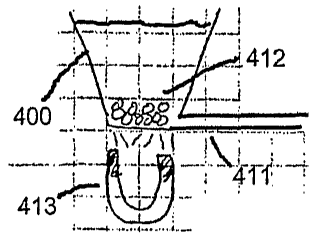
represented as magnet 413 in Figure 4B.
Since the applied magnetic field retains the magnetic beads 412 at the bottom
of well 400, fluid can be removed and added to the well without displacing the
beads.
Thus, the supernatant portion of the raw sample can be removed from the well
400, and
wash buffer can be repeatedly added and removed from the well 400, to remove
the
unwanted portion of the raw sample so that only the component of interest
bound to the
beads remains. The fluid removal and addition steps are schematically
represented in
Figure 4C.
In some embodiments, the fluid can be removed and added to the well using
standard liquid handling equipment. Examples of commercially available
automated
liquid handling equipment that could be used in embodiments of the invention
are the
Genesis and Freedom EVO products sold by the Tecan Group, Ltd. (Switzerland),
and
the Biomek FX and Biomek 2000 products sold by Beckman Coulter, Inc.
(Fullerton,
CA). In the embodiment shown in Figure 4C, the instrument interfacing with the
microfluidic device containing the well controls the flow of fluid through an
inlet tube
414 and an outlet tube 415. As such, in the embodiment shown in Figure 4C, a
suitable
wash buffer can be cycled through wel1400 by introducing the wash buffer into
the well
400 through inlet 414, and then withdrawing the wash buffer through outlet
415. Note
that since the magnetic beads 412, which are bound to the component of
interest, remain
magnetically retained at the bottom of wel1400, the beads 412 are not
inadvertently swept
out of well 400 during the cycling of wash buffer therethrough.
CA 02621632 2008-03-06
WO 2007/041619 PCT/US2006/038745
14
After undesired components of the raw sample have been removed from the
well 400 by the wash buffer, the component of interest retained on the
magnetic beads
412 can be eluted. Two alternative methods. of introducing the elution buffer
that releases
the component of interest from the magnetic beads 412 are shown in Figures 4D
and 4E.
In Figure 4D, the elution buffer is introduced into the well from outside the
microfluidic
device. As was the case with the wash buffer, the elution buffer could be
introduced into
we11400 with standard liquid handling equipment or, as specifically shown in
Figure 4D,
through an inlet tube 414 whose flow is controlled by the instrument
interfacing witli the
microfluidic device containing the well.
Alternatively, as represented in Figure 4E, the elution buffer could be
introduced through channe1411 into the wel1400. In the embodiment of Figure
4E, the
elution buffer would be stored in another well (not shown) on the microfluidic
device,
and the instrument interfacing with the microfluidic device would direct flow
from that
well, through channel 411, into well 400. The conceptual embodiment shown in
Figure
4E is particularly appealing as elution buffer is caused to percolate through
beads 412 as
the beads are magnetically retained at the bottom of we11410.
To help the elution buffer release the maximum amount of the component
bound to the beads, the beads can be agitated during the elution step. As
shown in Figure
4F, the beads can be agitated by moving the beads within the well by
manipulating the
magnetic field generated by magnet 413. For example, Figure 4F schematically
illustrates repositioning the magnet 413 generating the field so that the
magnetic
particles 412 are moved to one side of the wel1412.
Under the standard Dynabead protocol, the time required to accomplish
elution is on the order of 5 minutes. Once the elution is complete, the
component of
interest will be present in the elution buffer either in suspension or in
solution. As shown
in Figure 4G, the elution buffer containing the component of interest can be
directed into
channel 411 by the flow control system in the instrument interfacing with the
microfluidic
device. Note that a magnetic field is still being applied to the magnetic
beads 412, so the
beads will be retained within the wel1400. Once the fluid containing the
component of
interest is directed into channe1411, the flow control system can direct the
fluid into other
areas of the microfluidic device where it can undergo fiirther processing
steps such as
PCR amplification and/or detection.
CA 02621632 2008-03-06
WO 2007/041619 PCT/US2006/038745
In an alternative embodiment, the elution steps shown in Figures 4F and 4G
can be replaced by an elution process in which elution buffer is flowed under
pressure
into well 400, as shown in Figure 4E, while an electric field is applied
across the length of
channel 411 that transports the inherently negatively charged DNA-molecules
eluted from
5 the beads into channel 411 against the flow of elution buffer. This
alternative elution
process is based on the selective ion extraction technology disclosed in, for
example, U.S.
Published Patent Application No. 2003/0230486.
An alternative embodiment in which the wash buffer and elution buffer are
introduced into the well through one or more microfluidic channels is shown in
Figures
10 5A-5G. In the embodiments of Figures 5A-5G, a single channel 511 is
connected both
to a well containing wash buffer and to a well containing elution buffer. The
initial
situation shown in Figure 5A is identical to the situation depicted in Figure
4A: a raw
sample and a suspension containing magnetic beads is introduced into well 500,
while a
flow control system maintains a zero flow rate through channel 511. Once
again, in this
15 example embodiment, the raw biological sample is blood, and the reagents
and beads
used to extract the component of interest (DNA) from the raw sample are the
components
of the commercially available Dynabeads DNA DIRECTTM Universal product kit.
Thu,
in this embodiment the magnetic beads 512 are suspended in a buffer containing
a lysing
agent.
After the appropriate incubation period, the magnetic beads 512 are
subsequently retained at the bottom of well 500 in the same manner as shown in
Figure
5B. The step shown in Figure 5B is essentially identical to the step
represented by Figure
4B in the previously described embodiment. The step represented in Figure 5C,
however,
differs from the step shown in Figure 4C. In Figure 5C, wash buffer is
introduced into
well 500 through channel 511. This is accomplished by having the flow control
system in
the instrument (not shown) interfacing with the microfluidic device direct
flow from a
well containing wash buffer (not shown) through channel 511 into well 500. In
contrast,
in the previously described embodiments shown in Figure 4C the wash buffer was
introduced into well 500 from a source external to the microfluidic device. In
the
embodiment shown in Figure 5C, where the wash fluid is introduced at the
bottom of well
500, poor mixing between the supernatant portion of the raw sample and the
wash buffer
causes the supernatant sample to be displaced from the bottom of the well by
the
CA 02621632 2008-03-06
WO 2007/041619 PCT/US2006/038745
16
incoming wash buffer. As shown in Figure 5C, a sufficient amount of wash
buffer can be
introduced into the wel1500 so that the beads 512 at the bottom of the well
400 are
completely immersed in wash buffer. At this point, it may be desirable to
reposition the
magnet 513 to manipulate the field applied to the beads so that the beads are
agitated
within the wash buffer. This agitation step, which is represented in Figure
5D, can
enhance the effectiveness of the wash buffer in removing unwanted portions of
the raw
sainple from the vicinity of the beads 512.
As was the case in the embodiment shown in Figures 4A-4G, in the
embodiment shown in Figures 5A-5G the wash step is followed by the
introduction of an
elution buffer. As shown in Figure 5E, in the current embodiment the elution
buffer is
introduced tlirough channel 511. This is accomplished by having the flow
control system
in the instrument (not shown) interfacing with the microfluidic device direct
flow from a
well containing elution buffer (not shown), through channel 511 into well 500.
Once
again, the poor mixing between the elution buffer and the wash buffer will
cause the
incoming elution buffer to displace the wash buffer from the bottom of
we11500. Figure
5E represents the situation in wel1500 after a sufficient amount of elution
buffer has been
introduced into well 500 to displace the wash buffer from the vicinity of the
beads 512.
As shown in Figure 5F, the beads can be agitated to increase exposure of the
surfaces of
the beads to the elution buffer. After the elution step is complete, the
elution buffer
containing the component of interest can be withdrawn from well 500 through
channel
511 as shown in Figure 5G.
Not surprisingly, other variations on the present theme can be employed in
carrying out this inventive method. A third embodiment of the invention is
schematically
represented in Figures 6A-6D and in Figure 7. In this embodiment, well 600
consists of
an aperture in a cover layer 620, wllich is bordered by an opening 625 in the
top surface
of the cover layer 620, and two ports 615 in the main body 610 of the
microfluidic device
encompassed by the aperture. A top view of the microfluidic device illustrated
in Figures
6A-6D can be seen in Figure 7, where the aperture opening 625 in the cover
layer
encompasses the two ports 615,616 in the underlying main body of the device.
The steps
in the embodiment in Figures 6A-6D are quite similar to the steps in the
embodiment
shown in Figures 5A-5G, with the main difference being that the well 600 in
Figures 6A-
6D is in fluid communication with two channels 611,617 instead of just one
channel, e.g.,
CA 02621632 2008-03-06
WO 2007/041619 PCT/US2006/038745
17
511. The presence of the second channel in the embodiment of Figures 6A-6D
allows
undesired material, such as supernatant sample and used wash buffer, to be
removed from
the well 600.
Figure 6A represents the application of a magnetic field by a magnet 613 to
collect the magnetic beads 612 within one of the ports 615 after the magnetic
beads 612
have been incubated with the raw sample solution so that the cells in the raw
sample are
lysed to release the component of interest from the cells, and so the released
component
of interest can then bind to the ligands on the surface of the magnetic beads.
As
previously discussed, if a commercially available magnetic bead kit is
employed, the
standard conditions for lysing and binding specified for the kit can be used.
As shown in Figure 6B, after the beads are retained within the portion of the
well 600 defined by port 615, wash buffer can be introduced into the well
through
channel 611 and withdrawn from the well 600 through channel 617. Withdrawal of
the
used wash buffer from the well 600 should aid in the removal of undesired
material from
the vicinity of the beads 612.
After the washing step in Figure 6B is complete, elution solution can be
introduced through channel 611 as shown in Figure 6C. As shown in Figure 7,
channel
611 is in fluid communication with a wel1750 that contains wash buffer and a
well 760
that contains elution buffer. Known methods of controlling flow in a
microfluidic device
can be used to selectively direct flow from either well 750 or well 760
through channel
611 into well 600. In the embodiment shown in Figure 7, fluid withdrawn from
well 600
through channel 617 can be directed by a flow control system into a waste well
consisting
of port 771 and aperture 772.
After the required incubation period for elution has transpired, the elution
buffer containing the component of interest can be withdrawn from wel1600
through
channel 611 as shown in Figure 6D. As schematically illustrated in Figure 7,
flow from
channel 611 can be directed into channel 780, where the component of interest
can be
subjected to further processing. For example, wells 785 and 786 could contain
reagents
that will react with the component of interest as it travels through
channe1780 towards
waste we11790.
When the component of interest is genetic material such as DNA, the further
processing that takes place after sample purification will often include PCR
amplification
CA 02621632 2008-03-06
WO 2007/041619 PCT/US2006/038745
18
of the DNA. So, for example, the PCR process described in U.S. Published
Patent
Application No. 2002/0197630 could be performed on a sample purified using
methods in
accordance with the invention.
In methods in accordance with the invention, the entire process of removing a
component of interest, i.e., purifying, a raw biological sample takes place
within a well in
a microfluidic device. Since these methods do not require that the sample be
introduced
into the channels or chambers within the interior of the microfluidic device,
the problems
associated with flowing a raw sample through those channels or chambers are
completely
eliminated. Nevertheless, since the well is connected to the network of
microfluidic
channels in the device, the integration and automation provided by
microfluidic
technology can still be exploited.
The invention can be embodied in other specific forms without departing from
the spirit or essential characteristics thereof. The present embodiments,
therefore, are to
be considered in all respects as illustrative and not restrictive, the scope
of the invention
being indicated by the appended claims rather than by the foregoing
description, and all
changes which come within the meaning and range of equivalency of the claims
are
therefore intended to be embraced therein.