Note: Descriptions are shown in the official language in which they were submitted.
CA 02621863 2008-02-20
-~.-
APPARATUS FOR INDICATING THE BONE THICKNESS
BETWEEN A CAVITY IN A BONE AND THE BONE SURFACE
This invention relates to apparatus for indicating the bone thickness between
the predetermined distal end of a bone cavity located on a proximal/distal
axis and
the outer surface of the bone.
When preparing cavities in a bone, for example to receive the insert portion
of
a prosthetic component, it is necessary to drill into the bone. When fitting a
prosthetic component to the proximal end of a femur when carrying out proximal
epiphyseal replacement technique surgery, two proximal axially extending holes
are
drilled, one at an angle to the other, which can subsequently be joined by
reaming to
present a V-shaped cavity. When preparing such a cavity it is essential that
one or
other of the drill holes does not approach too close, or even break through,
the wall
of the bone. It is also common to prepare cavities, again, for example, at the
end of a
femur which are wide and if not accurately dimensioned, for example with
regard to
the width or the depth, can be too close to the wall of the bone, especially
in the neck
of the femur.
The present invention is intended to provide apparatus which will assist in
determining the thickness of the bone between the distal end of a bone cavity
and
the outer surface of the bone.
According to the present invention apparatus for indicating the bone thickness
between the predetermined distal end of a bone cavity located on a
proximal/distal
axis and the outer surface of the bone comprises a support member provided
with
means for location on the bone and having adjustable distance indicating means
which can be adjusted in relation to said support means to contact the outer
surface
of the bone.
With this apparatus and knowing the accurate dimensions of the apparatus
itself it is possible to measure the bone thickness concerned.
CA 02621863 2008-02-20
-2-
The apparatus may include a proximal/distal bone axis indicator which can,
for example, be in the form of a guide wire. With this in place the guide wire
can
provide a datum which can be used to set up the apparatus.
In a preferred form the guide wire is provided by a guide pin which forms part
of the means for location of the support member on the bone. Thus, the
apparatus
can be located on this accurately placed pin.
In an alternative arrangement the means for location of the support member
on the bone may comprise a collar adapted to fit on the bone at a
predetermined
location.
The apparatus can also include extension means which can be adjusted to
vary the proximal/distal length of the support member and which in one
embodiment
may comprise an adjustable carrier on which the adjustable distance indicating
means is supported.
In an alternative construction the extension means may comprise a series of
slots provided on said support member and adapted to locate on co-operating
connection means carried on the means for location on the bone, use of
alternate
slots effectively increasing or decreasing the effective proximal/distal
length of the
support member.
In a preferred embodiment the adjustable distance indicating means include
a sliding pin provided with distance indicating indicea which can be adjusted
in
relation to the support member.
The invention is not limited to operations relating to a femur and it can be
used on any bone in which the distance between an opening of the wall is
required.
CA 02621863 2008-02-20
-3-
The invention can be performed in various ways and two embodiments will
now be described by way of example and with reference to the accompanying
drawings in which :
Figure 1 is a diagrammatic side elevation of apparatus for indicating
the bone thickness between the predetermined distal end of a bone
cavity located on the proximal/distal axis and the outer surface of the
bone according to the invention;
Figure 2 is a diagrammatic representation of the proximal end of a
femur showing how it is prepared to carry out proximal epiphyseal
replacement technique surgery;
Figure 3 is a diagrammatic cross-sectional representation of a prosthetic
component for use in proximal epiphyseal replacement technique surgery
and for which a prosthetic stem cavity can be prepared using the
apparatus according to the invention;
Figure 4 is a diagrammatic cross-sectional view of an alternative form of
apparatus according to the invention;
Figure 5 is an exploded isometric view of the apparatus shown in Figure 4;
Figure 6 is a side elevation of an alternative construction according to the
invention;
Figure 7 is an exploded view of parts of the support member;
Figure 8 is an isometric view of the construction shown in Figure 6; and,
Figure 9 is a cross-sectional plan view on the line IX-IX of Figure 8.
CA 02621863 2008-02-20
-4-
The apparatus according to the invention is particularly, although not
exclusively, for use with proximal epiphyseal replacement technique surgery.
An
example of this type of surgery is explained and shown in EP 1138 283 and
includes
resecting a femur at a position on the proximal side of its neck to locate a
prosthetic
femoral component which has a tapered insert portion and a proximal head
portion.
The insert portion is adapted for location in a prepared socket which,
effectively, has
inclined side to 5 provide a tapering opening.
In order to carry out this type of surgery the end of a femur, indicated by
reference numeral I in Figure 2, the femoral head, indicated by reference
numeral 2,
is first prepared by machining it with a cylindrical cutter (not shown) to
provide a
cylindrical portion 3. In order to accurately locate the cylindrical cutter a
proximal/distal opening is drilled in the head to receive a guide wire and
this opening
is subsequently enlarged to receive a guide pin 5 on which the cy(indrical
cutter is
located. This pin can also be used in the apparatus to be described herein.
The neck of the femur is indicated by reference numeral 4.
Figure 3 shows diagrammatically how a typical femoral head component for
this type of surgery has a tapered insert portion 11 and a head 12 is fitted
into a
cavity 13 in the proximal end of the femur 1 which, in this Figure, is
indicated by
chain lines.
In order to provide the cavity 13 a hole is drilled into the prepared femur on
a
axis 14 which is substantially co-axial with the proximal/distal axis of the
end of the
femur. A second opening is also drilled along the line of another
proximal/distal axis
15 which is an angle to the line 14 to provide the basis for the tapering
socket.
By careful measurement of the femur and knowing the dimensions of the
prosthetic component it is possible to accurately determine the position of
the tip 16
CA 02621863 2008-02-20
-5-
of the component in the bone and the present apparatus can be used for
indicating
the bone thickness between the predetermined distal end of the bone cavity
which is
located on aproximal/distal axis and the outer surface of the bone, indicated
by
reference numeral 17. Reference numeral 18 indicates the calcar which can be
used as a reference point.
Apparatus according to the invention is shown in Figure 1 and the same
reference numerals are used for similar parts, as indicated in Figure 2. In
this Figure
however the apparatus is being employed before the cylindrical cutter (not
shown) is
used to provide a cylindrical portion 3 and the guide pin 5 is used in
connection with
the present apparatus.
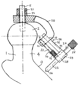
Apparatus according to the invention comprises a support member 20
provided with means for location on the bone in the form of a prepared socket
21
provided in a boss 22 which is dimensioned to be a close but sliding fit on
the guide
pin 5 which acts as a proximal/distal axis indicator. Thus the guide pin which
was
established on the guide wire provides a datum for setting up the apparatus.
The
support member 20 has an adjustable distance indicating means in the form of a
sliding pin 23 which is provided with distance indicating indicea 24. The
sliding pin
23 can be adjusted in relation to the support member 20 and carries an
operating
knob 25 for this purpose. As will be seen from Figure 1 the distal end 26 of
the
sliding pin 23 can be pushed into engagement with the surface 17 of the bone.
The apparatus also includes extension means 27 which can be adjusted to
vary the proximal/distal length of the support member 20. These extension
means
comprise an adjustable carrier 28 on which the sliding pin 23 is supported in
a bore
29. The carrier 28 is connected to the C-shaped support member 20 by a screw
threaded shaft 30. The proximal end 31 of the screw-threaded shaft 30 is
rotatable
in a screw-threaded bore 32 and the distal end of the shaft 30 is located in
the
carrier 28 but is free to rotate. The shaft has an enlarged knurled portion 33
to
assist in rotating it.
CA 02621863 2008-02-20
-6-
Location of the shaft 30 causes it to progress through the screw-threaded
opening 32 so that the position of the carrier 28 relative to the support
member 20
can be adjusted.
The carrier 28 also has a location strut 36 which is rigidly secured thereto
and
extends from the carrier through an opening 37 in the support member 20. An
indicator fin 38 is provided on the support member 20 which aligns with
indicea 39
provided on the location strut 36. With this arrangement therefore the carrier
28
accurately located on the support member 20 so that its proximal/distal
position can
be adjusted.
With this arrangement therefore, knowing the precise dimensions of the
apparatus, it can be used to measure the distance from the predetermined
position
of a hole to be drilled in the head to measure the thickness of bone at the
predetermined position.
In most circumstances this apparatus will merely confirm that the thickness of
bone is adequate but if it proves to be too small then it will be known to the
surgeon
that he cannot use the normal surgical technique to drill the holes and it may
be
necessary to use some different technique for applying the prosthetic insert.
Thus
the apparatus can prove valuable in overcoming the difficulty of knowing
whether a
particular technique can be used or not. If, for example, proximal epiphyseal
replacement technique surgery was intended and it was found that there was
insufficient bone a different technique could be used whilst the bone was
still
undrilled. Initial drilling without previously measuring the bone thickness
can do
considerable damage to the head of the bone which could cause further
difficulties if
an alternative technique was subsequently to be employed.
Figures 4 and 5 show an alternative construction and in which similar
reference numerals are used to indicate similar parts of the bone. This
apparatus
CA 02621863 2008-02-20
-7-
can conveniently be used on a head which has been prepared as shown in Figure
2
but in this case the pin 5 need not be employed because the support member 40
is
located on the bone by a collar 41. The collar can be held in place by screws
(not
shown) being passed through screw-threaded holes 42. The collar also has a
downwardly projecting pointer 43 which can be used to align the collar 41 in
the
desired position.
In this construction the support member 40 has a series of three slots 45 (as
shown in Figure 5) which are adapted to be a close sliding fit on co-operating
connecting means 46 which are in the form of a projecting lug 47 which fits
into the
slots 45. A spring loaded locking pin 48 is provided which can engage an
enlarged
opening 49 in the lug 47to rigidly hold the support member 40 in position. As
the
slots 45 are arranged in a proximal/distal direction alternative use of the
slots can
effectively extend or contract the length of the support member 40 in relation
to the
collar 41.
The distal end of the support member 40 carries a sliding pin 50 which is
provided with indicea 51 and has an operating knob 52. This sliding pin 50
operates
in a similar manner to the sliding pin 23 in the construction shown in Figure
1.
Figures 6, 7, 8 and 9 show another alternative construction which can be
used in place of that shown in Figure 1. In Figure 6 the femur is indicated by
chain
dot lines and the same reference numerals are used to indicate similar parts
as
those shown in Figure 1. The apparatus comprise a support member 60 provided
with means for location on the bone in the form of a clamp 62 which is
designed to
engage and clamp on a guide wire 61.
The clamp 62 comprises a cylindrical portion 63 provided on the support
member 60 and the distal end of which is provided with a claw-shaped portion
64
CA 02621863 2008-02-20
-8-
which has an inner curved surface 65 shaped to engage the guide wire 61. The
guide wire is held in place by a clamping sleeve 66 which is bifurcated to
forma
shaped slot 67 at its proximal end and a flat flange 68 at its distal end.
The bore 69 of the clamping sleeve 66 is dimensioned to be a sliding fit on
the cylindrical portion 63.
From Figure 7 it will be seen that the clamping sleeve 66 is assembled over
the cylindrical portion 63 with a compression coil spring 70 which also fits
over the
cylindrical portion 63 and is located between the flange 68 on the clamping
sleeve
66 and the end of an enlarged portion 72 of the support member 60. In order to
retain the clamping sleeve 66 in place a pin 75 is provided which is located
in a hole
76 in the cylindrical portion 63. The pin 75 is long enough to project from
one side of
the hole 76 and engage at that end in a slot 77 which extends through the wall
of the
clamping sleeve. As the slot 77 is elongated it acts to hold the sleeve 66 in
place but
allows sliding movement for the length of the slot between the parts.
Figure 9 shows how the shaped slot 67 in the clamping sleeve 66 extends
around the claw 62 and, when a guide wire 61 is in place, acts to clamp it
against the
inner curved surface 65 the claw 62. To release the wire 61 it is merely
necessary to
move the clamping sleeves 66 against the action 62 of the spring 70 along the
cylindrical portion 63 so that the wire 61 can be withdrawn from the inner
curved
surface 65 of the claw 64. Thus the guide wire 61 provides a datum for setting
up
the apparatus.
The support member 60 has an adjustable distance indicating means similar
to that shown in Figure 1 in that it includes a sliding pin 23 which is
provided with
distance indicating indicea 24. The sliding pin 23 can be adjusted in relation
to the
support member 60 and carries an operating knob 25 for this purpose. As will
be
seen from Figure 6 the distal end 26 of the sliding pin 23 can be pushed into
engagement with the surface 17 of the bone.
CA 02621863 2008-02-20
-9-
The adjustable distance indicating means is connected to the support
member 60 by extension means 80 which can be adjusted to vary the
proximal/distal
length of the support member 60. These extension means comprise an adjustable
carrier 81 on which the sliding pin 23 is supported in a bore 82. The carrier
81 is
connected to the support member 60 by passing through a slot 83, best shown in
Figure 7. The carrier means 81 is in the form of a flat bar 81 and is provided
with a
series of linked openings 84. The bar 81 can be locked in a number of
proximal/distal positions by operation of a locking pin 85 which is assembled
in a
bore 86 in the enlarged portion 72 of the support member 60. As will be seen
from
Figure 9 the pin passes through the bore 86 and extends on the far side where
it is
engaged by an operating button 87. The button is screw threaded onto the end
of
the locking pin 85 and a compression spring 88 is located between the
operating
button 87 and the end of the bore 86 so that the button can be moved towards
the
bore thus causing the pin 85 to move with it and allow a waisted portion 89 of
the
pin to be aligned with the slot 72. The dimensions of the waisted portion 89
of the
pin are slightly less than the gaps 100 between the openings 84 in the flat
bar 81
and this enables the bar to be moved lengthwise within the slot 72 to vary its
operative length. The selected length can be clamped into position by
releasing the
operating button 86 to allow the locking pin 85 to move into its innermost
position,
as shown in Figure 9, where an enlarged portion 101 of the locking pin 85 is
engaged
in one of the openings 84 of the bar 81 and thus holding it in position.
Suitable indicea 103 is provided on the edge of the extension so that its
precise length can be ascertained by the operator.
The apparatus is operated in the same way as that described with regard to
the apparatus shown in Figure 1.
CA 02621863 2008-02-20
-ZD-
The apparatus is not exclusively for use when carrying out proximal epiphyseal
replacement technique surgery but can be used in many other operations in
which a
hole has to be drilled into a bone and where it is desirable to know the bone
thickness between the end of the hole and the surface of the bone.