Note: Descriptions are shown in the official language in which they were submitted.
CA 02622544 2008-03-13
WO 2007/040645 PCT/US2006/018237
SYSTEMS AND METHODS FOR NON-INVASIVE DETECTION
AND MONITORING OF CARDIAC AND BLOOD PARAMETERS
Reference to Priority Applications
This application claims priority to U.S. Application No. 11/234,914 filed
September 26, 2005. This patent application is incorporated herein by
reference in its
entirety.
Technical Field of the Invention
In one aspect, the present invention relates to methods and systems for
monitoring
physiological parameters such as respiration, cardiac and/or vascular
parameters, events
and anomalies, such as embolic events, on an intermittent or continuous basis,
using
systems that are portable and ambulatory, over an extended period of time.
Blood flow
parameters, events and anomalies are monitored and detected using non-invasive
ultrasound techniques. Cardiac parameters, events and anomalies are monitored,
for
example, using non-invasive pressure-sensing and ECG technologies, as well as
ultrasound techniques. Ambulatory monitoring systems incorporate data
recording,
processing and storage capabilities for recording and/or storing acquired
data, optionally
processing acquired data to deterniine and output one or more physiological
parameters,
uploading and downloading data and/or instruction sets, inputting patient
data, and
triggering one or more alarms or notifications. Data analysis may be performed
by the
ambulatory device and/or by a companion analytical system to which data is
uploaded.
Back2roand of the Invention
Systems for monitoring numerous physiological parameters are well known and
are used widely in health care settings. These systems provide a generally
high level of
data collection and analysis but few of these systezns are ambulatory and few
provide
long term monitoring and data analysis over a period of several days, months
or years.
Yet, many physiological irregularities manifest only periodically or may be
asymptomatic
and are difficult to detect during routine patient evaluation, for example,
during an
appointment with a health care professional or during a hospital stay.
Ambulatory heart
rate monitors are available commercially and are used for fitness training,
cardiac
CA 02622544 2008-03-13
WO 2007/040645 PCT/US2006/018237
rehabilitation"arid lhe'l'ike. Some data storage and analytical features are
provided, alarms
may be programmed or programmable, and various levels of information may be
displayed. These systems generally don't have the capability and aren't
intended to
provide recording and storage of heart rate data for an extended time period.
Heart rate
monitors typically use a chest band having one or more electrodes to detect
heart rate,
althougli monitoring at sites other than the chest using other modalities can
be done.
For patients having cardiac irregularities or symptoms that occur sporadically
or
are asymptomatic, cardiac ECG monitoring is performed over a period of time
using
portable, battery-operated Holter monitoring or cardiac event monitoring
devices and
techniques. Holter monitoring is a common type of ambulatory ECG monitoring in
which the electrical cardiac signals are detected by electrodes contacting the
chest and
connected to a recording device. A patient typically keeps a detailed diary of
activities
and symptoms for a 24 or 48 hour period, during which time the cardiac
monitoring takes
place so that irregularities are detected and associated witli patient
activities and
symptoms. Holter monitoring is used to identify cardiac arrhythmias as well as
transient
ischemic episodes and silent myocardial ischemia.
Holter monitors generally record every heartbeat for a recording period,
providing
continuous cardiac ECG data over the recording period and are typically worn
for 24 to
48 hours. Presyniptom (looping memory) cardiac event monitors constantly
monitor and
provide short-term recording of ECG signals. When symptoms occur, the patient
presses
a button that makes a permanent recording of the ECG data both prior to and
following
activation of the button. Patient-activated looping memory monitors are
typically worn
for.30 days, but only patient-initiated events are permanently recorded. A
postsymptom
event monitor is generally used only when symptoms of a heart problem occur.
The
patient activates the system to start an ECG recording following the onset of
symptoms.
Recorded Holter and event monitor data are generally analyzed off-line using
dedicated
diagnostics systems and services. Programmable, auto-trigger monitors are
available for
arrhythmia detection. Such devices have been found to be particularly useful
for
monitoring events that are asymptomatic, such as asymptomatic arrhythmias,
Tachycardia, Bradycardia and Pauses.
Although Holter and cardiac event monitors are being used in attempts to
diagnose and monitor various cardiac irregularities that are asymptomatic or
infrequently
experienced, their limited data storage and analysis capabilities have reduced
their
application for wider ranging diagnostic and monitoring applications. The
success rate is
rather low with these devices, since the Holter monitor seldom captures rare
events in the
2
CA 02622544 2008-03-13
WO 2007/040645 PCT/US2006/018237
5" "'typicY, 'fe"latYte1~''shdi'tL'term recording period and the event monitor
is patient-triggered
and user dependent. These systems could be improved with more substantial
recording
and data storage capability and better analytical systems. The Holter and
cardiac event
monitors also are typically operated as stand-alone devices and are not
interfaced with
other devices collecting clinically useful patient data. Nonetheless, Holter
and cardiac
event monitoring are the only longer-term cardiac event monitoring systems
presently
available.
Doppler ultrasound techniques measure the frequency shift (the "Doppler
Effect")
of reflected sound, which indicates the velocity of the reflecting material.
Long-standing
applications of Doppler ultrasound include monitoring of the fetal heart rate
during labor
and delivery and evaluating blood flow in the carotid artery. The use of
Doppler
ultrasound has expanded greatly in the past two decades, and Doppler
ultrasound is now
used in many medical specialties, including cardiology, neurology, radiology,
obstetrics,
pediatrics, and surgery. Transcranial Doppler (TCD) technology today allows
detection of
blood flow in intracranial arteries and is used for intraoperative monitoring,
to detect
intracranial stenoses, to measure dynamic cerebrovascular responses, and to
detect
emboli.
Transcranial Doppler (TCD) techniques require application of the ultrasound to
those areas of the skull where the bone is relatively thin. The frequency of
the Doppler
signal is also adjusted, and pulsed wave rather than continuous wave
ultrasound is used to
augment the transmission of ultrasound waves through the skull. Blood flow
velocities
from the cerebral arteries, carotid arteries, the basilar and the vertebral
arteries can be
sampled by altering the transducer location and angle, and the instrument's
deptli setting.
The most common windows in the cranium are located in the orbit (of the eye),
and in the
temporal and suboccipital regions. Using TCD ultrasoriography, cerebrovascular
responsiveness to various physiological and pharmacological challenges can be
assessed
instantaneously, and various cerebral circulatory tests can be repeated
frequently and
safely. Rapid changes of cerebral perfusion over time can be easily followed,
documented
and analyzed and emboli and other blood flow irregularities can be detected
with a high
degree of sensitivity.
Emboli produce high intensity, transient Doppler ultrasound signals when they
traverse sample volumes of a Doppler ultrasound instrument, and emboli may be
detected
directly as changes in Doppler signal amplitude. U.S. Patent 5,348,015, for
example,
discloses methods and apparatus for ultrasonically detecting, counting and/or
characterizing emboli in either arterial or venous circulation.
3
CA 02622544 2008-03-13
WO 2007/040645 PCT/US2006/018237
,. .. , .::.. ..:... . ... ..
~ .S. ftterit"6;T9'6,972 relates to a pulse Doppler ultrasound system for
monitoring
blood flow including a graphical information display that simultaneously
displays depth-
mode and spectrogram data. The depth-mode display indicates various positions
along
the ultrasound beam axis at which blood flow is detected, with color
indicating the
direction of blood flow and varying intensity indicating the Doppler
ultrasound signal
amplitude or detected blood flow velocity.
Disturbances such as patient and probe movement and non-embolic debris in
circulation reduce the sensitivity and accuracy of emboli detection using
Doppler
ultrasound techniques. Data processing techniques have been developed to
increase the
accuracy of Doppler ultrasound emboli detection methodologies. Several
teclmiques are
described in Wang et al., Enaboli detection usirzg the Doppler ultrasound
technique,
Technical Acoustics Vol. 22 No.lE, pp. 15-18, 2003. U.S. Patent 6,547,736
discloses a
pulse Doppler ultrasound system for monitoring blood flow and detecting emboli
in
which subtraction of various background or artifact elements of the detected
Doppler
signals is provided to reduce false positive identifications of embolic
events.
U.S. Patent 6,616,611 discloses a Doppler ultrasound technique using clutter
filtering to subtract out signals that may be intense but are low velocity and
hence
represent tissue rather than embolic events. A depth-mode display assists the
user in
determining whether a desired vessel has been located and a simultaneously
displayed
spectrogram is used for successfully and reliably locating and orienting the
ultrasound
probe and determining an appropriate sample volume depth.
One drawback of using acoustic techniques for measuring physiological
parameters and detecting anomalies such as emboli using standard Doppler
techniques is
that localization of a desired CNS target area using an acoustic transducer is
challenging
and generally requires a trained, experienced sonographer to find and
(acoustically)
illuminate the desired target area, such as the middle cerebral artery (MCA).
After
locating the desired target area, the sonographer generally attaches a
cumbersome and
uncomfortable headset to the transducer that stabilizes the transducer
position and reduces
the effects of patient movement and other disturbances on the position of the
transducer.
The sonographer may be required to monitor acoustic readings and reposition or
reorient
the transducer interinittently to maintain the focus on the desired data
acquisition area.
This generally limits the use of Doppler ultrasound detection techniques to in-
hospital and
in-clinic situations where a trained sonographer is available.
There is increasing evidence that asyinptomatic emboli are more frequent than
clinical embolic events and are an important and detectable risk factor for
transient
4
CA 02622544 2008-03-13
WO 2007/040645 PCT/US2006/018237
isciieniic' attack "aYid"strbke. TCD monitoring for asymptomatic cerebral
emboli has been
limited to relatively short recordings by equipment size and complexity and
because
probe fixation and operation typically requires a trained sonographer, as
noted above.
Several systems for extended TCD monitoring have been proposed. U.S. Patent
6,682,483 discloses methods and devices that provide three dimensional imaging
of blood
flow using long-term, unattended Doppler ultrasound techniques. Doppler
ultrasound
blood velocity data is collected in a three-dimensional region using a planar
phased array
of piezoelectric elements that lock onto and track points in the three-
dimensional region
that produce the locally maximum blood velocity signals. The automated
tracking
process may be used to provide a three-dimensional map of blood vessels and
provide a
display that can be used to select multiple points of interest for expanded
data collection
for long-term, continuous and unattended blood flow monitoring.
Long-term ambulatory monitoring for cerebral emboli using TCD using an
ambulatory TCD system is described in Mackinnon et al., "Long-Term Ambulatory
Monitoring for Cerebral Emboli Using Transcranial Doppler Ultrasound," Stroke,
74-78,
January 2004. The middle cerebral artery (MCA) Doppler signal was obtained via
the
transtemporal window with a conventional Doppler unit, with the ainbulatory
probe
positioned at the transtenlporal window. Both a proprietary elastic headband
and glasses
were initially evaluated as methods of probe fixation. The software monitored
the
Doppler signal quality and implemented an auto-search module that attempted to
restore
vessel insonation during recording when the signal dropped below a preset
level. The
search mode was activated at regular intervals to optimize insonation.
Spencer Technologies (Seattle, WA) has developed a TCD probe fixation system
employing a headframe having a Doppler ultrasound probe mounted for contacting
a
subject's teinporal region to access the teinporal window for extended
surgical
monitoring, embolus detection monitoring and physiologic testing. The goal of
the
headfraine is to prevent movement of the probe. The preferred methodology
requires first
locating and assessing the temporal window using a hand held ultrasound probe
and then
positioning and orienting the probe on the headframe at the desired temporal
window
location. It is recommended that the headframe be completely loosened or
removed for
30-60 minutes every 3 hours of monitoring.
Deep vein thromboses in the peripheral vascular system, and particularly in
the
deep veins of the calves and thighs, produce narrowing of vessels that may
interfere with
circulation and may also embolize to produce embolic events in the heart,
lungs, brain
and other organs. Doppler ultrasound techniques are used to assess deep vein
5
CA 02622544 2008-03-13
WO 2007/040645 PCT/US2006/018237
thromboses, but conventional techniques and devices do not provide long term
monitoring, are not ambulatory, and suffer many of the disadvantages of
Doppler
ultrasound systems described above.
There is thus a significant need for methods and systems that provide long
term,
ambulatory monitoring of physiological parameters such as respiration, cardiac
and/or
blood flow parameters, events and anomalies and applicants' systems and
methods are
directed to addressing this need.
Summary of the Invention
The present invention provides ambulatory, noninvasive monitoring systems for
acquiring and storing data relating to one or more of the following
physiological
parameters: respiration, heart rate, body temperature, skin or tissue '
conductance,
electrical heart activity (electrocardiogram - ECG), myocardial tissue
stiffening, tension,
strain or strain rate for assessing myocardial contractility, myocardial
ischemia and
infarction, ventricular filling and atrial pressures, as well as diastolic
functions, blood
flow velocity, blood flow volume, blood pressure, intracranial pressure
("ICP"), presence
of einboli in the blood stream and other blood flow-related irregularities,
such as stenoses
or vasospasm, electrical brain activity (electroencephalogram - EEG), and
blood oxygen
coniposition or partial pressure (02, C02). Non-invasive pressure sensing
devices such as
electro-optical sensors, strain gauges and pressure transducers, for example,
may be used
to acquire data relating to respiration and heart rate, and conventional ECG
techniques
and electrodes may be used to acquire data relating to heart rate, blood
oxygen
composition, and electrical heart activity. Pulse oximetry techniques using,
for example,
electro-optical sensors, may be used to acquire data relating to heart rate
and blood gas
coinposition. Standard non-invasive blood pressure detection techniques using
pressure
cuffs or pressure transducers may be used to acquire data relating to blood
pressure. EEG
electrodes and data acquisition techniques are preferably used to acquire data
relating to
brain activity. Non-invasive ultrasound techniques are preferably used to
acquire data
relating to myocardial tissue properties and anomalies, blood flow properties,
blood
velocity, ICP, blood flow anomalies, the presence of emboli, and the like, and
may also
be used to acquire data relating to blood pressure. These systems may also
incorporate
movement detection devices to document the occurrence of motor seizures.
Monitoring systems of the present invention comprise one or more data
acquisition devices such as one or more of the devices described above that,
when placed
in proximity to and/or in contact with a subject, acquires data relating to
one or more of
6
CA 02622544 2008-03-13
WO 2007/040645 PCT/US2006/018237
.~Y
tlie esiredpara, .meters. Each of the data acquisition devices is in data
trans er
communication, via electrical leads or using a wireless data transfer
protocol, with a
patient data recording and storage device. The data acquisition device may be
housed in a
single module with the data recording and storage device, or these functions
may be
housed in multiple modules.
The patient data recording and storage device has robust data storage capacity
and
may have data processing, analytical and display capabilities. Data recorded
and stored is
identified with a unique identifier corresponding to the individual subject
for whom data
is being acquired. Recorded and stored data is also identified with time and
date
information and a time and date display may be provided. A microphone and
audio or
mechanical recording activator may also be provided, enabling the subject to
record
observations, activities and events as desired. Patient initiated information
may also be
input into the patient data recording and storage device using patient
selectable menu
choices and other data input mechanisms.
In one embodiment, the patient data recording and storage device may be
provided as a portable module designed for ambulatory subjects having an
integrated
power source and data transfer capabilities. Power sources that are
rechargeable using
electrically powered recharge devices are preferred. In another einbodiment,
the data
recording and storage device may be provided as a typically stationary, table-
top module
designed for patients who have limited mobility, with power provided from
external
sources. Collected data maybe directly transferred to, stored and analyzed at
one or more
remote locations, or a local patient data recording and storage device may
haved data
transfer capabilities that enable transfer of data from the storage device to
a separate, data
processing and analytical system, and/or to a larger capacity data archiving
facility. Data
transfer may be accomplished by physically removing a data storage subassembly
from
the data storage device, or using data transfer techniques employing a cable
or a wireless
protocol. Data transfer may be performed on a substantially real-time basis
witli
substantially continuous or frequent transfers of data from the patient
recording and
storage device and/or data acquisition devices to a remote data processing and
analytical
system for substantially real-time monitoring. Alternatively, data transfer
may be
performed periodically and at intervals determined by the subject or
professional
caregiver or at data transfer intervals programmed into the device.
The patient data recording and storage device may be operated to collect
and/or
store data continuously or intermittently and may optionally have analytical
and/or
display capabilities as well. In one embodiment, manual activation and shut-
off
7
CA 02622544 2008-03-13
WO 2007/040645 PCT/US2006/018237
= r, P i;:d,,, "=af .j.: ~~ 6~t" tr r
iriec anismspf'~vided, enabling a subject to activate and inactivate the data
acquisition devices and record and store data. In another embodiment, one or
more data
acquisition routines is programmed into the patient data recording and storage
device and
desired data acquisition routines may be selectable by the subject or pre-set
by a health
care professional. Data acquisition routines may involve, for example,
acquiring data
from one or more data acquisition devices at certain time intervals or during
certain
physiological states, acquiring data for certain time intervals, and
transmitting and storing
the data in specified databases or in one or more storage location(s).
The system may be programmed or programmable to compare real-time, acquired
data with predetermined or programmable standards and identify anomalies.
Alarm
and/or notification triggers may be preset or programmable at predetermined
limits and
alarms and notifications may be delivered locally, to the subject, or remotely
to a
monitoring service or health care provider. Certain data acquisition and
analysis
functions and capabilities may be selected and programmed by health care
professionals
and certain functions and capabilities may be programmable or selectable by
users. The
ambulatory devices may be provided with individual identifiers and may have
data
transmit-receive capabilities that enable acquired data to be transmitted to a
remote data
storage and/or analysis system, and that enable control systems, data
acquisition and
analysis routines, limits, and the like to be transmitted from a remote
location to the
ambulatory device.
Ambulatory devices may also have localization capabilities incorporating VHF,
GPS, satellite and/or triangulation location systems. These systems are
capable of
notifying care-givers or services having a companion receiver, in real time,
of anomalies
in a subject's physiology, location or the like, thus allowing the monitoring
entity to take
action to find and assist the subject. The inventive system may thus function
as a rapid
alarm, providing identification of the subject, the location of the subject
and an indication
of the problem the subject is having. The system may be applied, for example,
to
children, hikers, at-risk persons with known medical conditions, and
ambulatory, as well
as bed-ridden, patients.
A separate data processing and analytical system generally provides data
retrieval
and sophisticated data analysis when desired by a health care professional and
incorporates or is used in conjunction with a display system for presenting
visual
representations of the analyzed data. Substantial efficiencies are achieved
because a
single analytical system may be located remotely from the subject being
monitored and
used to evaluate patient data for a relatively large patient population. This
analytical
8
CA 02622544 2008-03-13
WO 2007/040645 PCT/US2006/018237
sys't"effi' used"liy'd'Oct'6A and other health care professionals to evaluate
the condition of
a patient and formulate diagnoses, prognoses, etc. Subject data may also be
transferred,
from the patient data recording and storage device and/or from the separate
data
processing and analytical system to a remote data storage and archiving
facility.
A standard cardiac monitor with event capability provides continuous recording
of
respiration, heart rate and event-triggered ECG. The measurements are compared
periodically to a calibrated norm and recording of the ECG data is activated
for the
duration of an event or for a predetermined time period when acquired
measurements
deviate from the norn7 by a predetermined amount. This device may be used by
athletes,
runners, cyclists, trekkers, climbers, patients undergoing cardiac
rehabilitation and
subjects at risk for or evidencing symptoms of cardiac irregularities. A
calculation of the
amount of calories lost during a measurement or exercise period may be
performed and
displayed and a body temperature reading may be measured and displayed as
well. The
inclusion of a location identifying technology such as GPS and wireless
communication
capability enables this system to also serve as an alarm and provide speedy
location of the
subject. A beacon function may be included to facilitate this safety-related
use where
wireless operation is not possible.
Systems of the present invention may be employed as a highly effective child
and
infant monitor. Such a monitoring device may incorporate many of the functions
identified above. The child's respiration may be continuously monitored and
any
meaningful deviation from a predetermined or empirically determined standard
may
trigger an audible alarm both at the data acquisition device and at the
matched receiver
device. This type of child monitoring device may additionally incorporate
heart rate
and/or ECG monitoring capability that may be automatically activated and
monitored or
that may be activatable by a companion receiver/controller device. This system
may be
set up so that a parent or supervisor may monitor location and communicate
(two-way)
with the child at any time by remote. In the event of anyone tainpering with
the child, the
child could push an alarm button activating the alarm to the parent and
turning on the
VHF transmitter and/or GPS and microphone. This would also occur automatically
if
anyone tried to tamper with or remove child's monitoring system. An on-site
alarm and
beacon may be incorporated for added safety.
Systems of the present invention that monitor respiration and/or heart rate
and/or
ECG may also be used for detection of sleep apnea without requiring a subject
to stay at a
specialized laboratory or wear unconifortable breathing monitors. The system
described
herein allows detection of apnea and otb.er abnormalities in a subject's own
home, at a
9
CA 02622544 2008-03-13
WO 20 07/040645 PCT/US2006/018237
. ,.. ..
fow cost, "and"cari "beuged to monitor the success of any therapy instituted.
The system
may also detect respiratory depression in infants and children, and can
therefore be used
to detect and prevent SIDS by monitoring the breathing status of children
during sleep.
Systems of the present invention may also be employed to monitor cardiac
tissue
properties and cardiac parameters using non-invasive techniques, such as
ultrasound
techniques. Such a system may, for example, provide monitoring of myocardial
tissue
stiffness, tension, strain, strain rate and the like, for assessment of
myocardial
contractility, myocardial ischemia and infarction, ventricular filling and
atrial pressures,
as well as diastolic functions. Methods for making these types of assessments
are
disclosed in U.S. Patent 7,022,077 B2, which is incorporated herein by
reference in its
entirety.
Another aspect of methods and 'systems of the present invention relates to
monitoring devices that, in addition or alternatively to having one or more
cardiac
monitoring functions, have the capacity to acquire data relating to blood and
blood flow
parameters using non-invasive techniques and similarly analyze, report,
trigger alarms,
and provide effective long term and remote monitoring of blood flow conditions
and
anomalies. Systems of the present invention incorporating a noninvasive
ultrasound
detection device are useful for providing long term monitoring of circulation,
blood
pressure and blood flow velocities, ICP, and for detecting blood and blood
vessel
anomalies such as stenoses, vasospasm and emboli.
In one embodiment, a "long term" emboli detection trace corresponding to data
acquired over a time period of at least several hours and up to several days
or months is
provided to illustrate trends and fluctuations in emboli over time that may be
predictive of
risk for pulmonary embolism, stroke, transient ischeinic attacks, and the
like. These
systems are based on Doppler or other acoustic measurements, such as acoustic
scatter,
taken from a target site on or within or in proximity to a blood vessel such
as the MCA, a
carotid artery, another cranial blood vessel or, for peripheral blood
monitoring
applications, a peripheral blood vessel. Monitoring systems incorporating
ultrasound data
acquisition devices preferably incorporate an automated target vessel locating
and
focusing feature that scans a tissue volume and identifies and focuses on
blood vessel(s)
and blood vessel volume(s) exhibiting desired acoustic properties relating to
desired
blood flow characteristics. This automated target vessel locating and focusing
feature
preferably updates and adjusts the focus and/or orientation of one or more
acoustic data
acquisition devices at regular intervals during long term monitoring
operations.
CA 02622544 2008-03-13
WO 2007/040645 _ PCT/US2006/018237
5' blood flow anomaly detection and monitoring is preferably
accomplished using an ambulatory ultrasound source/receiver system that may be
mounted on or applied to a patient's skull, neck, leg, trunk or the like, and,
during
operation, it preferably locates and maintains focus on a desired vessel or
another three-
dimensional target area with little or no assistance from an operator. An
initial
environmental assessment may be made, if desired, to assess the
characteristics of the
environment between the acoustic source and the target vessel site, and
calibration or
programming of the data acquisition device for use with a particular blood
vessel may be
facilitated by a health care professional. The initial environmental
assessment may be
determinative of various method and system parameters. Environmental
assessments
may additionally be updated at intervals throughout a diagnostic or monitoring
procedure.
A property of blood flow, such as acoustic scatter or flow velocity, may be
determined in any blood vessel. For determination of ICP and emboli detection
applications, arteries that traverse, or enter or exit CNS tissue
(collectively, "cranial blood
vessels") are preferred. Peripheral veins in the leg or tlligh are preferred
for detection of
emboli that are predictive of risk for pulmonary embolism. Blood flow
properties are
preferably detected using ultrasound techniques such as Doppler and
Transcranial
Doppler (TCD) ultrasound techniques, which are well known in the art.
Doppler ultrasound techniques may be used to acquire data relating to blood
flow
velocity and ICP and may be used, as well, to detect stenoses, vasospasm,
emboli and
other blood flow anomalies. In addition or alternatively, acoustic properties
of tissue,
including blood, blood vessel walls, tissue in proximity to blood flow, and
other tissue
sites, may be assessed, for example, by collecting acoustic scatter data using
an
ultrasound transducer aimed at or having a focus on a blood vessel, and/or at
another
target site. For purposes of detecting emboli, the target vessel site is
preferably a cranial
blood vessel or a blood vessel that leads to or traverses the brain, or a
peripheral blood
vessel such as a deep vein in an extremity. Cranial blood vessels may be
accessed by
contacting an ultrasound transducer to the temporal window through the skull
or by
contacting an ultrasound transducer to a location on the neck or upper chest
where
acoustic access to a cranial blood vessel such as a carotid artery is
available.
Monitoring of at least one of the common carotid arteries, cervical internal
carotid
arteries, middle cerebral arteries, subclavian arteries, vertebral arteries
and basilar arteries
is preferred for cerebral blood flow monitoring and emboli detection. In one
preferred
system, monitoring of a carotid artery that traverses the neck is provided
using a portable
ultrasound transducer mounted on an elastic band attachable around a subject's
neck.
11
CA 02622544 2008-03-13
WO 2007/040645 PCT/US2006/018237
"Syste'"r'fi9'"'bt_ tYid pl'egenti -mvention incorporating emboli detection
features may be used to
assess a subject's risk for stroke and other blood flow abnormalities and to
assess the
efficacy of treatment regimen. Monitoring of a deep venous vessel in the
peripheral
vascular system, such as deep veins in the legs, is preferred for peripheral
blood flow
monitoring and emboli detection and may be used to assess a patient's risk for
pulmonary
embolism and other blood flow abnormalities, as well as assess the efficacy of
a treatment
regimen.
Methods and systems of the present invention provide spatial location of
desired
target areas based on their acoustic properties, automated focusing of an
acoustic source
at one or more desired target area(s) and if desired, mapping of one or more
desired target
areas, such as a blood vessel. Multiple target vessels or multiple target
locations within
multiple vessels or multiple locations within a single vessel may be monitored
simultaneously or sequentially using ultrasound data acquisition techniques.
Suitable
source/detector combinations and transducer assemblies for scanning and
locating desired
target areas are described.
Blood flow monitoring and emboli detection methods and systems that monitor a
carotid artery, for example, may operate in one or more modes. A carotid
artery
monitoring regimen may involve acoustically illuminating (scanning) a
relatively large
tissue volume and analyzing received acoustic signals from a relatively large
tissue
volume to identify the location of the artery within a larger region of
tissue. Thereafter, a
focused acoustic beam may be aimed to acoustically illuminate substantially an
entire
cross-section of the artery, or one or more focused acoustic beams may be
aimed
simultaneously or sequentially to illuininate distinct smaller volumes within
the cross-
section of the artery. Acoustic detection patterns may match the transmit
patterns or may
differ from the transmit patterns. A multi-frequency acoustic array may be
used in
conjunction with multi-frequency transmit and detection schemes to provide
enhanced
detection of desired events and conditions, such as the presence of emboli.
Systems of the present invention may also incorporate three dimensional
locating
and/or mapping functions tat associate a point or area in three-dimensional
space with
various determinations made, anomalies identified, and the like. The location
and
mapping function may be displayed locally or remotely.
Systems of the present invention provide long term monitoring of ambulatory
patients to identify events and abnormalities that are asymptomatic and/or
infrequently
experienced and also provide effective assessment of treatment regimen. They
are
suitable for use with ambulatory subjects and may also be used in non-
ambulatory
12
CA 02622544 2008-03-13
WO 2007/040645 PCT/US2006/018237
applicatioris subli-as "iri "hospital rooms, surgical suites, ambulances,
nursing and other
long term care facilities, and the like. Integrated monitoring systems, for
example, may
be employed to provide comprehensive patient monitoring within a hospital or
institution
at a fraction of the cost of conventional monitoring equipment. At present,
hospitals have
only a fraction of their beds monitored, and the only monitoring systems are
cardiac
monitoring devices that require operation by trained nurses. A very small
percentage of
cardiac arrest patients in-hospital survive, due to the very critical few
minutes before the
code team gets to them. Alarm and notification systems of the present
invention alert
nurses or other care-givers in a residential or hospital facility, or
monitoring professionals
in a remote monitoring facility and expedite the delivery of essential and
appropriate care
and intervention. Methods and systems of the present invention can be used to
notify
medical staff at the very early moments of a respiratory or cardiac arrest or
of a major
embolic event or blood flow abnormality, thereby greatly increasing the
chances of a
successful outcome.
Brief Description of the Fi2ures
Fig. 1 illustrates a schematic diagram showing various ambulatory components
of
systems of the present invention.
Fig. 2 is a schematic flow diagram depicting data acquisition, processing and
communication functions of systems of the present invention.
Fig. 3 is a schematic diagram illustrating one embodiment of a patient data
recording and storage device.
Detailed Description of the Invention
Methods and systems of the present invention may comprise numerous
combinations of features and capabilities, as described herein. As illustrated
schematically in Fig. 1, a system of the present invention comprises one or
more
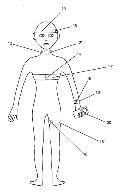
noninvasive data acquisition devices 10, 12, 14, 16, 18 or a sirriilar device
provided in
proximity to or in contact with a patient's skin or outer surface. In one
embodiment, data
acquisition devices 10, 12, 14, 16 and 18 are mounted or incorporated in or
integrated
with flexible, elastomeric bands 10', 12', 14', 16', 18' or alternative
mounting systems
sized to fit snugly around one or more features of a patient's anatomy. One or
more of
the bands may be adjustable to facilitate snug fitting of the band and contact
or close
proximity of the data acquisition device with a surface of the subject. The
data
acquisition devices may be provided at a fixed position on the respective
band, or they
13
CA 02622544 2008-03-13
WO 2007/040645 PCT/US2006/018237
may be mova6Te "ariti"guyustable on the respective bands to tacilitate
positioning ot tine
device at desired locations. In another embodiment, not illustrated, one or
more data
acquisition devices may be provided in connection with a garment or another
form-fitting
assembly.
In the embodiment illustrated schematically in Fig. 1, data acquisition device
10 is
intended for mounting on a subject's skull in proximity to a temporal window,
data
acquisition device 12 is intended for mounting on a subject's neck for data
acquisition
from blood vessels traversing the neck, such as a carotid artery, and data
acquisition
device 18 is intended for mounting on a subject's extremity, such as a thigh,
for data
acquisition from blood vessels such as deep veins, traversing the extremity.
Each of these
data acquisition devices preferably comprises an ultrasound transducer or
transducer array
capable of insonating and scanning a tissue target site to identify a target
vessel of
interest, focusing on one or more desired volume(s) of the vessel of interest,
and
acquiring acoustic data from the vessel of interest that relates to blood
pressure, blood
flow velocity and/or blood flow anomalies such as emboli.
Data acquisition device 14, intended for mounting on a subject's chest,
preferably
comprises one or more pressure sensing devices such as a pressure transducer
or strain
gauge for detection of respiration and measurenlent of heart rate and/or one
or more
electrodes for acquisition of ECG signals. Pressure sensing devices for
acquisition of
respiration and heart rate data may alternatively or additionally be mounted
on another
portion of the subject's trunk or provided in a data acquisition device 16
intended for
mounting on a subject's arm.
In one embodiment of the system of the present invention an elastic, pressure-
sensing material such as KINOTEX or another type of polymer foam consisting
of a
layer of thin cellular elastomers of urethane or silicon that electro-
optically measures
continuous mechanical respiration and/or heartbeat, is implemented as a data
acquisition
device. The polymer foam may be provided in the form of an elastic band or a
close-
fitting garment and may include an inner and/or outer skin of cotton or other
comfortable
material providing a patient contacting or wear surface. One or more ECG
sensors and/or
leads may be used in conjunction or integrated with a pressure sensing band or
garment
for acquisition of data relating to respiration, heart rate and ECG from the
same wearable,
ambulatory device.
As shown schematically in Fig. 2, each of the data acquisition devices 10, 12,
14,
16, 18 or the like, is in data flow communication with a data recording and
storage device
20. Data may be acquired in one or more of the acquisition devices on a
substantially
14
CA 02622544 2008-03-13
WO 2007/040645 PCT/US2006/018237
corifiiiiiotiis baO"Wirit''rinittently, and is conveyed to a data recording
and storage device
wirelessly or via electrical leads. Alternatively, data acquisition may be
initiated or
terminated by the user or a health care professional. Data acquisition times
and patterns
may be programmed or programmable via the data recording and storage device 20
and/or via another external programming input controller.
Data recording and storage device 20 may, in addition to data recording and
storage capabilities, have data analysis capabilities provided, for example,
in software or
firmware. High capacity data recording and storage may be provided in a
variety of
formats, such as Smart Media Cards, Flash Cards, in embedded Flash caches or
other
types of embedded digital storage media and may be provided as a removable
data
storage medium or as an embedded medium. For ambulatory applications, data
recording
and storage device 20 is preferably a relatively small, portable, battery
operated device
that can be easily carried by the user in a pocket or bag, worn on a user's
belt, placed in
proximity to the subject, or the like. Data acquired from data recording and
storage
device 20 is preferably marked with a unique identifier assigned to the
patient using the
device.
Data processing and analysis capabilities provided in recording and storage
device
20 may be programmed or programinable. In one embodiment, data acquired may be
processed in device 20 to determine heart rate, respiratory rate, body
temperature, calories
burned, or the like, for example, which may be displayed continuously or
intermittently
on a device display. Acquired data may be averaged over programmed or
programmable
time periods and otherwise processed according to methods that are well known
in the art.
Data recording and storage device 20 may also be prograinmed or programmable
by the
end-user or a medical professional using selectable embedded programs and
limits or
using an auxiliary programming input device 30. Device 20 may be programmed or
programmable to incorporate threshold limits or value ranges and data
processing
routines activating an alarm or notification, locally or remotely, when
acquired data
exceeds a programmed limit or falls outside a predetermined range.
Data acquisition and storage device 20 is illustrated schematically in Figs. 1
and 3
as a portable, ambulatory device, but it will be recognized that the data
acquisition and
storage device that interfaces with the patient data acquisition devices may
alternatively
be provided as a stationary, table-top type system designed for use in
hospital and
residential care facilities. A stationary system may have enhanced data
processing and
display functions compared to the ambulatory device and may provide longer
term
storage capabilities and enhanced alarm and notification functions.
CA 02622544 2008-03-13
WO 2007/040645 PCT/US2006/018237
Data'stofetl"Yri'd'evice 20 is preferably transferable to a separate
analytical device
40 for more sophisticated data processing, analysis, patient diagnosis, and
the like.
Analytical device 40 may be installed, for example, at a health care or
monitoring facility
and operated by health care professionals. Data may be transferred by removing
a
removable data storage medium and physically transferring the stored data to
analytical
device 40, or data may be transmitted using wireless or wired techniques from
device 20
to a remote analytical device 40 for data processing and analysis. Data
processing,
analysis and monitoring services may thus be centralized and receive and
analyze data
from numerous data acquisition and storage devices used by numerous patients.
Data stored in device 20 and/or data and analytical information generated by
analytical device 40 is preferably transferable to a data storage or archiving
facility 50
that is separate and optionally remote from data storage device 20 and
analytical device
40. When a separate data storage or archiving facility 50 is used, data is
preferably
transferable between archiving facility 50 and data analysis device 40 upon
command.
Device 20 may also have transmit/receive capability to analytical device 40
for relaying
alarms or notifications, for example, or to an independent matched
transmit/receive
device 60. VHF, GPS, satellite and triangulation location methodologies may be
implemented.
Fig. 3 illustrates a highly schematic diagram illustrating one embodiment of a
data
recording and storage device 20. Device 20 incoiporates a time/date display
22, and a
data display 24 for displaying cardiac and/or blood flow parameters calculated
using data
acquired from the data acquisition devices. Data relating to one or more of
respiration
rate, body temperature, heart rate, blood oxygen content, calories burned,
blood flow
velocity, ICP and blood pressure may be displayed, for example, for viewing by
the user.
Alarms and notifications may also be displayed. A display actuator 26 is
preferably
provided for manually activating and inactivating the display. Data storage
capability
may be incorporated as an integral part of device 20, or one or more
insertable and
removable data storage subassemblies 28 may be provided for data storage. High
capacity data storage capabilities are preferred.
Data recording and storage device 20 may additionally incorporate a manual
data
recording activator mechanism 32 that may be activated by a user, for example,
upon a
user's perception of symptoms or unusual conditions, to record and store data
during
and/or prior to an activation period. A data recording and storage inactivator
mechanism
34 may also be provided to permit the user to manually terminate data
recording and
storage upon return to perceived normal physiological conditions. A data
input/download
16
CA 02622544 2008-03-13
WO 2007/040645 PCT/US2006/018237
tunctiow5b may"a'rsorvvc'provided to allow the user or a medical professional
to input data
or information or to download progranuning or analytical data processing
capabilities to
the data recording and storage device 20. A voice recording actuator 42 may be
provided,
allowing a user or medical professional to record voice or auditory input to
device 20
through microphone 44. Audible alarms or notifications may be provided through
amplifier 46 and visual alarms and notifications may be provided through
visual alarm 48.
It will be appreciated that many modifications to device 20 as it is
illustrated in Fig. 3
may be made to provide the various features described herein and to deliver
relevant data
in a fashion that is most useful to both the subject and a medical
professional.
In one embodiment, a system of the present invention may incorporate one or
more ultrasound transducer or transducer array data acquisition devices
mounted in a
patient affixation device such as a headset or an elastic band suitable for
mounting on a
subject's skull, neck or extremity. Acoustic data is used, in this embodiment,
to detect
and monitor blood flow parameters such as blood flow velocity, changes in
blood flow
and blood flow parameters, arterial blood pressure, ICP, and the presence of
blood flow
anomalies such as emboli and the like. All of these blood-related parameters
may be
detected using techniques that are known in the art and the device may be
programmed or
programmable to activate one or more alarms or notifications when the data
acquired is
outside predetermined limits or ranges. Data indicative of blood flow velocity
and ICP
may also be acquired and analyzed to provide real-time data relating to values
for blood
flow velocity and ICP and changes in blood flow velocity and ICP, which are
clinically
useful parameters.
Ultrasound sources and detectors may be employed in a transmission mode, or in
a variety of reflection or scatter modes, including modes that examine the
transference of
pressure waves into shear waves, and vice versa. Detection techniques
involving
measurement of values for or changes in acoustic scatter, such as back scatter
or forward
scatter, or reflection, and particularly backscatter, are preferred for use in
many
embodiments of methods and systems of the present invention. Exemplary
acoustic data
that may be used to determine blood flow parameters and identify anomalies
according to
the present invention include: values for or changes in acoustic scatter,
including values
of and changes in the amplitude, phase and/or frequency of acoustic signals,
values for or
changes in length of scattered signals relative to the interrogation signal,
values for or
changes in the primary and/or other maxima and/or minima amplitudes of an
acoustic
signal within a cardiac and/or respiratory cycle; values for or changes in
ratios of the
maximum and/or minimum amplitude to that of the mean or variance or
distribution of
17
CA 02622544 2008-03-13
WO 2007/040645 PCT/US2006/018237
subsequeri't si'gridls' cvi'lllin a cardiac cycle, values for or changes in
temporal or spatial
variance of scattered or emitted signals at different times in the same target
location
and/or at the same time in different target locations, values for or changes
in endogenous
and/or induced brain tissue displacement or relaxation, and rates of change
for such
displacements, such as the velocity or acceleration of displacement, and the
like, and
combinations of these data.
Multiple acoustic interrogation signals may be employed, at the same or
different
frequencies, pulse lengths, pulse repetition frequencies and intensities, and
the multiple
interrogation signals may be emitted from the same location, or from multiple
locations,
eitller simultaneously or sequentially. Acoustic scatter data may be
collected, for
example, from a blood vessel at different points along the vessel, within or
outside the
cranial cavity, or from multiple sites at or in proximity to different
vessels. Scatter from
single or multiple interrogation signals may be detected at single or at
multiple
frequencies, at single or multiple time points, and at single or multiple
locations. In one
embodiment, methods and systems of the present invention may be used to
localize blood
flow abnormalities and anomalies within different tissue samples, thereby
localizing areas
of trauma or dysfunction.
In one embodiment, Doppler techniques are used to measure flow velocity and to
detect blood flow anomalies such as emboli in a desired blood vessel, such as
the MCA
(V mca), a carotid artery, or a peripheral vein. Doppler is a preferred
ultrasound
technique and can provide substantially continuous measurement of flow
velocity. Many
types of Doppler devices are known in the art. The Spencer Technologies TCD
100M
Power M-Mode Digital Transcranial Doppler device is one such device that is
suitable for
collecting acoustic data from cranial blood vessels.
In addition to blood flow velocity in one or more selected vessel(s), acoustic
data
may also be acquired and processed to provide real-time determination of blood
pressure,
particularly arterial blood pressure (ABP). ABP may be determined using
acquired
acoustic data and techniques described in PCT International Publication No. WO
02/43564, which is incorporated by reference herein in its entirety. ICP may
also be
determined, in real time and during long term monitoring, using acoustic data
acquired as
described herein. Several methods and systems for determining ICP are
described, for
example, in PCT International Publication No. WO 2004/107963 A2, which is
incorporated by reference herein in its entirety.
If ABP, ICP, blood flow velocity and flow anomaly data are acquired in an
integrated data acquisition device such as an ultrasound transducer array as
described
18
CA 02622544 2008-03-13
WO 2007/040645 PCT/US2006/018237
.,.~~ . ==r ~:.,, ~,:;ar . aa al .,=
h, the ~d is+~onveniently synchronous with respect to acquisition time,
substantially reducing or eliminating the need for data synchronization. In
other
embodiments, ABP, flow velocity and flow anomaly data may be acquired using
different
devices and/or synchronization rates, with the data being collected and
processed in an
integrated processing unit that provides data synchronization as necessary.
ABP may also
be monitored non-invasively, for exainple, using a conventional arm or leg
cuff or
another non-invasive device, such as the VASOTRAC device manufactured by
Medwave, Inc., 4382 Round Lake Road West, St. Paul, MN 55112-3923. Blood
vessel
and/or blood flow characteristics and ABP may be measured on a substantially
continuous or an intermittent basis using acoustic data.
Various data processing techniques may be used to condition acquired acoustic
data. These include, for example, downsampling and/or resampling of telemetry
and
Doppler flow data to provide that each linear signal record occupies the same
amount of
space so that standard signal processing techniques may be einployed more
easily. Data
cleaning may also be implemented to ensure that all signal records are
continuous, within
expected physiologic ranges, and appropriate for further processing. Anomalies
may
trigger an alarm or notification to provide monitoring information and alert
the user or a
monitoring professional that a blood flow anomaly has occurred or that the
data
acquisition device is no longer operating properly. Phase alignment of cardiac
cycle
boundaries is generally implemented to ensure the input data is in phase with
regard to
cardiac cycle boundaries.
If pulse-domain transformation is performed, the data may require alignment,
such
as through cross-correlation spectrum analysis or other methodologies.
Transformation
of the linear, phase-aligned, time-domain telemetry and Doppler flow records
to two-
dimensional, normalized pulse-domain records may be desirable. This is a multi-
step
process and may involve calculation and storage of beat-to-beat instantaneous
heart rate,
normalization of each cardiac cycle to a fixed number of samples, and moving
pulse-
window smoothing or envelope calculation for the V mca Doppler flow data.
Systems of
the present invention for monitoring blood flow parameter and blood flow
anomaly
events preferably provide trend analysis and data display features. One
suitable output
display provides: (1) one or more trace(s) of embolic events over a "long
term" period of
time of at least several minutes and up to several hours or days to illustrate
trends in
patient embolic activity; (2) a trace of "instantaneous" or "short term" flow
anomalies,
determined over several cardiac cycles; and (3) additional graphical
representations that
may aid in guidance of an acoustic transducer or transducer array, as
described below.
19
CA 02622544 2008-03-13
WO 2007/040645 PCT/US2006/018237
5' "' ...... .... g" "A"caYYbMYonStep using a measure of blood pressure taken
with a conventional
blood pressure device may be incorporated in a system having the capability of
making
blood pressure determinations using acoustic data. Acoustic proxies for the
pulsatility of
the blood vessel - such as oscillation rate of the blood vessel wall - may be
substituted
for direct measures of those quantities. In this method, the spontaneous
changes in the
diameter (or other geometric property) of the vessel being monitored are
assessed using
ultrasound, and this information is related (e.g., using correlation
techniques) to
synchronous Doppler flow measurements within the same vessel. Since the
diameter (or
other geometric property) of the vessel is a function of the pressure being
exerted against
the wall of the vessel by blood, and since the velocity of blood flow is
dependent on the
diameter (or radius) of the vessel through which the blood travels, blood
pressure can be
calculated from flow velocity measured by Doppler. By simultaneously measuring
the
pulsatility of the blood vessel of interest and the Doppler flow velocity
proximal and
distal to this site, continuous blood pressure can be determined.
One aspect of the present invention relates to the use of acoustic
source/detector
assemblies for acquiring data relating to blood flow parameters and for
detecting blood
flow anomalies. In operation, an acoustic source/detector combination, such as
a Doppler
source/detector, is stably mounted, or held, in proximity to a patient's body
surface, such
that the focus of the acoustic source(s) is adjustable to provide an acoustic
focal point on
a blood vessel or other target site within the patient's body. For CNS target
sites, the
acoustic source/detector is stably mounted, or held, in proximity to a cranial
window,
such that the focus of the acoustic source(s) is adjustable to provide an
acoustic focal
point on a cranial blood vessel. For vessel target sites such as the carotid
arter(ies), the
acoustic source/detector is stably mounted on a surface of the neck to provide
an acoustic
focal point on and/or within the vessel(s) of interest. Similarly, for
peripheral target sites,
the acoustic source/detector is stably mounted on a surface of the extremity,
such as on
the tliigh, to provide an acoustic focal point on and/or with the peripheral
vessel(s) of
interest.
The acoustic source/detector combination is preferably provided as a unitary
component, but separate acoustic source and detector components may also be
used. The
acoustic source/detector combination may be provided in connection with a
mounting
structure or accessory that provides temporary adherence to desired patient
sampling
locations and may be provided as a single use component.
Various types of acoustic transducers and acoustic transducer arrays may be
used
as acoustic source/detector assemblies and acoustic data acquisition
components of the
CA 02622544 2008-03-13
WO 2007/040645 PCT/US2006/018237
present'ifiVeritfoYi:"KA sfn'gle acoustic transducer, or a singer acoustic
transducer array may
be operated both as a source and a detector, or separate source and detector
transducers or
transducer arrays may be provided. Conventional PZT acoustic transducers may
be
implemented as acoustic data acquisition components in methods and systems of
the
present invention. Acoustic transducer arrays composed of cMUT and PVDF cells
or
elements may also be used and are preferred for many implementations. PZT,
eMUT and
PVDF acoustic transducers and arrays may be combined in various data
acquisition
components and operated in acoustic source and/or receiver modes in yet other
embodiments.
In one embodiment, the acoustic source/detector combination may be mounted on
a stabilizer, or on or in a structure,* such as a helmet-type structure or
headband or
neckband or legband that may be mounted on the patient at a location providing
acoustic
access to the desired blood vessel. An applicator containing an acoustically
transmissive
material, such as an acoustic gel, may be placed between the surface of the
acoustic
source/detector combination and the patient's skin. Steering of the acoustic
device may
be accomplished manually or using automated mechanisms, such as mechanical or
electronic steering mechanisms. Such mechanisms are well known in the art.
Methods and systems of the present invention incorporate systems and methods
forlocating and acoustically illuminating and/or probing a desired target area
in a reliable
and automated fashion, without requiring a trained sonographer. Major cerebral
vessels,
including the middle cerebral artery (MCA), are standard targets for
transcranial Doppler
procedures, and targets for acoustic measurements used in the methodology
employed for
detecting blood flow parameters and anomalies described above. The anterior
cerebral
arteries, anterior communicating artery, internal carotid artery and posterior
cominunicating artery are potential targets. In one embodiment of a scanning
mode, an
acoustic source/detector assembly of the present invention emits acoustic
interrogation
signals in a wide beam as described below, in which a relatively large target
area is
acoustically illuminated prior to the focusing and localization of acoustic
signals on one
or more smaller target site(s). In another embodiment of a scanning mode, an
acoustic
source/detector assembly emits a plurality of independent beams, separated in
time and/or
target focus and, based on referred signals, focuses and localizes acoustic
signals on one
or more target sites.
Thus, another aspect of the present invention relates to methods and systems
for
locating and acoustically illuminating and/or probing a desired target site in
aii automated
fashion using an array comprising a plurality of acoustic source and/or
detector elements.
21
CA 02622544 2008-03-13
WO 2007/040645 PCT/US2006/018237
Ari' acou"s'tic traris~&7rkeiver array may be employed in a scanning mode, for
example,
to acquire acoustic data from numerous sites within a larger target area.
Based on the
acoustic data collected in the scanning mode, localized sites within the
target area may be
selected as target sites for focused acoustic illumination and/or probing.
Localized target
sites may be selected, or predetermined, based on any aspect of the acoustic
data collected
in the scanning mode, such as acoustic scatter amplitude, phase and/or
frequency maxima
or minima, tissue stiffness properties, minimum resolvable variances, maximum
variances, spectral averages, cardiac averages, radial and/or vector blood
velocity, blood
flow volume, maximum, minimum, mean or any variance measurement of acoustic
brightness, endogenous and/or induced tissue displacement properties, rates of
change of
such properties, and various spatial and/or temporal distributions of any of
these values.
Focusing elements of an acoustic transducer/receiver array on selected target
sites
may be accomplished in an automated fashion, using mechanical or electronic
beam
steering and other automated acoustic focusing methodologies. In another
embodiment,
an automated system is provided that locates a desired target site within a
larger target
area in a scanning mode, focuses on the desired target site for acquisition of
acoustic data,
and tllereafter periodically scans the target area and repositions the
acoustic focus, if
necessary, to maintain the focus of the acoustic source at the desired target
site. Multiple
target sites may also be located in a scanning mode and focused on
sequentially and/or
simultaneously for acoustic data acquisition from multiple target sites using
acoustic
transducer/receiver array assemblies of the present invention. Systems
incorporating
suitable arrays of acoustic source and/or detector elements are disclosed.
A scanning acoustic transducer assembly of the present invention acoustically
illuminates and acquires acoustic data from multiple points within a broad
target area,
such as a large portion of the cerebral blood vessel complex, in a scanning
mode. Based
on the acoustic data acquired in the scanning mode, localized target sites
within the
scanned area may be identified and elements of the transducer assembly are
focused on
localized target site(s) for acquisition of acoustic data from the desired
target site(s).
Selection of localized target site(s) may be predetermined based on various
acoustic
properties, including the amplitude (or any amplitude derivative) of acoustic
scatter data,
Doppler analysis of acoustic scatter data, phase or frequency of acoustic
data, changes in
the primary and/or other maxima and/or minima amplitude, phase or frequency of
acoustic signals within a cardiac and/or respiratory cycle or other period, or
determinations derived from acoustic data, such as flow velocity, tissue
stiffness
properties, endogenous and/or induced tissue displacement properties, acoustic
emissions
22
CA 02622544 2008-03-13
WO 2007/040645 PCT/US2006/018237
"5"' 6s's'6&Wd' vhtfi,)' gueli" -displacements, rates of change of such
properties, minimum
resolvable and maximum variance(s), spectral average(s), cardiac average(s),
radial
and/or vector blood velocity and/or volume, maximum, minimum, mean and
variance of
acoustic brightness, and spatial and temporal distributions of any of these
quantities.
For monitoring blood flow parameters and anomalies using methods of the
present
invention, the selection of a desired localized target site, such as the MCA,
a carotid
artery, a peripheral vein, or another blood vessel, is preferably accomplished
by scanning
the desired target area, and determining the localized site of highest
amplitude acoustic
scatter, or highest Doppler or flow velocity values, which represents the
vessel of interest.
Acoustic elements of the acoustic source/receiver data acquisition component
may then
be focused on one or more localized blood vessel sites for acoustic data
acquisition.
Other sites having unique acoustic properties may also be located. Coordinates
for target
vessel volume location and values for acoustic properties may be recorded and
stored,
over time, mapped and displayed in a variety of formats.
Various noninvasive, non-acoustic detection modalities may be employed
alternatively or additionally to locate internal physiological structures,
including blood
vessels such as the MCA, prior to acquisition of acoustic data. Near infra-red
spectroscopy (NIRS), magnetic resonance, and other techniques are known and
used, for
example, to image and locate internal physiological structures. Such
techniques may be
used in association with the metliods and systems of the present invention for
locating
internal physiological structures prior to assessment of acoustic properties.
Using methodologies and assemblies described below, an acoustic
source/detector
combination, preferably an acoustic transducer array comprising multiple
transducer
elements, is operable in both a scanning mode and a focusing mode. One or more
acoustic source element(s) of the acoustic data acquisition component
acoustically
illuminates a relatively broad desired target area in a scanning mode to
identify target
sites having predetermined or desired acoustic properties, thus identifying
the target
site(s) as blood vessel(s). When the acoustic source has identified one or
more target sites
having the predetermined or desired acoustic properties, one or more of the
acoustic
source(s) may be manually or automatically focused on the desired target
site(s) for
operation in an acoustic interrogation or data acquisition mode. The acoustic
source may
also be programmed to monitor acquired acoustic data and to adjust the
positioning
and/or focus of the source to maintain the focus of selected or predetermined
acoustic
source(s) on the desired target site. Similarly, acoustic source(s) may be
programmed to
23
CA 02622544 2008-03-13
WO 2007/040645 PCT/US2006/018237
acquire data trom a" *pturality of predetermined or programmed target sites at
predetermined time points.
Having identified the location of the target vessel in a scanning mode, one or
more
target vessel volumes may be selected for data acquisition and analysis. For
methods and
systems involving data acquisition from the MCA, as described above, the
acoustic focus
and data acquisition volume generally represents substantially the entire
cross-section of
the target MCA vessel. For methods and systems involving data acquisition from
a
carotid artery or a peripheral vessel, it may similarly be desirable to
acquire acoustic data
in a volume that represents substantially the entire cross-section of the
target carotid or
peripheral vessel. In some embodiments, the focus and beam size of the
acoustic
source(s) may substantially match the focus and beam size of the acoustic
detector(s), so
that acoustic data is acquired from substantially the entire vessel volume
that was
acoustically illuminated.
For blood vessels having a relatively large cross-sectional volume, such as
the
carotid arteries and peripheral veins, for example, multiple sample volumes
that are
volumetrically smaller than a sample containing the entire vessel cross-
sectional volume
may be monitored simultaneously and/or sequentially. In a relatively large
vessel such as
a carotid artery or peripheral vessel, for example, it is desirable for some
applications to
acduire data from one or more relatively small vessel volumes at or near the
center of the
vessel and from one or more relatively small vessel volumes at or near the
periphery of
the vessel. Data from numerous vessel volumes may be acquired simultaneously
or
sequentially. The focus and beam size of an acoustic source may be
substantially larger
than that of one or more acoustic detectors to acoustically illuminate a
relatively large
vessel volume and provide data collection from one or more smaller vessel
volumes
within the larger acoustically interrogated volume. Alternatively, the vessel
volume
interrogated may substantially match the vessel volume from which acoustic
data is
acquired. In one approach, numerous vessel voluines are acoustically
illuminated,
simultaneously or sequentially. Alternatively, or additionally, a vessel
volume may be
monitored substantially, continuously, or at frequent intervals, particularly
in monitoring
applications to identify blood flow anomalies.
Monitoring of blood vessels such as a carotid artery may be accomplished using
a
generally higher frequency array than may be used for emboli detection, for
example, in
the MCA. Acoustic frequencies of from about 0.5 MHz to 15 MHz, more preferably
from about 1.0 - 10 MHz, may be used for carotid artery monitoring to provide
high
resolution acoustic data with a generally low level of artifacts. Vessel
monitoring may
24
CA 02622544 2008-03-13
WO 2007/040645 PCT/US2006/018237
5' a'Ts6 b"e'2ZdoriipfYslied"tYsing multiple frequencies for acoustically
interrogating annror tor
acoustic data acquisition over time and/or over vessel sample volumes to
facilitate
enhanced detection of blood flow parameters and anomalies. Acoustic transducer
source
and detector elements of the present invention may, in fact, be programmed to
collect one
or more types of acoustic data from a single or multiple target sites, at one
or more
frequencies and at one or more times. Acquisition of acoustic data, using
methods and
systems of the present invention, is preferably accomplished in an automated
fashion.
Methodologies for scanning and locating desired target areas based on their
acoustic properties may be based on "range-Doppler" search methodologies that
were
originally developed, for example, for prograinming torpedoes to hunt targets
such as
submarines. Range-Doppler processing is an efficient implementation of matched
filtering that has been used in the radar and sonar signal processing
community for many
years. It is a robust technique, in part because it makes very few assumptions
about the
statistical nature of the environment and targets that it encounters. Range-
Doppler
processing provides a useful decomposition of the spatial and temporal (i.e.
Doppler)
scattering properties of the target of interest. Sensor time series data are
divided into
frames, often overlapped, multiplied by the transmitted waveform replica and
then
transformed into the frequency domain via the Fast Fourier Transform (FFT)
algorithm.
These operations implement, very efficiently, a bank of matched filters, each
matched to a
narrow range of Doppler shifts. Range-Doppler processing affords separation of
targets
in terms of their range and speed relative to the acoustic device.
Intracranially, MCA
flow is by far the largest target, which makes it a natural for this 'search
and home in'
approach.
Other methodologies for finding and maintaining an acoustic focus on a desired
target area are also applicable. Acoustic holography techniques such as those
described
in Porter, R. P., P. D. Mourad, and A. Al-Kurd (1992), Wavefront
reconstruction in
variable, multimode waveguides. J. Opt. Soc. Ariz., A9(11) 1984-1990 and
Mourad, P.
D., D. Rouseff, R. P. Porter, and A. Al-Kurd (1992), Source localization using
a reference
wave to correct for oceanic variability, J. Acoust. Soc. Am., 92(1) 1031-1039,
may also
be used. Using acoustic holography techniques, signals from a target are
combined in a
convolution with signals from a reference source after each is measured on an
acoustic
array. The net result is a formula whose maximum occurs at the target site. To
determine
ICP using acoustic holography techniques, for example, all of the acoustic
fields may be
replaced by the Fourier transform of the acoustic field, or a component of the
Fourier
transform of the acoustic field, e.g. the Doppler signal. In this embodiment,
the Fourier
CA 02622544 2008-03-13
WO 2007/040645 PCT/US2006/018237
tr'ari0o''rm"6f t"adi5UMd backscatter from an acoustic array serves as the
target signal,
and the forward scatter from a TCD or array placed on the opposite temple may
be used
as the reference source. These signals would be mathematically combined to
find and
maintain an acoustic focus on a desired target area.
In another embodiment, it would be useful to have the option for the user to
have
the opportunity to assist the automated targeting, user independent aspects of
the present
invention. This may be useful, for example, for cases where systems for
automatically
identifying the feature of interest may not be uniquely converging on that
feature, or so
that the user can validate whether or not the feature chosen by the computer
is, in their
opinion, the optimal feature. The key idea is that the feature of interest
will be known to
represent a local if not global miniinuin or maximum among a spatial
distribution of
values of the feature of interest. We will use the example of finding the
maximum flow
velocity in the middle cerebral artery, where the velocity in the middle
cerebral artery is
known to have a range of values spatially distributed along the middle
cerebral artery,
with the understanding that this technology is not limited to this
application.
An exemplary acoustic system providing an automated targeting feature while
allowing user participation in targeting may utilize conventional TCD systems
made by
DWL, Spencer Technologies, Nicolet, or the like, wliere the acoustic sensor
consists of a
single transducer element, and where the acoustic system provides information
only along
the beam of the single transducerfor a given orientation of that transducer.
Here, the user
manually manipulates the transducer so that it insonifies different portions
of the cerebral
architecture, and electronically steers the depth along the transducer beam
axis. The user
would be guided by the real-time display of information, along with the user's
memory of
what the display has shown in the preceding moments, to seek out the maximum
in flow
velocity in the desired vessel. One portion of the display may provide the
real time value
of the variable of interest at a position relative to the face of the
transducer (reported in
absolute units, or arbitrary units, since the actual deptli is not important)
that is chosen by
the user with a cursor designated for this purpose. The display may provide,
for example,
the real time value of flow velocity in the MCA, otherwise known as the
spectrogram of
the flow.
. Another portion of the display may provide a graphical image designed to
communicate to the user, at any given orientation of the transducer, the
direction of larger
values of flow in the MCA relative to the real time position of the cursor.
This may take
the form of two arrows pointing in different directions, e.g. one pointing
'up' one
pointing 'down,' where up and down are known to the user to represent deeper
relative to
26
CA 02622544 2008-03-13
WO 2007/040645 PCT/US2006/018237
'tlie"pr'eseri't sr6f the cursor, and more shallow relative to the present
position of the
cursor, respectively. If there are local maxima in flow velocity in both
directions, the
direction in which a greater maximum exists would be designated by having a
brighter
arrow pointing in that direction. These flow velocity gradients may be
calculated within
the associated controller component by measuring the Doppler shift along all
of the points
insonified at a given moment by the transducer to provide a real-time
calculation of the
local gradient of the flow velocity. This calculation may be performed using a
variety of
well-known mathematical formulae (one-sided differences, centered differences
to a
variety of orders, etc). The absolute position of the local flow velocity
maximum in flow
in the MCA need not be known or reported or displayed to the user.
What the user gains from this analysis is a direction, relative to the current
position of the cursor, which position need not be defined, of the local
maximum in flow
velocity. The user may then manipulate the cursor to report the spectrogram at
a deeper
or a shallower position along the acoustic beam and judge for themselves
whether they
have achieved a local maximum in flow velocity. By providing guided
exploration of the
flow velocity along the beam axis in this fashion, in combination with
physical
manipulation of the relative position or angle of the transducer, the user
will be able to
locate the flow velocity maximum in a guided fashion.
Standard TCD devices also allow for the device to emit sound whose amplitude
is
tied to the flow velocity at a given point along the beam of the transducer,
the one, in
particular, whose spectrogranl is shown to the user. Such supplemental
information
would be of interest to the user of the present invention. In addition, one
could designate
the intensity of the display to increase or decrease as the absolute value of
the flow
velocity increased or decreased as the cursor was manipulated along the beam
of the
transducer. In this way visual information would supplement the aural
information
already available to the user.
Using an acoustic array comprising a relatively dense distribution of acoustic
transducers rather than a single transducer or a sparse array, one may have,
at any given
moment, information relating to the relative spatial distribution of flow
velocity or other
blood parameters in depth at a variety of angles from the center of the
acoustic beam. A
user assist feature may provide a display showing the direction of the local
flow velocity
maximum. Using a transducer array, however, locational information relating to
the
direction of maximum flow velocity may be provided in additional dimensions,
and the
user may be guided by an arrow pointing in each of the three possible
directions of cursor
movement relative to the real time cursor position. One set of arrows may
indicate the
27
CA 02622544 2008-03-13
WO 2007/040645 PCT/US2006/018237
5' "1'oc"''ma'x""imurri"3s"'ctddpbr than, or shallower than, the present
cursor position. Another set
of arrows may indicate that the local maximum is more anterior or posterior to
the present
cursor position. Yet another set of arrows may indicate that the local maximum
in flow
velocity is more superior or inferior to the present cursor position. This
information may
be calculated as described above, using Doppler analysis of acoustic
backscatter from the
field of positions insonified by the transducer array. The user's positioning
of the array
may be guided by this information, along with supplemental aural and visual
information
as described above, including the instantaneous spectrogram at the position of
interest, to
move the cursor, and re-examine the spectrogram.
Acoustic systems and transducer assemblies for locating and illuminating one
or
more desired target site(s) on or within a blood vessel are described below.
The acoustic
methods and systems described below may be useful for any application in which
collecting data relating to an acoustic property of a desired target site is
required.
Acoustic transducer arrays of the present invention are generally thin and
generally
comprise a single layer or thickness of transducer elements. Stacked,
inultiple layer
transducer cells, or elements, may be used for some applications. The
transducer
elements or cells may be arranged on a single plane to form a generally flat,
planar array,
or they may be arranged to form a curved or a geometrically stepped array.
Transducer a--rays having various configurations and structures may be useful
for
applications contemplated, in this disclosure. For applications involving
monitoring of a
carotid artery, a rectangular array having more cells aligned in one direction
than in the
other is generally preferred to facilitate monitoring of a vessel volume along
a length of
the carotid artery. For monitoring applications involving monitoring of
niultiple vessel
volumes simultaneously or sequentially, fewer cells may be employed in a
transmit mode
to acoustically illuminate a generally broad target area and more cells may be
employed
in a receive mode to acquire acoustic data from a plurality of different
vessel vohimes.
In one embodiment, data acquisition components comprising acoustic
source/detector coinbinations of the present invention comprise a plurality of
capacitive
micromachined ultrasound transducer (cMUT) cells. cMUT ultrasound transducers
are
manufactured using semiconductor processing techniques and have sufficient
power and
sensitivity to transmit and receive at diagnostic ultrasound energy levels,
which is
necessary and sufficient for purposes of the present invention. The transducer
elements
are fabricated using small capacitive diaphragm structures mounted on a
silicon substrate.
cMUT transducer arrays have the potential of being produced very
inexpensively, and
may also have the support electronics integrated onto the same chip.
28
CA 02622544 2008-03-13
WO 2007/040645 PCT/US2006/018237
-"' ""' cMU 1" 'U1tM90'ui1d transducer cells comprise a positive eiectroae,
generaiiy
provided as the top electrode and a negative electrode, generally provided as
the bottom
electrode. The top electrode is generally provided on or in connection with a
flexible
membrane and the bottom electrode is generally provided on or in connection
with a
substrate, such as a silicon substrate. Insulating supports are provided to
form a sealed
chamber between the positive and negative electrodes. The chamber may contain
a gas
or liquid or gel-like substance, or it may be provided as an evacuated
chamber. The
diaphragm structures of the cMUT ultrasound transducer convert acoustic
vibrations into
a modulated capacitance signal, or vice versa. A DC bias voltage is applied
and an AC
signal is either imposed on the DC signal in transmission or measured in
reception. In
general, cMUT transducer elements may be operated in various modes of transmit
and
receive operation, including unbiased mode, non-collapsed mode, collapsed mode
and
collapsed snapback mode (transmit only). One advantage of using cMUT
transducer
cells, elements and arrays is that the electronics may be provided on or in
the cell
structure, greatly simplifying the electronic communication to and from the
array and
facilitating programmable array features.
A cMUT transducer array is composed of multiple individual cMUT ultrasound
transducer cell structures arrayed as elements, with the elements arrayed in
rows and/or
columns and/or smaller divisions forming the array. The number of cV1UT
transducer
cells forming each transducer element, and the number of elements forming an
array may
be varied, depending on the array application. cMUT transducer arrays having
various
configurations may be assembled and used in the present invention. cMUT
transducer
arrays can be configured and operated to achieve acoustic transmission and
sensitivity
levels sufficient to perform as acoustic transmit/receive devices suitable for
use in
medical devices, such as TCD devices. More specifically, eMUT transducer
arrays
having a plurality of cMUT element columns operated at an 80V bias, 28 Vac to
transmit
acoustic energy to CNS target sites at intensities of up to 1.75 W/cm2, while
typical
transmission intensities of only about 0.6-0.7 w/cm2 are required for
determining cerebral
blood flow using conventional TCD acoustic devices. cMUT transducer arrays
operated
experimentally at an 80Vbias and at a gain of 60 and 80 dB to receive signals
from CNS
target sites in a range of less than 4 to greater than 6 cm from the array at
a level
sufficient to make Doppler determinations.
cMUT transducer cells and elements may be arranged in different combinations
to
provide cMUT transducer arrays having different capabilities. If each of the
cMUT cells
is nrovided with inrienenclentlv controlled or controllablv electronics. each
of the cMUT
29
CA 02622544 2008-03-13
WO 2007/040645 PCT/US2006/018237
cells inay ac as a" aiis ucer element and an array may be provi e as a p ura i
independently controlled or independently controllable cMUT cells. More
typically, a
transducer element comprises a plurality of cMUT cells that is electronically
controlled or
controllable as a unit. Thus, each of the elements composed of multiple cMUT
transducer
cells are controlled or controllable as a unit. Alternatively, a plurality of
the elements,
such as elements forming a row or a column, may be electronically controlled
or
controllable as a unit to provide a cMUT transducer array comprising a
plurality of row or
column transducer elements. A one-dimensional (1D) array may be composed of a
single
transducer element comprising multiple cells, while a two-dimensional (2D)
array is
composed of multiple transducer elements arranged in a generally planar, two-
dimensional configuration.
In one embodiment, two cMUT acoustic arrays, each composed of a single or
multiple transducer elements, are aligned in a "Mills Cross" configuration in
which two
transducer arrays are arranged generally orthogonal to one another, which
allows one
array to sweep vertically in send and receive modes and the other to sweep
horizontally in
receive and send modes. In this implementation, a first linear cMUT transmit
array may
be steerable in a first direction, such as a vertical direction and a second
linear cMUT
receive array is arranged generally orthogonal to the first linear array and
may be
steerable in a direction orthogonal. to the first direction. The two, crossed
linear cMUT
arrays alternatively transmit and receive ultrasound beams while steering the
sending and
listening beams, to identify and focus on acoustic signals having the desired
property.
In another embodiment, an acoustic array comprising PVDF (polyvinylidene
fluoride) film transducers is used as an acoustic detector array, alone or in
combination
with a cMUT array or a single element PZT transducer employed as the source.
In an
exemplary embodiment comprising a PVDF array in combination with another
transducer
or array, the source transducer or array transmits sound through the PVDF
array,
sweeping the sound in a single dimension generally perpendicular to the
arrangement of
the PVDF array. The PVDF array serves as the acoustic detector, receiving and
processing acoustic signals.
Acoustic transducer arrays suitable for use in systems of the present
invention
may alternatively comprise a combination PVDF/cMUT array(s). The combined
depth of
the arrays is generally quite small and may be on the order of about 1 cm. A
cMUT array
may be arranged below a PVDF array, for example, with the PVDF array arranged
closest
to the subject's surface during use. In this configuration, the cMUT array is
operated as
the acoustic source and transmits acoustic beams through the PVDF array. The
cMUT
CA 02622544 2008-03-13
WO 2007/040645 PCT/US2006/018237
'la41a~l Ma~ ~e''~o'4npd'se'd of a 1D or 2D array comprising one or more cMUT
acoustic
elements. The PVDF array may also be provided as a 1D array or as a 2D array.
When
acoustic source(s) and/or detector(s) are provided as 2D arrays, they are
capable of
sending and/or detecting acoustic signals in two dimensions, rather than a
single
direction.
Acoustic arrays suitable for use in systems of the present invention may also
comprise one or more combination(s) of PVDF array(s) and PZT transducer(s). A
cMUT
array may similarly be used in combination with a PZT transducer. The PVT
transducer
is generally mounted below the PVDF or cMUT array and transmits as an acoustic
source
through the PVDF or cMUT array in a single, broad beam. In these embodiments,
the
PZT transducer generally serves as the acoustic source and the PVDF or cMUT
array
generally serves as the acoustic detector. Each of the aligned transducer
elements in the
cMUT array is controlled or controllable as a unit.
One of the advantages of employing ultrasound transducer array components as
described above in systems of the present invention is that multifunctional
arrays may be
provided in a relatively high power, yet inexpensive system. Such arrays are
very
versatile, are capable of performing multiple acoustic functions and may be
pre-
programmed or programmable to provide desired functions, and may be provided
as
disposable or single-use elements of an integrated clinical diagnostic system.
In one
embodiment, acoustic arrays of the present invention are provided as a single-
use acoustic
data acquisition component of a medical device, such as a blood flow
monitoring system,
comprising one or more acoustic transducer arrays in operative communication
with a
controller component having data processing, storage and/or display
capability. The one
or more acoustic transducer arrays may coinmunicate with the controller
component by
means of one or more detachable cables, or using a radio frequency, infrared
or other
wireless technology. The transducer array(s) may be steerable and may be
progranuned
to scan one or more target areas having certain boundaries or parameters, and
locate one
or more desired target site(s) based on preselected or selectable acoustic
properties. The
transducer array(s) may furthermore be programmed and/or controllable to
establish and
maintain a focus by directing ultrasound beams having a preselected intensity,
anlplitude,
phase, frequency, etc., to the target site(s) in an automated fashion.
Transducer arrays of
the present invention may also be programmed to collect acoustic data from
multiple
target sites simultaneously, or at different times. In one embodiment, a
transducer array,
or a plurality of arrays, may be programmed to operate alternatively as
acoustic sources
and detectors. In one embodiment, inultiple transducer arrays used for
monitoring
31
CA 02622544 2008-03-13
WO 2007/040645 PCT/US2006/018237
multip1 e patieiiWoWide data to and communicate with a single data processing,
storage
and display device.
One exemplary embodiment of acoustic data acquisition components comprising
acoustic source/detector systems, such as acoustic arrays, is described below.
The
acoustic data acquisition components may incorporate both disposable and non-
disposable elements. In preferred systems, costly elements of the acoustic
system are
provided as non-disposable components, while less costly components, which
require
close interaction with a patient and, perhaps, sterilization, are provided as
a single-use
component.
In general, an acoustic data acquisition component comprising an acoustic
transducer array interfaces with an array electronics component and an
acoustic
transmission component that facilitates high fidelity acoustic transmission
between
transducer array and a subject's body surface. The acoustic transmission
component
preferably conlprises a sealed enclosure containing an acoustically
transmissive material,
such as an acoustic gel having iuiiform properties and being substantially
free from
acoustically significant discontinuities, such as bubbles. The acoustic
transmission
component may incorporate an adhesive substance on a least a portion of an
exposed
surface to facilitate temporary adherence of the data acquisition component to
a subject's
body surface. An exposed surface bearing an adhesive substance may be
protected by a
detacliable cover that may be removed prior to placement on a subject's body
surface.
The transducer array and array electronics component may be permanently
mounted in or on a support structure that facilitates communication of data
and/or power
to and/or from a controller component. The support structure may incorporate
control
and/or power features or may provide operable connection of the transducer
array and
array electronics to control and/or power features that are housed in a
separate controller.
component. A data acquisition component may communicate with a controller
component through the support structure and a cable, or communication may be
provided
using alternative communications methodologies, such as RF or other wireless
communications systems. If a transducer array and array electronics component
are
mounted permanently or semi-permanently in the support structure, an acoustic
transmission component may be provided as a single use component and may be
affixed
to an exposed surface of the transducer array prior to mounting on a subject's
body
surface.
Alternatively, an acoustic transducer array, array electronics component(s),
and
acoustic transmission coinponent(s) may be provided as a single use acoustic
data
32
CA 02622544 2008-03-13
WO. 2007/040645 j,_ ,,,f ,. . ..., , PCT/US2006/018237
acquisitic~Yi' comlio~cnt'. ~' ~ A single use acoustic data acquisition
component has an
electronics interface component that provides communication between the array
and the
array electronics component and electronics and/or power capabilities provided
in the
support structure or in a remote controller component. The electronics
interface
component may be a hard-wired interface component that relies on contact with
a mating
interface component in the support structure, or it may be provided as a
wireless interface
communications coinponent. Single use data acquisition components may be
packaged in
a sterile or non-sterile fashion.
An acoustic array may be provided as part of a single use or disposable system
element, in combination with a patient interface conzponent. The acoustic
array is
preferably in contact witli acoustically transmissive material, such as an
acoustic gel, that
provides high fidelity acoustic transmission into and from the target area.
The
acoustically transmissive material is preferably interfaced with a contact
material, such as
an adhesive material, that facilitates temporary positioning and affixation of
the
disposable system element to a patient's skin. The patient contact material
may be
protected by a removable cover, which is removable at the time of use. The
disposable
system element, including the acoustic array, may be provided as a unitary
element that
may be sterilized and packaged for one-time use.
Alternative single use systems and elements may also be employed. In one such
alternative system, acoustically transmissive material layers may be provided
as a
separately sterilized, packaged component that is designed to interface with a
non-
disposable component including the acoustic array(s). Such layers may be
provided with ,
an adhesive layer on one side for contact with the patient's skin. Or, a
recess may be
provided for manual application of acoustically transmissive material. It will
be evident
that many different embodiments and arrangements of disposable and non-
disposable
elements may be employed.
This compact, disposable array element may be placed in contact with the
temple
of the patient and, when activated, electronically scans a target area of
interest, such as
the area of cerebral blood vessels, and then focuses the acoustic source(s)
and detector(s)
on the target site of interest, such as the MCA, the carotid artery, or a
peripheral vein.
The acoustic array monitors and stays focused on the target area of interest
during
operation. In this embodiment, the acoustic array forms part of a disposable
assembly
including an acoustic gel, or another acoustic material that facilitates
transmission of
acoustic signals at the interface with the patient's skin during operation.
The exposed
surface of the acoustic gel is preferably i.nterfaced with one or more
adhesive elements
33
CA 02622544 2008-03-13
.5 ,. WO i00 a7~040a4e ~tbffipordry placement on anct consistent contact with
aCaeslrea0 paileni~
surface. A removable cover may be provided over the acoustic gel to preserve
the
acoustic array and other components.
These elements may be provided as a disposable unit, as shown in Fig.6B, that
is
mountable on non-disposable elements of the system. Non-disposable elements of
the
system may include mounting hardware, one or more cables or wireless
transmission
interfaces, and a data processing, storage and display device (not shown).
Placement of the acoustic source(s) and detector(s) on a subject for
assessment of
acoustic properties of a target blood vessel may be at known "acoustic
windows" in the
cranium for detection of blood flow parameters and anomalies in cranial
vessels such as
the MCA. Placement of the acoustic source(s) and detector(s) for assessment of
acoustic
properties and detection of blood flow parameters and/or anomalies of a
carotid vessel is
preferably on the neck or upper chest of a subject. Placement of the acoustic
source(s)
and detector(s) for assessment of acoustic properties and detection of blood
flow
parameters and/or anomalies of a peripheral vein is preferably on the thigh or
calf of a
subject. The placement of the source(s) with respect to the detector(s) will
depend on the
acoustic data desired - e.g., for collection of back scatter acoustic data,
the source(s) and
detector(s) are in proximity to one another, while the source(s) and
detector(s) are
positioned generally opposite one another for collection of forward scatter
acoustic data.
Acoustic scatter or reflection data may be collected at various angles by
placing the
source(s) and detector(s) at various locations on the patient.
Methods and systems of the present invention may be used in a variety of
settings,
including emergency medicine settings such as ambulances, emergency rooms,
intensive
care units, and the like, surgical settings, in-patient and out-patient care
settings,
residences, airplanes, trains, ships, public places, and the like. The
techniques used are
non-invasive and do not irreversibly damage the target tissue. They may thus
be used as
frequently as required without producing undesired side effects. The metliods
and
systems of the present invention do not require patient participation, and
patients that are
incapacitated may also take advantage of these systems. The methods and
systems for
assessing tissue properties, including ICP, may be used on a continuous or
intermittent
basis for monitoring tissue properties or ICP.
All of the publications described herein, including patent and non-patent
publications, are hereby incorporated herein by reference in their entireties.
34