Note: Descriptions are shown in the official language in which they were submitted.
DEMANDE OU BREVET VOLUMINEUX
LA PRESENTE PARTIE DE CETTE DEMANDE OU CE BREVET COMPREND
PLUS D'UN TOME.
CECI EST LE TOME 1 DE 2
CONTENANT LES PAGES 1 A 32
NOTE : Pour les tomes additionels, veuillez contacter le Bureau canadien des
brevets
JUMBO APPLICATIONS/PATENTS
THIS SECTION OF THE APPLICATION/PATENT CONTAINS MORE THAN ONE
VOLUME
THIS IS VOLUME 1 OF 2
CONTAINING PAGES 1 TO 32
NOTE: For additional volumes, please contact the Canadian Patent Office
NOM DU FICHIER / FILE NAME:
NOTE POUR LE TOME / VOLUME NOTE:
CA 02622719 2008-03-14
WO 2007/084192 PCT/US2006/036101
A Colorimetric Bio-Barcode Amplification Assay for Analyte Detection
Inventors: Jwa-Min Nam and John T. Groves
CROSS-REFERENCE TO RELATED APPLICATIONS
[001] This application claims benefit of priority to U.S. Provisional Patent
Application No.
60/717,851, filed on September 16, 2005, hereby incorporated by reference in
its entirety for
all purposes.
STATEMENT OF GOVERNMENTAL SUPPORT
[002] This invention was made during work supported by the U.S. Department of
Energy at
Lawrence Berkeley National Laboratory under Contract No. DE-AC02-05CH11231.
The
government has certain rights in this invention.
REFERENCE TO SEQUENCE LISTING
[003] This application incorporates by reference the attached sequence listing
found in paper
and electronic form.
BACKGROUND OF THE INVENTION
Field of the Invention
[004] The present invention relates to a sensitive screening method for
detecting for the
presence or absence of one or more target analytes in a sample. In particular,
the present
invention relates to a method that utilizes reporter oligonucleotides as
biochemical barcodes
for detecting one or more analytes in a solution.
Related Art
[005] Numerous high sensitivity biomolecule detection methods have been
developed, but
few have achieved the sensitivity of the polymerase chain reaction (PCR). The
bio-barcode
amplification assay is the only bio-detection method that has the PCR-like
sensitivity for both
protein and nucleic acid targets without a need for enzymatic amplification.
However,
current bio-barcode detection schemes still require microarrayer-based
immobilization of
oligonucleotide on a glass chip, surface passivation chemistry to minimize
nonspecific
binding, silver-enhancement of immobilized gold nanoparticles on a chip, light-
scattering
Page 1
CA 02622719 2008-03-14
WO 2007/084192 PCT/US2006/036101
measurement, and a quantification step. Such screening methods and detection
schemes have
been described by one of the inventors and others in US Pat. Appl. No. 10/
877,750,
published as US20050037397; U.S. Pat. Appl. No. 10/788,414, published as
US20050009206; and U.S. Pat. Appl. No. 10/108211, issued as U.S. Pat. No.
6,974,669, all
of which are hereby incorporated by reference for all purposes.
[006] Importantly, sophisticated instruments such as microarrayers and chip-
imaging tools
limit portability, and the assay cost is bound to be expensive. It would be
beneficial if one
can obviate or minimize the above requirements without sacrificing attomolar
sensitivity of
the bio-barcode assay.
[007] Others in the art have described colorimetric assays using gold
nanoparticle probes
capped with oligonucleotides including, Robert Elghanian, et al., Selective
Colorimetric
Detection of Polynucleotides Based on the Distance-Dependent Optical
Properties of Gold
Nanoparticles, Science 22 August 1997; 277: 1078-1081 (in Reports); James J.
Storhoff, et al,
One-Pot Colorimetric Differentiation of Polynucleotides with Single Base
Imperfections
Using Gold Nanoparticle Probes, J. Ain. Chem. Soc.; (Article); 1998; 120(9);
1959-1964.
However, typical detection limit of gold nanoparticle-based colorimetric
detection method is
nM.
[008] Bio-barcode amplification assays have become a powerful tool in
detecting tens to
hundreds of biological targets such as proteins and nucleic acids in the
entire sample.
However, current bio-barcode detection schemes still require many experimental
steps
including microarrayer-based immobilization of oligonucleotides on a glass
chip, silver-
enhancement of immobilized gold nanoparticles on a chip, and light-scattering
measurement.
Thus, there is a need to develop a bio-barcode assay capable of minimizing the
above
requirements while achieving attomolar sensitivity.
SUMMARY OF THE INVENTION
[009] The present invention provides a method for the detection of analytes in
a sample. In
one embodiment, the method comprises providing a sample suspected of
containing an
analyte of interest, contacting a porous particle probe and a magnetic probe
particle with the
sample, and allowing both the porous particle probe and magnetic probe
particle to bind to
the analyte of interest. The porous microparticle probe comprises a first
ligand that
specifically binds the analyte of interest and a barcode oligonucleotide. The
magnetic
nanoparticle probe comprises a second ligand that also specifically binds the
target analyte of
interest. If the analyte of interest is present in the sample, a complex is
formed between the
Page 2
CA 02622719 2008-03-14
WO 2007/084192 PCT/US2006/036101
analyte of interest, the porous microparticle probe and the magnetic
nanoparticle probe. The
complex is separated from the sample, the barcode oligonucleotide is released
and collected
from the complex, and the barcode oligonucleotide is detected.
[010] In some embodiments, the analyte of interest is a nucleic acid, a
protein, a peptide, a
metal ion, a hapten, a drug, a metabolite, a pesticide or a pollutant.
[011] In some embodiments, the analyte of interest is a cytokine.
[012] In some embodiments, the analyte of interest is a chemokine.
[013] In some embodiments, the porous microparticle probe is comprised of a
material
including polystyrene, cellulsose, silica, iron oxide, polyacrylamide, or
various
polysaccharides, dextran, agarose, cellulose, and derivatives and combinations
thereof.
[014] In some embodiments, the porous microparticle probe is modified with an
amine.
[015] In some embodiments, the microparticle has a size of about 0.1
micrometers to about
5000 micrometers, preferably a size of about 0.5 micrometers to about 10
micrometers, and
even more preferably a size of about 3 micrometers to about 5 micrometers.
[016] In some embodiments, the porous microparticle probe has a pore size of
about 50
angstroms to about 150 angstroms, and more preferably about 90 angstroms to
about 110
angstroms.
[017] In some embodiments, the porous microparticle probe has a surface area
of about
300m2/g to about 500m2/g, and more preferably about 400mz/g to about 450m2/g.
[018] In some embodiments, the barcode oligonucleotide is a gene, viral RNA or
DNA,
bacterial DNA, fungal DNA, mammalian DNA, cDNA, mRNA, RNA or DNA fragments,
natural and synthetic nucleic acids, or aptamers.
[019] In some embodiments, the barcode oligonucleotide is modified with a
detectable label.
The detectable label may be a biotin, a radiolabel, a fluorescent label, a
chromophore, a
redox-active group, a group with an electronic signature, a catalytic group,
or a Raman label.
[020] In some embodiments, the barcode oligonucleotide and microparticle are
members of
a universal probe.
[021] In some embodiments, the ligand is a monoclonal or polyclonal antibody.
[022] In some embodiments, detection of the barcode oligonucleotide is
performed by a
colorimetric assay. In some embodiments, the colorimetric assay comprises
detecting the
barcode oligonucleotide by providing a solution comprising a first and second
particle probe,
wherein the first particle probe comprises a capture oligonucleotide
complementary to one
end of the barcode oligonucleotide, and wherein the second particle probe
comprises a
capture oligonucleotide complementary to an opposite end of the barcode
oligonucleotide;
Page 3
CA 02622719 2008-03-14
WO 2007/084192 PCT/US2006/036101
contacting the barcode oligonucleotide with the solution and allowing
hybridization of the
barcode oligonucleotide to the first and second particle probes, whereby the
first and second
particle probes assemble an aggregate, wherein a color change in the solution
indicates
formation of said aggregates; and detecting the color change in said solution.
BRIEF DESCRIPTION OF THE DRAWINGS
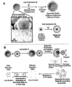
[023] Figure 1. Colorimetric Bio-Barcode Assay. A. Probe Preparation and
Electron
Micrograph Images of Amine-Modified Porous Silica Beads (Inset). B.
Interleukin-2
Detection Scheme.
[024] Figure 2. Quantification Method for Gold Nanoparticle Aggregates Spotted
on a
TLC Plate. Spot Intensity value is proportional to the number of barcode DNA
(the more gold
nanoparticles aggregated, the less color appeared) and the number of barcode
DNA is
proportional to the amount of target proteins present.
[025] Figure 3. Gold Nanoparticle-Based Colorimetric Barcode DNAa' 2 Detection
(Top:
Quantification Data; Bottom: Gold Nanoparticle Spots on a TLC Plate). A. In
Buffer. B. In
Human Serum Samples.
[026] Figure 4. Multiplexed Colorimetric Bio-Barcode Assay. A. Scheme showing
the
assay steps. B. Multiple types of nanoparticles that may be used in the assay.
DETAILED DESCRIPTION OF THE INVENTION
1. Introduction
[027] The present invention provides for a simple, ultrasensitive bio-barcode
method for
detecting an analyte of interest. This bio-barcode approach to analyte
detection is important
for the following reasons. First, this new method has shown that one can
dramatically
increase the number of barcode DNA per probe by adjusting surface and size of
barcode
probe. This allows for various embodiments to detect barcode DNA. In one
embodiment, as
shown in the examples, a colorimetric assay is used. Second, the detection
limit for this assay
is orders of magnitude better than other conventional immunoassays. Third,
this bio-barcode
method does not require complicated instrumentation or experiment steps.
Simple mixing
and separation of probe solutions would result in attomolar sensitivity
without using a
microarrayer, complicated signal amplification steps such as enzymatic
amplification and
silver-enhancement, or sophisticated signal measurement tools. Since the
readout is based on
color change, minimal expertise is required to perform the assay. Fourth, a
quantification
Page 4
CA 02622719 2008-03-14
WO 2007/084192 PCT/US2006/036101
metnocl using graphic software was developed for quantitative colorimetric
barcode DNA
detection assay, which was not possible with previous gold nanoparticle-based
colorimetric
DNA detection schemes. Finally, this assay should be suitable for point-of-
care applications
with the requirement only for probe solutions and TLC plates.
II. Definitions
[028] As used throughout the invention "barcode", "biochemical barcode",
"biobarcode",
"barcode oligonucleotide", "barcode DNA", "DNA barcode", "reporter barcode",
"reporter
barcode DNA", etc. are all interchangeable with each other and have the same
meaning. The
DNA barcode may be a nucleic acid such as deoxyribonucleic acid or ribonucleic
acid.
Preferably, the DNA barcode is an oligonucleotide of a predefined sequence. If
desired, the
DNA barcode may be labeled, for instance, with biotin, a radiolabel, or a
fluorescent label.
[029] The term "particle" refers to a small piece of matter that can
preferably be composed
of metals, silica, silicon-oxide, or polystyrene. A "particle" can be any
shape, such as
spherical or rod-shaped. The term "particle" as used herein specifically
encompasses both
nanoparticles and microparticles.
[030] The term "complex" or "probe complex" or "particle complex probe" refers
to a
conjugate comprised of a porous microparticle comprising a reporter
oligonucleotide and a
ligand specific for a target analyte conjugated to a magnetic probe particle
comprising a
ligand specific for the same target analyte, having the target analyte bound
thereto to both
ligands.
[031] The term "analyte", "analyte of interest", or "target analyte" refers to
the compound or
composition to be detected, including drugs, metabolites, pesticides,
pollutants, and the like.
The analyte can be comprised of a member of a specific binding pair (sbp) and
may be a
ligand, which is monovalent (monoepitopic) or polyvalent (polyepitopic),
preferably antigenic
or haptenic, and is a single compound or plurality of compounds, which share
at least one
common epitopic or determinant site. The analyte can be a part of a cell such
as bacteria or a
cell bearing a blood group antigen such as A, B, D, etc., or an HLA antigen or
a
microorganism, e.g., bacterium, fungus, protozoan, or virus. If the analyte is
monoepitopic,
the analyte can be further modified, e.g. chemically, to provide one or more
additional
binding sites. In practicing this invention, the analyte has at least two
binding sites.
[032] The term "ligand" refers to any organic compound for which a receptor
naturally
exists or can be prepared. The term ligand also includes ligand analogs, which
are modified
ligands, usually an organic radical or analyte analog, usually of a molecular
weight greater
Page 5
CA 02622719 2008-03-14
WO 2007/084192 PCT/US2006/036101
than 100, which can compete with the analogous ligand for a receptor, the
modification
providing means to join the ligand analog to another molecule. The ligand
analog will
usually differ from the ligand by more than replacement of a hydrogen with a
bond, which
links the ligand analog to a hub or label, but need not. The ligand analog can
bind to the
receptor in a manner similar to the ligand. The analog could be, for example,
an antibody
directed against the idiotype of an antibody to the ligand.
[033] The term "receptor" or "antiligand" refers to any compound or
composition capable of
recognizing a particular spatial and polar organization of a molecule, e.g.,
epitopic or
determinant site. Illustrative receptors include naturally occurring
receptors, e.g., thyroxine
binding globulin, antibodies, enzymes, Fab fragments, lectins, nucleic acids,
nucleic acid
aptamers, avidin, protein A, barsar, complement component C1q, and the like.
Avidin is
intended to include egg white avidin and biotin binding proteins from other
sources, such as
streptavidin.
[034] The term "specific binding pair (sbp) member" refers to one of two
different
molecules, which specifically binds to and can be defined as complementary
with a particular
spatial and/or polar organization of the other molecule. The members of the
specific binding
pair can be referred to as ligand and receptor (antiligand). These will
usually be members of
an immunological pair such as antigen-antibody, although other specific
binding pairs such as
biotin-avidin, enzyme-substrate, enzyme-antagonist, enzyme-agonist, drug-
target molecule,
hormones-hormone receptors, nucleic acid duplexes, IgG-protein A/protein G,
polynucleotide
pairs such as DNA-DNA, DNA-RNA, protein-DNA, lipid-DNA, lipid-protein,
polysaccharide-lipid, protein-polysaccharide, nucleic acid aptamers and
associated target
ligands (e.g., small organic compounds, nucleic acids, proteins, peptides,
viruses, cells, etc.),
and the like are not immunological pairs but are included in the invention and
the definition
of sbp member. A member of a specific binding pair can be the entire molecule,
or only a
portion of the molecule so long as the member specifically binds to the
binding site on the
target analyte to form a specific binding pair.
[035] The term "specific binding" refers to the specific recognition of one of
two different
molecules for the other compared to substantially less recognition of other
molecules.
Generally, the molecules have areas on their surfaces or in cavities giving
rise to specific
recognition between the two molecules. Exemplary of specific binding are
antibody-antigen
interactions, enzyme-substrate interactions, polynucleotide interactions, and
so forth.
[036] As used herein, a polynucleotide or fragment thereof is "substantially
homologous"
("substantially similar") to another if, when optimally aligned (with
appropriate nucleotide
Page 6
CA 02622719 2008-03-14
WO 2007/084192 PCT/US2006/036101
insertions or deletions) with the other polynucleotide (or its complementary
strand), using
BLASTN (Altschul, S.F., Gish, W., Miller, W., Myers, E.W. & Lipman, D.J.
(1990) "Basic
local alignment search tool." J. Mol. Biol. 215:403-410) there is nucleotide
sequence identity
in at least about 80%, preferably at least about 90%, and more preferably at
least about 95-
98% of the nucleotide bases. To determine homology between two different
polynucleotides,
the percent homology is to be determined using an alignment program such as
the BLASTN
program "BLAST 2 sequences". This program is available for public uses from
the National
Center for Blotechnolgoy Information (NCBI) over the Internet (Tatiana A.
Tatusova,
Thomas L. Madden (1999), "Blast 2 sequences - a new tool for comparing protein
and
nucleotide sequences", FEMS Microbiol Lett. 174:247-250). The parameters that
can be used
are whatever combination of the following yields the highest calculated
percent homology (as
calculated below) with the default parameters shown in parentheses:
Program - blastn
Reward for a match - 0 or 1 (1)
Penalty for a mismatch - 0, -1, -2 or -3 (-2)
Open gap penalty - 0, 1, 2, 3, 4 or 5 (5)
Extension gap penalty - 0 or 1 (1)
Gap x_dropoff - 0 or 50 (50)
Expect - 10
Word size -11
Filter - low complexity.
[037] The term "antibody" refers to an immunoglobulin which specifically binds
to and is
thereby defined as complementary with a particular spatial and polar
organization of another
molecule. The antibody can be monoclonal or polyclonal and can be prepared by
techniques
that are well known in the art such as immunization of a host and collection
of sera
(polyclonal) or by preparing continuous hybrid cell lines and collecting the
secreted protein
(monoclonal), or by cloning and expressing nucleotide sequences or mutagenized
versions
thereof coding at least for the amino acid sequences required for specific
binding of natural
antibodies. Antibodies may include a complete immunoglobulin or fragment
thereof, which
immunoglobulins include the various classes and isotypes, such as IgA, IgD,
IgE, IgGl,
IgG2a, IgGZb and IgG3, IgM, etc. Fragments thereof may include Fab, Fv and
F(ab [prime]) 2,
Fab[prime], and the like. In addition, aggregates, polymers, and conjugates of
immunoglobulins or their fragments can be used where appropriate so long as
binding affinity
for a particular molecule is maintained.
Page 7
CA 02622719 2008-03-14
WO 2007/084192 PCT/US2006/036101
III. Method for Detecting Analytes in a Sample
[038] Referring now to Figure 1B, one embodiment of the invention provides
methods for
detecting analytes of interest from a sample. The method comprises providing a
sample
suspected of containing an analyte of interest, contacting a porous particle
probe and a
magnetic probe particle with the sample, and allowing both the porous particle
probe and
magnetic probe particle to bind to the analyte of interest. The porous
particle (i.e.
microparticle or nanoparticle) probe comprises a first ligand that
specifically binds the analyte
of interest and a barcode oligonucleotide. The magnetic probe particle (i.e.
nanoparticle)
comprises a second ligand that also specifically binds the target analyte of
interest. If the
analyte of interest is present in the sample, a complex is formed between the
analyte of
interest, the porous particle probe and the magnetic probe particle. The
complex is separated
from the sample, the barcode oligonucleotide is released and collected from
the complex, and
the barcode oligonucleotide is detected.
[039] As shown in Figure 1B, the porous microparticle probe and the magnetic
probe
particle, both of which are functionalized with a ligand to capture the
analyte of interest, are
mixed with the sample suspected of containing the analyte of interest. Upon
mixing, the
analyte of interest, if present, binds to the ligands on both the magnetic
probe particle and the
porous microparticle probe to form a probe complex comprising the magnetic
probe particle
and the porous microparticle probe linked together by the ligands bound to the
analyte of
interest.
[040] In one embodiment, the method utilizes binding events of an analyte of
interest to a
particle labeled with oligonucleotides, and the subsequent detection of those
binding events.
The final step of the method described herein relies on the surface chemistry
of ordinary
DNA. Therefore, it can incorporate many of the high sensitivity aspects of
state-of-the-art
particle DNA detection methods but allows one to detect a variety of
biomolecules, such as
proteins, rather than DNA without having the proteins present during the
detection event. For
surface assays, proteins are typically more difficult to work with than short
oligonucleotides
because they tend to exhibit greater nonspecific binding to solid supports,
which often leads
to higher background signals. Finally, for the homogeneous assay, the
unusually sharp
melting profiles associated with these nanoparticle structures will allow one
to design more
biobarcodes than what would be possible with probes that exhibit normal and
broad DNA
melting behavior.
Page 8
CA 02622719 2008-03-14
WO 2007/084192 PCT/US2006/036101
[041] The present invention contemplates the use of any suitable particle
having
oligonucleotides attached thereto that are suitable for use in detection
assays. As described
herein, each microparticle, magnetic probe particle and nanoparticle will have
a plurality of
oligonucleotides attached to it. As a result, each particle-oligonucleotide
conjugate can bind
to a plurality of oligonucleotides or nucleic acids having the complementary
sequence.
[042] The oligonucleotides are contacted with the particles in aqueous
solution for a time
sufficient to allow at least some of the oligonucleotides to bind to the
nanoparticles by means
of the functional groups. Such times can be determined empirically. For
instance, it has been
found that a time of about 12 to 24 hours gives good results. In some
embodiments wherein
detection is in the clinic, a preferred time for hybridization may be 10
minutes to 12 hours.
Other suitable conditions for binding of the oligonucleotides can also be
determined
empirically. For instance a concentration of about 10-20nM nanoparticles and
incubation at
room temperature gives good results.
[043] The probe complex is separated from the sample after formation of the
probe
complex. In a preferred embodiment, this is carried out by magnetic separation
facilitated by
exposing the sample to a magnetic field (e.g., via a magnetic separation
device) which attracts
the magnetic particles in the probe complex and allows isolation or separation
from the
sample. Thus, in one aspect of the invention, the particle probe complex
comprises a
microparticle having barcode oligonucleotides and a ligand, wherein the ligand
is bound to a
specific analyte of interest and the analyte of interest is also bound to
another ligand on the
magnetic probe particle.
[044] After separation from the sample, the barcode oligonucleotide attached
to the porous
microparticle in the probe complex is released and captured for further
detection or analysis.
The barcodes can be released for the particles to which they are attached by a
chemical
releasing agent that will disrupt binding of the barcode to the surface of the
particle. Such
agents include, but are not limited to, any molecule that will preferentially
bind to a particle
through a thiol link such as other thiol- or disulfide-containing molecules,
dithiothreitol
(DTT), dithioerythritol (DTE), mercaptoethanol and the like, and reducing
agents such as
sodium borohydride that will cleave a disulfide linkage thereby releasing
barcodes from the
particles to which they are attached. The barcodes can also be released from
the particles by
exposing the barcodes to conditions under which the barcodes will dehybridize
from
oligonucleotides by which the barcodes were attached to the particles.
[045] The barcodes or reporter oligonucleotides may then be detected by any
suitable
means. Generally, the barcodes are released via dehybridization from the
complex prior to
Page 9
CA 02622719 2008-03-14
WO 2007/084192 PCT/US2006/036101
detection. Any suitable solution or media may be used that dehybridize and
release the
barcode from the complex. A representative medium is water.
a. Analyte of Interest
[046] The analyte of interest may be nucleic acid molecules, proteins,
peptides, haptens,
metal ions, drugs, metabolites, pesticide or pollutant. The method can be used
to detect the
presence of such analytes as toxins, hormones, enzymes, lectins, proteins,
signaling
molecules, inorganic or organic molecules, antibodies, contaminants, viruses,
bacteria, other
pathogenic organisms, idiotopes or other cell surface markers. It is intended
that the present
method can be used to detect the presence or absence of an analyte of interest
in a sample
suspected of containing the analyte of interest.
[047] In some embodiments, the target analyte is comprised of a nucleic acid
and the
specific binding complement is an oligonucleotide. Alternatively, the target
analyte is a
protein or hapten and the specific binding complement is an antibody
comprising a
monoclonal or polyclonal antibody. Alternatively, the target analyte is a
sequence from a
genomic DNA sample and the specific binding complement are oligonucleotides,
the
oligonucleotides having a sequence that is complementary to at least a portion
of the genomic
sequence. The genomic DNA may be eukaryotic, bacterial, fungal or viral DNA.
[048] In one embodiment, detection of a particular cytokine can be used for
diagnosis of
cancer. Specific analytes of interest include cytokines, such as IL-2 as shown
in the
examples. Cytokines are important analytes of interest in that cytokines play
a central role in
the regulation of hematopoiesis; mediating the differentiation, migration,
activation and
proliferation of phenotypically diverse cells. Improved detection limits of
cytokines will
allow for earlier and more accurate diagnosis and treatments of cancers and
immunodeficiency-related diseases and lead to an increased understanding of
cytokine-related
diseases and biology, because cytokines are signature biomarkers when humans
are infected
by foreign antigens.
[049] Chemokines are another important class of analytes of interest.
Chemokines are
released from a wide variety of cells in response to bacterial infection,
viruses and agents that
cause physical damage such as silica or the urate crystals. They function
mainly as
chemoattractants for leukocytes, recruiting monocytes, neutrophils and other
effector cells
from the blood to sites of infection or damage. They can be released by many
different cell
types and serve to guide cells involved in innate immunity and also the
lymphocytes of the
adaptive immune system. Thus, improved detection limits of chemokines will
allow for
Page 10
CA 02622719 2008-03-14
WO 2007/084192 PCT/US2006/036101
earlier and more accurate diagnosis and treatments, i.e. for bacterial
infections and viral
infections.
[050] In some embodiments, the target analyte may be a variety of pathogenic
organisms
including, but not limited to, sialic acid to detect HIV, Chlamydia, Neisseria
meningitides,
Streptococcus suis, Saltnonella, mumps, newcastle, and various viruses,
including reovirus,
sendai virus, and myxovirus; and 9-OAC sialic acid to detect coronavirus,
encephalomyelitis
virus, and rotavirus; non-sialic acid glycoproteins to detect cytomegalovirus
and measles
virus; CD4, vasoactive intestinal peptide, and peptide T to detect HIV;
epidermal growth
factor to detect vaccinia; acetylcholine receptor to detect rabies; Cd3
complement receptor to
detect Epstein-Barr virus; P-adrenergic receptor to detect reovirus; ICAM-1, N-
CAM, and
myelin-associated glycoprotein MAb to detect rhinovirus; polio virus receptor
to detect polio
virus; fibroblast growth factor receptor to detect herpes virus; oligomannose
to detect
Escherichia coli; ganglioside GMl to detect Neisseria meningitides; and
antibodies to detect a
broad variety of pathogens (e.g., Neisseria gonorrhoeae, V. vulnificus, V.
parahaemolyticus,
V. cholerae, and V. alginolyticus).
[051] In some embodiments, multiple analytes of interest can be detected by
utilizing
multiple ligands specific to different analytes of interest and utilizing
distinct barcode
oligonucleotides corresponding to each analyte of interest.
b. Sample
[052] The analyte of interest may be found directly in a sample such as a body
fluid from a
host. The host may be a mammal, reptile, bird, amphibian, fish, or insect. In
a preferred
embodiment, the host is a liuman. The body fluid can be, for example, urine,
blood, plasma,
serum, saliva, semen, stool, sputum, cerebral spinal fluid, tears, mucus, pus,
phlegm, and the
like. The particles can be mixed with live cells or samples containing live
cells.
[053] Where the sample is live cells or samples containing live cells, a cell
surface protein
or other molecule may serve as the analyte of interest. This allows for the
detection of cell
activation and proliferation events, cellular interactions, multiplexing, and
other
physiologically relevant events.
c. Porous Microparticle Probe
[054] In a preferred embodiment, the present method utilizes porous
microparticles and a
metal nanoparticle-based colorimetric DNA detection scheme for straightforward
readout
(Figure 1). In a preferred embodiment, the porous microparticle probe should
feature a ligand
Page 11
CA 02622719 2008-03-14
WO 2007/084192 PCT/US2006/036101
to capture a target analyte and a barcode oligonucleotide, which is a specific
barcode DNA
sequence.
[055] In one embodiment, the microparticle is a porous particle having a
defined degree of
porosity and comprised of pores having a defined size range, wherein the
barcode
oligonucleotides are impregnated within the pores of the microparticle. The
use of a porous
microparticle can accommodate millions of barcode DNA per particle, thus
allowing the use
of a colorimetric barcode DNA detection scheme with attomolar sensitivity.
This is an
important advance because this scheme has the attomolar (10-18 M) sensitivity
of the bio-
barcode amplification method as well as the simplicity, portability and low
cost of gold
nanoparticle-based colorimetric detection methods.
[056] In some embodiments, the porous microparticle probe can be comprised of
materials
including silica and iron oxide. The term "microparticle" as used herein is
intended to
encompass any particulate bead, sphere, particle or carrier, whether
biodegradable or
nonbiodegradable, comprised of naturally-occurring or synthetic, organic or
inorganic
materials that is porous. In particular, the microparticle includes any
particulate bead, sphere,
particle, or carrier having a diameter of about 0.1 to about 5000 micrometers,
more preferably
about 1-5 m in diameter, and even more preferably between about 3-4 m in
diameter. The
term "about" as used herein is meant to include up to 1 unit of the provided
range. In
another embodiment, porous silica microparticles (1.57 x 10⁹ m1-1 diameter:
3.53 0.49 ,um)
are used.
[057] The microparticles of the invention are comprised of polystyrene,
silica, iron oxide,
polyacrylamide, and various polysaccharides including dextran, agarose,
cellulose and
modified, crosslinked and derivatized embodiments thereof. Specific examples
of the
microparticles of the invention include polystyrene, cellulose, dextran
crosslinked with
bisacrylamide (Biogel.TM., Bio-Rad, U.S.A.), agar, glass beads and latex
beads. Derivatized
microparticles include microparticles derivatized with carboxyalkyl groups
such as
carboxymethyl, phosphoryl and substituted phosphoryl groups, sulfate,
sulfhydryl and
sulfonyl groups, and amino and substituted animo groups.
[058] The size, shape and chemical composition of the particles will
contribute to the
properties of the resulting probe including the barcode DNA. These properties
include
optical properties, optoelectronic properties, electrochemical properties,
electronic properties,
stability in various solutions, ability to separate bioactive molecules while
acting as a filter,
etc. The use of mixtures of particles having different sizes, shapes and/or
chemical
Page 12
CA 02622719 2008-03-14
WO 2007/084192 PCT/US2006/036101
compositions and the use of particles having uniform sizes, shapes and
chemical composition,
are contemplated.
[059] In some embodiments, the microparticle is amino-functionalized and then
reacted
with the ligand and the barcode oligonucleotide. In a preferred embodiment,
the porous
microparticle probes are comprised of silica and iron oxide and functionalized
with amine
groups for further modification with other biomolecules. For example, such
particles can be
obtained from PHENOMENEX (Torrance, CA). Analogous glutaraldehyde linker
chemistry
has been extensively used by others to affect protein linking to amino
functionalized particles.
[060] In another embodiment, the methods to functionalize the nanopartices as
described
infra may be used to functionalize the porous microparticle probe. In some
embodiments, the
silica coated magnetic particles are functionalized amino-silane molecules to
functionalize the
silica surface with amines.
[061] Other properties of the porous microparticles that affect the number of
barcode
oligonucleotides which can be incorporated onto the probe, and therefore
sensitivity, include:
surface area, pore size, interconnectivity of the pores, hydrophilicity and
pore distribution.
i. Surface Area
[062] The number of barcode oligonucleotides per probe is dramatically
increased by
adjusting the surface and size of the barcode probe which also allows for
various
embodiments to detect more than one barcode oligonucleotide. In the bio-
barcode approach,
the number of barcode oligonucleotide per probe is important because the final
detection
signal is proportional to the amount of captured barcode DNA.
[063] In some embodiments, the surface area of the porous particles is about
300 m2/g to
about 500 mz /g, more preferably about 400 m2/g to about 450 m2lg.
[064] In a preferred embodiment, the large size (a few micrometers) and
porosity of probe
result in significantly increased barcode oligonucleotide loading relative to
past approaches
(tens-of-nanometer particle without pores). Using UV-Vis spectroscopy (the UV
absorption
peak for single stranded DNA is at 260 nm), it was determined the average
total number of
barcode oligonucleotides per -3.5 micrometer bead to be - 3.6 x10⁶. Compared
with other
nanoparticle-based barcode probes which can host only hundreds of barcode DNA
per
nanoparticle probe, the present microparticles result in several orders of
magnitude more
amplification in terms of the number of barcode oligonucleotides per barcode
probe.
ii. Pore Size
Page 13
CA 02622719 2008-03-14
WO 2007/084192 PCT/US2006/036101
[065] Pore size is also an important aspect of the porous particles. The pore
size must be
large enough such that the barcode oligonucleotides can enter the pore during
binding of the
barcode to the particle and exit the pore when releasing the barcode
oligonucleotides for
detection.
[066] Therefore, in some embodiments, the pore size is about 50 angstroms to
about 150
angstroms, more preferably from about 90 angstroms to about 110 angstroms.
iii. Interconnectivity
[067] Interconnectivity of the pores within the porous particles allows sample
or effluent to
flow throughout the porous particle. These "channels" provides means for
preparing and
releasing the barcode DNA from within the pores. Also, by having channels, it
prevents air
pockets from forming within pores which can interfere with barcode DNA
entrance and
release.
[068] Thus, in a preferred embodiment, the porous particles have channels to
afford greater
accommodation of barcode DNA and better binding and release of the barcode DNA
from the
particle.
iv. Hydrophilicity
10691 In a preferred embodiment, the porous particle is hydrophilic and has
little to no
hydrophobicity. Hydropholic porous particles allows for effective probe
preparation and
effective release of barcode DNA for detection.
v. Pore Distribution
[070] In a preferred embodiment, the porous particle will have the greatest
number of pores
that can be incorporated onto the particle without negatively affecting the
structural integrity
of each particle.
[071] Pore distribution or the number of pores per particle can also affect
the number of
barcode DNA that can be accommodated onto the particle. The number of pores
has a direct
effect on the surface area of each particle. There is, however, a limit to the
number of pores
that a particle can have. The structural integrity of the particle may be
compromised if too
many pores are incorporated into each particle.
d. Ligands
Page 14
CA 02622719 2008-03-14
WO 2007/084192 PCT/US2006/036101
[072] The ligands attached to capture an analyte of interest may be attached,
removeably
attached, covalently or non-covalently attached to the porous particle probe
and magnetic
particle probe.
[073] Both the ligand attached to the porous particle probe and the ligand
attached to the
magnetic particle probe specifically bind to an analyte of interest. Thus, in
a preferred
embodiment, the analyte of interest has at least two binding sites allowing
for each ligand to
specifically bind.
[074] A ligand can be any molecule or material having a known analyte as a
specific binding
pair member. Thus, each member of the specific binding pair may be a nucleic
acid, an
oligonucleotide, a peptide nucleic acid, a polypeptide, an antigen, a
carbohydrate, an amino
acid, a hormone, a steroid, a vitamin, a virus, a polysaccharide, a lipid, a
lipopolysaccharide, a
glycoprotein, a lipoprotein, a nucleoprotein, an albumin, a hemoglobin, a
coagulation factor,
a peptide hormone, a non-peptide hormone, a biotin, a streptavidin, a
cytokine, a chemokine,
a peptide compromising a tumor-specific epitope, a cell, a cell surface
molecule, a
microorganism, a small molecules, an enzyme, a receptor, a channel, a
chromophore, a
chelating compound, a phosphate and reactive group, a molecular recognition
complex, a
dinitrophenol, an electron donor or acceptor group, a hydrophobic compound, a
hydrophilic
compound, an organic molecule, and an inorganic molecule.
[075] In some embodiments, the ligand is a monoclonal antibody or polyclonal
antibody
where the analyte of interest is a protein, hapten or peptide. Where
antibodies are used as the
ligands, the epitopes of the antibodies used to functionalize the magnetic
probe particle are
different from those of the antibodies used to prepare the microparticle
probes by using a
different coupling chemistry. Therefore in a preferred embodiment, the
antibodies chosen as
the ligands are already developed antibodies with two different epitopes. For
important
disease markers, many high quality antibodies with different epitopes are
readily available
through academic and commercial means. Furthermore, it is recognized in the
art that
antibodies can be raised to a ligand by one with skill in the art.
[076] In some embodiments, where the analyte of interest is a nucleic acid,
the ligand is an
oligonucleotide having a sequence that is complementary to at least a portion
of the sequence
of the nucleic acid.
[077] In some embodiments, where the analyte of interest is from a genomic DNA
sample,
the ligand is an oligonucleotide having a sequence that is complementary to
the genomic
sequence.
Page 15
CA 02622719 2008-03-14
WO 2007/084192 PCT/US2006/036101
10781 Amino-functionalized magnetic particles were linked to ligands for the
target analyte.
In a preferred embodiment where antibodies are used as the ligand, the
epitopes of the
antibodies are different from those of the antibodies used to prepare the
barcode DNA using
glutaraldehyde-amine coupling chemistry.
e. Barcode Oligonucleotide
[079] In a preferred embodiment, the barcode oligonucleotides attached to the
porous
microparticle probe to capture a target analyte may be attached, removeably
attached,
covalently or non-covalently attached.
[080] Any suitable method for attaching oligonucleotides onto the nanosphere
surface
may be used. A particularly preferred method for attaching oligonucleotides
onto a surface is
based on an aging process described in U.S. application Ser. No. 09/344,667,
filed Jun. 25,
1999; Ser. No. 09/603,830, filed Jun. 26, 2000; Ser. No. 09/760,500, filed
Jan. 12, 2001; Ser.
No. 09/820,279, filed Mar. 28, 2001; Ser. No. 09/927,777, filed Aug. 10, 2001;
and in
International application nos. PCT/US97/12783, filed Jul. 21, 1997;
PCT/USOO/17507, filed
Jun. 26, 2000; PCT/US01/01190, filed Jan. 12, 2001; PCT/USO1/10071, filed Mar.
28, 2001,
the disclosures which are incorporated by reference in their entirety. The
aging process
provides nanoparticle-oligonucleotide conjugates with unexpected enhanced
stability and
selectivity.
[081] In one embodiment, the method comprises providing barcode
oligonucleotides
preferably having covalently bound thereto a moiety comprising a functional
group which can
bind to the nanoparticles. The moieties and functional groups are those that
allow for binding
(i.e., by chemisorption or covalent bonding) of the oligonucleotides to
nanoparticles. For
instance, oligonucleotides having an alkanethiol, an alkanedisulfide or a
cyclic disulfide
covalently bound to their 5' or 3' ends can be used to bind the
oligonucleotides to a variety of
nanoparticles, including gold nanoparticles. Methods of attaching
oligonucleotides to
nanoparticles are futher described in U.S. Pat. Appl. Serial No. 10/877,750,
published as
US20050037397, hereby incorporated by reference.
[082] In some embodiments, the barcode oligonucleotides are attached to the
microparticle
by means of a linker. There are many amine-reactive linkers (for covalent
linking) available
commercially. Therefore, it is contemplated that the microparticles are
commonly modified
with amines. Preferably, the linker further comprises a hydrocarbon moiety
attached to the
cyclic disulfide. Suitable hydrocarbons are available commercially, and are
attached to the
cyclic disulfides. Preferably the hydrocarbon moiety is a steroid residue.
Oligonucleotide-
Page 16
CA 02622719 2008-03-14
WO 2007/084192 PCT/US2006/036101
particle conjugates prepared using linker comprising a steroid residue
attached to a cyclic
disulfide have unexpectedly been found to be remarkably stable to thiols
(e.g., dithiothreitol
used in polymerase chain reaction (PCR) solutions) as compared to conjugates
prepared using
alkanethiols or acyclic disulfides as the linker. Indeed, others have found
the oligonucleotide-
particle conjugates of the invention have been found to be 300 times more
stable. See U.S.
Pat. Appl. Serial No. 10/877,750. This stability is likely due to the fact
that each
oligonucleotide is anchored to a microparticle through two sulfur atoms,
rather than a single
sulfur atom. In particular, it is thought that two adjacent sulfur atoms of a
cyclic disulfide
would have a chelation effect which would be advantageous in stabilizing the
oligonucleotide-microparticle conjugates. The large hydrophobic steroid
residues of the
linkers also appear to contribute to the stability of the conjugates by
screening the
microparticles from the approach of water-soluble molecules to the surfaces of
the
nanoparticles.
[083] In another embodiment, the barcode oligonucleotides are bound to the
microparticles
using sulfur-based functional groups. U.S. Pat. Appl. Serial No. 09/760,500
and 09/820,279
and international application nos. PCT/US01/01190 and PCT/US01/10071 describe
oligonucleotides functionalized with a cyclic disulfide which are useful in
practicing this
invention. The cyclic disulfides preferably have 5 or 6 atoms in their rings,
including the two
sulfur atoms. Suitable cyclic disulfides are available commercially or may be
synthesized by
known procedures. The reduced form of the cyclic disulfides can also be used.
[084] In one embodiment, ethanolamine is used to passivate all unreacted
reaction sites on
the microparticles. A proteiii such as bovine serum albumin can also be used
in addition or
instead to further passivate inactive regions on the microparticle surface.
[085] As described in the definitions, the DNA barcode may be a nucleic acid
such as
deoxynucleic acid or ribonucleic acid. Preferably, the DNA barcode is an
oligonucleotide of
a predefined sequence. The DNA barcode oligonucieotide may comprise genes;
viral RNA
and DNA; bacterial DNA; fungal DNA; mammalian DNA, cDNA, mRNA, RNA and DNA
fragments; oligonucleotides; synthetic oligonucleotides; modified nucleotides;
single-
stranded and double-stranded nucleic acids; natural and synthetic nucleic
acids; and aptamers.
[086] Methods of making oligonucleotides of a predetermined sequence are well
known.
See, e.g., Sambrook et al., Molecular Cloning: A Laboratory Manual (2"' ed.
1989) and F.
Eckstein (ed.) Oligonucleotides and Analogues, lst Ed. (Oxford University
Press, New York,
1991). Solid-phase synthesis methods are preferred for both
oligoribonucleotides and
oligodeoxyribonuclotides (the well-known methods of synthesizing DNA are also
useful for
Page 17
CA 02622719 2008-03-14
WO 2007/084192 PCT/US2006/036101
synthesizying RNA). Oligonucleotides can also be prepared enzymatically. For
oligonucleotides having a specific binding complement to a target analyte
bound thereto, any
suitable method of attaching the specific binding complement, such as
proteins, to t1Te
oligonucleotide may be used.
[087] The present invention contemplates using sequences designed by
techniques known to
those of skill in the art including, optimization for annealing temperatures,
the specificity of
the sequence to the template, and length of sequence. The design of the
sequences can be
done using primer prediction software such as OIigo6 (Molecular Biology
Insights, Inc.,
Cascade, CO). Custom scripts and software for primer design can also be used.
[088] Any unique oligonucleotide sequence and its complementary sequence can
be used for
the barcode oligonucleotide. It is preferred that the oligonucleotide
sequences used as
barcode oligonucleotides hybridize their complementary sequences under
stringent
conditions. The term "stringent conditions" as used herein refers to
conditions under which a
sequence will hybridize to its target subsequence or complement, but to no
other sequences.
Stringent conditions are sequence-dependent and will be different in different
circumstances.
Longer sequences hybridize specifically at higher temperatures. Generally,
stringent
conditions are selected to be about 15 C lower than the thermal melting point
(Tm) for the
specific sequence at a defined ionic strength and pH. The Tm is the
temperature (under
defined ionic strength, pH, and nucleic acid concentration) at which 50% of
the probes
complementary to the target sequence hybridize to the target sequence at
equilibrium. (As the
target sequences are generally present in excess, at Tm, 50% of the probes are
occupied at
equilibrium.)
[089] In some embodiments, the barcode oligonucleotide is modified with a
detectable label.
Examples of detectable labels include biotin, radiolabels, fluorescent labels,
chromophores,
redox-active groups, groups with electronic signatures, catalytic groups and
Raman labels.
[090] Examples of such specific barcode DNA sequences can be found e.g. in
Multiplexed
Detection of Protein Cancer Markers with Biobarcoded Nanoparticle Probes,
Stoeva et al.,
128 J. Am. Chem. Soc. 8378-8379 (2006); Bio-Bar-Code-Based DNA Detection with
PCR-
like Sensitivity, Nam et al. 126 J. Am. Chem. Soc. 5932-5933 (2004); and
Multiplexed DNA
Detection witlz Biobarcoded Natzoparticle Pr=obes, Soteva et al., 45 Angew.
Chem. Int. Ed.
3303-3306 (2006), hereby incorporated by reference.
[091] In a preferred embodiment, barcode DNA are 3' amino-functionalized bar-
code DNA
complements having a defined sequence (e.g., as an identification tag) to
identify the
Page 18
CA 02622719 2008-03-14
WO 2007/084192 PCT/US2006/036101
microparticle as being used to detect a specific target analyte, thereby
permitting the detection
of multiple target analytes in a sample.
[092] In one embodiment, the method utilizes oligonucleotides as biochemical
barcodes for
detecting a single or multiple analytes in a sample. The approach takes
advantage of
recognition elements (e.g., proteins or nucleic acids) functionalized either
directly or
indirectly with nanoparticles and the previous observation that hybridization
events that result
in the aggregation of gold nanoparticles can significantly alter their
physical properties (e.g.
optical, electrical, meclianical). The general idea is that each recognition
element can be
associated with a different oligonucleotide sequence (i.e. a DNA barcode) with
discrete and
tailorable hybridization and melting properties and a physical signature
associated with the
nanoparticles that change upon melting to decode a series of analytes in a
multi-analyte assay.
Therefore, one can use the melting temperature of a DNA-linked aggregate and a
physical
property associated with the nanoparticles that change upon melting to decode
a series of
analytes in a multiple analyte assay. The barcodes herein are different from
the ones based on
physical diagnostic markers such as nanorods, fluorophore-labeled beads, and
quantum dots,
in that the decoding information is in the form of chemical information stored
in a
predesigned oligonucleotide sequence.
f. Magnetic Probe Particle
[093] The magnetic probe particle can be comprised of magnetic materials
including iron
oxide and other ferromagnetic materials. The magnetic probe particle can be
coated with
silica, or polymers such as polyacrylamide, polystyrene, etc. with the surface
functionalized as
described for the porous microparticles.
[094] In a preferred embodiment, the magnetic probe particles can be
nanoparticles or
microparticles having a diameter of about 0.1 nanometers to about 5000
micrometers.
Suitable magnetic particles are widely used in the art and can be obtained
from such vendors
as Dynal Biotech (newly acquired by Invitrogen).
[095] In one embodiment, the magnetic particles are prepared as described in
the Examples
using glutaraldehyde-amine coupling chemistry.
[096] Microparticles and nanoparticles useful in the practice of the invention
include metal
(e.g., gold, silver, copper and platinum), semiconductor (e.g., CdSe, CdS, and
CdS or CdSe
coated with ZnS) and magnetic (e.g., ferromagnetite) colloidal materials.
Other nanoparticles
useful in the practice of the invention include ZnS, ZnO, Ti02, AgI, AgBr,
HgI2, PbS, PbSe,
ZnTe, CdTe, In2S3, In2Se3, Cd3P2, Cd3As2, InAs, and GaAs. The size of the
nanoparticles
Page 19
CA 02622719 2008-03-14
WO 2007/084192 PCT/US2006/036101
is preferably from about 5 nm to about 150 nm (mean diameter), more preferably
from about
to about 50 nm, most preferably from about 10 to about 30 nm. The
nanoparticles may also
be rods, prisms, or tetrahedra.
[097] Methods of making metal, semiconductor and magnetic nanoparticles are
well-known
in the art, See, e.g., Schmid, G. (ed.) Clusters and Colloids (VCH, Weinheim,
1994); Hayat,
M. A. (ed.) Colloidal Gold: Principles, Methods, and Applications (Academic
Press, San
Diego, 1991); Massart, R., IEEE Taransactions On Magnetics, 17, 1247 (1981);
Ahmadi, T. S.
et al., Science, 272, 1924 (1996); Henglein, A. et al., J. Phys. Chem., 99,
14129 (1995);
Curtis, A. C., et aI., Angew. Chem. Int. Ed. Engl., 27, 1530 (1988).
[098] Methods of making ZnS, ZnO, Ti02, AgI, AgBr, HgI2, PbS, PbSe, ZnTe,
CdTe,
In2S3, In2Se3, Cd3P2, Cd3As2, InAs, and GaAs nanoparticles are also known in
the art. See,
e.g., Weller, Angew. Chem. Int. Ed. Engl., 32, 41 (1993); Henglein, Top. Curr.
Chem., 143,
113 (1988); Henglein, Chem. Rev., 89, 1861 (1989); Brus, Appl. Phys. A., 53,
465 (1991);
Bahncmann, in Photochemical Conversion and Storage of Solar Energy (eds.
Pelizetti and
Schiavello 1991), page 251; Wang and Herron, J. Phys. Chem., 95, 525 (1991);
Olshavsky et
al., J. Am. Chem. Soc., 112, 9438 (1990); Ushida et al., J. Phys. Chem., 95,
5382 (1992).
[099] Suitable nanoparticles are also commercially available from, e.g., Ted
Pella, Inc.
(gold), Amersham Corporation (gold) and Nanoprobes, Inc. (gold).
[0100] Presently preferred for use in detecting nucleic acids are gold
nanoparticles. Gold
colloidal particles have high extinction coefficients for the bands that give
rise to their
beautiful colors. These intense colors change with particle size,
concentration, interparticle
distance, and extent of aggregation and shape (geometry) of the aggregates,
making these
materials particularly attractive for colorimetric assays. For instance,
hybridization of
oligonucleotides attached to gold nanoparticles with oligonucleotides and
nucleic acids
results in an immediate color change visible to the naked eye.
[0101] The particles or the oligonucleotides, or both, are functionalized in
order to attach the
oligonucleotides to the particles. Such methods are known in the art. For
instance,
oligonucleotides functionalized with alkanethiols at their 3'-ternaini or 5'-
termini readily
attach to gold nanoparticles. See Whitesides, Proceedings of the Robert A.
Welch Foundation
39th Conference on Chemical Research Nanophase Chemistry, Houston, Tex., pages
109-121
(1995). See also, Mucic et al. Chem. Commun. 555-557 (1996) (describes a
method of
attaching 3' thiol DNA to flat gold surfaces; this method can be used to
attach
oligonucleotides to nanoparticles). The alkanethiol method can also be used to
attach
oligonucleotides to other metal, semiconductor and magnetic colloids and to
the other
Page 20
CA 02622719 2008-03-14
WO 2007/084192 PCT/US2006/036101
nanoparticles listed above. Other functional groups for attaching
oligonucleotides to solid
surfaces include phosphorothioate groups (see, e.g., U.S. Pat. No. 5,472,881
for the binding
of oligonucleotide-phosphorothioates to gold surfaces), substituted
alkylsiloxanes (see, e.g.
Burwell, Chemical Technology, 4, 370-377 (1974) and Matteucci and Caruthers,
J. Am.
Chem. Soc., 103, 3185-3191 (1981) for binding of oligonucleotides to silica
and glass
surfaces, and Grabar et al., Anal. Chem., 67, 735-743 for binding of
aminoalkylsiloxanes and
for similar binding of inercaptoaklylsiloxanes). Oligonucleotides terminated
with a 5'
thionucleoside or a 3' thionucleoside may also be used for attaching
oligonucleotides to solid
surfaces. The following references describe other methods which may be
employed to
attached oligonucleotides to nanoparticles: Nuzzo et al., J. Am. Chem. Soc.,
109, 2358 (1987)
(disulfides on gold); Allara and Nuzzo, Langmuir, 1, 45 (1985) (carboxylic
acids on
aluminum); Allara and Tompkins, J. Colloid Interface Sci., 49, 410-421 (1974)
(carboxylic
acids on copper); Iler, The Chemistry Of Silica, Chapter 6, (Wiley 1979)
(carboxylic acids on
silica); Timmons and Zisman, J. Phys. Chem., 69, 984-990 (1965) (carboxylic
acids on
platinum); Soriaga and Hubbard, J. Am. Chem. Soc., 104, 3937 (1982) (aromatic
ring
compounds on platinum); Hubbard, Acc. Chem. Res., 13, 177 (1980) (sulfolanes,
sulfoxides
and other functionalized solvents on platinum); Hickman et al., J. Am. Chem.
Soc., 111, 7271
(1989) (isonitriles on platinum); Maoz and Sagiv, Langmuir, 3, 1045 (1987)
(silanes on
silica); Maoz and Sagiv, Langmuir, 3, 1034 (1987) (silanes on silica);
Wasserman et al.,
Langmuir, 5, 1074 (1989) (silanes on silica); Eltekova and Eltekov, Langmuir,
3, 951 (1987)
(aromatic carboxylic acids, aldehydes, alcohols and methoxy groups on titanium
dioxide and
silica); Lec et al., J. Phys. Chem., 92, 2597 (1988) (rigid phosphates on
metals).
g. Universal Probes
[0102] In some embodiments, the barcode oligonucleotide and porous particle
are members
of a universal probe which may be used in an assay for any target nucleic acid
that comprises
at least two portions. This "universal probe" comprises oligonucleotides of a
single "capture"
sequence that is complementary to at least a portion of a reporter
oligonucleotide (e.g.
barcode DNA), and to a portion of a target recognition oligonucleotide. The
target
recognition oligonucleotides comprise a sequence having at least two portions;
the first
portion comprises complementary sequence to the capture sequence attached to
the porous
particle, and the second portion comprises complementary sequence to the first
portion of the
particular target nucleic acid sequence. Various types of target recognition
oligonucleotides
can be used to great advantage with the universal probe, such that a library
of target
Page 21
CA 02622719 2008-03-14
WO 2007/084192 PCT/US2006/036101
recognition oligonucleotides can be switched or interchanged in order to
select for particular
target nucleic acid sequences in a particular test solution. A capture
oligonucleotide, which
comprises sequence complementary to the second portion of the target nucleic
acid is
attached to the magnetic probe particle.
[0103] These universal probes can be manipulated for increased advantage,
which depend on
the particular assay to be conducted. The probes can be "tuned" to various
single target
nucleic acid sequences, by simply substituting or interchanging the target
recognition
oligonucleotides, such that the second portion of the universaI probe
comprises
complementary sequence to different target nucleic acid of interests.
Similarly, if multiple
target nucleic acid sequences are to be assayed in a single test solution, the
reporter
oligonucleotides can comprise a sequence that is specific for each target
nucleic acid,
whereby, detection of the reporter oligonucleotide of known and specific
sequence would
indicate the presence of the particular target nucleic acid in the test
solution. A capture
oligonucleotide, which comprises sequence complementary to the second portion
of the target
nucleic acid is attached to the nanoparticle.
h. Dendrimers
[0104] In one aspect of this embodiment of the invention, particles conjugated
with
dendrimers labeled with at least two types of oligonucleotides are provided.
Dendritic
molecules are structures comprised of multiple branching unit monomers, and
are used in
various applications. See, e.g. Barth et al., Bioconjugate Chemistry 5:58-66
(1994); Gitsov &
Frechet, Macromolecules 26:6536-6546 (1993); Lochmann et al., J. Amer. Chem.
Soc.
115:7043-7044 (1993); Miller et al., J. Amer. Chem. Soc. 114:1018-1025 (1992);
Mousy et al.,
Macromolecuels 25:2401-2406 (1992); Naylor et al., J. Amer. Chem Soc. 111:2339-
2341
(1989); Spindeler & Frechet, Macro aolecules 26:4809-4813 (1993); Turner et
al.,
Macromolecules 26:4617-4623 (1993); Wiener et al., Magnetic Resonance Med.
31(1):1-8
(1994); Service, 267:458-459 (1995); Tomalia, Sci. Amer. 62-66 (1995); and
U.S. Pat. Nos.
4,558,120; 4,507,466; 4,568,737; 4,587,329; 4,857,599; 5,527,524; 5,338,532 to
Tomalia,
and U.S. Pat. No. 6,274,743 to Nilsen, all of which are incorporated by
reference in their
entirety, for all purposes. Dendritic molecules provide important advantages
over other types
of supermolecular architectures, such as contacting a maximum volume a minimum
of
structural units, ability to more easily control size, weight, and growth
properties, and the
multiple termini can be derivatized to yield highly labeled molecules with
defined spacing
between the labels, or provide sites of attachment for other molecules, or
mixtures thereof.
Page 22
CA 02622719 2008-03-14
WO 2007/084192 PCT/US2006/036101
See generally U.S. Pat. No. 6,274,723 and the above cited references for
methods of synthesis.
Nucleic acid dendrimers that are useful in the methods of the invention are
any of those
known in the art that can be functionalized with nucleic acids or generated
from nucleic
acids/oligonucleotides. Such dendrimers can be synthesized according to
disclosures such as
Hudson et al., "Nucleic Acid Dendrimers: Novel Biopolymer Structures," Am.
Chem. Soc.
115:2119-2124 (1993); U.S. Pat. No. 6,274,723; and U.S. Pat. No. 5,561,043 to
Cantor.
IV. Colorimetric Method
[0105] In a preferred embodiment, the present invention provides for a simple,
ultrasensitive
colorimetric bio-barcode assay. The screening methods and detection schemes of
the present
invention are based upon those described by one of the inventors and others in
US Pat. Appl.
No. 10/ 877,750, published as US20050037397; U.S. Pat. Appl. No. 10/788,414,
published as
US20050009206; and U.S. Pat. App1. No. 10/108211, published as US20020192687,
again
all of which are hereby incorporated by reference for all purposes. In a
preferred embodiment,
the present bio-barcode assay provides an improved bio-barcode approach to
analyte
detection by providing a colorimetric assay having improved amplification of
bio-barcode
DNA, and quantification and multiplexing capability.
[0106] In one embodiment, as shown in the examples, a colorimetric assay is
used to detect
barcode DNA because it does not require complicated instrumentation or
experiment steps.
Simple mixing and separation of probe solutions would result in attomolar
sensitivity without
using a microarrayer, complicated signal amplification steps such as enzymatic
amplification
and silver-enhancement, or sophisticated signal measurement tools. Since the
readout is
based on color change, minimal expertise is required to perform the assay.
[0107] In some embodiments, the color change can be detected and quantified by
use of an
image analysis means. In another embodiment, the color change can be visually
detected by
eye.
[0108] In some embodiments, detection of the barcode oligonucleotide is
performed by a
colorimetric assay. In some embodiments, the colorimetric assay comprises
detecting the
barcode oligonucleotide by providing a solution comprising a first and second
particle probe,
wherein the first particle probe comprises a capture oligonucleotide
complementary to one
end of the barcode oligonucleotide, and wherein the second particle probe
comprises a
capture oligonucleotide complementary to an opposite end of the barcode
oligonucleotide;
contacting the barcode oligonucleotide with the solution and allowing
hybridization of the
barcode oligonucleotide to the first and second particle probes, whereby the
first and second
Page 23
CA 02622719 2008-03-14
WO 2007/084192 PCT/US2006/036101
particle probes assemble an aggregate, wherein a color change in the solution
indicates
formation of said aggregates; and detecting the color change in said solution.
[0109] The colorimetric detection of barcode DNA is carried out by visual
detection of
aggregated nanoparticles. Each type of nanoparticle contains a predetermined
capture
oligonucleotide complementary to specific barcode oligonucleotide for a
particular target
analyte. In the presence of target analyte, probe complexes are produced as a
result of the
binding interactions between the microparticles, magnetic particles and the
target analyte.
The barcode oligonucleotides are released from the complex and can be isolated
and analyzed
by any suitable means, e.g., thermal denaturation, to detect the presence of
one or more
different types of reporter oligonucleotides. However, it is contemplated that
furtber
amplification is not necessary for colorimetric detection.
[01101 In a preferred embodirnent, the method further comprises contacting a
solution
containing the particle capture probes with the barcode oligonucleotides under
conditions
effective to allow specific binding interactions between the oligonucleotides
to form an
aggregate complex to signal the presence of the target analyte in the sample;
detecting for the
presence or absence of a color change. In one embodiment, particle probes are
used in the
step to detect barcode DNA separated from the probe complex.
[01111 Presently preferred for use in detecting nucleic acids are gold or
silver nanoparticles.
Gold and silver colloidal particles have high extinction coefficients for the
bands that give
rise to their beautiful colors. These intense colors change with particle
size, concentration,
interparticle distance, and extent of aggregation and shape (geometry) of the
aggregates,
making these materials particularly attractive for colorimetric assays. For
instance,
hybridization of oligonucleotides attached to gold nanoparticles with
oligonucleotides and
nucleic acids results in an immediate color change visible to the naked eye
(see, e.g., the
Examples and Figure 4B). Suitable nanoparticles are also commercially
available from, e.g.,
Ted Pella, Inc. (gold), Amersham Corporation (gold) and Nanoprobes, Inc.
(gold).
[01121 Methods for using such nanoparticles for colorimetric detection have
also been
described by Chad A. Mirkin, Robert L. Letsinger, Robert C. Mucic, James J.
Storhoff, A
DNA-based method for rationally assembling nanoparticles into macroscopic
materials,
Nature 382, 607-609 (15 Aug 1996) and Selective Colorimetric Detection of
Polynucleotides
Based on the Distance-Dependent Optical Properties of Gold Nanoparticles,
Science 22
August 1997; 277: 1078-1081. In a preferred embodiment where gold
nanoparticles probes
are used, the color change is observed from red to purple.
Page 24
CA 02622719 2008-03-14
WO 2007/084192 PCT/US2006/036101
[0113] Referring to Figure 4B, the method can be multiplexed. Multiplexing
herein refers to
the simultaneous detection of many different targets in one solution. This
multiplexing can
be done as shown in Figure 4A. One kind of nanostructure (e.g. 13 nm gold
nanoparticle) can
be used with different spot positions (this is a simpler format). However,
multiplexing with
multiple labels would be more beneficial (this is true multiplexing since you
detect several
markers from one test tube by performing one experiment and you can
differentiate target by
looking color readout). The main idea here is to use different nanostructures
(shape,
composition, and size are variables) that present different optical
properties, and these
properties allow for labeling targets molecules with different nanostructures
that exhibit many
different colors.
[0114] Again, referring to Figure 4B, the method can also be performed using
silver
nanoparticles and other quantum dots for the readout. In embodiments where
silver
nanoparticle probes are used, the color change can be from orange, yellow or
green and
depending on the size, shape, etc of the particles, generally to a darker
shade of yellowish or
greenish color.
a. Colorimetric Detection of Barcode Oligonucleotide
[0115] The DNA barcodes or reporter oligonucleotides once released by
dehybridization
from the porous microparticles in the probe complex may then be detected by
any suitable
means. Generally, the DNA barcodes are released via dehybridization from the
complex prior
to detection. Any suitable solution or media may be used that dehybridize and
release the
DNA barcode from the complex. A representative medium is water.
[0116] In a preferred embodiment, the barcode DNA oligonucleotide is detected
by: (a)
providing a solution comprising a first and second nanoparticle probe, wherein
the first
nanoparticle probe is functionalized with a capture oligonucleotide
complementary to one end
of said specific DNA sequence of said barcode oligonucleotide, and wherein the
second
nanoparticle probe is functionalized with a capture oligonucleotide
complementary to the
opposite end of said specific DNA sequence of said barcode oligonucleotide;
(b) mixing said
barcode oligonucleotide separated from the probe complex with said solution to
allow
hybridization of said barcode oligonucleotide to said nanoparticle probes and
the assembly of
aggregates of said nanoparticle probes, wherein a color change in the solution
reflects the
formation of said aggregates; (c) spotting said solution on a substrate; (d)
detecting a color
change in said solution as compared to a control.
Page 25
CA 02622719 2008-03-14
WO 2007/084192 PCT/US2006/036101
101171 In another embodiment, the detectable change (the signal) can be
amplified and the
sensitivity of the assay increased by employing a substrate having the
nanoparticle probes
bound or attached thereto. A solution containing the barcode oligonucleotides
is then
deposited on the substrate for subsequent detection.
[01181 In a preferred embodiment, nanoparticle probes functionalized with a
capture
oligonucleotide complementary to a portion of said specific DNA sequence are
provided in a
solution. Two sets of nanoparticle probes are provided; each is functionalized
with a capture
oligonucleotide complementary to one of two ends of a specific DNA sequence of
the
barcode oligonucleotide released from the probe complexes. Thus, the capture
oligenucleotides attached to the one set of nanoparticle probes has a sequence
complementary
to the 5' end of the sequence of the barcode oligonucleotides to be detected,
while the other
set of nanoparticle probes has a sequence complementary to the 3' end of the
sequence of the
barcode oligonucleotides to be detected. The barcode oligonucleotide is then
contacted with
the two sets of nanoparticle probes under conditions effective to allow
hybridization of the
capture oligonucleotides on the nanoparticle probes with the barcode
oligonucleotides. In this
manner the barcode oligonucleotide becomes bound to at least two nanoparticle
probes
permitting assembly of aggregates of nanoparticle probes. The formation of
aggregates of
nanoparticle probes is thereby reflected in a colorimetric change of the
solution containing the
capture nanoparticle probe aggregates. The solution can then be spotted or
delivered to a
substrate for subsequent detection.
[0119] If sufficient complex is present in the solution, the complex can be
observed visually
with or without a background substrate. Any substrate can be used which allows
observation
of the detectable change. Suitable substrates include transparent solid
surfaces (e.g., glass,
quartz, plastics and other polymers), opaque solid surface (e.g., white solid
surfaces, such as
TLC silica plates, filter paper, glass fiber filters, cellulose nitrate
membranes, nylon
membranes), and conducting solid surfaces (e.g., indium-tin-oxide (ITO)). The
substrate can
be any shape or thickness, but generally will be flat and thin. Preferred are
transparent
substrates such as glass (e.g., glass slides) or plastics (e.g., wells of
microtiter plates). In a
preferred embodiment, the substrate is a TLC plate.
[0120] In one embodiment where the detection of the colorimetric change is
used for
diagnosis of a disease state of a patient, to insure against a false positive
rate of occurrence,
multiple panels or array should be provided to test. For example, a high-
throughput
microplate is provided, containing multiple wells each having the same
solution of specific
barcode and magnetic particle probes to identify target analytes. In another
embodiment, the
Page 26
CA 02622719 2008-03-14
WO 2007/084192 PCT/US2006/036101
detection step of the method is performed multiple times for each single
marker or analyte.
For example, a clinician would make five spots for barcode analysis, removing
the spots of
the highest and the lowest spot intensities, and use the other three spots for
the final
quantification and diagnosis.
[0121] It is also contemplated that the two sets of nanoparticle probes
provided for detection
may be the same or different types of nanoparticles. This may further permit
multiplexing for
the purposes of identifying one or more to many different target analytes
present in a sample.
Referring to Figure 4A, multiplexing with multiple labels would be more
beneficial allowing
detection of several target analytes in one sample well. Multiplexing with a
heterogeneous
mixture of nanoparticles may require detection using Rayleigh Light-Scattering
or Raman
spectroscopy for detection of the specific optical signature or wavelength of
each nanoparticle,
as is known and practiced in the art.
[0122] The present invention also contemplates providing an array to detect
more than one
target analyte present in a sample. For example, providing a high-throughput
microplate
containing multiple wells each having solutions containing specific probes to
identify target
analytes.
[0123] In another embodiment, microfluidics are employed to automate and make
massively
parallel arrays. A suitable microfluidics device can be based on that
described by one of the
inventors and others in Proc. Natl. Acad. Sci. USA, 102, 9745 (2005), which is
hereby
incorporated by reference in its entirety.
[0124] Referring now to Figure 2, the present invention further provides a
quantification
method for a quantitative colorimetric barcode DNA detection assay, which was
not possible
with previous gold nanoparticle-based colorimetric DNA detection schemes. This
quantification method can be carried out using graphic software developed
using a method
comprising the steps: (a) acquire a digital image of the aggregate spots on
the substrate; (b)
select a spot for analysis; (c) calculate the spot intensity as compared to a
control spot. In one
embodiment, step (b) further comprises the step of adjust contrast for better
visualization and
characterization. In a preferred embodiment, where gold nanoparticles are
used, the
duantification of aggregates and thereby the amount of analyte present in a
sample is
calculated according to the following:
Spot Intensity =(Mean Value of Histogram through RED channel for the Control s
aot)
(Mean Value of Histogram through RED channel for a Given spot)
Page 27
CA 02622719 2008-03-14
WO 2007/084192 PCT/US2006/036101
[0125] Spot intensity is proportional to the nwrnber of barcode DNA
oligonucleotides, i.e., the
more nanoparticles aggregated, the less red color appeared; and the number of
barcode DNA
oligonucleotides is proportional to the amount of target proteins present.
[0126] In a preferred embodiment, after adding barcode DNA to gold
nanoparticle probes, the
solution is spotted and dried on a TLC plate. The plate is scanned to acquire
a digital scan of
the plate. The scanned image contrast is adjusted using a graphic program such
as ADOBE
PHOTOSHOP software. Each nanoparticle spot is then selected, and the selected
area is
quantified using a quantification function such as the Histogram function in
PHOTOSHOP
with red channel option. The mean value from the Histogram window is used to
calculate the
spot intensity of each spot.
[0127] Finally, this assay should be suitable for point-of-care applications
with the
requirement only for probe solutions and TLC plates. Efforts to optimize the
detection
system for better quantification, and multiplex the system with other
cytokines are currently
ongoing. It is contemplated that the present embodiments described can be
varied or
optimized according to concentrations of probe solutions, probe size, reaction
time,
synthesizing more monodispersed porous microparticles, or by minimizing cross-
reactivity
for multiplexing (e.g., by further probe passivation or adjusting reaction
time).
V. Kit for Detecting Analytes
[0128] In one embodiment, the invention provides for a kit to carry out the
present method
comprising a high-throughput microplate, containing an array of wells, each
well having the
same or different solution of specific barcode and magnetic particle probes to
identify an
analyte of interest. An aliquot of the sample is mixed with each well in the
array, thereby
allowing the assay to be performed in parallel wells. In another embodiment,
the detection
step of the method is performed multiple times for each single marker or
analyte. For
example, a clinician would make five spots for barcode analysis, removing the
spots of the
highest and the lowest spot intensities, and use the other three spots for the
final
quantification and diagnosis.
101291 Optionally, in one embodiment, the invention provides for a device to
carry out the
image analysis comprised of a means for obtaining digital signal, such as a
flatbed scanner or
CCD camera, and a means for analysis, such as a computer having graphic
software that can
analyze pixel intensity. In a preferred embodiment, the device is comprised of
a plain flatbed
scanner and a computer having software such as ADOBE PHOTOSHOP (Adobe Systems,
San Jose, CA) to analyze pixel intensity.
Page 28
CA 02622719 2008-03-14
WO 2007/084192 PCT/US2006/036101
VI. Examples
101301 The following examples are meant to exemplify and illustrate, but not
to Iimit the
invention.
Example 1: Materials and Methods
[0131] Electron Micrographs. LEO 1550 Scanning Electron Micro-scope (SEM) at
UC
Berkeley Microlab facility has been used. The images were taken using 3 kV
acceleration
voltage at a working distance of 3 mm after vapor deposition of - 3 nm
Chromium onto the
sample.
[0132] Barcode Probe Preparation. To prepare the barcode probes, 1 ml of an
aqueous
suspension of the amino-functionalized porous silica microparticles (1.57 x
109 ml"1 diameter:
3.53 0.49 m; obtained from Phenomenex, Torrance, CA) was centrifuged for 5
min at
10,000 rpm, and the supernatant was removed. The particles were re-suspended
in PBS
solution, and the centrifugation step was repeated once more. The resulting
polystyrene
particle pellet was re-suspended in 1 ml of 8 % glutaraldehyde in PBS solution
at pH 7.4.
The solution was mixed for 5 hrs on a rocking shaker. Centrifugation followed
for 5 min at
10,000 rpm, and the supernatant was discarded (this step was repeated two more
times). The
resulting pellet was re-suspended in PBS, and 5 g of monoclonal antibody for
IL-2 was
added to the solution. The amount of antibody (5 g) is much less than the
amount of
antibody recommended by Polysciences, Inc. to fully modify the particle
surface (antibodies
were purchased from Abcam, Inc, Cambridge, MA). The solution was left on a
shaker
overnight to link the anti-IL-2 to the activated polystyrene particles.
Analogous
glutaraldehyde linker chemistry has been extensively used by others to effect
protein linking
to amino functionalized particles. 3'Amino-functionalized bar-code DNA
complements (1
ml at 100 M; 5' CGTCGCATTCAGGATTCTCAACTCGTAGCT-Alo-C6-amine 3' (SEQ
ID NO: 1)) were then added to the monoclonal antibody-modified silica
particles, and the
centrifugation step was repeated twice. The resulting pellet was re-suspended
in I ml of 0.2
M ethanolamine to passivate all unreacted glutaraldehyde sites on the
microparticles for 30
min at room temperature. Centrifugation was performed to remove supernatant.
Bovine
serum albumin solution (10% BSA) was subsequently added to further passivate
the protein-
inactive regions of the particle surface. The centrifugation step was repeated
twice, and the
supernatant was removed. The resulting pellet was re-suspended in I ml of 0.15
M PBS
solution.
Page 29
CA 02622719 2008-03-14
WO 2007/084192 PCT/US2006/036101
[0133] Magnetic Probe Preparation. Amino-functionalized magnetic particles
(Dynal
Biotech, Brown Deer, WI) were linked to monoclonal antibodies for IL-2. The
epitopes of
these antibodies are different from those of the antibodies used to prepare
the barcode probes
(Abcam, Cambridge, MA) using glutaraldehyde-amine coupling chemistry. Amino-
functionalized magnetic particles in 0.05 mM EDTA solution (5 ml solution at 1
mg/ml) were
washed with 10 ml of pyridine wash buffer. The resulting solution was
magnetically
separated, and the supernatant was removed (repeated two more times). The
magnetic
particles were then activated with 5 ml of 5% glutaraldehyde in pyridine wash
buffer for 3
hrs at room temperature. The activated magnetic particles were then
magnetically separated,
and the supernatant was removed. This magnetic separation step was repeated
twice, and the
magnetic particles were re-suspended in 10 ml of pyridine wash buffer. The
monoclonal anti-
IL-2 in pyridine wash buffer (]. ml at 750 g/ml) was then added to magnetic
particles, and
the solution was mixed for 10 hrs at room temperature. Then, 1 mg of BSA was
added to the
magnetic particle solution, and the solution was mixed for an additional 10
hrs at room
temperature. The magnetic separation step was repeated twice, and the magnetic
particles
were re-suspended in 5 ml of pyridine wash buffer. Then 3 ml of glycine
solution (1 M at pH
8.0) was added to the resulting solution to quench all of the unreacted
aldehyde sites, and the
resulting solution was stirred for 30 min. After the magnetic separation step,
5 ml of wash
buffer was added to the monoclonal antibody-functionalized magnetic particles
and mixed
vigorously (this step is repeated two more times). The magnetic particles were
then
magnetically separated and the supernatant was removed. This washing step was
repeated
three more times. Finally, the magnetic probes were re-suspended in 0.15 M PBS
solution.
[0134] Barcode DNA Quantification. After adding barcode DNA to gold
nanoparticle
probes, the solution was spotted and dried on a TLC plate. The plate was
scanned using a
flatbed scanner, and the scanned image was adjusted using Adobe Photoshop
software (all the
spots were adjusted together). Each nanoparticle spot was then selected, and
the selected area
was quantified using the Histrogram function with red channel option of the
Adobe
Photoshop (Adobe Systems Incorporated, San Jose, CA). The mean value from the
Histogram window was used to calculate the spot intensity of each spot (Figure
2).
Example 2: Colorimetric Bio-Barcode AmpliBcation Assay for Cytokines
[0135] In this work, our assay target is interleukin-2 (IL-2). IL-2 is a
secreted human
cytokine protein that mediates local interactions between white blood cells
during
inflammation and immune responses. Cytokines play a central role in the
regulation of
Page 30
CA 02622719 2008-03-14
WO 2007/084192 PCT/US2006/036101
hematopoiesis; mediating the differentiation, migration, activation and
proliferation of
phenotypically diverse cells.Z1'ZZ Improved detection limits of cytokines will
allow for earlier
and more accurate diagnosis and treatments of cancers and immunodeficiency-
related
diseases and lead to an increased understanding of cytokine-related diseases
and biology,
because cytokines are signature biomarkers when humans are infected by foreign
antigens.
Conventional cytokine detection assays have a detection limit of -50 fM and
the detection
limit of enzyme-based rolling-circle amplification method is -500 aM.
[0136] In a typical bio-barcode colorimetric bio-barcode assay, two types of
probes were
prepared (Figure IA). The first is the barcode probe, a 3 m porous silica
particle modified
with anti-IL-2 and the oligonucleotide which is complementary to a bar-code
sequence (5'
AGCTACGAGTTGAGAATCCTGAATGCGACG 3' (SEQ ID NO: 2)) that is a unique
identification tag for the target molecule. The second probe is a 2.8 gm iron
oxide magnetic
probe particle, which has a magnetic iron oxide core with an amine-modified
silane coating
(Dynal Biotech, Brown Deer, WI). These particles were functionalized with anti-
IL-2
molecules that can capture IL2 targets.
[0137] The detection limit for this assay is orders of magnitude better than
other conventional
immunoassays. In one embodiment, the assay is three orders of magnitude better
in detecting
IL-2 (e.g., 30 aM IL-2 in PBS buffer solution). Significantly, in this
embodiment, the
detection limit is -15 times more sensitive than an enzyme-based amplification
method in
detecting IL-2.
[0138] In the IL-2 detection assay (Figure IB), 15 L of magnetic probe
solution (1.5x10⁹
beads/mi) was added to 20 l of IL-2 solution, followed by the addition of 15
l of barcode
probe solution (1x10g beads/ml). The resulted solution was incubated at 37 C
for 50 min on
an orbital shaker. Next, the solution was placed in a magnetic separator
(Dynal Biotech,
Brown Deer, WI), and the supernant was removed. Then the probe complex
solution was
washed with 0.15 M PBS solution three more times. Finally, 50 l of NANOpure
water (18
megohm) was added to the magnetically separated complexes to release the
barcode DNA
and the complexes were kept on a rocking shaker at 70 C for 10 min. After
magnetic
separation, the supernatant including free barcode DNA strands was collected
for barcode
DNA detection. To detect the barcode DNA, 30 nm gold nanoparticle probes (25
~,1 at 1 nM
for both probe 1 and 2) functionalized for barcode DNA capture (barcode
capture probe 1: 5'
TCTCAACTCGTAGCTAAAAAAAAAA-triethylene glycol-SH 3' (SEQ ID NO: 3); barcode
capture probe 2: 5' SH-triethylene glycol-AAAAAAAAAACGTCGCATTCAGGAT 3' (SEQ
ID NO: 4)) were added to the barcode DNA in 0.15 M PBS solution. The resulting
solution
Page 31
CA 02622719 2008-03-14
WO 2007/084192 PCT/US2006/036101
was kept at room temperature for one and half hours. The solution was then
centrifuged to
increase the concentration of probe complexes and to collect small
nanoparticle aggregates
(10,000 rpm for 5 min), and the supernatant was discarded. Although a
centrifugation step is
used here, this step may not be essential for actual implementation of the
assay after further
optimization. Finally, 5 l of nanoparticle probe solution from the
concentrated nanoparticle
solution was spotted on a reverse-phase silica TLC plate (EMD Chemicals, Inc.,
Gibbstown,
NJ) for target verification and quantification (Figure 2A). The spot test was
ranged from 30
aM to 300 fM and included a control sample where no IL-2 is present. This
assay can detect
as low as 30 aM IL-2 targets in the presence of background proteins (1 l of 5
gM anti-biotin
and 1 l of 5 gM anti-fibronectin per sample). Spotted dots show not only
different colors
but also different intensities. Each spot intensity was quantified using image
analysis
software based on the red color intensity that reflects the aggregation of
gold nanoparticles
(Adobe Photoshop, Adobe Systems Incorporated, San Jose, CA). Because this
colorimetric
assay is based on the color change from red (without barcode DNA) to purple
(with barcode
DNA), a lower mean red color channel value is indicative of more barcode DNA
present in
solution (Figure 2). Spot intensity herein is defined by the mean red channel
value of a
control spot divided by the mean red channel value of a given sample spot.
These spot
intensity values are plotted in Figure 3A (experiments were repeated five
times, and the
highest and the lowest values were not used for the final spot intensity
calculation). The spot
intensity of a 30 aM target solution is higher than that of the control spot,
and the dynamic
range of this assay ranges from 30 aM to 300 tM (Figure 3A).
[0139] To validate this colorimetric bio-barcode system for real samples, IL-2
molecules in
human serum samples (Cambrex Corp., East Rutherford, NJ) were tested with the
same
protocol that was used for IL-2 detection in PBS buffer solution. Nanoparticle-
based barcode
detection spots for 300 aM, 3 fM, 30 fM, and 300 fM IL-2 samples were
distinctively
different from the control spot (Figure 3B). The spot intensity rapidly
saturates after 30 fM.
[0140] Any patents, patent publications, publications, or GenBank Accession
numbers cited
in this specification are indicative of levels of those skilled in the art to
which the patent
pertains and are hereby incorporated by reference to the same extent as if
each was
specifically and individually incorporated by reference.
Page 32
DEMANDE OU BREVET VOLUMINEUX
LA PRESENTE PARTIE DE CETTE DEMANDE OU CE BREVET COMPREND
PLUS D'UN TOME.
CECI EST LE TOME 1 DE 2
CONTENANT LES PAGES 1 A 32
NOTE : Pour les tomes additionels, veuillez contacter le Bureau canadien des
brevets
JUMBO APPLICATIONS/PATENTS
THIS SECTION OF THE APPLICATION/PATENT CONTAINS MORE THAN ONE
VOLUME
THIS IS VOLUME 1 OF 2
CONTAINING PAGES 1 TO 32
NOTE: For additional volumes, please contact the Canadian Patent Office
NOM DU FICHIER / FILE NAME:
NOTE POUR LE TOME / VOLUME NOTE: