Note: Descriptions are shown in the official language in which they were submitted.
CA 02622982 2008-03-18
WO 2007/044361 PCT/US2006/038701
LDI/MALDI SOURCE FOR ENHANCED SPATIAL RESOLUTION
BACKGROUND
Field of the Invention
[0001] The invention is in the field of mass spectrometry and more
specifically in the
field of ionization sources for mass spectrometry.
Related ANt
[0002] Laser-based ionization techniques, which include laser
desorption/ionization
(LDI) and matrix-assisted laser desorption/ionization (MALDI), are useful
tools for mass
spectrometric analysis. These techniques involve irradiating a sample
containing an analyte
substance with a short pulse of radiation, typically emitted by a laser. The
radiation is
absorbed by the sample, resulting in the desorption and ionization of analyte
molecules from
the sample. In the MALDI process, the sample is prepared by associating the
analyte
substance witll a matrix material, which is highly absorbent at the
irradiation wavelength and
which assists in the desorption and ionization of the analyte molecules. MALDI
is a
particularly useful technique for the analysis of large biological molecules,
such as peptides
or proteins, that may undergo fragmentation when subjected to alternative
ionization
methods. Furthermore, MALDI tends to produce singly-charged ions, thereby
facilitating
interpretation of the resultant mass spectra. The ions produced by the LDI or
MALDI source
(or prodiict ions derived therefrom) may be analyzed using any one or
combination of mass
analyzers known in the art, including quadrupole mass filters, quadrupole ion
traps, time-of-
flight analyzers, Fourier transform ion cyclotron resonance cells, and
electrostatic traps.
[0003] Recently, there has been growing interest in the use of LDI/MALDI mass
spectrometry to generate spatially resolved maps of analyte concentrations in
a biological
material, such as a tissue sample. This process, which is often referred to as
mass spectral
tissue imaging, offers great promise as a tool for the study of drug
absorption and excretion
by selected tissues. Because analyte concentrations in a tissue sample may
exhibit large
spatial gradients, it is generally desirable to perform tissue imaging
experiments at high
spatial resolution in order to gain useful information regarding analyte
concentration profiles
at areas of interest within the sample.
[0004] The miniinum spatial resolution that can be obtained using a MALDI or
LDI
source will be partially determined by the spot size, i.e., the area of the
sample that is
irradiated by the laser or other irradiation source. In most commercially
available MALDI
sources, the spot size has a diameter of around 100 m, which is too large for
some tissue
1
CA 02622982 2008-03-18
WO 2007/044361 PCT/US2006/038701
imaging applications. The spot size may be reduced by more tightly focusing
the radiation
beam at the sample surface, e.g., by using a beam-focusing lens having a
shorter focal length
and typically a larger diameter. However, the presence and positioning in the
ionization
source chamber of the ion guide or other optics, which transport the ions from
the sample
location to the mass analyzer, will often interfere with the placement of a
short focal length
lens, thereby making it difficult or impossible to focus the beam to the
desired size. The
placement of a short focal length and large numerical aperture lens may also
be rendered
more difficult by the presence of discrete viewing optics employed to acquire
an image of the
sample. In addition, in the case of angled laser delivery on the sample plate,
the laser spot is
no longer circular, typically being elliptical in shape, the spot being
stretched in one
dimension more than another. The stretching of the laser spot in this manner
can create a
different energy density within the laser spot.
[0005] In view of the above discussion, there is a need in the art for an LDI
or MALDI
source that allows for reduction of the radiation spot size and facilitates
tissue imaging or
other applications that require high spatial resolution.
SUMMARY
[0006] According to embodiments of the present invention, an LDI or MALDI
source is
provided in which a sample is arranged on a front surface of a sample plate
that is at least
locally transparent at the irradiation wavelength. In various implementations,
the
transparency may be achieved by fabricating the sample support from a
transparent material,
or by fabricating the sample support from a non-transparent material and
adapting the sample
support with openings or transparent windows in the region or regions
underlying the
sample(s). An ion optical device, such as a multipole ion guide, is positioned
adjacent the
sample support front surface for transporting the ions emitted from the
sample. Beam-
focusing optics, which may include one or more short focal length lenses, are
positioned
adjacent the rear surface of the sample support. The radiation beam, focused
by the beam-
focusing optics, traverses the transparent sample plate and impinges upon the
sample as a
tightly-focused spot to desorbs and ionize the sample.
[0007] In some embodiments, viewing optics are disposed adjacent the rear
surface of the
sample support to enable viewing of an image of the sample by the operator
(via, for
example, a video camera or other imaging device).
[0008] By positioning the beam-focusing optics and/or the imaging optics on a
different
side of the sample support from the ion optical device, the design of the
LDI/MALDI source
2
CA 02622982 2008-03-18
WO 2007/044361 PCT/US2006/038701
is less constrained by the limited space around the sample, thereby permitting
use of a short
focal length beam-focusing lens that must be positioned at close proximity to
the sample; in
some embodiments possibly peipendicular to the sample plate. Use of a short
focal length
lens produces a smaller beam spot than would be possible using prior art
LDI/MALDI system
architectures, which in turn allows for acquisition of mass spectral images at
higher
resolutions.
3
CA 02622982 2008-03-18
WO 2007/044361 PCT/US2006/038701
BRIEF DESCRIPTION OF THE DR.AWINGS
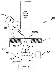
[0009] Figure 1 is an illustration of an LDI/MALDI source according to one
embodiment
of the invention, wlierein beam-focusing optics and an ion optical device are
disposed on
opposite sides of the sample support.
[0010] Figure 2 is an illustration of another embodiment of an LDI/MALDI
source,
wherein the viewing optics are disposed on the same side of the sample support
as the beam-
focusing optics.
[0011] Figures 3A aiid 3B are illustrations of further embodiments of
LDI/MALDI
sources, wherein the ion optical device and beam-focusing optics are located
on the same side
of the sample support and the viewing optics are located on an opposite side
of the sample
support.
[0012] Figure 4 is an illustration of a transparent sample support, according
to various
embodiments of the invention.
[0013] Figure 5 illustrates a metllod of analyzing a sample using a mass
spectrometer
having an LDI/MALDI source, according to various embodiments of the invention.
DETAILED DESCRIPTION
[0014] In one aspect of the invention, a laser desorption/ionization source or
matrix-
assisted laser desorption/ionization source (referred to collectively as an
LDI/MALDI source)
is provided which accommodates a sample support configured to support one or
more
sample(s) on a front surface thereof. The sample support is at least locally
transparent at the
wavelength of the irradiation beam. Transparency may be provided by the
modification of a
non-transparent sample support with transparent windows or openings that
underlie the
sample(s); alternatively, the entire sample support may be constructed from a
transparent
material such as quartz. Beam focusing optics and/or viewing optics may be
disposed
adjacent a rear surface of the sample support for, respectively, focusing a
beam of radiation
onto the sample and acquiring an image of the sample. An ion optical device,
such as a
multipolb ion guide, is disposed adjacent the front surface of the sample
support and
functions to collect and guide ions produced by irradiation of the sample.
[0015] Figures 1-3 illustrate different embodiments of an LDI/MALDI source
having
various aiTangements of beam-focusing and viewing optics. In each of these
embodiments,
the beam-focusing optics optionally includes a short focal length lens that
generates a
compact beam spot on the sample.
4
CA 02622982 2008-03-18
WO 2007/044361 PCT/US2006/038701
[0016] Figure 1 is an illustration of an LDI/MALDI source generally designated
100.
LDI/MALDI source 100 accommodates a sample support 110, and includes beam-
focusing
optics 120, viewing optics 130 and an ion optical device 140. Sample support
110 includes a
front surface 116, on which one or more sainples are deposited, and a rear
surface 115. Front
surface 116 may be flat and featureless, or may optionally include a
conductive coating for
application of an offset voltage, one or more chemical reagents configured to
react with the
analyte, and/or indentations configured to receive and hold the sample.
[0017] As noted above, each embodiment of the invention makes use of a
transparent
sample support. As used herein, the terms "transparent" or "transparency" are
not intended to
require coinplete transparency; rather, any sample support may be utilized
that allows
substantial transmission tllerethrough of radiation having the wavelength(s)
of interest.
Furthermore, the sample support may be only locally transparent, i.e., may be
transparent
only at regions thereof that underlie the sa.inple(s), and the remaining
portions of the sample
support may be opaque.
[0018] In some embodiments, sample support 110 is supported by a positioning
stage 117
that is moved with respect to ion optical device 140 and beam-focusing optics
120. A
positioning stage driver 119 is configured to move (e.g., translate or rotate)
positioning stage
117. Positioning stage driver 119 may includes a stepper motor, piezoelectric
device or
mechanism known in the art that is capable of precise control of the sample
support position.
In some embodiments, positioning stage driver 119 is configured to nlove
positioning stage
117 such that a selected one of a plurality of samples on sample support 110
is aligned with
the radiation beam and the proximal end of ion optical device 140. In various
embodiments,
positioning stage driver 119 is configured to move positioning stage 117 with
lateral (i.e., in
the X-Y plane defined by the sample support) resolutions of 10 micrometers, 5
micrometers,
3 micrometers, 1 micrometer, or less.
[0019] Beam-focusing optics 120 are disposed adjacent to (and as illustrated
perpendicular to) rear surface 115 of sample support 110. As used herein, the
term "adjacent"
does not require immediate adjacency, i.e., the beain-focusing optics should
still be
considered to be disposed adjacent to rear surface 115 even if one or more
structures are
interposed between the beam-focusing optics 120 and rear surface 115, or if
they are
separated by a substantial distance. Rather, the beam-focusing optics should
be considered
adjacent to the rear surface 115 if they are located in a region that is
closer to rear surface 115
than front surface 116. Beam-focusing optics 120 will typically include at
least one lens that
focuses a beam of radiation 122, which may be supplied by a radiation source,
for example
CA 02622982 2008-03-18
WO 2007/044361 PCT/US2006/038701
laser 124, onto a sample disposed on or near sample support 110 front surface
116. It is noted
that beam-focusing optics 120 may, without limitation, consist of a single
lens, as depicted in
the figures. Laser 124 will typically take the form of a nitrogen or solid-
state laser capable of
emitting short pulses of radiation at a wavelength or wavelengths that are
strongly absorbed
by the sample and matrix. In various embodiments, beam-focusing optics 120 are
configured
to produce a beam spot (the area of the sample impinged by the radiation beam)
having a
diameter of 10 micrometers, 5 micrometers, 3 micrometers, 2 micrometers, 1
micrometer, or
less. In various embodiments, beam-focusing optics 120 have a focal length of
15
millimeters, 12 millimeters, 10 millimeters, 8 millimeters, 5 millimeters, or
less. Beam-
focusing optics 120 are optionally positioned such that a major axis 125 is
approximately
parallel to surface front 116 and a center axis 126 is approximately
perpendicular to front
surface 116. In some embodiments, a combination of laser pulse power and focal
length may
be selected to effect single-shot desorption/ionization of the irradiated
region of the sample.
That is, substantially the entire thickness of the sample can be desorbed and
ionized at a
predetermined location with a single shot of a laser. This could allow for
more efficient use
of limited sample volumes, enabling results to be attained from a relatively
small amount of
analyte, and for numerous results to be attained from a single small sample
volume.
[0020] In some embodiments, laser 124 may operate in a selected one of two
modes. In
the first mode, the laser illuminates some, or all, of the sample for
subsequent visual image
acquisition via UV sensitive cameras, for example. In the second mode, the
laser irradiates a
target region of the sample for production of ions. Operation of the laser in
the first mode
may be employed, for example, to acquire and display an image that can be
viewed by the
instrument operator for use in selecting a portion of the sample to be
analyzed. Typically, the
illumination mode includes a lower beam flux than the ionization mode.
[0021] In some embodiments, beam-focusing optics 120 or a portion thereof are
mechanically coupled to a lens manipulator 127 configured to move beam-
focusing lens 120
relative to transparent sample support 110. For example, in some embodiments
lens
manipulator 127 is configured to move beam-focusing optics 120 toward or away
from front
surface 116. In some embodiments, lens manipulator 127 is configured to move
beam-
focusing optics 120 or other ionization optic parallel to first surface 116.
In these
embodiments, lens manipulator 127 is optionally used to move the beam spot
small distances
between different target locations on the sample. Lens manipulator 127 may be
operated in
conjunction with positioning stage 117 to achieve highly precise control of
the beam spot
position; for example, movement of positioning stage 117 may provide gross
control of the
6
CA 02622982 2008-03-18
WO 2007/044361 PCT/US2006/038701
beam spot position, and movement of lens manipulator 127 may provide fine
control of the
beam spot position. In various embodiments, lens manipulator 127 is configured
to move the
focal point by 20 micrometers, 10 micrometers, 5 micrometers, 3 micrometers, 2
micrometers, 1 micrometer, or less than 1 micrometer.
[0022] Viewing optics 130 are configured for viewing (i.e., acquiring an image
of) at
least a portion of the sa.inple disposed on sample support 110. An image
obtained using
viewing optics 130 can be displayed to the operator and used to select a
portion of interest of
the sample (e.g., a region within a tissue sample) for mass spectral analysis.
[0023] Viewing optics 130 typically include at least a focusing element such
as a lens
132, reflector, or the like, and a viewing element such as an eye piece or CCD
camera 134.
For exasnple, in some embodiments, imaging optics 130 includes CCD camera 134,
lens 132
a.nd a microscope aperture (not shown). In some embodiments, viewing optics
130 are
configured to detect the incidence of laser bea.in 122 on the sample. Viewing
optics 130
optionally include a visual distance indicator (not shown) configured to
assist an operator in
manipulatiiig beam-focusing optics 120 using lens manipulator 127 to focus on
a desired
location within the sample. One or more illumination sources (not depicted in
the figures)
may be provided to illuminate the sample for viewing and/or image acquisition.
[0024] Ion optical device 140 is configured to collect ions desorbed from a
MALDI
sample disposed on front surface 116 of sample support 110. Ion optical device
140 may
comprise, for example, a multipole ion guide to which appropriate AC and DC
voltages are
applied in order to confine the ions and/or draw the ions along the
longitudinal axis of the ion
guide. In a typical mass spectrometer architecture, ion optical device 140
transports ions
toward a mass analyzer, such as a quadrupole mass filter, ion trap, time-of-
flight analyzer, or
electrostatic trap, which separates ions according to their mass-to-charge
ratios for
subsequent detection and/or fragmentation. One or more intermediate chambers
as well as
various ion optics may be interposed in the ion path between ion optical
device 140 and the
mass analyzer.
[0025] Figure 2 is an illustration of an LDI/MALDI source 200, which is an
alternative
embodiment of LDI/MALDI source 100. In this embodiment, both beam-focusing
optics 120
and viewing optics 130 are disposed adjacent to (and as illustrated
perpendicular to) rear
surface 115 of sample support 110. Viewing optics 130 are configured to
acquire an image
(typically normal and undistorted) of a sample disposed on front surface 116
of sample
support 110. In this embodiment, beam-focusing optics 120 also functions to
focus the
sample image, in conjunction with partial reflector 210. Partial reflector 210
is preferably
7
CA 02622982 2008-03-18
WO 2007/044361 PCT/US2006/038701
highly reflective at the wavelength of laser 124 so as to direct the laser
beam onto the sample
and is at least partially transmissive at the wavelength range of visible
light so as to enable
viewing of the sample image therethrough by camera 134. The wavelength-
selective
reflection/transmission of partial reflector 210 may be achieved, for example,
by application
of suitable dielectric layers to one or both surfaces of the relector. In an
alternative
configuration, the relative positions of laser 124 and imaging optics 130 are
exchanged
relative to partial reflector 210.
[0026] Figure 3A is an illustration of a MALDI source 300, which is an
alternative
embodiment of MALDI source 100. In MALDI source 300, imaging optics 130 are
disposed
adjacent to rear surface 115 of sample support 110, and ion optical device 140
and beam-
focusing optics 120 are disposed adjacent to front surface 116 of sample
support 110. In this
embodiment, ion optical device 140 optionally includes a skimmer configured to
collect ions
desorbed from a sample disposed on front surface 116. Beam-focusing optics 120
is
optionally configured to focus laser beam 122 onto front surface 116 at a
perpendicular angle
to front surface 116. This orientation will typically produce the minimum spot
size of laser
beam 122 on the sample. However, in alternative embodiments, beam-focusing
optics 120
are configured to focus laser beam 122 onto front surface 116 at other angles
of incidence.
One example of this arrangement is illustrated in Figure 3B.
[0027] Figure 4 is a cross-sectional view of an exemplary implementation of
sample
support 110, wherein local transparency is achieved by adapting a substrate
420 with
openings 410 that underlie the samples 430. Each opening 410 narrows upwardly
to a
reduced-diameter well 413 having a diaineter indicated as 415. A sample 430
may be
deposited on sample support 110 by spotting a liquid solution containing the
analyte material
(and optionally a matrix substance) onto wells 413 and evaporating the
solvent. The well
diameter 415 should be sufficiently small to allow the liquid solution to be
retained in the
well by surface tension forces. In various embodiments, wells 413 have a
diameter 415 of
less than 50 micrometers, 25 micrometers, 10 micrometers or 8 micrometers. In
some
embodiments, wells 413 are each configured to hold a single cell.
[0028] Figure 5 illustrates a method of analyzing a sample, according to
various
embodiments of the invention. In a Prepare MALDI Sample step 510 a MALDI
sample is
deposited on front surface 116 of sample support 110, for example by adhering
a thin tissue
layer on the front surface and thereafter applying (e.g., by electrospraying)
a matrix layer
overlying the tissue.
8
CA 02622982 2008-03-18
WO 2007/044361 PCT/US2006/038701
[0029] In an optional View Sample step 520, viewing optics 130 are used to
view the
sample prepared in Prepare Sample step 510. The sample can either be viewed
directly
through a microscope aperture, viewed as an image captured using a digital
camera, or the
like. Typically, the sample is viewed in a magnified form. For example, in
some
embodiments the view may be in sufficient detail to identify areas of interest
within the
sample.
[0030] In an Ionize First Area step 530, laser 124 is operated to desorb and
ionize a part
of the MALDI sample located at the focal point of beam-focusing optics 120.
Ionization may
include simultaneous desorption and ionization or desorption followed by gas
phase
ionization.
[0031] In an Observe First Area step 540, the location of the area of the
sample ionized in
Ionize First Area step 530 is observed using viewing optics 130. This
observation can occur
eitller during the ionization process by imaging the ionization event or
following the
ionization process by imaging a change (e.g., loss of material) in the sample.
[0032] In a Change Locations step 550, the location of the focal point of beam-
focusing
optics 120 on the sample is moved. This relative movement may be accomplished
by moving
positioning stage 117 using positioning stage driver 119 and/or by moving beam-
focusing
optics 120 using lens manipulator 127. Change Locations step 550 is optionally
performed
while observing the sample through viewing optics 130 and/or using a distance
measurement
made using viewing optics 130.
[0033] Change Locations step 550 is optionally performed while operating laser
124 in
the illumination mode. For example, in one embodiment, Change Locations step
550
includes monitoring the position of the focal point of beam-focusing optics
120 by observing
light of laser beam 122 striking the sample, while laser beam 122 is operated
below a
desorption/ionization threshold of the MALDI sample. During this observation,
the focal
point is optionally moved to a specific part of the MALDI sample to be
analyzed. In various
embodiments, the change in location of the focal point of beam-focusing lens ,
that occurs in
Change Locations step 550, is less than or equal to 15 micrometers, 10
micrometers, 8
micrometers, 5 micrometers, 3 micrometers or 2 micrometers. In some
embodiments,
Change Locations step 550 includes moving the focal point of beam-focusing
optics 120
from one area of interest in a tissue sample to another.
[0034] In an Ionize Second Area step 560, laser 124 is operated in the
ionization mode to
desorb and ionize a second area of the sample. This second area is that part
of the MALDI
9
CA 02622982 2008-03-18
WO 2007/044361 PCT/US2006/038701
sample to which the focal point of beam-focusing lens 120 was directed to in
Change
Relative Locations step 550.
[0035] In a Determine M/Z step 570, the mass-to-charge ratios of ions
generated in Ionize
Second Area step 560 is determined using a mass analyzer to which ions are
transported by
ion optical device 140 (or which is incorporated into ion optical device 140).
These mass-to-
charge ratios are optionally used to forln a mass spectrum associated with the
ionized part of
the sample. By repeating Change Locations step 550 and Ionize Second Part step
560, mass
spectra associated with different areas of a tissue sample, or other sample,
are generated. In
alternative embodiments, an instance of Determine M/Z step 150 also follows
Ionize First
Part step 530.
[0036] The embodiments discussed herein are illustrative of the present
invention. As
these embodiments of the present invention are described with reference to
illustrations,
various modifications or adaptations of the methods and or specific structures
described may
become apparent to those skilled in the art. All such modifications,
adaptations, or variations
that rely upon the teachings of the present invention, and through which these
teachings have
advanced the art, are considered to be within the spirit and scope of the
present invention.
Hence, these descriptions and drawings should not be considered in a limiting
sense, as it is
understood that the present invention is in no way limited to only the
embodiments
illustrated.