Note: Descriptions are shown in the official language in which they were submitted.
CA 02623648 2014-08-26
SYSTEMS, COMPOSITIONS, AND METHODS FOR LOCAL IMAGING
AND TREATMENT OF PAIN
=
BACKGROUND OF THE INVENTION
30 1. Field of the Invention
[0005] This invention pertains generally to imaging of tissues
associated with
skeletal joints. More particularly, it relates to identification and/or
-1-
CA 02623648 2008-03-20
WO 2007/035906 PCT/US2006/036943
characterization of localized factors associated with musculoskeletal pain
using labeled markers and related imaging tools.
2. Description of Related Art
[0006] Chronic back pain (i.e. generally persisting longer than 12
weeks) is
among the most prevalent and expensive non-lethal conditions in the United
States, and is believed to be the most common cause of disability in persons
under 45 years old. The number of people suffering from chronic back pain is
estimated to exceed 25% of the overall population. Every year, about 3-4% of
the U.S. population is estimated to be disabled temporarily, and about 1% of
lo the working age population is estimated to be disabled totally and
permanently, due to intractable back pain. An estimated 11.7 Million patients
present medically with chronic back pain. National disability expenses for
this
prevalent condition range from $30-$70 billion per year. Effectively treating
this prevalent condition remains among the largest unmet clinical needs in
medicine. Properly diagnosing and localizing the source of pain also remains
a significant shortcoming on the critical path toward providing such therapy
in
a targeted manner with predictably successful outcomes.
[0007] Diagnosis of the location, mode, and extent of disc
degeneration is
often used as a precursor tool to drive therapy for treating back pain.
However, such measures are often not specific enough to localize the exact
site in or around a degenerating disc where pain is being experienced. Also, a
direct correspondence is not always found between disc degeneration and
back pain. Consequently, existing imaging modalities that identify (and even
quantify) disc anatomy, such as CT or MRI, are not always helpful at
localizing
sources of back pain in many cases.
[0008] Accordingly, there is still a substantial need for new imaging
modalities
to objectively, accurately, and specifically identify and localize source(s)
of
pain, and in particular back pain, and still more particularly lower lumbar
back
pain. There is in particular such a need with respect to identifying painful
discs in an improved way, and to localize within or around those discs the
specific site of injury or source of pain in an improved, predictable,
dependable manner.
-2-
CA 02623648 2008-03-20
WO 2007/035906 PCT/US2006/036943
BRIEF SUMMARY OF THE INVENTION
[0009] Accordingly, certain aspects of the present invention provide
a system,
composition of matter, and method that better describe, diagnose, and
localize of the sources of pain in and around musculoskeletal joints, and in
particular beneficial modes in and around spinal discs in relation to back
pain.
[0010] Among the various modes employed according to this aspect, one
particular beneficial mode involves artificially labeling substances locally
in the
area of back pain, such as in a particular beneficial example the spinal
motion
segment, that are known suspects to pain generation and transmission, such
as for example disc, facet joints, and vertebral bodies.
[0011] Two particularly beneficial embodiments according to this
mode, useful
either alone or in combination, include: (a) labeling nerves, and in
particular
beneficial embodiments nociceptors, and (b) labeling chemical factors that
irritate nerves, (c) labeling cells that produce chemical factors that
irritate
nerves; and (d) labeling blood vessels that are typically in close
approximation
to nerves.
[0012] In addition to the significant benefit provided by these
approaches for
clinical diagnosis, they are also considered highly beneficial in providing
new
avenues to drive choices for therapeutic approaches.
[0013] One aspect of the invention is a method for conducting a medical
procedure related to a localized, active source of pain at a location within a
patient. This method includes artificially labeling a pain factor at the
location in
a manner substantially increasing the ability to image the pain factor with an
imaging tool. The labeled pain factor is then labeled in a manner sufficient
to
selectively differentiate a first concentration of the labeled pain factor at
the
location versus a second concentration of the labeled pain factor in tissue
adjacent to the location.
[0014] According to one highly beneficial mode, the location is
associated with
a skeletal joint.
[0015] Another mode of this aspect further includes delivering a
substantially
targeted label into the patient that is adapted to differentially bind to and
label
a pain factor associated with the source of pain at the location. The pain
-3-
CA 02623648 2008-03-20
WO 2007/035906 PCT/US2006/036943
factor at the location is artificially labeled by binding the pain factor with
the
targeted label.
[0016] According to one embodiment, the differential binding
comprises
specific binding to the pain factor.
6 [0017] According to another embodiment, the differential
binding comprises
non-specific binding to the pain factor.
[0018] According to another mode, the pain factor comprises at least
one of a
nerve factor, an inflammatory factor, a cellular factor, or a blood vessel
factor,
or a combination thereof.
[0019] In one more particular mode, the pain factor comprises a nerve
factor.
[0020] According to one embodiment of this mode, the nerve factor
comprises
at least one substance associated with at least one of a nerve fiber or a
cellular structure associated with the nerve fiber.
[0021] In another embodiment, the nerve factor comprises a substance
associated with a nerve fiber. According to one particularly beneficial
embodiment, the substance is in particular associated with nociceptors.
[0022] In another more particular mode, the pain factor comprises a
blood
vessel factor.
[0023] According to one embodiment of this mode, the blood vessel
factor
comprises at least one of a blood vessel or a substance or structure
associated with the blood vessel.
[0024] In another embodiment of this mode, the blood vessel factor
comprises
a substance or structure associated with microvessels.
[0025] According to another more particular mode, the pain factor
comprises a
cellular factor.
[0026] According to one embodiment of this particular mode, the
cellular factor
is associated with a cell that produces at least one inflammatory factor.
[0027] In another embodiment, the cellular factor is associated with
at least
one inflammatory factor.
[0028] In another embodiment, the cellular factor is associated with cells
actively producing inflammatory factors.
[0029] In another embodiment, the cellular factor is associated with
an
-4-
CA 02623648 2008-03-20
WO 2007/035906 PCT/US2006/036943
inflammatory cell of a type that is attracted to a second pain factor at the
location. According to one particular variation of this embodiment, the
inflammatory cell comprises a leukocyte or macrophage.
[0030] According to another more particular mode, the pain factor
comprises
an inflammatory factor.
[0031] According to another mode, the pain factor comprises a
cytokine.
[0032] According to another mode of the present aspect, the pain
factor
comprises substance P or an analog or derivative or binding agent or antibody
thereof.
[0033] According to another mode, the pain factor comprises CGRP or an
analog or derivative or binding agent or antibody thereof.
[0034] According to another mode, the pain factor comprises receptor
tyrosine
kinase A (TrkA) or an analog or derivative thereof.
[0035] According to another mode, the pain factor comprises a TrkA
binding
agent or antibody.
[0036] According to another mode, the pain factor comprises a TrkA
receptor
or a binding agent or antibody thereof.
[0037] According to another mode, the pain factor comprises nerve
growth
factor (NGF) or an analog or derivative thereof.
[0038] According to another mode, the pain factor comprises an NGF binding
agent or antibody.
[0039] According to another mode, the pain factor comprises an NGF
antagonist or an analog or derivative thereof.
[0040] According to another mode, the pain factor comprises an NGF-
antagonist binding agent or anti-NGF antagonist antibody.
[0041] According to another mode, the pain factor comprises a nerve
binding
agent or antibody or an analog or derivative thereof.
[0042] According to another mode, the pain factor comprises protein
gene
product 9.5 (PGP 9.5) or an analog or derivative or binding agent or antibody
thereof.
[0043] According to another mode, the pain factor comprises SYN or an
analog or derivative or binding agent or antibody thereof.
-5-
CA 02623648 2008-03-20
WO 2007/035906 PCT/US2006/036943
[0044] According to another mode, the pain factor comprises
peripherin or an
analog or derivative or binding agent or antibody thereof.
[0045] According to another mode, the pain factor comprises
Neurofilament
200kD (NF200) or an analog or derivative or binding agent or antibody
thereof.
[0046] According to another mode, the pain factor comprises tissue
necrosis
factor alpha (INF-a) or an analog or derivative or binding agent or antibody
thereof.
[0047] According to another mode, the pain factor comprises a TNF-a
blocker
or binding agent or antibody thereof.
[0048] According to another mode, the pain factor comprises
macrophage
migration inhibitory factor (MIF) or an analog or derivative or binding agent
or
antibody thereof.
[0049] According to another mode, the pain factor comprises
infliximab, or an
analog or derivative thereof, or a binding agent or an antibody thereof.
[0050] According to another mode, the pain factor comprises PECAM or
an
analog or derivative or binding agent or antibody thereof.
[0051] According to another mode, the pain factor comprises CD34 or
an
analog or derivative or binding agent or antibody thereof.
[0052] According to another mode, the pain factor comprises vascular cell
adhesion molecule-1 (VCAM-1) or an analog or derivative or binding agent or
antibody thereof.
[0053] According to another mode, the pain factor comprises an
interleukin or
an analog or derivative or binding agent or antibody thereof.
[0054] According to one embodiment of this mode, the interleukin comprises
IL-1 or an analog or derivative or binding agent or antibody thereof.
[0055] According to another embodiment, the interleukin comprises IL-
6 or an
analog or derivative or binding agent or antibody thereof.
[0056] According to another embodiment, the interleukin comprises IL-
8 or an
analog or derivative or binding agent or antibody thereof.
[0057] According to another mode of the present aspect, the pain
factor
comprises prostaglandin E2 (PGE2) or an analog or derivative or binding
-6-
CA 02623648 2008-03-20
WO 2007/035906 PCT/US2006/036943
agent or antibody thereof.
[0058] According to another mode, the pain factor comprises a factor
associated with pH in tissue or a binding agent or an antibody thereof.
[0059] According to one embodiment of this mode, the labeled pain
factor is
indicative of a relatively low pH below a predetermined threshold at the
location.
[0060] According to another mode, the pain factor comprises a factor
associated with p02 in tissue or a binding agent or an antibody thereof.
[0061] In one embodiment according to this mode, the labeled pain
factor is
indicative of a relatively low p02 at the location.
[0062] According to another mode, the pain factor comprises glial
fibrillary
acidic protein (GFAP) or an analog or derivative or binding agent or antibody
thereof.
[0063] According to another mode, the pain factor comprises synuclein
(SYN)
or an analog or derivative or binding agent or antibody thereof.
[0064] According to another mode of the present aspect, the targeted
label
comprises at least one of a nerve factor, a blood vessel factor, a cellular
factor, an inflammatory factor, or an antibody thereof.
[0065] According to one embodiment of this mode, the targeted label
comprises a nerve factor or a binding agent or an antibody thereof.
[0066] In one variation according to this embodiment, the nerve
factor
comprises at least one substance associated with at least one of a nerve fiber
or a cellular structure associated with the nerve fiber or an antibody
thereof.
[0067] In another variation, the nerve factor comprises a substance
associated
with a nerve fiber or a binding agent or an antibody thereof.
[0068] In another embodiment, the targeted label comprises a blood
vessel
factor or a binding agent or an antibody thereof.
[0069] In one variation of this embodiment, the blood vessel factor
comprises
a substance associated with a structure of a blood vessel or a binding agent
or an antibody thereof.
[0070] In another variation, the blood vessel factor comprises a
substance
associated with a structure of a microvessel or a binding agent or an antibody
-7-
CA 02623648 2008-03-20
WO 2007/035906 PCT/US2006/036943
thereof.
[0071] According to another embodiment, the targeted label comprises
a
cellular factor or a binding agent or an antibody thereof.
[0072] In one variation, the cellular factor is associated with a
cell that
produces at least one inflammatory factor, or a binding agent or an antibody
thereof.
[0073] In another variation, the cellular factor is associated with
at least one
inflammatory factor or a binding agent or an antibody thereof.
[0074] In another variation, the cellular factor is associated with
an
intervertebral disc cell that is actively producing inflammatory factors, or a
binding agent or an antibody thereof.
[0075] In another variation, the cellular factor is associated with
an
inflammatory cell of a type that is attracted to the pain factor at the
location, or
a binding agent or an antibody thereof.
[0076] According to one feature of this variation, the inflammatory cell
comprises a leukocyte, or a binding agent or an antibody thereof.
[0077] According to another embodiment, the targeted label comprises
an
inflammatory factor, or a binding agent or an antibody thereof.
[0078] In one variation of this embodiment, the inflammatory factor
comprises
a cytokine, or an analog or derivative thereof, or a binding agent or an
antibody thereof.
[0079] According to another mode of the present aspect, the targeted
label
comprises a binding agent or antibody to substance P.
[0080] According to another mode, the targeted label comprises a
binding
agent or antibody to calcitonin gene-related peptide (CGRP).
[0081] According to another mode, the targeted label comprises a TrkA
antibody or binding agent.
[0082] According to another mode, the targeted label comprises nerve
growth
factor (NGF), or an analog or derivative thereof.
[0083] According to another mode, the targeted label comprises a NGF
binding agent or an anti-NGF antibody.
[0084] According to another mode, the targeted label comprises a NGF
-8-
CA 02623648 2008-03-20
WO 2007/035906 PCT/US2006/036943
antagonist or a binding agent or an antibody thereof.
[0085] According to another mode, the targeted label comprises an
anti-NGF
antagonist antibody or binding agent.
[0086] According to another mode, the targeted label comprises a
nerve
antibody or binding agent.
[0087] According to another mode, the targeted label comprises PGP
9.5, or
an analog or derivative thereof, or a binding agent or an antibody thereof.
[0088] According to another mode, the targeted label comprises a
binding
agent or antibody to peripherin.
[0089] According to another mode, the targeted label comprises
Neurofilament
200kD (NF200), or an analog or derivative thereof, or a binding agent or an
antibody thereof.
[0090] According to another mode, the targeted label comprises TNF-a,
or an
analog or derivative thereof, or a binding agent or an antibody thereof.
[0091] According to another mode, the targeted label comprises a TNF-a
blocker.
[0092] According to another mode, the targeted label comprises
infliximab, or
an analog or derivative thereof, or a binding agent or an antibody thereof.
[0093] According to another mode, the targeted label comprises a
PECAM
binding agent or antibody. .
[0094] According to another mode, the targeted label comprises a
binding
agent or antibody to CD34.
[0095] According to another mode, the targeted label comprises an
interleukin
binding agent or antibody.
[0096] In one embodiment of this mode, the interleukin binding agent or
antibody comprises an IL-1 binding agent or antibody.
[0097] In another embodiment of this mode, the interleukin binding
agent or
antibody comprises an IL-6 binding agent or antibody.
[0098] In another embodiment of this mode, the interleukin binding
agent or
antibody comprises an IL-8 binding agent or antibody.
[0099] According to another mode of the present aspect, the targeted
label
comprises a binding agent or antibody to PGE2.
-9-
CA 02623648 2008-03-20
WO 2007/035906 PCT/US2006/036943
[00100] According to another mode, the targeted label comprises a
binding
agent or antibody to MIF.
[00101] According to another mode, the targeted label comprises an
antibody
or binding agent to a factor associated with pH in tissue.
6 [00102] According to one embodiment of this mode, the labeled
pain factor is
indicative of a relatively low pH below a predetermined threshold at the
location.
[00103] According to another mode, the targeted label comprises an
antibody
or binding agent to a factor associated with p02 in tissue.
[00104] According to one embodiment of this mode, the labeled pain factor
is
indicative of a relatively low p02 at the location..
[00105] According to another mode, the targeted label comprises a
radioactive
material.
[00106] According to one embodiment of this mode, the targeted label
comprises a radio-labeled TN F-a antibody, or an analog or derivative thereof.
[00107] According to another embodiment, the targeted label comprises
radiolabeled iodine. In one variation of this embodiment, the radiolabeled
iodine comprises 1-125.
[00108] According to another mode, the targeted label comprises a
nanoparticle.
[00109] According to another mode, the targeted label comprises gold.
[00110] According to another mode, the targeted label comprises iron
oxide.
[00111] According to another mode, the targeted label comprises
gadolinium.
[00112] According to another mode of the present aspect, the method
further
includes imaging the labeled pain factor using an imaging tool that comprises
a phosphor imaging plate.
[00113] According to another mode, the method includes imaging the
labeled
pain factor using MRI.
[00114] According to another mode, a first binding agent is delivered
into the
body that is adapted to bind to a first pain factor. The targeted label is
delivered into the patient's body after the first binding agent is bound to
the
first pain factor. The targeted label is adapted to bind to a site located on
the
-10-
CA 02623648 2008-03-20
WO 2007/035906 PCT/US2006/036943
bound combination of the first binding agent and the first pain factor.
[00115] According to one embodiment, the first binding agent comprises
a bi-
specific antibody with a first binding site adapted to bind to the first pain
factor
and a second binding site adapted to bind to the targeted label.
[00116] According to another mode, the targeted label comprises a cell
bound
to an antibody having an exposed binding site that is adapted to bind to the
pain factor.
[00117] According to another mode, the method further includes
conducting a
therapeutic procedure in a substantially localized manner to the location
lo where the targeted labeled pain factor is locally imaged.
[00118] In one embodiment of this mode, the therapeutic procedure is
adapted
to substantially alleviate generation or transmission of pain at the location.
[00119] According to another embodiment, the therapeutic procedure is
adapted to substantially ablate at least one nerve at the location.
[00120] In another embodiment, the therapeutic procedure comprises
delivering
at least one therapeutic chemical in a substantially localized manner to the
location.
[00121] In another embodiment, the therapeutic procedure comprises
delivering
a therapeutic dose of energy in a substantially localized manner to the
location.
[00122] In one variation of this embodiment, the therapeutic procedure
further
comprises ablating at least one nerve at the location with the therapeutic
dose
of energy.
[00123] In another variation, the therapeutic procedure further
comprises
delivering ultrasound energy to the location. In a further variation, the
method
further includes delivering the ultrasound energy in a directed manner locally
into the location from a second location. In still a further variation, the
second
location is outside of the patient, and the ultrasound energy is delivered via
high intensity focused ultrasound (HIFU) that is adapted to focus the
ultrasound energy to the location. In yet another variation, the second
location is adjacent to the location within the patient, and the ultrasound
energy is delivered via a directional ultrasound probe. In still a further
feature
-11-
CA 02623648 2008-03-20
WO 2007/035906 PCT/US2006/036943
of this variation, the second location is adjacent to an intervertebral disc
and
the location receiving the directional ultrasound therapy is within the
intervertebral disc.
[00124] According to another variation of the present embodiment, the
therapeutic dose of energy comprises thermal energy.
[00125] According to another variation, the therapeutic dose of energy
comprises electrical energy. In one further variation, the method involves
delivering the electrical energy via a radiofrequency (RF) probe.
[00126] According to another variation, the therapeutic dose of energy
comprises microwave energy.
[00127] According to another variation, the therapeutic dose of energy
comprises light energy.
[00128] According to another mode of the present aspect, the location
comprises at least a portion of an intervertebral disc.
[00129] According to another mode, the location comprises a region of
tissue
located within only a portion that is equal to less than an entire
circumference
of an intervertebral disc.
[00130] In one embodiment of this mode, the portion comprises a region
of
tissue located within less than or equal to one-half of the circumference of
the
intervertebral disc.
[00131] In one variation of this embodiment, the portion comprises a
region of
tissue located within less than or equal to one-quarter of a circumference of
the intervertebral disc.
[00132] According to another mode of the present aspect of the
invention, the
location comprises an end-plate associated with a vertebral body.
[00133] According to another mode, the location comprises a facet
joint.
[00134] The method of the present aspect according to another mode
includes
delivering the targeted label in a localized manner to the location.
[00135] One embodiment of this mode further includes injecting the
targeted
label into a region of tissue associated with the location using a local
injection
assembly.
[00136] Another embodiment includes delivering the targeted label
systemically
-12-
CA 02623648 2008-03-20
WO 2007/035906 PCT/US2006/036943
to the patient.
[00137] One further embodiment includes injecting the targeted label
into the
patient's systemic blood circulation.
[00138] Another further embodiment includes delivering the targeted
label into
the patient's gastrointestinal system.
[00139] Another mode includes artificially labeling the pain factor at
multiple
said locations by binding the pain factor with the targeted label delivered
into
the patient. The labeled pain factor is then imaged with an imaging tool
adapted to image at least one of the targeted label or the labeled pain factor
and in a manner sufficient to differentiate a first concentration of the
labeled
pain factor at the multiple said locations versus a second concentration of
the
labeled pain factor in tissue adjacent to the multiple said locations.
[00140] According to one embodiment of this mode, the method further
includes conducting at least one therapeutic procedure in a substantially
localized manner to each of the locations where the targeted labeled pain
factor is locally and selectively imaged.
[00141] Another aspect of the invention involves a system for treating
pain at a
location within a body of a patient. This aspect includes a targeted label
that
is adapted to bind to and label a pain factor associated with a source of pain
at the location. Also included is a delivery assembly that is adapted to
deliver
the targeted label into the patient. An imaging system also included in the
system is adapted to image at least one of the targeted label or the labeled
pain factor and in a manner sufficient to selectively differentiate a first
concentration of the labeled pain factor at the location versus a second
concentration of the labeled pain factor in tissue adjacent to the location. A
therapeutic device assembly is also included, and is adapted to provide
therapy in a substantially localized manner that is substantially isolated to
the
location.
[00142] According to one mode of this aspect, the targeted label is
adapted to
bind and label a pain factor associated musculoskeletal joint pain, and the
location is associated with at least one musculoskeletal joint.
[00143] According to one embodiment, the therapeutic device assembly
-13-
CA 02623648 2008-03-20
WO 2007/035906 PCT/US2006/036943
comprises an energy delivery assembly that is adapted to deliver a
therapeutic dose of energy in a substantially localized manner that is
substantially isolated to the location associated with the musculoskeletal
joint.
[00144] According to one further embodiment, the energy delivery
assembly is
adapted to be delivered into the patient to a position at or adjacent to the
location.
[00145] According to another further embodiment, an introducer is
provided in
the system and is adapted to deliver the energy delivery assembly to the
location.
[00146] In one variation of this embodiment, the introducer comprises a
needle
assembly. This may provide the additional feature in that the needle
assembly is adapted to be advanced through bone and to deliver the
therapeutic device assembly to a position within the bone. According to
another further feature, the therapeutic device assembly may be adapted to
ablate an intraosseous nerve within the bone and that is associated with pain
related to the labeled pain factor visualized at the location. In another
further
beneficial feature, the needle assembly is adapted to be advanced through
bone of a vertebral body and to deliver the therapeutic device assembly to a
position within the vertebral body associated with a basivertebral nerve, and
the therapeutic device assembly is adapted to ablate the basivertebral nerve
from the position.
[00147] According to another mode of the present aspect, the
therapeutic
device assembly comprises a radiofrequency (RF) current ablation assembly.
[00148] In one embodiment, the RF current ablation assembly comprises
a first
electrode and a second electrode adapted to be positioned at first and second
positions adapted to straddle at least a portion of the basivertebral nerve.
The
RF current ablation assembly is adapted to deliver the RF current between the
first and second electrodes sufficient to ablate nerve tissue between the
first
and second positions.
[00149] According to one variation of this embodiment, the RF current
ablation
assembly comprises a delivery probe with an elongated body that carries the
first and second electrodes in a bipolar lead assembly arrangement.
-14-
CA 02623648 2008-03-20
WO 2007/035906 PCT/US2006/036943
[00150] According to another mode of the present embodiment, the
targeted
label is adapted to bind and label a pain factor comprising at least one of a
nerve factor, a blood vessel factor, a cellular factor, an inflammatory
factor, or
an antibody thereof.
[00151] It is to be appreciated that further more detailed particularly
beneficial
modes provided hereunder are contemplated with respect to the present
aspect described. In particular, further modes of the present aspect include
the various beneficial examples for pain factors and targeted labels described
for use under the method aspect of the invention described above
[00152] According to another mode, the system further includes an imaging
tool
that is adapted to image the labeled pain factor in a manner sufficient to
differentiate a first concentration at the location associated with pain
versus a
second concentration at a second location adjacent to the location and
associated with less pain that at the location.
[00153] Another aspect of the invention is a method for imaging and
identifying
a localized, active source of pain at a location associated with a region of
tissue in a patient, such as in particular beneficial further modes a skeletal
joint in a patient, and in still further beneficial more detailed modes spinal
joints in a patient. This method includes delivering a substantially
targeted label into the patient that is adapted to differentially bind to and
label
a pain factor. A pain factor that is resident at the location is artificially
labeled
by binding the pain factor with the targeted label delivered into the patient.
The labeled pain factor is imaged with an imaging tool adapted to image at
least one of the label or the labeled pain factor and in a manner sufficient
to
differentiate a first concentration of the labeled pain factor at the location
versus a second concentration of the labeled pain factor in tissue adjacent to
the location.
[00154] According to various modes of this aspect, the pain factor may
be
related to at least one of a nerve fiber, a substance associated with a nerve
fiber, a blood vessel, a substance associated with a blood vessel, a cell
actively producing at least one inflammatory factor, a cell attracted to
inflammation or other pain factors, or a chemo-inflammatory factor, or a
-15-
CA 02623648 2008-03-20
WO 2007/035906 PCT/US2006/036943
combination thereof.
[00155] Another aspect of the invention is a system for identifying or
characterizing a property of tissue associated with a skeletal joint. Such
aspect may further include any one or more of the various aspects, modes,
embodiments, variations, or features herein shown or described, or
combinations thereof.
[00156] According to one mode of this aspect, the system is adapted to
provide
information indicative of a degree of a property of at least a portion of an
intervertebral disc.
[00157] Another aspect is a system for identifying or characterizing a
property
of tissue associated with a skeletal joint in a patient. This includes
labeling at
least one of: pain factors, nerve factors, blood vessel factors, cellular
factors,
or inflammation factors. Or, the system may include a combination of one or
more of the foregoing.
[00158] According to one mode of this aspect, the information is related to
a
degree of a property of at least a portion of an intervertebral disc.
[00159] Another aspect of the invention is a system for characterizing
at least a
portion of an intervertebral disc with respect to a degree of a property of
that
disc, such as in particular related to pain or degeneration. This system
includes a labeled marker delivery system and a labeled marker imaging
system. The labeled marker imaging system provides information that is
useful to indicate at least in part the degree of the property.
[00160] According to one further embodiment of the foregoing aspects
and
modes, the respective system is adapted to produce the information based on
either or both of an annular portion or a nucleus portion of the
intervertebral
disc.
[00161] According to another embodiment, the system is adapted to
display a
geographical representation related to the spatial concentration of the
labeled
factor, and a portion of the geographical representation provides the
information.
[00162] According to another embodiment, the information is adapted to
distinguish a degree of degradation of the disc. According to one highly
-16-
CA 02623648 2008-03-20
WO 2007/035906 PCT/US2006/036943
beneficial further embodiment, the information is adapted to distinguish as to
the degree of degradation by reference to a Thompson scale.
[00163] According to another embodiment, the property comprises at
least one
of pain, or at least one factor that correlates with pain.
[00164] According to another embodiment, the information is related to
ratios of
concentration of one or more pain factors.
[00165] According to another embodiment, the information is related to
presence of secondary or other indirect materials that generally, though
indirectly, correlate well with presence of other more direct pain factors.
[00166] According to another embodiment, the information relates to at
least
one chemical constituent of an intervertebral disc.
[00167] According to another embodiment, the property comprises at
least one
of a degree of dehydration of the disc, a degree of breakdown of a
proteoglycan matrix of the disc, and a degree in a breakdown of a collagen
matrix.
[00168] According to another embodiment, the system further includes a
radiolabel imaging system that is adapted to produce the information.
[00169] Another aspect of the invention is a method for identifying or
characterizing a property of tissue associated with a skeletal joint. One or
more of the foregoing method aspects, modes, embodiments, variations, or
features herein described, or combinations thereof, may be employed to
advance this method.
[00170] One further mode of this aspect further includes providing
information
indicative of a degree of a property of at least a portion of an
intervertebral
disc.
[00171] Another aspect is a method for identifying or characterizing a
property
of tissue associated with a skeletal joint in a patient, and includes at least
one
of the following steps: labeling a pain factor in the tissue; imaging the
labeled
pain factor in the tissue; comparing different imaged regions having different
concentrations of the labeled pain factor; identifying a location of increased
presence of pain factors based upon the comparison; and treating the location
with local treatment modality based upon the identification. Or a combination
-17-
CA 02623648 2008-03-20
WO 2007/035906 PCT/US2006/036943
of one or more of the foregoing may be used.
[00172] One mode of this aspect includes determining a degree of a
property of
at least a portion of an intervertebral disc based upon the information.
[00173] Another aspect of the invention is a method for characterizing
at least a
portion of an intervertebral disc with respect to a degree of a property
thereof,
and includes capturing a signal related to the portion using a signal imaging
system. The signal imaging system provides information that indicates at
least in part the degree of the property.
[00174] According to one embodiment of the various method aspects and
lo modes just described, the information produced is based on either or
both of
an annular portion or a nucleus portion of the intervertebral disc.
[00175] In another embodiment, a curve is displayed that is related to
the
presence of the labeled pain factor, and wherein a portion of the curve
provides the information.
[00176] Another embodiment includes distinguishing a degree of degradation
of
the disc based upon the information. A still further embodiment includes
distinguishing the degree of degradation of the disc in relation to a Thompson
grade based upon the information.
[00177] Another embodiment includes correlating the disc with degree
of pain,
or at least one factor that correlates with pain, based upon the information.
[00178] According to another embodiment, the information is related to
a ratio
of magnitude of a signal imaged that corresponds with the amount of labeled
pain factor in a given area or volume of tissue.
[00179] According to another embodiment, the information is related to
a
cytokine, a precursor material thereof, an analog or derivative thereof, or a
metabolite or degradation product thereof.
[00180] According to another embodiment, the information relates to at
least
one chemical constituent of an intervertebral disc.
[00181] According to another embodiment, the property relates to at
least one
of a degree of dehydration of the disc, a degree of breakdown of a
proteoglycan matrix of the disc, and a degree in a breakdown of a collagen
matrix.
-18-
CA 02623648 2008-03-20
WO 2007/035906 PCT/US2006/036943
[00182] Another embodiment includes producing the information at least
in part
using a radiation imaging system.
[00183] Another aspect is a method for preparing a system for
performing a
medical procedure on a patient, comprising: diagnosing the patient with pain;
and based upon the diagnosis, preparing a volume of a targeted agent for
delivery into the patient. The prepared volume of targeted agent is configured
to differentially bind to a pain factor associated with the pain in a manner
adapted to enhance at least one of (i) diagnostic localization of the pain and
(ii) selective tissue therapy in an area associated with the bound pain factor
in
response to a delivered energy to the area.
[00184] Another aspect is a system for performing a medical procedure
on a
patient, comprising: a therapeutic volume of a targeted agent prepared for
delivery into a patient diagnosed with pain and that is configured to
differentially bind to a pain factor associated with the pain in a manner
adapted to enhance at least one of (i) diagnostic localization of the pain and
(ii) selective tissue therapy to a location containing the bound pain factor
in
response to a delivered energy to an area containing the location.
[00185] Another aspect is a method for selectively treating one or
more tissue
regions associated with pain in a patient, comprising delivering a targeted
agent into the patient configured to differentially bind to a pain factor
associated with the pain; and allowing the delivered targeted agent to
differentially bind to the pain factor so as to form a differentially bound
pain
factor; and delivering energy into the patient in a manner that differentially
treats the one or more regions associated with the differentially bound pain
factor.
[00186] Another aspect is a system for selectively treating one or
more tissue
regions associated with pain in a patient, comprising: a volume of targeted
agent; and an energy delivery system that is configured to deliver energy into
the patient. The volume of targeted agent is configured for delivery into a
patient and to differentially bind to a pain factor associated with the pain
in a
manner such that tissue regions containing a first concentration of the
differentially bound pain factor exhibit a differential and selective
therapeutic
-19-
CA 02623648 2008-03-20
WO 2007/035906 PCT/US2006/036943
response to the delivered energy versus other regions with lower
concentrations of the differentially bound pain factor.
[00187] Each aspect, mode, embodiment, variation, or feature herein
described
is considered independently beneficial without requiring combination with the
others. However, such further combinations and sub-combinations thereof
are also considered yet further beneficial independent aspects invention. For
example, where particular modes, embodiments, variations, or features are
herein described with respect to one aspect hereunder, it is to be appreciated
by one of ordinary skill that such description is further applicable to other
aspects also described though such particular combination may not be
specifically mentioned. In further example, a more detailed description
provided with respect to a method aspect may provide information that is to be
clearly combined as further development of a similar system-related aspect or
description, or visa versa.
[00188] Further aspects of the invention will be brought out in the
following
portions of the specification, wherein the detailed description is for the
purpose of fully disclosing preferred embodiments of the invention without
placing limitations thereon.
BRIEF DESCRIPTION OF THE SEVERAL VIEWS
OF THE DRAWING(S)
[00189] The invention will be more fully understood by reference to
the
following drawings which are for illustrative purposes only:
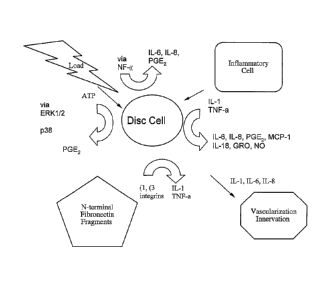
[00190] FIG. 1 shows a schematic of certain cascades associated with
inflammation and pain.
[00191] FIGS. 2A-D shows stained cross-sectioned histology slides
indicating
presence of certain factors associated with pain as follows, wherein "N" is
nucleus pulposus, "A" is annulus fibrosus, and "G" designates growth plate.
[00192] FIG. 2A shows a mid-sagittal section of normal mouse-tail disc
demonstrating TNF-alpha localization in periphery of nucleus pulposus (brown
stain).
[00193] FIG. 2B shows a normal mouse disc wherein localization of TNF-
alpha
is present in the hypertrophic zone of the growth plate as generally expected.
-20-
CA 02623648 2008-03-20
WO 2007/035906 PCT/US2006/036943
[00194] FIG. 2C shows in the compressed disc wherein increased amounts
of
TNF-alpha are apparent within the nucleus and inner annulus.
[00195] FIG. 2D shows increased TNF-alpha in the nucleus, inner
annulus and
irregularities in growth plate observed in compressed disc.
[00196] FIG. 3 shows a schematic view of a mouse 30 according to an
experimental model wherein the mouse tail 36 is injured by a fixture 40 for
evaluating pain factors.
[00197] FIG. 4 shows an experimental set-up related to the mouse
injury model
illustrated in FIG. 3, wherein a series of mice 30 are positioned for viewing
their respective tails via a phosphor imaging plate 50.
[00198] FIG. 5 shows an image 60 taken from a phosphor imaging plate
according to the set-up shown in FIG. 4 for four treatment mice and one
control mouse tail (located centrally in the figure).
[00199] FIG. 6 shows a schematic view of MAPK signaling pathways
associated with certain pain factors.
[00200] FIG. 7 shows a schematic view of a NF-K3 pathway associated
with
certain pain factors.
[00201] FIG. 8 shows a schematic view of a prostaglandin pathway
associated
with certain pain factors.
DETAILED DESCRIPTION OF THE INVENTION
[00202] Referring more specifically to the drawings, for illustrative
purposes the
present invention is embodied in the systems and methods generally shown in
or illustrated by reference to FIG. 1 through FIG. 8. It will be appreciated
that
the apparatus may vary as to configuration and as to details of the parts, and
that the method may vary as to the specific steps and sequence, without
departing from the basic concepts as disclosed herein.
[00203] Label Disc Features Associated With Pain
[00204] Discogenic pain is generally believed to be a multifactoral
phenomenon
in many cases. In particular, three illustrative factors are summarized in
varying levels of detail here as examples that are considered contributors in
various ways to (or otherwise indicative of) the generation or transmission of
discogenic pain. It is believed that these illustrative factors frequently act
as a
-21-
CA 02623648 2008-03-20
WO 2007/035906 PCT/US2006/036943
co-existent combination, often acting simultaneously. These types of factors
are summarized as follows.
[00205] One such factor type relates to the presence of nociceptors.
Normally,
intervertebral discs are substantially avascular and only sparsely innervated
at
the outer margins of the disc annulus. These unnnyelinated, substance P (SP)
or calcitonin gene-related peptide (CGRP) containing fibers are typically
unresponsive and termed silent nociceptors [Cavanaugh, 1996]. SP and
CGRP are believed to be the sensory transmitters of nociceptive information.
As degeneration proceeds, nerves can follow microvessels and grow deeper
into discs, which may occur for example either peripherally or via the
endplate.
This nerve and vessel in-growth is facilitated by degeneration-related
decreases in disc pressure and proteoglycan content.
[00206] A second such factor type is generally embodied by the need
for the
intradiscal nociceptors to be sensitized, and thus generally involves agents
providing such sensitization. This can occur for example via cytokines, which
are typically small, secreted proteins that mediate and regulate inflammation.
Elevated levels of certain cytokines have been measured in human discs, and
are associated with degeneration and pain. Such major cytokines have been
observed to include interleukin-1, -6, and -8, tissue necrosis factor-alpha
(TNF-a), macrophage migration inhibitory factor (MIF), and prostaglandin E2
(PGE2). The source of cytokines can be circulating inflammatory cells, such
as for example in the case of herniated discs, or disc cells, such as for
example in the case of contained disc degeneration. These pro-inflammatory
stimuli can trigger cells to initiate a number of catabolic programs meant to
stimulate tissue repair and remodeling that includes production of matrix
metalloproteinases 1, 9 and 13. During this wound healing process, cytokines
are also often involved in stimulating angiogenesis and granulation tissue
formation.
[00207] In one particular beneficial embodiment of the present
invention,
cytokines and/or their cell-surface receptors are imaged at sites of
inflammation in vivo using labeled markers, such as radiolabels. In particular
beneficial examples, cytokines are tagged with one or more of the following,
-22-
CA 02623648 2008-03-20
WO 2007/035906 PCT/US2006/036943
without limitation: iodine-123, iodine-125, iodine-131, technetium-99m,
fluorine-18, or indium-111. In addition, positron-emitting radioisotopes (for
example and without limitation fluorine-18) can be imaged using positron
emission tomography (PET) or positron emission tomography-computed
tomography (PET-CT). Other radiolabeled compounds can be imaged for
example using single photon emission computerized tomography (SPECT).
[00208] It is also to be appreciated that MRI may be employed
according to
further embodiments for visualizing or observing accumulation or binding of
various labeled markers variously herein described, such as for example in
applying gadolinium as a marker tagged to or conjugated with certain labels to
be bound to pain factors. Moreover, nanoparticles such as gold or iron oxide
may be used as labels or markers to bind and thereafter be viewed or
selectively targeted for therapy using appropriate visualization or treatment
modalities, respectively.
[00209] A third such factor related to discogenic back pain involves disc
depressurization that leads to mechanical instability while a pre-stress in
the
annulus and interspinal ligaments is diminished. Depressurization and
instability, in turn, lead to abnormal internal disc stress that may stimulate
nerves, leading to discogenic pain. Abnormal disc stress may also cause disc
cells to be pro-inflammatory, compounding the adverse effects of an abnormal
mechanical environment.
[00210] Labeling and Imaging Nerve Factors
[00211] According to certain particular embodiments, one or more
materials
associated with nerves in or around intmertebral discs are labeled with
markers that are imaged for localization of pain. This is premised in part on
the presence of certain such factors as indicators that pain may originate or
transmit in the area. These embodiments include, without limitation, labeling
structures or substances associated with nerves themselves. Further detailed
modes of this include labeling substances within nerves, such as in particular
but without limitation substance P or "CGRP". Other nerve fiber factors,
substances or components that may be labeled according to such further
embodiment(s) include, without limitation: TRK-a; anti-TRK-a antibody; nerve
-23-
CA 02623648 2008-03-20
WO 2007/035906 PCT/US2006/036943
growth factor (NGF); anti-NGF antibody; NGF antagonist; anti-NGF antagonist
antibody; POP 9.5; SYN; peripherin; or other form of nerve antibodies or
related materials in general. Other materials such as neurofilament 200kD
(NF200) [Johnson, 2001; Ashton, 1994] may also be the target of such
labeling and subsequent imaging.
[00212] As apparent from these highly beneficial illustrative
embodiments just
noted immediately above (and elsewhere herein), endogenous substances
such as TrkA or NGF may be targeted as the pain factor for labeling, or
related antibodies or other substances having particular binding affinity or
lo specificity to such resident materials may be bound to them in the area
of pain
and then thereafter provide the binding site for targeted labels to be
subsequently delivered. In this regard, it is to be appreciated that various
forms binding agents are broadly contemplated hereunder this description,
though they may not be particularly antibodies affecting function of the
target
for binding. For example but without limitation, an antibody mimetic may be
employed according to the present embodiments. Furthermore, various such
substances described hereunder as targeted pain factors may be themselves
labeled as markers and delivered to other targets. For example, NGF may be
labeled and artificially delivered as the agent to mark TrkA as the targeted
pain factor for imaging. In each of these different types of exemplary cases,
the ultimate target for labeling via a separately delivered agent (e.g.
whether
the target is an endogenous resident substance or an artificially delivered
substance) is considered a "nerve factor" as a pain factor according to the
present embodiments.
[00213] The following description provides further understanding of the
role of
these types of chemicals and other materials with respect to these present
embodiments. Further description of the benefits of various particular
illustrative examples are also provided elsewhere herein for a further
understanding.
[00214] The intervertebral disc is normally avascular and only sparsely
innervated at the outer layers of the annulus fibrosus and the vertebral
endplate [Fagan, 2003]. The outer 1/3 of the posterior annulus is believed to
-24-
CA 02623648 2008-03-20
WO 2007/035906 PCT/US2006/036943
be most typically innervated by the afferent fibers from the sinovertebral
nerve, which is considered a 'recurrent branch' of the ventral ramus of the
spinal nerve at the same level [Nakamura, 1996]. The ventral and lateral
aspects of the annulus are believed to be most typically innervated by the
dorsal root ganglion (DRG) [Aoki, 2004]. Also, it has been reported that
sensory fibers from upper level DRGs are believed to most typically innervate
the dorsal portion of discs via the paravertebral sympathetic trunk [Ohtori,
2001].
[00215] The endplate is also suggested to be innervated by the
basivertebral
lo nerve, which as further suggested may be a branch of the sinovertebral
nerve
entering the vertebral body through the posterior neurovascular foramen
[Antonacci, 1998].
[00216] Nerves usually accompany blood vessels, but can be found as
isolated
nerves in disc matrix. These non-vessel-associated fibers found in back pain
patients have been observed to express growth-associated protein 43
(GAP43) as well as SP [Freemont, 1997]. Small disc neurons contain CGRP
and also express the high-affinity nerve growth factor (NGF) receptor,
tyrosine
kinase A (trkA)[Aoki, 2004]. Disc inflammation has been observed to cause
an increase in CGRP positive neurons [Aoki, 2004]. A recent study showed
that NGF is expressed in microvascular blood vessels in a painful lumbar disc,
and that there are trkA (TRK-a) expressing nerve fibers adjacent to the
vessels that enter painful discs primarily through the endplate [Freemont,
2002; Brown, 1997]. Along with nerves growing into degenerated discs are
specialized nerve support cells termed gglia' or Schwann cells localized using
glial fibrillary acidic protein (GFAP) [Johnson, 2001].
[00217] Accordingly, various such materials may provide the requisite
binding
affinity or specificity to painful regions (or highly innervated regions) to
play the
role as the labeled marker agent for delivery to pain factor targets. Or,
these
materials may provide the particular target as the pain factor to be labeled
with selectively bound markers according to various embodiments of the
present invention. In one particular beneficial example, TrkA antibody (or
other binding agent) is labeled and delivered as a marker for binding and
-25-
CA 02623648 2014-08-26
visualization at a location associated with pain. In another beneficial
example,
NGF itself is labeled and delivered as a marker to itself bind to TrI<A. In
further embodiments, the resident quantities of these materials are treated as
the pain factors themselves for targeted labeling, e.g. using anti-bodies or
other agents with beneficial binding affinity and/or specificity to these
types of
resident compounds in painful regions.
[00218]
WO 2004/032870 to
Shelton et al. as "Inventors" and Rinat Neuroscience Corp. as "Applicant" for
"Methods for Treating Post-Surgical Pain By Administering a Nerve Growth
Factor Antagonist and Compositions Containing the Same"; WO 2004/058184
to Shelton et al. as "Inventors" and Rinat Neuroscience Corp. as "Applicant"
for "Anti-NGF Antibodies and Methods Using Same"; WO 2004/073653 to
Shelton et al. as "Inventors" and Rinat Neuroscience Corp. as "Applicant" for
"Methods for Treating Pain by Administering A Nerve Growth Factor
Antagonist and an NSAID And Compositions Containing The Same"; WO
2004/096122 to Shelton et al. as "Inventors" and Rinat Neuroscience Corp. as
"Applicant" for "Methods for Treating Pain By Administering A Nerve Growth
Factor Antagonist And An Opioid Analgesic and Compositions Containing The
Same"; and WO 2005/000194 to Shelton et al. as "Inventors" and Rinat
Neuroscience Corp. as "Applicant" for "Methods for Treating Post-Surgical
Pain By Administering An Anti-Nerve Growth Factor Antagonist Antibody and
Compositions Containing The Same."
[00219] The various compositions and methods described-
may be adopted where appropriate to one of ordinary skill as
label/marker vehicles and/or pain factor targets according to further
embodiments of the various aspects and modes of the present invention
herein described. For example without limitation, NGF antagonists, anti-NGF
antibodies, anti-NGF antagonist antibodies, and various combinations or
blends of these, or analog or derivatives thereof, may be so incorporated as
further embodiments of the aspects herein described. Moreover, additional
compounds may also be included in the agent delivery scheme, or as
-26-
CA 02623648 2008-03-20
WO 2007/035906 PCT/US2006/036943
additional targets tor labeled markers, such as for example opioids, NSAID, or
other molecules or drug agents related to pain therapy.
[00220] Labeling and Imaging Blood Vessel Factors
[00221] Since blood vessels typically run along side and co-existent
with
nerves, factors related to blood vessels may also be labeled and imaged as
indicia regarding vascularity itself, or as a measure of concomitant
innervation
in an area. Such constitutes a further embodiment contemplated hereunder,
and described in some further detail as follows. In one regard, PECAM and/or
CD34 [Freemont, 2002; Brown, 1997] may be appropriate targets as factors
lo related to blood vessels and thus indicating their presence in a
particular
location or region. Another example of an appropriate target includes GFAP
for endothelial cells [Johnson, 20011. Other microvessel-related factors are
considered as included, though not specifically listed here, as would be
apparent to one of ordinary skill based upon review of this disclosure and
other available information.
[00222] Labeling and Imaging Inflammatory Factors
[00223] According to still further embodiments contemplated hereunder,
inflammatory factors themselves may be labeled with targeted markers and
imaged as indicators of pain in a location or area. One exemplary type of
such factor includes cytokines, such as for example but without limitation
(though considered of particular benefit): tnf-a, or certain interleukins such
as
IL-1, 6, or 8 (or other interleukins). Another exemplary pro-inflammatory
factor
includes MIF and PGE2.
[00224] Other factors considered indicative of certain activities or
environmental
considerations believed linked to pain, and thus appropriate targets for
labeling and imaging using targeted markers, include: pH (e.g. in particular
marking low pH as indicator of pain; or 02 levels, e.g. in particular marking
low 02 as indicator of pain).
[00225] Cytokines, in the present context, are generally described as
small,
secreted proteins that mediate and regulate inflammation. They generally act
over short distances, short times, and at very low concentrations. They
typically function by binding to specific membrane receptors, which often then
-27-
CA 02623648 2008-03-20
WO 2007/035906 PCT/US2006/036943
signal the cell via second messengers (discussed below) to alter gene
expression. Responses to cytokines include increasing or decreasing
expression of membrane proteins (including cytokine receptors), cell
proliferation, and secretion of effector molecules. Cytokines may act on the
cells that secrete them (autocrine action), on nearby cells (paracrine
action),
or in some instances on distance cells (endocrine action). It is common for
different cells types to secrete the same cytokine or for a single cytokine to
act
on several different cell types (pleiotropy). Cytokines are redundant in their
activity, and are often produced in a cascade, as one cytokine stimulates its
target cells to make additional cytokines. Cytokines can also act
synergistically
or antagonistically.
[00226] Elevated levels of certain cytokines have been measured in
human
discs, and have been associated with degeneration and pain. Among the
major cytokines found are, for example and without limitation: interleukin-1, -
6,
and -8, tissue necrosis factor-alpha (TNF-a), and prostaglandin E2
(PGE2)[Miyamoto, 2000; Ahn, 2002; Olmarker, 1998; Weiler, 2005]. The
source of cytokines can be circulating inflammatory cells in the case of
herniated discs [Kawaguchi, 2002; Woertgen, 2000], or disc cells in the case
of contained disc degeneration [Burke, 2002].
[00227] For disc cells, inflammatory factor production may be stimulated
for
example as part of several signaling cascades (described below), by
fragments of degraded extracellular matrix, or matrix deformation (FIG. 1).
These exemplary pro-inflammatory stimuli can trigger cells to initiate a
number
of catabolic programs meant to stimulate tissue repair and remodeling that
includes production of matrix metalloproteinases 1, 9 and 13 [Anderson,
2002]. During this wound healing process, cytokines are also involved in
stimulating angiogenesis and granulation tissue formation [Gillitzer, 2001].
[00228] IL-1 and TNF-a
[00229] IL-lb and TNF-a have been observed to demonstrate overlapping
pro-
inflammatory effects, activate common signaling cascades, and induce similar
target genes (see ref in Faur). Effector cascades mediating inflammatory
responses to IL-1 and TNF-a include the mitogen-activated protein kinases
-28-
CA 02623648 2008-03-20
WO 2007/035906 PCT/US2006/036943
(MAPK), NF-K13, and prostaglandin signal transduction pathways (shalom-
barak). The signaling molecule nitric oxide may also form important
component of the inflammatory cascade.
[00230] Imaging via labeling tissue necrosis factor-alpha (TNF-a)
provides one
particular beneficial example of marking for imaging a pro-inflammatory
cytokine that can chemically hypersensitize the intervertebral disc and spinal
nerve roots, thereby contributing to low back pain. Studies have been
conducted that utilize immunohistochemistry to localize TNF-a in histologic
sections of normal and degenerated mouse-tail discs. These studies suggest
that the levels of TNF-a are increased after compression-induced
degeneration of the intervertebral disc (FIGS. 2A-D).
[00231] To demonstrate a TNF-a based localization modality of the
present
invention, compositions and methods have been developed that label TNF-
a antibodies with 1-125 so that variations in TNF-a content can be imaged in
viva An experiment was conducted to observe and confirm the beneficial
use of this approach as follows. Mice such as mouse 30 shown in FIG. 3
were subjected to conditions that initiate tail-disc degeneration (FIG. 3),
and
were then injected intravenously with 1-125 labeled TNF-a antibody. These
animals were then imaged with a phosphor imaging plate, such as plate 50
shown in FIG. 4. Use of this composition and imaging methods demonstrated
readily observed increased uptake in the regions of the injured discs, such as
seen in image 60 in FIG. 5 wherein four injured tails are shown in 2-group
sets
on either side of a centrally located control tail in the image that was not
injured though received similar labeled marker injection.
[00232] This particular experiment was performed using a particular radio-
labeled TNF-a blocker, more specifically infliximab (Trade name
"Remicade TM" commercially available from Johnson & Johnson), and
demonstrates one exemplary embodiment adapted for beneficial use
according to the present invention. While this particular modality is
considered highly beneficial in the specific mode described, it is also
exemplary of a number of broad aspects of the present invention that may be
illustrated by many alternative or combinatorial approaches that are herein
-29-
CA 02623648 2008-03-20
WO 2007/035906 PCT/US2006/036943
contemplated.
[00233] In one regard, the present illustrative embodiment provides an
example
of using a therapeutic compound that actually provides some pain-related
therapy (e.g. TNF-cc antibody or other form of blocker) that is also used to
image the location of the pain being treated (as the labeled marker, as
conducted in the illustrative experiment, or targeted factor itself). This
step
may be followed by additionally treating the imaged region thereafter with
additional spacially localized or directed therapies. Examples include,
without
limitation, directed energy therapies such as those elsewhere herein
described, or further localized injection of similar or other therapeutic
compound(s)).
[00234] In another more specific regard, TNF-a blockers or antibodies
are
contemplated as a class of therapeutic compounds beneficially adapted for
use according to the invention, within which infliximab or Remicade TM (or
analogs or derivatives thereof) is used in a particular beneficial embodiment
as just described. These provide the benefit of selective uptake at nerve
endings where pain may be occurring, and thus a particular beneficial target
agent for labeling to image pain. They also provide the benefit of some
therapeutic value to the pain itself.
[00235] Furthermore, it is to be appreciated that targeted agents, such as
antibodies as herein described by way of example, may provide the label for
imaging, or may take the form of the targeted factor (either by itself or by
virtue of its conjugation or binding with a first resident factor). In the
later
case, delivery of the first factor is then subjected to subsequent labeling by
delivery of a second agent as the labeled marker (again either by its
imagability itself or as bound, associated, or conjugated with the first
delivered
agent to the region imaged).
[00236] MAPK Pathway
[00237] MAPKs form an intracellular signaling pathway built upon a
self-
propagating phosphorylation system (FIG. 6). Activation of MAPKs are one of
the pivotal intracellular pathways triggered by cytokine receptors (Shalom-
berak). Three MAPK subgroups have been identified: extracellular signal
-30-
CA 02623648 2008-03-20
WO 2007/035906 PCT/US2006/036943
regulated kinase (ERK); the Jun NH2-terminal kinases (JNK); and p38 (geng,
others). In chondrocytes, ERK activation occurs in response to diverse
stimuli,
while JNK and p38 is only seen in response to IL-1 and TNF-a (Firestein,
liancini): this signaling pathway is thought responsible for cartilage
degradation (geng). JNK and p38 are collectively termed stress activated
protein kinases (SAPKs). The signal is initiated by membrane-proximal small
GTPases of the Rho family, activation of MLK, and phosphorylation and
activation of MKK3/6 that in turn phosphorylates and activates p38. Faur).
[00238] One important endpoint of MAPK activation is the production of the
phosphorylated active activator protein 1 (AP-1) transcription factor
(heterodimer of c-Jun and c-Fos), which in turn, can influence chondrocyte
collagenase activity (mengshol,Ferreria refs). AP-1 plays a central role in
the
transcriptional regulation of many MMP genes including collagenase and
stromelysin (mengshol refs, Firestein). Similarly, MIF activates the MAPK
pathway and AP-1 leading to cell proliferation, and PGE2 production, which
eventually promotes monocyte/macrophage activation. Certain published
data suggests that MIF is in particular upregulated under conditions of
chronic
emotional stress and can potentiate elevated levels of other inflammatory
factors such as for example those examples herein described. Accordingly,
labeling MIF provides yet a further embodiment of the various present
aspects.
[00239] JNK and p38 are essential for IL-1 induction of mmp-13, while ERK
pathway is not. p38 is essential for multiple inflammatory genes, including Il-
1, TNF-a, 11-6, stromelysin-1 (mmp-3) and mmp-1 (mengeshol).
[00240] It is to be appreciated that various such materials associated with
pathways or molecular cascades associated with pain may provide the target
for labeled markers and subsequent imaging as herein described, and various
such materials are provided here as beneficial examples which, though of
particular value, are also not intended to limit broad aspects contemplated
hereunder. In addition, such otherwise indigenous materials may also
demonstrate selective uptake in tissues associated with pain. In such case,
these otherwise indigenous materials (or synthetic or other biologic
constructs
-31-
CA 02623648 2008-03-20
WO 2007/035906 PCT/US2006/036943
similar to them, such as analogs or derivatives thereof) may also be
harnessed and labeled for delivery as the labeled marker. Moreover, due to
their selective uptake, particular accumulated concentrations of certain
molecules in areas of pain also render them viable targets as the pain factors
themselves for labeling with labeled markers that bind to them.
[00241] NF-x-fi Pathway
[00242] In addition to the MAPK induction, IL-1 and TNF-a activate NF-
icp. NF-
-K13 is a transcription factor that exists in a latent form in the cytoplasm
of
unstimulated cells and is composed of a transcriptionally active dimer (p65
and p50) bound to an inhibitor protein (14) (Bowie, Magnani). NF-K6 is
activated by a large number of different signals that include similar cell
stress
signals that activate SAPKs. IL-1 and TNF-a trigger the phosphorylation and
degradation of 14, resulting in the release of NF-K13 to enter the nucleus
(refs
in Shalom; Baeuerle). NF-x8 activation occurs through a cascade starting with
NF-4-inducing kinase (NIK), which then phosphorylates and activates the
inhibitor of NF-K13 (IK(3) kinases. Phosphorylation of IK6 results in
ubiquitination
and degradation of Ixf3 inhibitory subunit, allowing NF-K8 to translocate to
the
nucleus where it acts as a transcription factor and regulates its target
genes,
which include collagenase (MMP-1; Barchowsky) (Mengshol, magnani) and
COX-2 (Mifflin). FIG. 7 shows certain further details of this cascade and
relationship between components.
[00243] Prostaglandin Pathway
[00244] Eicosanoids are signaling molecules that act in an autocrine
fashion.
Pro-inflammatory stimuli can lead to increased phospholipid-derived
eicosanoid synthesis that involves a cascade of three enzyme reactions (FIG.
8). First, arachidonic acid (AA) is liberated from its phospholipid storage
sites
by phospholipase A2 (PLA2). The next rate-limiting step is conversion of AA
to prostaglandin H2 by cyclooxgenase (COX).
[00245] The prostaglandin pathway is stimulated by IL-1b. This
cytokine
increases the activity of PLA2 and induces COX-2 gene expression by binding
to a specific cell-surface receptor (IL-1 RI) that ultimately leads to
increases in
COX-2 promoter activity via the NF-43 pathway (Faur refs, geng). In
-32-
CA 02623648 2014-08-26
chondrocytes, COX activity is not increased by TNF-cc. Rather, TNF-a can
amplify COX activity in IL-1 stimulated cells. (Berenbaum).
[00246] Prostaglandin E2 (PGE2) stimulates the catabolism of
chondrocytes,
having both anti-proliferative and pro-apoptotic effects (berenbaum ref, also
goldring ref in liancici). An increase in PGE2 may therefore tip the balance
toward catabolism.
[00247] Nitric Oxide
[00248] Nitric oxide (NO) is a small signaling molecule that is part
of the
catabolic program in chondrocytes induced by 1L-1 and TNF-a (Lotz;
Goldring). It is produced within the cell by the inducible isoform of NO
synthase (iNOS), and then passes readily through the cell membrane to affect
neighboring cells. Because it has a short half-life (5 to 10 seconds) it acts
only
locally, yet it plays an important role in the pathophysiology of arthritic
disease
(Ferreira Mendes). It has been shown to: induce apoptosis (by stimulating
release of cytochrome c from mitochondria) and inflammatFon (by activating
COX and PLA2 (Vassalle, clancey)); suppress collagen and proteoglycan
synthesis; and upregulate MMP synthesis (Scheurwegh).
[00249] IL-1 and TNF-a increase the gene expression and synthesis of
iNOS,
through the transcription factors NF-Kp and AP-1. Activation of NF-K is an
essential step for iNOS induction (see Mendes refs). Also, there is some
evidence that the MAPK p38 may be involved in the activation of NF-K8 and
subsequent iNOS expression, since p38 is reported to be required for IL-1-
induced iNOS expression in chondrocytes (Mendes).
[00250] Labelinollmaginq Cellular Factors Associated with inflammation
[00251] Cells that produce or are associated with inflammatory factors can
also
be labeled with targeted markers and thereafter imaged as an indicator that
pain exists in the area. For example, disc cells that are actively
synthesizing
inflammatory factors may be labeled as such (or components thereof may be
labeled). Inflammatory cells that are attracted to painful discs, such as for
example leukocytes, may be labeled and imaged for this purpose.
[00252]
-33-
CA 02623648 2008-03-20
WO 2007/035906 PCT/US2006/036943
1. Haro H, Crawford, H. J. Clin. Invest. 2000; 105:143-150.
2. Mow V, Hayes, W. Basic Orthopaedic Biomechanics. In. New York:
Raven Press, 1991; 339-342.
3. Thompson JP, Pearce, R.H., Schechter, M.T., Adams, M.E., Tsang,
I.K., Bishop, P.B. Preliminary evaluation of a scheme for grading the
gross morphology of the human intervertebral disc. Spine 1990;
15:411-415.
4. latridis JC, Setton, L.A., Weidenbaum, M., Mow, V.C. Alterations in the
mechanical behavior of the human lumbar nucleus pulposus with
degeneration and aging. In:Journal of orthopaedic research, 1997; 318-
322.
5. Urban JP, McMullin, J.F. Swelling pressure of the intervertebral disc:
influence of proteoglycan and collagen contents. Biorheology 1985;
1985.
6. Beall PT, Amety, S.R. etal. States of Water in Biology: NMR Data
Handbook for Biomedical Applications. New York: Pergamon Press,
1984.
7. Boos N, Boesch, C. Quantitative magnetic resonance imaging of the
lumbar spine: potential for investigations of water content and
biochemical composition. Spine 1995:2358-2366.
8. Bottomley PA, Foster, T.H. et al. A review of normal tissue hydrogen
NMR relaxation times and relaxation mechanisms from 1-100 MHz:
dependence on tissue type, NMR frequency, temperature, species,
excision, and age. Medical Physics 1984:425-448.
9. Lyons G, Eisenstein, S.M. et al. Biochemical changes in intervertebral
disc degeneration. Biochim Biophys Acta 1981:443-453.
10. Majors AW, McDevitt, C.A. et al. A correlative analysis of T2, ADC and
MT ratios with water, hydroxyproline and GAG content in excised
human intervertebral disk. In:40th Annual Meeting Orthopaedic
Research Society. New Orleans, Louisiana: Orthopaedic Research
Society, 1994.
-34-
CA 02623648 2008-03-20
WO 2007/035906 PCT/US2006/036943
11. Maroudas A. The Biology of the Intervertebral Disc. In: Ghosh P, ed.
The Biology of the Intervertebral Disc. Boca Raton: CRC Press, 1988;
Ch. 9.
12. Pearce RH, Grimmer, B.J. etal. Degeneration and the chemical
composition of the human lumbar intervertebral disc. Journal of
orthopaedic research 1987:198-205.
13. Tertti M, Paajanen, H. et aL Disc degeneration in magnetic resonance
imaging: a comparative biochemical, histologic, and radiologic study in
cadaver spines. Spine 1991:629-634.
14. Chui E, David C. Newitt, Mark R. Segal, Serena S. Hu, Jeffrey C. Lotz,
Sharmila Majumdar. Magnetic Resonance Imaging Measurement of
Relaxation and Water Diffusion in the Human Lumbar Intervertebral
Disc Under Compression In Vitro. Spine 2001; 26:E437-444.
15. Gundry CR, Fritts, H.M. Magnetic resonance imaging of the
musculoskeletal system: Part 8. The spine. Clin Orthop Rel Res
1997:275-287.
16. Gunzburg RPRea. A cadaveric study comparing discography, magnetic
resonance imaging, histology and mechanical behavior of the human
lumbar disc. Spine 1991:417-423.
17. Modic MT, Pavlicek, W. etal. Magnetic resonance imaging of
intervertebral disc disease: clinical and pulse sequence considerations.
Radiology 1984:103-111.
18. Modic MT, Masaryk, T.J. etal. Lumbar herniated disk disease and
canal stenosis: prospective evaluation by surface coil MR, CT and
myelography. ANJR 1986:709-717.
19. Modic MT, Masaryk, T.J. et al. Imaging of degenerative disc disease.
Radiology 1988:177-186.
20. Sether LA, Yu, S. et al. Intervertebral disk: Normal age-related
changes
in MR signal intensity. Radiology 1990:385-388.
21. Pfirrmann C, Metzdorf, A., Zanetti, M. Magnetic Resonance
Classification of Lumbar Intervertebral Disc Degeneration. Spine 2001;
-35-
CA 02623648 2008-03-20
WO 2007/035906 PCT/US2006/036943
26:1873-1878.
22. Nieminen MT, Rieppo, J., Silvennoinen, J. et al. Spatial assessment of
articular cartilage proteoglycans with Gd-DTPA -enhanced T1 imaging.
Magnetic Resonance in Medicine 2002; 48:640-648.
23. Mosher TJ, Dardzinski, B.J., Smith, M.B. Human articular cartilage:
influence of aging and early symptomatic degeneration on the spatial
variation of T2-preliminary findings at 3 T. Radiology 2000; 214:259-
266.
24. Boos N, Wallin, A., Boesch, C.H., Aebi, M. Quantitative MR Imaging of
diurnal water content variations in lumbar intervertebral disc. In:38th
Annual Meeting, Orthopeadic Research Society. Washington, D.C.:
The Orthopaedic Research Society, 1992; 165.
25. Boos N, Wallin, A., Harms, S., Vock, P., Boesch, C.-., Aebi, M. Tissue
characterization of normal and herniated lumbar intervertebral discs by
quantitative MRI. In:39th Annual Meeting, Orthopaedic Research
Society. San Francisco, CA: Orthopaedic Research Society, 1993; 417.
26. Burstein D, Gray, M.L. et al. Diffusion of small solutes in cartilage
as
measured by nuclear magnetic resonance (NMR) spectroscopy and
imaging. Journal of orthopaedic research 1993:465-478.
27. Koh K, Kusaka, Y. et al. Self diffusion coefficient of water and its
anisotropic property in bovine intervertebral discs analyzed by pulsed
gradient NMR method. Orthop Trans 1992:483.
28. Koh K, Kusaka, Y. et al. Self diffusion coefficient of water in human
intervertebral discs analyzed by pulsed gradient NMR method. In:39th
Annual Meeting Orthopaedic Research Society. San Francisco, CA,
1993.
29. Abdulkarim JA, Dhingsa, R., Finlay, D.B. Magnetic Resonance Imaging
of the Cervical Spine: Frequency of Degenerative Changes in the
Intervertebral Disc with Relation to Age. Clinical Radiology 2003:980-
984.
30. Swanson MG, Vigneron DB, Tabatabai ZL, et al. Proton HR-MAS
-36-
CA 02623648 2008-03-20
WO 2007/035906 PCT/US2006/036943
spectroscopy and quantitative pathologic analysis of MRI/3D-MRSI-
targeted postsurgical prostate tissues. Magnetic Resonance in
Medicine 2003; 50:944-954.
31. Schiller J, Naji, L., Huster, D., Kaufmann, J., Arnold, K. 1H and 13C
HR-MAS NMR investigations on native and enzymatically digested
bovine nasal cartilage. Magnetic Resonance Materials in Physics,
Biology and Medicine 2001:19-27.
32. Carr HY, Purcell, E.M. Effects of Diffusion on Free Precession in
Nuclear Magnetic Resonance Experiments. Physical Review 1954;
94:630-638.
33. Kupce E. Applications of adiabatic pulses in biomolecular nuclear
magnetic resonance. In:Methods in Enzymology, 2001; 82-111.
34. Mucci A, Schenetti, L., Volpi, N. 1H and 13C nuclear magnetic
resonance identification and characterization of components of
chondroitin sulfates of various origin. Carbohydrate Polymers 2000:37-
45.
35. Goupille P, Jayson, M.I., Valat, J.P., Freemont, A.J. Matrix
metalloproteinases: the clue to intervertebral disc degeneration? Spine
1998; 23:1612-1626.
36. Kang JD, Stefanovic-Racic, M.,McIntyre, L.A., Georgescu, H.I., Evans,
C.H. Toward a biochemical understanding of human intervertebral disc
degeneration and herniation. Contributions of nitric oxide, interleukins,
prostaglandin E2, and matrix metalloproteinases. Spine 1997; 22:1065-
1073.
37. Weiler C, Nerlich, A.G., Zipperer, J., Bachmeier, B.E., Boos, N. 2002
SSE Award Competition in Basic Science: Expression of major matrix
metalloproteinases is associated with intervertebral disc degradation
and resorption. European Spine Journal 2002:308-320.
38. Urban JP, Roberts, S., Ralphs, J.R. The Nucleus of the Intervertebral
Disc from Development to Degeneration. In:American Zoologist, 2000;
53-61.
39. Weidenbaum M, Foster, R.J., Best, B.A., Saed-Nejad, F., Nickoloff, E.,
-37-
CA 02623648 2008-03-20
WO 2007/035906 PCT/US2006/036943
Newhouse, J., Ratcliffe, A., Mow, V.C. Correlating magnetic resonance
imaging with the biochemical content of the normal human
intei-vertebral disc. Journal of orthopaedic research 1992; 10:552.
40. El-Sayed, I.H., Huang, X., El-Sayed, M.A. "Surface Plasmon
Resonance Scattering and Absorption of anti-EGFR Antibody
Conjugated Gold Nanoparticles in Cancer Diagnostics: Applications in
Oral Cancer," Nano Letters 2005 Vol. 5 No. 5 829-834.
41. Herold, D.M., Das, I.J., Stobbe, C.C., lyer, R.V., Chapman, J.D., "Gold
microspheres: a selective technique for producing biologically effective
dose enhancement," Int.J.Radiat.Biol. 2000, Vol. 76, No. 10, 1357-
1364.
[00253] It is to be appreciated based upon the foregoing disclosure
that pain
factors are labeled and imaged in order to identify, with a useful degree of
geographic specificity, active pain sites in and around skeletal joints. Such
is
considered highly beneficial in particular for use in diagnosing the cause of
pain, and understanding where and how to treat for pain relief, such as for
example with local ablation or energy delivery systems, and/or local drug
delivery.
[00254] Various terms have been used herein of a certain technical nature,
and
should be given their standard technical meaning in the context of the
particular art to which this disclosure pertains, and in the context of their
use
in this description together with other accompanying disclosure, unless
otherwise given a specific meaning hereunder. Notwithstanding the foregoing,
it is understood that certain specific materials or types of materials are
identified, whereas other similar materials or types of materials are also
intended to be implicated within the broad scope intended for the current
invention. For example, "pain factors" are herein identified as playing a role
in
various of the present embodiments. Such terms are intended to mean any
and all materials, whether structural, chemical, or otherwise, that have an
association, either directly or indirectly, with pain such that binding them
provides a vehicle to enhance diagnosis or therapy in relation to the
-38-
CA 02623648 2008-03-20
WO 2007/035906 PCT/US2006/036943
associated pain. In one particular example, factors related to transmitting
pain
signals along or between nerves are to be included. Or, factors that stimulate
pain, such as "inflammatory" materials, are indicated. Materials related to
other points in a chemical or biological cascade related to pain are also
implicated, such as factors that relate to secondary or tertiary products or
components of such pain generation or transmission process. If a factor is
distinctly present (or absent) in a somewhat recognizable manner when and
where pain is present, and in a different level or manner than when and where
pain is not present, then it is considered a "pain factor" as herein
described.
This use of the term "factor" similarly applies in other contexts herein
provided, such as for example "inflammatory factors", "cellular factor(s)",
"nerve factors", etc.
[00255] In another regard, it is also contemplated that, where certain
specific
examples of chemicals or materials are herein provided, other related
compounds may be interposed in addition or in the alternative to such
specified compound. For example, agents related to a certain material may
be suitable substitutes and may include for example precursor materials, such
as a material that may be metabolized or otherwise altered to produce the
specified "factor" or "label" or other compound or material referenced.
Analogs or derivatives of the specified material may also be suitable in
similar
uses or preparations or systems. This includes for example modified
molecular forms of a specified material that retain the related binding or
other
activity of the specified material so as to perform as herein described as a
labeled pain factor or targeted label.
[00256] Moreover, use of a "marker" or "label" to tag or label a "factor"
is
generally herein described in fairly simple terms for the purpose of providing
a
general overall understanding of the broad aspects contemplated hereunder.
However, the actual steps and/or materials used in order to achieve such
"labeled pain factor" result may be more extensive than herein described,
though may be carried out by one of ordinary skilled in the art based upon
review of this disclosure in its entirety in combination with other available
related information and thus further contemplated hereunder. For example,
-39-
CA 02623648 2008-03-20
WO 2007/035906 PCT/US2006/036943
intermediary tagging, labeling, or binding may be beneficially used in order
to
achieve the labeled marking necessary to provide differential imaging of the
labeled result in a useful manner.
[00257] In one further exemplary embodiment, bi-specific antibodies
maybe
used in such a manner as follows. One binding site of the bi-specific
antibody provides a particular binding affinity for the pain factor being
targeted, and thus differentially binds to that factor. However, this is done
in a
manner leaving a second binding site exposed, and which second binding site
has binding specificity to a second material as a label agent. This second
material thereafter binds to the second binding site of the bi-specific
antibody
bound at the first site to the pain marker. The result provides a labeled
marker on the pain factor via the second material, which is tagged to the pain
factor via use of this intermediary bi-specific antibody.
[00258] It is also to be understood that the labeled marking of pain
factors
herein described is of particular benefit with respect to thereafter image the
result. While imaging the "labeled pain factor" may be generally described, it
is to be understood that what is imaged by the particular imaging modality
may include without limitation: the overall conjugate or combination of label-
plus-factor; the label itself; the factor itself (e.g. to the extent modified
in a
recognizable way by the labeled marking); or combinations of the above,
including in further modes use of intermediary binding materials such as for
example bi-specific antibodies as herein described.
[00259] One particular example of a labeled marker and pain factor
combination believed to be useful according to certain of the embodiments
herein described is provided in finer detail to provide a further
understanding.
This relates to radiolabeled TNF-a antibodies and related imaging tools herein
described. However, it is to be appreciated that this approach, though in
particular highly beneficial, is exemplary of broader aspects of the present
invention and other labeling and/or marker modalities, or targeted vehicles
such as without limitation antibodies, and/or imaging tools are contemplated
and may be used without departing from the intended broad scope according
to various aspects of the invention.
-40-
CA 02623648 2008-03-20
WO 2007/035906 PCT/US2006/036943
[00260] The invention according to further aspects provides a unique
ability to
direct therapy to pain, including without limitation pain associated with
musculoskeletal joints and in particular the spine. Accordingly, the systems
and methods of the invention according to further embodiments also include
therapeutic device assemblies for delivering such therapy. Such may include
local drug or other chemical delivery modalities. Or, therapeutic dosing of
energy may be delivered, such as for example radiofrequency (RF) energy
delivery probes, ultrasound probes, high intensity focused ultrasound (HIFU),
light energy (e.g. lasers for example), microwave energy, or cryovascular
therapeutic tools may be used. By identifying where treatment is required due
to the selectively visualized pain factors there, these tools may be used in a
more efficient manner. Accordingly, the compositions of labeled markers, the
visualization or imaging tools, and the therapeutic tools are thus used in an
overall symphony that together provides beneficial healthcare results in
treating pain.
[00261] This is in particular the case with respect to back pain. For
example, a
disc may be identified as a source of pain, whereas lack of further clarity
may
render it difficult to treat the pain in a selective way. Often, ablation of
the
entire disc is not desired. According to certain further embodiments, the
labeled marking of pain factors and related imaging is used to identify more
specifically where pain occurs. In one mode, at least one-half of the disc is
identified as the target for therapy. In another mode, the labeled marker
visualization localizes the target for therapy to one or more quarter
quadrants
of the disc. In still further embodiments, directionally localized energy
delivery, e.g. laser, ultrasound, or microwave, may be particularly beneficial
for isolating the therapy to the isolated region of visualized, labeled pain
factors. Furthermore, local injections of pain medication may be directed via
such targeted labeling and related imaging of pain localization.
[00262] In another highly beneficial aspect, pain factors that are
visualized with
targeted markers as described hereunder may relate to nerves that are
located at least in part within bones. This may be the case for example with
respect to bony end-plates that are innervated with nociceptive nerve fibers.
-41-
CA 02623648 2014-08-26
In one particular beneficial embodiment, pain factor imaging as herein
described is used to locally identify one or more particular end-plates of
vertebral bodies as the pain source. Accordingly in many such instances, a
basivertebral ablation tool set and method may be used to ablate the
basivertebral nerve that innervates that end-plate. This may be done for
example using a mono- or bi-polar electrode assembly that is delivered via
one or more needle or drill probes into the vertebral body that is used to RF
ablate the nerve closer to a root trunk section within the bone. Despite this
particular beneficial combination of tools and methods for treating pain in a
uniquely localized manner, however, it is to be appreciated that other
localized
pain sources may be selectively visualized using a variety of useful targeted
markers, and a variety of tools or methods may be used to direct therapy
accordingly, without departing from the present intended scope of the present
invention.
[00263]
5,391,197 to Burdette etal.; 6,074,352 to Hynynen etal.;
6,126,682 to Sharkey etal.; 6,231,528 to Kaufman etal.; 6,368,292 to Ogden
etal.; 6,470,220 to Kraus, Jr. etal.; 6,562,033 to Shah etal.; 6,575,969 to
Rittman III et al.; 6,699,242 to Heggeness; 6,736,835 to Pellegrino et al.;
6,827,716 to Ryan etal.; 6,907,884 to Pellegrino et al.WO 2003/059437
to Diederich etal.; and WO 03/061756 to Diederich etal.; US 2004/0064137 to
Pellegrino etal.; and US 2004/0064136 to Papineau etal.
[00264] Various different modes of "imaging" and related tools are
herein
contemplated, as apparent to one of ordinary skill to match the targeted
marker modalities employed to accomplish the general objectives hereunder.
In one regard, a variety of diagnostic tools may be used to acquire
information
related to the targeted pain factor(s) and related spacial location relative
to
surrounding tissues. This information may be processed and converted into a
-42-
CA 02623648 2014-08-26
representation that may be displayed or otherwise conveyed to a healthcare
provider in a manner sufficient and useful to understand the spacial location
of
the associated pain. Accordingly, various different types of sensors, data
acquisition systems, processors, and displays may be used in various
combinations to convert the labeled marking to useful information to such
healthcare providers. Many of these are commercially available in sufficient
form to readily integrate with the targeted marker agents and delivery systems
herein described (which may further include therapeutic aspects) in an overall
system sufficient to provide useful information in medical patient
management.
[00265] Although the description above contains many details, these
should not
be construed as limiting the scope of the invention but as merely providing
illustrations of some of the presently preferred embodiments of this
invention.
Therefore, it will be appreciated that the scope of the present invention
fully
encompasses other embodiments which may become obvious to those skilled
in the art, and that the scope of the present invention is accordingly to be
limited by nothing other than the appended claims, in which reference to an
element in the singular is not intended to mean "one and only one" unless
explicitly so stated, but rather "one or more." All structural, chemical, and
functional equivalents to the elements of the above-described preferred
embodiment that are known to those of ordinary skill in the art
are intended to be encompassed by the
present claims. Moreover, it is not necessary for a device or method to
address each and every problem sought to be solved by the present invention,
for it to be encompassed by the present claims. Furthermore, no element,
component, or method step in the present disclosure is intended to be
dedicated to the public regardless of whether the element, component, or
method step is explicitly recited in the claims. No claim element herein is to
be construed under the provisions of 35 U.S.C. 112, sixth paragraph, unless
the element is expressly recited using the phrase "means for."
-43-