Note: Descriptions are shown in the official language in which they were submitted.
CA 02624059 2008-03-27
WO 2007/041244 PCT/US2006/037923
MULTI-SITE BODY FLUID SAMPLING AND ANALYSIS CARTRIDGE
FIELD
The present invention relates to devices, arrangements and methods for
facilitating the sampling, collection and analysis of body fluids. In certain
embodiments, the present invention can be directed to a cartridge that can be
utilized
in conjunction with an integrated body fluid sampling and monitoring devices.
BACKGROUND
In the discussion that follows, reference is made to certain structures and/or
methods. However, the following references should not be construed as an
admission that these structures and/or methods constitute prior art.
Applicants
expressly reserve the right to demonstrate that such structures and/or methods
do not
qualify as prior art.
According to the American Diabetes Association, diabetes is the fifth-
deadliest disease in the United States and kills more than 213,000 people a
year, the
_
total economic cost of diabetes in 2002 was estimated at over $132 billion
dollars. -
One out of every 10 health care dollars is spent on diabetes and its
complications.
The risk of developing type I juvenile diabetes is higher than virtually all
other
chronic childhood diseases. Since 1987 the death rate due to diabetes has
increased
by 45 percent, while the death rates due to heart disease, stroke, and cancer
have
declined.
A critical component in managing diabetes is frequent blood glucose
monitoring. Currently, a number of systems exist for self-monitoring by the
patient.
Most fluid analysis systems, such as systems for analyzing a sample of blood
for
glucose content, comprise multiple separate components such as separate
lancing,
transport, and quantification portions. These systems are bulky, complicated
and
confusing for the user. The systems require significant user intervention to
perform
repeated testing.
- 1 -
CA 02624059 2008-03-27
WO 2007/041244
PCT/US2006/037923
Some attempts have been made to integrate some or all of these functions.
For instance, a device has been developed that contains a disposable array of
test
strips. This device integrates the functions of transport and quantification
only.
Another device attempts to integrate all three of the above-mentioned
functions. However this device is single use, and the user must reload a test
strip
and lancet for each test. The device is also very large and requires
significant user
intervention. For instance, this device has separate members to create and to
transport a sample. The wound is created with a lancet and a test strip
collects a
sample. This system uses several complicated mechanisms to bring the test
strip to
a position where it can collect the sample. Finally, the device is not
configured for
fingertip testing.
Another device contains an array of quantification strips and dispenses one
strip at a time, without the function of automated lancing or sample
transport.
Yet another device includes a disposable insert that may contain an array of
lancets and possibly test strips. Yet the device is large, cumbersome, and non-
wearable. The device may be expensive.
In addition, in those devices where such integration has been attempted, the
mechanism(s) for actuaing the skin-piercing members are provided in the
reusable
portion of the device and not in the cartridge. These actuation mechanisms are
overly complex and bulky so that their inclusion into a disposable cartridge
has been
impractical.
In summary, most current systems that are not integrated involve many
pieces that are not convenient and make the test difficult to perform
discreetly.
Other current devices may be somewhat integrated but still require significant
user
intervention, are not discreet, are overly complex and bulky and require more
than
one device to complete the test.
SUMMARY OF THE INVENTION
According to the present invention, there are provided body fluid sampling
and monitoring devices and methods that may address one or more of the
shortcomings noted above associated with conventional arrangements and
devices.
- 2 -
CA 02624059 2008-03-27
WO 2007/041244
PCT/US2006/037923
Although not required, the present invention can provide devices,
arrangements and techniques which possess one or more of the following
advantages:
Convenience and Simplicity - according to the principles of the present
invention the user can carry a single disposable cartridge which is capable of
completing multiple tests.
Reduced Risk of Infection and Cross-Contamination - a cartridge formed
according to the present invention ensures that the user cam access a fresh
lancet
and test strip for every testing event, and that contaminated articles are
contained
and stored within the cartridge which acts like a self-contained receptacle.
Reduced Environmental Contamination of the Reagent - conventional
systems protect test strips from environmental contamination by storing them
in a
plastic vial or other container. As soon as this container is opened, all the
strips are
exposed to the environment. This exposure can result in deterioration of the
reagent
contained in the test strips. According to the present invention, each reagent-
containing test strip can be shielded from the environment in a chambers
formed
within the cartridge.
Improved Reliability - rather than relying on intervention by the user to
deliver a sample to an analysis site (e.g., test strip), the present invention
can
automatically transfer a sample body fluid to an analysis site.
Automatic Calibration and Accuracy Verification - conventional systems
typically require the user to input a calibration code for each new series of
test
strips. This procedure can be confusing and is often performed incorrectly, or
ignored by the user. According to the present invention, calibration
information will
be provided on each cartridge and automatically read by an integrated meter or
device upon insertion of the cartridge therein. Similarly, each cartridge can
comprise one or more analysis sites which act as a control. For example, upon
reading and analyzing the control representing a known concentration of
analyte, the
results obtained by the integrated meter are then compared to this known
concentration. Any deviation therefrom can be accounted for and corrected by,
for
- 3 -
CA 02624059 2008-03-27
WO 2007/041244
PCT/US2006/037923
example, updating or modifying the algorithm utilized to calculate the
concentration
of analyte contained in the sample body fluid.
Automatic Algorithm and Software Update Capabilities - the cartridge of the
present invention may include the readable information (e.g., in the form of a
chip)
which can be utilized to automatically update the software, firmware,
algorithm
and/or analysis method of the integrated meter or device upon insertion of the
cartridge therein.
As used herein "digital" or "digit" means fingers or toes. "Digital body
fluid" means expression of body fluid from a wound created on the fingers or
toes,
and encompasses lancing sites on the dorsal or palm side of the distal finger
tips.
As used herein "alternate-site" means a location on the body other than the
digits, for example, the palm, forearm or thigh. "Alternate-site body fluid
sampling"
means expression of body fluid from the lancing site on a surface of the body
other
than the fingers or toes, and encompasses lancing sites on the palm, forearm,
and
.. thigh.
As used herein, "body fluid" encompasses whole blood, intestinal fluid, and
mixtures thereof.
As used herein "integrated device" or "integrated meter" means a device or
meter that includes all components necessary to perform sampling of body
fluid,
transport of body fluid, quantification of an analyte, and display of the
amount of
analyte contained in the sample of body fluid.
According to one aspect, the present invention is directed to an arrangement
comprising: a housing; a plurality of sampling and analysis sites contained
within
the housing, each of the sampling and analysis sites comprising: a skin-
penetration
.. member having a first end configured to pierce the skin, and a inner lumen
in
communication with the first end; an actuator operatively associated with the
skin-
penetration member; and an analyte quantification member in fluid
communication
with the inner lumen of the skin-penetration member.
According to another aspect, the present invention is directed to an
integrated meter or device comprising the above-identifed arrangement.
- 4 -
CA 02624059 2008-03-27
WO 2007/041244
PCT/US2006/037923
BRIEF DESCRIPTION OF THE DRAWING FIGURES
The following description of preferred embodiments can be read in
connection with the accompanying drawings in which like numerals designate
like
elements and in which:
Figure 1 is a perspective view of an arrangement constructed according to
the present invention.
Figure 2 is perspective view of a portion of the arrangement of Figure 1.
Figure 3 is an exploded view of the arrangement of Figure 1.
Figures 4A-4B are schematic illustrations of a control/calibration mechanism
which may be utilized in conjunction with the arrangement of Figure 1.
Figure 5 is a side view of a skin-piercing member, hub and actuator of the
arrangement of Figure 1.
Figure 6 is a top view of the skin-piercing member, hub and actuator of the
arrangement of Figure 4.
Figure 7 is a side view of a triggering mechanism for an actuator according
to one embodiment of the present invention.
Figure 8 is a side view of a triggering mechanism for an actuator according
to an alternative embodiment of the present invention.
Figure 9 is a side view of a triggering mechanism for an actuator according
to a further embodiment of the present invention.
Figures 10 is a top view of an optional sealing member for the triggering
mechanism of Figure 9 of the present invention.
Figure 11 is a top view of a triggering mechanism according to an optional
embodiment of the present invention.
Figure 12 is a top view of a triggering mechanism according to another
embodiment of the present invention.
Figure 13 is a top view of a triggering mechanism according to yet another
embodiment of the present invention.
Figure 14 is a top view of a triggering mechanism according to still another
embodiment of the present invention.
- 5 -
CA 02624059 2008-03-27
WO 2007/041244
PCT/US2006/037923
Figure 15A and 15B are side and detailed perspective views, respectively, of
a further embodiment of a triggering mechanism.
Figure 16 is a perspective view of a triggering mechanism formed according
to a further embodiment of the present invention.
Figure 17 is a magnified perspective view of a portion of Figure 16.
Figure 18 is a magnified perspective view of a portion of Figure 16.
Figure 19 is a magnified perspective view of a portion of Figure 16.
Figure 20 is a side view of a triggering mechanism for an actuator according
to a further alternative embodiment of the present invention.
Figure 21 is a perspective view of an integrated meter or device which can
incorporate arrangements formed according to the present invention.
Figure 22 is a perspective view of certain details of the integrated meter or
device of Figure 21.
Figure 23 is a perspective view with parts of the integrated meter or device
shown in transparency to reveal certain details contained therein.
Figure 24 is a perspective view of an alternative embodiment of an
integrated device which may include arrangements formed according to the
present
invention.
Figure 25 is a schematic illustration of an optical detection arrangement
.. formed according to one embodiment of the present invention.
Figure 26 is a schematic illustration of an optical detection arrangement
formed according to an alternative embodiment of the present invention.
Figure 27 is a schematic illustration of an optical detection arrangement
formed according to a further alternative embodiment of the present invention.
Figure 28 is a schematic illustration of an optical detection arrangement
formed according to another embodiment of the present invention.
Figure 29 is a schematic illustration of an optical detection arrangement
formed according to still another embodiment of the present invention.
=
- 6 -
CA 02624059 2008-03-27
WO 2007/041244
PCT/US2006/037923
DETAILED DESCRIPTION
According to a first aspect of the present invention, there are provided
arrangements and techniques for sampling and analyzing body fluid to determine
a
concentration of a target analyte contained therein. Target analytes include,
but are
not limited to, glucose, bilirubin, alcohol, controlled substances, toxins,
hormones,
proteins, etc. The arrangements and techniques are suitable for use in
sampling
body fluid from a digit or from an alternate site.
Generally, the arrangement of the present invention may comprise a
disposable arrangement. The disposable arrangement may be in the form of a
cartridge. The present invention may also comprise an integrated meter
comprising a
disposable arrangement (e.g., cartridge) as well as a reusable portion. The
cartridge
may include an array of skin piercing elements attached to guides, triggers
and/or
actuation mechanisms. The cartridge may also include mechanisms for
transporting
a sample of body fluid from the skin surface into other areas of the device.
According to certain embodiments, at least a portion of the transport
operation is
integrated into the skin-piercing elements. The cartridge may also include
analyte
quantification members that may be separate from or integrated with the
transport
member. The analyte quantification members may be designed to optically or
electrochemically indicate detectable changes when exposed to the analyte of
interest. The cartridge may also include one or more skin-interfacing members,
possibly a soft silicone footprint. The skin interfacing member(s) or
footprint(s) can
optionally be constructed of any material that facilitates sample acquisition
via
conditioning the skin prior to, during and/or after piercing. Alternatively,
the skin
interface member(s) may be included in the reusable portion of the device. The
.. disposable portion may include an energy source. The disposable portion may
also
include a housing designed to enclose, and/or seal the analyte medium. The
disposable portion may also include mechanisms, or be designed to allow for
user-
adjustable skin piercing depth. The disposable portion may also include vacuum
chambers as well as a means to provide an airtight seal against the skin.
Finally, the
disposable portion may contain readable information usable for calibration,
control
or software updating purposes.
- 7 -
CA 02624059 2008-03-27
WO 2007/041244
PCT/US2006/037923
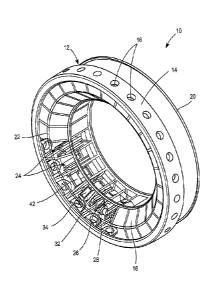
An arrangement formed according to one exemplary embodiment of the
present invention is illustrated in Figures 1-6. As illustrated therein, the
arrangement can be provided generally in the form of a replaceable cartridge
10.
The cartridge 10 comprises a housing 12. The housing 12 can be constructed of
any
suitable material. For example, a housing 12 can be constructed of a molded
polymeric material.
The housing 12 can be provided in any suitable form. One optional
configuration is illustrated in Figures 1-3. As illustrated, the housing 12
can
comprise a footprint ring 14. The footprint ring 14 comprises a plurality of
apertures 16 disposed about its circumference. The footprint ring 14 may
optionally
comprise a plurality of footprints 17 which surround respective apertures 16
and are
attached to the footprint ring 14. Each footprint 17 is configured to be
placed on the
surface of the skin of a user at a sampling site. The footprints 17 can be
annular in
shape according to the illustrated embodiment. However, the footprints are not
limited to this shape or configuration. Numerous shapes or configurations may
satisfy the function of providing a footprint around the site on the surface
of the skin
from which body fluid is to be expressed, i.e., the sampling site. According
to
certain embodiments, the footprints 17 are constructed from a material which
facilitates the formation of a seal between the skin and the footprints 17.
For
example, suitable materials for this purpose include a relatively soft
elastomeric
material, such as a silicone rubber. The footprints 17 can be formed having
any
appropriate size. For example, the footprints 17 can have a diameter, or
opening
having a major dimension, of about 3-8 mm. As an alternative to the above
described arrangement, a footprint can be provided for the same purpose as
part of
an integrated meter or device in which the arrangement or cartridge 10 can be
placed, as will be described in more detail herein.
According to the illustrated embodiment, the housing 12 further comprises a
transparent optical window 18. The transparent optical window 18 can be
provided,
for example, in order to permit optical communication between a detection
device
and one or more components located within the arrangement or cartridge 10.
- 8 -
CA 02624059 2008-03-27
WO 2007/041244
PCT/US2006/037923
The housing 12 can further include a top cover 20. An inner frame 22 can
also be provided. The inner frame 22 may help define a plurality of analysis
sites 24
within the cartridge 10.
One beneficial aspect of the arrangement or cartridge 10 of the present
invention is that it may be used to carry information which is readable by the
device
into which it is inserted. Such information can be used to update data and/or
code
utilized by the device, and can also be used for purposes of accuracy
verification and
calibration. Various mechanisms can be associated with the cartridge tend to
accomplish this purpose, as will be evident to those of ordinary skill in the
art. Two
exemplary mechanisms are illustrated in Figure 3. Namely, the cartridge 10 can
comprise a mechanism such as a readable memory chip 21 which carries
information and/or code which can be read by the device into which the
cartridge 10
is inserted. The manner in which the data and/or code is read from the chip 21
can
comprise any conventional arrangement for reading the information contained on
a
memory chip, such as electrical contacts and radio frequency
identification/transmission or direct optical communication such as a system
of
infrared emitters and detector. Another mechanism by which data and/or other
information can be provided to the device into which the cartridge 10 is
inserted is
illustrated in Figure 3 as comprising a barcode 23, or similar optically-
readable
mechanism. The barcode 23 is positioned on the exterior of the cartridge such
that
an optical sensor positioned within the integrated meter can read the
information
contained in the bars. The optical sensor and a processor within the
integrated
device can convert the pattern of bars into data as is commonly known in other
areas
such as point-of-sale scanners. The data read off of the barcode is used to
access
specific algorithms or lookup tables stored within memory in the integrated
meter.
This data allows the integrated device to adjust for any variances in the
manufacture
of the disposable cartridges. A suitable sensor/detector for reading the chip
21
and/or barcode 23 is schematically illustrated as element S/D in Figure 22.
Another beneficial aspect of the arrangement described above is the ability to
utilize one or more of the analysis sites 24 for calibration and control
purposes.
Generally, one or more of the analysis sites 24 can be used to verify the
accuracy of
- 9 -
CA 02624059 2008-03-27
WO 2007/041244
PCT/US2006/037923
test readings and automatically calibrate the system to compensate for any
variations
which may occur with operation of the device. One such technique and
arrangement
is illustrated in Figures 4A-4B. As illustrated therein, one and possibly
more, of the
analysis sites 24 are provided with a hub 32 containing a control assay pad
30'. The
control assay pad 30' is provided with three distinct regions, each producing
known
reflectance values. Namely, the first region X having a first darker color, a
second
uncolored region Y, and a third lightly colored region. As the control assay
pad 30'
is read by the detector D' through the transparent window 18, the pixels of
the
detector D' that correspond to each of the regions X, Y and Z produce
reflectance
readings. This detection is depicted in Figure 4B. As illustrated therein, the
reflectance values actually measured by the detector D' may differ from the
known
reflectance values of the control assay pad 30'. This difference can be
analyzed and
compensated for by any suitable technique. For instance, the algorithm
utilized to
calculate analyte concentration levels can be adjusted to compensate for the
difference, thereby leading to more accurate results. Such control and
calibration
operations can be carried out after each test, or after a number of tests.
As an alternative to the above control assay pad 30', a control fluid can be
released into an assay pad and allowed to react with a chemical reagent
contained
therein. Since the control fluid contains a known concentration of analyte,
the
measured concentration of analyte can then be compared to the known
concentration, and any differences analyzed and compensated for in the manner
described above.
Each sampling and analysis site 24 of the illustrated embodiment comprises
a skin penetration member 26. Each skin penetration member 26 can take any
suitable form. According to the illustrated embodiment, each skin penetration
member 26 is in the form of a hollow needle and has a first in the portion 26e
configured to pierce the skin, as well as an inner lumen 26f, (Figure 5). It
should be
understood that alternative skin penetration members may also be utilized
consistent
with the principles of the present invention (e.g., solid lancets, etc.). The
at least
one skin penetration member 26 can take any suitable form. For example, the at
least one skin penetration member can comprise a solid lancet or a hollow
needle.
- 10-
CA 02624059 2008-03-27
WO 2007/041244
PCT/US2006/037923
According to one embodiment, the skin-penetration member 26 is in the form of
a
so-called "microneedle." As the name implies, microneedles are characterizable
by
their relatively small outer diameters. For example, a microneedle, as the
term is
utilized herein, may encompass a skin-penetration member having an outside
diameter which is on the order of 40-200 gm. The inside diameter can vary, for
example, having an inside diameter on the order of 25-160 m. Needles are also
characterizable in the art by reference to the "gage." By way of illustration,
and
consistent with the above description, microneedles having a gage ranging from
26-
36 are clearly comprehended by the present invention. Certain advantages may
be
gleaned from the use of such microneedles as the skin-penetration member. In
particular, due to their small size, the size of the wound left upon entry
into the skin
is relatively small, thereby minimizing the pain associated with such needle
insertions and allowing for a quicker healing process. However, the present
invention is certainly not limited to the use of such microneedles. Thus, for
example, according to one possible alternative embodiment, the skin
penetration
member(s) comprise hollow needles having a gage of about 20-25, or comprising
hollow needles having an inner diameter of about 0.007 inches and an outer
diameter of about 0.020 inches.
The least one skin-penetration member can be formed of any suitable
material, such as metal, plastic, glass, etc.
Each skin-penetration member can be attached to a hub 32. Each hub 32 is,
in turn, attached to an actuator 28. It should be understood that a number of
different actuators may be utilized according to the principles of the present
invention. The actuators can be mechanical, electrical, pneumatic, etc.
According
to the illustrated embodiment, the actuator 28 is in the form of a torsional
spring.
Upon activation, the torsional spring drives the hub 32 and the attached skin
penetration member 26 through a respective aperture 16 and into the skin of
the
user. According to certain embodiments, each sampling and analysis site 24
further
comprises and analyte quantification member which produces a detectable signal
when contacted with a target analyte contained in a sample of body fluid. A
number
of suitable members are envisioned. The members may be based on conventional
- 11 -
CA 02624059 2008-03-27
WO 2007/041244
PCT/US2006/037923
technologies such as photometric or electrochemical analysis. According to the
illustrated embodiment, an assay pad 30 is provided on each hub 32 which can
generally comprises an absorbent material containing a chemical reagent which,
upon reaction with a target analyte, produces a chemical reaction that results
in a
.. detectable signal. The assay pad 30 is in fluid communication with the
inner lumen
22e of the skin piercing element 22. As noted above, the signal can be
detected
optically, electrochemically, or by other suitable means. According to one
embodiment, the assay pad 30, upon reaction with the target analyte, produces
a spot
which is optically detected by any suitable arrangement or technique. As
schematically illustrated, for example, in Figure 5, the assay pad 30 can be
located
on an exterior surface of the hub 32 and retained in position by a retaining
element
or cover 34. The retaining element or cover 34 can take any suitable form,
such as a
cap that snap fits onto the hub 23, or a strip of adhesive, The retaining
element or
cover 34 is preferable transparent. Thus, the spot produced on the assay pad
30 by
the above-mentioned reaction can be observed optically through the transparent
optical window 18 formed along the interior region of the illustrated
cartridge
housing 12.
Various mechanisms for triggering actuation of a hub 32 and attached skin
penetration member 26 will now be described.
In the exemplary, nonlimiting arrangement illustrated in Figures 5-6, the
actuator 28 is in the form of the torsional spring having a rear leg 36 and a
forward
leg 38. The forward leg 38 is fixedly attached to the hub 32 by any suitable
means,
such as the illustrated bore in the hub 32. The hub 32 is further provided
with a
mechanism for releasably capturing the rear leg 36 of the torsional spring.
According to the illustrated embodiment, the releasably capturing mechanism
comprises an open locking groove 40 which is configured to receive the rear
leg 36.
When the rear leg 36 is disposed within the releasably capturing mechanism, or
groove 40, the rear leg 36 and the forward leg 38 are urged toward one
another. In
this state, the torsional spring has a bias which tends to urge the rear leg
36 and the
forward leg 38 apart. Thus, in order to actuate the skin penetration member 26
and
the attached hub 32, the rear leg 38 is released from the open locking groove
40 by
- 12 -
CA 02624059 2008-03-27
WO 2007/041244
PCT/US2006/037923
any suitable mechanism or technique. As illustrated in Figure 6, the rear leg
36 is
urged out of communication with the groove 40 by moving it in the direction
indicated by arrow A. The rear leg 36 is prevented from significant movement
by
virtue of the fact that it is trapped within a wall W of the inner frame,
while the
forward leg 38 is relatively unrestrained. As a result of the natural bias of
the
torsion spring urging the rear and forward legs 36, 38 apart, the hub 32 and
the
attached skin penetration member 26 is urged in an arcing, downward movement
such that the skin penetration member 26 passes through a respective aperture
16,
and into the surface of the skin of the user. The hub 32 can rotate about the
pivot or
pin 42 upon actuation.
Figures 7-10 illustrate further optional aspects of the triggering mechanism
constructed according to the principles of the present invention. As
illustrated in
Figure 7, the triggering mechanism 50 is provided for the purpose of urging
the rear
leg 36 of the actuator 28 out of registry with the locking groove 40.
According to
the illustrative, nonlimiting embodiment, the triggering mechanism 50
comprises a
driving portion 52, such as a motor, solenoid, or servo device, and a driven
linear
actuator arm 54. In order to protect the components contained within the
cartridge
from environmental contamination, and in order to facilitate the creation of a
vacuum pressure at the analysis sites 24, it may be preferable according to
certain
optional aspects of the present invention to seal each analysis site. While it
is noted
at the arrangement illustrated in Figure 7 has an opening 16 corresponding to
the
aperture contained in the footprint ring 14, this opening will be sealed when
the
cartridge 10 is applied to the surface of the skin in the manner described
above. As
illustrated, for example, in Figure 7, and opening 55 is provided in the frame
22 in
order to permit introduction of the linear actuator arm 54. This opening 55
can be
sealed by means of a flexible solid membrane 56. The membrane 56 is flexible
enough to permit the necessary degree of movement of a linear actuator arm 54
in
order to disengage the rear leg 36 of the actuator 28 from the locking groove
40,
without being penetrated or broken by this movement.
A similar configuration is illustrated in Figure 8. However, in the
embodiment illustrated in Figure 8, the opening 55 is sealed by the
combination of
- 13 -
CA 02624059 2008-03-27
WO 2007/041244
PCT/US2006/037923
and apertured membrane 58 which has an opening to permit passage of the linear
actuator arm 54 therethrough, in combination with a secondary seal 60 which is
disposed about the linear actuator arm 54. As illustrated, the secondary
sealed 60 is
designed to come into firm contact with the apertured membrane 58 upon
insertion
.. of the driven linear actuator arm 54 therethrough. Thus, a seal is
maintained
through this opening 55 in the frame 22 for the purposes described the above.
As
further illustrated in Figure 8, the opening(s) 16 in the cartridge may
optionally be
sealed by any suitable mechanism or member, such as a thin sealing film 17s.
This
seal 17s will allow each chamber to remain completely sealed until it is
punctured.
The seal can either be removed by the user when loading a new disposable or
actually punctured by the skin penetration member 26 as it penetrates the
user's skin.
It should be understood that this aspect of the embodiment illustrated in
Figure 8
can be applied to any of the various embodiments described in this
application.
A further variation of the above arrangements is depicted in Figures 9-10.
As illustrated therein, the opening 55 in the frame 22 is sealed by means of a
piercable membrane seal 62. The piercable membrane seal 62 is normally of a
solid
construction. However, the piercable membrane seal 62 can be provided with
weakened portions or perforations 64 (Figure 10) which facilitates the
creation of an
opening therein upon contact with the driven linear actuator arm 54. Upon
insertion
of the linear actuator arm 54 at the location of the weakened portion or
perforations
64, a passageway is formed within the piercable membrane seal 62. However, a
relatively tight contact is maintained between the newly formed aperture in
the
piercable membrane seal 62 and the linear actuator arm 54. This contact serves
to
maintain at least a significant sealing effect.
Further alternative embodiments of a triggering mechanism formed
according to the principles of present invention are illustrated in Figures 11-
20. As
illustrated in Figure 11, the linear actuator arm 54 travels through the
opening 55 in
the direction of arrow B. The opening 55 can be sealed by any suitable
mechanism
or construction, such as any of the previously described ceiling mechanisms.
The
arm 54 is provided within angular ramp surface 66 which is designed to
interact
with the rear leg 36 of the actuator in a manner that pushes it out of
engagement
=
- 14 -
CA 02624059 2008-03-27
WO 2007/041244
PCT/US2006/037923
with the locking groove 40, as indicated by the relative positions of the
linear
actuator arm 54 and rear leg 36 shown in broken lines in Figure 11.
A further modification of the arrangement of Figure 10 is illustrated in
Figure 12. According to this modification, the linear actuator arm 54 is
provided
with a curved or arcuate ramp surface 68 which is also designed to interact
with the
rear leg 36 of the actuator in a manner which pushes it out of engagement with
the
locking groove upon traveling a predetermined distance in the direction of
arrow C,
as indicated by the relative positions of the linear actuator arm 54 and the
rear leg 36
shown in broken lines in Figure 12. Again, the opening 55 can be sealed by any
.. suitable means, such as any of the previously-described sealing
constructions.
A further embodiment of the triggering mechanism formed according to the
present invention is illustrated in Figure 13. According to this embodiment, a
pivotable actuator arm 70 is provided for movement within the opening 55. The
opening 55 can be sealed by any suitable mechanism, such as any of the
previously
described sealing constructions. The pivotable arm 70 is constructed and
arranged
so as to translate or pivot in the direction indicated by arrow D, thereby
forcing the
rear leg 36 of the actuator out of communication with the locking groove 40,
as
indicated in the broken line portion of Figure 12. The pivotable arm 70 can be
driven by any suitable conventional mechanism, such as a motor, solenoid or
servo
device.
A triggering mechanism constructed to still another embodiment of the
present invention is illustrated in Figure 14. According to this embodiment, a
linear
actuator arm 72 is provided having a construction similar to that of the
linear
actuator arm 54 described in the previous embodiments. However, the linear
actuator arm 72 is oriented at a location which is offset 90 relative to the
location of
the previously described linear actuator arm 54. As illustrated in Figure 14,
the
linear actuator arm 72 is positioned to travel in the direction of arrow E,
thereby
directly engaging the second end 36 of the actuator at a position adjacent to
the
bottom of the locking groove 40 and pushing it out of engagement with the
locking
groove 40, as illustrated by the broken lines in Figure 14. As with the
previously
- 15 -
CA 02624059 2008-03-27
WO 2007/041244
PCT/US2006/037923
described embodiments, the opening 55 can be sealed by any suitable mechanism,
such as any of the previously described sealing arrangements.
As illustrated in Figures 15A-15B, a suitable alternative triggering
mechanism can be constructed by providing a pivotable actually arm 74 which
travels within the opening 55 in the direction indicated by arrow F. The
pivotable
actuating arm 74 is provided within angular ramp surface 76 which is
configured to
interact with the rear leg 36 of the actuator upon traveling in the direction
indicated
by arrow F in a manner which forces the second leg 36 out of communication
with
locking groove 40 in the direction indicated by arrows G. The opening 55 can
be
sealed by any suitable mechanism, such as any of the previously described
sealing
mechanisms.
A further alternative triggering or release mechanism and arrangement
formed according to the present invention is illustrated in Figures 16-19.
According
to this embodiment, the rear leg 36 of the actuator 28 is fixedly retained in
a locking
feature 80 (e.g., Fig. 18) in the pin or pivot 42. The forward leg 38 of the
actuator
28 is fixedly retained by the hub 32. The hub 32, actuator 28 and pin or pivot
42 is
mounted within a chamber 81 defined by cell walls 82, 84. According to the
illustrated embodiment, the pivot or pin 42, and the attached hub 32, actuator
28 is
retained between the cell walls 82, 84 via retaining grooves 90 disposed
therein.
The hub 32 is positioned within the chamber 81 such that the hub is initially
locked
in a cocked position (e.g., Figs. 16-17) by interaction between a locking
feature
associated with the hub 32 and a locking feature associated with the chamber
81.
According to the illustrative embodiment, the locking feature associated with
the
chamber 81 comprises a pair of projections 86, each extending from a
respective cell
wall 82, 84, and the locking feature associated with the hub 32 comprises a
pair of
laterally spaced grooves or recesses 88 configured to releasably mate with the
projections 86. Numerous modifications to the illustrated locking features are
contemplated. For instance, the location of the projections 86 and the grooves
88
can be switched. Additionally, the cooperating projections and grooves can
have a
multitude of different geometrical configurations.
- 16 -
CA 02624059 2008-03-27
WO 2007/041244
PCT/US2006/037923
When the hub 32 is positioned in the chamber 81 in a locked position, the
rear leg 36 and the forward leg 38 are biased away from one another, such that
upon
disengagement of the locking features 86, 88, (Fig. 19) the hub 32 and the
attached
skin penetration member 26 is urged and an arcing, downward movement such that
the skin penetration member 26 passes into the surface of the skin of the
user. The
locking features 86, 88 are disengaged by application of a force to the hub
32, as
indicated for example by the arrow F (Fig. 19). Any suitable mechanism may be
utilized to apply the force necessary to disengage the hub, such as those
mechanisms
previously described herein.
A further optional triggering mechanism constructed according to the
principles of the present invention is illustrated in Figure 20, the
triggering
mechanism 50 is provided for the purpose of severing a wire or fuse 92, having
one
end attached to the hub 32 and the other end attached to a relatively
stationary
surrounding member. According to the illustrative, nonlimiting embodiment, the
triggering mechanism 50 comprises a portion 94 which can comprise at least one
of
a cutting member or heating element, both capable of severing the restraining
wire
or fuse 93. The opening 55 can optionally be sealed by means of any of the
previously described sealing arrangements.
The arrangement 10 can form at least part of a device which functions only
to sample body fluid. For example, the arrangement 10 can be used to express
body
fluid in the form of a drop of blood which pools on the surface of the skin of
the
user. This drop of blood can then be transferred to another separate device
which
then transports and/or analyzes the sample for a target analyte.
Alternatively, the
arrangement 10 may express a sample of body fluid from the digit D, and then
transport the sample to a location which can then be accessed for further
analysis by
a separate device. For instance, the sample body fluid can be transported to a
reagent-containing pad, also contained within the arrangement 10. The sample
then
reacts with the reagent to produce a detectable spot or signal. The reagent
pad can
then be analyzed by a separate meter using photochemical, electrochemical, or
other
suitable techniques known per se to those skilled in the art. The reagent pad
can
remain within the arrangement 10 during the aforementioned analysis.
-17-
CA 02624059 2008-03-27
WO 2007/041244
PCT/US2006/037923
Alternatively, the reagent pad can be removed from the arrangement 10 and
inserted
into a separate device, such as an electrochemical or photometric meter.
According to a further aspect of the present invention, the above-described
arrangements and techniques as previously described herein, can form at least
part
of an integrated device. As previously noted, as used herein, the term
"integrated
device" or "integrated meter" means a device or meter that includes all
components
necessary to perform sampling of the body fluid, transport of the body fluid,
quantification of an analyte, and display of the amount of analyte contained
in the
sample body fluid. Thus, according to the principles of the present invention,
an
integrated device or meter can comprise one or more, or any combination, of
the
features previously described herein. According to further aspects of the
present
invention, and integrated meter or device can comprise additional components
and/or features, which are described as follows.
It should be understood that while not required, any of the above-described
triggering mechanisms can form part of a separate sampling only device or part
of
an integrated device into which the cartridge 10 is placed.
One such integrated meter is illustrated Figures 21-23. As illustrated
therein,
the integrated meter 100 generally comprises a housing 112. The integrated
meter
100 may further comprise a footprint 114 of the type previously described. A
door
116 can be provided on the housing 112. The door 116 is connected via a hinge
118
to the housing 112. As illustrated in Figures 22-23, the door 116 can be
opened to
reveal a cartridge 10 containing a plurality of skin-piercing elements and
analysis
sites, as previously described herein. In the illustrated embodiment, the
integrated
meter 100 further includes a display 120 for communicating the results of the
analysis on the sample body fluid for the presence and/or concentration of an
analyte
contained therein. The integrated meter 100 may further include one or more
buttons 122 which can be pressed by the user to engage various functions and
interfaces of the integrated meter 100.
Figure 22 is an illustration of the integrated meter 100 with the door 116
opened to reveal further details of the interior components of the integrated
meter
100. As illustrated therein, the housing 112 contains a cartridge 10 therein.
In the
- 18-.
CA 02624059 2008-03-27
WO 2007/041244
PCT/US2006/037923
illustrated embodiment, the cartridge 10 is circular and contains a plurality
of skin-
piercing elements and analysis sites. The cartridge 10 is mounted about a
central
hub 122 and is rotatable thereon. Thus, upon sampling a skin-piercing element
is
driven through an opening in the housing in registry with the footprint 114
and
pierces the skin of the user. Once the test has been completed, the cartridge
10 can
be rotated such that an unused skin-piercing element now comes into registry
with
the opening in the housing and the corresponding opening in the footprint 114
in
preparation for the next sampling event. It should be understood that the
present
invention is not limited to the illustrated circular cartridge having the
particular
.. configuration depicted in the drawing figures. To the contrary, a number of
alternative cartridge configurations are possible, such as a slidable linear
or
polygonal configuration (not shown). Also illustrated in Figure 22 is the
presence of
a light source 124 disposed on the back of the door 116. The light source 124
can
take any suitable form, such as a light emitting diode. It should be
understood that
.. alternative light sources may also be utilized. The function of the light
source 124
will be described in further detail below.
In this regard, light emitted from the light source 124 is incident upon an
assay pad (e.g., 30), and reflects off the surface thereof. Upon formation of
a
reaction spot on the surface of the assay pad, the amount of light reflected
off the
reaction spot differs from the light reflected off of other portions of the
reagent pad
containing no such reaction spot. This reflected light is picked up by the
detector
126. The detector 126 may comprise a lens 128 and optical detector element
130.
The optical detector element 130 generally comprises one or more detector
elements. According to one alternative construction, the detector element 130
comprises a plurality of detector elements formed in an array. The array can
take
any suitable configuration, and can be a linear array according to one
nonlimiting
example. The detector elements can comprise any suitable construction. For
example, the detector elements 130 can comprise a photo diode, CCD, or CMOS
based detector element. The signals transmitted to the detector element 130
are
passed on to suitable electronics contained within the housing 112 via
suitable
electrical connectors, such as flexible ribbons 131 (Figure 23). The specifics
of the
-19-
CA 02624059 2014-02-27
WO 2007/041244
PCT/US2006/037923 '
electronics and signal interpretation being familiar to those of ordinary
skill in the
art. While not necessary to enable practice of the presently claimed
invention,
further details concerning the construction, function and arrangement of the
analysis
sites, and components contained therein, can be gleaned from the disclosure
contained in U.S. Patent No. 8,382,618 entitled DEVICE FOR
FLUID ANALYSIS WITH SAMPLE EXTRACTION AND TRANSPORT.
Similarly, while not
necessary to enable practice of the presently claimed invention, further
details
concerning the structure, function, and arrangement of the detector 126, and
the
components contained therein, can be gleaned from the disclosure contained in
U.S.
Patent Publication No. 20060281187 Al, entitled ANALYTE DETECTION
DEVICES AND METHODS WITH HEMATOCRIT/VOLUME CORRECTION
AND FEEDBACK CONTROL.
An integrated meter incorporating an arrangement formed according to the
. _prpsentinvention can be configured for digital body fluid sampling and
analysis as
well as alternate-site body fluid sampling and analysis, which may be
performed at
either location at the election of the user.
As evident from Figures 21-23, the integrated meter 100 is configured for
handheld use. However, the invention is not limited to handheld devices. For
example, the present invention is also directed to integrated meters that are
wearable. An example of such a wearable device is illustrated in Figure 24.
The
wearable integrated device 200 illustrated therein can be generally composed
of a
functional portion 202 and a body-attachment portion 204. The functional
portion
can comprise an arrangement 10 of the type described herein. The functional
portion can also have one or more of the features and elements of the handheld
integrated meter described above.
As previously noted, according to certain embodiments of the present
invention, the concentration of an analyte contained in a sample of body fluid
can be
measured using a photometric technique wherein the assay pad is interrogated
with a
light source and a detector thereby producing a signal indicative of a color
change
-20-
CA 02624059 2008-03-27
WO 2007/041244
PCT/US2006/037923
caused by reaction between an analyte and reagent contained in the assay pad,
which
is then correlated to the concentration of analyte contained in the sample.
The present invention provides photometric analysis devices, arrangements
and techniques that facilitate their incorporation into devices and
arrangements of
the type described above that are compact, discrete, wearable or handheld, and
capable of performing multiple tests without reloading testing components.
According to a first embodiment, a photometric analysis arrangement
constructed to satisfy at least the above-noted objectives is illustrated in
Fig. 25. As
illustrated therein, the arrangement 300 generally comprises a platform or
stage 302,
a plurality of assay pads 304 containing chemical reagents, a single light
source 306,
and a single detector 308. The light source 306 may be provided by any
suitable
device, such as a light emitting diode (LED), similarly, the detector may
comprise
any suitable device, such as one or more CMOS, CCD, photodiode or infrared
detector elements. According to one embodiment, the detector 308 comprises an
array of CMOS detector elements.
According to the arrangement 300, the plurality of assay pads 304 are
provided at fixed locations relative to the platform or stage 302. Thus, no
relative
movement between the assay pads 304 and the platform 302 is possible. The
light
source 306 and the detector 308 are also provided at fixed locations
independent of
the platform or stage 302. The light source 306 is arranged to direct light
toward a
specific assay pad 304 when brought into registry therewith. Similarly, the
detector
308 is arranged to receive light reflected off the assay pad that is
positioned at a
predetermined location. The platform 302 is rotatable, as indicated by the
arrow
contained in Fig. 25, such that each of the plurality of assay pads 304 may be
indexed and brought into registry with light source 306 and the detector 308
for
analysis.
A variation of the arrangement 300 is depicted in Fig. 26. The arrangement
400 is constructed in a manner that shares many of the same features
previously
described in connection with the arrangement 300. According to the arrangement
400, platform 302 is fixed and is not rotatable. Both the light source 306 and
the
detector 308 are mounted on a second platform or stage 402. Both the light
source
- 21 -
CA 02624059 2008-03-27
WO 2007/041244
PCT/US2006/037923
306 and the detector 308 are provided at fixed locations relative to the
platform 402,
such that relative movement therewith is not permitted. According to the
arrangement 400, each of the individual assay pads 304 are indexed, or brought
into
registry the light source 306 and the detector 308 by rotating the second
platform
402 in the manner indicated by the arrow appearing in Fig. 26.
A further optional modification of the arrangements 300,400 is depicted in
Fig. 27. According to the arrangement 500, the platform 302 is fixed, and is
not
movable. Both the light source 306 and the detector 308 are mounted on an
indexing arm 502 in a fixed manner. According the arrangement 500, the light
source 306 and the detector 308 are indexed, or brought into registry with
each of
the assay pads 304 by rotating the movable indexing arm 502 in the manner
indicated by the arrow appearing in Fig. 27. Thus, the light source 306 and
the
detector 308 are brought to a position which is located above a selected assay
pad
304. According to this arrangement 500, light is emitted downwardly from the
light
source 306 toward the assay pad 308. At least a portion of this light is then
reflected
off the assay pad 304 in a generally upward direction such that it is then
received by
the detector 308.
In certain instances, it may be advantageous to eliminate the need to move
the assay pads 304 relative to the light source 306 and the detector 308 in
order to
.. selectively index or bring the components into registry therewith for
analysis.
Once such arrangement which accomplishes this objective is illustrated in
Fig. 28. According to the arrangement 600, each of a plurality of assay pads
304
may be individually interrogated without the necessity of providing relatively
movable components within the system. According to illustrated arrangement
600,
a plurality of light pipes or similar light transmitting elements 602 are
provided
which communicate between a single stationary light source 306 and each of a
plurality of assay pads 304. The detector 604 is positioned such that it may
receive
light reflected light off of each individual assay pads 304. In order to
accomplish
this objective, the detector 604 may be partitioned, or formed as an array of
discrete
.. detector elements, as illustrated in Fig. 28. Thus, the detector 604
comprises a
plurality of sections, each of which is committed to receive light reflected
off of a
-22 -
CA 02624059 2008-03-27
WO 2007/041244
PCT/US2006/037923
selected assay pad 304. The light emitted from the light source 306 may be
multiplexed or selectively transmitted to a particular assay pad 304. This
multiplexing can be accomplished by any suitable technique familiar to those
in the
art.
One possible variation of the arrangement 110 is depicted in Fig. 29.
According the arrangement 700, like the arrangement 600, a plurality of
analysis
sites may be interrogated without the use of relatively movable components.
According to the arrangement 700, a single light source 306 is provided which
transmits light to all of the assay pads 304 simultaneously. A plurality of
detector
elements 702, 704, 706 are provided, each of which is positioned in registry
with
light reflected off of a respective assay pad 304. Each of the individual
detector
elements 702, 704, 706 may be multiplexed, or selectively activated in order
to read
only the desired assay pad 304. This multiplexing may be accomplished by any
suitable means, as familiar to those in the art.
An exemplary body fluid sampling and analysis methodology or technique,
which may be utilized in conjunction with any of the above-mentioned
arrangements, devices or integrated meters, but is not necessarily limited
thereto, is
described as follows.
A user loads a fresh disposable cartridge containing a plurality of skin
penetration members and analysis sites into an integrated meter. The
integrated
meter then reads calibration data contained in or on the cartridge. This data
can be
read in any suitable manner. For example, a bar code may be placed on the
cartridge
which can be optically read by the optical assembly contained within the
meter.
Alternatively, the data is contained on a chip carried by the cartridge that
is read
upon insertion into the integrated meter. The integrated meter then selects
the
proper lookup table or algorithm to calculate an aggregate glucose measurement
taking into consideration the calibration data. The meter may then place
itself in a
ready mode waiting for a trigger to initiate sampling and testing. The user
then
either manually presses a button or trigger to initiate sampling and analysis,
or the
device verifies that it is properly positioned on the skin of the user and
ready to
begin the sampling and analysis procedure. Suitable sensors to accomplish this
- 23 -
CA 02624059 2014-02-27
WO 2007/041244 PCT/US2006/037923
include optical, capacitive or pressure sensors. The device may then initiate
a
catalyst which acts to facilitate the expression of body fluid. According to
one
alternative embodiment, the catalyst is an inflatable member that exerts
pressure on
a digit. Alternatively, the catalyst is vacuum pressure which generates
suction at the
sampling site. Sensors present in the meter may be used to monitor and control
the
positive or negative pressure of the catalyst. After achieving a target
pressure for a
desired period of time, the skin penetration member (e.g., a hollow needle) is
actuated and driven into the skin of the user to create a wound site. The skin
penetration member comes to rest in or directly on the wound created at the
sampling site where it is in the desired position for collecting a sample of
body fluid
expressed from the wound. The integrated meter may further include a mechanism
for detecting a whether a sufficient amount of sample has been expressed.
Details of
such suitable detection techniques are described in detail in U.S. Patent No.
7,052,652, entitled ANALYTE CONCENTRATION DETECTION DEVICES AND
METHODS. Once
_ 439 desired amount of body fluid has been obtained, the catalyst is
deactivated. A
_ _ . . _ . .
sample of body fluid is in fluid communication with a device or mechanism
which
creates a detectable signal upon reaction within analyte present in the sample
body
fluid. For example, one such suitable mechanism is an absorbent pad containing
a
chemical reagent which, upon reaction with the analyte produces a reaction
spot
which can be optically detected. An optical assembly which is an optical
communication with the above described signal generating mechanism is utilized
to
detect the signal created via reaction with the analyte and communicate the
signals
to supporting electronics contained within the meter. The concentration of a
target
analyte (e.g., glucose) can then be calculated using these signals as a basis.
Additional factors may be considered during these calculations, such as the
sample
size, levels of other substances contained in the sample (e.g. hematocrit),
etc. Such
optional calculation techniques are described in further detail in U.S. Patent
Publication No. 20060281187 Al, entitled ANALYTE DETECTION DEVICES
AND METHODS WITH HEMATOCRIT/VOLUME CORRECTION AND
FEEDBACK CONTROL.
- 24-
CA 02624059 2014-02-27
=
WO 2007/041244
PCT/US2006/037923
These calculations quantify the amount of analyte contained in the
sample body fluid. This quantity is displayed on a suitable display contained
within
the meter which can be easily read by the user. The integrated meter then
automatically indexes the disposable cartridge to present a fresh unused skin
penetration member which will be utilized to perform the next sampling and
analysis
event.
Numbers expressing quantities of ingredients, constituents, reaction
conditions, and so forth used in this specification are to be understood as
being
modified in all instances by the term "about". Notwithstanding that the
numerical
ranges and parameters setting forth, the broad scope of the subject matter
presented
herein are approximations, the numerical values set forth are indicated as
precisely
as possible. Any numerical value, however, inherently contains certain errors
necessarily resulting from the standard deviation found in their respective
measurement techniques.
Although the present invention has been described in connection With
preferred embodiments thereof, it will be appreciated by those skilled in the
art that
additions, deletions, modifications, and substitutions not specifically
described may
be made without department from the scope of the invention as defined herein.
-25 -