Note: Descriptions are shown in the official language in which they were submitted.
CA 02624233 2008-03-28
WO 2007/041070 PCT/US2006/037312
-1-
INTEGRATED INTRODUCER AND TRANSMITTER ASSEMBLY
AND METHODS OF USE
PRIORITY
This PCT application claims priority to United States Patent Application No.
11/240,259,
filed September 30, 2005 and is hereby incorporated by reference.
BACKGROUND
Continuous glucose monitoring systems generally include a sensor such as a
subcutaneous analyte sensor, at least a portion of which is inserted under the
skin for fluid
contact with interstitial fluid, for detecting analyte levels such as glucose
levels, a transmitter
(such as an RF transmitter) in communication with the sensor and configured to
receive the
sensor signals and to transmit them to a corresponding receiver unit by for
example, using RF
data transmission protocol. The receiver may be operatively coupled to a
glucose monitor that
performs glucose related calculations and data analysis.
The transmitter is in signal communication with the sensor. Generally, the
sensor is
configured to detect and measure the glucose levels of the patient over a
predetermined period of
time, and the transmitter is configured to transmit data corresponding to or
associated with the
measured glucose levels over the predetermined period of time for further
analysis. To initially
!o deploy the sensor so that the sensor contacts and electrodes are in fluid
contact with the patient's
analyte fluids, a separate deployment mechanism such as a sensor inserter or
introducer is used.
More specifically, the introducer includes a sharp needle shaped inserter that
is configured to
pierce through the skin of the patient and substantially concurrently guide
the sensor through the
patient's skin so as to place at least a portion of the sensor in fluid
contact with the target
5 biological fluid of the patient.
The sharp inserter is typically used only during the sensor insertion process,
and once the
sensor is properly and accurately positioned, the inserter and the introducer
are discarded. This
requires a level of care as the inserter is sharp and may damage other parts
of the patient's skin if
not properly discarded. Further, since the tip of the inserter has come into
fluid contact with the
CA 02624233 2008-03-28
WO 2007/041070 PCT/US2006/037312
-2-
patient's biological fluids, it is important to take particular precautions in
the handling of the
sharp inserter.
Further, to minimize data errors in the continuous or semi-continuous
monitoring system,
it is important to properly insert the sensor through the patient's skin and
securely retain the
sensor during the time that the sensor is configured to detect analyte levels.
In addition to
accurate positioning of the sensor through the skin of the patient, it is
important to minimize the
level of pain associated with the insertion of the sensor through the
patient's skin. Additionally,
for the period of continuous or semi-continuous monitoring which can include,
for example, 3
days, 5 days or 7 days, it is important to have the transmitter in proper
contact with the analyte
sensor so as to minimize the potential errors in the monitored data.
In view of the foregoing, it would be desirable to have method and apparatus
which
provides for simple handling of the sensor introducer during sensor deployment
and also after the
sensor deployment. More specifically, it would be desirable to have method and
apparatus that
minimizes the potential physical contact with the inserter mechanism and the
patient to minimize
the potential for disseminating the biological fluids that has come into
contact with the inserter,
and also, that provides for an easy to use sensor insertion mechanism that
minimizes thepain to
the patient.
SUMMARY OF THE INVENTION
o In one embodiment, there is provided a method and apparatus for providing an
integrated
sensor introducer mechanism and transmitter unit for use in continuous or semi-
continuous
monitoring systems such as glucose monitoring systems which includes a
disposable sensor
introducer provided within the integrated sensor/transmitter assembly and
which is retained
within the assembly during the time period of the sensor in active mode.
5 More specifically, in one embodiment of the present invention, there is
provided an
integrated sensor introducer mechanism which may be triggered by a single
depression or
activation of a switch to deploy the sharp sensor introducer through the skin
of the patient. The
single depression trigger mechanism is configured to return to its original
position upon firing
CA 02624233 2008-03-28
WO 2007/041070 PCT/US2006/037312
-3-
the sensor and the introducer, so that the sharp sensor introducer is removed
from the patient
after placing the sensor in the patient.
In this manner, the patient is not required to handle the cumbersome and
potentially
dangerous and sharp sensor introducer microneedle, for example. In this
manner, a convenient,
simple and sanitary sensor insertion and analyte monitoring system is
provided.
BRIEF DESCRIPTION OF THE DRAWINGS
FIG. lA illustrates a perspective view of the overall assembly including
integrated
introducer and transmitter coupled to the transmitter mount with the switch
cover in a closed
position in accordance with one embodiment of the present invention;
FIG. 1 B illustrates a perspective view of the overall assembly including
integrated
introducer and transmitter coupled to the transmitter mount with the switch
cover in an open
position in accordance with one embodiment of the present invention;
FIG. 2 illustrates a perspective view of the overall assembly including
integrated
introducer and transmitter coupled to the transmitter mount with the switch
cover in a closed
position provided on an adhesive patch in accordance with one embodiment of
the present
invention;
FIGS. 3A and 3B illustrate the open and closed positions, respectively, of the
transmitter
unit opening portion on the transmitter unit housing in accordance with one
embodiment of the
?o present invention;
FIG. 4 illustrates a close up perspective view of the switch opening in the
open position
exposing the sensor introducer trigger mechanism and mounted on the
transmitter unit base
portion in accordance with one embodiment of the present invention;
FIGS. 5A-5B illustrate a side view and a perspective view, respectively, of
the sensor
!5 introducer trigger mechanism with the sensor positioned in the pre-
deployment position in
accordance with one embodiment of the present invention;
FIG. 6 illustrates a perspective view of the sensor introducer trigger
mechanism and the
transmitter unit base portion in cooperation with the sensor in pre-deployment
position in
accordance with one embodiment of the present invention;
CA 02624233 2008-03-28
WO 2007/041070 PCT/US2006/037312
-4-
FIG. 7A illustrates a perspective view of the sensor introducer trigger
mechanism and the
transmitter unit base portion in post deployment position in accordance with
one embodiment of
the present invention;
FIG. 7B illustrates a side view of the sensor introducer trigger mechanism in
post sensor
deployment position in accordance with one embodiment of the present
invention;
FIG. 8A is a cross sectional view of the sensor introducer trigger mechanism
before the
sensor insertion in accordance with one embodiment of the present invention;
FIG. 8B is a cross sectional side view of the sensor introducer trigger
mechanism before
the sensor insertion in accordance with one embodiment of the present
invention; and
FIGS. 9A and 9B are cross sectional views of the sensor introducer trigger
mechanism
after sensor insertion and withdrawal of the introducer for retention within
the integrated sensor
introducer and transmitter assembly in accordance with one embodiment of the
present
invention.
DETAILED DESCRIPTION
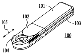
FIG. 1A illustrates a perspective view of the overall assembly including
integrated
introducer and transmitter coupled to the transmitter mount with the switch
cover in a closed
position in accordance with one embodiment of the present invention, and FIG.
lB illustrates a
perspective view of the overall assembly including integrated introducer and
transmitter coupled
.o to the transmitter mount with the switch cover in an open position in
accordance with one
embodiment of the present invention. Referring to FIGS. 1A-1B, integrated
sensor introducer
and transmitter assembly 100 in one embodiment of the present invention
includes a transmitter
unit 101, a transmitter unit opening portion 102, and transmitter mount base
portion 103. As
shown, the transmitter unit 101 is configured to physically couple to the
transmitter mount base
5 portion 103 so as to provide an integrated assembly. The transmitter mount
base portion 103 is
configured to be placed on the skin of a patient, and as will be discussed in
further detail, and
includes a sensor introducer and the sensor pre-assembled therein.
Referring to FIGS. lA-1B, the transmitter unit opening portion 102 is
configured in one
embodiment to be slidably displaced between an open position and a closed
position, along the
CA 02624233 2008-03-28
WO 2007/041070 PCT/US2006/037312
-5-
directional arrow 105. As can be seen, when the transmitter unit opening
portion 102 is in the
open position, a sensor introducer trigger mechanism 104 is exposed to the
patient. More
specifically, the patient is able to slidably move the transmitter unit
opening portion 102 between
the open position and the closed position to operate the sensor introducer
trigger mechanism 104,
and upon successfully deploying the sensor transcutaneously to the desired
position, the patient
may place the transmitter unit opening portion 102 in the closed position so
as to provide cover
and protection to the sensor introducer trigger mechanism 104, and also, to
avoid potential
inadvertent interaction with the sensor introducer trigger mechanism 104.
Within the scope of
the present invention, the sensor introducer trigger mechanism 104 may include
a plug or stopper
with latch mechanism.
Referring back to FIGS. lA-1B, while the transmitter unit opening portion 102
is shown
with a slidable movement so as to be displaced between the open position and
the closed
position, within the scope of the present invention, the transmitter unit
opening portion 102 may
include other types of mechanisms to open and close the area exposing the
sensor introducer
trigger mechanism 104. For example, the transmitter unit opening portion 102
may include a
hinge portion pivotally mounted to the transmitter unit 102 so that the
transmitter unit opening
portion 102 may pivotally move to expose and to close the sensor introducer
trigger mechanism.
Additionally, a latch mechanism may also be provided so as to securely place
the
transmitter unit opening portion 102 in a closed and latched position so as to
avoid potential
inadvertent exposure of the sensor introducer trigger mechanism 104. Within
the scope of the
present invention, the latch mechanism may include a Velcro type fastener, a
button type
latching mechanism, a tongue and groove type latch mechanism, a snap, a
detente, a hook, or any
other type of latching mechanism that would securely place the transmitter
unit opening portion
102 in the closed position in the event of inadvertent application of pressure
thereto.
?5 FIG. 2 illustrates a perspective view of the overall assembly including
integrated
introducer and transmitter coupled to the transmitter mount with the switch
cover in a closed
position provided on an adhesive patch in accordance with one embodiment of
the present
invention. Referring to FIG. 2, there is shown an adhesive patch 201 that is
configured to
receive the transmitter unit base portion 103 on its upper surface, while the
lower surface is
CA 02624233 2008-03-28
WO 2007/041070 PCT/US2006/037312
-6-
provided with an adhesive material, and where the lower surface is configured
to be securely
attached to the skin of the patient, thus effectively providing a firm and
secure mounting of the
integrated sensor introducer and transmitter assembly 100. As shown, the
adhesive patch 201 in
one embodiment of the present invention is substantially flexible and
configured to substantially
follow the contour of the patient's skin where the integrated sensor
introducer and transmitter
assembly 100 is to be placed for the predetermined period of time that the
patient will be wearing
the asseinbly 100 (for example, 3 days, 5 days, 7 days and so on). In this
manner, comfort can
be provided to the patient while not substantially hindering the patient's
daily activities.
FIGS. 3A and 3B illustrate the open and closed positions, respectively, of the
transmitter
unit opening portion on the transmitter unit housing, and FIG. 4 illustrates a
close-up,
perspective view of the switch opening in the open position exposing the
sensor introducer
trigger mechanism and mounted on the transmitter unit base portion in
accordance with one
embodiment of the present invention. Referring to FIGS. 3A-3B and 4, it can be
seen that the
sensor introducer trigger mechanism 104 is positioned substantially within the
housing of the
integrated sensor introducer and transmitter assembly 100, shown in one
embodiment as
including the transniitter unit 101 coupled with the transmitter unit base
portion 102.
Moreover, while the transmitter unit 101 is provided with a substantially
circular opening
corresponding to the position of the sensor introducer trigger mechanism 104,
within the scope
of the present invention, any suitable shape may be integrated to the housing
of the transmitter
!o unit 101 so as to effectively be opened and closed to respectively expose
and seal off the sensor
introducer trigger mechanism 104 as desired by the patient. For example, the
circular opening
on the transmitter unit 101 may alternatively be formed in an oblong shape, a
triangular shape, a
rectangular shape, and so on.
FIGS. 5A-5B illustrate a side view and a perspective view, respectively, of
the sensor
.5 introducer trigger mechanism with the sensor positioned in the pre-
deployment position in
accordance with one embodiment of the present invention. Referring to FIGS. 5A
and 5B, as
can be seen, the sensor introducer trigger mechanism 104 in one embodiment
includes a trigger
portion 501 operatively coupled to an introducer portion 502. As shown, the
trigger portion 501
of the sensor introducer trigger mechanism 104 is configured to displace the
introducer portion
CA 02624233 2008-03-28
WO 2007/041070 PCT/US2006/037312
-7-
502 in a substantially skin-inserting direction, e.g., a substantially
vertical direction relative to
the patient's skin surface. Further, as shown in the Figures, an analyte
sensor 503 is provided in
cooperation with the introducer portion 502 such that in one embodiment, when
the trigger
portion 501 is activated by the patient, for example, by the application of
downward pressure on
the outer surface of the trigger portion (the outer surface of the "dome
shaped" area), the
introducer por-tion 502 is in turn configured to be driven in a substantially
complimentary
direction to the direction of the applied pressure, and further, displacing at
least a portion of the
sensor 503 with the introducer portion 502. In other words, the introducer
portion 502 is
configured in one embodiment to transcutaneously place a portion of the sensor
503 so that the
portion of the sensor is in fluid contact with the biological fluid (for
example, interstitial fluid) of
the patient.
Referring to FIGS. 5A-5B, the sensor 503 is in one embodiment also provided
with one
or more contact points 504 which are- configured to be in electrical contact
with the
corresponding electrical contacts of the transmitter unit 101. That is, in the
case of analyte
5 sensors, the working, reference, and counter electrodes (in certain
embodiments an electrode
may function as both reference and counter electrodes) are each coupled to _a
respective one of
the contact points 504, and in turn, each of which are in electrical
communication with the
respective contacts on the transmitter unit 101.
In this manner, in one embodiment, the sensor detected analyte levels are
provided to the
0 transmitter unit 101, for example, as current signals, and which are in
turn, converted to
respective digital signals for transmission (including, for example, RF
transmission) to a receiver
unit for fiuther data processing and data analysis (including drug (e.g.,
insulin) therapy
management, infusion control, and health monitoring and treatment, for
example). That is, the
monitored analyte data may be used by the patient and/or the patient's
healthcare provider to
5 modify the patient's therapy such as an infusion protocol (such as basal
profile modifications in
the case of diabetics) as necessary to improve insulin infusion therapy for
diabetics, and further,
to analyze trends in analyte levels for better treatment.
While glucose is described as an example of the detected and/or monitored
analyte,
within the scope of the present invention, analytes that may be detected or
monitored also
CA 02624233 2008-03-28
WO 2007/041070 PCT/US2006/037312
-8-
include, for example, acetyl choline, amylase, bilirubin, cholesterol,
chorionic gonadotropin,
creatine kinase (e.g., CK-MB), creatine, DNA, fructosamine, glucose,
glutamine, growth
hormones, hormones, ketones, lactate, peroxide, prostate-specific antigen,
prothrombin, RNA,
thyroid stimulating hormone, and troponin. The concentration of drugs, such
as, for example,
antibiotics (e.g., gentamicin, vancomycin, and the like), digitoxin, digoxin,
drugs of abuse,
theophylline, and warfarin, may also be detected and/or monitored.
FIG. 6 illustrates a perspective view of the sensor introducer trigger
mechanism and the
transmitter unit base portion in cooperation with the sensor in pre-deployment
position in
accordance with one embodiment of the present invention. As shown, the one or
more contact
points 504 of the sensor 503 (which in one embodiment may correspond to a
respective one of
the working electrode, a counter electrode, and a reference electrode, for
example), are
configured to couple to a respective contacts on the transmitter unit 101
(FIGS. 3A-3B) such that
the sensor 503, which is in fluid contact with the patient's biological
fluids, is in electrical
conununication with the transmitter unit 101.
Furthermore, as can be seen from FIG. 6, the substantially dome shaped
inserter
introducer trigger mechanism 104 is configured to be collapsible when the
patient applies
downward pressure to drive the introducer portion 502 through the patient's
skin. Further, when
the downward pressure is removed from the dome shaped inserter introducer
trigger mechanism
104, the outer inserter introducer trigger mechanism 104 is configured to
return to substantially
0 the original shape, and concurrent therewith, removing the introducer
portion 502 from the
insertion site of the patient, while leaving behind the subcutaneous portion
of the sensor in fluid
contact with the patient's biological fluid. This can be seen also with FIGS.
7A-7B as described
below.
FIG. 7A illustrates a perspective view of the sensor introducer trigger
mechanism and the
5 transmitter unit base portion in post deployment position, and FIG. 7B
illustrates a side view of
the sensor introducer trigger mechanism in post sensor deployment position in
accordance with
one embodiment of the present invention. More specifically, it can be seem
from FIGS. 7A-7B
that when the downward pressure is applied upon the substantially dome shaped
inserter
introducer trigger mechanism 104, the upper conical portion of the inserter
introducer trigger
CA 02624233 2008-03-28
WO 2007/041070 PCT/US2006/037312
-9-
mechanism 104 takes a substantially inverted conical shape, and with the same
force, driving the
portion 701 of the sensor 503 (FIG. 5A) through the patient's skin. In one
embodiment, the
inserter introducer trigger mechanism 104 may be made of one of stainless
steel, rubber,
polyester, or PET film, or any other suitable material that is flexible and
provides the properties
described herein, including being reversibly collapsible.
FIG. 8A is a cross sectional view of the sensor introducer trigger mechanism
before the
sensor insertion, and FIG. 8B is a cross sectional side view of the sensor
introducer trigger
mechanism before the sensor insertion in accordance with one embodiment of the
present
invention. As can be seen from FIG. 8A, the portion 701 of the sensor 503 is
substantially
retained in cooperation with the introducer portion 502 (e.g., a microneedle),
all of which are
substantially retained within the integrated inserter introducer and
transmitter assembly 100
(FIGS. lA-1B).
Moreover, referring to FIG. 8B, the non-transcutaneously displaced portion of
the sensor
503 is also substantially completely retained within the integrated inserter
introducer and
transmitter assembly 100. That is, in one embodiment of the present invention,
the transmitter
unit 101 as well as the sensor insertion mechanism (e.g., sensor introducer
trigger mechanism
104) are provided within a single integrated housing. Furthermore, as
discussed in additional
detail below, after the deployment of the sensor 503 transcutaneously so as to
have a portion 701
thereof in fluid contact with the patient's biological fluid, the sensor
insertion mechanism is
;0 retained within the integrated housing itself, so that the patient is not
required to further handle
the sharp and contaminated needle portion of the sensor introducer assembly.
FIGS. 9A and 9B are cross sectional views of the sensor introducer trigger
mechanism
after sensor insertion and withdrawal of the introducer for retention within
the integrated sensor
introducer and transmitter assembly in accordance with one embodiment of the
present
5 invention. Referring to FIGS. 9A-9B, it can be seen that after the sensor
503 is placed
transcutaneously through the patient's skin at the intended location and in
fluid contact with the
patient's biological fluid, the introducer portion 502 is retained
substantially completely within
the dome shaped sensor introducer trigger mechanism 104 provided within the
integrated sensor
introducer and transmitter assembly 100.
CA 02624233 2008-03-28
WO 2007/041070 PCT/US2006/037312
-10-
Furthermore, while the sensor 503 is described as substantially
transcutaneously placed in
the patient, witliin the scope of the present invention, the sensor may be
wholly implantable
under the skin of the patient, or at least a portion of the sensor may be
provided under the skin of
the patient so as to be in fluid contact with the patient's analyte.
In one embodiment, the introducer portion 502 is configured to be retained
within the
assembly 100 during the entire duration of the sensor 503 in operation for
monitoring the
patient's analyte levels, and is discarded along with the sensor 503 after
use. In this manner,
while the transmitter unit 102 may be reusable, in one embodiment of the
present invention, the
base portion 103 and the sensor introducer trigger mechanism 104 along with
the sensor 503 are
discarded after each use.
Further, the detected analyte signals from the sensor 503 may be provided to
transmitter
unit 102, which is, in one embodiment, configured to wirelessly or otherwise
transmit data
corresponding to the detected analyte levels from the sensor 503 to a receiver
unit; where the
receiver unit may include an analyte, e.g., glucose, monitor unit and/or an
insulin pump unit
and/or a computer terminal and/or any other electronic device capable of being
configured for
wireless communication. A physical connection may be provided in certain
embodiments. _. -
Within the scope of the present invention, the receiver unit functions may be
integrated
into portable electronic devices such as a watch, a pager, a mobile telephone,
and a personal
digital assistant. Additional information on the detection, monitoring and
analysis of analyte
?o levels are described in f-urther detail in U.S. Patent No. 6,175,752
entitled "Analyte Monitoring
Device and Methods of Use" the disclosure of which is incorporated herein by
reference for all
purposes. In certain embodiments, the transmitter may also be capable of
wirelessly or otherwise
receiving signal from a receiver such that a receiver may also be capable of
transmitting
information to the transmitter.
:5 In a further embodiment, the transmitter unit 102 may includes a wireless
communication
unit for wireless transmission of the signal, where the wireless communication
unit may include
one or more of a radio frequency (RF) communication unit, a Bluetooth
communication unit, an
infrared communication unit, an 801.11x communication unit, or a Zigbee
communication unit.
CA 02624233 2008-03-28
WO 2007/041070 PCT/US2006/037312
-11-
Similarly, the receiver unit may be configured to support one more or of the
above-referenced
wireless communication protocols to communicate with the transmitter unit.
In the manner described above, an apparatus including an integrated sensor
insertion unit
includes a base unit, a sensor coupled to the base unit, and an insertion unit
disposed on the base
unit, the insertion unit operatively coupled to the sensor and configured to
place at least a portion
of the sensor under a skin of a patient, wlierein the insertion unit is
configured to remain
disposed on the base unit after sensor placement.
In one embodiment, a transmitter unit may be disposed on the base unit, where
the
transmitter unit is configured to operatively couple to the sensor. Also, the
transmitter unit may
include an opening portion configured to substantially cover the insertion
unit when the
transmitter unit is disposed on the base unit, where the opening portion is
slidably disposed on
the transmitter unit to selectively expose the insertion unit. Further, the
opening portion may
alternately pivotally mounted to the transmitter unit to selectively expose
the insertion unit.
Additionally the base unit, the sensor and the insertion unit in one
embodiment may be
.5 formed as an integrated disposable unit.
In a further embodiment, the insertion unit may include a sharp portion, the
sharp portion
configured to couple to a portion of the sensor, the sharp portion further
configured to pierce
through a skin of the patient to position at least the portion of the sensor
in the patient, where the
at least the portion of the sensor may be configured to be in fluid contact
with a biological fluid
0 of a patient. In one embodiment, the biological fluid includes one of
interstitial fluid or blood.
In an additional embodiment, the sensor is an analyte sensor, and which
includes a
glucose sensor.
An apparatus in still a further embodiment of the present invention includes a
transmitter
mount, a sensor coupled to the transmitter mount, the sensor configured to be
in fluid contact
5 with a biological fluid of a patient, and a sensor introducer coupled to the
sensor and configured
to place at least a portion of the sensor under the skin of the patient, the
sensor introducer
integrated with the transmitter mount such that the sensor introducer is
retained with the
transmitter mount after sensor placement.
CA 02624233 2008-03-28
WO 2007/041070 PCT/US2006/037312
-12-
Also provided a still a further embodiment is an adhesive layer where the
transmitter
mount disposed on the adhesive layer, and a transmitter unit coupled to the
transmitter mount on
the adhesive layer, where the transmitter unit further configured to be in
electrical
communication with the sensor.
The adhesive layer in one embodiment is positioned substantially around a
sensor
insertion location on a skin of the patient.
Moreover, the transmitter unit may include an opening portion, the opening
portion
configured to selectively provide access to the sensor introducer.
Further, the transmitter unit in yet another embodiment may be configured to
transmit
one or more signals corresponding to a respective one or more sensor signals,
where the one or
more sensor signals may correspond to or are associated with a respective one
or more of analyte
levels detected by the sensor.
The one or more analyte levels may include one of a glucose level, a lactate
level, or an
oxygen level.
In addition, in a further embodiment, there may be provided a receiver unit
configured to
receive the one or more signals from the transmitter unit.,
A method in yet still another embodiment includes the steps of placing at
least a portion
of a sensor under the skin of a patient, substantially covering the sensor
introducer mechanism,
and discarding the sensor introducer mechanism with the sensor.
?o An analyte detection apparatus for use with an analyte sensor in still
another embodiment
includes a transmitter, and an analyte sensor insertion unit operatively
coupled to the transmitter.
A system in accordance with still another embodiment includes a transmitter
mount, a
sensor coupled to the transmitter mount, the sensor configured to be in fluid
contact with a
biological fluid of a patient, a sensor introducer coupled to the sensor and
configured to place at
5 least a portion of the sensor under the skin of the patient, the sensor
introducer integrated with
the transmitter mount such that the sensor introducer is retained with the
transmitter mount after
sensor placement, and a transmitter unit coupled to the transmitter mount, the
transmitter unit
electrically coupled to the sensor, and configured to transmit one or more
signals associated with
the biological fluid levels of the patient.
CA 02624233 2008-03-28
WO 2007/041070 PCT/US2006/037312
- 13-
An insertion kit in one embodiment of the present invention includes a base
unit, a sensor
coupled to the base unit, and an insertion unit disposed on the base unit, the
insertion unit
operatively coupled to the sensor and configured to place at least a portion
of the sensor under a
skin of a patient, the insertion unit including a sensor introducer, wherein
the sensor introducer is
retained substantially disposed on the base unit after sensor placement.
Various other modifications and alterations in the structure and method of
operation of
this invention will be apparent to those skilled in the art without departing
from the scope and
spirit of the invention. Although the invention has been described in
connection with specific
preferred embodiments, it should be understood that the invention as claimed
should not be
unduly limited to such specific embodiments. It is intended that the following
claims define the
scope of the present invention and that structures and methods within the
scope of these claims
and their equivalents be covered thereby.