Note: Descriptions are shown in the official language in which they were submitted.
CA 02624636 2014-06-10
1
INSTANTANEOUS VISUALIZATION OF CONTRAST AGENT CONCENTRATION IN
IMAGING APPLICATIONS
Field of the invention
The present invention relates to the medical imaging field. More specifically,
the
present invention relates to contrast agent imaging applications.
Background of the invention
In the field of equipments for medical applications, imaging techniques are
well-
established for analyzing a body-part of a patient in a substantially non-
invasive manner; for
example, the imaging can be based on the recording of an echo signal that
results from the
application of ultrasound waves to the body-part. For this purpose, a contrast
agent (for
example, consisting of a suspension of phospholipid-stabilized gas-filled
microbubbles in
ultrasound applications) is typically administered to the patient; the
contrast agent acts as an
efficient (ultrasound) reflector, so that it enhances the visualization of
blood in a vascular
system within the body-part where it is present. Particularly, this technique
is commonly
exploited for the assessment of blood perfusion; indeed, as the contrast agent
flows at the
same velocity as the blood in the patient, its tracking provides information
about the
perfusion of the blood in the body-part under analysis.
Typically, the flow of the contrast agent is monitored by imaging the body-
part
during the perfusion process. More in detail, each image is defined by a
matrix of pixel
values indicative of the amplitude of the echo signal originating from
corresponding portions
of the body-part. For this purpose, the echo signal is usually compressed so
as to adjust its
amplitude to the smaller dynamic range that is commonly supported by video
monitors. In
order to obtain images with well-balanced contrast, the process always
involves a non-linear
compression; the operation is commonly based on a transfer function of the
logarithmic type
CA 02624636 2014-06-10
2
(and it is then referred to as log-compression).
Generally, in order to facilitate the tracking of the contrast agent, a
contribution of any
tissues of the body-part is at first reduced in the echo signal. This result
may be achieved by
acquiring the echo signal in a contrast-specific imaging mode. One example of
such contrast-
specific imaging is achieved by pulse inversion techniques. Other examples of
contrast-
specific imaging are achieved by power modulation techniques or by
combinations of pulse
inversion and power modulation techniques. Yet another example of contrast-
specific imaging
is disclosed in XP000764798 Arditi M. et al. "Preliminary Study in
Differential Contrast
Echography" Ultrasound in Medicine and Biology, Vol.23, No.8, pp.1185-1194
1997,
Elsevier. For this purpose, the cited document proposes processing the echo
signal in two
channels, whose signals are then subtracted; the processed signal so obtained
is typically
displayed in a linear gray-scale (i.e., with gray-levels proportional to the
amplitude of the
processed signal). In a specific implementation, the same processed signal is
superimposed
over an (unprocessed) log-compressed image.
In any case, the amplitude of either the video signal (i.e., the one obtained
by
compressing the amplitude of the echo signal) or of the contrast signal (i.e.,
the raw echo
signal obtained as described in the cited document or by any other known
contrast-specific
imaging technique) is not in direct proportion to the local concentration of
the contrast
agent. As a matter of fact, only the power of the echo signal (i.e., the echo-
power signal)
exhibits a direct proportionality with the local concentration of the contrast
agent.
With reference in particular to the video signal, as small differences in the
echo
signal (for example, with a ratio of 1.7 to 2.5, i.e., 20 = logio (1.7) = 5dB
to
20 = log10 (2.5) = 8dB) may be totally masked in the resulting compressed
images, blood flow
distributions with subtle variations or opacification heterogeneities (due to
perfusion
deficits) may thus be difficult to identify and can be easily overlooked. This
hinders the
detection of perfusion abnormalities, typically indicative of pathological
conditions.
In any case, the resulting images strongly depend on the specific log-
compression
that is implemented by each type of equipment. Moreover, this process
introduces
CA 02624636 2014-06-10
3
subjectivity due to the setting of the log-compression according to different
operator
preferences. Therefore, the results obtained cannot be compared among
operators using
different equipments or settings.
On the other hand, a quantitative assessment of the perfusion process is
provided by
parametric analysis techniques. In this case, the video signal is preferably
linearized so as to
make its amplitude directly proportional to the local concentration of the
contrast agent in
the corresponding portions of the body-part. For this purpose, an inverse log-
compression
function is applied to the video signal, and the result so obtained is squared
(so as to provide
a signal in direct proportion to the local power of the original echo signal).
The change over
time of the linearized signal for each single pixel (or group of adjacent
pixels) is fitted by a
mathematical function. The mathematical function can then be used to calculate
different
perfusion parameters, which are indicative of corresponding haemodynamic and
morphological characteristics of the corresponding portion of the body-part
(such as the
relative blood volume, its velocity, flow, and the like).
The result of the above-described analysis may also be represented graphically
by
means of a so-called parametric image (or map). The parametric image is built
by assigning
the respective values of a selected perfusion parameter to each pixel.
Typically, different
ranges of values of the perfusion parameter are coded with corresponding
colors; the pixel
values so obtained are then overlaid on one of the original images. In this
way, the
parametric image shows the spatial distribution of the perfusion parameter
throughout the
body-part under analysis.
However, although the parametric images may facilitate the identification of
possible
portions of the body-part that are abnormally perfused, they simply provide a
static
representation of the perfusion parameters. Therefore, the parametric images
do not allow a
direct visual perception of the perfusion process, which is normally provided
by the
playback of the original sequence of images.
In any case, the parametric analysis techniques usually require time-consuming
processing of the information that was recorded; therefore, the obtained
results are only
available off-line (i.e., not in real-time during the perfusion process).
CA 02624636 2014-06-10
4
Summary of the invention
The present invention provides a solution as set out in the independent
claims.
Advantageous embodiments of the invention are described in the dependent
claims.
In principle, the invention is based on the idea of dynamically representing
linearized
signals.
Particularly, an aspect of the invention proposes a system for imaging a body
part
including a tissue, while said body part is perfused with a contrast agent.
The system
includes means (such as an ultrasound scanner) for providing a sequence of
original images
(offering a digital representation over time of the body-part). Each original
image includes a
plurality of original values; each original value is indicative of a response
to an interrogation
signal (such as an echo signal resulting from insonifying ultrasound pulses)
of a
corresponding location of the body-part - which possibly includes the contrast
agent - with a
contribution of the tissue that is substantially reduced (for example, being
acquired in a
contrast-specific imaging mode). The system further includes means for
generating an
overlaid image for each one of a set of selected original images (such as all
of them or a
subset ensuing from a temporal sub-sampling). This result is achieved by
operating on a set
of selected locations, which set may include all the locations of the original
images or a
portion thereof (for example, in a region or interest, or ROI). For each
selected location, the
overlaid image includes an overlaid value. The overlaid value consists of a
linearized value,
derived from the corresponding original value, substantially proportional to a
concentration
of the contrast agent in the selected location when the linearized signal
reaches a predefined
threshold (for example, when said linearized value exceeds the threshold);
otherwise (i.e.,
when the linearized value is below the threshold), the overlaid value consists
of a
compressed value that depends non-linearly on the corresponding response to
the
interrogation signal. Means is further provided for displaying the overlaid
images in
CA 02624636 2014-06-10
succession.
Typically, the linearized value is proportional to a local echo power.
Moreover, the compressed value commonly depends on the echo signal according
to
a logarithmic law.
5 In a
preferred embodiment of the invention, the threshold is higher than a
corresponding residual contribution of the tissue.
Generally, the sequence of original images is obtained by applying one or more
insonifying ultrasound pulses, recording the corresponding radio-frequency
echo signals
(raw signals), processing them to substantially reduce the contribution of the
tissue, and then
generating the desired original images.
Advantageously, the pixels outside the ROI are assigned the corresponding
compressed values.
Generally, the compressed value is derived from the original value as well.
The proposed solution is commonly applied starting from compressed values; in
this
case, the overlaid image is obtained by replacing each compressed value within
the ROI
with its linearized value when it is necessary (with the linearized value that
is calculated
from the compressed value by applying an inverse log-compression function and
squaring
the obtained result).
For this purpose, in an implementation of the invention a linearized image is
generated by linearizing the compressed values for the pixels within the ROI;
the linearized
values so obtained are then compared with the threshold.
As a further enhancement, the linearized images are spatially sub-sampled
according
to their estimated resolution (for example, based on the size of speckle
grains that typically
occur in ultrasound imaging).
In a preferred embodiment of the invention, the linearized values are
represented
according to a color lookup table (distinct from the one of the compressed
values).
The proposed solution is particularly advantageous when the overlaid images
are
displayed substantially in real-time during the acquisition of the
corresponding original
CA 02624636 2014-06-10
6
images.
Another aspect of the present invention proposes a corresponding method for
imaging a body part perfused with a contrast agent.
A further aspect of the present invention proposes a computer program for
performing the method.
Brief description of the drawings
The invention itself, as well as further features and the advantages thereof,
will be best
understood with reference to the following detailed description, given purely
by way of a non-
restrictive indication, to be read in conjunction with the accompanying
drawings, in which:
Figure 1 is a pictorial representation of an ultrasound scanner in which the
solution
according to an embodiment of the invention is applicable;
Figure 2 is an explanatory diagram illustrating the effect of the log-
compression;
Figure 3 shows exemplary applications of the log-compression and of the
linearization;
Figure 4 shows an exemplary application of the solution according to an
embodiment
of the invention;
Figures 5a-5b show another exemplary application of the solution according to
an
embodiment of the invention; and
Figure 6 depicts the main software and hardware components that can be used
for
practicing the solution according to an embodiment of the invention.
Detailed description
With reference in particular to Figure 1, a medical imaging system consisting
of an
ultrasound scanner 100 is illustrated; the scanner 100 is used to analyze a
body-part 105 of a
CA 02624636 2014-06-10
7
patient 110, and especially to assess its blood perfusion (for example, for
diagnostic
purposes).
Particularly, the ultrasound scanner 100 includes a central unit 115 with a
hand-held
transmit-receive imaging probe 120 (such as of the array type). The imaging
probe 120
transmits ultrasound waves consisting of a sequence of insonifying ultrasound
pulses (for
example, having a center frequency between 2 and 10 MHz), and receives a (raw)
radio-
frequency (RF) echo signal resulting from the reflection of the ultrasound
pulses; for this
purpose, the imaging probe 120 is provided with a transmit/receive
multiplexer, which
allows using the imaging probe 120 in the pulse-echo mode.
The central unit 115 houses a motherboard 125, on which the electronic
circuits
controlling operation of the ultrasound scanner 100 (such as a microprocessor,
a working
memory and a hard-disk drive) are mounted. Moreover, one or more daughter
boards
(denoted as a whole with 130) are plugged on the motherboard 125; the daughter
boards 130
provide the electronic circuits for driving the imaging probe 120 and for
processing the
received echo signal. The ultrasound scanner 100 can also be equipped with a
drive 135 for
reading removable disks 140 (such as floppy-disks). A monitor 145 displays
images relating
to the analysis process that is in progress. Operation of the ultrasound
scanner 100 is
controlled by means of a keyboard 150, which is connected to the central unit
115 in a
conventional manner; preferably, the keyboard is provided with a trackball 155
that is used
to manipulate the position of a pointer (not shown in the figure) on a screen
of the monitor
145.
In order to assess the blood perfusion in the body-part 105, an Ultrasound
Contrast
Agent (UCA) is administered to the patient 110; the contrast agent is
preferably provided by
an intravenous injection, either as a continuous infusion (e.g., by means of
an infusion
pump) or as a bolus (typically by hand with a syringe).
Suitable contrast agents include suspensions of gas bubbles in a liquid
carrier;
typically, the gas bubbles have diameters on the order of 0.1-5 gm, so as to
allow them to
pass through the capillary bed of the patient. The gas bubbles are generally
stabilized by
entraining or encapsulating the gas or a precursor thereof into a variety of
systems, including
CA 02624636 2014-06-10
8
emulsifiers, oils, thickeners, sugars, proteins or polymers; stabilized gas
bubbles are referred
to as gas-filled microvesicles. The microvesicles include gas bubbles
dispersed in an
aqueous medium and bound at the gas/liquid interface by a very thin envelope
involving a
surfactant, i.e., an amphiphilic material (also known in this case as
microbubbles).
Alternatively, the microvesicles include suspensions in which the gas bubbles
are
surrounded by a solid material envelope formed of lipids or of
natural/synthetic polymers
(also known as microballoons or microcapsules). Another kind of contrast agent
includes
suspensions of porous microparticles of polymers or other solids, which carry
gas bubbles
entrapped within the pores of the microparticles. Examples of suitable aqueous
suspensions
of microvesicles, in particular microbubbles and microballoons, and of the
preparation
thereof are described in EP-A-0458745, WO-A-91/15244, EP-A-0554213, WO-A-
94/09829
and WO-A-95/16467. A commercial ultrasound contrast agent comprising gas-
filled
microvesicles is SonoVue by Bracco International By.
The imaging probe 120 is placed in contact with the skin of the patient 110 in
the
area of the body-part 105 to be analyzed. The echo signal that is recorded in
response to
ultrasound pulses over time results from the superimposition of a contribution
due to the
tissues of the body-part 105 and a contribution due to the contrast agent. The
ultrasound
scanner 100 operates in a contrast-specific imaging mode so as to
substantially reduce the
(linear) contribution of the tissues in the echo signal with respect to the
(non-linear)
contribution of the contrast agent; examples of contrast-specific imaging
modes include
harmonic imaging (HI), pulse inversion (PI), power modulation (PM) and
contrast pulse
sequencing (CPS) techniques, as described, for example, in "Rafter et al.,
Imaging
technologies and techniques, Cardiology Clinics 22 (2004), pp. 181-197".
Generally, the
reduction of the contribution of the tissues in the (processed) echo signal
with respect to the
original (unprocessed) echo signal is defined by the ratio of their amplitudes
(in dB);
preferably, the reduction is at least 40 dB, more preferably at least 50 dB,
and still more
preferably at least 60 dB. Therefore, in the normal practice, a residual
contribution of the
tissue is always present in the processed echo signal. This residual
contribution may be used
to display information on the anatomy of the body-part under evaluation. In
some preferred
CA 02624636 2014-06-10
9
embodiments, the contribution of the tissue may, however, be completely
removed. Thus,
only the contribution of the contrast agent is present in the processed echo
signal and no
information about the anatomy of the body part under analysis is available in
it. In this case,
possible information about the anatomy of the body part under analysis can be
derived, if
desired, from standard non contrast-specific echo signals, as illustrated in
the following of
the description.
The resulting echo signal is then converted into a sequence of digital images
(or
frames) that represent the body-part 105 at corresponding successive
acquisition instants (for
example, with an imaging rate of 10-30 images per second). Each image is
defined by a
bitmap comprised of a matrix (for example, with 512 rows and 512 columns) of
values for
respective visualizing elements, i.e., basic picture elements (pixels) or
basic volume elements
(voxels); each pixel (or voxel) corresponds to a location consisting of a
small portion of the
body-part 105. Typically, the pixel value is represented by a gray-scale level
(for example, of
8 bits) that defines the brightness of the pixel; the pixel value increases
according to the
intensity of the corresponding echo signal (representing the acoustical
response at the pixel
location), from 0 (black) to 255 (white).
During the above-mentioned process, the echo signal is subjected to a non-
linear
compression to improve the visualization quality of the images. Indeed, the
amplitude of the
echo signal has a large dynamic range, defined as the ratio between the
minimum and
maximum usable values of its (voltage) amplitude; for example, the dynamic
range of the
echo signal amplitude (DRE) can readily exceed 10,000 (i.e., 20 = logio (10,
000) = 80dB).
However, the dynamic range that an observer can generally perceive on the
monitor 145 is
less than 30 dB. Therefore, in order to allow visual perception of all the
useful information
contained in the echo signal, it is necessary to amplify the echo signal in a
non-linear
manner, so as to enhance the lower amplitude echo signals. This makes it
possible to obtain
images with well-balanced contrast, which convey useful anatomical information
about the
body-part 105 under analysis.
The desired result is usually achieved by compressing the echo signal through
a
transfer function of the logarithmic type. Each manufacturer of the scanner
100 has a
CA 02624636 2014-06-10
peculiar approach to the implementation of this log-compression. For example,
a video
signal to be displayed on the monitor 145 may be set equal to a compressed
signal obtained
by applying the following transfer function:
( Lc
M
AX
= Ac = ______________________ v = log. AE =102 , (0.1)
LC/ MAX E
/20
5 where AE is the amplitude of the echo signal, MAXE is the maximum
allowable amplitude of
the echo signal, LC is a parameter defining the desired compression factor (in
dB), Av is the
amplitude of the video signal, MAX v is the maximum allowable amplitude of the
video
signal, and Ac is the amplitude of the compressed signal.
The transfer function (0.1) was verified experimentally by using a phantom
material
10 mimicking a tissue (such as a polyurethane gel with solid scatterers
embedded therein). For a
given value of the compression factor LC, a series of images was acquired by
varying the gain
setting of the scanner (displayed in dB), which defined the amplitude of the
echo signal, so as
to cover its whole dynamic range DRE. The images were analyzed off-line by
measuring the
pixel values in a very small area contained within a single speckle grain.
The effect of the log-compression due to the application of the transfer
function (0.1)
is illustrated schematically in Figure 2. Particularly, this figure provides a
diagram that plots
the amplitude of the video signal (on the axis of the ordinates) against the
amplitude of the
echo signal (on the axis of the abscissas), both of them expressed in relative
terms from 0 to
100 (with the relative video signal amplitude equal to 211,=100 and the
relative echo signal
MAX v
____________ amplitude equal to AL = 100 ).
MAXL
As can be seen, a curve 210 (in solid line) indicates a linear relationship
(with the
video signal proportional to the echo signal), which would be obtained by
linearly mapping
the echo signal into the video signal (without any log-compression). In
contrast, a curve 220
(in dashed line) and a curve 230 (in dash-dotted line) represent non-linear
relationships
resulting from the application of the transfer function (0.1) with the
compression factor LC
CA 02624636 2014-06-10
11
equal to 30dB and 60dB, respectively.
It is evident that the log-compression actually amplifies the echo signal in a
non-linear
manner, so as to enhance lower values thereof. Particularly, the video signal
is always zero
for values of the echo signal below a minimum value MINE given by the
intersection of the
curves 220,230 with the axis of the abscissas (i.e., A v=0):
MAX E
MINE =
LC =
1020
For values of the echo signal above this minimum value MINE, the video signal
takes a
significant value even for a very small increase thereof. However, the desired
result implies
a loss of proportionally between the video signal and the echo signal; this
effect is more
apparent for higher values of the compression factor LC (i.e., in the curve
230 with respect
to the curve 220).
Linearization of the video signal is instead important for deriving correct
functional
information relating to the blood perfusion in the body-part under analysis.
As used
hereinafter, the term linearization indicates any processing that makes the
amplitude of the
video signal (i.e., the pixel values) directly proportional to the local
concentration of the
contrast agent in the corresponding pixel locations. This linearized signal
provides a direct
representation of the (relative) local blood volume in the respective portions
of the body-part
(since the contrast agent concentration is related, i.e., proportional, to the
blood volume). As
a consequence, the linearized signal allows a correct assessment of the blood
perfusion in
the body-part.
This result is obtained by calculating a local power of the echo signal. When
the echo
signal is directly accessible, this is simply obtained by squaring its
amplitude:
AL =(A)2, (0.2)
where AL is the amplitude of the linearized signal; in this case, the
linearized signal extends
from a minimum value M/NL =0 to a maximum value MAXL=(MAXE)2. However, in most
practical situations the video signal only is available; in this case, the
linearization is
obtained by applying an inverse log-compression (to reverse its effect) and
then squaring the
CA 02624636 2014-06-10
12
result so obtained, as described in WO-A-2004/110279. For example, when the
log-
compression is defined by the transfer function (0.1), the linearized signal
is calculated by
means of the following inverse function:
I,(' (A, -MAX, ) -2
AL mAx r .10 20 MAX (0.3)
In this case, the linearized signal amplitude extends from a minimum value
M/A/L to a
maximum value MAXL that are given by:
)2
LC (0-MAX, ) 2 - MAX -2 (
3 ,r3-3 I, ME
AX .1 20 MAX, = mAx .11-1 20 MAX, = MAX
___________________________________________________________ E and
-will',
LC (MAX, -MAX, ) -2
MAX = MAX =10 20 MAX, = (MAX ,)2 ,
respectively.
Exemplary applications of the log-compression and of the linearization to an
in-vivo
analysis are illustrated in Figure 3. For this purpose, three images were
acquired of a rabbit
kidney at the peak of the contrast agent concentration after a bolus
injection. The images
denoted with (A) and (B) were obtained by applying the log-compression based
on the
transfer function (0.1) with the compression factor LC equal to 83dB and 40dB,
respectively; conversely, the image denoted with (C) was obtained by
linearizing the video
signal. It is evident from this figure that the appearance of the images
strongly depends on
the applied processing.
Particularly, the image (A) obtained with the higher compression factor LC
provides
a well-balanced representation of the body-part under analysis; in this case,
the compressed
image (A) shows an apparent uniform opacification of the whole kidney cortex.
When the
compression factor LC is lowered, as shown in the compressed image (B), the
visualization
quality is degraded to some extent; however, a slight opacification
heterogeneity now
appears in the area corresponding at 4 o'clock. Conversely, the linearization
in the image (C)
makes it evident that the opacification is heterogeneous; however, this result
is achieved at
CA 02624636 2014-06-10
13
the cost of a very poor representation of the body-part under analysis.
Particularly, the linearized image (C) now allows detecting that the upper
part of the
kidney cortex is bright and uniform, while its lower-right part is much less
pacified. This
conclusion was confirmed by standard off-line quantification analysis of the
same image
(C). For this purpose, the pixel values were measured in an upper region, a
middle region
and a lower region of the linearized image (C); those pixel values were then
averaged in
each region. As can be seen, the mean pixel values in the middle region (22dB)
and in the
lower region (26dB) were significant lower than the mean pixel value (31dB) in
the upper
region. Assuming a uniform transducer sensitivity with depth, the
corresponding differences
(9dB and 5dB, respectively) are indicative of a reduced contrast agent
concentration (since
the linearized signal is proportional thereto); the associated deficit in the
blood perfusion
might signify a pathological state of this part of the kidney cortex.
The solution according to an embodiment of the present invention overlays the
linearized image on the original compressed image for their simultaneous
display. For this
purpose, a ROT is selected. Each pixel inside the ROT is assigned a
(linearized) value
substantially proportional to the local echo power (i.e., proportional to the
concentration of
the contrast agent when present in the corresponding portions of the body-part
under analysis)
when this linearized value reaches a predefined threshold TH; the other pixels
inside the ROI
(which linearized values are below the threshold TH) and the pixels outside
the ROI are
displayed as in the original compressed image.
The proposed overlay representation ensures that the anatomical information
about
the body-part under analysis is not lost in the images shown on the monitor.
At the same time,
this allows identifying blood flow distributions with subtle variations, or
opacification
heterogeneities; therefore, the detection of perfusion abnormalities
(typically indicative of
pathological conditions) is strongly facilitated.
Moreover, the obtained results are less dependent on the type of equipment
that is
used; in any case, any subjectivity (due to the setting of the log-
compression) is avoided. As
a consequence, it is now possible to compare the results among operators using
different
equipments or settings.
CA 02624636 2014-06-10
14
The proposed solution thus provides an animated representation of the
evolution over
time of the concentration of the contrast agent (during the perfusion
process).
It is emphasized that the desired result is available instantaneously, without
the need
of any time-consuming off-line analysis. Therefore, the obtained images may be
displayed
in real-time during the perfusion process. This allows making a first quick
diagnosis about
the location and the severity of possible pathologies; it is then possible to
decide
immediately whether any further investigation is needed (and possibly which
treatment
procedure must be followed).
Preferably, the threshold TH is set to be higher than a corresponding
(reduced)
contribution of the tissues in the linearized signal (i.e., higher than a
linearized limit, which is
obtained by linearizing an original limit of the contribution of the tissues
in the echo signal).
For this purpose, the threshold TH may be set to a predefined percentage of
the maximum
linearized signal; for example, the threshold TH is preferably chosen in the
range of 1-10%,
and more preferably in the range of 4-7%, such as equal to 5%, of the maximum
linearized
signal. In this way, the linearized values that are higher than the threshold
TH represent the
contrast agent only (when its concentration is significant). On the contrary,
the compressed
values for the other pixels ¨ whose linearized values are below the threshold
TH ¨ represent
the tissues only (with the possible addition of the contrast agent at very low
concentration).
As it will be appreciated by those skilled in the art, in those instances
where the contribution
of the tissue in the linearized signal can be substantially completely
removed, the threshold
TH may advantageously be set to zero.
Advantageously, the linearized images are scaled by an arbitrary gain factor
and
displayed according to a separate color lookup table. In this way, any
differences in the echo
signal are further emphasized.
Moving now to Figure 4, an exemplary application of the solution described
above is
illustrated using the same experimental data of Figure 3. Particularly, the
image on the left
(A) corresponds to the compressed image denoted with the same reference in
Figure 3
(obtained with the higher compression factor LC). Conversely, the image on the
right (B) is
obtained from the image (A) by overlaying the corresponding linearized image
on this
CA 02624636 2014-06-10
compressed image according to the method based on an embodiment of the
invention. The
pixels displayed as in the linearized image are easily recognized (by their
color coding) with
respect to the pixels displayed as in the compressed image (i.e., the original
gray-scale
levels). In this way, it is possible to identify a perfusion heterogeneity of
the lower-right part
5 of the kidney cortex immediately; at the same time, the obtained image
provides a well-
balanced representation of the body-part under analysis in the background.
Another exemplary application of the same solution is shown in Figure 5a. In
this
case, an image (A) was acquired by applying a standard log-compression. The
image (A)
represents a pig kidney at the steady state of the contrast agent
concentration during a
10 continuous infusion of the contrast agent; a 30% stenosis was induced in
the renal artery to
create an abnormal perfusion in the kidney cortex. The image (A) shows a
uniform
opacification of the whole kidney cortex, so that the perfusion abnormality
cannot be
detected.
A corresponding image denoted with (B) was obtained by applying the above-
15 described solution on a selected ROI, which is delimited by an
elliptical region in the figure.
As a result, the pixels inside the ROI are displayed as in the linearized
image or as in the
compressed image (when the corresponding linearized value is strictly higher
or lower than
the threshold TH, respectively); conversely, the pixels outside the ROI are
always displayed
as in the compressed image. As can be seen, a suspect region is now clearly
visible at the top
of the kidney cortex (without adversely affecting the anatomical
representation of the same
body-part).
The location of that perfusion abnormality was confirmed by standard analysis
of the
kidney cortex by means of fluorescent micro-spheres. Moreover, off-line
quantification
analysis of the perfusion was performed on a sequence of images of the kidney
cortex
acquired with a destruction-replenishment technique; particularly, Squared
Root-Mean-
Square (RMS2) values of the linearized video signal were calculated for the
pixels in a ROI
placed in the suspected region and in another ROI placed in a control region
at its left
(allegedly in a healthy condition). As shown in Figure 5b, the diagram denoted
with (A)
plots the RMS2 values over time for the suspected region (curve 610s) and for
the control
CA 02624636 2014-06-10
16
region (curve 610c) in a baseline condition before inducing the stenosis. As
can be seen, the
curves 610s and 610c are very similar. On the other hand, the diagram denoted
with (B)
plots the RMS2 values over time for the same suspected region (curve 620s) and
for the
same control region (curve 620c) in the stenotic condition. In this case, the
curve 620s of the
suspected region clearly differs from the curve 620c of the control region,
thereby
confirming the correct location of the perfusion abnormality.
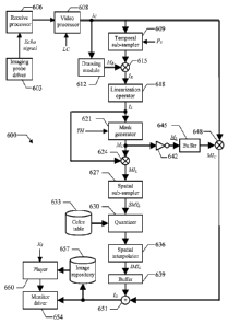
Moving now to Figure 6, the main software and hardware components that can be
used
for practicing the solution according to an embodiment of the invention are
denoted as a whole
with the reference 600. The information (programs and data) is typically
stored on the hard
disk and loaded (at least partially) into the working memory when the programs
are running,
together with an operating system and other application programs (not shown in
the figure).
The programs are initially installed onto the hard disk, for example, from CD-
ROM.
Particularly, a driver 603 controls the imaging probe (not shown in the
figure); for
example, the imaging probe driver 603 includes a transmit beam former and
pulsers for
generating the ultrasound pulses to be applied to the body-part under
analysis. The
corresponding (analog RF) echo signal that is received from said body-part is
supplied to a
receive processor 606. Typically, the receive processor 606 pre-amplifies the
analog RF echo
signal and applies a preliminary time-gain compensation (TGC); the analog RF
echo signal is
then converted into digital values by an Analog-to-Digital Converter (ADC),
and combined
into a focused signal through a receive beam former. The digital signal so
obtained is
preferably processed through further digital algorithms and other linear or
non-linear signal
conditioners (such as a post-beam-forming TGC). Particularly, the receive
processor 606
applies a contrast-specific algorithm to suppress the contribution of the
tissues (such as based
on the above-mentioned HI, PI, PM or CPS techniques). The digital signal so
processed is
passed to a video processor 608, wherein it is demodulated, log-compressed,
and scan-
converted into a video format. The process results in the recording of a
sequence of
compressed images Ic. For this purpose, the video processor 608 receives as
input the desired
compression factor LC.
The compressed images /c are provided to a temporal sub-sampler 609, which
also
CA 02624636 2014-06-10
17
receives a sub-sampling parameter Ps (for example, from 0 to 10). The temporal
sub-sampler
609 outputs one compressed image /c out of every Ps+I; for this purpose, the
temporal sub-
sampler 609 lets a compressed image lc pass through and then skips the next Ps
ones. In most
practical situations, the sub-sampling parameter Ps is set to 0 (so that every
compressed
image /c is taken into account); higher values of the sub-sampling parameter
Ps are instead
used to limit the number of compressed images /c to be processed (for example,
when the
ultrasound scanner works at ultra-high frame rates, such as 100-500 frames per
second).
A drawing module 612 is used to predefine a ROI for the analysis process on
the
compressed images Ic (from the video processor 608). The operation generates a
reduction
mask MR, which consists of a matrix of binary values with the same size as the
compressed
images /c (i.e., MxN); the binary values inside the ROI are assigned the logic
value 1,
whereas the binary values outside the ROI are assigned the logic value 0. A
multiplier
operator 615 receives the (possibly temporally sub-sampled) compressed images
lc from the
temporal sub-sampler 609 and the reduction mask MR from the drawing module
612. The
operator 615 multiplies each compressed image Ic by the reduction mask MR
pixel-by-pixel,
so as to generate a corresponding sequence of reduced images /R. As a result,
the reduced
images IR only include the pixel values of the compressed images /c that are
inside the ROI
(defined by the reduction mask MR), while the other pixel values are reset to
0.
Each reduced image IR is provided to a linearization operator 618, which
outputs a
corresponding linearized image IL. Particularly, the operator 618 linearizes
the reduced
image IR pixel-by-pixel, so as to make each pixel value of the linearized
image IL directly
proportional to the local echo power (i.e., proportional to the concentration
of the contrast
agent when present in the body part under analysis); in the example at issue,
this result is
achieved by applying the formula (0.3) to every pixel value of the reduced
image IR.
The linearized image /L, is then passed to a mask generator 621, which is
controlled by
the threshold TH. The mask generator 621 creates a corresponding linearization
mask ML;
the linearization mask ML is obtained from the linearized image IL by
assigning (to each
pixel) the logic value 1 if its value exceeds the threshold TH or the logic
value 0 otherwise.
A multiplier operator 624 receives the linearized image IL (from the
linearization operator
CA 02624636 2014-06-10
18
618) and the linearization mask ML (from the mask generator 621). The operator
624
multiplies the linearized image IL by the linearization mask ML pixel-by-
pixel, so as to
generate a corresponding masked (linearized) image MIL. As a result, the
masked image MIL
only includes the pixel values of the linearized image /L that exceed the
threshold TH, while
the other pixel values are reset to 0.
A spatial sub-sampler 627 receives the masked image MIL so obtained. The
module
627 sub-samples the masked image MIL according to a factor based on the
spatial frequency
content of one of the compressed images k (for example, according to the size
of speckle
grains that typically occur in ultrasound imaging, for example, equivalent to
2-6 pixels).
Preferably, the spatial sub-sampling comprises low-pass filtering followed by
sub-sampling.
The low-pass filtering has a cutoff frequency, which can be chosen as the
highest frequency
component containing significant energy in a selected one of the compressed
images k (for
example, determined by Fourier analysis). The sub-sampling is performed
according to a
factor that can be determined, for example, as a value resulting in a spatial
sub-sampling
frequency equal to twice the cutoff frequency. In this way, the masked image
MIL is
transformed into a corresponding sub-sampled masked image SM/L; each value of
the sub-
sampled masked image SM/L thus represents a cell corresponding to a group of
adjacent
pixels in the masked image MIL (which cell has a size defined according to the
above-
mentioned spatial resolution). This allows smoothing any irregularity in the
recorded
information (for example, due to any misalignments of the compressed images
/c).
The (sub-sampled) masked image SM/L is then provided to a quantizer 630. The
quantizer 630 is adapted to convert the cell values of the masked image SM/L
into
corresponding discrete values (for example, consisting of 64 or 128 levels
that are uniformly
distributed from 0 to the maximum video signal MAX), by possibly applying a
gain factor.
The quantizer 630 also accesses a color (look-up) table 633. The color table
633 associates all
the possible levels with the representation of corresponding colors (that are
preferably brighter
as the levels increase); for example, each color is defined by an index for
accessing a location
within a palette containing its actual specification. The quantizer 630
replaces each cell value
in the masked image SM/L with the corresponding color representation.
CA 02624636 2014-06-10
19
The masked image SM/L is provided to a spatial-interpolator 636. The spatial-
interpolator 636 restores the full-size of the masked image SM/L corresponding
to the size of
the compressed images lc (i.e., MxN) by means of interpolation techniques
(such as based on
the nearest neighbor, bilinear, or bicubic technique). For this purpose, the
value of each cell in
the masked image SM/L is replicated for the corresponding group of pixels
(nearest neighbor
interpolation method) and optionally filtered spatially (such as using a low-
pass 2D or 3D
spatial filter). The operation generates a corresponding (interpolated) masked
image /M/L. The
masked image /MIL is latched into a single-image buffer 639 (replacing its
previous
content). In this way, the masked image /MIL in the buffer 639 is updated
whenever a new
compressed image /c is output by the temporal sub-sampler 609, while it
remains
unchanged otherwise (so as to maintain the last calculated masked image /M/L).
Concurrently, the linearization mask ML is also supplied from the mask
generator 621
to an inverter 642, which generates a corresponding inverted (linearization)
mask ML (by
exchanging the logic values 0 and 1). The inverted mask ML is likewise latched
into a single-
image buffer 645 (replacing its previous content), so as to be always
synchronized with the
masked image /MIL in the buffer 639. A multiplier operator 648 receives the
inverted mask
(latched in the buffer 645) and a current compressed image /c (from the video
processor
608). The operator 648 multiplies the compressed image k by the inverted mask
ML pixel-
by-pixel, so as to obtain a corresponding masked (compressed) image M/c. As a
result, the
masked image M/c includes the pixel values of the corresponding compressed
image /c that
are outside the ROI and below the threshold TH within the ROI, while the other
pixel values
within the ROI are reset to 0.
An adder operator 651 receives the masked image /MIL (latched in the buffer
639)
and the masked image MI c (from the multiplier operator 648). The operator 651
adds the
masked image /MIL and the masked image M/c pixel-by-pixel (correctly
synchronized) so as
to obtain an overlaid image /o. In this way, each pixel value of the overlaid
image /0 within
the ROI is displayed as in the linearized image IL whenever that pixel value
(in the same
linearized image IL) is larger than the threshold TH; the other pixel values
within the ROT
that are below the threshold TH and all the pixel values outside the ROI are
instead
CA 02624636 2014-06-10
displayed as in the compressed image k.
The overlaid image /o is passed to a monitor driver 654, which controls its
visualization. The same operations described above are reiterated for each new
compressed
image /c that is recorded; as a result, the overlaid images /o are displayed
in succession on
5 the monitor of the ultrasound scanner in real-time; this means that the
overlaid images /o are
available substantially at the same time when the corresponding compressed
images /c are
acquired (or with a short delay, but in any case without the need to wait for
the completion
of their acquisition for starting the displaying).
In addition or in alternative, the sequence of overlaid images /0 so obtained
may also
10 be saved into a repository 657. The repository 657 is accessed by a
player 660; the player 660
also receives an index Xs, which is selected according to the desired
reproduction speed of the
overlaid images /0; for example, the speed index Xs is set to 1 for a
reproduction in real time,
to a value lower than 1 for a reproduction in slow-motion or to a value higher
than 1 for a
reproduction in accelerated-motion. The player 660 extracts the overlaid
images /0 in
15 succession from the repository 657. Each overlaid image /0 is then
passed to the monitor
driver 654 for its playback (with a frame rate corresponding to the selected
speed index Xs).
Modifications
Naturally, in order to satisfy local and specific requirements, a person
skilled in the
art may apply to the solution described above many modifications and
alterations.
Particularly, although the present invention has been described with a certain
degree of
particularity with reference to preferred embodiment(s) thereof, it should be
understood that
various omissions, substitutions and changes in the form and details as well
as other
embodiments are possible; moreover, it is expressly intended that specific
elements and/or
method steps described in connection with any disclosed embodiment of the
invention may
be incorporated in any other embodiment as a general matter of design choice.
CA 02624636 2014-06-10
21
For example, similar considerations apply if the ultrasound scanner has a
different
structure or includes other units (such as with an imaging probe of the linear-
, convex-,
phased-, or matrix- array type). Likewise, the solution of the invention lends
itself to be put
into practice with equivalent contrast agents (even administrated in other
ways, such as
intra-arterial). In addition, the devised solution may be used in applications
that do not relate
to the perfusion assessment; a typical example is the detection and the
quantification of the
contrast agent that is immobilized on a specific biological target, as
described in the co-
pending application No.PCT/EP06/068305 of 9 November 2006.
Moreover, any other technique may be used to reduce the contribution of the
tissues
in the echo signal (for example, by applying the algorithm described in the
above-cited
document by Arditi at al.). It should also be noted that the proposed
numerical examples for
the reduction of the contribution of the tissues in the echo signal are not to
be interpreted in
a limitative manner; particularly, the complete removal of the contribution of
the tissues
from the echo signal is within the scope of the invention.
In any case, nothing prevents the application of the proposed processing to
all the
available images (without any temporal sub-sampling).
Naturally, the above-described transfer function defining the log-compression
and
the formulas for linearizing the available images are merely illustrative;
similar
considerations apply to different transfer functions of the logarithmic type,
or more
generally to any other non-linear compression.
Moreover, the numerical examples for the threshold TH must not be interpreted
in a
limitative manner; more generally, nothing prevents setting the threshold TH
in other ways
(even independently of the residual contribution of the tissues). In any case,
a similar result
may be achieved in a system based on negative images (wherein the pixel values
decrease
with the intensity of the echo signal) by using a different (maximum)
threshold.
Although the technique of the invention has been specifically designed for
ultrasound
applications, nothing prevents its use in any other medical imaging
application, such as
based on Magnetic Resonance Imaging (MRI) or X-ray Computed Tomography (CT).
Alternatively, the same solution may also be applied in a system that consists
of an
CA 02624636 2014-06-10
22
ultrasound scanner and a distinct computer (or any equivalent data processing
entity); in this
case, the recorded information is transferred from the ultrasound scanner to
the computer for
its processing (for example, through the removable disk, a memory key, or a
network
connection).
According to an alternative embodiment, the pixel values outside the selected
ROT
may be reset to 0 (so that the portion of the overlaid image outside the ROI
is black);
however, the application of the proposed solution to the whole content of the
compressed
images is contemplated.
According to a different embodiment of the invention, the compressed values
for the
pixels inside the ROI whose linearized values are below the threshold TH, and
for the pixels
outside the ROT, may be obtained from any other signals. For example, the
compressed
values from signals obtained with a non contrast-specific imaging modality,
such as
fundamental B-mode imaging, can be advantageously employed. These values can
be
obtained, for instance, from the echo signals of the imaging probe driver. The
values so
obtained may thus be used as the compressed values in the overlaid images to
represent, for
instance, the anatomy of the body part under analysis. At the same time, the
linearized
values assigned to the pixels inside the ROI exceeding the threshold TH are
obtained from
signals wherein the contribution of the tissues has been reduced with respect
to the one of
the contrast agent. As previously mentioned, when the contribution of the
tissues in the
linearized signal is completely removed, the threshold TH may advantageously
be set to
zero.
Without departing from the principles of the invention, it is also possible to
apply the
thresholding to the compressed values (instead of the linearized values); in
this case, the
linearized values are only calculated for the compressed values that exceed
the threshold.
Moreover, when the (non-compressed) echo signal is accessible, the overlaid
image may
also be directly composed by linearizing each pixel value above the threshold
or by
compressing it otherwise.
In some embodiments, the linearized signal might be already available for
other
purposes (such as when parametric analysis techniques are implemented); in
this case, it is
CA 02624636 2014-06-10
23
possible to exploit the available information without any additional
linearization operation.
Similar considerations apply if the linearized images are spatially sub-
sampled with a
different procedure (for example, according to a predefined sub-sampling
factor), or if the
spatial sub-sampling is performed beforehand or afterward; in any case, the
application of
the proposed solution at the pixel level (instead of at the level of groups of
pixels defined by
the above-mentioned spatial sub-sampling) is not excluded.
It should also be noted that the step of applying the gain factor on the
linearized values
may be replaced by applying a differently scaled color lookup table; in any
case, a gray-scale
representation of the linearized values is within the scope of the invention.
As described above, even if the advantages of the present invention are more
clearly
perceived when the overlaid images are displayed in real-time, the application
of the devised
solution for analyzing the obtained results off-line is contemplated.
Similar considerations apply if the program (which may be used to implement
each
embodiment of the invention) is structured in a different way, or if
additional modules or
functions are provided; likewise, the memory structures may be of other types,
or may be
replaced with equivalent entities (not necessarily consisting of physical
storage media).
Moreover, the proposed solution lends itself to be implemented with an
equivalent method
(having similar or additional steps, even in a different order). In any case,
the program may
take any form suitable to be used by or in connection with any data processing
system, such
as external or resident software, firmware, or microcode (either in object
code or in source
code). Moreover, the program may be provided on any computer-usable medium;
the
medium can be any element suitable to contain, store, communicate, propagate,
or transfer
the program. Examples of such medium are fixed disks (where the program can be
pre-
loaded), removable disks, tapes, cards, wires, fibers, wireless connections,
networks,
broadcast waves, and the like; for example, the medium may be of the
electronic, magnetic,
optical, electromagnetic, infrared, or semiconductor type.
In any case, the solution according to the present invention lends itself to
be carried
out with a hardware structure (for example, integrated in a chip of
semiconductor material),
or with a combination of software and hardware.