Note: Descriptions are shown in the official language in which they were submitted.
DEMANDE OU BREVET VOLUMINEUX
LA PRESENTE PARTIE DE CETTE DEMANDE OU CE BREVET COMPREND
PLUS D'UN TOME.
CECI EST LE TOME 1 DE 2
CONTENANT LES PAGES 1 A 43
NOTE : Pour les tomes additionels, veuillez contacter le Bureau canadien des
brevets
JUMBO APPLICATIONS/PATENTS
THIS SECTION OF THE APPLICATION/PATENT CONTAINS MORE THAN ONE
VOLUME
THIS IS VOLUME 1 OF 2
CONTAINING PAGES 1 TO 43
NOTE: For additional volumes, please contact the Canadian Patent Office
NOM DU FICHIER / FILE NAME:
NOTE POUR LE TOME / VOLUME NOTE:
CA 02624648 2008-04-02
WO 2007/047969 PCT/US2006/041113
A-1009-PCT
METHODS OF DECREASING VASCULAR CALCIFICATION
USING IL-1 INHIBITORS
This application claims the benefit of U.S. Provisional Application No.
60/729,305, filed October 21, 2005, which is hereby incorporated by reference.
FIELD OF THE INVENTION
This invention relates generally to the field of medicine and, more
specifically, to methods of decreasing, treating or preventing vascular
calcification.
BACKGROUND OF THE INVENTION
The IL-1 system
One of the most potent inflammatory cytokines yet discovered is
interleukin-1 (IL-1). IL-1 is thought to be a key mediator in many diseases
arid
medical conditions. It is manufactured (though not exclusively) by cells of
the
macrophage/monocyte lineage and may be produced in two forms: IL-1 alpha
(IL-1a) and IL-1 beta (IL-1(3). A third cytokine in the system acts as an
antagonist
and is referred to as IL-1 receptor antagonist (IL-lra).
There are three known IL-1 receptor subunits. The active receptor complex
consists of the type I receptor (IL-1 RI) and IL-1 receptor accessoiy protein
(IL-
1RAcP). IL-1 RI is responsible for binding the three naturally occurring
ligands
(IL-1 alpha, IL-1 beta and IL-1 ra) and is able to do so in the absence of the
IL-
1RAcP. However, signal transduction requires interaction of IL-1 alpha or IL-1
beta with IL-1RAcP. IL-ira does not interact with the IL-1RAcP and hence
cannot
signal. A third receptor subunit, the type II receptor (IL-1 RII), binds IL-1
alpha or
IL-1 beta but cannot signal because it lacks an intracellular domain. Instead,
it
inhibits IL-1 bioactivity by acting as a decoy receptor in both a membrane-
bound
form and a cleaved, secreted form. See Dinarello (1996) Blood 87:2095-2147.
IL-1ra inlzibits IL-1 alpha and IL-1 beta by binding to IL-1 RI but not
transducing an intracellular signal or a biological response. IL-lra inhibits
the
CA 02624648 2008-04-02
WO 2007/047969 PCT/US2006/041113
A-1009-PCT
biological activities of IL-1 botli in vitro and in vivo, and has been shown
to be
effective in animal models of septic shock, rheumatoid arthritis, graft versus
host
disease, stroke, and cardiac ischemia. A recombinant form of IL-lra produced
in
E. coli is currently approved for pharmaceutical use in the United States and
Europe. This drug has the generic name anakinra and is marketed under the
trade
name Kineret .
Vascular calcification
Vascular calcification, a well-recognized and common complication of
chronic kidney disease (CKD), increases the risk of cardiovascular morbidity
and
mortality (Giachelli, C. J. Am. Soc. Nephrol. 15: 2959-64, 2004; Raggi, P. et
al.
J. Am. Coll. Cardiol. 39: 695-701, 2002). While the causes of vascular
calcification in CKD remain to be elucidated, associated risk factors include
age,
gender, hypertension, time on dialysis, diabetes and glucose intolerance,
obesity,
and cigarette smoking (Zoccali C. Nephrol. Dial. Transplant 15: 454-7, 2000).
These conventional risk factors, however, do not adequately explain the high
mortality rates from cardiovascular causes in the patient population. Recent
observations suggest that certain abnorinalities in calcium and phosphorus
metabolism, resulting in a raised serum calcium-phosphorus product (Ca x P)
contribute to the development of arterial calcification, and possibly to
cardiovascular disease, in patients with end-stage renal disease (Goodman, W.
et
al. N. Engl. J. Med. 342: 1478-83, 2000; Guerin, A. et al. Nephrol. Dial.
Transplant 15:1014-21, 2000; Vattikuti, R. & Towler, D. Am. J. Physiol.
Endocrinol. Metab., 286: E686-96, 2004).
Another hallmarlc of advanced CKD is secondary hyperparathyroidism
(HPT), characterized by elevated parathyroid horinone (PTH) levels and
disordered mineral metabolism. The elevations in calcium, phosphorus, and Ca x
P observed in patients with secondary HPT have been associated with an
increased
risk of vascular calcification (Chertow, G. et al. Kidney Int. 62: 245-52,
2002;
Goodman, W. et al. N. Engl. J. Med. 342: 1478-83, 2000; Raggi, P. et al. J.
Am.
Coll. Cardiol. 39: 695-701, 2002). Commonly used therapeutic interventions for
secondary HPT, such as calcium-based phosphate binders and doses of active
CA 02624648 2008-04-02
WO 2007/047969 PCT/US2006/041113
A-1009-PCT
vitamin D sterols can result in hypercalcemia and hyperphosphatemia (Chertow,
G. et al. Kidney Int. 62: 245-52, 2002; Tan, A. et al. KidneyInt 51: 317-23,
1997;
Gallieni, M. et al. Kidney 1 42: 1191-8, 1992), which are associated with the
development or exacerbation of vascular calcification.
Vascular calcification is an important and potentially serious complication
of cllronic renal failure. Two distinct patterns of vascular calcification
have been
identified (Proudfoot, D & Shanahan, C. Herz 26: 245-51, 2001), and it is
common for both types to be present in uremic patients (Chen, N. & Moe, S.
Semin Nephro124: 61-8, 2004). The first, medial calcification, occurs in the
media of the vessel in conjunction witll a phenotypic transformation of smooth
muscle cells into osteoblast-like cells, while the other, atherogenesis, is
associated
with lipid-laden macrophages and intimal hyperplasia.
Medial wall calcification can develop in relatively young persons with
chronic renal failure, and it is common in patients with diabetes mellitus
even in
the absence of renal disease. The presence of calcium in the medial wall of
arteries distinguishes this type of vascular calcification from that
associated with
atherosclerosis (Schinlce T. & Karsenty G. Nephrol Dial Transplant 15: 1272-4,
2000). Atherosclerotic vascular calcification occurs in atheromatous plaques
along the intimal layer of arteries (Farzaneh-Far A. JAMA 284: 1515-6, 2000).
Calcification is usually greatest in large, well-developed lesions, and it
increases
with age (Wexler L. et al. Circulation 94: 1175-92, 1996; Rumberger J. et al.
Mayo Clin Proc 1999; 74: 243-52.). The extent of arterial calcification in
patients
with atherosclerosis generally corresponds to severity of disease. Unlike
medial
wall calcification, atherosclerotic vascular lesions, whether or not they
contain
calcium, impinge upon the arterial lumen and compromise blood flow. The
localized deposition of calcium within atherosclerotic plaques may happen
because of inflamination due to oxidized lipids and other oxidative stresses
and
infiltration by monocytes and macrophages (Berliner J. et al. Circulation 91:
2488-
96, 1995).
Some patients with end-stage renal disease develop a severe form of
occlusive arterial disease called calciphylaxis or calcific uremic
arteriolopathy.
CA 02624648 2008-04-02
WO 2007/047969 PCT/US2006/041113
A-1009-PCT
This syndrome is characterized by extensive calcium deposition in small
arteries
(Gipstein R. et al. Arch Intern Med 136: 1273-80, 1976; Richens G. et al. J Am
Acad. Dermatol. 6: 537-9, 1982). In patients with this disease, arterial
calcification and vascular occlusion lead to tissue ischemia and necrosis.
Involvement of peripheral vessels can cause ulceration of the skin of the
lower
legs or gangrene of the digits of the feet or hands. Ischemia and necrosis of
the
skin and subcutaneous adipose tissue of the abdominal wall, thighs and/or
buttocks are features of a proximal form of calcific uremic arteriolopathy
(Budisavljevic M. et al. J Am Soc Nephrol. 7: 978-82, 1996; Ri.iggian J. et
al. Am.
J. Kidney Dis. 28: 409-14, 1996). This syndrome occurs more frequently in
obese
individuals, and women are affected more often than men for reasons that
remain
unclear (Goodman W. J. Nephrol. 15(6): S82-S85, 2002).
Current therapies to normalize serum mineral levels or to decrease, inhibit,
or prevent calcification of vascular tissues or implants are of limited
efficacy and
cause unacceptable side effects. Therefore, there exists a need for an
effective
method of inhibiting and preventing vascular calcification.
SUMMARY OF THE INVENTION
The present invention provides methods of inhibiting, decreasing, or
preventing vascular calcification in a subject comprising administering a
therapeutically effective amount of an IL-1 inhibitor to the subject. In one
aspect,
the vascular calcification can be atherosclerotic calcification. In another
aspect,
the vascular calcification can be medial calcification.
In one aspect, the subject can be suffering from chronic renal insufficiency
or end-stage renal disease. In another aspect, the subject can be pre-
dialysis. In a
further aspect, the subject can be suffering from uremia. In another aspect,
the
subject can be suffering from diabetes mellitus I or II. In another subject,
the
subject can be suffering from a cardiovascular disorder. In one aspect, the
subject
can be human:
In one aspect, the IL-1 inhibitor can be the molecule having the generic
name analcinra. In another aspect, the IL-i inhibitor can be a molecule having
the
CA 02624648 2008-04-02
WO 2007/047969 PCT/US2006/041113
A-1009-PCT
sequence shown in Figure 4 hereinafter (SEQ ID NO: 1), hereinafter referred to
as
"Fc-IL-1 ra." In yet another aspect, the IL-1 inhibitor can be an antibody to
the IL-
1 receptor. Preferred 'antibodies to the IL-1 receptor are described in U.S.
Pat.
App. 2004/0097712, published May 20, 2004 (U.S. Ser. No. 10/656,769). In a
still further aspect, the IL-1 inhibitor can be an IL-1 trap molecule.
In one aspect, the IL-1 inhibitor used in the methods of the invention can
be N- ((6-(methyloxy)-4'-(trifluoromethyl)-1,1'-biphenyl-3-yl) methyl)-1-
phenylethanamine, or a pharmaceutically acceptable salt thereof.
In one aspect, the invention provides methods of inhibiting, decreasing, or
preventing vascular calcification; wherein a vitamin D sterol had been
previously
administered to the subject. In one aspect, the vitamin D sterol can be
calcitriol,
alfacalcidol, doxercalciferol, maxacalcitol or paricalcitol. In one aspect,
the IL-1
inhibitor can be administered prior to or following administration of a
vitamin D
sterol. In another aspect, the IL-1 inhibitor can be administered in
combination
with a vitamin D sterol.
In one aspect, the IL-1 inhibitor can be administered in combination with
RENAGELO.
The invention further provides methods of decreasing serum creatinine
levels in a subject, comprising administering a therapeutically effective of
an IL-1
inhibitor to the subject. In one aspect, the subject can be suffering from
increased
serum creatinine levels induced by the administration of a vitamin D sterol to
the
subject.
BRIEF DESCRIPTION OF THE FIGURES
Figure 1 shows an experimental paradigm for adenine model of chronic
kidney disease (CKD) and secondary hyperparathyroidism (SHPT).
Figure 2 shows prevention of aortic vascular calcification with FC-IL-lra
in an animal model of CKD. The asterisk in Figure 2 indicates that under the
conditions tested (p=0.005; unpaired t-test; control (A5S) vehicle (red; n=4);
Fc-
IL-lra (blue hatched, n=7)), there was no calcification (0 g/cm2 bone mineral
density) in seven of seven Fc-IL-lra-treated animals.
CA 02624648 2008-04-02
WO 2007/047969 PCT/US2006/041113
A-1009-PCT
Figure 3 shows reduction of parathyroid gland size (A) and serum PTH (B)
by Fc-IL-lra in an animal model of CKD/SHPT. In Figure 3A, the asterisk (*)
denotes the parathyroid wild type/body wild type for A5S vehicle control vs.
Fc-
IL-lra; p=0.009; unpaired t-test; control (vehicle, A5S), red; n=8); Fc-IL-
lra,
(blue, n=13). In Figure 3B, the pound sign (#) denotes serum PTH for A5S
vehicle
control vs. Fc-IL-ira; p=0.028; unpaired t-test; control (vehicle, A5S), red;
n=4);
Fc-IL- l ra, (blue, n=7).
Figure 4 shows the aminoacid sequence (SEQ ID NO: 1) of the molecule
referred to herein as Fc-IL-1 ra.
Figure 5 shows the experimental paradigm for the 5/6-nephrectoiny model
of CKD and secondary HPT.
Figure 6 shows attenuation of calcitriol-induced aortic vascular
calcification in uremic rats treated with Fc-IL-lra.
DETAILED DESCRIPTION OF THE INVENTION
Recent scientific literature includes suggestions to study the effects of
cytokines on vascular calcification. Yao et al. (2004), Scandinavian J. Urol.
Nephrol. 38: 405-16, found what they considered a "strong association between
inflammation and increased oxidative stress and endothelial dysfunction" in
end-
stage renal disease (ESRD) patients. Malberti and Ravini (2005), Giomale
Italiano
di Nefrologia (22 Suppl.) 31: S47-52, noted that vascular calcifications are
more
frequent in dialysis patients than in the general population or in patients
with
cardiovascular disease with norinal renal function, which led these authors to
suggest study of the effects of anti-inflammatory treatments on the
nutritional and
cardiovascular status of ESRD patients. Moe and Chen (2005), Blood Purif. 23:
64-71, noted that cytokines and other mediators of inflammation may have a
direct
stimulatory effect on vascular calcification, leading them to suggest that
inhibition
of cytokine-mediated inflammation represents "a plausible therapeutic approach
to
limit vascular calcification." The literature does not identify, however,
which
inflammatory cytokines may mediate vascular calcification.
Elsewhere in the literature, Nicklin et al. (2000), J. Exper. Med. 191: 303-
11, found that IL-lra deficient mice develop lethal arterial inflammation in
CA 02624648 2008-04-02
WO 2007/047969 PCT/US2006/041113
A-1009-PCT
flexpoints and branch points of the aorta. Arai et al. (1998), J.
Toxicological
Sciences 23: 121-8, found that 1,25 dihydroxyvitamin D3 has been shown to
increase IL-1 synthesis. The literature does not clarify, however, a
definitive role
for IL-1 receptor antagonism in prevention of vascular calcification.
The present invention is directed to methods of reducing, inhibiting, or
preventing vascular calcification using IL-1 inhibitors.
IL-1 inhibitors in general
"IL-1" refers to IL-1 a and IL-1 (3.
"IL-1 inhibitors" as used throughout this specification refers to molecules
that decrease the bioactivity of IL-1 a, IL-1 (3, or IL-1 receptor type I(IL-1
RI),
whether by direct or indirect interaction with IL-1 a, IL-1 (3, IL-1 RI, IL-1
receptor
accessory protein (IL-1RacP), interleukin-1 converting enzyme (ICE), with
proteins that mediate signaling through a receptor for IL-1 a or (3, with
proteins
controlling the expression or release of IL-1 a, IL-1 (3, IL-1 RI or IL-1 RII.
Inhibition of IL-1 may result from a number of mechanisms, including down-
regulation of IL-1 transcription, expression, or release from cells that
produce IL-
1; binding of free IL-1; interference with binding of IL-1 to its receptor;
interference with formation of the IL-1 receptor complex (i.e., association of
the
IL-1 receptor type I with IL-1 RacP); and interference with modulation of IL-1
signaling after binding to its receptor. Thus, the term "IL-1 inhibitor"
includes,
but is not limited to, IL-1 beta inhibitors and IL-1 receptor antagonists (IL-
ira),
such as anal:inra and antibodies to IL-1 RI.
Classes of IL-1 inhibitors include the following, which are described in
detail further hereinbelow:
Interleukin-1 receptor antagonists such as IL-lra and anti-IL-1 receptor
monoclonal antibodies, as described below;
IL-1 binding proteins such as soluble IL-1 receptors, anti-IL-1 monoclonal
antibodies;
Inhibitors of interleukin-1 beta converting enzyme (ICE) or caspase I (e.g.,
WO 99/46248, WO 99/47545, and WO 99/47154, the disclosures of which are
CA 02624648 2008-04-02
WO 2007/047969 PCT/US2006/041113
A-1009-PCT
hereby incorporated by reference), which can be used to inhibit IL-1 beta
production and secretion;
Interleukin- l beta protease inhibitors;
and compounds and proteins that block in vivo synthesis or extracellular
release ofIL-1.
The term "IL-1 binding proteins" refers to molecules that bind to IL-1 and
thus prevent IL-1 beta from exerting bioactivity when bound to IL-1 RI. Thus,
IL-
1 beta inhibitors include, but are not limited to, IL-1 beta antibodies,
peptides that
bind to IL-1 beta, peptibodies that bind to IL-1 beta, soluble IL-1 receptor
molecules, and IL-1 trap molecules.
The term "IL-1 receptor antagonists" refers to molecules that bind to IL-1
RI or IL-1 RacP or otherwise,prevent the interaction of IL-1 RI and IL-1 RacP.
Thus, the term "IL-1 receptor antagonists" includes, but is not limited to
analcinra,
Fc-IL-lra, IL-1 RI antibodies, IL-1RacP antibodies, peptides that bind to IL-1
RI
or to IL-1RAcP, and peptibodies that bind to IL-1 RI or IL-1RAcP.
Exemplaiy IL-1 inliibitors are disclosed in the following references:
US Pat. Nos. 5,747,444; 5,359,032; 5,608,035; 5,843,905; 5,359,032;
5,866,576; 5,869,660; 5,869,315; 5,872,095; 5,955,480; 5,965,564;
International (WO) patent applications 98/21957, 96/09323, 91/17184,
96/40907, 98/32733, 98/42325, 98/44940, 98/47892, 98/56377, 99/03837,
99/06426, 99/06042, 91/17249, 98/32733, 98/17661, 97/08174, 95/34326,
99/36426, 99/36415;
European (EP) patent applications 534978 and 894795; and
French patent application FR 2762514.
IL-1 receptor antagonists ,
For purposes of the present invention, IL-lra and variants and derivatives
thereof as discussed hereinafter are collectively termed "IL-lra protein(s)".
The
molecules described in the above references and the variants and derivatives
thereof discussed hereinafter are collectively termed "IL-1 inhibitors."
IL-lra is a liuman protein that acts as a natural inhibitor of interleukin-1
and which is a member of the IL-1 family member that includes IL-la and IL-
1(3.
CA 02624648 2008-04-02
WO 2007/047969 PCT/US2006/041113
A-1009-PCT
Preferred receptor antagonists (including IL-lra and variants and derivatives
thereof), as well as methods of making and using thereof, are described in WO
91/08285; WO 91/17184; AU 9173636; WO 92/16221; W093/21946; WO
94/06457; WO 94/21275; FR 2706772; WO 94/21235; DE 4219626, WO
94/20517; WO 96/22793;WO 97/28828; WO 99/36541, and U.S. Patent Nos.
5,075,222 and 6,599,873 (incorporated herein by reference). The proteins
include
glycosylated as well as non-glycosylated IL-1 receptor antagonists.
Specifically, three useful forms of IL-lra and variants thereof are disclosed
and described in the 5,075,222 patent. The first of these, called "IL-li" in
the '222
10' patent, is characterized as a 22-23 1cD molecule on SDS-PAGE with an
approximate isoelectric point of 4.8, eluting from a Mono Q FPLC column at
around 52 mM NaCI in Tris buffer, pH 7.6. The second, IL-lra(3, is
characterized
as a 22-23 kD protein, eluting from a Mono Q column at 48 mM NaCl. Both IL-
lraa and IL-lra(3 are glycosylated. The third, IL-lrax, is characterized as a
20 kD
protein, eluting from a Mono Q colunui at 48 mM NaCl, and is non-glycosylated.
5,075,222 patent also discloses methods for isolating the genes responsible
for
coding the inhibitors, cloning the gene in suitable vectors and cell types,
and
expressing the gene to produce the inhibitors.
Those skilled in the art understand that many combinations of deletions,
insertions and substitutions (individually or collectively "variant(s)") can
be made
within the amino acid sequences of IL-lra, provided that the resulting
molecule is
biologically active (e.g., possesses the ability to inhibit IL-1). Particular
variants
are described in U.S. Pat. No. 5,075,222 and U.S. Ser. No. 11/097,453, which
are
hereby incorporated by reference.
The term "IL-1 receptor antagonist" further includes modified IL-lra and
fusion proteins comprising IL-lra. Exemplay fusion proteins include Fc-IL-lra
(Figure 4, SEQ ID NO: 1), and molecules as described in U.S. Pat. No.
6,294,170.
Antibodies
"IL-1 beta antibodies" and "antibodies to IL-I beta" refer to antibodies that
specifically bind to IL-1 beta. One example of an IL-1 beta antibody is known
as
MAb 201 and is commercially available. Additional IL-1 beta antibodies may be
CA 02624648 2008-04-02
WO 2007/047969 PCT/US2006/041113
A-1009-PCT
produced as described hereinafter. Further examples of IL-1 antibodies are
described in WO 9501997, WO 9402627, WO 9006371, EP 364778, EP 267611,
EP 220063, and U.S. Pat. No. 4,935,343 (incorporated by reference).
Antibodies having specific binding affinity for IL-1(3 can be produced
through standard methods. Alternatively, antibodies may be commercially
available, for example, from R&D Systems, Inc., Minneapolis, Minn. The terms
"antibody" and "antibodies" include polyclonal antibodies, monoclonal
antibodies,
humanized or chimeric antibodies, single chain Fv antibody fragments, Fab
fragments, and F(ab)2 fragments. Polyclonal antibodies are heterogeneous
populations of antibody molecules that are specific for a particular antigen,
which
are contained in the sera of the immunized animals. Polyclonal antibodies are
produced using well-known methods.
Likewise, "IL-1 RI antibodies" and "antibodies to IL-1 RI" refer to
antibodies that specifically bind to IL-1 RI. Examples of IL-1 RI antibodies
are
described in EP 623 674 and U.S. Pat. App. 2004/0097712, published May 20,
2004 (U.S. Ser. No. 10/656,769), the disclosure of which is hereby
incorporated
by reference. Additional IL-1 RI antibodies may be produced as described
hereinafter.
The terms "antibody" and "antibodies" as used herein refer to intact
antibody, or a binding fragment thereof that competes with the intact antibody
for
specific binding and includes chimeric, humanized, fully human, and bispecific
antibodies. In certain embodiments, binding fragments are produced by
recombinant DNA techniques. In additional einbodiments, binding fragments are
produced by enzymatic or chemical cleavage of intact antibodies. Binding
fragments include, but are not limited to, Fab, Fab', F(ab')2, Fv, and single-
chain
antibodies.
The term "heavy chain" includes a full-length heavy chain and fragments
thereof having sufficient variable region sequence to confer specificity for
IL-1R1.
A full-length heavy chain includes a variable region domain, VH, and three
constant region domains, CH1, CH2, and CH3. The VH domain is at the amino-
terminus of the polypeptide, and the CH3 domain is at the carboxyl-terminus.
CA 02624648 2008-04-02
WO 2007/047969 PCT/US2006/041113
A-1009-PCT
The term "light chain" includes a full-length light chain and fragments
thereof having sufficient variable region sequence to confer specificity for
IL-IR1.
A full-length light chain includes a variable region domain, VL, and a
constant
region domain, CL. Like the heavy chain, the variable region domain of the
light
chain is at the amino-terminus of the polypeptide.
Monoclonal antibodies, which are homogeneous populations of antibodies
to a particular epitope contairied within an antigen, can be prepared using
standard
hybridoma technology. In particular, monoclonal antibodies can be obtained by
any technique that provides for the production of antibody molecules by
continuous cell lines in culture such as described by Kohler, G. et al.,
Nature,
1975, 256:495, the human B-cell hybridoma technique (Kosbor et al.,
Immunology Today, 1983, 4:72; Cole et al., Proc. Natl. Acad. Sci. USA, 1983,
80:2026), and the EBV-hybridoma technique (Cole et al., Monoclonal Antibodies
and Cancer Therapy, Alan R. Liss, Inc., 1983, pp. 77-96). Such antibodies can
be
of any immunoglobulin class including IgG, IgM, IgE, IgA, IgD, and any
subclass
thereof. The hybridoma producing the monoclonal antibodies of the invention
can
be cultivated in vitro or in vivo.
A chimeric antibody is a molecule in wliich different portions are derived
from different animal species, such as those having a variable region derived
from
a murine monoclonal antibody and a human immunoglobulin constant region.
Chimeric antibodies can be produced through standard techniques.
Antibody fragments that have specific binding affinity for IL-1 P can be
generated by known techniques. For example, such fragments include, but are
not
limited to, F(ab')2 fragments that can be produced by pepsin digestion of the
antibody molecule, and Fab fragments that can be generated by reducing the
disulfide bridges of F(ab')2 fragments. Alternatively, Fab expression
libraries can
be constructed. See, for example, Huse et al., 1989, Science, 246: 1275.
Single
chain Fv antibody fragments are formed by linlcing the heavy and light chain
fragments of the Fv region via an amino acid bridge (e.g., 15 to 18 amino
acids),
CA 02624648 2008-04-02
WO 2007/047969 PCT/US2006/041113
A-1009-PCT
resulting in a single chain polypeptide. Single chain Fv antibody fragments
can be
produced through standard techniques. See, for example, U.S. Pat. No.
4,946,778.
A "Fab fragment" is comprised of one light chain and the CH1 and variable
regions of one heavy chain. The heavy chain of a Fab molecule cannot form a
disulfide bond with another heavy chain molecule.
A "Fab' fragment" contains one light chain and one heavy chain that
contairis more of the constant region, between the CH1 and CH2 domains, such
that
an interchain disulfide bond can be formed between two heavy chains to form a
F(ab')2 molecule.
A "F(ab')2 fragment" contains two light chains and two heavy chains
containing a portion of the constant region between the CH 1 and CH2 domains,
such that an interchain disulfide bond is forined between two heavy chains.
The "Fv region" comprises the variable regions from both the heavy and
light chains, but lacks the constant regions.
"Single-chain antibodies" are Fv molecules in which the heavy and light
chain variable regions have been connected by a flexible linker to form a
single
polypeptide chain, which forms an antigen-binding region. Single chain
antibodies are discussed in detail in International Patent Application
Publication
No. WO 88/01649 and U.S. Patent Nos. 4,946,778 and 5,260,203 (hereby
incorporated by reference).
A "bivalent antibody" other than a"multispecific" or "multifiinctional"
antibody, in certain embodiments, is understood to comprise binding sites
having
identical'antigenic specificity.
A "bispecific" or "bifunctional" antibody is a hybrid antibody having two
different heavy/light chain pairs and two different binding sites. Bispecific
antibodies may be produced by a variety of methods including, but not limited
to,
fusion of hybridomas or linking of Fab' fragments. See, e_g., Songsivilai &
Laclunann (1990), Clin. Exp. Immunol. 79:315-321; Kostelny et al. (1992),
Immunol. 148:1547-1553.
CA 02624648 2008-04-02
WO 2007/047969 PCT/US2006/041113
A-1009-PCT
Preferred antibodies are described in U.S. Pat. App. 2004/0097712,
published May 20, 2004 (hereinafter referred to as the '712 application).
Specifically preferred are antibodies having a heavy chain variable region
selected
from the following:
Met Glu Phe Gly Leu Ser Trp Val Phe Leu 10
Val Ala Leu Leu Arg Gly Val Gln Cys Gln 20
Val Gln Leu Val Glu Ser Gly Gly Gly Val 30
Val Gln Pro Gly Arg Ser Leu Arg Leu Ser 40
Cys Ala Ala Ser Gly Phe Thr Phe Ser Asn 50
Tyr Gly Met His Trp Val Arg Gln Ala Pro 60
Gly Lys Gly Leu Glu Trp Val Ala Gly Ile 70
Trp Asn Asp Gly Ile Asn Lys Tyr His Ala 80
His Ser Val Arg Gly Arg Phe Thr Ile Ser 90
Arg Asp Asn Ser Lys Asn Thr Leu Tyr Leu 100
Gln Met Asn Ser Pro Arg Ala Glu Asp Thr 110
Ala Val Tyr Tyr Cys Ala Arg Ala Arg Ser 120
Phe Asp Trp Leu Leu Phe Glu Phe Trp Gly 130
Gln Gly Thr Leu Val Thr Val Ser Ser 139
(SEQ ID NO: 408)
Met Glu Phe Gly Leu Ser Trp Val Phe Leu 10
Val Ala Leu Leu Arg Gly Val Gin Cys Gln 20
Val Gln Leu Val Glu Ser Gly Gly Gly Val 30
Val Gln Pro Gly Arg Ser Leu Arg Leu Ser 40
Cys Ala Val Ser Gly Phe Thr Phe Ser Asn 50
Tyr Gly Met His Trp Val Arg Gln Ala Pro 60
Gly Lys Gly Leu Glu Trp Val Ala Ala Ile 70
Trp Asn Asp Gly G1u Asn Lys His His Ala 80
Gly Ser Val Arg Gly Arg Phe Thr Ile Ser 90
Arg Asp Asn Ser Lys Asn Thr Leu Tyr Leu 100
Gln Met Asn Ser Leu Arg Ala Glu Asp Thr 110
CA 02624648 2008-04-02
WO 2007/047969 PCT/US2006/041113
A-1009-PCT
Ala Val Tyr Tyr Cys Ala Arg Gly Arg Tyr 120
Phe Asp Trp Leu Leu Phe Glu Tyr Trp Gly 130
Gin Gly Thr Leu Val Thr Val Ser Ser 139
(SEQ ID NO: 409)
Met Gly Ser Thr Ala Ile Leu Ala Leu Leu 10
Leu Ala Val Leu Gln Gly Val Cys Ala Glu 20
Val Gln Leu Met Gln Ser Gly Ala Glu Val 30
Lys Lys Pro Gly Glu Ser Leu Lys Ile Ser 40
Cys Lys Gly Ser Gly Tyr Ser Phe Ser Phe 50
His Trp Ile Ala Trp Val Arg Gln Met Pro 60
Gly Lys Gly Leu Glu Trp Met Gly Ile Ile 70
His Pro Gly Ala Ser Asp Thr Arg Tyr Ser 80
Pro Ser Phe Gln Gly Gln Val Thr Ile Ser 90
Ala Asp Asn Ser Asn Ser Ala Thr Tyr Leu 100
Gln Trp Ser Ser Leu Lys Ala Ser Asp Thr 110
Ala Met Tyr Phe Cys Ala Arg Gln Arg Glu 120
Leu Asp Tyr Phe Asp Tyr Trp Gly Gln Gly 130
Thr Leu Val Thr Val Ser Ser 137
(SEQ ID NO: 410)
The foregoing are SEQ ID NOS: 10, 14, and 16 of the '712 application.
Antibodies incorporating these sequences may be prepared as described therein.
Most preferred are antibodies having all or an immunologically functional
fragment of a heavy chain having a sequence selected from SEQ ID NOS: 20, 22,
24, 26, 28, 30, 32, 34, and 36 of the '712 application, all of which are
specifically
incorporated by reference.
Also specifically preferred are antibodies having a light chain variable
region selected from the following:
Met Glu Ala Pro Ala Gln Leu Leu Phe Leu 10
Leu Leu Leu Trp Leu Pro Asp Thr Thr Gly 20
Glu Ile Val Leu Thr Gln Ser Pro Ala Thr 30
CA 02624648 2008-04-02
WO 2007/047969 PCT/US2006/041113
A-1009-PCT
Leu Ser Leu Ser Pro Gly Glu Arg Ala Thr 40
Leu Ser Cys Arg Ala Ser Gln Ser Val Ser 50
Ser Tyr Leu Ala Trp Tyr Gln Gln Lys Pro 60 Gly Gln Ala Pro Arg Leu Leu Ile Tyr
Asp 70
Ala Ser Asn Arg Ala Thr Gly Ile Pro Ala 80
Arg Phe Ser Gly Ser Gly Ser Gly Thr Asp 90
Phe Thr Leu Thr Ile Ser Ser Leu Glu Pro 100
Glu Asp Phe Ala Val Tyr Tyr Cys Gln Gin 110
Arg Ser Asn Trp Pro Pro Leu Thr Phe Gly 120 10 Gly Gly Thr Lys Val Glu Ile Lys
128
(SEQ ID NO: 411)
Met Ser Pro Ser Gln Leu Ile Gly Phe Leu 10
Leu Leu Trp Va1 Pro Ala Ser Arg Gly G1u 20
Ile Val Leu Thr Gln Ser Pro Asp Phe Gln 30
Ser Val Thr Pro Lys Glu Lys Val Thr Ile 40
Thr Cys Arg Ala Ser Gln Ser Ile Gly Ser 50
Ser Leu His Trp Tyr Gln Glri Lys Pro Asp 60
Gln Ser Pro Lys Leu Leu Ile Lys Tyr Ala 70
Ser Gln Ser Phe Ser Gly Val Pro Ser Arg 80
Phe Ser Gly Ser Gly Ser Gly Thr Asp Phe , 90
Thr Leu Thr Ile Asn Ser Leu Glu Ala Glu 100
Asp Ala Ala Ala Tyr Tyr Cys His G1n Ser 110
Ser Ser Leu Pro Leu Thr Phe Gly Gly Gly 120
Thr Lys Val Glu Ile Lys 126
(SEQ ID NO: 412)
The foregoing are SEQ ID NOS: 12 and 18 of the '712 application.
Antibodies incorporating these sequences may be prepared as described therein.
Most preferred are antibodies having all or an immunologically functional
fragment of a light chain having a sequence selected from SEQ ID NOS: 38 and
40 of the '712 application, all of which are specifically incoiporated by
reference.
CA 02624648 2008-04-02
WO 2007/047969 PCT/US2006/041113
A-1009-PCT
Although SEQ ID NOS: 408 through 410 are described as heavy chain
variable regions, persons skilled in the art may employ those sequences for IL-
1R
binding at a different position in an antibody (e.g., as a light chain
variable region)
or in another molecular (e.g., an Fc fusion molecule). Likewise, although SEQ
ID
NOS: 411 and 412 are described as light chain variable regions, persons
skilled in
the art may employ those sequences for IL-1R binding at a different position
in an
antibody (e.g., as a heavy chain variable region) or in another molecular
(e.g., an
Fc fusion molecule). The same techniques can be used to prepare additional IL-
1
beta antibodies or molecules derived from IL-1 beta antibodies. Thus, the
MAb201 heavy and light chain variable regions can be used at different
positions
in an antibody frameworlc and in different molecular forms. All such molecules
described in this paragraph are within the scope of this invention.
Peptides and Peptibodies
Phage display peptide libraries have emerged as a powerful method in
identifying peptide agonists and antagonists of proteins of interest. See, for
example, Scott et al. (1990), Science 249: 386; Devlin et al. (1990), Science
249:
404; WO 96/40987, published Dec. 19, 1996; WO 98/15 833, published Apr. 16,
1998; and U.S. Pat. Nos. 5,223,409; 5,733,731; 5,498,530; 5,432,018;
5,338,665;
and 5,922,545 (each of which is incorporated by reference). In such libraries,
random peptide sequences are displayed by fusion with coat proteins of
filamentous phage. Typically, the displayed peptides are affinity-eluted
against an
antibody-immobilized extracellular domain of a receptor. The retained phages
may
be enriched by successive rounds of affinity purification and repropagation.
The
best binding peptides may be sequenced to identify key residues within one or
more structurally related families of peptides. See, e.g., Cwirla et al.
(1997),
Science 276: 1696-9, in which two distinct families were identified. The
peptide
sequences may also suggest that residues may be safely replaced by alanine
scanning or by mutagenesis at the DNA level. Mutagenesis libraries may be
created and screened to further optimize the sequence of the best binders.
Lowman
(1997), Ann. Rev. Biopliys. Biomol. Struct. 26: 401-24.
CA 02624648 2008-04-02
WO 2007/047969 PCT/US2006/041113
A-1009-PCT
Phage display and other techniques may be used to generate peptide IL-1
inhibitors. Such peptides have been generated as described in U.S. Pat. Nos.
5,608,035, 5,786,331, 5,880,096, and 6,660,843, each of which is hereby
incorporated by reference. Such peptides may be linlced to Fc domains,
polyethylene glycol, or other half-life extending moieties (see U.S. Pat. No.
6,660,843): Such peptides linked to Fe domains are referred to as
"peptibodies."
Peptibodies directed to targets other than IL-1 and IL-1 receptor have shown
efficacy in human clinical trials. A number of peptides suitable for use in
peptibodies are described in Table 1 below.
Table 1-IL-1 antagonist peptide sequences
Sequence/structure SEQ ID
NO:
TANVSSFEWTPYYWQPYALPL 2
SWTDYGYWQPYALPISGL 3
ETPFTWEESNAYYWQPYALPL 4
ENTYSPNWADSMYWQPYALPL 5
SVGEDHNFWTSEYWQPYALPL 6
DGYDRWRQSGERYWQPYALPL 7
FEWTPGYWQPY 8
FEWTPGYWQHY 9
FEWTPGWYQJY 10
AcFEWTPGWYQJY 11
FEWTPGWpYQJY 12
FAWTPGYWQJY 13
FEWAPGYWQJY 14
FEWVPGYWQJY 15
FEWTPGYWQJY 16
AcFEWTPGYWQJY 17
FEWTPaWYQJY 18
FEWTPSarWYQJY 19
FEWTPGYYQPY 20
FEWTPGWWQPY 21
, -,
CA 02624648 2008-04-02
WO 2007/047969 PCT/US2006/041113
A-1009-PCT
FEWTPNYWQPY 22
FEWTPvYWQJY 23
FEWTPecGYWQJY 24
FEWTPAibYWQJY 25
FEWTSarGYWQJY 26
FEWTPGYWQPY 27
FEWTPGYWQHY 28
FEWTPGWYQJY 29
AcFEWTPGWYQJY 30
FEWTPGW-pY-QJY 31
FAWTPGYWQJY 32
FEWAPGYWQJY 33
FEWVPGYWQJY 34
FEWTPGYWQJY 35
AcFEWTPGYWQJY 36
FEWTPAWYQJY 37
FEWTPSarWYQJY 38
FEWTPGYYQPY 39
FEWTPGWWQPY 40
FEWTPNYWQPY 41
FEWTPVYWQJY 42
FEWTPecGYWQJY 43
FEWTPAibYWQJY 44
FEWTSarGYWQJY 45
FEWTPGYWQPYALPL 46
1 NapEWTPGYYQJY 47
YEWTPGYYQJY 48
FEWVPGYYQJY 49
FEWTPSYYQJY 50
FEWTPNYYQJY 51
TKPR 52
RKSSK 53
RKQDK 54
CA 02624648 2008-04-02
WO 2007/047969 PCT/US2006/041113
A-1009-PCT
NRKQDK 55
RKQDKR 56
ENRKQDKRF 57
VTKFYF 58
VTKFY 59
VTDFY 60
SHLYWQPYSVQ 61
TLVYWQPYSLQT 62
RGDYWQPYSVQS 63
VHVYWQPYSVQT 64
RLVYWQPYSVQT 65
SRVWFQPYSLQS 66
NMVYWQPYSIQT 67
SWFWQPYSVQT 68
TFVYWQPYALPL 69
TLVYWQPYSIQR 70
RLVYWQPYSVQR 71
SPVFWQPYSIQI 72
WIEWWQPYSVQS 73
SLIYWQPYSLQM 74
TRLYWQPYSVQR 75
RCDYWQPYSVQT 76
MRVFWQPYSVQN 77
KIVYWQPYSVQT 78
RHLYWQPYSVQR 79
ALVWWQPYSEQI 80
SRVWFQPYSLQS 81
WEQPYALPLE 82
QLVWWQPYSVQR 83
DLRYWQPYSVQV 84
ELVWWQPYSLQL 85
DLVWWQPYSVQW 86
NGNYWQPYSFQV 87
CA 02624648 2008-04-02
WO 2007/047969 PCT/US2006/041113
A-1009-PCT
ELVYWQPYSIQR 88
ELMYWQPYSVQE 89
NLLYWQPYSMQD 90
GYEWYQPYSVQR 91
SRVWYQPYSVQR 92
LSEQYQPYSVQR 93
GGGWWQPYSVQR 94
VGRWYQPYSVQR 95
VHVYWQPYSVQR 96
QARWYQPYSVQR 97
VHVYWQPYSVQT 98
RSVYWQPYSVQR 99
TRVWFQPYSVQR 100
GRIWFQPYSVQR 101
GRVWFQPYSVQR 102
ARTWYQPYSVQR 103
ARVWWQPYSVQM 104
RLMFYQPYSVQR 105
ESMWYQPYSVQR 106
HFGWWQPYSVHM 107
ARFWWQPYSVQR 108
RLVYWQ PYAPIY 109
RLVYWQ PYSYQT 110
RLVYWQ PYSLPI 111
RLVYWQ PYSVQA 112
SRVWYQ PYAKGL 113
SRVWYQ PYAQGL 114
SRVWYQ PYAMPL 115
SRVWYQ PYSVQA 116
SRVWYQ PYSLGL 117
SRVWYQ PYAREL 118
SRVWYQ PYSRQP 119
SRVWYQ PYFVQP 120
CA 02624648 2008-04-02
WO 2007/047969 PCT/US2006/041113
A-1009-PCT
EYEWYQ PYALPL 121
IPEYWQ PYALPL 122
SRIWWQ PYALPL 123
DPLFWQ PYALPL 124
SRQWVQ PYALPL 125
IRSWWQ PYALPL 126
RGYWQ PYALPL 127
RLLWVQ PYALPL 128
EYRWFQ PYALPL 129
DAYWVQ PYALPL 130
WSGYFQ PYALPL 131
NIEFWQ PYALPL 132
TRDWVQ PYALPL 133
DSSWYQ PYALPL 134
IGNWYQ PYALPL 135
NLRWDQ PYALPL 136
LPEFWQ PYALPL 137
DSYWWQ PYALPL 138
RSQYYQ PYALPL 139
ARFWLQ PYALPL 140
NSYFWQ PYALPL 141
RFMYWQPYSVQR 142
AHLFWQPYSVQR 143
WWQPYALPL 144
YYQPYALPL 145
YFQPYALGL 146
YWYQPYALPL 147
RWWQPYATPL 148
GWYQPYALGF 149
YWYQPYALGL 150
IWYQPYAMPL 151
SNMQPYQRLS 152
TFVYWQPY AVGLPAAETACN 153
CA 02624648 2008-04-02
WO 2007/047969 PCT/US2006/041113
A-1009-PCT
TFVYWQPY SVQMTITGKVTM 154
TFVYWQPY SSHXXVPXGFPL 155
TFVYWQPY YGNPQWAIHVRH 156
TFVYWQPY VLLELPEGAVRA 157
TFVYWQPY VDYVWPIPIAQV 158
GWYQPYVDGWR 159
RWEQPYVKDGWS 160
EWYQPYALGWAR 161
GWWQPYARGL 162
LFEQPYAKALGL 163
GWEQPYARGLAG 164
AWVQPYATPLDE 165
MWYQPYSSQPAE 166
GWTQPYSQQGEV 167
DWFQPYSIQSDE 168
PWIQPYARGFG 169
RPLYWQPYSVQV 170
TLIYWQPYSVQI 171
RFDYWQPYSDQT 172
WHQFVQPYALPL 173
EWDS VYWQPYSVQ TLLR 174
WEQN VYWQPYSVQ SFAD 175
SDV VYWQPYSVQ SLEM 176
YYDG VYWQPYSVQ VMPA 177
SDIWYQ PYALPL 178
QRIWWQ PYALPL 179
SRIWWQ PYALPL 180
RSLYWQ PYALPL 181
TIIWEQ PYALPL 182
WETWYQ PYALPL 183
SYDWEQ PYALPL 184
SRIWCQ PYALPL 185
EIMFWQ PYALPL 186
-,,
CA 02624648 2008-04-02
WO 2007/047969 PCT/US2006/041113
A-1009-PCT
DYVWQQ PYALPL 187
MDLLVQ WYQPYALPL 188
GSKVIL WYQPYALPL 189
RQGANI WYQPYALPL 190
GGGDEP WYQPYALPL 191
SQLERT WYQPYALPL 192
ETWVRE WYQPYALPL 193
KKGSTQ WYQPYALPL 194
LQARMN WYQPYALPL 195
EPRSQK WYQPYALPL 196
VKQKWR WYQPYALPL 197
LRRHDV WYQPYALPL 198
RSTASI WYQPYALPL 199
ESKEDQ WYQPYALPL 200
EGLTMK WYQPYALPL 201
EGSREG WYQPYALPL 202
VIEWWQ PYALPL 203
VWYWEQ PYALPL 204
ASEWWQ PYALPL 205
FYEWWQ PYALPL 206
EGWWVQ PYALPL 207
WGEWLQ PYALPL 208
DYVWEQ PYALPL 209
AHTWWQ PYALPL 210
FIEWFQ PYALPL 211
WLAWEQ PYALPL 212
VMEWWQPYALPL 213
ERMWQPYALPL 214
NXXWXXPYALPL 215
WGNWYQPYALPL 216
TLYWEQPYALPL 217
VWRWEQPYALPL 218
LLWTQPYALPL 219
CA 02624648 2008-04-02
WO 2007/047969 PCT/US2006/041113
A-1009-PCT
SRIWXXPYALPL 220
SDIWYQPYALPL 221
WGYYXXPYALPL 222
TSGWYQPYALPL 223
VHPYXXPYALPL 224
EHSYFQPYALPL 225
XXIWYQPYALPL 226
AQLHSQPYALPL 227
WANWFQPYALPL 228
SRLYSQ YALPL 229
GVTFSQPYALPL 230
SIVWSQPYALPL 231
SRDLVQPYALPL 232
HWGHVYWQPYSVQ DDLG 233
SWHSVYWQPYSVQ SVPE 234
WRDSVYWQPYSVQ PESA 235
TWDAVYWQPYSVQ KWLD 236
TPPWVYWQPYSVQ SLDP 237
YWSSVYWQPYSVQ SVHS 238
YWYQ PYALG L 239
YWYQPY ALPL 240
EWIQPYATGL 241
NWEQPYAKPL 242
AFYQPYALPL 243
FLYQPYALPL 244
VCKQPYLEWC 245
ETPFTWEESNAYYWQPYALPL 246
QGWLTWQDSVDMYWQPYALPL 247
FSEAGYTWPENTYWQPYALPL 248
TESPGGLDWAKIYWQPYALPL 249
DGYDRWRQSGERYWQPYALPL 250
TANVSSFEWTPGYWQPYALPL 251
SVGEDHNFWTSEYWQPYALPL 252
CA 02624648 2008-04-02
WO 2007/047969 PCT/US2006/041113
A-1009-PCT
MNDQTSEVSTFPYWQPYALPL 253
SWSEAFEQPRNLYWQPYALPL 254
QYAEPSALNDWGYWQPYALPL 255
NGDWATADWSNYYWQPYALPL 256
THDEHIYWQPYALPL 257
MLEKTYTTWTPGYWQPYALPL 258
WSDPLTRDADLYWQPYALPL 259
SDAFTTQDSQAMYWQPYALPL 260
GDDAAWRTDSLTYWQPYALPL 261
AIIRQLYRWSEMYWQPYALPL 262
ENTYSPNWADSMYWQPYALPL 263
MNDQTSEVSTFPYWQPYALPL 264
SVGEDHNFWTSEYWQPYALPL 265
QTPFTWEESNAYYWQPYALPL 266
ENPFTWQESNAYYWQPYALPL 267
VTPFTWEDSNVFYWQPYALPL 268
QIPFTWEQSNAYYWQPYALPL 269
QAPLTWQESAAYYWQPYALPL 270
EPTFTWEESKATYWQPYALPL 271
TTTLTWEESNAYYWQPYALPL 272
ESPLTWEESSALYWQPYALPL 273
ETPLTWEESNAYYWQPYALPL 274
EATFTWAESNAYYWQPYALPL 275
EALFTWKESTAYYWQPYALPL 276
STP-TWEESNAYYWQPYALPL 277
ETPFTWEESNAYYWQPYALPL 278
KAPFTWEESQAYYWQPYALPL 279
STSFTWEESNAYYWQPYALPL 280
DSTFTWEESNAYYWQPYALPL 281
YIPFTWEESNAYYWQPYALPL 282
QTAFTWEESNAYYWQPYALPL 283
ETLFTWEESNATYWQPYALPL 284
VSSFTWEESNAYYWQPYALPL 285
CA 02624648 2008-04-02
WO 2007/047969 PCT/US2006/041113
A-1009-PCT
QPYALPL 286
Py-1-NapPYQJYALPL 287
TANVSSFEWTPG YWQPYALPL 288
FEWTPGYWQPYALPL 289
FEWTPGYWQJYALPL 290
FEWTPGYYQJYALPL 291
ETPFTWEESNAYYWQPYALPL 292
FTWEESNAYYWQJYALPL 293
ADVL YWQPYA PVTLWV 294
GDVAE YWQPYA LPLTSL 295
SWTDYG YWQPYA LPISGL 296
FEWTPGYWQPYALPL 297
FEWTPGYWQJYALPL 298
FEWTPGWYQPYALPL 299
FEWTPGWYQJYALPL 300
FEWTPGYYQPYALPL 301
FEWTPGYYQJYALPL 302
TANVSSFEWTPGYWQPYALPL 303
SWTDYGYWQPYALPISGL 304
ETPFTWEESNAYYWQPYALPL 305
ENTYSPNWADSMYWQPYALPL 306
SVGEDHNFWTSEYWQPYALPL 307
DGYDRWRQSGERYWQPYALPL 308
FEWTPGYWQPYALPL 309
FEWTPGYWQPY 310
FEWTPGYWQJY 311
EWTPGYWQPY 312
FEWTPGWYQJY 313
AEWTPGYWQJY 314
FAWTPGYWQJY 315
FEATPGYWQJY 316
FEWAPGYWQJY 317
FEWTAGYWQJY 318
CA 02624648 2008-04-02
WO 2007/047969 PCT/US2006/041113
A-1009-PCT
FEWTPAYWQJY 319
FEWTPGAWQJY 320
FEWTPGYAQJY 321
FEWTPGYWQJA 322 FEWTGGYWQJY 323
FEWTPGYWQJY 324
FEWTJGYWQJY 325
FEWTPecGYWQJY 326
FEWTPAibYWQJY 327
FEWTPSarWYQJY 328
FEWTSarGYWQJY 329
FEWTPNYWQJY 330
FEWTPVYWQJY 331
FEWTVPYWQJY 332
AcFEWTPGWYQJY 333
AcFEWTPGYWQJY 334
INap-EWTPGYYQJY 335
YEWTPGYYQJY 336
FEWVPGYYQJY 337
FEWTPGYYQJY 338
FEWTPsYYQJY 339
FEWTPnYYQJY 340
SHLY-Nap-QPYSVQM 341
TLVY-Nap-QPYSLQT 342
RGDY-Nap-QPYSVQS 343
NMVY-Nap-QPYSIQT 344
VYWQPYSVQ 345
VY-Nap-QPYSVQ 346
TFVYWQJYALPL 347
FEWTPGYYQJ-Bpa 348
XaaFEWTPGYYQJ-Bpa 349
FEWTPGY-Bpa-QJY 350
AcFEWTPGY-Bpa-QJY 351
CA 02624648 2008-04-02
WO 2007/047969 PCT/US2006/041113
A-1009-PCT
FEWTPG-Bpa-YQJY 352
AcFEWTPG-Bpa-YQJY 353
AcFE-Bpa-TPGYYQJY 354
AcFE-Bpa-TPGYYQJY 355
Bpa-EWTPGYYQJY 356
AcBpa-EWTPGYYQJY 357
VYWQPYSVQ 358
RLVYWQPYSVQR 359
RLVY-Nap-QPYSVQR 360
RLDYWQPYSVQR 361
RLVWFQPYSVQR 362
RLVYWQPYSIQR 363
DNSSWYDSFLL 364
DNTAWYESFLA 365
DNTAWYENFLL 366
PARE DNTAWYDSFLI WC 367
TSEY DNTTWYEKFLA SQ 368
SQIP DNTAWYQSFLL HG 369
SPFI DNTAWYENFLL TY 370
EQIY DNTAWYDHFLL SY 371
TPFI DNTAWYENFLL TY 372
TYTY DNTAWYERFLM SY 373
TMTQ DNTAWYENFLL SY 374
TI DNTAWYANLVQ TYPQ 375
TI DNTAWYERFLA QYPD 376
HI DNTAWYENFLL TYTP 377
SQ DNTAWYENFLL SYKA 378
Ql DNTAWYERFLL QYNA 379
NQ DNTAWYESFLL QYNT 380
TI DNTAWYENFLL NHNL 381
HY DNTAWYERFLQ QGWH 382
ETPFTWEESNAYYWQPYALPL 383
YIPFTWEESNAYYWQPYALPL 384
l1O
CA 02624648 2008-04-02
WO 2007/047969 PCT/US2006/041113
A-1009-PCT
DGYDRWRQSGERYWQPYALPL 385
pY-INap-pY-QJYALPL 386
TANVSSFEWTPGYWQPYALPL 387
FEWTPGYWQJYALPL 388
FEWTPGYWQPYALPLSD 389
FEWTPGYYQJYALPL 390
FEWTPGYWQJY 391
AcFEWTPGYWQJY 392
AcFEWTPGWYQJY 393
AcFEWTPGYYQJY 394
AcFEWTPaYWQJY 395
AcFEWTPaWYQJY 396
AcFEWTPaYYQJY 397
FEWTPGYYQJYALPL 398
FEWTPGYWQJYALPL 399
FEWTPGWYQJYALPL 400
TANVSSFEWTPGYWQPYALPL 401
AcFEWTPGYWQJY 402
AcFEWTPGWYQJY 403
AcFEWTPGYYQJY 404
AcFEWTPAYWQJY 405
AcFEWTPAWYQJY 406
AcFEWTPAYYQJY 407
The peptides above correspond to the peptides of Table 4 and SEQ ID
NOS: 212, 907-910, 917, 979, 213 to 271, 671 to 906, and 911 to 978, and 980
to
1023 (incorporated herein by reference) of the aforementioned U.S. Pat. No.
6,660,843 and may be prepared by methods known in the art. Such peptides are
within the scope of IL-1 inhibitors in this invention.
Peptides such as those described in Table 1 may be used to make
molecules of the formulae
CA 02624648 2008-04-02
WO 2007/047969 PCT/US2006/041113
A-1009-PCT
I
(X')a-F' -(X2)b
II
X1-F1
III
Fl -X2
IV
Fl-(Ll)c-Pl
v
F1-(L1)c-P1-(L2)d-P2
and inultimers thereof wherein:
Fl is a half-life extending vehicle, such as polyethylene glycol (PEG),
dextran, or preferably an Fc domain;
X1 and X2 are each'independently selected from -(L1),-P1, -(L1)(,-PI-(L2)a -
P2, -(L1),-P1-(L2)d-P2-(L3)e-P3, and -(L)c-P1-(L2 )d-P2-(L3)e -P3-(L4)f_P4
PI, PZ, P3, and P4 are each independently sequences of pharmacologically
active IL-1 antagonist peptides;
L', L2, L3, and L4 are each independently linkers; and
a, b, c, d, e, and f are each independently 0 or 1, provided that at least one
ofaandbis 1.
Molecules of the foregoing formulae in which F1 is an Fc domain have
been named "peptibodies." Peptibodies may be prepared as described in the
aforementioned U.S. Pat. No. 6,660,843. All vehicle-linked peptide molecules,
including peptibodies, are IL-1 inhibitors within the meaning of this
specification.
Soluble IL-1 receptors
"Soluble IL-1 receptor molecules" refers to soluble IL-1 RI (sIL-1 RI),
soluble IL-1 RII (sIL-1 RII), and soluble IL-1RacP (sIL-1RacP); fragments of
sIL-
1 RI, sIL-1 RII, and sIL-1RacP; and fusion proteins of sIL-1 RI, sIL-1 RII,
sIL-
1 RacP and fragments of any thereof, including "IL-1 trap" molecules and
fusion
proteins with human serum albumin, transthyretin or an Fc domain; and
CA 02624648 2008-04-02
WO 2007/047969 PCT/US2006/041113
A-1009-PCT
derivatives of any of the foregoing (e.g., soluble receptor linked to
polyethylene
glycol). Soluble IL-1 receptor molecules are described in U. S. Pat. Nos.
5,492,888; 5,488,032; 5,464,937; 5,319,071; and 5,180,812, the disclosures of
which are hereby incorporated by reference.
Fragments of the IL-1 receptor include, but are not limited to, synthetic
polypeptides corresponding to residues 86-93 of the human type I IL-1
receptor,
which bind IL-la and (3 and inhibit IL-1 activity in vitro and in vivo. See
Tanihara
et al. (1992) Biochem. Biophys. Res. Commun. 188: 912.
IL-1 tran
The IL-1 trap is as essentially described in U.S. Pat. No. 5,844,099, which
is hereby incorporated by reference. Briefly, the IL-1 trap is a fusion
protein
comprising the human cytokine receptor extracellular domains and the Fc
portion
of human IgGI. The IL-1 trap incorporates into a single molecule the
extracellular
domains of both receptor components required for IL-1 signaling; the IL-1 Type
I
receptor (IL-1RI) and the IL-1 receptor accessory protein (AcP). Since it
contains
both receptor coinponents, the IL-1 trap binds IL-la and IL-1 P with picomolar
affinity, while the IL-1RI alone in the absence of AcP binds with about 1 nM
affinity. The IL-1 trap was created by fusing the sequences encoding the
extracellular domains of the AcP,.IL-1RI, and Fc in line without any
intervening
linlcer sequences. An expression construct encoding the fusion protein is
transfected into Chinese hamster ovary (CHO) cells, and high producing lines
are
isolated that secrete the IL-1 trap into the medium. The IL-1 TRAP is a
dimeric
glycoprotein with a protein molecular weight of 201 kD and including
glycosylation has a total molecular weight of .about.252 kD. Disulfide bonds
in
the Fc region covalently link the dimer.
Vascular calcification
"Vascular calcification," as used herein, means formation, growth or
deposition of extracellular matrix hydroxyapatite (calcium phosphate) crystal
deposits in blood vessels. Vascular calcification encompasses coronary,
valvular,
aortic, and other blood vessel calcification. The term includes
atherosclerotic and
medial wall calcification.
CA 02624648 2008-04-02
WO 2007/047969 PCT/US2006/041113
A-1009-PCT
"Atherosclerotic calcification" means vascular calcification occurring in
atheromatous plaques along the intimal layer of arteries.
"Medial calcification," "medial wall calcification," or "Monckeberg's
sclerosis," as used herein, means calcification characterized by the presence
of
calcium in the medial wall of arteries.
"Inhibiting," in connection with inhibiting vascular calcification, is
intended to mean preventing, retarding, or reversing formation, growth or
deposition of extracellular matrix'hydroxyapatite crystal deposits.
The terin "treatment" or "treating" includes the administration, to a person
in need, of an amount of an IL-1 inhibitor, which will inhibit or reverse
development of a pathological vascular calcification condition.
The term "prevention" or "preventing" includes either preventing the onset
or preventing / slowing the progression of clinically evident vascular
calcification
disorders altogether or preventing the onset of a preclinically evident stage
of
vascular calcification disorder in individuals. This includes prophylactic
treatment
of those at risk of developing a vascular calcification disorder.
The phrase "therapeutically effective amount" is the amount of the IL-1
inhibitor that will achieve the goal of improvement in disorder severity and
the
frequency of incidence. The improvement in disorder severity includes the
reversal of vascular calcification, as well as slowing down the progression of
vascular calcification: In one aspect, "therapeutically effective amount"
means the
amount of the IL-1 inhibitor that decreases serum creatinine levels or
prevents an
increase in serum creatinine levels.
As used herein, the term "subject" is intended to mean a human or other
mainmal, exhibiting, or at risk of developing, calcification. Such an
individual
can have, or be at risk of developing, for example, vascular calcification
associated with conditions such as atherosclerosis, stenosis, restenosis,
renal
failure, diabetes, prosthesis implantation, tissue injury or age-related
vascular
disease. The prognostic and clinical indications of these conditions are known
in
the art. An individual treated by a method of the invention can have a
systemic
CA 02624648 2008-04-02
WO 2007/047969 PCT/US2006/041113
A-1009-PCT
mineral imbalance associated witll, for example, diabetes, chronic kidney
disease,
renal failure, kidney transplantation or kidney dialysis.
Animal models that are reliable indicators of human atherosclerosis, renal
failure, hyperphosphatemia, diabetes, age-related vascular calcification and
other
conditions associated with vascular calcification are known in the art. For
example, Yamaguchi et al., Exp. Path., describe an experimental model of
calcification of the vessel wall. 25: 185-190, 1984.
Assessment of vascular calcification
Methods of detecting and measuring vascular calcification are well known
in the art. In one aspect, methods of measuring calcification include direct
methods of detecting and measuring extent of calcium-phosphorus depositions in
blood vessels.
In one aspect, direct methods of measuring vascular calcification comprise
in vivo imaging methods such as plain film roentgenography, coronary
arteriography; fluoroscopy, including digital subtraction fluoroscopy;
cinefluorography; conventional, helical, and electron beam computed
tomography;
intravascular ultrasound (IVUS); magnetic resonance imaging; and transthoracic
and transesophageal echocardiography. Persons skilled in the art most commonly
use fluoroscopy and EBCT to detect calcification noninvasively. Coronary
interventionalists use cinefluorography and IVUS to evaluate calcification in
specific lesions before angioplasty.
In one aspect, vascular calcification can be detected by plain film
roentgenography. The advantage of this method is availability of the film and
the
low cost of the method, however, the disadvantage is its low sensitivity.
Kelley M.
& Newell J. Cardiol Clin. 1: 575-595, 1983.
In another aspect, fluoroscopy can be used to detect calcification in
coronary arteries. Although fluoroscopy can detect moderate to large
calcifications, its ability to identify small calcific deposits is low.
Loecker et al. J.
Am. Coll. Cardiol. 19: 1167-1172, 1992. Fluoroscopy is widely available in
both
inpatient and outpatient settings and is relatively inexpensive, but it has
several
disadvantages. In addition to only a low to moderate sensitivity, fluoroscopic
CA 02624648 2008-04-02
WO 2007/047969 PCT/US2006/041113
A-1009-PCT
detection of calcium is dependent on the skill and experience of the operator
as
well as the number of views studied. Other important factors include
variability of
fluoroscopic equipment, the patient's body habitus, overlying anatomic
structures,
and overlying calcifications in structures such as vertebrae and valve annuli.
With
fluoroscopy, quantification of calcium is not possible, and film documentation
is
not commonly obtained.
In yet anotlier aspect, vascular detection can be detected by conventional
computed tomography (CT). Because calcium attenuates the x-ray beam,
computed tomography (CT) is extremely sensitive in detecting vascular
calcification. While conventional CT appears to have better capability than
fluoroscopy to detect coronary artery calcification, its limitations are slow
scan
times resulting in motion artifacts, volume averaging, breathing
misregistration,
and inability to quantify amount of plaque. Wexler et al. Circulation 94: 1175-
1192, 1996.
In a further aspect, calcification can be detected by helical or spiral
computer tomography, which has considerably faster scan times than
conventional
CT. Overlapping sections also improve calcium detection. Shemesh et al.
reported
coronary calcium imaging by helical CT as having a sensitivity of 91 % and a
specificity of 52% when compared with angiographically significant coronary
obstructive disease. Shemesh et al. Radiology 197: 779-783, 1995. However,
other preliminary data have shown that even at these accelerated scan times,
and
especially with single helical CT, calcific deposits are blurred due to
cardiac
motion, and small calcifications may not be seen. Baskin et al. Circulation
92(suppl I): I-651, 1995. Thus, helical CT remains superior to fluoroscopy and
conventional CT in detecting calcification. Double-helix CT scanners appear to
be more sensitive than single-helix scanners in detection of coronary
calcification
because of their higher resolution and thinner slice capabilities. Wexler et
al.,
supra.
In another aspect, Electron Beam Computed Tomography (EBCT) can be
used for detection of vascular calcification. EBCT uses an electron gun and a
stationary tungsten "target" rather than a standard x-ray tube to generate x-
rays,
CA 02624648 2008-04-02
WO 2007/047969 PCT/US2006/041113
A-1009-PCT
permitting very rapid scanning times. Originally referred to as cine or
ultrafast
CT, the term EBCT is now used to distinguish it from standard CT scans because
modern spiral scanners are also achieving subsecond scanning times. For
purposes of detecting coronary calcium, EBCT images are obtained in 100 ms
with a scan slice thickness of 3 mm. Thirty to 40 adjacent axial scans are
obtained
by table incrementation. The scans, which are usually acquired during one or
two
separate breath-holding sequences, are triggered by the electrocardiographic
signal
at 80% of the RR interval, near the end of diastole and before atrial
contraction, to
minimize the effect of cardiac motion. The rapid image acquisition time
virtually
eliminates motion artifact related to cardiac contraction. The unopacified
coronary
arteries are easily identified by EBCT because the lower CT density of
periarterial
fat produces marked contrast to blood in the coronary arteries, while the
mural
calcium is evident because of its high CT density relative to blood.
Additionally,
the scanner software allows quantification of calcium area and density. An
arbitrary scoring system has been devised based on the x-ray attenuation
coefficient, or CT number measured in Hounsfield units, and the area of
calcified
deposits. Agatston et al. J. Am. Coll. Cardiol. 15:827-832, 1990. A screening
study for coronary calcium can be completed within 10 or 15 minutes,
requiring,
only a few seconds of scamiing time. Electron beam CT scanners are more
20. expensive than conventional or spiral CT scanners and are available in
relatively,
fewer sites.
In one aspect, intravascular ultrasound (IVUS) can be used for detecting
vascular calcification, in particular, coronary atherosclerosis. Waller et al.
Circulation 85: 2305-2310, 1992. By using transducers with rotating reflectors
mounted on the tips of catheters, it is possible to obtain cross-sectional
images of
the coronary arteries during cardiac catheterization. The sonograms provide
information not only about the lumen of the artery but also about the
thiclcness and
tissue characteristics of the arterial wall. Calcification is seen as a
hyperechoic
area with shadowing: fibrotic noncalcified plaques are seen as hyperechoic
areas
without shadowing. Honye et al. Trends Cardiovasc Med. 1: 305-311, 1991. The
disadvantages in use of IVUS, as opposed to other imaging modalities, are that
it
CA 02624648 2008-04-02
WO 2007/047969 PCT/US2006/041113
A-1009-PCT
is invasive and currently performed only in conjunction with selective
coronary
angiography, and it visualizes only a limited portion of the coronary tree.
Although invasive, the technique is clinically important because it can show
atherosclerotic involvement in patients with normal findings on coronary
arteriograms and helps define the morphological characteristics of stenotic
lesions
before balloon angioplasty and selection of atherectomy devices. Tuzcu et al.
J.
Am. Coll. Cardiol. 27: 832-838, 1996.
In another aspect, vascular calcification can be measured by magnetic
resonance imaging (MRI). However, the ability of MRI to detect coronary
calcification is somewhat limited. Because microcalcifications do not
substantially alter the signal intensity of voxels that contain a large amount
of soft
tissue, the net contrast in such calcium collections is low. Therefore, MRI
detection of small quantities of calcification is difficult, and there are no
reports or
expected roles for MRI in detection of coronaiy artery calcification. Wexler
et al.,
5upr a.
In another aspect, vascular calcification can be measured by transthoracic
(surface) echocardiography, which is particularly sensitive to detection of
mitral
and aortic valvular calcification; however, visualization of the coronary
arteries
has been documented only on rare occasions because of the limited available
external acoustic windows. Transesophageal echocardiography is a widely
available methodology that often can visualize the proximal coronary arteries.
Koh et al. Int. J. Cardiol. 43: 202-206, 1994. Fernandes et al. Circulation
88:
2532-2540, 1993.
In another aspect, vascular calcification can be assessed ex vivo by Van
Kossa method. This method relies upon the principle that silver ions can be
displaced from solution by carbonate or phosphate ions due to their respective
positions in the electrochemical series. The argentaffin reaction is
photochemical
in nature and the activation energy is supplied from strong visible or ultra-
violet
light. Since the demonstrable forms of tissue carbonate or phosphate ions are
invariably associated with calcium ions the method may be considered as
demonstrating sites of tissue calcium deposition.
CA 02624648 2008-04-02
WO 2007/047969 PCT/US2006/041113
A-1009-PCT
Other methods of direct measuring calcification may include, but not
limited to, immunofluorescent staining and densitometry. In another aspect,
methods of assessing vascular calcification include methods of measuring
determinants and/or risk factors of vascular calcification. Such factors
include, but
are not limited to, serum levels of phosphorus, calcium, and calcium x
phosphorus
product, parathyroid hormone (PTH), low-density lipoprotein cholesterol (LDL),
high-density lipoprotein cholesterol (HDL), triglycerides, and creatinine.
Methods
of measuring these factors are well known in the art. Other methods of
assessing
vascular calcification include assessing factors of bone formation. Such
factors
include bone formation markers such as bone-specific alkaline phosphatase
(BSAP), osteocalcin (OC), carboxyterminal propeptide of type I collagen
(PICP),
and aminoterininal propeptide of type I collagen (PINP); serum bone resorption
markers such as cross-linlced C-telopeptide of type I collagen (ICTP),
tartrate-
resistant acid phosphatase, TRACP and TRAP5B, N-telopeptide of collagen cross-
links (NTx), and C-telopeptide of collagen cross-linlcs (CTx); and urine bone
resorption markers, such as hydroxyproline, free and total pyridinolines
(Pyd), free
and total deoxypyridinolines (Dpd), N-telopeptide of collagen cross-links
(NTx),
and C-telopeptide of collagen cross-links (CTx).
Methods of treatment
In one aspect, the invention provides a method of inhibiting, decreasing or
preventing vascular calcification in an individual. The method comprises
administering to the individual a therapeutically effective amount of the IL-1
inhibitor of the invention. In one aspect, administration of the compound of
the
invention retards or reverses the formation, growtli or deposition of
extracellular
matrix hydroxyapatite crystal deposits. In another aspect of the invention,
administration of the compound of the invention prevents the formation, growth
or
deposition of extracellular matrix hydroxyapatite crystal deposits.
Methods of the invention may be used to prevent or treat atherosclerotic
calcification and medial calcification and other conditions characterized by
vascular calcification. In one aspect, vascular calcification may be
associated with
chronic renal insufficiency or end-stage renal disease. In another aspect,
vascular
CA 02624648 2008-04-02
WO 2007/047969 PCT/US2006/041113
A-1009-PCT
calcification may be associated with pre- or post-dialysis or uremia. In a
further
aspect, vascular calcification may, be associated with diabetes mellitus I or
II. In
yet anotlier aspect, vascular calcification may be associated with a
cardiovascular
disorder.
In one aspect, administration of an effective amount of an IL-1 ii-Azibitor
can reduce serum PTH without causing aortic calcification. In another aspect,
administration of an IL-1 inhibitor can reduce serum creatinine level or can
prevent increase of serum creatinine level. In another aspect, administration
of an
IL-1 inhibitor can attenuates parathyroid (PT) hyperplasia.
In one aspect of combination therapy, the IL-1 inhibitors of the invention
may be used with calcimimetics, vitamins and their analogs, such as vitamin D
and analogs thereof (including vitamin D sterols such as calcitriol,
alfacalcidol,
doxercalciferol, maxacalcitol and paricalcitol), antibiotics, lanthanum
carbonate,
lipid-lowering agents, such as LIPITOR , anti-hypertensives, anti-
inflainmatory
agents (steroidal and non-steroidal), inhibitors of pro-inflammatory cytokine
(ENBRELcD, KINERET ), and cardiovascular agents. vitamin D sterols and/or
RENAGEL . In one aspect, the compositions of the invention may be
administered before, concurrently, or after administration of calcimimetics,
vitainin D sterols and/or RENAGEL . The dosage regimen for treating a disease
condition with the combination therapy of this invention is selected in
accordance
with a variety of factors, including the type, age, weight, sex and medical
condition of the patient, the severity of the disease, the route of
administration,
and the particular compound employed, and thus may vary widely.
In accordance with this invention, IL-1 inhibitors may be administered
alone or in combination with other drugs for treating vascular calcification,
such
as vitamin D sterols and/or RENAGEL . Vitamin D sterols can include
calcitriol, alfacalcidol, doxercalciferol, maxacalcitol or paricalcitol. In
one aspect,
IL-i inhibitors can be administered before or after administration of vitainin
D
sterols. In another aspect, IL-1 inhibitors can be co-administered with
vitamin D
sterols. The methods of the invention can be practiced to attenuate the
mineralizing effect of calcitriol on vascular tissue. In one aspect, the
methods of
CA 02624648 2008-04-02
WO 2007/047969 PCT/US2006/041113
A-1009-PCT
the invention can be used to reverse the effect of calcitriol of increasing
the serum
levels of calcium, phosphorus and Ca x P product thereby preventing or
inhibiting
vascular calcification. In another aspect, the methods of the invention can be
used
to stabilize or decrease serum creatinine levels. In one aspect, in addition
to
creatinine level increase due to a disease, a further increase in creatinine
level can
be due to treatment with vitamin D sterols such as calcitriol.
In addition, IL-1 inhibitors may'be administered in conjunction with
surgical and non-surgical treatments. In one aspect, the methods of the
invention
can be practiced in injunction with dialysis.
The following examples are offered to more fully illustrate the invention,
but are not to be construed as limiting the scope thereof.
Example 1
Adenine-induced secondary hyperparathyroidism (SHPT) and calcification
in rats and prevention of aortic vascular calcification by Fc-IL-lra
This experiment used the protocol shown in Figure 1.
Adenine, included as a dietary supplement (0.75%), was fed to adult, male
Sprague-Dawley rats. Blood for chemistry analyses (total sei~.un calcium,
phosphorous, blood urea nitrogen [BUN], creatinine, PTH) was collected before
and again on drug treatment days 0 (pretreatment) and 21 from the retro-
orbital
sinus of anesthetized .rats. Blood (0.5 ml) was collected for PTH levels into
SST
(clot activator) brand blood tubes and allowed to clot. Serum was removed and
stored at -70 C until assayed. PTH levels were quantified according to the
vendor's instructions using rat PTH (1_34) immunoradiometric assay kit
(Immutopics, San Clemente, CA). Calcium and phosphorous were measured
using a blood chemistry analyzer (AU 400; Olyinpus, Melville, NY).
Vascular calcification was assessed by quantifying the bone mineral
density from fixed (formalin, PBS), isolated aortas using a Dxa scanner
(Piximus
densitomer, GE Healthcare).
In this model, CKD/SHPT induced by dietaiy adenine leads to significant
renal impairment (increased BUN, creatinine), and aortic vascular
calcification.
CA 02624648 2008-04-02
WO 2007/047969 PCT/US2006/041113
A-1009-PCT
Fc-IL-lra prevented the development of aortic vascular calcification
(decreased
bone mineral density content of the aorta) in this model (Figure 2), but did
not
affect BUN, creatinine levels. In addition, Fc-IL-lra reduced the size of the
parathyroid gland in this model (Figure 3A) and decreased serum PTH levels
(Figure 3B).
Example 2
Effect of IL-lra on vitamin D-induced vascular calcification
The protocol used in this experiment may be summarized as follows.
PILOT: IL-1 ra effects on vascular calcification.
Vitainin D 100 ng x 3 weeks + IL-1Ra (subcutaneous) ---> aortas for VC
Female, Lewis rats 250 g (pump implantation)
Vitainin D3, supplied as 1 a, 25-dihydroxycholecalciferol from Sigma-
Aldrich, Corp (St. Louis, MO), was dissolved in 90% ethanol to create a 1mM
stock solution that was stored at -20 C until final dilution in phosphate
buffered
saline (PBS). Vitamin D3 (0.1 g, in a dose volume of 0.2 ml PBS) was
administered by subcutaneous (s.c.) injection.
IL-lra
Dose 5mg/kg/h SC infusion
Endpoint: Von Kossa
Groups (n=6/group)
1. Vitamin D 100ng
2. Vitamin D 100ng + IL1-Ra (5mg/kg/h SC)
3. Vitamin D 100ng + vehicle (pump)
4. vehicle (n=2)
21 days treatment
Sacrifice: remove aortas for von Kossa staining
Example 3
Effect of Fc-IL-lra on vitamin D-induced vascular calcification
The protocol used in this experiment may be suinmarized as follows.
PILOT: Fc IL-lra effects on vascular calcification
CA 02624648 2008-04-02
WO 2007/047969 PCT/US2006/041113
A-1009-PCT
Vitamin D 100ng x 3 weeks + Fc IL-1ra (subcutaneous) ---> aortas for VC
Female, Lewis rats 250 g and/or male, SD rats (250g)
Vitamin D3, supplied as la, 25-dihydroxycholecalciferol from Sigma-
Aldrich, Corp (D-1530 -.1mg, St. Louis, MO), was dissolved in 90% ethanol to
create a 1mM stock solution that was stored at -20 C until final dilution in
phosphate buffered saline (PBS). Vitamin D3 (0.1 g, in a dose volume of 0.2
ml
PBS) was administered by subcutaneous (s.c.) injection.
Fc IL-lra
Dose 100 mg/kg SC/day
Endpoint: Von Kossa
Groups (n=6/group)
1. Vitamin D 100ng
2. Vitamin D 100ng + Fc IL1-Ra (100mg/kg/d SC)
3. Vitamin D lOOng + vehicle.(pump)
4. vehicle (n=2)
21 days treatment
Sacrifice: remove aortas for von Kossa staining
Example 4
Attenuation of calcitriol (vitamin D3) -induced aortic vascular calcification
with IL-lra-Fc (inhibitor of IL-1)
We administered the IL-1 receptor antagonist Fc-IL-lra, calcitriol, the
combination of Fc-IL-Ira + calcitriol) or their corresponding vehicles to a
rodent
animal model of chronic kidney disease (CKD) and secondary
hyperparathyroidism (SHPT) induced by subtotal nephrectoiny (5/6Nx). The
experimental paradigm is shown in Figure 5, with the treatment groups set out
in
Table 2 below.
Table 2 30 Treatment groups (n=10/group): 5/6 Nx, male, SD rats (n=45)
= FC-IL-lra 100 mg/kg/day s.c. in A5S buffer n=10 at week 4
CA 02624648 2008-04-02
WO 2007/047969 PCT/US2006/041113
A-1009-PCT
= Vehicle (A5S buffer; lml/kg/day s.c) n=10 at week 4
= FC-IL-Ira 100 mg/Icg/day s.c. + Calcitriol (30ng) n=10 at week 4
= Calcitriol (30ng) n=10 at week 4
= Calcitriol Vehicle n=5
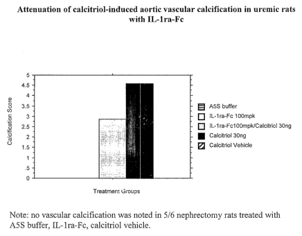
Aortas were removed at treatment week 4 (trt wk4), fixed and stained for
mineralization (Von Kossa) and the severity determined and scored by a
pathologist blinded to the treatments (Calcification Scores: 0=no
calcification;
1=minimal; 2=mild; 3=moderate; 4=marked; 5=severe).
Fc-IL-1ra administered systemically to a rodent animal model of
established chronic kidney disease accompanied with secondary
hyperparatllyroidism (induced by subtotal (5/6) nephrectomy) did not cause
vascular calcification, whereas calcitriol caused marked to severe aortic
calcification. Fc-IL-lra attenuated calcitriol (Vitamin D3)-induced aortic
vascular
calcification, in this model whereby uremia was established for 8 weeks before
treatments were started (Figure 6). In addition, the incidence of severe
calcification in calcitriol-treated uremic subjects (4/8 or 50%) was reduced
in
animals receiving calcitriol in combination with Fc-IL-lra (1/8 or 12.5%).
****~~~~*~
The foregoing specification includes numerous definitions. These
definitions apply to the terms as used throughout this specification, unless
otherwise limited in specific instances. Definitions apply equally to the
plural and
singular forms of each term.
All publications, patents and patent applications cited in this specification
are herein incorporated by reference as if each individual publication or
patent
application were specifically and individually indicated to be incorporated by
reference.
Although the foregoing invention has been described in some detail by way
of illustration and example for puiposes of clarity of understanding, it will
be
readily apparent to those of ordinary skill in the art in light of the
teachings of this
CA 02624648 2008-04-02
WO 2007/047969 PCT/US2006/041113
A-1009-PCT
invention that certain changes and modifications may be made thereto without
departing from the spirit or scope of the appended claims.
DEMANDE OU BREVET VOLUMINEUX
LA PRESENTE PARTIE DE CETTE DEMANDE OU CE BREVET COMPREND
PLUS D'UN TOME.
CECI EST LE TOME 1 DE 2
CONTENANT LES PAGES 1 A 43
NOTE : Pour les tomes additionels, veuillez contacter le Bureau canadien des
brevets
JUMBO APPLICATIONS/PATENTS
THIS SECTION OF THE APPLICATION/PATENT CONTAINS MORE THAN ONE
VOLUME
THIS IS VOLUME 1 OF 2
CONTAINING PAGES 1 TO 43
NOTE: For additional volumes, please contact the Canadian Patent Office
NOM DU FICHIER / FILE NAME:
NOTE POUR LE TOME / VOLUME NOTE: