Note: Descriptions are shown in the official language in which they were submitted.
CA 02625467 2008-04-09
WO 2007/048831 PCT/EP2006/067837
Biodegradable Scaffold with ECM Material
Field of the invention
The present invention relates to a scaffold comprising a biodegradable layer
having ECM
material in the form of flakes, fibres, particles, powder or the like
incorporated in the
biodegradable layer.
Background
Scaffolds are structures used to guide the organization, growth and
differentiation of cells
in the process of forming new functional tissue.
To achieve the goal of tissue reconstruction, scaffolds must meet some
specific
requirements. A high porosity and an adequate pore size are necessary to
facilitate cell
growth and diffusion throughout the whole structure of both cells and
nutrients.
Biodegradability is essential since scaffolds need to be absorbed by the
surrounding
tissues without the necessity of a surgical removal.
Many different materials (natural and synthetic, biodegradable and permanent)
have been
investigated for use as scaffolds. Most of these materials have been known in
the medical
field before the advent of tissue engineering as a research topic, being
already employed
as bioresorbable sutures. Examples of these materials are collagen or some
linear
aliphatic polyesters.
However, when testing laboratory made scaffolds in vivo, it is often seen,
that the cells do
not grow readily into these scaffolds, maybe due to the fact that no
biological signal
molecules, e.g. growth factors, are found in synthetically made scaffolds.
In order to improve the biological properties of the scaffolds and to
accelerate wound
healing, several labs have added growth factors to a synthetic scaffold and
seen
beneficial effects on wound healing. In all of these publications a single
growth factor has
been incorporated in a sheet or hydrogel. The growth factors examined have
been FGF-2
(1; 2), R-FGF-2 tested in a concentration of 25pg/cm2 (2), FGF-1 (3; 4), EGF
(5)(14), or
TGF-R (6; 7) in a concentration of 2pg/cm2 . Acellular extracellular matrices
(ECM) from
warm-blooded vertebras are used extensively in tissue engineering and plastic
surgery
(8). It has been shown that acellular ECM contains several growth factors (9-
11). ECMs
contain a lot of biologic molecules and it has been shown that cells readily
populate these
SUBSTITUTE SHEET (RULE 26)
CA 02625467 2008-04-09
WO 2007/048831 PCT/EP2006/067837
2
sheets of concentrated ECM (12; 13). The ECMs on the market today are of human
or
porcine origin. The cells are removed from the tissue and the tissue is
subsequently
lyophilized and cut into sheets. The sheets of porcine origin come in
different sizes. The
price of these sheets is very high. The sheets are fairly stiff when un-
hydrated. An
example is the sheets from the company Acell. They sell sheets of ECM (Urinary
Bladder
Matrix, UBM) that accelerate the wound healing. Such sheet (7x5 cm) weighs
about
100mg and has a density of about 190mg/cm3.
Use of ECMs or ECM proteins in wound care is known. These products are in the
form of
sheets or hydrogels. Examples of sheet products are OASIS from Healthpoint
(lyophilized
porcine ECM sheet) and Graftjacket from Wright medical (lyophilized human ECM
sheet).
The sheets provide both a scaffold as well as a complex mixture of proteins to
the cells of
the wound. Examples of non-scaffold products containing ECM proteins on the
market, is
Xelma from Molnlycke, which is a hydrogel that contains a protein extract from
ECM of
developing pig teeth.
Summary
The present application discloses that the growth promoting effects of ECM is
maintained
if the ECM is incorporated into a scaffold. We demonstrate that when using
scaffolds
containing ECM material, higher concentrations of ECM surprisingly do not give
better cell
morphology. Concentrations lower than 60% is sufficient to obtain the best
cell
morphology and distribution. In addition it is shown that by varying the
concentration of
discrete ECM material in scaffolds the physical characteristic of the scaffold
changes but
that the changes are depending on the material of the scaffold. The present
application
takes this knowledge to the patient by showing a sterilisation strategy that
maintains the
biological activity of the ECM material after sterilisation.
Detailed Disclosure
The present invention relates to a temporary composite scaffold comprising
discrete ECM
particles.
By adding discontinuous regions of ECM to a scaffold it is possible to combine
the range
of physical properties (e.g. strength, softness, flexibility, durability) the
scaffold can offer
with the reconstructive properties of the ECM. In addition, the price of such
scaffold will be
CA 02625467 2008-04-09
WO 2007/048831 PCT/EP2006/067837
3
lower than other ECM scaffolds both because the powder is a waste-product from
the
production of acellular ECM sheets and because the optimal amount of discrete
ECM
material for each application can be determined and equally distributed in the
dressing
hence avoiding unnecessary high concentrations of ECM. In addition to the
effect of the
ECM, the porous structure of the base material provides the cells with a
structure for in-
growth. In one embodiment a discontinuous region of ECM is obtained by adding
discrete
ECM material, such as particles, flakes, fibres or powder.
A discrete phase of ECM material means material of ECM that is distinguished
in their
form and density from the ground material that they are embedded in. This can
be
demonstrated by histology sections as seen in example 5 or by scanning
electron
microscope (SEM) seen in example 6. By adding discontinuous regions of ECM, we
can
control the concentration of ECM. As shown in the examples (e.g. examples 2
and 3), it is
important that concentration is controlled to optimise cell growth.
It is preferred, that the ECM material is added to the scaffold before
scaffold formation
(e.g. freeze-drying). In this way, the ECM material is homogeneously
distributed in the
scaffold. That is, in the time it takes to solidify the scaffold (e.g. during
freezing) the
density of ECM material might be somewhat higher in one end of the scaffold
than the
other. However, in the present context a homogeneous distribution allows for
such density
gradient through the scaffold provided that the density in the centre of the
scaffold is >0.
Thus, a preferred embodiment relates to a temporary, continuous scaffold
comprising
homogeneously distributed discontinuous regions of ECM wherein the
concentration of
discontinuous regions of ECM is between 20%(w/w) and 60%(w/w).
In the present context, a temporary scaffold means a scaffold that disappears;
is
hydrolysed, is broken down, is biodegraded / bioresorbable / bioabsorbable, is
dissolved
or in other ways vanish from the wound site. This is a huge clinical advantage
as there is
nothing to remove from the wound. Thus, the newly formed tissue is not
disturbed or
stressed by removal of the temporary scaffold. It is typically preferred that
the scaffold is
broken down during 1 day to 10 weeks - depending on the application. For open
wound
applications, it is preferred that the scaffold is broken down during 1-10
days, such as 2-7
days. In one aspect of the invention, the scaffold is biodegradable.
In one embodiment the scaffold is a continuous scaffold. That is a scaffold of
a continues
phase. A continuous scaffold with discontinuous regions results in a composite
material.
CA 02625467 2008-04-09
WO 2007/048831 PCT/EP2006/067837
4
As with other composite materials, this is an engineered material made from
two or more
constituent materials with significantly different physical or chemical
properties and which
remains separate and distinct within the finished structure.
Extracellular matrix (ECM) is the non-cellular portion of animal or human
tissues. The
ECM is hence the complex material that surrounds cells. Consequently, it is
preferred that
the discontinuous regions of ECM are cell free regions. Cell free regions are
obtained by
the use of physical, enzymatic, and/or chemical methods. Layers of cells can
be removed
physically by e.g. scraping the tissue. Detergents and enzymes may be used to
detach
the cells from one another in the tissue. Water or other hypotonic solutions
may also be
used, since hypotonicity will provoke the cells in the tissue to burst and
consequently
facilitate the decellularization process.
Another way to obtain cell free regions is by adding the ECM powder
(discontinuous
regions of ECM) to the scaffold matrix. A cell-free product minimizes the risk
any immune
rejection once implanted, since components of cells may cause an immunogenic
response.
In broad terms there are three major components in ECMs: fibrous elements
(particularly
collagen, elastin, or reticulin), link proteins (e.g. fibronectin, laminin),
and space-filling
molecules (usually glycosaminoglycans). ECMs are known to attract cells and to
promote
cellular proliferation by serving as a reservoir of growth factors and
cytokines (9; 10). A
temporary scaffold containing particulate ECMs used in a wound will be
populated by cells
both from the wound edges as well as cells from the circulating blood. As the
cells invade
the scaffold, the scaffold material will be degraded and eventually the
scaffold will be
replaced with new tissue.
The concentration of the discontinuous regions of ECM is preferably higher
than
15%(w/w), that is higher than 20%(w/w), such as higher than 30%(w/w). The
concentration of the discontinuous regions of ECM is preferably lower than
95%(w/w), that
is lower than 90%(w/w), such as lower than 80%(w/w), or lower than 70%(w/w).
In a
particular preferred embodiment of the invention the concentration is between
20%(w/w)
and 60%(w/w), such as between 20%(w/w) and 40%(w/w).
CA 02625467 2008-04-09
WO 2007/048831 PCT/EP2006/067837
The skin of humans comprises an upper layer of epidermis, formed by inter alia
keratinocytes. Below epidermis is dermis, formed by inter alia fibroblasts,
but also
endothelial cells.
When promoting growth of fibroblasts, the present examples (e.g. example 3)
show that
5 increasing the concentration of ECM from 0%(w/w) to about 60%(w/w) results
in a marked
improvement in the number of cells on the surface of the scaffold and in the
cell
morphology. Thus, one aspect of the invention relates to a wound care device
comprising
40%(w/w) to 60%(w/w) ECM to promote growth of fibroblasts.
When promoting growth of keratinocytes, the present examples using gelatine
scaffolds
show that increasing the concentration of ECM from 0%(w/w) to about 25%(w/w)
results in
a marked improvement in the ability of the cells to grow together (as
keratinocytes should
do), in the cell morphology and in the total number of cells. However,
increasing the
concentration of ECM above 40%(w/w) results in a decrease in the promotion of
cell
growth in terms of number of cells on the surface, their morphology and the
number of
cells. Thus, one aspect of the invention relates to a wound care device
comprising
20%(w/w) to 30%(w/w) ECM to promote growth of keratinocytes.
The wound dressing of the present invention may comprise multiple layers.
These layers
could include 1 or more layers of biodegradable material, which all optionally
comprise
ECM. If ECM is incorporated in more than one layer the dose may vary across
the layers.
In one embodiment, the first layer comprises 40%(w/w) to 60%(w/w) ECM; the
second
layer 20%(w/w) to 30%(w/w) ECM.
In another embodiment, the scaffold is designed for growth stimulation of
different cell-
types. That is, for growth stimulation of fibroblasts the optimal
concentration is 40%(w/w) -
60%(w/w) ECM, the optimal concentration for endothelial cells is 30%(w/w) -
60%(w/w),
whereas for growth stimulation of keratinocytes, the optimal concentration is
20%(w/w) to
30%(w/w). One embodiment of the invention relates to a wound care device
comprising
two scaffolds, a first scaffold for stimulation of fibroblasts with a
concentration of
discontinuous regions of ECM of 40%(w/w) - 60%(w/w), and a second scaffold for
stimulation of keratinocytes with a concentration of discontinuous regions of
ECM of
20%(w/w) to 30%(w/w). A third scaffold can be added to the wound care device
with a
concentration of discontinuous regions of ECM material of 20%(w/w) to
30%(w/w).
CA 02625467 2008-04-09
WO 2007/048831 PCT/EP2006/067837
6
The present data enable use of a scaffold comprising 40%(w/w) - 60%(w/w) of
discontinuous regions of ECM for stimulation of fibroblast growth. The present
data also
enable use of a scaffold comprising 20%(w/w) to 30%(w/w) of discontinuous
regions of
ECM for stimulation of keratinocyte growth.
It is our experience, that when promoting growth of fibroblasts, the growing
fibroblasts will
excrete growth factors inducing growth of keratinocytes. Thus, a preferred
aspect of the
invention relates to a scaffold wherein the concentration of discontinuous
regions of ECM
is between 40%(w/w) and 50%(w/w). Hereby, fibroblast growth is promoted such
that
keratinocyte growth is subsequently promoted and the wound is healed.
The concentration of ECM in the scaffold structure is calculated as
weight/weight percent.
That is: concentration (w/w) = MECM/(MECM+Mscaffold)x100%, where MECM is the
mass in
gram of ECM and Mscaffo,d is the mass in gram of scaffold (not containing
ECM).
In a dissolvable scaffold (e.g. MPEG-PLGA) you dissolve the scaffold in
solvent and filter
the ECMs. After freezedrying, the material is weighted.
In a non-dissolvable scaffold the material is embedded in an appropriate
embedding
material (e.g. paraffin), sectioned in a statically representative number and
stained using a
appropriate stain which only stains the ECMs and not the scaffold materials.
Using image
analysis the amount of ECMs are calculated in relation to scaffold.
Preferred ECM materials contain bioactive ECM components derived from the
tissue
source of the materials. For example, they may contain Fibroblast Growth
Factor-2 (basic
FGF), Transforming Growth Factor-beta (TGF-beta) and vascular endothelial
growth
factor (VEGF). It is also preferred that ECM base materials of the invention
contain
additional bioactive components including, for example, one or more of
collagens,
glycosaminoglycans, glycoproteins and/or proteoglycans. The ECM may include
the
basement membrane, which is made up of mostly type IV collagen, laminins and
proteoglycans. The ECM material of the invention is preferably prepared from
tissue
harvested from animals raised for meat production, including but not limited
to, pigs, cattle
and sheep. Other warm-blooded vertebrates are also useful as a source of
tissue, but the
greater availability of such tissues from animals used for meat production
makes such
tissue preferable. Pigs that are genetically engineered to be free of the
galacatosyl, alpha
CA 02625467 2008-04-09
WO 2007/048831 PCT/EP2006/067837
7
1,3 galactose (GAL epitope) may be used as the source of tissues for
production of the
ECM material. In a preferred embodiment the ECM will be of porcine origin.
The ECM material can be obtained from any animal. It could be derived from,
but not
limited to, intestinal tissue, bladders, liver, spleen, stomach, lymph nodes
or skin. ECM
derived from human cadaver skin, porcine urinary bladder submucosa (UBS),
porcine
urinary bladder matrix (UBM), or porcine small intestinal submucosa (SIS) are
particularly
preferred.
Human tissue is preferably avoided to minimize transfer of diseases. Thus, in
a preferred
embodiment the discontinuous regions of ECM are obtained from animal tissues.
Due to
species similarity, it is preferred to use ECM from warm-blooded mammal.
In a particular preferred embodiment the discontinuous regions of ECM are UBM
(Urinary
Bladder Matrix) particles. The UBM material comprise a unique cocktail of ECM
proteins
of which a few have been quantified: TGF-R 293 8pg/g, b-FGF 3862 170pg/g,
and
VEGF 475 22pg/g (that is pg VEGF/g UBM). With an average density of 3
mg/cm2, the
concentration is about TGF-9: 0,9 pg/cm2 in an ECM sheet, b-FGF: 11,6pg/cm2
and
VEGF 1,4pg/cm2.
One aspect of the invention is to provide a scaffold with constant dosing of
growth factors.
One property of the scaffold used in the present invention is to distribute
the discontinuous
regions of ECM within the porous base material, such that the ECM is
accessible for the
cells. When the cells migrate through the scaffold matrix, the discontinuous
regions of
ECM are exposed to protease activity and degraded which are believed to result
in
release of the biologically active components from the discontinuous regions
of ECM (14).
Thus, the release of biologically active components can be kept somewhat
constant
throughout the period of use, thereby providing a somewhat constant dosing to
the wound
bed and cells. In one embodiment, the discontinuous regions of ECM are equally
distributed within the temporary scaffold.
ECM comes in several micronized forms: e.g. as particles, flakes, fibres or
powder. All of
these are considered discontinuous regions of ECM, i.e. discrete ECM
materials.
A preferred form of discontinuous regions of ECM is ECM particles. Preferably
particles
with a mean diameter of approximately 150pm. This is determined by a
Mastersizer 200
CA 02625467 2008-04-09
WO 2007/048831 PCT/EP2006/067837
8
from Malvern Instrument for volume weighted mean. For example, a surface
weighted
mean of 100pm, can have the smallest particles of 3pm, the largest particles
of 750pm. A
volume weighted mean would, in this case be 250 pm.
By distributing the discontinuous regions of ECM in a porous scaffold, it is
possible to
optimise the physical properties (e.g. strength, softness, flexibility,
durability) of the
scaffold without major impact from the discontinuous regions of ECM.
In order to obtain both the beneficial effect of the ECMs, and the physical
properties a
porous scaffold can offer, particulate ECM can be included in a wound dressing
such as a
scaffold and be used for tissue engineering (e.g. remodelling of soft tissue
engineering,
bone, cartilage, ligaments and tendons) or dental applications. This porous
scaffold
should preferably be of a material that is biodegradable. The temporary
scaffold may be
either in a lyophilised form, in a fibrous form (woven or non-woven), in a
foamed form or
as a film. In all forms the discontinuous regions of ECM are accessible to the
cells on both
the outer and inner surface of porous/fibrous structure.
The material used for the scaffold may be any biodegradable material, from
both synthetic
and of natural sources. Of the scaffolds constructed from natural materials,
particular
preferred are those based on derivatives of the extracellular matrix. Examples
of such
materials are protein materials, such as collagen or fibrin, and
polysaccharidic materials,
like chitosan or glycosaminoglycans (GAGs).
In one embodiment the biodegradable scaffold is made of protein containing
substances.
This will enable degradation by proteolytic enzymes. Such scaffolds are
preferably made
of proteins such as collagen, keratin, fibrin, elastin, laminin, vimentin,
vitronectin,
fibronectin, fibrinogen and derivatives of these and the like or denaturated
proteins such
as gelatin.
By making scaffolds using polymer materials such as gelatin, fibrin,
hyalouronic acid,
collagen, chitin, chitosan, keratin, alginate, PLA and PLGA it is possible to
vary the
scaffolds physical characteristics (strengths, softness, flexibility) through
combinations
and modifications.
In another embodiment the biodegradable scaffold is made of carbohydrate/
polysaccharide containing substances. This will enable degradation by
hydrolysis and
CA 02625467 2008-04-09
WO 2007/048831 PCT/EP2006/067837
9
enzymatic degradation of the polysaccharides. Such scaffolds are preferably
made of
polysaccharides such as heparan sulfate, chondroitin sulfate, dermatan
sulfate, heparin,
keratan sulfate and derivatives of these, alginates, HSC cellulose and
cellulose
derivatives (CMC), some alginates, chitosan, chitin, pectin and pectin
derivatives,
hyaluronic acid and proteoglycans (mucopolysaccharides) and derivatives of
these.
In another aspect the temporary scaffold is synthetic. Such scaffolds are
mainly degraded
by hydrolysis in combination with enzymatic digestion. These scaffolds are
preferably
made from materials selected from the group consisting of PLA (polylactide),
PGA
(polyglycolide), PLGA (poly (lactide-co-glycolide)), MPEG-PLGA, PCL
(polycaprolactone),
poly ortho esters, polydioxanone, polyanhydrides, polyhydroxyalkanoate, and co-
polymers
of the above-mentioned materials.
Examples of well known natural scaffolds/gels are collagen based (3; 6),
fibrin based (4),
chitosan based (1), or gelatine based (2; 7).
A commonly used synthetic material is PLA - polylactic acid. This is a
polyester, which
degrades within the human body to form lactic acid, a naturally occurring
chemical that is
easily removed from the body. Similar materials are polyglycolic acid (PGA)
and
polycaprolactone (PCL): their degradation mechanism is similar to that of PLA,
but they
exhibit respectively a faster and a slower rate of degradation compared to
PLA. Such
MPEG-PLGA polymer can be synthesized as follows: MPEG, DL-lactide, glycolide
and
4%(w/v) stannous octanoate in toluene are added to a vial in a glove box with
nitrogen
atmosphere. The vial is closed, heated and shaken until the contents are clear
and
homogeneous and then placed in an oven at 120-200 CC for 1 min-24h. The
synthesis
can also be made in a solution in a suitable solvent (e.g. dioxane) to
facilitate the
subsequent purification. Then MPEG, DL-lactide, glycolide, 4% Stannous 2-
ethylhexanoate and dioxane are added to a vial in a glove box with nitrogen
atmosphere,
and treated as above.
The polymer can be purified as follows: The polymer is dissolved in a suitable
solvent
(e.g. dioxane, tetrahydrofuran, chloroform, acetone), and precipitated with
stirring in a
non-solvent (e.g. water, methanol, ethanol, 1-propanol or 2-propanol) at a
temperature of
-40 C - 40 C. The polymer is left to settle, solvent discarded and the polymer
is dried in a
vacuum oven at 40 C - 120 C/overnight.
CA 02625467 2008-04-09
WO 2007/048831 PCT/EP2006/067837
As illustrated in the foregoing, the base material of the scaffold can be made
of material of
synthetic and/or natural origin - including combinations thereof. Hence the
scaffold can
comprise combinations of proteins, polysaccharides and synthetic polymers.
5 One function of the scaffold used in the present invention is to provide a
matrix promoting
cell growth. One criterion to promote cell in-growth into the scaffold is a
scaffold that is
solid at room temperature. That is, the scaffold has a fixed physical
structure, a bi-
continuous structure. By this structure, cells are helped to migrate through
the scaffold
and form new tissue.
Another criterion to promote cell growth is a scaffold that has open pores, or
at least a
porosity that allows cell migration.
Porosity is defined as P=1-p (V/M)
where P is the scaffold porosity, p the density of the polymeric system used,
M the weight,
and V the volume of the fabricated scaffolds.
One embodiment of the invention relates to a porous scaffold comprising
discontinuous
regions of ECM as described herein. As illustrated in the examples, a porosity
of more
than 50% enables cell growth. Thus, an a preferred embodiment the scaffold as
described
comprises a porosity of more than 50%, such as >80%, even more than 90%, or as
porous at 95%.
It is preferred that the porous scaffold has open interconnected pores.
In a preferred embodiment of the invention, the temporary scaffold has a
thickness
between 0.1 to 8 mm, preferably 0.3 to 3 mm, and even more preferred 0.5 to 2
mm. In a
particular preferred embodiment of the invention the thickness of the
biodegradable layer
is approximately 1 mm. In a preferred embodiment of the invention the
biodegradable
layer is in direct contact with the wound.
The scaffold according to the present invention is aimed for use as a wound
dressing.
One factor to bear in mind in wound dressings is to make it soft and
conformable. By soft
and conformable, in this context, is meant that it is not unpleasant or
painful, when applied
in an open wound, as the edges will not cut through and stress the sensitive
wound
CA 02625467 2008-04-09
WO 2007/048831 PCT/EP2006/067837
11
surroundings, and the dressing will bend with the curvatures of the wound.
This also
secures direct contact between the wound area and the ECM containing scaffold.
One example of such soft and conformable matrix is a chitosan, prepared in a 1-
2% (w/w)
solution and freeze-dried. The result is an open matrix that is soft, that is
a scaffold with
open interconnected pores. This matrix is also sufficiently open-pored to
allow cell growth
and migration.
In one set of embodiments, the dressing according to the invention is used for
acute
wounds, burn wounds, chronic wounds, and/or surgical wounds.
In another embodiment, the dressing according to the invention is used in
plastic surgery.
In a related embodiment, the scaffold comprising discontinuous regions of ECM
is used
for tissue engineering (e.g. remodeling of soft tissue, bone, cartilage,
ligaments and
tendons) or dental applications.
In many of these uses, it is a requirement that the dressing according to the
invention is
sterilized. One embodiment of the invention relates to a sterilised,
temporary, continuous
scaffold comprising discontinuous regions of ECM. This is typically expressed
as a
temporary, continuous scaffold comprising discontinuous regions of ECM
packaged
bacterial tight, with a marking on the packaged that this product is
sterilized. As illustrated
in Example 4, sterilisation by e.g. radiation maintains the biological effect
of ECM -
dependent on scaffold type. Bacterial tight materials are well known to the
skilled person.
CA 02625467 2008-04-09
WO 2007/048831 PCT/EP2006/067837
12
References
1. Obara, K., Ishihara, M., Fujita, M., Kanatani, Y., Hattori, H., Matsui, T.,
Takase, B.,
Ozeki, Y., Nakamura, S., Ishizuka, T., Tominaga, S., Hiroi, S., Kawai, T., &
Maehara,
T. 2005, "Acceleration of wound healing in healing-impaired db/db mice with a
photocrosslinkable chitosan hydrogel containing fibroblast growth factor-2",
Wound.Repair Regen., vol. 13, no. 4, pp. 390-397.
2. Miyoshi, M., Kawazoe, T., Igawa, H. H., Tabata, Y., Ikada, Y., & Suzuki, S.
2005,
"Effects of bFGF incorporated into a gelatin sheet on wound healing",
J.Biomater.Sci.Polym.Ed, vol. 16, no. 7, pp. 893-907.
3. Pandit, A., Ashar, R., Feldman, D., & Thompson, A. 1998, "Investigation of
acidic
fibroblast growth factor delivered through a collagen scaffold for the
treatment of full-
thickness skin defects in a rabbit model", Plast.Reconstr.Surg., vol. 101, no.
3, pp.
766-775.
4. Pandit, A. S., Wilson, D. J., & Feldman, D. S. 2000, "Fibrin scaffold as an
effective
vehicle for the delivery of acidic fibroblast growth factor (FGF-1)",
J.Biomater.Appl.,
vol. 14, no. 3, pp. 229-242.
5. Ulubayram, K., Nur, C. A., Korkusuz, P., Ertan, C., & Hasirci, N. 2001,
"EGF
containing gelatin-based wound dressings", Biomaterials, vol. 22, no. 11, pp.
1345-
1356.
6. Pandit, A., Ashar, R., & Feldman, D. 1999, "The effect of TGF-beta
delivered
through a collagen scaffold on wound healing", J.Invest Surg., vol. 12, no. 2,
pp. 89-
100.
7. Yamamoto, M., Tabata, Y., Hong, L., Miyamoto, S., Hashimoto, N., & Ikada,
Y.
2000, "Bone regeneration by transforming growth factor betal released from a
biodegradable hydrogel", J.Control Release, vol. 64, no. 1-3, pp. 133-142.
CA 02625467 2008-04-09
WO 2007/048831 PCT/EP2006/067837
13
8. Badylak, S. F. 2002, "The extracellular matrix as a scaffold for tissue
reconstruction",
Semin.Cell Dev.8io1.2002.Oct.;13(5):377.-83., vol. 13, pp. 377-383.
9. Hodde, J. P., Record, R. D., Liang, H. A., & Badylak, S. F. 2001, "Vascular
endothelial growth factor in porcine-derived extracellular matrix",
Endothelium
2001;8.(1):11-24., vol. 8, pp. 11-24.
10. Voytik-Harbin, S. L., Brightman, A. 0., Kraine, M. R., Waisner, B., &
Badylak, S. F.
1997, "Identification of extractable growth factors from small intestinal
submucosa",
J. Ce118iochem. , vol. 67, pp. 478-491.
11. McDevitt, C. A., Wildey, G. M., & Cutrone, R. M. 2003, "Transforming
growth factor-
betal in a sterilized tissue derived from the pig small intestine submucosa",
J.Biomed.Mater.Res.2003.Nov.1; 67A. (2):637.-40., vol. 67A, pp. 637-640.
12. Badylak, S. F., Record, R., Lindberg, K., Hodde, J., & Park, K. 1998,
"Small
intestinal submucosa: a substrate for in vitro cell growth",
J.Biomater.Sci.Polym.Ed,
vol. 9, pp. 863-878.
13. Lindberg, K. & Badylak, S. F. 2001, "Porcine small intestinal submucosa
(SIS): a
bioscaffold supporting in vitro primary human epidermal cell differentiation
and
synthesis of basement membrane proteins", Burns 2001 May.;27.(3):254.-66.,
vol.
27, pp. 254-266.
14. Li, F., Li, W., Johnson, S., Ingram, D., Yoder, M., & Badylak, S. 2004,
"Low-
molecular-weight peptides derived from extracellular matrix as
chemoattractants for
primary endothelial cells", Endothelium 2004.May.-Aug.;11(3-4):199.-206., vol.
11,
pp. 199-206.
CA 02625467 2008-04-09
WO 2007/048831 PCT/EP2006/067837
14
Figures
Figure 1: LDH measurements of scaffolds of gelatine seeded with human primary
fibroblasts in three different concentrations (Cell/cm2). The bars represent
the growth at
day 3, measured as Abs.
Figure 2: LDH measurements of scaffolds of MPEG-PLGA seeded with human primary
fibroblasts in three different concentrations (Cell/cm2). The bars represent
the growth at
day 3, measured as Abs.
Figure 3: LDH measurements of scaffolds of gelatine seeded with human primary
keratinocytes in three different concentrations (Cell/cm2). The bars represent
the growth at
day 3, measured as Abs.
Figure 4: LDH measurements of scaffolds of MPEG-PLGA seeded with human primary
keratinocytes in three different concentrations (Cell/cm2). The bars represent
the growth at
day 3, measured as Abs.
Figure 5: LDH measurements of scaffolds of gelatine seeded with human
umbilical vein
endothelial cells in three different concentrations (Cell/cm2). The bars
represent the
growth at day 3, measured as Abs
Figure 6: LDH measurements of scaffolds of MPEG-PLGA seeded with human
umbilical
vein endothelial cells in three different concentrations (Cell/cm2). The bars
represent the
growth at day 3, measured as Abs.
Figure 7: Digital images of the distribution of ECM particles in the MPEG-PLGA
scaffold.
Figure 8: SEM picture of MPEG-PLGA scaffold (Magnification 250x).
Figure 9: SEM picture of MPEG-PLGA containing 40% ECM particles (Magnification
250x).
Figure 10: Digital image of endothelial growth in MPEG-PLGA scaffold.
CA 02625467 2008-04-09
WO 2007/048831 PCT/EP2006/067837
Figure 11: Digital image of endothelial growth in MPEG-PLGA containing 23% ECM
particles.
Figure 12: Digital image of endothelial growth in MPEG-PLGA containing 23% ECM
particles showing a mmagnification of capillary-like morphology in the deeper
layers of the
5 scaffold.
CA 02625467 2008-04-09
WO 2007/048831 PCT/EP2006/067837
16
Examples
Example 1: In-growth of primary human fibroblasts in synthetic scaffolds
with and without ECM particles
Scaffolds made of biodegradable polyesters containing UBM (Acell) particles
(mean
diameter of approximately 150 m) at 40%(w/w) were compared with scaffolds
without the
ECM particles in a test of cell morphology and 3D growth.
Metoxy-polyethylene glycol - Poly(lactide-co-glycolide) (Mn 2.000-30.000, L:G
1:1) was
dissolved in 1,4-dioxane to a 1.5% solution. For the UBM containing scaffold,
0.03 g UBM
was added to 3 ml polymer solution (40% w/w drymatter), highspeed-mixed and
poured in
3x3 cm mould. The solution was frozen at -5 C and lyophilized at -20 C for 5h
and 20 C
for approx 60h. The samples were subsequently placed in draw (hydraulic pump)
in a
desiccator for 5h.
The test of growth and morphology of seeded primary fibroblasts on the surface
of the two
scaffolds were evaluated.
Results from day 1, 3 and 7 were graded from 1-5, with 1 corresponding to
worst case
and 5 being the best. In the scaffold mixed with ECM particles the
distribution and growth
of cells was given a grade 5 at all days and were better than the control
scaffold (graded
21/2 at all days).
Conclusion: The biological activity of the powdered ECM matrix retains
activity after
incorporation in a synthetic scaffold, and causes a considerably better growth
on the
scaffold when compared to scaffold alone.
Example 2: Cell morphology and 3D growth in gelatine-ECM composites
containing 5 different concentrations of ECM.
A study of cell morphology and 3D growth of primary fibroblasts seeded on the
surface of
gelatine-ECM scaffolds. The gelatine scaffolds were cross-linked by heating
and
contained increasing concentrations of UBM (0, 12%(w/w), 26%(w/w), 41 %(w/w),
51 %
(w/w) and 58%(w/w)). The concentrations were calculated as amount UBM in
relation to
CA 02625467 2008-04-09
WO 2007/048831 PCT/EP2006/067837
17
total amount of solids meaning a 58%(w/w) scaffold contained 0.05g polymer and
0.07g
UBM corresponding to 13.8 mg UBM/cm3.
Gelatin from porcine skin, type A, bloom 175 (Sigma) was dissolved in milli-Q
water and t-
BuOH (95:5) to a 1% solution. For UBM containing samples, the UBM was added to
the
solution while stirring (0, 12, 26, 41, 51, 58% w/w: 0, 0.007, 0.018, 0.035,
0.053, 0.07
g/scaffold). 5 ml of the UBM containing gelatin solution was poured into the
mould (D=5
cm). The mould with the solution was placed in +5 C for 1 h, then frozen at -
20 C and
lyophilized at -20 C for 5h and at 20 C for 36h. The samples were subsequently
cross-
linked in vacuum oven at 120 C for 15h.
This study showed that by increasing the concentration of UBM in the composite
scaffold
the fibroblasts were better distributed and had a higher proliferation rate
compared to
without UBM. In the first couple of days of the study, it was seen that on
scaffolds without
UBM, cells were only growing in the area where they were applied, while cells
were better
and more evenly distributed on the surface of scaffolds where the
concentration of the
UBM were above 26%(w/w). From day 14 it was clear that a lower number of cells
were
found at the gelatine scaffold without UBM in comparison with composite
scaffolds
containing UBM. With respect to the cell morphology the fibroblasts had a
rounded but
adherent morphology at the plain gelatine scaffolds but with increasing amount
of UBM an
increasing fibroblastic morphology were observed. From 41 %(w/w) UBM the best
morphology and distribution of the fibroblasts were observed. Increasing the
concentration
of UBM above 41 %(w/w) did not result in better morphology or distribution of
the cells in
the composite scaffolds.
Example 3: Preparation and cell morphology and 3D growth in composite
scaffolds of MPEG-PLGA or gelatine holding 6 different concentrations of
ECM particles.
Preparation of composite scaffolds of gelatine and ECM: Gelatin from porcine
skin, type
A, Gelita pharmagrade 832 was dissolved in milli-Q water and t-BuOH (95:5) to
a 1%
solution. For UBM containing samples, the UBM was added to the solution while
stirring;
0, 0.006, 0.013, 0.021, 0.033, 0.05, 0.075 g/scaffold (0, 10, 20, 30, 40, 50,
60% w/w). 5 ml
of the UBM containing gelatin solution was poured into the mould (D=5 cm). The
mould
with the solution was placed in +5 C for 1 h, then frozen at -20 C and
lyophilized at -20 C
CA 02625467 2008-04-09
WO 2007/048831 PCT/EP2006/067837
18
for 5h and at 20 C for 18h. The samples were subsequently cross-linked in
vacuum oven
at 130 C for 15h.
Preparation of composite scaffolds of MPEG-PLGA and ECM: Metoxy-polyethylene
glycol
- Poly(lactide-co-glycolide) (Mn 2.000-30.000, L:G 1:1) was dissolved in 1,4-
dioxane to a
1.5% solution. For UBM containing samples, the UBM was added to the solution
while
stirring; 0, 0.017, 0.038, 0.064, 0.1, 0.15, 0.225 g/scaffold (0, 10, 20, 30,
40, 50, 60%
w/w), highspeed-mixed and 10 ml poured in 7.3x7.3 cm mould. The solution was
frozen at
-5 C and lyophilized at -20 C for 5h and 20 C for approx 15h. The samples were
subsequently placed in draw (hydraulic pump) in a desiccator for 24h.
In order to evaluate the cell morphology and 3D growth of the composite
scaffolds,
biopsies were punched out of each type of the scaffolds and seeded with
primary human
fibroblasts (passage 3), human umbilical vein endothelial cells (HUVEC,
passage 4), or
primary keratinocytes (passage 5) on the surface of the scaffolds with a
density of 2.5 x
104 cells/cm2 in a small volume of growth medium (primary fibroblasts in 10%
FCS in
DMEM; HUVEC's in EGM-2; primary keratinocytes in KGM-2) containing antibiotics
(penicillin, streptomycin and Amphotericin B). The scaffolds were incubated at
37 C at 5%
CO2 before additional growth medium was added. Evaluation of the cells
attachment,
morphology, growth and population of the scaffold were preformed on day 1, 3
and 7 by
staining the cells with neutral red followed by evaluation using an Leica
DMIRE2 inverted
microscope fitted with a Evolution MP cooled colour camera (Media
Cybernetics). Digital
images were taken using Image Pro Plus 5.1 software (Media Cybernetics). The
number
of cells was calculated by using Cytotoxicity Detection Kit (LDH, Roche
Diagnostics
GmbH). Cells were seeded in three different concentrations (1.25x10⁴, 2.5x10⁴
and 5x10⁴
cells/cm2) on top of the different scaffold types in the same way as above.
The scaffolds
were evaluated on day 1, 3 and 7 by washing the scaffolds with PBS before cell
lysis
using 0,5% CHAPS for 20h at 4 C on a flat shaker. Supernatant were transferred
to a
micro-plate and the amount of LDH measured according to manufactures
instructions.
Fibroblasts
The quantitative measurement of fibroblasts showed increasing number of cells
with
increasing time but no effect was seen between the different concentrations of
UBM in the
gelatine scaffolds (figure 1).
CA 02625467 2008-04-09
WO 2007/048831 PCT/EP2006/067837
19
In the MPEG-PLGA scaffolds cells were both increasing with time and amount of
UBM in
the scaffold (figure 2). The cell morphology and 3D growth in the gelatine
scaffold without
UBM showed in the first days of the study adherent cells growing with a
rounded
morphology and staying where they were applied. These cells became a little
more
spindle-shaped during the rest of the study. Adding 10%(w/w) UBM to the
scaffold did not
change this pattern but at 20%(w/w) UBM an increasing number of cells were
becoming
spindle-shaped cells with normal fibroblastic morphology and the cells
beginning to
spread more on the surface. At 30-40%(w/w) a maximum in the ratio of spindle-
shaped
cells were observed and a change from cells growing on the surface of the
scaffolds to a
growth where cells were growing in depth of the scaffolds with a more 3D
morphology of
the cells were seen. In the MPEG-PLGA the same pattern were observed with a
few
exceptions: Increasing the concentration of UBM gives an increasing number of
cells on
and in the scaffolds. At 20 - 30%(w/w) UBM an increasing spreading of cells
are seen and
more cells with spindle shaped morphology. From 40%(w/w) and above the cells
are
beginning to grow with 3D morphology and were instead of growing on the
surface now
growing into the depth of the scaffold.
Keratinocytes
Seeding primary keratinocytes on top of gelatine scaffold with 10%(w/w) UBM
showed an
increase in the number of cells compared to the scaffold without UBM. A
maximum in the
number of cells were seen at 10-20%(w/w) UBM in the first days of the study
but later in
the study 20-30%(w/w) UBM showed maximum effect. Increasing the concentration
of
UBM resulted in decreasing number of cells on the scaffold (figure 3).
On the MPEG-PLGA scaffold the largest number of cells were seen on the
scaffold
without UBM. Addition of UBM resulted in a decrease in the number of cells
found in the
scaffold with increasing number of UBM. This effect became more marked at the
end of
the study. Generally a relative large variation was seen between duplicates
due to overlap
in concentrations (figure 4). The cell morphology and 3D growth showed fine
single
growing keratinocytes adhering to the surface of the gelatine scaffold. In the
10%(w/w)
UBM scaffold an increased number of cells were growing in close connection
with each
other, like in small sheets. This effect seemed to be more pronounced with
time.
Increasing the concentration to 20-30%(w/w) UBM were still giving rise to the
coherent
growing but not as close as in 10%(w/w) UBM. Above 20-30%(w/w) UBM a more
single
cell growth with increased spreading of the cells were seen together with a
decreasing
CA 02625467 2008-04-09
WO 2007/048831 PCT/EP2006/067837
number of cells. In the MPEG-PLGA scaffolds without UBM cells were collected
at the
centre of the scaffold growing close together almost like in a sheet.
Increasing the
concentration of UBM resulted in an increasing spreading of the cells together
with a
decreasing number of cells. In the two highest concentrations dead cells were
found
5 trapped into the scaffold structure.
Endothelial cells
Gelatine scaffolds seeded with Huvec's showed a tendency to increase in number
to an
optimum around 20-30%(w/w) UBM where after a decrease was seen (figure 5).
MPEG-
PLGA with Huvec's showed that increasing concentrations of UBM resulted in
increasing
10 number of cells. Generally large variations were seen with overlaps in
adjacent
concentrations at both types of scaffolds using Huvec's (figure 6). The cell
morphology
and 3D growth showed that increasing the UBM to 30%(w/w) UBM in the gelatine
scaffold
gives an increasing ability of the Huvec's to adhere to the surface growing
with a flattening
morphology with normal short extensions. From 40%(w/w) and up the cells were
growing
15 with a more rounded morphology and a decreasing number of cells. In the
MPEG-PLGA
scaffold without UBM the Huvec's were low in number and growing with rounded
morphology resulting in no cells at day 7. Adding 10-20%(w/w) UBM to the
scaffold gives
a spreading effect of the cells but no effect on the morphology. Increasing
the
concentration above 30-40%(w/w) gives the optimum cell morphology and
increasing the
20 concentration further gives even more 3D growth of the cells. Generally
variations were
also seen in the cell morphology and 3D growth of both the gelatine and MPEG-
PLGA
scaffolds containing UBM. Summery of the effect of increasing UBM
concentrations on
primary fibroblasts, primary keratinocytes and human endothelial cells:
CA 02625467 2008-04-09
WO 2007/048831 PCT/EP2006/067837
21
Fibroblasts Keratinocytes Endothelial
Gelatine MPEG- Gelatine MPEG- Gelatine MPEG-
PLGA PLGA PLGA
cell 0% = 10% 20-60% 10% max 0% max Increasing 0%
morphology 20% spreading sheet-like sheet-like 0-30% decreasing
and 3D beginning and spindle growth of growth of Max 30% cells
growth shaped cells cells
spindle- cells 40-60% 10-20 /
shaped 20-30% 10-60% decreasing spreading
cells 40-60% sheet-like decreasing effect
Max 30- gives 3D- 40-60% number of 30-40%
40% growth single cells cells and max
sheet-like morphology
formation
50-60%
gives more
3D-growth
Number of No effect Increasing Max 20- Decreasing Max 20- Increasing
cells 30% 30%
40-60% 40-60%
decreasing decreasing
Example 4: Effect of sterilisation of ECM +/- incorporation in scaffolds on
the
cell morphology and 3D growth of primary fibroblasts.
Metoxy-polyethylene glycol - Poly(lactide-co-glycolide) (Mn 2.000-30.000, L:G
1:1) was
dissolved in 1,4-dioxane to a 1.5% solution. For UBM containing samples,
0.045g non-
sterilized UBM was added to 10 ml polymer solution (23% w/w drymatter),
highspeed-
mixed and poured in 7x7 cm mould. The solution was frozen at -5 C and
lyophilized at -
20 C for 5h and 20 C for approx 18h. The samples were subsequently placed in
draw
(hydraulic pump) in a desiccator for 15h.
The samples with and without UBM were beta radiated by 0, 1x25kGy and 2x25kGy.
Another sample was prepared in the same way, but a pre-sterilized UBM (2x25kGy
beta
radiation) was used (0.045g/5mi solution) and the sample was not sterilized
after
preparation.
Gelatin from porcine skin, type A, bloom 175 (Sigma) was dissolved in milli-Q
water and t-
BuOH (95:5) to a 1% solution. 0.015g non-sterilized UBM was added to 5 ml
solution
(23% w/w drymatter) while stirring and poured into the mould (D=5 cm). The
mould with
the solution was placed in +5 C for 2h, then frozen at -20 C and lyophilized
at -20 C for
CA 02625467 2008-04-09
WO 2007/048831 PCT/EP2006/067837
22
5h and at 20 C for 20h. The samples were subsequently cross-linked in vacuum
oven at
120 C for 15h. The samples with and without UBM were beta radiated by 0 and
1x25kGy
and 2x25kGy. Another sample was prepared in the same way without UBM. The
samples
were sterilized after preparation at 0, 1x25 kGy and 2x25kGy.
Gelatin from porcine skin, type A, bloom 175 (Sigma) was dissolved in milli-Q
water and t-
BuOH (95:5) to a 1% solution. 0.015g pre-sterilized UBM (1x25 kGy) was added
to 5 ml
solution (23% w/w drymatter) while stirring and poured into the mould (D=5
cm). The
mould with the solution was placed in +5 C for 1 h, then frozen at -20 C and
lyophilized at
-20 C for 5h and at 20 C for 50h. The samples were subsequently cross-linked
in vacuum
oven at 130 C for 15h.
The cell morphology and 3D growth study showed that an increasing radiation of
UBM
sheets reduced the number of cells on the UBM sheets but with no effect on the
morphology of the cells. In the gelatine scaffold and gelatine with 30%(w/w)
UBM a
decreasing number of cells and a change in morphology from typical
fibroblastic cells to a
more rounded one was seen with the largest effect seen in the gelatine
scaffold.
Sterilisation of UBM particles before incorporation in gelatine scaffolds
gives a better cell
morphology and 3D growth compared to incorporation of UBM particles before
sterilisation
of the scaffold. In the MPEG-PLGA an increasing radiation resulted in an
increased
number of cells with fibroblastic morphology due to increased moistening of
the scaffold.
Radiation of scaffolds of MPEG-PLGA containing 30%(w/w) UBM resulted in an
even
higher number of cells and a more 3D morphology of the fibroblasts also
compared with
scaffold where the UBM particles were radiated before incorporation into the
scaffold.
This study showed that the highest biological activity was achieved in the non-
radiated
gelatine scaffold and that radiation decreased the activity. On the contrary
the highest bio-
logical activity was found when the UBM particles were incorporated in the
MPEG-PLGA
scaffold, and subsequently sterilized. It is believed that radiation decreases
the biological
activity of UBM. Radiation can affect the scaffold material in a negative or
positive way
depending on the material in relation to biological activity. There are
indications showing
that the scaffold material (e.g. MPEG-PLGA) can have a protective effect of
the UBM dur-
ing sterilization.
CA 02625467 2008-04-09
WO 2007/048831 PCT/EP2006/067837
23
Example 5: Discrete particles of ECM in MPEG-PLGA.
Scaffolds of MPEG-PLGA containing 41%(w/w) of UBM particles were seeded with
primary fibroblasts on the surface of the scaffolds with a density of 2.5 x
104 cells/cm2 in a
small volume of growth medium (10% FCS in DMEM containing antibiotics
(penicillin,
streptomycin and Amphotericin B). The scaffolds were incubated at 37 C at 5%
C02
before additional growth medium was added. After 7 days the scaffolds were
placed in
Lillys fixative for 3 days before embedding in paraffin, sectioning into 8 pm
slices and
staining by Meyer's haematoxylin erosion (HE). Digital images (4x and 20x
magnifications) were collected using a BX-60 Olympus microscope fitted with an
Evolution
MP cooled colour camera (Media Cybernetics) and digital image were taken using
Image
Pro Plus 5.1 software.
Digital images of the distribution of ECM particles in the MPEG-PLGA scaffold
showed
discrete UBM particles stained red by HE and distinguish from the scaffold
material.
Fibroblasts growing in the scaffold were stained blue (figure 7).
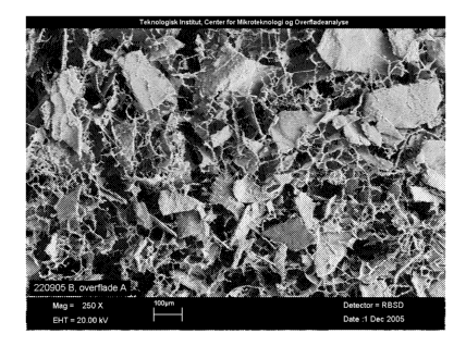
Example 6: Discrete particles of UBM in MPEG-PLGA shown by SEM.
Scaffolds were prepared as described in Example 1.
The SEM pictures are showing MPEG-PLGA scaffolds with (figure 9) and without
(figure
8) UBM particles. The pictures are taken at the top surface of the scaffold at
a magnitude
of 250. The SEM pictures were taken at the Danish technological institute
(2005-160).
Example 7: Three dimensional endothelial growth and differentiation in
scaffolds holding ECM particles.
Metoxy-polyethylene glycol - Poly(lactide-co-glycolide) (Mn 2.000-30.000, L:G
1:1) was
dissolved in 1,4-dioxane to a 1.5% solution. For UBM containing samples,
0.045g non-
sterilized UBM was added to 10 ml polymer solution (23% w/w drymatter),
highspeed-
mixed and poured in 7x7 cm mould. The solution was frozen at -5 C and
lyophilized at -
20 C for 5h and 20 C for approx 16h. The samples were subsequently placed in
draw
(hydraulic pump) in a desiccator for 15h.
Primary human endothelial cells from umbilical cord were co-cultured with
primary human
dermal fibroblasts on the surface of MPEG-PLGA scaffolds and scaffold
containing
CA 02625467 2008-04-09
WO 2007/048831 PCT/EP2006/067837
24
23%(w/w) UBM. The constructs were cultured submerged in defined endothelial
growth
medium for 6-10 days after which they are airlifted and cultured for another 9
days. On the
final day of culture constructs were fixed with 4% formalin buffer, bisected
and paraffin
embedded.
By immunohistochemical peroxidase staining of CD31/PECAM (platelet endothelial
cell
adhesion molecule) endothelial cells were visualized on 5pm sections.
Identifying fibro-
blasts, parallel sections were stained with PECAM peroxidase combined with a
haema-
toxylin counterstain. As endothelial growth and differentiation is influenced
by fibroblast
performance, all scaffold materials were tested with 2 different fibroblast
populations but
were not giving rise to different results.
All MPEG-PLGA scaffolds support fibroblast and endothelial growth. Fibroblasts
were
found throughout the entire volume of all MPEG-PLGA scaffolds. UBM particles
were
homogenously distributed and scaffolds remain intact during culture. Culturing
endothelial
cells and fibroblasts on MPEG-PLGA scaffolds however brings endothelial
surface growth
only - endothelial cells proliferate within a matrix produced by the
neighboring fibroblasts
on top of the scaffold. Adding UBM particles promote fibroblast and
endothelial growth in
the deeper layers of the scaffolds and endothelial cells adopt capillary-like
morphology.
Endothelial cells are guided along the surface of UBM particles rather than
migrating into
them. Therefore we find that including UBM particles in scaffolds lead to a
very distinct
improvement in endothelial growth and differentiation. The different
fibroblast populations
were not giving rise to different results.
MPEG-PLGA scaffolds (figure 10) and 23%(w/w) UBM in MPEG-PLGA (figure 11) show
growth of endothelial cells in the surface of the MPEG-PLGA scaffold where the
growth is
into the depth holding UBM particles (endothelium is stained red (shown black)
-
fibroblasts are not visible).
Capillary-like morphology of endothelial cells were seen in the deeper layer
of MPEG-
PLGA scaffold holding 23%(w/w) UBM (figure 12). These structures were not seen
in the
MPEG-PLGA scaffold.
CA 02625467 2008-04-09
WO 2007/048831 PCT/EP2006/067837
Example 8: Physical and mechanical properties of scaffolds containing
different concentrations of ECM particles.
Samples prepared:
Freeze-dried scaffold with gelatin matrix
5 Freeze-dried scaffold with gelatin matrix and 40 w/w UBM particles
Freeze-dried scaffold with gelatin matrix and 80 w/w UBM particles.
Gelatin from porcine skin, type A (PG-832-6 Gelita) was dissolved in milli-Q
water and t-
BuOH (95:5) to a 1% solution. For UBM containing samples, UBM was added to the
solution while stirring (40%w/w: 0,033g/5m1, 80%w/w: 0,2g/5ml). 5 ml of the
UBM
10 containing gelatin solution was poured into the mould (D=5 cm). The mould
with the
solution was placed in +5 C for 2.5h, then frozen at -20 C and lyophilized at -
20 C for 5h
and at 20 C for 100h. The samples were subsequently cross-linked in vacuum
oven at
130 C for 15h.
Samples prepared:
15 Freeze-dried scaffold with PLGA matrix
Freeze-dried scaffold with PLGA matrix and 40%(w/w) UBM particles
Freeze-dried scaffold with PLGA matrix and 80%(w/w) UBM particles.
Metoxy-polyethylene glycol - Poly(lactide-co-glycolide) (Mn 2.000-30.000, L:G
1:1) was
dissolved in 1,4-dioxane to a 1.5% solution. For UBM containing samples, UBM
was
20 added to the polymer solution (40%(w/w): 0,1g/10ml, 80%(w/w): 0,6g/lOml),
highspeed-
mixed and poured in 7x7 cm mould. The solution was frozen on a 1,4-dioxane
layer at -
5 C and lyophilized at -20 C for 5h and 20 C for approx 100h. The samples were
subsequently placed in draw (hydraulic pump) in a desiccator for 15h.
Physical properties and mechanical testing
25 Depending on the matrix material and the amount of UBM added, different
physical and
mechanical properties can be achieved.
= The porosity decreases with the amount of UBM added, thus the density
increases.
= If the matrix is hydrophobic, the UBM will provide increased wet ability.
CA 02625467 2008-04-09
WO 2007/048831 PCT/EP2006/067837
26
= Gelatin scaffolds retain its tensile strength up to at least 40%(w/w) UBM,
after
which it decreases, whereas PLGA scaffolds are slightly strengthened by the
UBM
particles. The low material concentration in combination with the freeze-
drying
process gives low tensile strength, which is also the case for the samples in
this
example.
Gelatin Height (mm) Density (mg/cm3) Porosity (%) Wet ability (min)
- - - - - - - - - - - - - - - - - - - - - - - - - - - - - - - - - - - - - - - -
- - - - - - - - - - - - - - - - - - - - - - - - - - - - - - - - - - - - - - -
- - - - - - - - - - - - - - - - - - - - - - - - - - - - - - - - - - - - - - - -
- - - - - - - - - - - - - - - - - - - - - - - - - - - - - - - - - - - - - - -
- - - - - - - - - - - - - - - - - - - - - - - - - - - - - - - - - - - - - - - -
- - - - - - - - - - - - - - - - - - - -
0% UBM 1.6 ( 0.1) 15 ( 1) 99 ( 0) instant
40% UBM 1.6 ( 0.0) 24 ( 1) 97 ( 0) instant
80% UBM 1.6 ( 0.0) 73 ( 0) 72 ( 0) instant
. . . . . . . . . . . . . . . . . . . . . . . . . . . . . . . . . . . . . . .
. . . . . . . . . . . . . . . . . . . . . . . . . . . . . . . . . . . . . . .
. . . . . . . . . . . . . . . . . . . . . . . . . . . . . . . . . . . . . . .
. . . . . . . . . . . . . . . . . . . . . . . . . . . . . . . . . . . . . . .
. . . . . . . . . . . . . . . . . . . . . . . . . . . . . . . . . . . . . . .
. . . . . . . . . . . . . . . . . . . . . . . ,
. . . . . . . . . . . . . . . . . . . . . . . . . . . . . . . . . . . . . . .
. . . . . . . . . . . . . . . . . . . . . . . . . . . . . . . . . . . . . . .
. . . . . . . . . . . . . . . . . . . . . . . . . . . . . . . . . . . . . . .
. . . . . . . . . . . . . . . . . . . . . . . . . . . . . . . . . . . . . . .
. . . . . . . . . . . . . . . . . . . . . . . . . . . . . . . . . . . . . . .
. . . . . . . . . . . . . . . . . . . . . . . ,
PLGA Height (mm) Density (mg/cm3) Porosity (%) Wet ability (min)
- - - - - - - - - - - - - - - - - - - - - - - - - - - - - - - - - - - - - - - -
- - - - - - - - - - - - - - - - - - - - - - - - - - - - - - - - - - - - - - -
- - - - - - - - - - - - - - - - - - - - - - - - - - - - - - - - - - - - - - - -
- - - - - - - - - - - - - - - - - - - - - - - - - - - - - - - - - - - - - - -
- - - - - - - - - - - - - - - - - - - - - - - - - - - - - - - - - - - - - - - -
- - - - - - - - - - - - - - - - - - - -
0% UBM 1.2 ( 0.0) 26 ( 3) 98 ( 0) >45
40% UBM 1.3 ( 0.0) 36 ( 2) 96 ( 0) 5<x<15
80% UBM 1.6 ( 0.0) 94 ( 8) 65 ( 3) <2
- - - - - - - - - - - - - - - - - - - - - - - - - - - - - - - - - - - - - - - -
- - - - - - - - - - - - - - - - - - - - - - - - - - - - - - - - - - - - - - -
- - - - - - - - - - - - - - - - - - - - - - - - - - - - - - - - - - - - - - - -
- - - - - - - - - - - - - - - - - - - - - - - - - - - - - - - - - - - - - - -
- - - - - - - - - - - - - - - - - - - - - - - - - - - - - - - - - - - - - - - -
- - - - - - - - - - - - - - - - - - - -
Height is measured with a slide gauge.
Density is calculated as:
Density =Mass/ (Area x Height)
Porosity is calculated as:
Porosity = (polymer density - sample density)/polymer density.
The polymer density is weight adjusted according to added UBM (3 mg/cm3).
Wet ability is calculated as the amount of time for a droplet of water to be
fully absorbed
by the sample, photo monitored.
Gelatin Tensile
Force max (N)
0% UBM 0.20 ( 0.07)
40% UBM 0.20 ( 0.02)
80% UBM 0.02 ( 0.00)
CA 02625467 2008-04-09
WO 2007/048831 PCT/EP2006/067837
27
PLGA Tensile
Force max (N)
-------------------------------------------------------------------------------
-
0% UBM 0.01 ( 0.00)
40% UBM 0.02 ( 0.00)
80% UBM 0.04 ( 0.01)
- - - - - - - - - - - - - - - - - - - - - - - - - - - - - - - - - - - - - - - -
- - - - - - - - - - - - - - - - - - - - - - - - - - - - - - - - - - - - - - -
-
Tensile testing was carried out on a Texture Analyzer from Stable Micro
Systems.