Note: Descriptions are shown in the official language in which they were submitted.
CA 02625744 2008-04-10
WO 2007/044860 PCT/US2006/039963
DIABETES-ASSOCIATED MARKERS AND METHODS OF USE
THEREOF
INCORPORATION BY REFERENCE
This application claims priority from U.S. Provisional Application Serial No.
60/725,462, filed on October 11, 2005.
Each of the applications and patents cited in this text, as well as each
document or
reference cited in each of the applications and patents (including during the
prosecution of
each issued patent; "application cited documents"), and each of the U.S. and
foreign
applications or patents corresponding to and/or claiming priority from any of
these
applications and patents, and each of the documents cited or referenced in
each of the
application cited documents, are hereby expressly incorporated herein by
reference. More
generally, documents or references are cited in this text, either in a
Reference List before the
claims, or in the text itself; and, each of these documents or references
("herein-cited
references"), as well as each document or reference cited in each of the
herein-cited
references (including any manufacturer's specifications, instructions, etc.),
is hereby
expressly incorporated herein by reference. Documents incorporated by
reference into this
text may be employed in the practice of the invention.
FIELD OF THE INVENTION
The present invention relates generally to the identification of biological
markers
associated with an increased risk of developing Diabetes, as well as methods
of using such
biological markers in diagnosis and prognosis of Diabetes.
BACKGROUND OF THE INVENTION
Diabetes Mellitus describes a metabolic disorder characterized by chronic
hyperglycemia with disturbances of carbohydrate, fat and protein metabolism
that result from
defects in insulin secretion, insulin action, or both. The effects of Diabetes
Mellitus include
long-term damage, dysfunction and failure of various organs. Diabetes may be
present with
characteristic symptoms such as thirst, polyuria, blurring of vision, chronic
infections, slow
wound healing, and weight loss. In its most severe forms, ketoacidosis or a
non-ketotic
hyperosmolar state may develop and lead to stupor, coma and, in the absence of
effective
1
CA 02625744 2008-04-10
WO 2007/044860 PCT/US2006/039963
treatment, death. Often symptoms are not severe, not recognized, or may be
absent.
Consequently, hyperglycemia sufficient to cause pathological and functional
changes may be
present for a long time, occasionally up to ten years, before a diagnosis is
made, usually by
the detection of high levels of glucose in urine after overnight fasting
during a routine
medical work-up. The long-term effects of Diabetes Mellitus include
progressive
development of complications such as retinopathy with potential blindness,
nephropathy that
may lead to renal failure, neuropathy, microvascular changes, and autonomic
dysfunction.
People with Diabetes are also at increased risk of cardiovascular, peripheral
vascular, and
cerebrovascular disease (together, "arteriovascular" disease). There is also
an increased risk
of cancer. Several pathogenetic processes are involved in the development of
Diabetes.
These include processes which destroy the insulin-secreting beta cells of the
pancreas with
consequent insulin deficiency, and changes in liver and smooth muscle cells
that result in the
resistance to insulin uptake. The abnormalities of carbohydrate, fat and
protein metabolism
are due to deficient action of insulin on target tissues resulting from
insensitivity to insulin or
lack of insulin.
Regardless of the underlying cause, Diabetes Mellitus is subdivided into Type
1
Diabetes and Type 2 Diabetes. Type 1 Diabetes results from autoimmune mediated
destruction of the beta cells of the pancreas. The rate of destruction is
variable, and the
rapidly progressive form is commonly observed in children, but may also occur
in adults.
The slowly progressive form of Type 1 Diabetes generally occurs in adults and
is sometimes
referred to as latent autoimmune Diabetes in adults (LADA). Some patients,
particularly
children and adolescents, may exhibit ketoacidosis as the first manifestation
of the disease.
Others have modest fasting hyperglycemia that can rapidly change to severe
hyperglycemia
and/or ketoacidosis in the presence of infection or other stress. Still
others, particularly
adults, may retain residual beta cell function sufficient to prevent
ketoacidosis for many
years. Individuals with this form of Type 1 Diabetes often become dependent on
insulin for
survival and are at risk for ketoacidosis. Patients with Type 1 Diabetes
exhibit little or no
insulin secretion as manifested by low or undetectable levels of plasma C-
peptide. However,
there are some forms of Type 1 Diabetes which have no known etiology, and some
of these
patients have permanent insulinopenia and are prone to ketoacidosis, but have
no evidence of
autoimmunity. These patients are referred to as "Type 1 idiopathic."
Type 2 Diabetes is the most common form of Diabetes and is characterized by
disorders of insulin action and insulin secretion, either of which may be the
predominant
2
CA 02625744 2008-04-10
WO 2007/044860 PCT/US2006/039963
feature. Both are usually present at the time that this form of Diabetes is
clinically
manifested. Type 2 Diabetes patients are characterized with a relative, rather
than absolute,
insulin deficiency and are resistant to the action of insulin. At least
initially, and often
throughout their lifetime, these individuals do not need insulin treatment to
survive. Type 2
Diabetes accounts for 90-95% of all cases of Diabetes. This form of Diabetes
can go
undiagnosed for many years because the hyperglycemia is often not severe
enough to
provoke noticeable symptoms of Diabetes or symptoms are simply not recognized.
The
majority of patients with Type 2 Diabetes are obese, and obesity itself may
cause or
aggravate insulin resistance. Many of those who are not obese by traditional
weight criteria
may have an increased percentage of body fat distributed predominantly in the
abdominal
region (visceral fat). Ketoacidosis is infrequent in this type of Diabetes and
usually arises in
association with the stress of another illness. Whereas patients with this
form of Diabetes
may have insulin levels that appear normal or elevated, the high blood glucose
levels in these
diabetic patients would be expected to result in even higher insulin values
had their beta cell
function been normal. Thus, insulin secretion is often defective and
insufficient to
compensate for the insulin resistance. On the other hand, some hyperglycemic
individuals
have essentially normal insulin action, but markedly impaired insulin
secretion.
Diabetic hyperglycemia may be decreased by weight reduction, increased
physical
activity, and/or pharmacological treatment. There are several biological
mechanisms that are
associated with hyperglycemia such as insulin resistance, insulin secretion,
and
gluconeogenesis, and there are orally active drugs available that act on one
or more of these
mechanisms. With lifestyle and/or drug intervention, glucose levels can return
to near-
normal levels, but this is usually temporary. With time, additional second-
tier drugs are often
required additions to the treatment approach. Often with time, even these
multi-drug
approaches fail, at which point insulin injections are instituted.
Over 18 million people in the United States have Type 2 Diabetes, and of
these, about
5 million do not know they have the disease. These persons who do not know
they have the
disease and who do not exhibit the classic symptoms of Diabetes present a
major diagnostic
and therapeutic challenge.
There is a large group in the United States, nearly 41 million persons, who
are at
significant risk of developing Type 2 Diabetes. They are broadly referred to
in the literature
as "pre-diabetics." A "pre-diabetic" or a subject with pre-Diabetes represents
any person or
population with a greater risk than the broad population for conversion to
Type 2 Diabetes in
3
CA 02625744 2008-04-10
WO 2007/044860 PCT/US2006/039963
a given period of time. The risk of developing Type 2 Diabetes increases with
age, obesity,
and lack of physical activity. It occurs more frequently in women with prior
gestational
Diabetes, and in individuals with hypertension and/or dyslipidemia. Its
frequency varies in
different ethnic subgroups. Type 2 Diabetes is often associated with strong
familial, likely
genetic, predisposition, however the genetics of this form of Diabetes are
complex and not
clearly defined.
Pre-diabetics often have fasting glucose levels between normal and frank
diabetic
levels. Occasionally in research, these persons are tested for their tolerance
to glucose.
Abnormal glucose tolerance, or "impaired glucose tolerance" can be an
indication that an
individual is on the path toward Diabetes; it requires the use of a 2-hour
oral glucose
tolerance test for its detection. However, it has been shown that impaired
glucose tolerance is
by itself entirely asymptomatic and unassociated with any functional
disability. Indeed,
insulin secretion is typically greater in response to a mixed meal than in
response to a pure
glucose load; as a result, most persons with impaired glucose tolerance are
rarely, if ever,
hyperglycemic in their daily lives, except when they undergo diagnostic
glucose tolerance
tests. Thus, the importance of impaired glucose tolerance resides exclusively
in its ability to
identify persons at increased risk of future disease (Stem et al, 2002). In
studies conducted
by Stem and others, the sensitivity and false-positive rates of impaired
glucose tolerance as a
predictor of future conversion to Type 2 Diabetes was 50.9% and 10.2%,
respectively,
representing an area under the Receiver-Operating Characteristic Curve of
77.5% and a p-
value of 0.20. Because of its cost, reliability, and inconvenience, the oral
glucose tolerance
test is seldom used in routine clinical practice. Moreover, patients whose
Diabetes is
diagnosed solely on the basis of an oral glucose tolerance test have a high
rate of reversion to
normal on follow-up and may in fact represent false-positive diagnoses. Stem
and others
reported that such cases were almost 5 times more likely to revert to non-
diabetic status after
7 to 8 years of follow-up compared with persons meeting conventional fasting
or clinical
diagnostic criteria. Clearly, there is a need for improved methods of
assessing the risk of
future Diabetes.
Often a person with impaired glucose tolerance will be found to have at least
one or
more of the common arteriovascular disease risk factors. This clustering has
been termed
"Syndrome X," or "Metabolic Syndrome" by some researchers and can be
indicative of a pre-
diabetic state. Alone, each component of the cluster conveys increased
arteriovascular and
diabetic disease risk, but together as a combination they become much more
significant. This
4
CA 02625744 2008-04-10
WO 2007/044860 PCT/US2006/039963
means that the management of persons with hyperglycemia and other features of
Metabolic
Syndrome should focus not only on blood glucose control but also include
strategies for
reduction of other arteriovascular disease risk factors. Furthermore, such
risk factors are non-
specific for Diabetes or pre-Diabetes and are not in themselves a basis for a
diagnosis of
Diabetes, or of diabetic status.
It should furthermore be noted that an increased risk of conversion to
Diabetes
implies an increased risk of converting to arteriovascular disease and events.
Diabetes itself
is one of the most significant single risk factors for arteriovascular
disease, and is in fact
often termed a "coronary heart disease equivalent" by itself, indicating a
greater than 20
percent ten-year risk of an arteriovascular event, in a similar range with
stable angina and just
below the most significant independent risk factors, such as survivorship of a
previous
arteriovascular event. The same is true of other arteriovascular disease, such
as peripheral
artery disease or cerebrovascular disease.
It is well documented that pre-Diabetes can be present for ten or more years
before
the detection of glycemic disorders like Diabetes. Treatment of pre-diabetics
with drugs
such as acarbose, metformin, troglitazone and rosiglitazone can postpone or
prevent
Diabetes; yet few pre-diabetics are treated. A major reason, as indicated
above, is that no
simple laboratory test exists to determine the actual risk of an individual to
develop Diabetes.
Thus, there remains a need in the art for methods of identifying and
diagnosing these
individuals who are not yet diabetics, but who are at significant risk of
developing Diabetes.
SUMMARY OF THE INVENTION
The present invention relates in part to the discovery that certain biological
markers,
such as proteins, nucleic acids, polymorphisms, metabolites, and other
analytes, as well as
certain physiological conditions and states, are present in subjects with an
increased risk of
developing Diabetes Mellitus or a pre-diabetic condition such as, but not
limited to,
Metabolic Syndrome (Syndrome X), conditions characterized by impaired glucose
regulation
and/or insulin resistance, such as Impaired Glucose Tolerance (IGT) and
Impaired Fasting
Glycemia (IFG), but where such subjects do not exhibit some or all of the
conventional risk
factors of these conditions, or subjects who are asymptomatic for these
conditions.
Accordingly, the invention provides biological markers of Diabetes or pre-
diabetic
conditions that can be used to monitor or assess the risk of subjects
experiencing such
diabetic or pre-diabetic conditions, to diagnose or identify subjects with a
diabetic or pre-
5
CA 02625744 2008-04-10
WO 2007/044860 PCT/US2006/039963
diabetic condition, to monitor the risk for development of a diabetic or pre-
diabetic condition,
to monitor subjects that are undergoing therapies for Diabetes or a pre-
diabetic condition, to
differentially diagnose disease states associated with Diabetes or a pre-
diabetic condition
from other diseases, or within sub-classifications of Diabetes or pre-diabetic
conditions, to
evaluate changes in the risk of Diabetes or pre-diabetic conditions, and to
select therapies for
use in treating subjects with Diabetes or a pre-diabetic condition, or for use
in treating
subjects who are at risk for developing Diabetes or a pre-diabetic condition.
Preferably, the
present invention provides use of biological markers, some of which are
unrelated to Diabetes
or have not heretofore been identified as related to Diabetes, but are related
to early
biological changes that can lead to the development of Diabetes or a pre-
diabetic condition,
to detect and identify subjects who exhibit none of the symptoms for Diabetes,
i.e., who are
asymptomatic for Diabetes or pre-diabetic conditions or have only non-specific
indivators of
potential pre-diabetic conditions, such as arteriovascular risk factors, or
who exhibit none or
few of the conventional risk factor of Diabetes. Significantly, many of the
biomarkers
disclosed herein have shown little individual significance in the diagnosis of
Diabetes, but
when used in combination (in "panels") with other disclosed markers and
combined with the
herein disclosed mathematical classification algorithms, becomes significant
discriminates of
the pre-Diabetes patient or population from one who is not pre-diabetic.
Accordingly, in one aspect, the present invention provides a method with a
predetermined level of predictability for assessing a risk of development of
Diabetes Mellitus
or a pre-diabetic condition in a subject comprising: measuring the level of an
effective
amount of one or more, preferably two or more DBRISKMARKERS selected from the
group
consisting of DBRISKMARKERS 1-260 in a sample from the subject, and measuring
a
clinically significant alteration in the level of the one or more, preferably
two or more
DBRISKMARKERS in the sample, wherein the alteration indicates an increased
risk of
developing Diabetes Mellitus or a pre-diabetic condition in the subject.
In one embodiment, the Diabetes Mellitus comprises Type 1 Diabetes, Type 2
Diabetes, or gestational Diabetes. In other embodiments, the pre-diabetic
condition
comprises IFG, IGT, Metabolic Syndrome, or Syndrome X.
The level of DBRISKMARKERS can be measured electrophoretically or
immunochemically. Where the detection is immunochemical, the detection can be
by
radioimmunoassay, immunofluorescence assay or by an enzyme-linked
immunosorbent
assay. The detection can also be achieved by specific oligonucleotide
hybridization.
6
CA 02625744 2008-04-10
WO 2007/044860 PCT/US2006/039963
In some embodiments, the subject has not been previously diagnosed or
identified as
having the Diabetes Mellitus or the pre-diabetic condition. In other
embodiments, the subject
is asymptomatic for the Diabetes Mellitus or the pre-diabetic condition.
The sample as defined by the present invention can be serum, blood plasma,
blood
cells, endothelial cells, tissue biopsies, ascites fluid, bone marrow,
interstitial fluid, sputum,
or urine.
In one embodiment of the present invention, the level of expression of five or
more
DBRISKMARKERS is measured, but can also encompass measurement of ten or more,
twenty-five or more, or fifty or more DBRISKMARKERS.
In another aspect, a method with a predetermined level of predictability for
diagnosing or identifying a subject having Diabetes Mellitus or a pre-diabetic
condition is
provided, comprising measuring the level of an effective amount of one or
more, preferably
two or more DBRISKMARKERS selected from the group consisting of DBRISKMARKERS
1-260 in a sample from the subject, and comparing the level of the effective
amount of the
one or more (or two or more) DBRISKMARKERS to a reference value.
In one embodiment, the reference value is an index value. The reference value
can
also be derived from one or more risk prediction algorithms or computed
indices for the
Diabetes or pre-diabetic condition.
Another aspect of the present invention provides a method with a predetermined
level
of predictability for assessing a risk of impaired glucose tolerance in a
subject comprising
measuring the level of an effective amount of one or more, preferably two or
more
DBRISKMARKERS selected from the group consisting of DBRISKMARKERS 1-260 in a
sample from the subject, and measuring a clinically significant alteration in
the level of the
one or more (or two or more) DBRISKMARKERS in the sample, wherein the
alteration
indicates an increased risk of impaired glucose tolerance in the subject.
In one embodiment, the subject has not been previously diagnosed as having
impaired
glucose tolerance. In another embodiment, the subject is asymptomatic for the
impaired
glucose tolerance.
In another aspect, a method with a predetermined level of predictability for
diagnosing or identifying a subject having impaired glucose tolerance is
provided, comprising
measuring the level of an effective amount of one or more, preferably two or
more
DBRISKMARKERS selected from the group consisting of DBRISKMARKERS 1-260 in a
sample from the subject, and comparing the level of the effective amount of
the one or more
7
CA 02625744 2008-04-10
WO 2007/044860 PCT/US2006/039963
(preferably two or more) DBRISKMARKERS to a reference value. The reference
value can
be an index value.
Alternatively, the reference value can be derived from one or more risk
prediction
algorithms or computed indices for impaired glucose tolerance.
Another aspect of the invention provides a method with a predetermined level
of
predictability for assessing the progression of Diabetes Mellitus or a pre-
diabetic condition in
a subject, comprising detecting the level of an effective amount of one or
more, preferably
two or more DBRISKMARKERS selected from the group consisting of DBRISKMARKERS
1-260 in a first sample from the subject at a first period of time; detecting
the level of an
effective amount of one or more, preferably two or more DBRISKMARKERS in a
second
sample from the subject at a second period of time; and comparing the level of
the effective
amount of the one or more (or two or more) DBRISKMARKERS detected in step (a)
to the
amount detected in step (b), or to a reference value.
In one embodiment, the subject has previously been diagnosed or identified as
suffering from the Diabetes Mellitus or the pre-diabetic condition. In another
embodiment,
the subject has previously been treated for the Diabetes Mellitus or the pre-
diabetic condition.
In yet another embodiment, the subject has not been previously diagnosed or
identified as
suffering from the Diabetes Mellitus or the pre-diabetic condition. In other
embodiments, the
subject is asymptomatic for the Diabetes Mellitus or the pre-diabetic
condition.
In the context of the invention, the first sample can be taken from the
subject prior to
being treated for the Diabetes Mellitus or the pre-diabetic condition. The
second sample can
taken from the subject after being treated for the Diabetes Mellitus or the
pre-diabetic
condition. The reference value can be derived from one or more subjects who
have suffered
from Diabetes Mellitus or a pre-diabetic condition.
In another aspect of the present invention, a method with a predetermined
level of
predictability for assessing the progression of impaired glucose tolerance
associated with
Diabetes Mellitus or a pre-diabetic condition in a subject is provided,
comprising detecting
the level of an effective amount of one or more, preferably two or more
DBRISKMARKERS
selected from the group consisting of DBRISKMARKERS 1-260 in a first sample
from the
subject at a first period of time; detecting the level of an effective amount
of one or more,
preferably two or more DBRISKMARKERS in a second sample from the subject at a
second
period of time; and comparing the level of the effective amount of the one or
more (or two or
8
CA 02625744 2008-04-10
WO 2007/044860 PCT/US2006/039963
more) DBRISKMARKERS detected in step (a) to the amount detected in step (b),
or to a
reference value.
The subject can be one who has previously been treated for the Diabetes
Mellitus or
the pre-diabetic condition. The subject can also be one who has not been
previously
diagnosed or identified as having impaired glucose tolerance or suffering from
the Diabetes
Mellitus or the pre-diabetic condition. Alternatively, the subject can be
asymptomatic for the
impaired glucose tolerance, or is asymptomatic for the Diabetes Mellitus or
the pre-diabetic
condition.
In yet another aspect, a method with a predetermined level of predictability
for
monitoring the effectiveness of treatment for Diabetes Mellitus or a pre-
diabetic condition is
provided, comprising detecting the level of an effective amount of one or
more, preferably
two or more DBRISKMARKERS selected from the group consisting of DBRISKMARKFRS
1-260 in a first sample from the subject at a first period of time; detecting
the level of an
effective amount of one or more, preferably two or more DBRISKMARKERS in a
second
sample from the subject at a second period of time; and comparing the level of
the effective
amount of the one or more (or two or more) DBRISKMARKERS detected in step (a)
to the
amount detected in step (b), or to a reference value, wherein the
effectiveness of treatment is
monitored by a change in the level of the effective amount of one or more,
preferably two or
more DBRISKMARKERS from the subject.
In one embodiment, the treatment for the Diabetes Mellitus or the pre-diabetic
condition comprises exercise regimens, dietary supplements, therapeutic
agents, surgical
intervention, and prophylactic agents. In another embodiment, the reference
value is derived
from one or more subjects who show an improvement in Diabetes risk factors as
a result of
one or more treatments for the Diabetes Mellitus or the pre-diabetic
condition.
The effectiveness of treatment can be additionally monitored by detecting
changes in body
mass index (BMI), insulin levels, blood glucose levels, HDL levels, systolic
and/or diastolic
blood pressure, or combinations thereof. Changes in blood glucose levels can
be detected by
an oral glucose tolerance test.
Another aspect of the present invention provides a method with a predetermined
level
of predictability for selecting a treatment regimen for a subject diagnosed
with or at risk for
Diabetes Mellitus or a pre-diabetic condition comprising detecting the level
of an effective
amount of one or more, preferably two or more DBRISKMARKERS selected from the
group
consisting of DBRISKMARKERS 1-260 in a first sample from the subject at a
first period of
9
CA 02625744 2008-04-10
WO 2007/044860 PCT/US2006/039963
time; optionally detecting the level of an effective amount of one or more,
preferably two or
more DBRISKMARKERS in a second sample from the subject at a second period of
time;
and comparing the level of the effective amount of the one or more (or two or
more)
DBRISKMARKERS detected in step (a) to a reference value, or optionally, to the
amount
detected in step (b).
The present invention also provides a Diabetes Mellitus reference expression
profile,
comprising a pattern of marker levels of an effective amount of one or more,
preferably two
or more markers selected from the group consisting of DBRISKMARKERS 1-260,
taken
from one or more subjects who do not have the Diabetes Mellitus.
An impaired glucose tolerance reference expression profile is also provided by
the
invention, comprising a pattern of marker levels of an effective amount of one
or more,
preferably two or more markers selected from the group consisting of
DBRISKMARKERS
1-260, taken from one or more subjects who do not have impaired glucose
tolerance.
In another aspect, a Diabetes Mellitus subject expression profile is provided,
comprising a pattern of marker levels of an effective amount of one or more,
preferably two
or more markers selected from the group consisting of DBRISKMARKERS 1-260
taken
from one or more subjects who have the Diabetes Mellitus, are at risk for
developing the
Diabetes Mellitus, or are being treated for the Diabetes Mellitus.
In another aspect, an impaired glucose tolerance subject expression profile is
provided, comprising a pattern of marker levels of an effective amount of one
or more,
preferably two or more markers selected from the group consisting of
DBRISKMARKERS
1-260 taken from one or more subjects who have impaired glucose tolerance, are
at risk for
developing impaired glucose tolerance, or are being treated for impaired
glucose tolerance.
The present invention also provides a kit comprising a plurality of
DBRISKMARKER detection reagents that detect the corresponding DBRISKMARKERS
selected from the group consisting of DBRISKMARKERS 1-260, sufficient to
generate the
profiles of the invention. The detection reagent can comprise one or more
antibodies or
fragments thereof. Alternatively, or additionally, the detection reagent can
comprise one or
more oligonucleotides or one or more aptamers.
The present invention also provides, in another aspect, a machine readable
media
containing one or more Diabetes Mellitus reference expression profiles
according to the
invention, or one or more Diabetes Mellitus subject expression profiles
according to the
invention, and optionally, additional test results and subject information.
CA 02625744 2008-04-10
WO 2007/044860 PCT/US2006/039963
A machine readable media containing one or more impaired glucose tolerance
reference expression profiles according to invention is also contemplated, or
one or more
impaired glucose tolerance subject expression profiles according to the
invention, and
optionally, additional test results and subject information.
In another aspect, a DBRISKMARKER panel comprising one or more
DBRISKMARKERS that are indicative of a physiological and/or biochemical
pathway
associated with Diabetes Mellitus or a pre-diabetic condition is provided. In
one
embodiment, the physiological and biochemical pathways comprise autoimmune
regulation,
inflammation and endothelial function (including cytokine-cytokine receptor
interactions, cell
adhesion molecules (CAMs), focal adhesions, leukocyte transendothelial
migration, natural
killer cell mediated cytotoxicity, regulation of the actin cytoskeleton,
adherens/tight/gap
junctions, and extracellular matrix (ECM)-receptor interaction), adipocyte
development and
maintenance (including adipocytokines, cell cycle, apoptosis, and neuroactive
ligand-receptor
interaction) as well as hematopoietic cell lineage, complement and coagulation
cascades,
intra- and extracellular cell signaling pathways (including the mTOR, TGF-(3,
MAPK,
insulin, GnRH, Toll-like receptor, Jak-STAT, PPAR, T-cell receptor, B-cell
receptor, FcsRI,
calcium, Wnt, and VEGF signaling pathways and other cell communication
mechanisms), in
addition to those pathways that are commonly associated with Type 1 and Type 2
Diabetes
Mellitus.
A DBRISKMARKER panel comprising one or more DBRISKIVIARKERS that are
indicative of a site associated with Diabetes Mellitus or a pre-diabetic
condition is also
provided, wherein the site can comprise beta cells, endothelial cells,
skeletal and smooth
muscle, or peripheral, cardiovascular, or cerebrovascular arteries.
In other aspects, a DBRISKMARKER panel comprising one or more
DBRISKMARKERS that are indicative of the progression of Diabetes Mellitus or a
pre-
diabetic condition is provided.
The present invention further provides a DBRISKMARKER panel comprising one or
more DBRISKMARKERS that are indicative of the speed of progression of Diabetes
Mellitus or a pre-diabetic condition. The invention also concerns a
DBRISKMARKER panel
comprising one or more DBRISKMARKERS that are specific to one or more types of
Diabetes Mellitus and a DBRISKMARKER panel comprising one or more
DBRISKMARKERS that are specific to a pre-diabetic condition.
11
CA 02625744 2008-04-10
WO 2007/044860 PCT/US2006/039963
A DBRISKMARKER panel comprising one or more DBRISKMARKERS selected
from mathematical classification algorithms and factor analysis approach is
provided,
utilizing a relevant past cohort of subjects, or calculated indices which were
developed in
such past cohorts. In particular, a DBRISKMARKER panel of one or more,
preferably two
or more DBRISKMARKERS selected from a subset of the disclosed DBRISKMARKERS
comprising Leptin (LEP), Haptoglobin (HP), Insulin-like growth factor binding
protein 3
(ILGFBP3), Resistin (RETN), Matrix Metallopeptidase 2 (MMP-2), Angiotensin I
converting
enzyme (peptidyl dipeptidase A)-l (ACE), complement component 4A (Rogers blood
group)(C4A), CD14 molecule (CD14), selectin E (endothelial adhesion
molecule)(SELE),
colony stimulating factor 1(macrophage) (CSF1), and vascular endothelial
growth factor
(VEGF), c-reactive protein, pentraxin-related (CRP), Tumor Necrosis Factor
Receptor
Superfamily Member 1A (TNFRSFIA), RAGE (Advanced Glycosylation End Product-
specific Receptor [AGER]), CD26 (dipeptidyl peptidase 4; DPP4), and their
statistical and/or
functional equivalents within mathematical classification algorithms using one
or more of
these DBRISKMARKERS.
A method for treating one or more subjects at risk for developing Diabetes
Mellitus or
a pre-diabetic condition is also contemplated by the present invention,
comprising detecting
the presence of increased levels of at least one, preferably two different
DBRISKMARKERS
present in a sample from the one or more subjects; and treating the one or
more subjects with
one or more Diabetes-modulating drugs until altered levels of the at least
one, preferably two
different DBRISKMARKERS return to a baseline value measured in one or more
subjects at
low risk for developing the Diabetes Mellitus or the pre-diabetic condition,
or a baseline
value measured in one or more subjects who show improvements in Diabetes risk
markers as
a result of treatment with one or more Diabetes-modulating drugs.
The Diabetes-modulating drugs can comprise sulfonylureas; biguanides; insulin,
insulin analogs; peroximsome proliferator-activated receptor-y (PPAR-y)
agonists; dual-
acting PPAR agonists; insulin secretagogues; analogs of glucagon-like peptide-
1 (GLP-1);
inhibitors of dipeptidyl peptidase IV (DPP4); pancreatic lipase inhibitors; a-
glucosidase
inhibitors; and combinations thereof. In one embodiment, the improvements in
Diabetes risk
markers as a result of treatment with one or more Diabetes-modulating drugs
comprise a
reduction in body mass index (BMI), a reduction in blood glucose levels, an
increase in
insulin levels, an increase in HDL levels, a reduction in systolic and/or
diastolic blood
pressure, or combinations thereof.
12
CA 02625744 2008-04-10
WO 2007/044860 PCT/US2006/039963
In another aspect, a method of evaluating changes in the risk of impaired
glucose
tolerance in a subject diagnosed with or at risk for developing a pre-diabetic
condition is
provided, comprising detecting the level of an effective amount of one or
more, preferably
two or more DBRISKMARKERS selected from the group consisting of DBRISKMARKRRS
1-260 in a first sample from the subject at a first period of time; optionally
detecting the level
of an effective amount of one or more, preferably two or more DBRISKMARKERS in
a
second sample from the subject at a second period of time; and comparing the
level of the
effective amount of the one or more (or two or more) DBRISKMARKERS detected in
step
(a) to a reference value, or optionally, the amount in step (b).
The present invention further provides a method of differentially diagnosing
disease
states associated with Diabetes Mellitus or a pre-diabetic condition in a
subject comprising
detecting the level of an effective amount of one or more, preferably two or
more
DBRISKMARKERS selected from the group consisting of DBRISKMARKERS 1-260 in a
sample from the subject; and comparing the level of the effective amount of
the one or more
(or two or more) DBRISKMARKERS detected in step (a) to the Diabetes Mellitus
disease
subject expression profile of the invention, to the impaired glucose tolerance
subject
expression profile of the invention, or to a reference value.
Further, in a method of diagnosing or identifying a subject at risk for
developing
Diabetes or a pre-diabetic condition by analyzing Diabetes risk factors, the
present invention
provides an improvement comprising measuring the level of an effective amount
of one or
more, preferably two or more DBRISKMARKERS selected from the group consisting
of
DBRISKMARKERS 1-260 in a sample from the subject, and measuring a clinically
significant alteration in the level of the one or more (or two or more)
DBRISKMARKERS in
the sample, wherein the alteration indicates an increased risk of developing
Diabetes Mellitus
or a pre-diabetic condition in the subject.
In yet another aspect of the present invention, in a method of diagnosing or
identifying a subject at risk for developing Diabetes or a pre-diabetic
condition by analyzing
Diabetes risk factors, the present invention provides an improvement
comprising: measuring
the level of an effective amount of one or more DBRISKMARKERS selected from
the group
consisting of: Leptin (LEP), Haptoglobin (HP), Insulin-like growth factor
binding protein 3
(ILGFBP3), Resistin (RETN), Matrix Metallopeptidase 2 (MMP-2), Angiotensin I
converting
enzyme (peptidyl dipeptidase A)-1 (ACE), complement component 4A (C4A), CD14
molecule (CD14), selectin E (SELE), colony stimulating factor 1 (macrophage;
CSF1), and
13
CA 02625744 2008-04-10
WO 2007/044860 PCT/US2006/039963
vascular endothelial growth factor (VEGF), c-reactive protein, pentraxin-
related (CRP),
Tumor Necrosis Factor Receptor Superfamily Member 1A (TNFRSF1A), RAGE
(Advanced
Glycosylation End Product-specific Receptor [AGER]), and CD26 (dipeptidyl
peptidase 4;
DPP4), and measuring a clinically significant alteration in the level of the
one or more
DBRISKMARKERS in the sample, wherein the alteration indicates an increased
risk of
developing Diabetes Mellitus or a pre-diabetic condition in the subject.
In a method of diagnosing or identifying a subject at risk for developing
Diabetes or a
pre-diabetic condition by analyzing Diabetes risk factors, the present
invention provides an
improvement comprising: measuring the level of an effective amount of two or
more
DBRISKMARKERS selected from the group consisting of: Leptin (LEP), Haptoglobin
(HP),
Insulin-like growth factor binding protein 3 (ILGFBP3), Resistin (RETN),
Matrix
Metallopeptidase 2 (MMP-2), Angiotensin I converting enzyme (peptidyl
dipeptidase A)-1
(ACE), complement component 4A (C4A), CD14 molecule (CD14), selectin E (SELE),
colony stimulating factor 1 (macrophage; CSF1), and vascular endothelial
growth factor
(VEGF), c-reactive protein, pentraxin-related (CRP), Tumor Necrosis Factor
Receptor
Superfamily Member 1A (TNFRSFIA), RAGE (Advanced Glycosylation End Product-
specific Receptor [AGER]), and CD26 (dipeptidyl peptidase 4; DPP4), and
measuring a
clinically significant alteration in the level of the two or more
DBRISKMARKERS in the
sample, wherein the alteration indicates an increased risk of developing
Diabetes Mellitus or
a pre-diabetic condition in the subject.
Unless otherwise defined, all technical and scientific terms used herein have
the same
meaning as commonly understood by one of ordinary skill in the art to which
this invention
pertains. Although methods and materials similar or equivalent to those
described herein can
be used in the practice of the present invention, suitable methods and
materials are described
below. All publications, patent applications, patents, and other references
mentioned herein
are expressly incorporated by reference in their entirety. In cases of
conflict, the present
specification, including definitions, will control. In addition, the
materials, methods, and
examples described herein are illustrative only and are not intended to be
limiting.
Other features and advantages of the invention will be apparent from and are
encompassed by the following detailed description and claims.
BRIEF DESCRIPTION OF THE DRAWINGS
14
CA 02625744 2008-04-10
WO 2007/044860 PCT/US2006/039963
The following Detailed Description, given by way of example, but not intended
to
limit the invention to specific embodiments described, may be understood in
conjunction
with the accompanying Figures, incorporated herein by reference, in which:
Figure 1 is a flow chart depicting DBRISKMARKER physiological and biological
pathways and categories in the context of the disease progression from Normal
to Pre-
Diabetes to Diabetes.
Figure 2 is an illustration depicting classes and desirable characteristics of
DBRISKMARKERS, and illustrating several differing illustrative patterns of
markers that are
useful in the diagnosis of subjects having Pre-Diabetes, and Diabetes as
compared to normal.
Figures 3A - 3RR are graphic illustrations of the KEGG pathways highlighting
three
or more DBRISKMARKERS in each disclosed pathway.
Figure 3A depicts neuroactive ligand-receptor interactions.
Figure 3B depicts cytokine-cytokine receptor interactions.
Figure 3C depicts the adipocytokine signaling pathway.
Figure 3D shows the mitogen-activated protein kinase (MAPK) signaling pathway.
Figure 3E shows the insulin signaling pathway.
Figure 3F shows the Type II Diabetes Mellitus pathway.
Figure 3G depicts the apoptosis signaling pathway.
Figure 3H depicts the complement and coagulation cascades.
Figure 31 depicts the Jak-STAT signaling pathway.
Figure 3J is a representation of the hematopoietic cell lineage.
Figure 3K shows the PPAR signaling pathway.
Figure 3L is the Toll-like receptor signaling pathway.
Figure 3M shows the T-cell receptor signaling pathway.
Figure 3N depicts the focal adhesion signaling pathway.
Figure 30 shows the Type I Diabetes Mellitus pathway.
Figure 3P is the pancreatic cancer signaling pathway.
Figure 3Q depicts the mTOR signaling pathway.
Figure 3R shows the TGF-(3 signaling pathway.
Figure 3S is the calcium signaling pathway.
Figure 3T shows the natural killer cell-mediated cytotoxicity pathway.
Figure 3U shows the B-cell receptor signaling pathway.
Figure 3V shows the FcsRI signaling pathway.
CA 02625744 2008-04-10
WO 2007/044860 PCT/US2006/039963
Figure 3W depicts the pathway of leukocyte transendothelial migration.
Figure 3X depicts the arachidonic acid metabolic pathway.
Figure 3Y depicts the Wnt signaling pathway.
Figure 3Z shows the VEGF signaling pathway.
Figure 3AA depicts cell adhesion molecule interactions.
Figure 3BB is a schematic showing regulation of the actin cytoskeleton.
Figure 3CC depicts interactions relating to glioma.
Figure 3DD depicts nicotinate and nicotinamide metabolism.
Figure 3EE shows the signaling pathway of adherens junctions.
Figure 3FF is a schematic showing the signaling pathway of tight junctions.
Figure 3GG depicts interactions relating to antigen processing and
presentation.
Figure 3HH shows interactions relating to long-term potentiation.
Figure 311 shows the GnRH signaling pathway.
Figure 3JJ shows the interactions relating to colorectal cancer.
Figure 3KK shows the interactions at cell junctions.
Figure 3LL is a schematic showing the pathways involved in neurodegenerative
disorders.
Figure 3MM depicts the cell cycle signaling pathway.
Figure 3NN shows ECM-receptor interactions.
Figure 300 shows the interactions involved in circadian rhythms.
Figure 3PP is a schematic showing the interactions involved in long-term
depression.
Figure 3QQ depicts the interactions relating to Huntington's Disease.
Figure 3RR shows the signaling pathways involved in Helicobacter. pylori
infection.
Figures 4A - 4F are listings of KEGG pathways with only one or two
DBRISKMARKERS each within them.
Figures 5A - 5E are examples of Pre-Diabetes classification performance
characteristics of selected individual DBRISKMARKERS as shown in ANOVA
analysis of
said markers between patient samples from Normal, Pre-Diabetes, and Diabetes
cohorts.
Figure 6 is a tabular example depicting the additive ROC performance
characteristics
of pairs of DBRISKMARKERS in classification of pre-Diabetes from normal
cohorts absent
a mathematical algorithm indicating the tradeoff of increased sensitivity at
the cost of
reduced specificity.
16
CA 02625744 2008-04-10
WO 2007/044860 PCT/US2006/039963
Figure 7 is a graph depicting the change in classification algorithm
performance, as
measured by R2 versus the Reference Diabetes Conversion Risk with the addition
of multiple
DBRISKIVIARKERS utilizing a forward selection algorithm.
Figure 8 is a graph depicting a three-dimensional rendering of the performance
characteristics of the entire set of possible three marker combinations of a
group of 50
DBRISKMARKERS, highlighting the highest performing combinations.
Figure 9 is a histogram depicting the distribution of the performance across
the entire
set of possible three marker combinations shown in Figure 6,
Figure 10 is a mathematical clustering and classification tree showing the
Euclidean
standardized distance the DBRISKMARKERS shown in Figure 6.
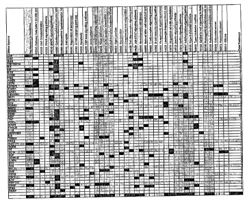
Figure 11 presents tables of selected DBRISKMARKERS by eight Position
Categories useful for the construction of panels selecting DBRISKMARKRRS
according to
the method disclosed herein.
Figure 12 is a listing of 25 high performing DBRISKMARKER panels using three
DBRISKMARKERS selected from Position Categories according to the method
disclosed
herein. Logistic regression algorithms using said panels had calculated RA2
values ranging
from 0.300 to 0.329 when employed on samples in the described example and non-
diabetic
patient cohort.
Figure 13 is a listing of 25 high performing DBRISKMAKER panels using eight
DBRISKMARKERS selected from Position Categories according to the method
disclosed
herein. Logistic regression algorithms using said panels had calculated RA2
values ranging
from 0.3 10 to 0.475 when employed on samples in the described example and non-
diabetic
patient cohort.
Figure 14 is a listing of 25 high performing DBRISKMAKER panels using eighteen
DBRISKMARKERS selected from Position Categories according to the method
disclosed
herein. Logistic regression algorithms using said panels had calculated RA2
values ranging
from 0.523 to 0.6105 when employed on samples in the described example and non-
diabetic
patient cohort.
Figure 15 is a graph ROC curve and AUC statistics for the highest performing
three,
eight, and eighteen DBRISKMARKER panels respectively when employed on samples
in the
described example and non-diabetic patient cohort.
17
CA 02625744 2008-04-10
WO 2007/044860 PCT/US2006/039963
Figure 16 is an ROC curve and AUC statistics indicating the three relative
highest
performing individual DBRISKMARKERS markers when employed on samples in the
described example and non-diabetic patient cohort.
Figure 17 is a standard curve demonstrating a typical result from the methods
of the
present invention. Once a working standard curve is demonstrated, the assay is
typically
applied to 24 serum samples to determine the normal distribution of the target
analyte across
clinical samples.
Figure 18 depicts a graph exemplifying single molecule detection data across
92
samples for 25 biomarkers.
DETAILED DESCRIPTION OF THE INVENTION
The present invention relates to the identification of biomarkers associated
with
subjects having Diabetes or a pre-diabetic condition, or who are pre-disposed
to developing
Diabetes or a pre-diabetic condition. Accordingly, the present invention
features methods for
identifying subjects who are pre-disposed to developing Diabetes or a pre-
diabetic condition,
including those subjects who are asymptomatic for Diabetes or a pre-diabetic
condition by
detection of the biomarkers disclosed herein. These biomarkers are also useful
for
monitoring subjects undergoing treatments and therapies for Diabetes or pre-
diabetic
conditions, and for selecting therapies and treatments that would be
efficacious in subjects
having Diabetes or a pre-diabetic condition, wherein selection and use of such
treatments and
therapies slow the progression of Diabetes or pre-diabetic conditions, or
substantially delay
or prevent its onset.
"Diabetes Mellitus" in the context of the present invention encompasses Type 1
Diabetes, both autoimmune and idiopathic and Type 2 Diabetes (together,
"Diabetes"). The
World Health Organization defines the diagnostic value of fasting plasma
glucose
concentration to 7.0 mmol/1(126 mg/dl) and above for Diabetes Mellitus (whole
blood 6.1
mmol/1 or 110 mg/dl), or 2-hour glucose level _11.1 mmol/L (_200 mg/dL). Other
values
suggestive of or indicating high risk for Diabetes Mellitus include elevated
arterial pressure >_
140/90 mm Hg; elevated plasma triglycerides (>1.7 mmol/L; 150 mg/dL) and/or
low HDL-
cholesterol (<0.9 mmol/L, 35 mg/dl for men; <1.0 mmol/L, 39 mg/dL women);
central
obesity (males: waist to hip ratio >0.90; females: waist to hip ratio > 0.85)
and/or body mass
index exceeding 30 kg/m2; microalbuminuria, where the urinary albumin
excretion rate _20
g/min or albumin:creatinine ratio _ 30 mg/g).
18
CA 02625744 2008-04-10
WO 2007/044860 PCT/US2006/039963
"Pre-diabetic condition" refers to a metabolic state that is intermediate
between
normal glucose homeostasis and metabolism and states seen in frank Diabetes
Mellitus. Pre-
diabetic conditions include, without limitation, Metabolic Syndrome ("Syndrome
X"),
Impaired Glucose Tolerance (IGT), and Impaired Fasting Glycemia (IFG). IGT
refers to
post-prandial abnormalities of glucose regulation, while IFG refers to
abnormalities that are
measured in a fasting state. The World Health Organization defines values for
IFG as a
fasting plasma glucose concentration of 6.1 mmol/L (100 mg/dL) or greater
(whole blood 5.6
mmol/L; 100 mg/dL), but less than 7.0 mmol/L (126 mg/dL)(whole blood 6.1
mmol/L; 110
mg/dL). Metabolic syndrome according to the National Cholesterol Education
Program
(NCEP) criteria are defined as having at least three of the following: blood
pressure _ 130/85
mm Hg; fasting plasma glucose _6.1 mmol/L; waist circumference >102 cm (men)
or >88 cm
(women); triglycerides _1.7 mmol/L; and HDL cholesterol <1.0 mmol/L (men) or
1.3
mmol/L (women).
"Pre-Diabetes" in the context of the present invention indicates the
physiological
state, in an individual or in a population, of having a higher than normal
expected rate of
disease conversion to frank Type 2 diabetes mellitus. Such absolute expected
rate of
conversion to frank Type 2 diabetes in Pre-Diabetes populations may be up to 1
percent or
more per annum, and preferably 2 percent per annum or more. It may also be
stated in terms
of a relative risk from normal between quartiles of risk or as a likelihood
ratio between
differing biomarker and index scores, including those coming from the
invention. Unless
otherwise noted, and without limitation, when a categorical positive diagnosis
of Pre-
Diabetes is stated here, it is defined experimentally by the group of patients
with an expected
conversion rate to Type 2 Diabetes of two percent (2%) per annum over the
coming 7.5 years,
or fifteen percent (15%) of those testing at a given threshold value (the
selected Pre-Diabetes
clinical cutoff). When a continuous measure of Pre-Diabetes conversion risk is
produced,
having a "pre-diabetic condition" encompasses any expected annual rate of
conversion above
that seen in a normal reference or general unselected normal prevalence
population.
"Impaired glucose tolerance" (IGT) is defined as having a blood glucose level
that is
higher than normal, but not high enough to be classified as Diabetes Mellitus.
A subject with
IGT will have two-hour glucose levels of 140 to 199 mg/dL (7.8 to 11.0 mmol)
on the 75-g
oral glucose tolerance test. These glucose levels are above normal but below
the level that is
diagnostic for Diabetes. Subjects with impaired glucose tolerance or impaired
fasting glucose
19
CA 02625744 2008-04-10
WO 2007/044860 PCT/US2006/039963
have a significant risk of developing Diabetes and thus are an important
target group for
primary prevention.
"Insulin resistance" refers to a condition in which the cells of the body
become
resistant to the effects of insulin, that is, the normal response to a given
amount of insulin is
reduced. As a result, higher levels of insulin are needed in order for insulin
to exert its effects.
"Normal glucose levels" is used interchangeably with the term "normoglycemic"
and
refers to a fasting venous plasma glucose concentration of less than 6.1
mmol/L (110 mg/dL).
Although this amount is arbitrary, such values have been observed in subjects
with proven
normal glucose tolerance, although some may have IGT as measured by oral
glucose
tolerance test (OGTT).
Two hundred and sixty biomarkers have been identified as being found to have
altered or modified presence or concentration levels in subjects who have
Diabetes, or who
exhibit symptoms characteristic of a pre-diabetic condition, or have Pre-
Diabetes (as defined
herein) such as those subjects who are insulin resistant, have altered beta
cell function or at
risk of developing Diabetes based upon known clinical parameters or risk
factors, such as
family history of Diabetes, low activity level, poor diet, excess body weight
(especially
around the waist), age greater than 45 years, high blood pressure, high levels
of triglycerides,
HDL cholesterol of less than 35, previously identified impaired glucose
tolerance, previous
Diabetes during pregnancy ("gestational Diabetes Mellitus") or giving birth to
a baby
weighing more than nine pounds, and ethnicity.
The biomarkers and methods of the present invention allow one of skill in the
art to
identify, diagnose, or otherwise assess those subjects who do not exhibit any
symptoms of
Diabetes or a pre-diabetic condition, but who nonetheless may be at risk for
developing
Diabetes or experiencing syinptoms characteristic of a pre-diabetic condition.
The term "biomarker" in the context of the present invention encompasses,
without
limitation, proteins, nucleic acids, polymorphisms of proteins and nucleic
acids, elements,
metabolites, and other analytes. Biomarkers can also include mutated proteins
or mutated
nucleic acids. The term "analyte" as used herein can mean any substance to be
measured and
can encompass electrolytes and elements, such as calcium. Finally, biomarkers
can also refer
to non-analyte physiological markers of health status encompassing other
clinical
characteristics such as, without limitation, age, ethnicity, diastolic and
systolic blood
pressure, body-mass index, and resting heart rate.
CA 02625744 2008-04-10
WO 2007/044860 PCT/US2006/039963
Proteins, nucleic acids, polymorphisms, and metabolites whose levels are
changed in
subjects who have Diabetes or a pre-diabetic condition, or are predisposed to
developing
Diabetes or a pre-diabetic condition are summarized in Table 1 and are
collectively referred
to herein as, irater alia, "Diabetes risk-associated proteins", "DBRISKMARKER
polypeptides", or "DBRISKMARKER proteins". The corresponding nucleic acids
encoding
the polypeptides are referred to as "Diabetes risk-associated nucleic acids",
"Diabetes risk-
associated genes", "DBRISKMARKER nucleic acids", or "DBRISKMARKER genes".
Unless indicated otherwise, "DBRISKMARKER", "Diabetes risk-associated
proteins",
"Diabetes risk-associated nucleic acids" are meant to refer to any of the
sequences disclosed
herein. The corresponding metabolites of the DBRISKMARKER proteins or nucleic
acids
can also be measured, as well as any of the aforementioned conventional risk
marker
metabolites previously disclosed, including, without limitation, such
metabolites as
dehydroepiandrosterone sulfate (DHEAS); c-peptide; cortisol; vitamin D3; 5-
hydroxytryptamine (5-HT; serotonin); oxyntomodulin; estrogen; estradiol; and
digitalis-like
factor, herein referred to as "DBRISKMARKER metabolites". Non-analyte
physiological
markers of health status (e.g., such as age, ethnicity, diastolic or systolic
blood pressure,
body-mass index, and other non-analyte measurements commonly used as
conventional risk
factors) are referred to as "DBRISKMARKER physiology". Calculated indices
created from
mathematically combining measurements of one or more, preferably two or more
of the
aforementioned classes of DBRISKMARKERS are referred to as "DBRISKIVIARKER
indices". Proteins, nucleic acids, polymorphisms, mutated proteins and
niutated nucleic
acids, metabolites, and other analytes are, as well as common physiological
measurements
and indices constructed from any of the preceding entities, are included in
the broad category
of "DBRISKMARKERS".
A"subject" in the context of the present invention is preferably a mammal. The
mammal can be a human, non-human primate, mouse, rat, dog, cat, horse, or cow,
but are not
limited to these examples. Mammals other than humans can be advantageously
used as
subjects that represent animal models of Diabetes Mellitus or pre-Diabetes
conditions. A
subject can be male or female. A subject can be one who has been previously
diagnosed or
identified as having Diabetes or a pre-diabetic condition, and optionally has
already
undergone treatment for the Diabetes or pre-diabetic condition. Alternatively,
a subject can
also be one who has not been previously diagnosed as having Diabetes or a pre-
diabetic
condition. For example, a subject can be one who exhibits one or more risk
factors for
21
CA 02625744 2008-04-10
WO 2007/044860 PCT/US2006/039963
Diabetes or a pre-diabetic condition, or a subject who does not exhibit
Diabetes risk factors,
or a subject who is asymptomatic for Diabetes or pre-Diabetes. A subject can
also be one
who is suffering from or at risk of developing Diabetes or a pre-diabetic
condition.
A "sample" in the context of the present invention is a biological sample
isolated from
a subject and can include, for example, serum, blood plasma, blood cells,
endothelial cells,
tissue biopsies, lymphatic fluid, ascites fluid, interstitital fluid (also
known as "extracellular
fluid" and encompasses the fluid found in spaces between cells, including,
inter alia, gingival
crevicular fluid), bone marrow, sputum, or urine.
One or more, preferably two or more DBRISKMARKERS can be detected in the
practice of the present invention. For example, two (2), five (5), ten (10),
fifteen (15), twenty
(20), forty (40), fifty (50), seventy-five (75), one hundred (100), one
hundred and twenty five
(125), one hundred and fifty (150), one hundred and seventy-five (175), two
hundred (200),
two hundred and ten (210), two hundred and twenty (220), two hundred and
thirty (230), two
hundred and forty (240), two hundred and fifty (250) or more DBRISKMARKERS can
be
detected. In some aspects, a11260 DBRISKMARKERS disclosed herein can be
detected.
Preferred ranges from which the number of DBRISKMARKERS can be detected
include
ranges bounded by any minimum selected from between one and 260, particularly
two, five,
ten, twenty, fifty, seventy-five, one hundred, one hundred and twenty five,
one hundred and
fifty, one hundred and seventy-five, two hundred, two hundred and ten, two
hundred and
twenty, two hundred and thirty, two hundred and forty, two hundred and fifty,
paired with
any maximum up to the total known DBRISKMARKERS, particularly five, ten,
twenty, fifty,
and seventy-five. Particularly preferred ranges include two to five (2-5), two
to ten (2-10),
two to fifty (2-50), two to seventy-five (2-75), two to one hundred (2-100),
five to ten (5-10),
five to twenty (5-20), five to fifty (5-50), five to seventy-five (5-75), five
to one hundred (5-
100), ten to twenty (10-20), ten to fifty (10-50), ten to seventy-five (10-
75), ten to one
hundred (10-100), twenty to fifty (20-50), twenty to seventy-five (20-75),
twenty to one
hundred (20-100), fifty to seventy-five (50-75), fifty to one hundred (50-
100), one hundred to
one hundred and twenty-five (100-125), one hundred and twenty-five to one
hundred and
fifty (125-150), one hundred and fifty to one hundred and seventy five (150-
175), one
hundred and seventy-five to two hundred (175-200), two hundred to two hundred
and ten
(200-210), two hundred and ten to two hundred and twenty (210-220), two
hundred and
twenty to two hundred and thirty (220-230), two hundred and thirty to two
hundred and forty
22
CA 02625744 2008-04-10
WO 2007/044860 PCT/US2006/039963
(230- 240), two hundred and forty to two hundred and fifty (240-250), and two
hundred and
fifty to more than two hundred and fifty (250+).
Diagnostic and Prognostic Methods
The risk of developing Diabetes or Pre-Diabetes can be detected with a "pre-
determined level of predictability" by examining an "effective amount" of
DBRISKMARKER proteins, nucleic acids, polymorphisms, metabolites, and other
analytes
in a test sample (e.g., a subject derived sample) and comparing the effective
amounts to
reference or index values, often utilizing mathematical algorithms in order to
combine
information from results of multiple individual DBRISKMARKERS into a single
measurement or index. Subjects identified as having an increased risk of
Diabetes or a pre-
diabetic condition can optionally be selected to receive treatment regimens,
such as
administration of prophylactic or therapeutic compounds such as "diabetes-
modulating
drugs" as defined herein, or implementation of exercise regimens or dietary
supplements to
prevent or delay the onset of Diabetes or Pre-Diabetes. A sample isolated from
the subject
can coniprise, for example, blood, plasma, blood cells, endothelial cells,
tissue biopsies,
lymphatic fluid, serum, bone marrow, ascites fluid, interstitial fluid
(including, for example,
gingival crevicular fluid), urine, sputum, or other bodily fluids.
The amount of the DBRISKMARKER protein, nucleic acid, polymorphism,
metabolite, or other analyte can be measured in a test sample and compared to
the normal
control level. The term "normal control level", means the level of a
DBRISKMARKER
protein, nucleic acid, polymorphism, metabolite, or other analyte, or
DBRISKMARKER
physiology or indices, typically found in a subject not suffering from
Diabetes or a pre-
diabetic condition and not likely to have Diabetes or a pre-diabetic
condition, e.g., relative to
samples collected from young subjects who were monitored until advanced age
and were
found not to develop Diabetes or a pre-diabetic condition. Alternatively, the
normal control
level can mean the level of a DBRISKMARKER protein, nucleic acid,
polymorphism,
metabolite, or other analyte typically found in a subject suffering from
Diabetes or a pre-
diabetic condition. The normal control level can be a range or an index.
Alternatively, the
normal control level can be a database of patterns from previously tested
subjects. A change
in the level in the subject-derived sample of a DBRISKMARKER protein, nucleic
acid,
polymorphism, metabolite, or other analyte compared to the normal control
level can indicate
that the subject is suffering from or is at risk of developing Diabetes or a
pre-diabetic
23
CA 02625744 2008-04-10
WO 2007/044860 PCT/US2006/039963
condition. In contrast, when the methods are applied prophylactically, a
similar level
compared to the normal control level in the subject-derived sample of a
DBRISKMARKER
protein, nucleic acid, polymorphism, metabolite, or other analyte can indicate
that the subject
is not suffering from, is not at risk or is at low risk of developing Diabetes
or a pre-diabetic
condition.
The difference in the level of DBRISKMARKERS is preferably statistically
significant. By "statistically significant", it is meant that the alteration
is greater than what
might be expected to happen by chance alone. Statistical significance can be
determined by
any method known in the art. For example, statistical significance can be
determined byp-
value. The p-value is a measure of probability that a difference between
groups during an
experiment happened by chance. (P(z>zobserved)). For example, a p-value of
0.01 means
that there is a 1 in 100 chance the result occurred by chance. The lower the p-
value, the more
likely it is that the difference between groups was caused by treatment. An
alteration is
statistically significant if the p-value is at least 0.05. Preferably, the p-
value is 0.04, 0.03,
0.02, 0.01, 0.005, 0.001 or less. As noted below, and without any limitation
of the invention,
achieving statistical significance generally but not always requires that
combinations of
several DBRISKMARKERS be used together in panels and combined with
mathematical
algorithms in order to achieve a statistically significant DBRISKMARKER index.
The "diagnostic accuracy" of a test, assay, or method concerns the ability of
the test,
assay, or method to distinguish between subjects having Diabetes or a pre-
diabetic condition,
or at risk for Diabetes or a pre-diabetic condition is based on whether the
subjects have a
"clinically significant presence" or a "clinically significant alteration" in
the levels of a
DBRISKMARKER. By "clinically significant presence" or "clinically significant
alteration",
it is meant that the presence of the DBRISKMARKER (e.g., mass, such as
milligrams,
nanograms, or mass per volume, such as milligrams per deciliter or copy number
of a
transcript per unit volume) or an alteration in the presence of the
DBRISKMARKER in the
subject (typically in a sample from the subject) is higher than the
predetermined cut-off point
(or threshold value) for that DBRISKMARKER and therefore indicates that the
subject has
Diabetes or a pre-diabetic condition for which the sufficiently high presence
of that protein,
nucleic acid, polymorphism, metabolite or analyte is a marker.
The present invention may be used to make categorical or continuous
measurements
of the risk of conversion to Type 2 Diabetes, thus diagnosing a category of
subjects defined
as Pre-Diabetic.
24
CA 02625744 2008-04-10
WO 2007/044860 PCT/US2006/039963
In the categorical scenario, the methods of the present invention can be used
to
discriminate between Normal and Pre-Diabetes subject cohorts. In this
categorical use of the
invention, the terms "high degree of diagnostic accuracy" and "very high
degree of diagnostic
accuracy" refer to the test or assay for that DBRISKMARKER (or DBRISKMARKER
index;
wherein DBRISKMARKER value encompasses any individual measurement whether from
a
single DBRISKMARKER or derived from an index of DBRISKMARKERS) with the
predetermined cut-off point correctly (accurately) indicating the presence or
absence of Pre-
Diabetes. A perfect test would have perfect accuracy. Thus, for subjects who
have Pre-
Diabetes, the test would indicate only positive test results and would not
report any of those
subjects as being "negative" (there would be no "false negatives"). In other
words, the
"sensitivity" of the test (the true positive rate) would be 100%. On the other
hand, for
subjects who did not have Pre-Diabetes, the test would indicate only negative
test results and
would not report any of those subjects as being "positive" (there would be no
"false
positives"). In other words, the "specificity" (the true negative rate) would
be 100%. See,
e.g., O'Marcaigh AS, Jacobson RM, "Estimating The Predictive Value Of A
Diagnostic Test,
How To Prevent Misleading Or Confusing Results," Clin. Ped. 1993, 32(8): 485-
491, which
discusses specificity, sensitivity, and positive and negative predictive
values of a test, e.g., a
clinical diagnostic test. In other embodiments, the present invention may be
used so as to
discriminate Pre-Diabetes from Diabetes, or Diabetes from Normal. Such use may
require a
different DBRISKMARKER panel, mathematical algorithm, and/or cut-off point,
but be
subject to the same aforementioned measurements of diagnostic accuracy for the
intended
use.
In the categorical diagnosis of a disease, changing the cut point or threshold
value of a
test (or assay) usually changes the sensitivity and specificity, but in a
qualitatively inverse
relationship. For example, if the cut point is lowered, more subjects in the
population tested
will typically have test results over the cut point or threshold value.
Because subjects who
have test results above the cut point are reported as having the disease,
condition, or
syndrome for which the test is conducted, lowering the cut point will cause
more subjects to
be reported as having positive results (e.g., that they have Diabetes, Pre-
Diabetes, or a pre-
diabetic condition). Thus, a higher proportion of those who have Diabetes or
Pre-Diabetes
will be indicated by the test to have it. Accordingly, the sensitivity (true
positive rate) of the
test will be increased. However, at the same time, there will be more false
positives because
more people who do not have the disease, condition, or syndrome (e.g., people
who are truly
CA 02625744 2008-04-10
WO 2007/044860 PCT/US2006/039963
"negative") will be indicated by the test to have DBRISKMARKER values above
the cut
point and therefore to be reported as positive (e.g., to have the disease,
condition, or
syndrome) rather than being correctly indicated by the test to be negative.
Accordingly, the
specificity (true negative rate) of the test will be decreased. Similarly,
raising the cut point
will tend to decrease the sensitivity and increase the specificity. Therefore,
in assessing the
accuracy and usefulness of a proposed medical test, assay, or method for
assessing a subject's
condition, one should always take both sensitivity and specificity into
account and be mindful
of what the cut point is at which the sensitivity and specificity are being
reported because
sensitivity and specificity may vary significantly over the range of cut
points.
There is, however, an indicator that allows representation of the sensitivity
and
specificity of a test, assay, or method over the entire range of test (or
assay) cut points with
just a single value. That indicator is derived from a Receiver Operating
Characteristics
("ROC") curve for the test, assay, or method in question. See, e.g., Shultz,
"Clinical
Interpretation Of Laboratory Procedures," chapter 14 in Teitz, Fundamentals of
Clinical
Chemistry, Burtis and Ashwood (eds.), 4th edition 1996, W.B. Saunders Company,
pages
192-199; and Zweig et al., "ROC Curve Analysis: An Example Showing The
Relationships
Among Serum Lipid And Apolipoprotein Concentrations In Identifying Subjects
With
Coronory Artery Disease," Clin. Chem., 1992, 38(8): 1425-1428.
An ROC curve is an x-y plot of sensitivity on the y-axis, on a scale of zero
to one
(e.g., 100%), against a value equal to one minus specificity on the x-axis, on
a scale of zero to
one (e.g., 100%). In other words, it is a plot of the true positive rate
against the false positive
rate for that test, assay, or method. To construct the ROC curve for the test,
assay, or method
in question, subjects can be assessed using a perfectly accurate or "gold
standard" method
that is independent of the test, assay, or method in question to determine
whether the subjects
are truly positive or negative for the disease, condition, or syndrome (for
example, coronary
angiography is a gold standard test for the presence of coronary
atherosclerosis). The subjects
can also be tested using the test, assay, or method in question, and for
varying cut points, the
subjects are reported as being positive or negative according to the test,
assay, or method. The
sensitivity (true positive rate) and the value equal to one minus the
specificity (which value
equals the false positive rate) are determined for each cut point, and each
pair of x-y values is
plotted as a single point on the x-y diagram. The "curve" connecting those
points is the ROC
curve.
26
CA 02625744 2008-04-10
WO 2007/044860 PCT/US2006/039963
The ROC curve is often used in order to determine the optimal single clinical
cut-off
or treatment threshold value where sensitivity and specificity are maximized;
such a situation
represents the point on the ROC curve which describes the upper left corner of
the single
largest rectangle which can be drawn under the curve.
The total area under the curve ("AUC") is the indicator that allows
representation of
the sensitivity and specificity of a test, assay, or method over the entire
range of cut points
with just a single value. The maximum AUC is one (a perfect test) and the
minimum area is
one half (e.g. the area where there is no discrimination of normal versus
disease). The closer
the AUC is to one, the better is the accuracy of the test. It should be noted
that implicit in all
ROC and AUC is the definition of the disease and the post-test time horizon of
interest.
By a "high degree of diagnostic accuracy", it is meant a test or assay (such
as the test
of the invention for determining the clinically significant presence of
DBRISKMARKERS,
which thereby indicates the presence of Diabetes or a pre-diabetic condition)
in which the
AUC (area under the ROC curve for the test or assay) is at least 0.70,
desirably at least 0.75,
more desirably at least 0.80, preferably at least 0.85, more preferably at
least 0.90, and most
preferably at least 0.95.
By a "very high degree of diagnostic accuracy", it is meant a test or assay in
which
the AUC (area under the ROC curve for the test or assay) is at least 0.80,
desirably at least
0.85, more desirably at least 0.875, preferably at least 0.90, more preferably
at least 0.925,
and most preferably at least 0.95.
Alternatively, in low disease prevalence tested populations (defined as those
with less
than 1% rate of occurrences per annum), ROC and AUC can be misleading as to
the clinical
utility of a test, and absolute and relative risk ratios as defined elsewhere
in this disclosure
can be employed to determine the degree of diagnostic accuracy. Populations of
subjects to
be tested can also be categorized into quartiles, where the top quartile (25%
of the
population) comprises the group of subjects with the highest relative risk for
developing or
suffering from Diabetes or a pre-diabetic condition and the bottom quartile
comprising the
group of subjects having the lowest relative risk for developing Diabetes or a
pre-diabetic
condition. Generally, values derived from tests or assays having over 2.5
times the relative
risk from top to bottom quartile in a low prevalence population are considered
to have a"high
degree of diagnostic accuracy," and those with five to seven times the
relative risk for each
quartile are considered to have a very high degree of diagnostic accuracy.
Nonetheless,
values derived from tests or assays having only 1.2 to 2.5 times the relative
risk for each
27
CA 02625744 2008-04-10
WO 2007/044860 PCT/US2006/039963
quartile remain clinically useful are widely used as risk factors for a
disease; such is the case
with total cholesterol and for many inflammatory markers with respect to their
prediction of
future cardiovascular events.
The predictive value of any test depends on the sensitivity and specificity of
the test,
and on the prevalence of the condition in the population being tested. This
notion, based on
Bayes' theorem, provides that the greater the likelihood that the condition
being screened for
is present in a subject or in the population (pre-test probability), the
greater the validity of a
positive test and the greater the likelihood that the result is a true
positive. Thus, the problem
with using a test in any population where there is a low likelihood of the
condition being
present is that a positive result has limited value (i.e., more likely to be a
false positive).
Similarly, in populations at very high risk, a negative test result is more
likely to be a false
negative. By defining the degree of diagnostic accuracy, i.e., cut points on a
ROC curve,
defining an acceptable AUC value, and determining the acceptable ranges in
relative
concentration of what constitutes an effective amount of the DBRISKMARKERS of
the
invention allows for one of skill in the art to use the DBRISKMARKERS to
diagnose or
identify subjects with a pre-determined level of predictability.
Alternative methods of determining diagnostic accuracy must be used with
continuous
measurements of risk, which are commonly used when a disease category or risk
category
(such as Pre-Diabetes) has not yet been clearly defined by the relevant
medical societies and
practice of medicine.
"Risk" in the context of the present invention can mean "absolute" risk, which
refers
to that percentage probability that an event will occur over a specific time
period. Absolute
risk can be measured with reference to either actual observation post-
measurement for the
relevant time cohort, or with reference to index values developed from
statistically valid
historical cohorts that have been followed for the relevant time period.
"Relative" risk refers
to the ratio of absolute risks of a subject's risk compared either to low risk
cohorts or average
population risk, which can vary by how clinical risk factors are assessed.
Odds ratios, the
proportion of positive events to negative events for a given test result, are
also commonly
used (odds are according to the formula p/(1-p) where p is the probability of
event and (1- p)
is the probability of no event) to no-conversion. Alternative continuous
measures which may
be assessed in the context of the presentt invention include time to Diabetes
conversion and
therapeutic Diabetes conversion risk reduction ratios.
28
CA 02625744 2008-04-10
WO 2007/044860 PCT/US2006/039963
For such continuous measures, measures of diagnostic accuracy for a calculated
index
are typically based on linear regression curve fits between the predicted
continuous value and
the actual observed values (or historical index calculated value) and utilize
measures such as
R squared, p values and confidence intervals. It is not unusual for predicted
values using
such algorithms to be reported including a confidence interval (usually 90% or
95% CI)
based on a historical observed cohort's predictions, as in the test for risk
of future breast
cancer recurrence commercialized by Genomic Health (Redwood City, California).
The ultimate determinant and gold standard of true risk conversion to Diabetes
is a
actual conversions within a sufficiently large population and observed over
the length of time
claimed. However, this is problematic, as it is necessarily a retrospective
point of view,
coming after any opportunity for preventive interventions. As a result,
subjects suffering
from or at risk of developing Diabetes or a pre-diabetic condition are
commonly diagnosed or
identified by methods known in the art, and future risk is estimated based on
historical
experience and registry studies. Such methods include, but are not limited to,
measurement
of systolic and diastolic blood pressure, measurements of body mass index, in
vitro
determination of total cholesterol, LDL, HDL, insulin, and glucose levels from
blood
samples, oral glucose tolerance tests, stress tests, measurement of human
serum C-reactive
protein (hsCRP), electrocardiogram (ECG), c-peptide levels, anti-insulin
antibodies, anti-beta
cell-antibodies, and glycosylated hemoglobin (HbAIc). Additionally, any of the
aforementioned methods can be used separately or in combination to assess if a
subject has
shown an "improvement in Diabetes risk factors." Such improvements include,
without
limitation, a reduction in body mass index (BMI), a reduction in blood glucose
levels, an
increase in HDL levels, a reduction in systolic and/or diastolic blood
pressure, an increase in
insulin levels, or combinations thereof.
The oral glucose tolerance test (OGTT) is principally used for diagnosis of
Diabetes
Mellitus or pre-diabetic conditions when blood glucose levels are equivocal,
during
pregnancy, or in epidemiological studies (Definition, Diagnosis and
Classification of
Diabetes Mellitus and its Complications, Part 1, World Health Organization,
1999). The
OGTT should be administered in the morning after at least 3 days of
unrestricted diet (greater
than 150 g of carbohydrate daily) and usual physical activity. A reasonable
(30-50 g)
carbohydrate-containing meal should be consumed on the evening before the
test. The test
should be preceded by an overnight fast of 8-14 hours, during which water may
be consumed.
After collection of the fasting blood sample, the subject should drink 75 g of
anhydrous
29
CA 02625744 2008-04-10
WO 2007/044860 PCT/US2006/039963
glucose or 82.5 g of glucose monohydrate in 250-300 ml of water over the
course of 5
minutes. For children, the test load should be 1.75 g of glucose per kg body
weight up to a
total of 75 g of glucose. Timing of the test is from the beginning of the
drink. Blood samples
must be collected 2 hours after the test load. As previously noted, a
diagnosis of impaired
glucose tolerance (IGT) has been noted as being only 50% sensitive, with a
>10% false
positive rate, for a 7.5 year conversion to diabetes when used at the WHO cut-
off points.
This is a significant problem for the clinical utility of the test, as even
relatively high risk
ethnic groups have only a 10% rate of conversion to Diabetes over such a
period unless
otherwise enriched by other risk factors; in an unselected general population,
the rate of
conversion over such periods is typically estimated at 5-6%, or less than 1%
per annum.
Other methods of measuring glucose in blood include reductiometric methods
known
in the art such as, but not limited to, the Somogyi-Nelson method, methods
using hexokinase
and glucose dehydrogenase, immobilized glucose oxidase electrodes, the o-
toluidine method,
the ferricyanide method and the neocuprine autoanalyzer method. Whole blood
glucose
values are usually about 15% lower than corresponding plasma values in
patients with a
normal hematocrit reading, and arterial values are generally about 7% higher
than
corresponding venous values. Subjects taking insulin are frequently requested
to build up a
"glycemic profile" by self-measurement of blood glucose at specific times of
the day. A "7-
point profile" is useful, with samples taken before and 90 minutes after each
meal, and just
before going to bed.
A subject suffering from or at risk of developing Diabetes or a pre-diabetic
condition
may also be suffering from or at risk of developing arteriovascular disease,
hypertension or
obesity. Type 2 Diabetes in particular and arteriovascular disease have many
risk factors in
common, and many of these risk factors are highly correlated with one another.
The
relationships among these risk factors may be attributable to a small number
of physiological
phenomena, perhaps even a single phenomenon. Subjects suffering from or at
risk of
developing Diabetes, arteriovascular disease, hypertension or obesity are
identified by
methods known in the art. For example, Diabetes is frequently diagnosed by
measuring
fasting blood glucose levels or insulin. Normal adult glucose levels are 60-
126 mg/dl.
Normal insulin levels are 7 mU/mL 3mU. Hypertension is diagnosed by a blood
pressure
consistently at or above 140/90. Risk of arteriovascular disease can also be
diagnosed by
measuring cholesterol levels. For example, LDL cholesterol above 137 or total
cholesterol
above 200 is indicative of a heightened risk of arteriovascular disease.
Obesity is diagnosed
CA 02625744 2008-04-10
WO 2007/044860 PCT/US2006/039963
for example, by body mass index. Body mass index (BMI) is measured (kg/m2 (or
lb/in2 X
704.5)). Alternatively, waist circumference (estimates fat distribution),
waist-to-hip ratio
(estimates fat distribution), skinfold thickness (if measured at several
sites, estimates fat
distribution), or bioimpedance (based on principle that lean mass conducts
current better than
fat mass (i.e. fat mass impedes current), estimates % fat) is measured. The
parameters for
normal, overweight, or obese individuals is as follows: Underweight: BMI
<18.5; Normal:
BMI 18.5 to 24.9; Overweight: BMI = 25 to 29.9. Overweight individuals are
characterized
as having a waist circumference of >94 cm for men or >80 cm for women and
waist to hip
ratios of > 0.95 in men and > 0.80 in women. Obese individuals are
characterized as having a
BMI of 30 to 34.9, being greater than 20% above "normal" weight for height,
having a body
fat percentage > 30% for women and 25% for men, and having a waist
circumference >102
cm (40 inches) for men or 88 cm (35 inches) for women. Individuals with severe
or morbid
obesity are characterized as having a BMI of > 35. Because of the
interrelationship between
Diabetes and arteriovascular disease, some or all of the individual
DBRISKMARKERS and
DBRISKMARKER panels of the present invention may overlap or be encompassed by
biomarkers of arteriovascular disease, and indeed may be useful in the
diagnosis of the risk of
arteriovascular disease.
Risk prediction for Diabetes Mellitus or a pre-diabetic condition can also
encompass
risk prediction algorithms and computed indices that assess and estimate a
subject's absolute
risk for developing Diabetes or a pre-diabetic condition with reference to a
historical cohort.
Risk assessment using such predictive mathematical algorithms and computed
indices has
increasingly been incorporated into guidelines for diagnostic testing and
treatment, and
encompass indices obtained from and validated with, inter alia, multi-stage,
stratified
samples from a representative population. A plurality of conventional Diabetes
risk factors
are incorporated into predictive models. A notable example of such algorithms
include the
Framingham Heart Study (Kannel, W.B., et al, (1976) Am. J. Cardiol. 38: 46-5
1) and
modifications of the Framingham Study, such as the National Cholesterol
Education Program
Expert Panel on Detection, Evaluation, and Treatment of High Blood Cholesterol
in Adults
(Adult Treatment Panel III), also know as NCEP/ATP III, which incorporates a
patient's age,
total cholesterol concentration, HDL cholesterol concentration, smoking
status, and systolic
blood pressure to estimate a person's 10-year risk of developing
arteriovascular disease,
which is commonly found in subjects suffering from or at risk for developing
Diabetes
31
CA 02625744 2008-04-10
WO 2007/044860 PCT/US2006/039963
Mellitus, or a pre-diabetic condition. The same Framingham algorithm has been
found to be
modestly predictive of the risk for developing Diabetes Mellitus, or a pre-
diabetic condition.
Other Diabetes risk prediction algorithms include, without limitation, the San
Antonio
Heart Study (Stem, M.P. et al, (1984) Am. J. Epidemiol. 120: 834-851; Stem,
M.P. et al,
(1993) Diabetes 42: 706-714; Burke, J.P. et al, (1999) Arch. Intern. Med. 159:
1450-1456),
Archimedes (Eddy, D.M. and Schlessinger, L. (2003) Diabetes Care 26(11): 3093-
3101;
Eddy, D.M. and Schlessinger, L. (2003) Diabetes Care 26(11): 3102-3110), the
Finnish-based
Diabetes Risk Score (Lindstr6m, J. and Tuomilehto, J. (2003) Diabetes Care
26(3): 725-731),
and the Ely Study (Griffin, S.J. et al, (2000) Diabetes Metab. Res. Rev. 16:
164-171), the
contents of which are expressly incorporated herein by reference.
Archimedes is a mathematical model of Diabetes that simulates the disease
state
person-by-person, object-by-object and comprises biological details that are
continuous in
reality, such as the pertinent organ systems, more than 50 continuously
interacting biological
variables, and the major symptoms, tests, treatments, and outcomes commonly
associated
with Diabetes.
Archimedes includes many diseases simultaneously and interactively in a single
integrated physiology, enabling it to address features such as co-morbidities,
syndromes,
treatments and other multiple effects. The Archimedes model includes Diabetes
and its
complications, such as coronary artery disease, congestive heart failure, and
asthma. The
model is written in differential equations, using object-oriented programming
and a construct
called "features". The model comprises the anatomy of a subject (all simulated
subjects have
organs, such as hearts, livers, pancreases, gastrointestinal tracts, fat,
muscles, kidneys, eyes,
limbs, circulatory systems, brains, skin, and peripheral nervous systems), the
"features" that
determine the course of the disease and representing real physical phenomena
(e.g., the
number of milligrams of glucose in a deciliter of plasma, behavioral
phenomena, or
conceptual phenomena (e.g., the "progression" of disease), risk factors,
incidence, and
progression of the disease, glucose metabolism, signs and tests, diagnosis,
symptoms, health
outcomes of glucose metabolism, treatments, complications, deaths from
Diabetes and its
complications, deaths from other causes, care processes, and medical system
resources. For a
typical application of the model, there are thousands of simulated subjects,
each with a
simulated anatomy and physiology, who will get simulated diseases, can seek
care at
simulated health care facilities, will be seen by simulated health care
personnel in simulated
facilities, will be given simulated tests and treatments, and will have
simulated outcomes. As
32
CA 02625744 2008-04-10
WO 2007/044860 PCT/US2006/039963
in reality, each of the simulated patients is different, with different
characteristics,
physiologies, behaviors, and responses to treatments, all designed to match
the individual
variations seen in reality.
The model is built by development of a non-quantitative or conceptual
description of
the pertinent biology and pathology - the variables and relationships - as
best they are
understood with current information. Studies are then identified that pertain
to the variables
and relationships, and typically comprise basic research, epidemiological, and
clinical studies
that experts in the field identify as the foundations of their own
understanding of the disease.
That information is used to develop differential equations that relate the
variables. The
development of any particular equation in the Archimedes model involves
finding the form
and coefficients that best fit the available information about the variables,
after which the
equations are programmed into an object-oriented language. This is followed by
a series of
exercises in which the parts of the model are tested and debugged, first one
at a time, and
then in appropriate combinations, using inputs that have known outputs. The
entire model
can then be used to simulate a complex trial, which demonstrates not only the
individual parts
of the model, but also the connections between all the parts. The Archimedes
calculations are
performed using distributed computing techniques. Archimedes has been
validated as a
realistic representation of the anatomy, pathophysiology, treatments and
outcomes pertinent
to Diabetes and its complications (Eddy, D.M. and Schlessinger, L. (2003)
Diabetes Care
26(11) 3102-3110).
The Finland-based Diabetes Risk Score is designed as a screening tool for
identifying
high-risk subjects in the population and for increasing awareness of the
modifiable risk
factors and healthy lifestyle. The Diabetes Risk Score was determined from a
random
population sample of 35- to 64-year old Finnish men and women with no anti-
diabetic drug
treatment at baseline, and followed for 10 years. Multivariate logistic
regression model
coefficients were used to assign each variable category a score. The Diabetes
Risk Score
comprises the sum of these individual scores and validated in an independent
population
survey performed in 1992 with a prospective follow-up for 5 years. Age, BMI,
waist
circumference, history of anti-hypertensive drug treatment and high blood
glucose, physical
activity, and daily consumption of fruits, berries, or vegetables were
selected as categorical
variables.
The Finland-based Diabetes Risk Score values are derived from the coefficients
of the
logistic model by classifying them into five categories. The estimated
probability (p) of
33
CA 02625744 2008-04-10
WO 2007/044860 PCT/US2006/039963
drug-treated Diabetes over a 10-year span of time for any combination of risk
factors can be
calculated from the following coefficients:
e(Ro + Rlx i+ 0 2x2 +...)
p(Diabetes) = ----------------------------
1 +e O+R1x1+R2x2+...>
where (3o is the intercept and (31, (32, and so on represent the regression
coefficients of
the various categories of the risk factors x1, x2, and so on.
The sensitivity relates to the probability that the test is positive for
subjects who will
get drug-treated Diabetes in the future and the specificity reflects the
probability that the test
is negative for subjects without drug-treated Diabetes. The sensitivity and
the specificity
with 95% confidence interval (CI) were calculated for each Diabetes Risk Score
level in
differentiating the subjects who developed drug-treated Diabetes from those
who did not.
ROC curves were plotted for the Diabetes Risk score, the sensitivity was
plotted on the y-axis
and the false-positive rate (1-specificity) was plotted on the x-axis. The
more accurately
discriminatory the test, the steeper the upward portion of the ROC curve, and
the higher the
AUC, the optimal cut point being the peak of the curve.
Statistically significant independent predictors of future drug-treated
Diabetes in the
Diabetes Risk Score are age, BMI, waist circumference, antihypertensive drug
therapy, and
history of high blood glucose levels. The Diabetes Risk Score model comprises
a concise
model that includes only these statistically significant variables and a full
model, which
includes physical activity and fruit and vegetable consumption.
The San Antonio Heart Study is a long-term, community-based prospective
observational study of Diabetes and arteriovascular disease in Mexican
Americans and non-
Hispanic Caucasians. The study initially enrolled 3,301 Mexican-American and
1,857 non-
Hispanic Caucasian men and non-pregnant women in two phases between 1979 and
1988.
Participants were 25-64 years of age at enrollment and were randomly selected
from low,
middle, and high-income neighborhoods in San Antonio, Texas. A 7-8 year follow-
up exam
followed approximately 73% of the surviving individuals initially enrolled in
the study.
Baseline characteristics such as medical history of Diabetes, age, sex,
ethnicity, BMI, systolic
and diastolic blood pressure, fasting and 2-hour plasma glucose levels,
fasting serum total
cholesterol, LDL, and HDL cholesterol levels, as well as triglyceride levels,
were compiled
and assessed. A multiple logistic regression model with incident Diabetes as
the dependent
variable and the aforementioned baseline characteristics were applied as
independent
34
CA 02625744 2008-04-10
WO 2007/044860 PCT/US2006/039963
variables. Using this model, univariate odds ratios can be computed for each
potential risk
factor for men and women separately and for both sexes combined. For
continuous risk
factors, the odds ratios can be presented for a 1 -SD increment. A
multivariate predicting
model with both sexes combined can be developed using a stepwise logistic
regression
procedure in which the variables that had shown statistically significant odds
ratios when
examined individually were allowed to enter the model. This multivariable
model is then
analyzed by ROC curves and 95% CIs of the areas under the ROC curves estimated
by non-
parametric algorithms such as those described by DeLong (DeLong E.R. et al,
(1988)
Biometrics 44: 837-45). The results of the San Antonio Heart Study indicate
that pre-diabetic
subjects have an atherogenic pattern of risk factors (possibly caused by
obesity,
hyperglycemia, and especially hyperinsulinemia), which may be present for many
years and
may contribute to the risk of macrovascular disease as much as the duration of
clinical
Diabetes itself.
Despite the numerous studies and algorithms that have been used to assess the
risk of
Diabetes or a pre-diabetic condition, the evidence-based, multiple risk factor
assessment
approach is only moderately accurate for the prediction of short- and long-
term risk of
manifesting Diabetes or a pre-diabetic condition in individual asymptomatic or
otherwise
healthy subjects. Such risk prediction algorithms can be advantageously used
in combination
with the DBRISKMARKERS of the present invention to distinguish between
subjects in a
population of interest to determine the risk stratification of developing
Diabetes or a pre-
diabetic condition. The DBRISKMARKERS and methods of use disclosed herein
provide
tools that can be used in combination with such risk prediction algorithms to
assess, identify,
or diagnose subjects who are asymptomatic and do not exhibit the conventional
risk factors.
The data derived from risk prediction algorithms and from the methods of the
present
invention can be compared by linear regression. Linear regression analysis
models the
relationship between two variables by fitting a linear equation to observed
data. One variable
is considered to be an explanatory variable, and the other is considered to be
a dependent
variable. For example, values obtained from the Archimedes or San Antonio
Heart analysis
can be used as a dependent variable and analyzed against levels of one or more
DBRISKMARKERS as the explanatory variables in an effort tomore fully define
the
underlying biology implicit in the calculated algorithm score (see Examples).
Alternatively,
such risk prediction algorithms, or their individual inputs, which are
generally
DBRISKMARKERS themselves, can be directly incorporated into the practice of
the present
CA 02625744 2008-04-10
WO 2007/044860 PCT/US2006/039963
invention, with the combined algorithm compared against actual observed
results in a
historical cohort.
A linear regression line has an equation of the form Y = a + bX, where X is
the
explanatory variable and Y is the dependent variable. The slope of the line is
b, and a is the
intercept (the value of y when x = 0). A numerical measure of association
between two
variables is the "correlation coefficient,"or R, which is a value between -1
and 1 indicating
the strength of the association of the observed data for the two variables.
This is also often
reported as the square of the correlation coefficient, as the "coefficient of
determination" or
R2; in this form it is the proportion of the total variation in Y explained by
fitting the line.
The most common method for fitting a regression line is the method of least-
squares. This
method calculates the best-fitting line for the observed data by minimizing
the sum of the
squares of the vertical deviations from each data point to the line (if a
point lies on the fitted
line exactly, then its vertical deviation is 0). Because the deviations are
first squared, then
summed, there are no cancellations between positive and negative values.
After a regression line has been computed for a group of data, a point which
lies far
from the line (and thus has a large residual value) is known as an outlier.
Such points may
represent erroneous data, or may indicate a poorly fitting regression line. If
a point lies far
from the other data in the horizontal direction, it is known as an influential
observation. The
reason for this distinction is that these points have may have a significant
impact on the slope
of the regression line. Once a regression model has been fit to a group of
data, examination
of the residuals (the deviations from the fitted line to the observed values)
allows one of skill
in the art to investigate the validity of the assumption that a linear
relationship exists. Plotting
the residuals on the y-axis against the explanatory variable on the x-axis
reveals any possible
non-linear relationship among the variables, or might alert the skilled
artisan to investigate
"lurking variables." A "lurking variable" exists when the relationship between
two variables
is significantly affected by the presence of a third variable which has not
been included in the
modeling effort.
Linear regression analyses can be used, inter= alia, to predict the risk of
developing
Diabetes or a pre-diabetic condition based upon correlating the levels of
DBRISKMARKERS
in a sample from a subject to that subjects' actual observed clinical
outcomes, or in
combination with, for example, calculated Archimedes risk scores, San Antonio
Heart risk
scores, or other known methods of diagnosing or predicting the prevalence of
Diabetes or a
pre-diabetic condition. Of particular use, however, are non-linear equations
and analyses to
36
CA 02625744 2008-04-10
WO 2007/044860 PCT/US2006/039963
determine the relationship between known predictive models of Diabetes and
levels of
DBRISKMARKERS detected in a subject sample. Of particular interest are
structural and
synactic classification algorithms, and methods of risk index construction,
utilizing pattern
recognition features, including established techniques such as the Kth-Nearest
Neighbor,
Boosting, Decision Trees, Neural Networks, Bayesian Networks, Support Vector
Machines,
and Hidden Markov Models. Most commonly used are classification algorithms
using
logistic regression, which are the basis for the Framingham, Finnish, and San
Antonio Heart
risk scores. Furthermore, the application of such techniques to panels of
multiple
DBRISKMARKERS is encompassed by or within the ambit of the present invention,
as is the
use of such combination to create single numerical "risk indices" or "risk
scores"
encompassing information from multiple DBRISKMARKER inputs. An example using
logistic regression is described herein in the Examples.
Factor analysis is a mathematical technique by which a large number of
correlated
variables (such as Diabetes risk factors) can be reduced to fewer "factors"
that represent
distinct attributes that account for a large proportion of the variance in the
original variables
(Hanson, R.L. et al, (2002) Diabetes 51: 3120-3127). Thus, factor analysis is
well suited for
identifying components of Diabetes Mellitus and pre-diabetic conditions such
as IGT, IFG,
and Metabolic Syndrome. Epidemiological studies of factor "scores" from these
anlyses can
further determine relations between components of the metabolic syndrome and
incidence of
Diabetes. The premise underlying factor analysis is that correlations observed
among a set of
variables can be explained by a small number of unique unmeasured variables,
or "factors".
Factor analysis involves two procedures: 1) factor extraction to estimate the
number of
factors, and 2) factor rotation to determine constituents of each factor in
terms of the original
variables.
Factor extraction can be conducted by the method of principal components.
These
components are linear combinations of the original variables that are
constructed so that each
component has a correlation of zero with each of the other components. Each
principal
component is associated with an "eigen-value," which represents the variance
in the original
variables explained by that component (with each original variable
standardized to have a
variance of 1). The number of principal components that can be constructed is
equal to the
number of original variables. In factor analysis, the number of factors is
customarily
determined by retention of only those components that account for more of the
total variance
than any single original variable (i.e., those components with eigen-values of
>1).
37
CA 02625744 2008-04-10
WO 2007/044860 PCT/US2006/039963
Once the number of factors has been established, then factor rotation is
conducted to
determine the composition of factors that has the most parsimonious
interpretation in terms of
the original variables. In factor rotation, "factor loadings," which represent
correlations of
each factor with the original variables, are changed so that these factor
loadings are made as
close to 0 or 1 as possible (with the constraint that the total amount of
variance explained by
the factors remains unchanged). A number of methods for factor rotation have
been
developed and can be distinguished by whether they require the final set of
factors to remain
uncorrelated with one another (also known as "orthogonal methods") or by
whether they
allow factors to be correlated ("oblique methods"). In interpretation of
factor analysis, the
pattern of factor loadings is examined to determine which original variables
represent
primary constituents of each factor. Conventionally, variables that have a
factor loading of
>0.4 (or less than -0.4) with a particular factor are considered to be its
major constituents.
Factor analysis can be very useful in constructing DBRISKMARKER panels from
their
constituent components, and in grouping substitutable groups of markers.
Levels of an effective amount of DBRISKMARKER proteins, nucleic acids,
polymorphisms, metabolites, or other analytes also allows for the course of
treatment of
Diabetes or a pre-diabetic condition to be monitored. In this method, a
biological sample can
be provided from a subject undergoing treatment regimens, e.g., drug
treatments, for
Diabetes. Such treatment regimens can include, but are not limited to,
exercise regimens,
dietary supplementation, surgical intervention, and treatment with
therapeutics or
prophylactics used in subjects diagnosed or identified with Diabetes or a pre-
diabetic
condition. If desired, biological samples are obtained from the subject at
various time points
before, during, or after treatment. Levels of an effective amount of
DBRISKMARKER
proteins, nucleic acids, polymorphisms, metabolites, or other analytes can
then be determined
and compared to a reference value, e.g. a control subject or population whose
diabetic state is
known or an index value or baseline value. The reference sample or index value
or baseline
value may be taken or derived from one or more subjects who have been exposed
to the
treatment, or may be taken or derived from one or more subjects who are at low
risk of
developing Diabetes or a pre-diabetic condition, or may be taken or derived
from subjects
who have shown improvements in Diabetes risk factors as a result of exposure
to treatment.
Alternatively, the reference sample or index value or baseline value may be
taken or derived
from one or more subjects who have not been exposed to the treatment. For
example,
samples may be collected from subjects who have received initial treatment for
Diabetes or a
38
CA 02625744 2008-04-10
WO 2007/044860 PCT/US2006/039963
pre-diabetic condition and subsequent treatment for Diabetes or a pre-diabetic
condition to
monitor the progress of the treatment. A reference value can also comprise a
value derived
from risk prediction algorithms or computed indices from population studies
such as those
disclosed herein.
The DBRISKMARKERS of the present invention can thus be used to generate a
"reference expression profile" of those subjects who do not have Diabetes or a
pre-diabetic
condition such as impaired glucose tolerance, and would not be expected to
develop Diabetes
or a pre-diabetic condition. The DBRISKMARKERS disclosed herein can also be
used to
generate a "subject expression profile" taken from subjects who have Diabetes
or a pre-
diabetic condition like impaired glucose tolerance. The subject expression
profiles can be
compared to a reference expression profile to diagnose or identify subjects at
risk for
developing Diabetes or a pre-diabetic condition, to monitor the progression of
disease, as
well as the rate of progression of disease, and to monitor the effectiveness
of Diabetes or pre-
Diabetes treatment modalities. The reference and subject expression profiles
of the present
invention can be contained in a machine-readable medium, such as but not
limited to, analog
tapes like those readable by a VCR, CD-ROM, DVD-ROM, USB flash media, among
others.
Such machine-readable media can also contain additional test results, such as,
without
limitation, measurements of conventional Diabetes risk factors like systolic
and diastolic
blood pressure, blood glucose levels, insulin levels, BMI indices, and
cholesterol (LDL and
HDL) levels. Alternatively or additionally, the machine-readable media can
also comprise
subject information such as medical history and any relevant family history.
The machine-
readable media can also contain information relating to other Diabetes-risk
algorithms and
computed indices such as those described herein.
Differences in the genetic makeup of subjects can result in differences in
their relative
abilities to metabolize various drugs, which may modulate the symptoms or risk
factors of
Diabetes or a pre-diabetic condition. Subjects that have Diabetes or a pre-
diabetic condition,
or at risk for developing Diabetes or a pre-diabetic condition can vary in
age, ethnicity, body
mass index (BMI), total cholesterol levels, blood glucose levels, blood
pressure, LDL and
HDL levels, and other parameters. Accordingly, use of the DBRISKMARKERS
disclosed
herein allow for a pre-determined level of predictability that a putative
therapeutic or
prophylactic to be tested in a selected subject will be suitable for treating
or preventing
Diabetes or a pre-diabetic condition in the subject.
39
CA 02625744 2008-04-10
WO 2007/044860 PCT/US2006/039963
To identify therapeutics or drugs that are appropriate for a specific subject,
a test
sample from the subject can be exposed to a therapeutic agent or a drug, and
the level of one
or more of DBRISKMARKER proteins, nucleic acids, polymorphisms, metabolites or
other
analytes can be determined. The level of one or more DBRISKMARKERS can be
compared
to sample derived from the subject before and after treatment or exposure to a
therapeutic
agent or a drug, or can be compared to samples derived from one or more
subjects who have
shown improvements in Diabetes or pre-Diabetes risk factors as a result of
such treatment or
exposure. Examples of such therapeutics or drugs frequently used in Diabetes
treatments,
and may modulate the symptoms or risk factors of Diabetes include, but are not
limited to,
sulfonylureas like glimepiride, glyburide (also known in the art as
glibenclamide), glipizide,
gliclazide; biguanides such as metformin; insulin (including inhaled
formulations such as
Exubera), and insulin analogs such as insulin lispro (Humalog), insulin
glargine (Lantus),
insulin detemir, and insulin glulisine; peroxisome proliferator-activated
receptor-y (PPAR-y)
agonists such as the thiazolidinediones including troglitazone (Rezulin),
pioglitazone (Actos),
rosiglitazone (Avandia), and isaglitzone (also known as netoglitazone); dual-
acting PPAR
agonists such as BMS-298585 and tesaglitazar; insulin secretagogues including
metglitinides
such as repaglinide and nateglinide; analogs of glucagon-like peptide-1 (GLP-
1) such as
exenatide (AC-2993) and liraglutide (insulinotropin); inhibitors of dipeptidyl
peptidase IV
like LAF-237; pancreatic lipase inhibitors such as orlistat; a-glucosidase
inhibitors such as
acarbose, migitol, and voglibose; and combinations thereof, particularly
metformin and
glyburide (Glucovance), metformin and rosiglitazone (Avandamet), and metformin
and
glipizide (Metaglip). Such therapeutics or drugs have been prescribed for
subjects diagnosed
with Diabetes or a pre-diabetic condition, and may modulate the symptoms or
risk factors of
Diabetes or a pre-diabetic condition.
A subject sample can be incubated in the presence of a candidate agent and the
pattern
of DBRISKMARKER expression in the test sample is measured and compared to a
reference
profile, e.g., a Diabetes reference expression profile or a non-Diabetes
reference expression
profile or an index value or baseline value. The test agent can be any
compound or
composition or combination thereof. For example, the test agents are agents
frequently used
in Diabetes treatment regimens and are described herein.
Table 1 comprises the two-hundred and sixty (260) DBRISKMARKERS of the
present invention. One skilled in the art will recognize that the
DBRISKMARKERS
presented herein encompasses all forms and variants, including but not limited
to,
CA 02625744 2008-04-10
WO 2007/044860 PCT/US2006/039963
polymorphisms, isoforms, mutants, derivatives, precursors including nucleic
acids, receptors
(including soluble and transmembrane receptors), ligands, and post-
translationally modified
variants, as well as any multi-unit nucleic acid, protein, and glycoprotein
structures
comprised of any of the DBRISKMARKERS as constituent subunits of the fully
assembled
structure.
Table 1: DBRISKMARKERS
DBRISKMARKER Official Name Common Name Entrez
Gene Link
1 ATP-binding cassette, sub-family C sulfonylurea receptor (SURl), ABCC8
(CFTR/MRP), member 8 HI; SUR; HHF1; MRP8;
PHHI; SUR1; ABC36; HRINS
2 ATP-binding cassette, sub-family C sulfonylurea receptor (SUR2a), ABCC9
(CFTR/MRP), member 9 SUR2; ABC37; CMD1O;
FLJ36852
3 angiotensin I converting enzyme angiotensin-converting enzyme ACE
(peptidyl-dipeptidase A) 1 (ACE) - ACE1, CD143, DCP,
DCP1, CD143 antigen;
angiotensin I converting
enzyme; angiotensin converting
enzyme, somatic isoform;
carboxycathepsin; dipeptidyl
carboxypeptidase 1; kininase
II; peptidase P; peptidyl-
dipeptidase A; testicular ECA
4 adenylate cyclase activating adenylate cyclase activating ADCYAP1
polypeptide 1 (pituitary) polypeptide
5 adiponectin, C1Q and collagen Adiponectin - ACDC, ADIPOQ
domain containing ACRP30, APM-1, APMI,
GBP28, adiponectin, adipocyte,
C1Q and collagen domain
containing; adipocyte, C1Q and
collagen domain-containing;
adiponectin; adipose most
abundant gene transcript 1;
gelatin-binding protein 28
6 adiponectin receptor 1 G Protein Coupled Receptor ADIPORI
AdipoRl - ACDCRI, CGI-45,
PAQR1, TESBPIA
7 adiponectin receptor 2 G Protein Coupled Receptor ADIPOR2
AdipoR2 - ACDCR2, PAQR2
8 adrenomedullin adrenomedullin - AM, ADM
preproadrenomedullin
9 adrenergic, beta-2-, receptor, surface G Protein- Coupled Beta-2 ADRB2
Adrenoceptor - ADRB2R,
ADRBR, B2AR, BAR,
BETA2AR, beta-2 adrenergic
receptor; beta-2 adrenoceptor;
catecholamine receptor
advanced glycosylation end product- RAGE - advanced AGER
specific receptor glycosylation end product-
specific receptor RAGE3;
41
CA 02625744 2008-04-10
WO 2007/044860 PCT/US2006/039963
DBRISKMARKER Official Name Common Name Entrez
Gene Link
advanced glycosylation end
product-specific receptor
variant sRAGEl; advanced
glycosylation end product-
specific receptor variant
sRAGE2; receptor for
advanced glycosylation end-
products; soluble receptor
11 agouti related protein homolog AGRT, ART, ASIP2, & AGRP
(mouse) Agouti-related transcript,
mouse, homolog of; agouti
(mouse) related protein; agouti
related protein homolog
12 angiotensinogen (serpin peptidase angiotensin I; pre- AGT
inhibitor, clade A, member 8) angiotensinogen; angiotensin II
precursor; angiotensinogen
(serine (or cysteine) peptidase
inhibitor, clade A, member 8);
angiotensinogen (serine (or
cysteine) proteinase inhibitor,
clade A (alpha-1 antiproteinase,
antitrypsin), member 8)
13 angiotensin II receptor, type 1 G protein-Coupled Receptor AGTR1
AGTRIA - AG2S, AGTRIA,
AGTRIB, AT1, AT1B,
AT2R1, AT2R1A, AT2RIB,
HAT 1 R, angiotensin receptor
1; angiotensin receptor 1B;
type-1B angiotensin II receptor
14 angiotensin II receptor-associated angiotensin II - ATRAP, ATI AGTRAP
protein receptor-associated protein;
angiotensin II, type I receptor-
associated protein
15 alpha-2-HS-glycoprotein A2HS, AHS, FETUA, HSGA, AHSG
Alpha-2HS-glycoprotein;
fetuin-A
16 v-akt murine thymoma viral Ser/Thr kinase Akt - PKB, AKT1
oncogene homolog I PRKBA, RAC, RAC-ALPHA,
RAC-alpha serine/threonine-
protein kinase; murine
thymoma viral (v-akt)
oncogene homolog-1; protein
kinase B; rac protein kinase
alpha
17 v-akt murine thyinoma viral PKBBETA, PRKBB, RAC- AKT2
oncogene homolog 2 BETA, Murine thymoma viral
(v-akt) homolog-2; rac protein
kinase beta
18 albumin Ischemia-modified albumin ALB
(IMA) - cell growth inhibiting
protein 42; growth-inhibiting
protein 20; serum albumin
19 Alstrom syndrome I ALSS ALMS1
20 archidonate 12-lipoxygenase LOG12, 12(S)-lipoxygenase; ALOX12
platelet-type 12-
lipoxygenase/arachidonate 12-
42
CA 02625744 2008-04-10
WO 2007/044860 PCT/US2006/039963
DBRISKMARKER Official Name Common Name Entrez
Gene Link
lipoxygenase
21 ankyrin repeat domain 23 DARP, MARP3, Diabetes ANKRD23
related ankyrin repeat protein;
muscle ankyrin repeat protein 3
22 apelin, AGTRL I Ligand XNPEP2, apelin, peptide APLN
ligand for APJ receptor
23 apolipoprotein A-I apolipoproteins A-1 and B, APOA1
amyloidosis; apolipoprotein A-
I, preproprotein; apolipoprotein
A1; preproapolipoprotein
24 apolipoprotein A-II Apolipoprotein A-II APOA2
25 apolipoprotein B (including Ag(x) apolipoproteins A-1 and B - APOB
antigen) Apolipoprotein B, FLDB,
apoB-100; apoB-48;
apolipoprotein B;
apolipoprotein B48
26 apolipoprotein E APO E- AD2, apoprotein, APOE
Alzheimer disease 2
(APOE*E4-associated, late
onset); apolipoprotein E
precursor; apolipoprotein E3
27 aryl hydrocarbon receptor nuclear dioxin receptor, nuclear ARNT
translocator translocator; hypoxia-inducible
factor 1, beta subunit
28 Aryl hydrocarbon receptor nuclear Bmal l, TIC; JAP3; MOP3; ARNTL
translocator-like BMAL1; PASD3; BMALlc;
bHLH-PAS protein JAP3;
member of PAS superfamily 3;
ARNT-like protein 1, brain and
muscle; basic-helix-loop-helix-
PAS orphan MOP3
29 arrestin, beta 1 beta arrestin - ARB1, ARR1, ARRB1
arrestin beta 1
30 arginine vasopressin (neurophysin II, copeptin - ADH, ARVP, AVP- AVP
antidiuretic hormone, Diabetes NPII, AVRP, VP, arginine
insipidus, neurohypophyseal) vasopressin-neurophysin II;
vasopressin-neurophysin II-
copeptin, vasopressin
31 bombesin receptor subtype 3 G-protein coupled receptor; BRS3
bombesin receptor subtype 3
32 betacellulin betacellulin BTC
33 benzodiazepine receptor (peripheral) PBR - DBI, IBP, MBR, PBR, BZRP
PKBS, PTBR, mDRC, pk18,
benzodiazepine peripheral
binding site; mitochondrial
benzodiazepine receptor;
peripheral benzodiazapine
receptor; peripheral
benzodiazepine receptor;
peripheral-type benzodiazepine
receptor
34 complement component 3 complement C3 - acylation- C3
stimulating protein cleavage
product; complement
component C3, ASP; CPAMDI
43
CA 02625744 2008-04-10
WO 2007/044860 PCT/US2006/039963
DBRISKMARKER Official Name Common Name Entrez
Gene Link
35 complement component 4A (Rodgers complement C4 - C4A C4A
blood group) anaphylatoxin; Rodgers form
of C4; acidic C4; c4
propeptide; complement
component 4A; complement
component C4B
36 complement component 4B (Childo C4A, C4A13, C4A91, C4B1, C4B
blood group) C4B12, C4B2, C4B3, C4B5,
C4F, CH, C04, CPAMD3, C4
complement C4d region; Chido
form of C4; basic C4;
complement C4B; complement
component 4B; complement
component 4B, centromeric;
complement component 4B,
telomeric; complement
component C4B
37 complement component 5 anaphylatoxin C5a analog - C5
CPAMD4
38 Calpain-10 calcium-activated neutral CAPN10
protease
39 cholecystokinin cholecystokinin CCK
40 cholecystokinin (CCK)-A receptor CCK-A; CCK-A; CCKRA; CCKAR
CCKl-R; cholecystokinin-1
receptor; cholecystokinin
type-A receptor
41 chemokine (C-C motif) ligand 2 Monocyte chemoattractant CCL2
protein-1 (MCP-1) - GDCF-2,
GDCF-2 HC11, HC11,
HSMCR30, MCAF, MCP-1,
MCP1, SCYA2, SMC-CF,
monocyte chemoattractant
protein-1; monocyte
chemotactic and activating
factor; monocyte chemotactic
protein 1, homologous to
mouse Sig-je; monocyte
secretory protein JE; small
inducible cytokine A2; small
inducible cytokine A2
(monocyte chemotactic protein
1, homologous to mouse Sig-
je); small inducible cytokine
subfamily A (Cys-Cys),
member 2
42 CD14 molecule CD14 antigen - monocyte CD14
receptor
43 CD163 molecule CD163 - M130, MM130 - CD163
CD163 antigen; macrophage-
associated antigen,
macrophage-specific antigen
44 CD36 molecule (thrombospondin fatty acid translocase, FAT; CD36
receptor) GP4; GP3B; GPIV; PASIV;
SCARB3, PAS-4 protein;
collagen type I; glycoprotein
IlIb; cluster determinant 36;
fatty acid translocase;
44
CA 02625744 2008-04-10
WO 2007/044860 PCT/US2006/039963
DBRISKMARKER Official Name Common Name Entrez
Gene Link
thrombospondin receptor;
collagen type I receptor;
platelet glycoprotein IV;
platelet collagen receptor;
scavenger receptor class B,
member 3; leukocyte
differentiation antigen CD36;
CD36 antigen (collagen type I
receptor, thrombospondin
receptor)
45 CD38 molecule T10; CD38 antigen (p45); CD38
cyclic ADP-ribose hydrolase;
ADP-ribosyl cyclase/cyclic
ADP-ribose hydrolase
46 CD3d molecule, delta (CD3-TCR CD3-DELTA, T3D, CD3D CD3D
coinplex) antigen, delta polypeptide;
CD3d antigen, delta
polypeptide (TiT3 complex);
T-cell receptor T3 delta chain
47 CD3g molecule, gamma (CD3-TCR T3G; CD3-GAMMA, T3G, CD3G
complex) CD3G gamma; CD3g antigen,
gamma polypeptide (TiT3
complex); T-cell antigen
receptor complex, gamma
subunit of T3; T-cell receptor
T3 gamma chain; T-cell surface
glycoprotein CD3 gamma
chain precursor
48 CD40 molecule, TNF receptor Bp50, CDW40, TNFRSF5, CD40
superfamily member 5 p50, B cell surface antigen
CD40; B cell-associated
molecule; CD40 antigen; CD40
antigen (TNF receptor
superfamily member 5); CD40
type II isoform; CD40L,
receptor; nerve growth factor
receptor-related B-lymphocyte
activation molecule; tumor
necrosis factor receptor
superfamily, member 5
49 CD401igand (TNF superfamily, ' CD40 Ligand (CD40L) (also CD40LG
member 5, hyper-IgM syndrome) called soluble CD40L vs.
platelet-bound CD40L),
CD154, CD40L, HIGM1, IGM,
IMD3, T-BAM, TNFSF5,
TRAP, gp39, hCD40L, CD40
antigen ligand; CD401igand;
T-B cell-activating molecule;
TNF-related activation protein;
tumor necrosis factor (ligand)
superfamily member 5; tumor
necrosis factor (ligand)
superfamily, member 5 (hyper-
IgM syndrome); tumor necrosis
factor ligand superfamily
member 5
50 CD68 molecule GP110; SCARDI; macrosialin; CD68
CA 02625744 2008-04-10
WO 2007/044860 PCT/US2006/039963
DBRISKMARKER Official Name Common Name Entrez
Gene Link
CD68 antigen; macrophage
antigen CD68; scavenger
receptor class D, member 1
51 cyclin-dependent kinase 5 PSSALRE; cyclin-dependent CDK5
kinase 5
52 complement factor D (adipsin) ADN, DF, PFD, C3 convertase CFD
activator; D component of
complement (adipsin); adipsin;
complement factor D;
properdin factor D
53 CASP8 and FADD-like apoptosis FLIP - caspase 8 inhibitor, CFLAR
regulator CASH; FLIP; MRIT; CLARP;
FLAME; Casper; c-FLIP;
FLAME-1; I-FLICE;
USURPIN; c-FLIPL; c-FLIPR;
c-FLIPS; CASP8AP1, usurpin
beta; FADD-like anti-
apoptotic molecule; Inhibitor of
FLICE; Caspase-related
inducer of apoptosis; Caspase
homolog; Caspase-like
apoptosis regulatory protein
54 Clock homolog (mouse) clock protein; clock (mouse) CLOCK
homolog; circadian locomoter
output cycles kaput protein
55 chymase 1, mast cell chymase 1 - CYH, MCT1, CMA1
chymase 1 preproprotein
transcript E; chymase 1
preproprotein transcript I;
chymase, heart; chymase, mast
cell; mast cell protease I
56 cannabinoid receptor 1 (brain) cannabinoid receptor 1 - CNRl
CANN6, CB-R, CB1, CB1A,
CB1K5, CNR, central
cannabinoid receptor
57 cannabinoid receptor 2 (macrophage) cannabinoid receptor 2 CNR2
(macrophage), CB2, CX5
58 cortistatin CST-14; CST-17; CST-29; CORT
cortistatin-14; cortistatin-17;
cortistatin-29; preprocortistatin
59 carnitine palmitoyltransferase I CPT1; CPT1-L; L-CPT1, CPT1A
carnitine palmitoyltransferase I;
liver
60 carnitine palmitoyltransferase II CPTl, CPTASE CPT2
61 complement component (3b/4b) complement receptor CRl; CRl
receptor 1 KN; C3BR; CD35; CD35
antigen; C3b/C4b receptor; C3-
binding protein; Knops blood
group antigen; complement
component receptor 1;
complement component
(3b/4b) receptor 1, including
Knops blood group system
62 complement component (3d/Epstein complement receptor CR2; CR2
Barr virus) receptor 2 C3DR; CD21
46
CA 02625744 2008-04-10
WO 2007/044860 PCT/US2006/039963
DBRISKMARKER Official Name Common Name Entrez
Gene Link
63 CREB binding protein (Rubinstein- Cbp; CBP; RTS; RSTS, CREBBP
Taybi syndrome) CREB-binding protein
64 C-reactive protein, pentraxin-related C-Reactive Protein, CRP, CRP
PTX1
65 CREB regulated transcription Torc2 (transcriptional CRTC2
coactivator 2 coactivator); transducer of
regulated cAMP response
element-binding protein
(CREB) 2
66 colony stimulating factor I M-CSF - colony stimulating CSF1
(macrophage) factor 1; macrophage colony
stimulating factor
67 cathepsin B cathepsin B - procathepsin B, CTSB
APPS; CPSB, APP secretase;
amyloid precursor protein
secretase; cathepsin 131;
cysteine protease;
preprocathepsin B
68 cathepsin L CATL, MEP, major excreted CTSL
protein
69 cytochrome P450, family 19, ARO, ARO1, CPV1, CYAR, CYP19A1
subfamily A, polypeptide 1 CYP19, P-450AROM,
aromatase; cytochrome P450,
family 19; cytochrome P450,
subfamily XIX (aromatization
of androgens); estrogen
synthetase; flavoprotein-linked
monooxygenase; microsomal
monooxygenase
70 Dio-2, death inducer-obliterator 1 death associated transcription DIDO1
factor 1; BYE1; DIO1;
DATF1; DIDO2; DIDO3;
DIO-1
71 dipeptidyl-peptidase 4 (CD26, dipeptidylpeptidase IV - DPP4
adenosine deaminase complexing ADABP, ADCP2, CD26,
protein 2) DPPIV, TP103, T-cell
activation antigen CD26;
adenosine deaminase
complexing protein 2;
dipeptidylpeptidase IV;
dipeptidylpeptidase IV (CD26,
adenosine deaminase
complexing protein 2)
72 epidermal growth factor (beta- URG - urogastrone EGF
urogastrone)
73 early growth response 1 zinc finger protein 225; EGRl
transcription factor ETR103;
early growth response protein
1; nerve growth factor-induced
protein A
74 epididymal sperm binding protein 1 E12, HE12, epididymal ELSPBPI
secretory protein
75 ectonucleotide ENPP1 - M6S1, NPP1, NPPS, ENPPI
pyrophosphatase/phosphodiesterase PC-1, PCA1, PDNPI, Ly-41
1 antigen; alkaline
phosphodiesterase 1;
47
CA 02625744 2008-04-10
WO 2007/044860 PCT/US2006/039963
DBRISKMARKER Official Name Common Name Entrez
Gene Link
membrane component,
chromosome 6, surface marker
1; phosphodiesterase
I/nucleotide pyrophosphatase 1;
plasma-cell membrane
glycoprotein 1
76 E1A binding protein p300 p300, E1A binding protein EP300
p300, E 1 A-binding protein,
300kD; E1A-associated protein
p300
77 coagulation factor XIII, AI Coagulation Factor XIII - F13A1
polypeptide Coagulation factor XIII A
chain; Coagulation factor XIII,
A polypeptide; TGase;
(coagulation factor XIII, Al
polypeptide); coagulation
factor XIII A1 subunit; factor
XIIIa, coagulation factor XIII
A1 subunit
78 coagulation factor VIII, procoagulant Factor VIII, AHF, F8 protein, F8
component (hemophilia A) F8B, F8C, FVIII, HEMA,
coagulation factor VIII;
coagulation factor VIII,
isoform b; coagulation factor
VIIIc; factor VIII F8B;
procoagulant component,
isoform b
79 fatty acid binding protein 4, fatty acid binding protein 4, FABP4
adipocyte adipocyte - A-FABP
80 Fas (TNF receptor superfamily, soluble Fas/APO-1 (sFas), FAS
member 6) ALPSIA, APO-1, APT1, Apo-
1 Fas, CD95, FAS1, FASTM,
TNFRSF6, APO-1 cell surface
antigen; CD95 antigen; Fas
antigen; apoptosis antigen 1;
tumor necrosis factor receptor
superfamily, member 6
81 Fas ligand (TNF superfamily, Fas ligand (sFasL), APTILG1, FASLG
member 6) CD178, CD95L, FASL,
TNFSF6, CD951igand;
apoptosis (APO-1) antigen
ligand 1; fas ligand; tumor
necrosis factor (ligand)
superfamily, member 6
82 free fatty acid receptor I G protein-coupled receptor 40 - FFARI
FFA I R, GPR40, G protein-
coupled receptor 40
83 fibrinogen alpha chain Fibrin, Fib2, fibrinogen, A FGA
alpha polypeptide; fibrinogen,
alpha chain, isoform alpha
preproprotein; fibrinogen,
alpha polypeptide
84 forkhead box A2 (Foxa2); HNF3B; TCF3B; FOXA2
hepatic nuclear factor-3-beta;
hepatocyte nuclear factor 3,
beta
48
CA 02625744 2008-04-10
WO 2007/044860 PCT/US2006/039963
DBRISKMARKER Official Name Common Name Entrez
Gene Link
85 forkhead box OIA FKH1; FKHR; FOXO1; FOXOIA
forkhead (Drosophila) homolog
I (rhabdomyosarcoma);
forkhead, Drosophila, homolog
of, in rhabdomyosarcoma
86 ferritin FTH; PLIF; FTHL6; PIG15; FTHI
apoferritin; placenta
immunoregulatory factor;
proliferation-inducing protein
87 glutamate decarboxylase 2 glutamic acid decarboxylase GAD2
(GAD65) antibodies;
Glutamate decarboxylase-2
(pancreas); glutamate
decarboxylase 2 (pancreatic
islets and brain, 65kD)
88 galanin GALN; GLNN; galanin-related GAL
peptide
89 gastrin gastrin - GAS GAST
90 glucagon glucagon-like peptide-1, GLP- GCG
1, GLP2, GRPP, glicentin-
related polypeptide; glucagon-
like peptide 1; glucagon-like
peptide 2
91 glucokinase hexokinase 4, maturity to onset GCK
Diabetes of the young 2; GK;
GLK; HK4; HHF3; HKIV;
HXKP; MODY2
92 gamma-glutamyltransferase 1 GGT; GTG; CD224; glutamyl GGT1
transpeptidase; gamma-
glutamyl transpeptidase
93 growth hormone I growth hormone - GH, GH-N, GH1
GHN, hGH-N, pituitary growth
hormone
94 ghrelin/obestatin preprohormone ghrelin - MTLRP, ghrelin, GHRL
obestatin, ghrelin; ghrelin
precursor; ghrelin, growth
hormone secretagogue receptor
ligand; motilin-related peptide
95 gastric inhibitory polypeptide glucose-dependent GIP
insulinotropic peptide
96 gastric inhibitory polypeptide GIP Receptor GIPR
receptor
97 glucagon-like peptide 1 receptor glucagon-like peptide I GLP1R
receptor
98 guanine nucleotide binding protein G-protein beta-3 subunit - G GNB3
(G protein), beta polypeptide 3 protein, beta-3 subunit; GTP-
binding regulatory protein beta-
3 chain; guanine nucleotide-
binding protein G(I)/G(S)/G(T)
beta subunit 3; guanine
nucleotide-binding protein,
beta-3 subunit; hypertension
associated protein; transducin
beta chain 3
49
CA 02625744 2008-04-10
WO 2007/044860 PCT/US2006/039963
DBRISKMARKER Official Name Common Name Entrez
Gene Link
99 glutamic-pyruvate transaminase glutamic-pyruvate GPT
(alanine aminotransferase) transaminase (alanine
aminotransferase), AAT1,
ALT1, GPT1
100 gastrin releasing peptide (bombesin) bombesin; BN; GRP-10; GRP
proGRP; preproGRP;
neuromedin C; pre-progastrin
releasing peptide
101 gelsolin (amyloidosis, Finnish type) gelsolin GSN
102 hemoglobin CD31; alpha-1 globin; alpha-l- HBA1
globin; alpha-2 globin; alpha-2-
globin; alpha one globin;
hemoglobin alpha 2;
hemoglobin alpha-2;
hemoglobin alpha-1 chain;
hemoglobin alpha 1 globin
chain, NCBI Reference
Sequences (RefSeq)
103 hemoglobin, beta HBD, beta globin HBB
104 hypocretin (orexin) neuropeptide orexin A; OX; PPOX HCRT
precursor
105 hepatocyte growth factor Hepatocyte growth factor HGF
(hepapoietin A; scatter factor) (HGF) - F-TCF, HGFB, HPTA,
SF, fibroblast-derived tumor
cytotoxic factor; hepatocyte
growth factor; hepatopoietin A;
lung fibroblast-derived
mitogen; scatter factor
106 hepatocyte nuclear factor 4, alpha hepatocyte nuclear factor 4 - HNF4A
HNF4, HNF4a7, HNF4a8,
HNF4a9, MODY, MODY1,
NR2A1, NR2A21, TCF,
TCF14, HNF4-alpha; hepatic
nuclear factor 4 alpha;
hepatocyte nuclear factor 4
alpha; transcription factor-14
107 haptoglobin haptoglobin - hp2-alpha HP
108 hydroxysteroid (11-beta) Corticosteroid 11-beta- HSD11B1
dehydrogenase 1 dehydrogenase, isozyme 1;
HDL; 11-DH; HSD11;
HSD11B; HSD11L; 11-beta-
HSD1
109 heat shock 70kDa protein 1B HSP70-2, heat shock 70kD HSPAIB
protein 1B
110 islet amyloid polypeptide Amylin - DAP, IAP, Islet IAPP
amyloid polypeptide (Diabetes-
associated peptide; amylin)
111 intercellular adhesion molecule 1 soluble intercellular adhesion ICAMI
(CD54), human rhinovirus receptor molecule-1, BB2, CD54, P3.58,
60 bp after segment 1; cell
surface glycoprotein; cell
surface glycoprotein P3.58;
intercellular adhesion molecule
1
112 interferon, gamma IFNG: IFG; IFI IFNG
CA 02625744 2008-04-10
WO 2007/044860 PCT/US2006/039963
DBRISKMARKER Official Name Common Name Entrez
Gene Link
113 insulin-like growth factor I IGF-1: somatomedin C. IGFl
(somatomedin C) insulin-like growth factor-1
114 insulin-like growth factor 2 IGF-II polymorphisms IGF2
(somatomedin A) (somatomedin A) - C11orf43,
INSIGF, pp9974, insulin-like
growth factor 2; insulin-like
growth factor II; insulin-like
growth factor type 2; putative
insulin-like growth factor II
associated protein
115 insulin-like growth factor binding insulin-like growth factor IGFBPI
protein 1 binding protein-1 (IGFBP-1) -
AFBP, IBP1, IGF-BP25, PP12,
hIGFBP-1, IGF-binding protein
1; alpha-pregnancy-associated
endometrial globulin; amniotic
fluid binding protein; binding
protein-25; binding protein-26;
binding protein-28; growth
hormone independent-binding
protein; placental protein 12
116 insulin-like growth factor binding insulin-like growth factor IGFBP3
protein 3 binding protein 3: IGF-binding
protein 3 - BP-53, IBP3, IGF-
binding protein 3; acid stable
subunit of the 140 K IGF
complex; binding protein 29;
binding protein 53; growth
hormone-dependent binding
protein
117 inhibitor of kappa light polypeptide ikk-beta; IKK2; IKKB; IKBKB
gene enhancer in B-cells, kinase beta NFKBIKB; IKK-beta; nuclear
factor NF-kappa-B inhibitor
kinase beta; inhibitor of nuclear
factor kappa B kinase beta
subunit
118 interleukin 10 IL-10, CSIF, IL-10, IL10A, IL10
TGIF, cytokine synthesis
inhibitory factor
119 interleukin 18 (interferon-gamma- IL- 18 - IGIF, IL- 18, IL-lg, IL18
inducing factor) IL1F4, IL-1 gamma; interferon-
gamma-inducing factor;
interleukin 18; interleukin-1
gamma; interleukin-18
120 interleukin 1, alpha IL 1- IL-lA, ILl, IL1- IL1A
ALPHA, IL1F1, IL1A (IL1F1);
hematopoietin-1; preinterleukin
1 alpha; pro-interleukin-l-alpha
121 interleukin 1, beta interleukin-1 beta (IL-1 beta) - IL1B
IL-1, IL1-BETA, IL1F2,
catabolin; preinterleukin 1 beta;
pro-interleukin-l-beta
122 interleukin 1 receptor antagonist interleukin-1 receptor IL1RN
antagonist (IL-1Ra) - ICIL-
1RA, IL-lra3, IL1F3, IL1RA,
IRAP, IL1RN (IL1F3);
51
CA 02625744 2008-04-10
WO 2007/044860 PCT/US2006/039963
DBRISKMARKER Official Name Common Name Entrez
Gene Link
intracellular IL-1 receptor
antagonist type II; intracellular
interleukin-1 receptor
antagonist (icIL-lra); type II
interleukin-1 receptor
antagonist
123 interleukin 2 interleukin-2 (IL-2) - IL-2, IL2
TCGF, lymphokine, T cell
growth factor; aldesleukin;
interleukin-2; involved in
regulation of T-cell clonal
expansion
124 interleukin 6 (interferon, beta 2) Interleukin-6 (IL-6), BSF2, IL6
HGF, HSF, IFNB2, IL-6
125 interleukin 6 receptor interleukin-6 receptor, soluble IL6R
(sIL-6R) - CD126, IL-6R-1, IL-
6R-alpha, IL6RA, CD126
antigen; interleukin 6 receptor
alpha subunit
126 interleukin 8 Interleukin-8 (IL-8), 3-lOC, IL8
AMCF-I, CXCL8, GCP-1,
GCP1, IL-8, K60, LECT,
LUCT, LYNAP, MDNCF,
MONAP, NAF, NAP-1, NAP1,
SCYB8, TSG-1, b-ENAP,
CXC chemokine ligand 8;
LUCT/interleukin-8; T cell
chemotactic factor; beta-
thromboglobulin-like protein;
chemokine (C-X-C motif)
ligand 8; emoctakin;
granulocyte chemotactic
protein 1; lymphocyte-derived
neutrophil-activating factor;
monocyte derived neutrophil-
activating protein; monocyte-
derived neutrophil chemotactic
factor; neutrophil-activating
factor; neutrophil-activating
peptide 1; neutrophil-activating
protein 1; protein 3-lOC; small
inducible cytokine subfamily
B, member 8
127 inhibin, beta A (activin A, activin activin A - EDF, FRP, Inhibin, INHBA
AB alpha polypeptide) beta-1; inhibin beta A
128 insulin insulin, proinsulin INS
129 insulin receptor CD220, HHF5 INSR
130 insulin promoter factor-1 IPF- 1, PDX-1 (pancreatic and IPFI
duodenal homeobox factor-1)
131 insulin receptor substrate 1 HIRS-1 IRS1
132 insulin receptor substrate-2 IRS2 IRS2
133 potassium inwardly-rectifying ATP gated K+ channels, Kir KCNJ11
channel, subfamily J, member 11 6.2; BIR; HHF2; PHHI;
IKATP; KIR6.2
134 potassium inwardly-rectifying ATP gated K+ channels, Kir KCNJ8
channel, subfamily J, member 8 6.1
52
CA 02625744 2008-04-10
WO 2007/044860 PCT/US2006/039963
DBRISKMARKER Official Name Common Name Entrez
Gene Link
135 klotho klotho KL
136 kallikrein B, plasma (Fletcher factor) kallikrein 3 - KLK3 - KLKB 1
1 Kallikrein, plasma; kallikrein 3,
plasma; kallikrein B plasma;
kininogenin; plasma kallikrein
B1
137 leptin (obesity homolog, mouse) leptin - OB, OBS, leptin; leptin LEP
(murine obesity homolog);
obesity; obesity (murine
homolog, leptin)
138 leptin receptor leptin receptor, soluble - LEPR
CD295, OBR, OB receptor
139 legumain putative cysteine protease 1- LGMN
AEP, LGMN1, PRSC1,
asparaginyl endopeptidase;
cysteine protease 1; protease,
cysteine, 1 (legumain)
140 lipoprotein, Lp(a) lipoprotein (a) [Lp(a)], AK38, LPA
APOA, LP, Apolipoprotein
Lp(a); antiangiogenic AK38
protein; apolipoprotein(a)
141 lipoprotein lipase LPL - LIPD LPL
142 v-maf musculoaponeurotic MafA (transcription factor) - MAFA
fibrosarcoma oncogene homolog A RIPE3b1, hMafA, v-maf
(avian) musculoaponeurotic
fibrosarcoma oncogene
homolog A
143 mitogen-activated protein kinase 8 IB1, JIP-1, JIP1, PRKM8IP, MAPK8IP1
interacting protein I JNK-interacting protein 1;
PRKM8 interacting protein;
islet-brain 1
144 mannose-binding lectin (protein C) COLECI, HSMBPC, MBL, MBL2
2, soluble (opsonic defect) MBP, MBP1, Mannose-binding
lectin 2, soluble (opsonic
defect); mannan-binding lectin;
mannan-binding protein;
mannose binding protein;
mannose-binding protein C;
soluble mannose-binding lectin
145 melanocortin 4 receptor G protein coupled receptor MC4R
MC4
146 melanin-concentrating hormone G Protein-Coupled Receptor 24 MCHR1
receptor 1 - GPR24, MCH1R, SLC1, G
protein-coupled receptor 24; G-
protein coupled receptor 24
isoform 1, GPCR24
147 matrix metallopeptidase 12 Matrix Metalloproteinases MMP12
(macrophage elastase) (MMP), HME, MME,
macrophage elastase;
macrophage metalloelastase;
matrix metalloproteinase 12;
matrix metalloproteinase 12
(macrophage elastase)
148 matrix metallopeptidase 14 Matrix Metalloproteinases MMP14
(membrane-inserted) (MMP), MMP-X1, MT1-
MMP, MTMMP1, matrix
53
CA 02625744 2008-04-10
WO 2007/044860 PCT/US2006/039963
DBRISKMARKER Official Name Common Name Entrez
Gene Link
metalloproteinase 14; matrix
metalloproteinase 14
(membrane-inserted);
membrane type 1
metalloprotease; membrane-
type matrix metalloproteinase
1; membrane-type-1 matrix
metalloproteinase
149 matrix metallopeptidase 2 (gelatinase Matrix Metalloproteinases MMP2
A, 72kDa gelatinase, 72kDa type IV (MMP), MMP-2, CLG4,
collagenase) CLG4A, MMP-II, MONA,
TBE-1, 72kD type IV
collagenase; collagenase type
IV-A; matrix metalloproteinase
2; matrix metalloproteinase 2
(gelatinase A, 72kD gelatinase,
72kD type IV collagenase);
matrix metalloproteinase 2
(gelatinase A, 72kDa
gelatinase, 72kDa type IV
collagenase); matrix
metalloproteinase-II; neutrophil
gelatinase
150 matrix metallopeptidase 9 (gelatinase Matrix Metalloproteinases MMP9
B, 92kDa gelatinase, 92kDa type IV (MMP), MMP-9, CLG4B,
collagenase) GELB, 92kD type IV
collagenase; gelatinase B;
macrophage gelatinase; matrix
metalloproteinase 9; matrix
metalloproteinase 9 (gelatinase
B, 92kD gelatinase, 92kD type
IV collagenase); matrix
metalloproteinase 9 (gelatinase
B, 92kDa gelatinase, 92kDa
type IV collagenase); type V
collagenase
151 nuclear receptor co-repressor 1 NCoR; thyroid hormone- and NCOR1
retinoic acid receptor-
associated corepressor 1
152 neurogenic differentiation 1 neuroD (transcription factor) - NEURODI
BETA2, BHF-1, NEUROD
153 nuclear factor of kappa light nuclear factor, kappa B NFKB 1
polypeptide gene enhancer in B-cells (NFKB); DNA binding factor
1(p105) KBF1; nuclear factor NF-
kappa-B p50 subunit; nuclear
factor kappa-B DNA binding
subunit
154 nerve growth factor, beta polypeptide B-type neurotrophic growth NGFB
factor (BNGF) - beta-nerve
growth factor; nerve growth
factor, beta subunit
155 non-insulin-dependent Diabetes NIDDMI NIDDMI
Mellitus (common, type 2) 1
156 non-insulin-dependent Diabetes NIDDM2 NIDDM2
Mellitus (common, t)pe 2) 2
157 Noninsulin-dependent Diabetes NIDDM3 NIDDM3
54
CA 02625744 2008-04-10
WO 2007/044860 PCT/US2006/039963
DBRfSKMARKER Official Name Common Name Entrez
Gene Link
Mellitus 3
158 nischarin (imidazoline receptor) imidazoline receptor; IRAS; I-1 NISCH
receptor candidate protein;
imidazoline receptor candidate;
imidazoline receptor antisera
selected
159 NF-kappaB repressing factor NRF; ITBA4 gene; NKRF
transcription factor NRF; NF-
kappa B repressing factor;
NF-kappa B-repressing factor
160 neuronatin Peg5 NNAT
161 nitric oxide synthase 2A NOS, type II; nitric oxide NOS2A
synthase, macrophage
162 Niemann-Pick disease, type C2 epididymal secreting protein 1 - NPC2
HE1, NP-C2, epididymal
secretory protein; epididymal
secretory protein E1; tissue-
specific secretory protein
163 natriuretic peptide precursor B B-type Natriuretic Peptide NPPB
(BNP), BNP, brain type
natriuretic peptide, pro-BNP?,
NPPB
164 nuclear receptor subfamily 1, group Human Nuclear Receptor NR1D1
D, member I NR1D1 - EARl, THRA1,
THRAL, ear-1, hRev, Rev-erb-
alpha; thyroid hormone
receptor, alpha-like
165 nuclear respiratory factor 1 NRFl; ALPHA-PAL; alpha NRFI
palindromic-binding protein
166 oxytocin, prepro- (neurophysin I) oxytocin - OT, OT-NPI, OXT
oxytocin-neurophysin I;
oxytocin-neurophysin I,
preproprotein
167 purinergic receptor P2Y, G-protein G Protein Coupled Receptor P2RY10
coupled, 10 P2Y 10 - P2Y10, G-protein
coupled purinergic receptor
P2Y10; P2Y purinoceptor 10;
P2Y-like receptor
168 purinergic receptor P2Y, G-protein G Protein- Coupled Receptor P2RY12
coupled, 12 P2Y12 - ADPG-R, HORK3,
P2T(AC), P2Y(AC),
P2Y(ADP), P2Y(cyc), P2Y12,
SP1999, ADP-glucose
receptor; G-protein coupled
receptor SP1999; Gi-coupled
ADP receptor HORK3; P2Y
purinoceptor 12; platelet ADP
receptor; purinergic receptor
P2RY12; purinergic receptor
P2Y, G-protein coupled 12;
purinergic receptor P2Y12;
putative G-protein coupled
receptor
169 purinergic receptor P2Y, G-protein Purinoceptor 2 Type Y(P2Y2) P2RY2
coupled, 2 - HP2U, P2RU1, P2U, P2U1,
CA 02625744 2008-04-10
WO 2007/044860 PCT/US2006/039963
DBRISKMARKER Official Name Common Name Entrez
Gene Link
P2UR, P2Y2, P2Y2R, ATP
receptor; P2U nucleotide
receptor; P2U purinoceptor 1;
P2Y purinoceptor 2; purinergic
receptor P2Y2; purinoceptor
P2Y2
170 progestagen-associated endometrial glycodelin-A; glycodelin-F; PAEP
protein (placental protein 14, glycodelin-S; progesterone-
pregnancy-associated endometrial associated endometrial protein
alpha-2-globulin, alpha uterine
protein)
171 paired box gene 4 Pax4 (transcription factor) - PAX4
paired domain gene 4
172 pre-B-cell colony enhancing factor 1 visfatin; nicotinamide PBEF1
phosphoribosyltransferase
173 phosphoenolpyruvate carboxykinase PEPCKI; PEP carboxykinase; PCK1
1 (PEPCKI) phosphopyruvate carboxylase;
phosphoenolpyruvate
carboxylase
174 proprotein convertase proprotein convertase 1(PC1, PCSK1
subtilisin/kexin type 1 PC3, PCSK1, cleaves pro-
insulin)
175 placental growth factor, vascular placental growth factor - PGF
endothelial growth factor-related PLGF, PIGF-2
protein
176 phosphoinositide-3-kinase, catalytic, P13K, p110-alpha, P13-kinase PIK3CA
alpha polypeptide p110 subunit alpha; Ptdlns-3-
kinase p110;
phosphatidylinositol 3-kinase,
catalytic, 110-KD, alpha;
phosphatidyl inositol3-kinase,
catalytic, alpha polypeptide;
phosphatidylinositol-4,5-
bisphosphate 3-lcinase catalytic
subunit, alpha isoform
177 phosphoinositide-3-kinase, phophatidylinositol 3-kinase; PIK3R1
regulatory subunit 1(p85 alpha) phosphatidylinositol 3-kinase,
regulatory, 1;
phosphatidylinositol 3-kinase-
associated p-85 alpha;
phosphoinositide-3-kinase,
regulatory subunit, polypeptide
1 (p85 alpha);
phosphatidylinositol 3-kinase,
regulatory subunit, polypeptide
1 (p85 alpha)
178 phospholipase A2, group XIIA PLA2GI2, group XII secreted PLA2GI2A
phospholipase A2; group XIIA
secreted phospholipase A2
179 phospholipase A2, group IID phospholipase A2, secretory - PLA2G2D
SPLASH, sPLA2S, secretory
phospholipase A2s
180 plasminogen activator, tissue tissue Plasminogen Activator PLAT
(tPA), T-PA, TPA, alteplase;
plasminogen activator, tissue
type; reteplase; t-plasminogen
56
CA 02625744 2008-04-10
WO 2007/044860 PCT/US2006/039963
DBRISKMARKER Official Name Common Name Entrez
Gene Link
activator; tissue plasminogen
activator (t-PA)
181 patatin-like phospholipase domain Adipose tissue lipase, ATGL - PNPLA2
containing 2 ATGL, TTS-2.2, adipose
triglyceride lipase; desnutrin;
transport-secretion protein 2.2;
triglyceride hydrolase
182 proopiomelanocortin proopiomelanocortin - beta- POMC
(adrenocorticotropin/ beta-lipotropin/ LPH; beta-MSH; alpha-MSH;
alpha-melanocyte stimulating gamma-LPH; gamma-MSH;
hormone/ beta-melanocyte corticotropin; beta-endorphin;
stimulating hormone/ beta- met-enkephalin; lipotropin
endorphin) beta; lipotropin gamma;
melanotropin beta; N-terminal
peptide; melanotropin alpha;
melanotropin gamma; pro-
ACTH-endorphin;
adrenocorticotropin; pro-
opiomelanocortin;
corticotropin-lipotrophin;
adrenocorticotropic hormone;
alpha-melanocyte-stimul ating
hormone; corticotropin-like
intermediary peptide
183 paraoxonase 1 ESA, PON, paraoxonase - ESA, PON, PON1
Paraoxonase Paraoxonase
184 peroxisome proliferative activated Peroxisome proliferator- PPARA
receptor, alpha activated receptor (PPAR),
NR1C1, PPAR, hPPAR, PPAR
alpha
185 peroxisome proliferative activated Peroxisome proliferator- PPARD
receptor, delta activated receptor (PPAR),
FAAR, NR1C2, NUC1, NUCI,
NUCII, PPAR-beta, PPARB,
nuclear hormone receptor 1,
PPAR Delta
186 peroxisome proliferative activated Peroxisome proliferator- PPARG
receptor, gamma activated receptor (PPAR),
HUMPPARG, NR1C3,
PPARGI, PPARG2, PPAR
gamma; peroxisome
proliferative activated receptor
gamma; peroxisome
proliferator activated-receptor
gamma; peroxisome
proliferator-activated receptor
gamma 1; ppar gamma2
187 peroxisome proliferative activated Pgc1 alpha; PPAR gamma PPARGCIA
receptor, gamma, coactivator I coactivator-1; ligand effect
modulator-6; PPAR gamma
coactivator variant form3
188 protein phosphatase 1, regulatory PP1G, PPPIR3, protein PPPIR3A
(inhibitor) subunit 3A (glycogen and phosphatase 1 glycogen-
sarcoplasmic reticulum binding associated regulatory subunit;
subunit, skeletal muscle) protein phosphatase 1
glycogen-binding regulatory
57
CA 02625744 2008-04-10
WO 2007/044860 PCT/US2006/039963
DBRISKMARKER Official Name Common Name Entrez
Gene Link
subunit 3; protein phosphatase
type-1 glycogen targeting
subunit; serine /threonine
specific protein phosphatase;
type-1 protein phosphatase
skeletal muscle glycogen
targeting subunit
189 protein phosphatase 2A, regulatory protein phosphatase 2A - PPP2R4
subunit B' (PR 53) PP2A, PR53, PTPA, PP2A,
subunit B'; phosphotyrosyl
phosphatase activator; protein
phosphatase 2A, regulatory
subunit B'
190 protein kinase, AMP-activated, beta on list as adenosine PRKABl
1 non-catalytic subunit monophosphate kinase? -
AMPK, HAMPKb, 5'-AMP-
activated protein kinase beta-1
subunit; AMP-activated protein
kinase beta 1 non-catalytic
subunit; AMP-activated protein
kinase beta subunit; AMPK
beta -1 chain; AMPK beta 1;
protein kinase, AMP-activated,
noncatalytic, beta-1
191 protein kinase, cAMP-dependent, PKA (kinase) - PKACA, PKA PRKACA
catalytic, alpha C-alpha; cAMP-dependent
protein kinase catalytic subunit
alpha; cAMP-dependent
protein kinase catalytic subunit
alpha, isoform 1; protein kinase
A catalytic subunit
192 protein kinase C, epsilon PKC-epsilon - PKCE, nPKC- PRKCE
epsilon
193 proteasome (prosome, macropain) Bridge-1; homolog of rat PSMD9
26S subunit, non-ATPase, 9 (Bridge- Bridge 1; 26S proteasome
1) regulatory subunit p27;
proteasome 26S non-ATPase
regulatory subunit 9
194 prostaglandin E synthase mPGES - MGST-IV, MGST1- PTGES
LI, MGSTILI, PGES, PIG12,
PP102, PP1294, TP53112
Other Designations: MGST1-
like 1; glutathione S-transferase
1-like 1; microsomal
glutathione S-transferase 1-like
1; p53-induced apoptosis
protein 12; p53-induced gene
12; tumor protein p53 inducible
protein 12
195 prostaglandin-endoperoxide synthase Cyclo-oxygenase-2 (COX-2) - PTGS2
2 (prostaglandin G/H synthase and COX-2, COX2, PGG/HS,
cyclooxygenase) PGHS-2, PHS-2, hCox-2,
cyclooxygenase 2b;
prostaglandin G/H synthase
and cyclooxygenase;
prostaglandin-endoperoxide
58
CA 02625744 2008-04-10
WO 2007/044860 PCT/US2006/039963
DBRISKMARKER Official Name Common Name Entrez
Gene Link
synthase 2
196 protein tyrosine phosphatase, PTPMTI - PLIP, PNAS-129, PTPMT1
mitochondrial 1 NB4 apoptosis/differentiation
related protein; PTEN-like
phosphatase
197 Peptide YY PYY1 PYY
198 retinol binding protein 4, plasma RBP4; retinol-binding protein RBP4
(RBP4) 4, plasma; retinol-binding
protein 4, interstitial
199 regenerating islet-derived 1 alpha regenerating gene product REG1A
(pancreatic stone protein, pancreatic (Reg); protein-X; lithostathine
thread protein) 1 alpha; pancreatic thread
protein; regenerating protein I
alpha; islet cells regeneration
factor; pancreatic stone protein,
secretory; islet of langerhans
regenerating protein
200 resistin resistin - ADSF, FIZZ3, RETN
RETN1, RSTN, XCP1, C/EBP-
epsilon regulated myeloid-
specific secreted cysteine-rich
protein precursor 1; found in
inflammatory zone 3
201 ribosomal protein S6 kinase, 90kDa, S6-kinase 1- HU-1, RSK, RPS6KA1
polypeptide I RSKI, S6K-alpha 1,
(ribosomal protein S6 kinase,
90kD, polypeptide 1); p90-
RSK 1; ribosomal protein S6
kinase alpha 1; ribosomal
protein S6 kinase, 90kD, 1;
ribosomal protein S6 kinase,
90kD, polypeptide 1
202 Ras-related associated with Diabetes RAD, RAD1, REM3, RAS RRAD
(RAD and GEM) like GTP
binding 3
203 serum amyloid A1 Serum Amyloid A (SAA), SAA1
PIG4, SAA, TP5314, tumor
protein p53 inducible protein 4
204 selectin E (endothelial adhesion E-selectin, CD62E, ELAM, SELE
molecule 1) ELAM1, ESEL, LECAM2,
leukocyte endothelial cell
adhesion molecule 2; selectin
E, endothelial adhesion
molecule 1
205 serpin peptidase inhibitor, clade A corticosteroid-binding globulin;
SERPINA6
(alpha-1 antiproteinase, antitrypsin), transcortin; corticosteroid
member 6 binding globulin; serine (or
cysteine) proteinase inhibitor,
clade A (alpha-I antiproteinase,
antitrypsin), member 6
206 serpin peptidase inhibitor, clade E plasminogen activator SERPINEI
(nexin, plasminogen activator inhibitor-1 - PAI, PAI-1, PAI1,
inhibitor type 1), member I PLANHI, plasminogen
activator inhibitor, type I;
plasminogen activator
59
CA 02625744 2008-04-10
WO 2007/044860 PCT/US2006/039963
DBRISKMARKER Official Name Common Name Entrez
Gene Linir,
inhibitor-1; serine (or cysteine)
proteinase inhibitor, clade E
(nexin, plasminogen activator
inhibitor type 1), member 1
207 serum/glucocorticoid regulated Serum/Glucocorticoid SGK
kinase Regulated Kinase 1 - SGKI,
serine/threonine protein kinase
SGK; serum and glucocorticoid
regulated kinase
208 sex hormone-binding globulin sex hormone-binding globulin SHBG
(SHBG) - ABP, Sex hormone-
binding globulin (androgen
binding protein)
209 thioredoxin interacting protein Sirtl; SIR2alpha; sir2-like 1; SIRT1
sirtuin type 1; sirtuin (silent
mating type information
regulation 2, S. cerevisiae,
homolog) 1
210 solute carrier family 2, member 10 glucose transporter 10 SLC2A10
(GLUT 10); ATS
211 solute carrier family 2, member 2 glucose transporter 2 (GLUT2) SLC2A2
212 solute carrier family 2, member 4 glucose transporter 4 (GLUT4) SLC2A4
213 solute carrier family 7 (cationic ERR - ATRC1, CAT-1, ERR, SLC7A1
amino acid transporter, y+ system), HCAT1, RECIL, amino acid
member 1(ERR) transporter, cationic 1;
ecotropic retroviral receptor
214 SNFI-like kinase 2 Sik2; salt-inducible kinase 2; SNFILK2
salt-inducible serine/threonine
kinase 2
215 suppressor of cytokine signaling 3 CIS3, Cish3, SOCS-3, SSI-3, SOCS3
SSI3, STAT induced STAT
inhibitor 3; cytokine-induced
SH2 protein 3
216 v-src sarcoma (Schmidt-Ruppin A-2) ASV, SRC1, c-SRC, p60-Src, SRC
viral oncogene homolog (avian) proto-oncogene tyrosine-
protein kinase SRC;
protooncogene SRC, Rous
sarcoma; tyrosine kinase
pp60c-src; tyrosine-protein
kinase SRC-1
217 sterol regulatory element binding sterol regulatory element- SREBFI
transcription factor 1 binding protein Ic (SREBP-lc)
218 solute carrier family 2, member 4 SMST, somatostatin-14, SST
somatostatin-28
219 somatostatin receptor 2 somatostatin receptor subtype 2 SSTR2
220 somatostatin receptor 5 somatostatin receptor 5 - SSTR5
somatostatin receptor subtype 5
221 transcription factor 1, hepatic; LF- HNFIa; albumin proximal TCF1
B1, hepatic nuclear factor (HNFI) factor; hepatic nuclear factor 1;
maturity onset Diabetes of the
young 3; Interferon production
regulator factor (HNFl)
222 transcription factor 2, hepatic; LF- hepatocyte nuclear factor 2 - TCF2
B3; variant hepatic nuclear factor FJHN, HNF1B, HNFlbeta,
HNF2, LFB3, MODY5,
CA 02625744 2008-04-10
WO 2007/044860 PCT/US2006/039963
DBRISKMARKER Official Name Common Name Entrez
Gene Link
VHNF1, transcription factor 2
223 transcription factor 7-like 2 (T-cell TCF7L2 - TCF-4, TCF4 TCF7L2
specific, HMG-box)
224 transforming growth factor, beta 1 TGF-beta: TGF-beta 1 protein; TGFB1
(Camurati-Engelmann disease) diaphyseal dysplasia 1,
progressive; transforming
growth factor beta 1;
transforming growth factor,
beta 1; transforming growth
factor-beta 1, CED, DPD1,
TGFB
225 transglutaminase 2 (C polypeptide, TG2, TGC, C polypeptide; TGM2
protein-glutamine-gamma- TGase C; TGase-H; protein-
glutamyltransferase) glutamine-gamma-
glutamyltransferase; tissue
transglutaminase;
transglutaminase 2;
transglutaminase C
226 thrombospondin 1 thrombospondin - THBS, TSP, THBS1
TSP 1, thrombospondin- lp 180
227 thrombospondin, type I, domain TMTSP, UNQ3010, THSD1
containing 1 thrombospondin type I domain-
containing 1; thrombospondin,
type I, domain 1;
transmembrane molecule with
thrombospondin module
228 tumor necrosis factor (TNF TNF-alpha (tumour necrosis TNF
superfamily, member 2) factor-alpha) - DIF, TNF-alpha,
TNFA, TNFSF2, APC I
protein; TNF superfamily,
member 2; TNF, macrophage-
derived; TNF, monocyte-
derived; cachectin; tumor
necrosis factor alpha
229 tumor necrosis factor (TNF tumor necrosis factor receptor 2 TNF
superfamily, member 2) - DIF, TNF-alpha, TNFA,
TNFSF2, APC1 protein; TNF
superfamily, member 2; TNF,
macrophage-derived; TNF,
monocyte-derived; cachectin;
tumor necrosis factor alpha
230 tumor necrosis factor receptor tumor necrosis factor receptor 1 TNFRSFIA
superfamily, member IA gene R92Q polymorphism -
CD120a, FPF, TBP1, TNF-R,
TNF-R-I, TNF-R55, TNFAR,
TNFR1, TNFR55, TNFR60,
p55, p55-R, p60, tumor
necrosis factor binding protein
1; tumor necrosis factor
receptor 1; tumor necrosis
factor receptor type 1; tumor
necrosis factor-alpha receptor
231 tumor necrosis factor receptor soluble necrosis factor receptor TNFRSFIB
superfamily, member 1B - CD120b, TBPII, TNF-R-II,
TNF-R75, TNFBR, TNFR2,
61
CA 02625744 2008-04-10
WO 2007/044860 PCT/US2006/039963
DBRISKMARKER Official Name Common Name Entrez
Gene Link
TNFR80, p75, p75TNFR, p75
TNF receptor; tumor necrosis
factor beta receptor; tumor
necrosis factor binding protein
2; tumor necrosis factor
receptor 2
232 tryptophan hydroxylase 2 enzyme synthesizing serotonin; TPH2
neuronal tryptophan
hydroxylase, NTPH
233 thyrotropin-releasing hormone thyrotropin-releasing hormone TRH
234 transient receptor potential cation vanilloid receptor 1- VRI, TRPV 1
channel, subfamily V, member 1 capsaicin receptor; transient
receptor potential vanilloid la;
transient receptor potential
vanilloid lb; vanilloid receptor
subtype 1, capsaicin receptor;
transient receptor potential
vanilloid subfamily 1 (TRPV1)
235 thioredoxin interacting protein thioredoxin binding protein 2; TXNIP
upregulated by 1,25-
dihydroxyvitamin D-3
236 thioredoxin reductase 2 TR; TR3; SELZ; TRXR2; TR- TXNRD2
BETA; selenoprotein Z;
thioredoxin reductase 3;
thioredoxin reductase beta
237 urocortin 3 (stresscopin) archipelin, urocortin III, SCP, UCN3
SPC, UCNIII, stresscopin;
urocortin 3
238 uncoupling protein 2 (mitochondrial, UCPH, uncoupling protein 2; UCP2
proton carrier) uncoupling protein-2
239 upstream transcription factor 1 major late transcription factor 1 USF1
240 urotensin 2 PR01068, U-II, UCN2, UII UTS2
241 vascular cell adhesion molecule 1 (soluble) vascular cell adhesion VCAM1
molecule-1, CD106, INCAM-
100, CD 106 antigen, VCAM-1
242 vascular endothelial growth factor VEGF - VEGFA, VPF, VEGF
vascular endothelial growth
factor A; vascular permeability
factor
243 vimentin vimentin VIM
244 vasoactive intestinal peptide vasoactive intestinal peptide - VIP
PHM27
245 vasoactive intestinal peptide receptor vasoactive intestinal peptide VIPR1
1 receptor 1- HVR1, II, PACAP-
R-2, RCD1, RDC1, VIPR,
VIRG, VPAC1, PACAP type II
receptor; VIP receptor, type I;
pituitary adenylate cyclase
activating polypeptide receptor,
type II
246 vasoactive intestinal peptide receptor Vasoactive Intestinal Peptide VIPR2
2 Receptor 2 - VPAC2
247 von Willebrand factor von Willebrand factor, VWF
FBVWF, VWD, coagulation
factor VIII VWF
62
CA 02625744 2008-04-10
WO 2007/044860 PCT/US2006/039963
DBRISKMARKER Official Name Common Name Entrez
Gene Link
248 Wolfram syndrome 1 (wolframin) DFNA14, DFNA38, DFNA6, WFS1
DIDMOAD, WFRS, WFS,
WOLFRAMIN
249 X-ray repair complementing Ku autoantigen, 70kDa; Ku XRCC6
defective repair in Chinese hamster autoantigen p70 subunit;
cells 6 thyroid-lupus autoantigen p70;
CTC box binding factor 75 kDa
subunit; thyroid autoantigen
70kD (Ku antigen); thyroid
autoantigen 70kDa (Ku
antigen); ATP-dependent DNA
helicase II, 70 kDa subunit
250 c-peptide c-peptide
251 cortisol cortisol - hydrocortisone is the
synthetic form
252 vitamin D3 vitamin D3
253 estrogen estrogen
254 estradiol estradiol
255 digitalis-like factor digitalis-like factor
256 oxyntomodulin oxyntomodulin
257 dehydroepiandrosterone sulfate dehydroepiandrosterone sulfate
(DHEAS) (DHEAS)
258 serotonin (5-hydroxytryptamine) serotonin (5-
hydroxytryptamine)
259 anti-CD38 autoantibodies anti-CD38 autoantibodies
260 gad65 autoantibody gad65 autoantibody epitopes
One skilled in the art will note that the above listed DBRISKMARKERS come from
a
diverse set of physiological and biological pathways, including many which are
not
commonly accepted to be related to diabetes. For convenience and ease of
analysis, a
representative subset of approximately fifty of the disclosed DBRISKMARKERS
was
studied in depth in order to elucidate the more important pathways. Figure 1
is a matrix
depicting DBRISKMARKER physiological and biological pathways and categories,
with
reference to the Kyoto University Encyclopedia of Genes and Genomes (KEGG)
pathway
numbers and descriptions. These database inquiries to KEGG (and subsequent
literature
searches to update that database) were combined with experimental work
interrogating actual
human serum samples from relevant populations cohorts, as detailed below in
the Examples
section. This was done in order to ascertain the actual levels of expression,
translation and
blood serum precense of this representative group of DBRISKMARKERS, so as to
calibrate
the DBRISKMARKER results with respect to Normal, Pre-Diabetes, and Diabetes
cohorts.
In Figure 1, the highlighted horizontal rows of the matrix indicate the most
significant
biomarker signals and algorithm contributors to the DBRISKMARKER panels that
constitute
63
CA 02625744 2008-04-10
WO 2007/044860 PCT/US2006/039963
the invention. The highlighted vertical columns indicate the KEGG pathways
numbers and
their descriptions which have representation by the most statistically
significant
DBRISKMARKERS for the classification of individuals or cohorts with Pre-
Diabetes, or
prediabetic conditions, from those within Normal non-diabetic populations. The
total counts
in the bottom row of the figure indicate mentions only due to the highlighted
DBRISKMARKERS. Although there was broad general representation across most of
the
listed pathways by one or another DBRISKMARKERS, a differing concentration of
pathways appear evident in the more statistically significant DBRISKMARKERS
versus less
significant DBRISKMARKERS. As will be detailed below, these groupings of
different
DBRISKMARKERS even within those high significance segments may presage
differing
signals of the stage or rate of the progression of the disease.
The strongest signal comes from inflammatory markers concentrated on the
cytokine-
cytokine receptor and adipocytokine signaling pathways, and significantly the
Jak-STAT
signalling pathway, which is concentrated in a group of markers including LEP
(Leptin) and
HP (Haptoglobin). Another overlapping signal also covers the MAPK and insulin
signaling
pathways and, interestingly, the mTOR signaling pathway, coming from
DBRISKMARKERS including ILGFBP3 (Insulin-like growth factor binding protein 3)
and
such DBRISKMARKERS as VEGF. This group also has the overlapping involvement of
ECM-receptor interaction and cell adhesion molecule (CAMs) pathways, together
with
complement and coagulation cascades and hematopoietic cell lineages and toll-
like receptor
pathways, perhaps indicating endothelial and vascular changes, and is further
represented by
CD14 and CSF 1(M-CSF). A final signal, involving the DBRISKMARKERS such as
VEGF
and SELE (E-Selectin), is concentrated on focal adhesion, ECM and other
pathways related
to vascular and endothelial remodeling. The kinetics of these expression
relative to status of
pre-diabetic risk remains to be ascertained and validated, but it is believed
that such distinct
patterns may allow a more biologically detailed and clinically useful signal
from the
DBRISKMARKERS as well as opportunities for pattern recognition within the
DBRISKMARKER panel algorithms combining the biomarker signals.
The above discussion for convenience focuses on a subset of the
DBRISKMARKERS; other DBRISKMARKERS and even biomarkers which are not listed in
the above table but related to these physiological and biological pathways may
prove to be
useful given the signal and information provided from these studies. To the
extent that other
participants within the total list of DBRISKMARKERS are also relevant pathway
64
CA 02625744 2008-04-10
WO 2007/044860 PCT/US2006/039963
participants in Pre-Diabetes they may be functional equivalents to the
biomarkers thus far
disclosed. DBRISKMARKERS provided they additionally share certain defined
characteristics of a good biomarker, which would include both this biological
process
involvement and also analytically important characterisitics such as the
bioavailability of said
markers at a useful signal to noise ration, and in a useful sample matrix such
as blood serum.
Such requirements typically limit the usefulness of many members of a
biological KEGG
pathway, as this is unlikely to be generally the case, and frequently occurs
only in pathway
members that constitute secretory substances, those accessible on the plasma
membranes of
cells, as well as those that are released into the serum upon cell death, due
to apotosis or for
other reasons such as endothelial remodeling or other cell turnover or cell
necrotic processes,
whether or not said is related to the disease progression of Pre-Diabetes and
Diabetes.
However, the remaining and future biomarkers that meet this high standard for
DBRISKMARKERS are likely to be quite valuable. Our invention encompasses such
functional and statistical equivalents to the aforelisted DBRISKMARKERS.
Furthermore,
the statistical utility of such additional DBRISKMARKERS is substantially
dependent on the
cross-correlation between markers and new markers will often be required to
operate within a
panel in order to elaborate the meaning of the underlying biology.
As is shown in Figure 2, many DBRISKMARKERS within the aforementioned
representative set of fifty (50) are closely correlated and clustered in
groups that thus rise or
fall in their concentration with each other (or in opposite directions to each
other). While this
may offer multiple opportunities for new and useful DBRISKMARKERS within known
and
previously disclosed biological pathways, our invention hereby anticipates and
claims such
useful biomarkers that are functional or statistical equivalents to those
listed, and such
correlations and DBRISKMARKER concentrations are disclosed hereby referenced
and
disclosed herein, as are the potential identies of other biological pathway
members in.
Figure 2 also illustrates several differing patterns of markers that are
useful in the
diagnosis in subjects of Pre-Diabetes and Diabetes from Normal; several
specific clusters of
markers are clearly observable from the aforementioned human sample data and
in the figure.
As earlier mentioned, individual DBRISKMARKERS provide differing pathway and
physiological information, and one aspect of the invention are methods of
arriving at
DBRISKMARKER panels which provide sufficient information for improvements in
performance over traditional risk assessment techniques. Figure 3 and 4
encompasses the
listing of the KEGG pathways with three or more (in the case of Figure 3), or
only one or two
CA 02625744 2008-04-10
WO 2007/044860 PCT/US2006/039963
(in the case of Figure 4) of the DBRISKMARKERS listed above respectively
highlighted
within the relevant pathways as colored icons.
It was previously noted that many of the individual markers listed, when used
alone
and not as a member of a multi-marker panel of DBRISKMARKERS, have little or
no
statistically significant differences in their concentration levels between
Normal, Pre-
Diabetes, and Diabetes populations, and thus cannot reliably be used alone in
classifying any
patient between those three states (Normal, Pre-Diabetes, or Diabetes). As
also previously
mentioned, a common measure of statistical significance is the p value, which
indicates the
probability that an observation has arisen by chance rather than correlation
or causation;
preferably, such p values are 0.05 or less, representing a 95% chance that the
observation of
interest arose by other than chance. Figure 5 details such statistical
analysis for our entire
representative list of fifty DBRISKMARKERS, disclosing the DBRISKMARKER
concentrations and studying the variances between and within patient samples
across all three
subject populations, based on established one -factor ANOVA (analysis of
variance)
statistical techniques. It is particularly noteworthy that only one (IL- 18)
of the fifty studied
DBRISKMARKERS has a p value under 0.05 indicating reliable utility in disease
classification; in many cases the p values indicate very significant odds of
random chance
having had the predominant role in describing the observed concentration
variances between
and within the subject populations. It can be concluded that when taken
individually such
DBRISKMARKERS are of limited use in the diagnosis of Diabetes or Pre-Diabetes.
Despite this individual marker performance, it is the subject matter of our
invention
that certain specific combinations of two or more DBRISKMARKERS of the present
invention can also be used as multi-marker panels comprising combinations of
DBRISKMARKERS that are known to be involved in one or more physiological or
biological pathways, and that such information can be combined and made
clinically useful
through the use of various statistical classification algorithms, including
those commonly
used such as logistic regression. In fact, it is the further detailed subject
matter of the
invention, that such algorithms, when optimized for their best clinical
classification
performance (as measured by line fitting statistics such as R2) across a
reasonably large group
of potentially contributing DBRISKMARKERS as continuous measurements of the
risk of
conversion to Type 2 Diabetes, will commonly share one of of a discrete number
of
multimarker components motifs and combinations. These include, solely within
the
representative group of DBRISKMARKERS previously assayed, strong significance
around
66
CA 02625744 2008-04-10
WO 2007/044860 PCT/US2006/039963
groupings aound the marker LEPTIN (LEP), and in particular the various
permutations and
component combinations of LEPTIN, HAPTOGLOBIN (HP), INSULIN-LIKE GROWTH
FACTOR BINDING PROTEIN 3 (IGFBP3), and RESISTIN (RETN) including, without
limitation, the various subsets of two or more of each of the foregoing
markers, and the
combination of those sets with additional markers. An alternative general
strategy to that of
using LEPTIN and its supporting cluster of partner markers involves the use of
TNFR1 and
CD26, typically together as a cluster, but either alone or with other markers
(including with
the use of LEPTIN and any of the other individually mentioned family of
markers in panels
of three or more DBRISKMARKERS). A third, generally lower performing strategy
than
that of LEP is to use more generalized markers of inflammation, such as C-
REACTIVE
PROTEIN (CRP), RECEPTOR FOR ADVANCED GLYCOSYLATION ENDPRODUCTS
(RAGE, now AGER), and general cytokines, adipocytokines, and complement and
coagulation cascade members such as IL-18, ADIPONECTIN (ADIPOQ), ADIPISIN (aka
COMPLEMENT FACTOR D or CFD), and PAI-1 (SERPINE1), among the others disclosed,
in larger numbers or in combination with more specific DBRISKMARKERS.
The general concept of how two less specific or lower performing biomarkers
are
combined into novel and more useful combinations for the purpose of diagnosing
PRE-
DIABETES, is a subject and key aspect of the invention. An illustrative
example, Figure 6
presents individual marker performance for LEPTIN and HAPTOGLOBIN in the top
two
panels showing each marker alone. In the lower left panel, the two tests are
shown used
together combined in a simple clinical classification rule, where the tested
subject is
considered a positive panel test for disease if either marker is above its
individual ROC
defined clinical cut-off level (those used in the previous panels). This type
of "either A or
B" rule is very commonplace in medicine; for example, a patient is considered
dylipidemic if
any one of the three total cholesterol, HDL or triglycerides measurements are
above certain
individual cut-offs for each test.
As the lower left panel indicates, while the test has maintained its
sensitivity (a larger
patient cohort might show an improvement, but LEPTIN had excellent starting
performance,
and only one false negative remains). However, specificity has declined
dramatically, to a
level worse than either marker alone, due to the higher number of false
positives called (58
together versus 29 for LEPTIN alone or 45 for HAPTOGLOBIN alone). More
typically, an
improvement in sensitivity at the cost of a drop in specificity is expected
when two markers
are used in this way together.
67
CA 02625744 2008-04-10
WO 2007/044860 PCT/US2006/039963
In contrast, in the lower right panel, the same two markers are tested
together when
combined using a standard logistic regression algorithm. In this scenario,
sensitivity remains
maintained, but specificity has increased to a higher level than either marker
is capable of
alone. The logistic algorithm scenario is shown across all cut-offs in the
following ROC
curve, and has the a higher AUC than either marker alone (unfortunately, again
due to the
small sample size of the disease cohort, this AUC difference does not quite
make statistical
significance; however, it is clear from the preceeding categorical analysis
that the
combination is a superior test, with a lower false positive rate and false
negative rate)).
This example illustrated several concepts. The first is that multiple markers
can often
yield better performance than the individual components when proper
mathematical and
clinical algorithms are used; this is often evident in both sensitivity and
specificity, and
results in a greater AUC. The second key concept is that there is often novel
unperceived
infonnation in existing markers, as was necessary in order to achieve the new
algorithm
conibined level of specificity. The final concept is that this hidden
information may hold true
even for markers which are generally regarded to have suboptimal clinical
performance on
their own, as did the HAPTOGLOBIN in the example, at only 62.5% sensitivity
and 41.5%
specificity, a conclusion which would not be obvious prior to testing the two
markers
together with an algorithm. In fact, the suboptimal performance in terms of
high false
positive rates on the individual test in may very well be the indicator that
some important
additional information is contained within the tests results - information
which would not be
elucidated absent the combination with a second marker and a mathematical
algorithm. The
example in Figure 6 was shown using actual patient marker data and calculated
diabetes risk
outcomes.
Figure 7 is a further demonstration of the synergy and often unforeseeable
benefits
and impacts of multi-marker approach. It demonstrates that a marker which is
perceived as a
valuable and heavily weighted determinant when used alone, or even with one or
several
other markers, may significantly change in its contribution with the addition
of new
information in the form of additional biomarkers. The graph depicts the change
in the
logistic regression coefficient (or marker loading) for the first marker as a
second through
fourty-eighth marker is added. It indicates that the weighing of the marker
has changed with
the provision of more markers and more information to the re-optimized
algorithm. This is
again using an actual example of how the inventors developed several of the
multimarker
approaches disclosed here by using a search algorithm which seeks the best
additional marker
68
CA 02625744 2008-04-10
WO 2007/044860 PCT/US2006/039963
from the group of fifty to add to the algorithm at each step, improving the
algorithm output or
clinical index measurement with each additional marker. Figure 8 presents that
same
improved performance at each DBRISKMARKER addition, as measured by R2 versus
the
calculated reference Diabetes expected conversion rate curve, through the
addition of
multiple DBRISKMARKERS step by step utilizing a "forward selection" algorithm
comparing all possible remaining additions to the panel at each given panel
size, and then
choosing the one with the highest improvement in performance.
The disadvantages of such forward selection techniques is the possibility of
non-step
wise solutions, where synergistic information can be gained by also testing a
"step
backwards" in order to reassess each existing markers remaining contribution
(as noted, the
beta coefficients do change) and to test for such synergies that might be
cloaked by the legacy
steps taken to get to the current panel size. This forward and backwards
technique can be
combined with a balancing factor providing input as to when the additional
complexity of
more markers outweighs the incremental gain to further marker additions, a
searching
technique commonly called a "stepwise." searching algorithm. It is clear from
the R2 graph
in Figure 8 that the return to additional markers decreases over time if each
step is taken in an
optimal manner - eventually, there just is no more relevant clinical
information to be fed to
the algorithm, and additional markers largely bring complexity and redundant
information,
decreasing algorithm usability and reliability.
Several techniques can be used to generate such best marker addition
algorithms,
building the optimal DBRISKMARKER additions at each step. Figure 9 is a
depiction of a
non-stepwise technique - total enumeration of ALL of the possibilities, which
is increasingly
possible given advances in computer power - but not typically employed for
problems over a
certain size and complexity. The graph depicts a three dimensional cube with a
list of all the
markers on each of the x, y, and axes. Marker combinations are depicted by
interior cubes
within the interior of the base cube; an interactive user interface allows the
viewer to
highlight algorithms with specific members or levels of performance. The cube
pictured
represented a total of over two hundred thousand individual logistic
regression calculations
covering all possible combinations of approximately sixty DBRISKMARKERS, each
with
intercept, coefficients and R2 calculated. The "rods" suspended within the
cube represent
high contribution markers, such as LEPTIN, TNFR1, and CD26, which for one or
more
second marker partners, have a much higher than average algorithm performance,
irrespective of their third partners, thus describing a straight line through
the cube. Such a
69
CA 02625744 2008-04-10
WO 2007/044860 PCT/US2006/039963
technique, which is an inherent part of the invention, comprising a mechanism
for selecting
the best DBRISKMARKER combination, has the added benefit of allowing complete
trends
to be seen across the entire space of probabilities - trends which may be
continued in larger
panels and enable deeper insight into marker interrelationships and biology,
ultimately and
allowing higher effectiveness in DBRISKMARKER panel construction.
Figure 10 is a histogram depicting the distribution of the R2 performance
measure
across the entire set of possible three marker combinations shown previously
in Figure 9.
Clearly there is a division of a relatively small minority of high performance
algorithms,
regardless of the technique used in panel construction.
Other statistical tools such as factor and cross-marker correlation/covariance
analysis
allow more rationale approaches to panel construction. Figure 11 is a
mathematical
clustering and classification tree showing the Euclidean standardized distance
between the
DBRISKMARKERS as shown in Figure 1 and 2. While such grouping may or may not
give direct insight into the biology and desired informational content targets
for ideal Pre-
Diabetes algorithms, it is the result of a method of factor analysis intended
to group
collections of markers with similar information content (see Examples below
for more
statistical techniques commonly employed).
Figure 12 presents tables of selected DBRISKMARKERS dividing them into two key
classes of Key Individual Markers and Key Combination Markers, useful in
constructing
various categories of DBRISKMARKER Panels. As previously noted, the position
of the
individual DBRISKMARKER on the panel is closely related to its provision of
incremental
information content for the algorithm, so the order of contribution is highly
dependent on the
other constituent DBRISKMARKERS in the panel.
A DBRISKMARKER panel is comprised of a series of individual DBRISKMARKER
components. Within our study using 50 representative DBRISKMARKERS, there are
three
core marker approaches which can be used independently or, when larger panels
are desired,
in combination in order to achieve high performance in a DBRISKMARKER panel:
the first,
which we term the Key Individual Marker approach in Figure 11, centers first
on LEPTIN as
the core marker with the highest individual contribution to R2 and continues
through a rank
ordering of other Key Indivdual Marker positions such as HAPTOGLOBIN, ILGFBP,
RESISTIN, MMP2, ACE, COMPC4, and CD14. While substitution is possible with
this
approach, several forward search algorithms have demonstrated and confirmed
the order of
the core markers shown below as a high relative contribution to RZ in
representative
CA 02625744 2008-04-10
WO 2007/044860 PCT/US2006/039963
populations for the diagnoses of Pre-Diabetes from Normal. In general, for
smaller panels,
the higher performing panels are generally chosen first from the core marker
listed under Key
Individual Markers, with highest levels of performance when each of the eight
marker
positions is occupied. The earlier positions such as Marker Position 1
(LEPTIN) have the
typically have the potential for the highest contributions.
An second alternative approach is to begin building a DBRISKMARKER panel using
what we have defined in Figure 11 as Key Combination Markers, which achieve
high
performance primarily through their close interaction in sets, most
particularly the TNFR1 -
CD26 pairing, but also pairing and supporting various members of the Key
Individual
Markers, where they are identified as common substitution strategies that
still arrive at high
overall DBRISKMARKER panel performance, In fact, as a group, some
substitutions of
Key Individual Markers for Key Combination Markers is beneficial for panels
over a certain
size, particularly when Key Marker substitution has already occurred, or when
panel size is
beyond the core eight Key Marker positions.
Key Combination Markers do not have a set order of hierarchy or order beyond
the
common upfront pairing of TNFR1 and CD26, and of several of the other members
with Key
Individual Markers, notably E-Selctin, MCSF, and VEGF. Often the Key
Combination
Markers are added late in a DBRISKMARKER panel construction approach, of when
factor
and information redundancy makes multiple statistically similar high
performance solutions
to the optimal DBRISKMARKER panel possible.
A final, third approach is to work within the group of more generalized
inflammation
cytokine, adipokine and coagulation markers, including CRP, RAGE, IL- 18,
ADIPONECTIN, ACTIVIN A, and ADIPISIN, This is a common fill-in strategy for
approaches begun with Key Individual or Key Combination Markers, as the more
generalized
and broad information content of some of these multi-potent markers (such as
CRP and
RAGE in particular) makes them amenable to being added to many different panel
combinations without creating information redundancy.
Examples of specific DBRISKMARKER panel construction using the above general
techniques are also disclosed herein, without limitation of the foregoing, our
techniques of
marker panel construction, or the applicability of alternative DBRISKMARKERS
or
biomarkers from functionally equivalent classes which are also involved in the
same
constituent physiological and biological pathways.
71
CA 02625744 2008-04-10
WO 2007/044860 PCT/US2006/039963
KEY INDNIDUAL MARKERS
Core Marker LEPfIN HAPTOOLOBIN ILGFBP] RESISTIN MMP2 ACE COMPC4 CD14
3 4
FrequentlyCambinedWith HAPTOGLOBIN LEP LEP LEP LEP LEP LEP LEP
ILGFBP] ILGFBP] HAPTOGLOBIN HAPTOGLOBIN HAPTOGLOBIN HAPTOGLOBIN HAPTOGLOBIN
HAPTOGLOBIN
"'CD~26'' TNFRI RESISTIN TNFR1-C6_26RESISTIN RESISTIN TNFRt:'CD=28 TNFR7=CD-28
RAGE ADIPONECTIN TNFRi CRP TNFR1=CD=26TNFRt=CD=20 - RAGE CRP AOIPONECTIN CRP
CRPRAGE I1i16 PAL1IL_16 RAGE
ADIPSIN ADIPSIN IL_18 ADIPONECTIN IL_1B
PAL7 ADIPSIN CRP ADIPONECTIN
CRP CRP
KeyMarker TNFR1=CO=2C TNFRt:C628 TNFRI:CD0.20 MCSF $,SELECTINCD,1.G "
F,,;SECECTIN E_SELECTIN
SubatllutionStrate0lea CRPII,_18 E_SELECTIN TNFRL=CP26 MCSF FLSELECTIN VEGF
MCBF
IL65OLR RAGE VEGF ADIPSIN FIBRINOGEN FIBRINOGEN MCBF CD,78
ADIPSIN FETUINf ADIPSIN ACTIVIR A ACTIVINf. ICAM
C040 LIGAND ADIPONECTIN ICAM INFGAMMA C_PEPTIDE FAS
HGF CD40 LIGAND ICAM CD41LSOLUBLE
RAGE
C_PEPTIDE
ADIPONECTIN
KEY COMBINATION MARKER6
SuballtutlunMarker E_SEtECTIN MGBF VEOF TNFR1 CD_2e FIBRINOGEN AGRP TNFALPHA
Altemativa ADIPSIN ICAM CD4U LIGAND ICAM HGF ACTIVN,LA MMP2 C040 LIGAND
SubatitutlonStrateBlea IL65OLR ADIPSIN ADIPSIN ACTIVINJ ACTIVIILA CD40_LIGAND
INFGAMMA ADIPSIN
MMP2 ACTIVIN A INFGAMMA HGF ICAM ICAM ACTIVI" IL_B
Suba111u11onMarker MCP 1 TOFB1 COMP CS AI(Ti
Alternalive ACTIVINjI ICAM ICAM ACTIVINJ
SubatItutlonStraleDlea MMP2 C PEPiIDE ADIPSIN ADIPSIN
IL 6 ACTIVII4_A MMP2 ICAM
Figure 12 is a listing of 25 high performing DBRISKIVIARKER panels using three
DBRISKMARKERS selected from Position Categories according to the method
disclosed
herein. Logistic regression algorithms using said panels had calculated RA2
values ranging
from 0.300 to 0.329 when employed on samples in the described example and non-
diabetic
patient cohort.
Figure 13 is a listing of 25 high performing DBRISKIVIAKER panels using eight
DBRISKMARKERS selected from Position Categories according to the method
disclosed
herein, using single marker substitution from a base set of markers assembled
using a
backwards seeking algorithm with an AIC feedback loop (see methods below).
Logistic
regression algorithms using said panels had calculated R2 values ranging from
0.3 10 to 0.475
when employed on samples in the described example and non-diabetic patient
cohort.
Figure 14 is a listing of 55 high performing DBRISKMAKER panels using eighteen
DBRISKlV1ARKERS selected from Position Categories according to the method
disclosed
herein, using single marker substitution with three options from a starting
set of markers
previously assembled using a backwards seeking algorithm with an AIC feedback
loop (see
methods below). Logistic regression algorithms using said panels had
calculated R2 values
ranging from 0.523 to 0.6105 when employed on samples in the described example
and non-
diabetic patient cohort.
Levels of the DBRISKMARKERS can be determined at the protein or nucleic acid
level using any method known in the art. For example, at the nucleic acid
level, Northern and
72
CA 02625744 2008-04-10
WO 2007/044860 PCT/US2006/039963
Southern hybridization analysis, as well as ribonuclease protection assays
using probes which
specifically recognize one or more of these sequences can be used to determine
gene
expression. Alternatively, expression can be measured using reverse-
transcription-based
PCR assays (RT-PCR), e.g., using primers specific for the differentially
expressed sequence
of genes. Expression can also be determined at the protein level, e.g., by
measuring the
levels of peptides encoded by the gene products described herein, or
activities thereof. Such
methods are well known in the art and include, e.g., immunoassays based on
antibodies to
proteins encoded by the genes, aptamers or molecular imprints.. Any biological
material can
be used for the detection/quantification of the protein or its activity.
Alternatively, a suitable
method can be selected to determine the activity of proteins encoded by the
marker genes
according to the activity of each protein analyzed.
The DBRISKMARKER proteins, polypeptides, mutations, and polymorphisms
thereof can be detected in any suitable manner, but is typically detected by
contacting a
sample from the subject with an antibody which binds the DBRISKIVIARKER
protein,
polypeptide, mutation, or polymorphism and then detecting the presence or
absence of a
reaction product. The antibody may be monoclonal, polyclonal, chimeric, or a
fragment of
the foregoing, as discussed in detail above, and the step of detecting the
reaction product may
be carried out with any suitable immunoassay. The sample from the subject is
typically a
biological fluid as described above, and may be the same sample of biological
fluid used to
conduct the method described above.
Immunoassays carried out in accordance with the present invention may be
homogeneous assays or heterogeneous assays. In a homogeneous assay the
immunological
reaction usually involves the specific antibody (e.g., anti- DBRISKMARKER
protein
antibody), a labeled analyte, and the sample of interest. The signal arising
from the label is
modified, directly or indirectly, upon the binding of the antibody to the
labeled analyte. Both
the immunological reaction and detection of the extent thereof can be carried
out in a
homogeneous solution. Immunochemical labels which may be employed include free
radicals, radioisotopes, fluorescent dyes, enzymes, bacteriophages, or
coenzymes.
In a heterogeneous assay approach, the reagents are usually the sample, the
antibody,
and means for producing a detectable signal. Samples as described above may be
used. The
antibody can be immobilized on a support, such as a bead (such as protein A
and protein G
agarose beads), plate or slide, and contacted with the specimen suspected of
containing the
antigen in a liquid phase. The support is then separated from the liquid phase
and either the
73
CA 02625744 2008-04-10
WO 2007/044860 PCT/US2006/039963
support phase or the liquid phase is examined for a detectable signal
employing means for
producing such signal. The signal is related to the presence of the analyte in
the sample.
Means for producing a detectable signal include the use of radioactive labels,
fluorescent
labels, or enzyme labels. For example, if the antigen to be detected contains
a second binding
site, an antibody which binds to that site can be conjugated to a detectable
group and added to
the liquid phase reaction solution before the separation step. The presence of
the detectable
group on the solid support indicates the presence of the antigen in the test
sample. Examples
of suitable immunoassays are oligonucleotides, immunoblotting,
immunofluorescence
methods, chemiluminescence methods, electrochemiluminescence or enzyme-linked
immunoassays.
Those skilled in the art will be familiar with numerous specific immunoassay
formats
and variations thereof which may be useful for carrying out the method
disclosed herein. See
generally E. Maggio, Enzyme-Immunoassay, (1980) (CRC Press, Inc., Boca Raton,
Fla.); see
also U.S. Pat. No. 4,727,022 to Skold et al. titled "Methods for Modulating
Ligand-Receptor
Interactions and their Application," U.S. Pat. No. 4,659,678 to Forrest et al.
titled
"Immunoassay of Antigens," U.S. Pat. No. 4,376,110 to David et al., titled
"Immunometric
Assays Using Monoclonal Antibodies," U.S. Pat. No. 4,275,149 to Litman et al.,
titled
"Macromolecular Environment Control in Specific Receptor Assays," U.S. Pat.
No.
4,233,402 to Maggio et al., titled "Reagents and Method Employing Channeling,"
and U.S.
Pat. No. 4,230,767 to Boguslaski et al., titled "Heterogenous Specific Binding
Assay
Employing a Coenzyme as Label."
Antibodies can be conjugated to a solid support suitable for a diagnostic
assay (e.g.,
beads such as protein A or protein G agarose, microspheres, plates, slides or
wells formed
from materials such as latex or polystyrene) in accordance with known
techniques, such as
passive binding. Antibodies as described herein may likewise be conjugated to
detectable
labels or groups such as radiolabels (e.g., 35S, I25I, 131I), enzyme labels
(e.g., horseradish
peroxidase, alkaline phosphatase), and fluorescent labels (e.g., fluorescein,
Alexa, green
fluorescent protein) in accordance with known techniques.
Antibodies can also be useful for detecting post-translational modifications
of
DBRISKMARKER proteins, polypeptides, mutations, and polymorphisms, such as
tyrosine
phosphorylation, threonine phosphorylation, serine phosphorylation,
glycosylation (e.g., 0-
G1cNAc). Such antibodies specifically detect the phosphorylated amino acids in
a protein or
proteins of interest, and can be used in immunoblotting, immunofluorescence,
and ELISA
74
CA 02625744 2008-04-10
WO 2007/044860 PCT/US2006/039963
assays described herein. These antibodies are well-known to those skilled in
the art, and
commercially available. Post-translational modifications can also be
determined using
metastable ions in reflector matrix-assisted laser desorption ionization-time
of flight mass
spectrometry (MALDI-TOF) (Wirth, U. et al. (2002) Proteomics 2(10): 1445-51).
For DBRISKMARKER proteins, polypeptides, mutations, and polymorphisms known
to have enzymatic activity, the activities can be determined in vitro using
enzyme assays
known in the art. Such assays include, without limitation, kinase assays,
phosphatase assays,
reductase assays, among many others. Modulation of the kinetics of enzyme
activities can be
determined by measuring the rate constant KM using known algorithms, such as
the Hill plot,
Michaelis-Menten equation, linear regression plots such as Lineweaver-Burk
analysis, and
Scatchard plot.
Using sequence information provided by the database entries for the
DBRISKMARY-ER sequences, expression of the DBRISKMARKER sequences can be
detected (if present) and measured using techniques well known to one of
ordinary skill in the
art. For example, sequences within the sequence database entries corresponding
to
DBRISKMARKER sequences, or within the sequences disclosed herein, can be used
to
construct probes for detecting DBRISKMARKER RNA sequences in, e.g., Northern
blot
hybridization analyses or methods which specifically, and, preferably,
quantitatively amplify
specific nucleic acid sequences. As another example, the sequences can be used
to construct
primers for specifically amplifying the DBRISKMARKER sequences in, e.g.,
amplification-
based detection methods such as reverse-transcription based polymerase chain
reaction (RT-
PCR). When alterations in gene expression are associated with gene
amplification, deletion,
polymorphisms, and mutations, sequence comparisons in test and reference
populations can
be made by comparing relative amounts of the examined DNA sequences in the
test and
reference cell populations.
Expression of the genes disclosed herein can be measured at the RNA level
using any
method known in the art. For example, Northern hybridization analysis using
probes which
specifically recognize one or more of these sequences can be used to determine
gene
expression. Alternatively, expression can be measured using reverse-
transcription-based
PCR assays (RT-PCR), e.g., using primers specific for the differentially
expressed sequences.
Alternatively, DBRISKMARKER protein and nucleic acid metabolites can be
measured. The term "metabolite" includes any chemical or biochemical product
of a
metabolic process, such as any compound produced by the processing, cleavage
or
CA 02625744 2008-04-10
WO 2007/044860 PCT/US2006/039963
consumption of a biological molecule (e.g., a protein, nucleic acid,
carbohydrate, or lipid).
Metabolites can be detected in a variety of ways known to one of skill in the
art, including the
refractive index spectroscopy (RI), ultra-violet spectroscopy (UV),
fluorescence analysis,
radiochemical analysis, near-infrared spectroscopy (near-IR), nuclear magnetic
resonance
spectroscopy (NMR), light scattering analysis (LS), mass spectrometry,
pyrolysis mass
spectrometry, nephelometry, dispersive Raman spectroscopy, gas chromatography
combined
with mass spectrometry, liquid chromatography combined with mass spectrometry,
matrix-
assisted laser desorption ionization-time of flight (MALDI-TOF) combined with
mass
spectrometry, ion spray spectroscopy combined with mass spectrometry,
capillary
electrophoresis, NMR and IR detection. (See, WO 04/056456 and WO 04/088309,
each of
which are hereby incorporated by reference in their entireties) In this
regard, other
DBRISKMARKER analytes can be measured using the above-mentioned detection
methods,
or other methods known to the skilled artisan.
Kits
The invention also includes a DBRISKMARKER-detection reagent, e.g., nucleic
acids that specifically identify one or more DBRISKMARKER nucleic acids by
having
homologous nucleic acid sequences, such as oligonucleotide sequences,
complementary to a
portion of the DBRISKMARKER nucleic acids or antibodies to proteins encoded by
the
DBRISKMARKER nucleic acids packaged together in the form of a kit. The
oligonucleotides can be fragments of the DBRISKMARKER genes. For example the
oligonucleotides can be 200, 150, 100, 50, 25, 10 or less nucleotides in
length. The kit may
contain in separate containers a nucleic acid or antibody (either already
bound to a solid
matrix or packaged separately with reagents for binding them to the matrix) ,
control
formulations (positive and/or negative), and/or a detectable label.
Instructions (e.g., written,
tape, VCR, CD-ROM, etc.) for carrying out the assay may be included in the
kit. The assay
may for example be in the form of a Northern hybridization or a sandwich ELISA
as known
in the art.
For example, DBRISKMARKER detection reagents can be immobilized on a solid
matrix such as a porous strip to form at least one DBRISKMARKER detection
site. The
measurement or detection region of the porous strip may include a plurality of
sites
containing a nucleic acid. A test strip may also contain sites for negative
and/or positive
controls. Alternatively, control sites can be located on a separate strip from
the test strip.
Optionally, the different detection sites may contain different amounts of
immobilized
76
CA 02625744 2008-04-10
WO 2007/044860 PCT/US2006/039963
nucleic acids, e.g., a higher amount in the first detection site and lesser
amounts in
subsequent sites. Upon the addition of test sample, the number of sites
displaying a
detectable signal provides a quantitative indication of the amount of
DBRISKMARKERS
present in the sample. The detection sites may be configured in any suitably
detectable shape
and are typically in the shape of a bar or dot spanning the width of a test
strip.
Alternatively, the kit contains a nucleic acid substrate array comprising one
or more
nucleic acid sequences. The nucleic acids on the array specifically identify
one or more
nucleic acid sequences represented by DBRISKMARKERS 1-260. In various
embodiments,
the expression of 2, 3,4, 5, 6, 7, 8, 9, 10, 15, 20, 25, 40, 50, 100, 125,
150, 175, 200, 210, 220,
230, 240 or more of the sequences represented by DBRISKMARKERS 1-260 can be
identified by virtue of binding to the array. The substrate array can be on,
e.g., a solid
substrate, e.g., a "chip" as described in U.S. Patent No.5,744,305.
Alternatively, the substrate
array can be a solution array, e.g., xMAP (Luminex, Austin, TX), Cyvera
(Illumina, San
Diego, CA), CellCard (Vitra Bioscience, Mountain View, CA) and Quantum Dots'
Mosaic
(Invitrogen, Carlsbad, CA).
The skilled artisan can routinely make antibodies, nucleic acid probes, e.g.,
oligonucleotides, aptamers, siRNAs, antisense oligonucleotides, against any of
the
DBRISKMARKERS in Table 1.
EXAMPLES
Example 1
The protein biomarker panels were determined by analyzing 64 proteins in human
serum samples derived from a group of 96 normal, pre-diabetic, and diabetic
persons.
Source Reagents: A large and diverse array of vendors that were used to source
immunoreagents as a starting point for assay development, such as, but not
limited to,
Abazyme, Abnova, Affinity Biologicals, AntibodyShop, Biogenesis, Biosense
Laboratories,
Calbiochem, Cell Sciences, Chemicon International, Chemokine, Clontech,
Cytolab, DAKO,
Diagnostic BioSystems, eBioscience, Endocrine Technologies, Enzo Biochem,
Eurogentec,
Fusion Antibodies, Genesis Biotech, GloboZymes, Haematologic Technologies,
Immunodetect, Immunodiagnostik, Immunometrics, Immunostar, Immunovision,
Biogenex,
Invitrogen, Jackson ImmunoResearch Laboratory, KMI Diagnostics, Koma Biotech,
LabFrontier Life Science Institute, Lee Laboratories, Lifescreen, Maine
Biotechnology
Services, Mediclone, MicroPharm Ltd., ModiQuest, Molecular Innovations,
Molecular
77
CA 02625744 2008-04-10
WO 2007/044860 PCT/US2006/039963
Probes, Neoclone, Neuromics, New England Biolabs, Novocastra, Novus
Biologicals,
Oncogene Research Products, Orbigen, Oxford Biotechnology, Panvera,
PerkinElmer Life
Sciences, Pharniingen, Phoenix Pharmaceuticals, Pierce Chemical Company,
Polymun
Scientific, Polysiences, Inc., Promega Corporation, Proteogenix, Protos
Immunoresearch,
QED Biosciences, Inc., R&D Systems, Repligen, Research Diagnostics,
Roboscreen, Santa
Cruz Biotechnology, Seikagaku America, Serological Corporation, Serotec,
SigmaAldrich,
StemCell Technologies, Synaptic Systems GmbH, Technopharm, Terra Nova
Biotechnology,
TiterMax, Trillium Diagnostics, Upstate Biotechnology, US Biological, Vector
Laboratories,
Wako Pure Chemical Industries, and Zeptometrix. A search for capture
antibodies, detection
antibodies, and analytes was performed to configure a working sandwich
immunoassay. The
reagents were ordered and received into inventory.
Immunoassays were developed in three steps: Prototyping, Validation, and Kit
Release. Prototyping was conducted using standard ELISA formats when the two
antibodies
used in the assay were from different host species. Using standard conditions,
anti-host
secondary antibodies conjugated with horse radish peroxidase were evaluated in
a standard
curve. If a good standard curve was detected, the assay proceeded to the next
step. Assays
that had the same host antibodies went directly to the next step (e.g., mouse
monoclonal
sandwich assays).
Validation of a working assay was performed using the Zeptosense detection
platform
from Singulex, Inc. (St. Louis, MO). The detection antibody was first
conjugated to the
fluorescent dye Alexa 647. The conjugations used standard NHS ester chemistry,
for
example, according to the manufacturer. Once the antibody was labeled, the
assay was tested
in a sandwich assay format using standard conditions. Each assay well was
solubilized in a
denaturing buffer, and the material was read on the Zeptosense platform.
FIG. 1 shows a typical result for a working standard curve. Once a working
standard
curve was demonstrated, the assay was typically applied to 24 serum samples to
determine
the normal distribution of the target analyte across clinical samples. The
amount of serum
required to measure the biomarker within the linear dynamic range of the assay
was
determined, and the assay proceeded to kit release. For the initial 39
validated assays, 0.004
microliters were used per well on average.
Each component of the kit including manufacturer, catalog numbers, lot
numbers,
stock and working concentrations, standard curve, and serum requirements were
compiled
78
CA 02625744 2008-04-10
WO 2007/044860 PCT/US2006/039963
into a standard operating procedures for each biomarker assay. This kit was
then released for
use to test clinical samples.
Samples were collected from several sources. In all cases, sufficient clinical
annotations were available to calculate risk factors using the model developed
by Stem et al.
(2002). Typically, a minimum of the following clinical annotations were
available from each
study: Date of collection, age, sex, height, weight, waist, BMI, ethnicity,
family history,
diastolic and systolic blood pressure, fasting glucose levels, cholesterol.
The samples were
collected using standard protocols, and were stored at -80C from the time of
collection.
Clinical samples arrived frozen on dry ice, and each sample was stored at -
80C. Each
sample typically had many clinical annotations associated with it. The
clinical annotations
associated with each sample set were brought into a standardized nomenclature
prior to
import. All of the clinical annotations associated with each sample were then
imported into a
relational database.
The frozen aliquots of clinical samples were thawed and aliquotted for use in
the
laboratory. Each clinical sample was thawed on ice, and aliquots were
dispensed into
barcoded tubes (daughter tubes). Each daughter tube was stored at -80C until
it was needed
for the immunoassays. The daughter tubes were then arrayed into sample plates.
Each
barcoded daughter tube to be assayed was arrayed into barcoded 96 or 384 well
plates
(sample plates). The daughter tube to sample plate well mapping was tracked by
the
relational database.
Each sample plate was prepared for immunoassay analysis. The 384 well barcoded
assay plates were dedicated to one biomarker per plate. Typically, 4-12 assay
plates were
derived from each sample plate depending upon the amount of serum required for
each assay.
The sample plate went through a series of dilutions to ensure that the
clinical samples were at
an appropriate dilution for each immunoassay. The clinical samples were then
deposited into
the assay plate wells in triplicate for each marker. Again, tracking of each
sample plate well
to assay plate well was tracked in the relational database. The assays were
then be processed
using standard immunoassay procedures, and the assay plate was read on the
Zeptosense
instrument. Each run contained data for a single biomarker across about 384
clinical
samples. The resulting data files were then imported back into the relational
database, where
standard curves were calculated and the concentration values for each
biomarker for each
sample were calculated. FIG. 2 shows an example of single molecule detection
data across
92 samples for 25 biomarkers.
79
CA 02625744 2008-04-10
WO 2007/044860 PCT/US2006/039963
The biomarker values assigned to each clinical sample were reassociated with
the
original clinical annotations. The quantitative biomarker data were correlated
to the clinical
annotations associated with each sample. Diabetes risk over 7.5 years was
calculated using
the model developed by Stem et al. (2002). The clinical model is of the form
of a logistic
equation
p = 1/(1 +e X),
where
x= -13.415 + 0.028(age) + 0.661(sex) + 0.412(MA) + 0.079(FG) + 0.018(SBP) -
0.03 9(HDL) + 0.070(BMI) + 0.481(family history).
In this equation, p= the probability of developing diabetes over the 7.5 year
follow-up
period; age is in years; sex = 1 if female, 0 if male; MA = 1 if Mexican
American, 0 if non-
Hispanic white; FG = fasting glucose in mg/dL; SBP = systolic blood pressure
in mm Hg;
HDL = high-density lipoprotein cholesterol level in mg/dL; BMI = body mass
index in
kg/m2; and family history = 1 if at least one parent or sibling has diabetes
or 0 if not (Stem et
al. 2002).
In order to estimate risk for the cohort patient samples, the following
modifications
were made to these parameters. First, African Americans and Hispanics were
included in the
high risk group with Mexican Americans and patients with a diagnosis of
hypertension were
assumed to have a SBP=150 and patients without an SBP=125. The rest of the
data were
available in the clinical record. Raw concentration data for each marker were
logio
transformed and used as the inputs for several linear regression models on the
logit transfom
of risk (x in the above equation).
Linear regression of x on the loglo biomarker concentration on each
univariate,
bivariate, and tri-variate basis by marker sets was performed via a complete
search of all
combinations. The quality of models was judged on the basis of the coefficient
of
determination, R2.
Models larger than three markers were developed using forward, backward, and
stepwise selection based on Akaike Information Criterion (AIC). Alternatives
to these marker
sets were identified by eliminating each marker and searching the remaining
set for the best
replacement, where 'best' is the marker with the highest R2 value.
CA 02625744 2008-04-10
WO 2007/044860 PCT/US2006/039963
A full model was also created by adding a single variable to the null model
one by
one until all markers were used. Each marker was selected based on the
coefficient of
determination of the complete marker set being used up to that point. Selected
fits of these
models were used to calculate sensitivity and specificity of any individual
model.
The uniqueness/similarity of biomarker concentrations was investigated using
principle components analysis (PCA), Hierarchical clustering, and simple
correlation. The
results of the PCA were evaluated graphically using scree plots, bi-plots, and
sample
projections to quantify how much independent variation existed among these
markers.
Hierarchical clustering, using the standardized (mean=0, sd=1) concentrations,
was based on
euclidian distance as a distance metric and Ward's method as the means of
agglomeration.
Clusters were used to identify markers behaving similarly.
The following is an illustrative example of a method that was used in
developing
protein biomarker tests in accordance with the invention.
Assay Analyte: C-Reactive Protein
Table 2: Components
Component Vendor Catalog Number Lot Number
C-Reactive Protein US Biologicals C7907-26A L5042910
Capture Antibody US Biologicals C7907-09 L4030562
Detection Antibody US Biologicals C7907-10 L2121306M
Each individual well on a NUNC Maxisorp 384-well plate was coated with 20 l
of
capture antibody diluted in coating buffer (0.05 M carbonate, pH 9.6; diluted
to 1 g/mL and
prepared immediately before use) and incubated overnight at room temperature.
The plate
was then washed three times in 100 l of Wash buffer A (PBS with 0.1% Tween
20), and
blocked in 30 l PBS buffer containing 1% BSA, 5% sucrose, 0.05% NaN3 for
analyte
capture for at least two hours at room temperature. After incubation, blocking
buffer was
removed and the blocked plates air-dried for at least 5 hours at room
temperature and
prepared for storage at 4 C or for Zeptosense assay.
Samples were diluted 1:400 in Assay Buffer (BS buffer containing 1% BSA, 0.1%
Triton X-100. To the blocked and dried plate, 20 1/well of standards and
diluted unknown
samples were added and allowed to incubate overnight at room temperature.
After
incubation, the plate was washed five times in wash buffer B (BS buffer with
0.02% Triton
X-100 and 0.0001% BSA), detection antibody A647 was diluted to 50 ng/ml in
assay buffer
81
CA 02625744 2008-04-10
WO 2007/044860 PCT/US2006/039963
and was added to the wells in an amount of 20 l/well. The detection antibody
was allowed
to bind for 2 hours at room temperature, after which the plate was washed five
times in 100
gl of wash buffer B. A standard curve was generated using a control diluted to
100 ng/ml in
a calibrator. Serial dilutions from 100 ng/ml to 0.01 pg/ml in calibrator
diluent (assay buffer
+ additional 5% BSA) were prepared. FIG 1 is a representative standard curve
using IL-1
receptor antagonist. Elution buffer (4 M urea, 1X BS with 0.02% Triton X-100,
and 0.001%
BSA) was added in an amount of 20 l/well and incubated for half an hour at
room
temperature, after which the samples were analyzed on a Zeptosense instrument.
It is to be understood that while the invention has been described in
conjunction with
the detailed description thereof, the foregoing description is intended to
illustrate and not
limit the scope of the invention, which is defined by the scope of the
appended claims. Other
aspects, advantages, and modifications are within the scope of the following
claims.
REFERENCES
1. Aalto, K., L. Korhonen, et al. (2002). "Nerve growth factor in serum of
children with
systemic lupus erythematosus is correlated with disease activity." C okine
20(3):
136-9.
2. Abate, N., M. Chandalia, et al. (2005). "ENPP1/PC-1 K121Q polymorphism and
genetic susceptibility to type 2 Diabetes." Diabetes 54(4): 1207-13.
3. Adam, T. C., J. Jocken, et al. (2005). "Decreased glucagon-like peptide 1
release after
weight loss in overweight/obese subjects." Obes Res 13(4): 710-6.
4. Aguilar-Bryan, L., C. G. Nichols, et al. (1995). "Cloning of the beta cell
high-affinity
sulfonylurea receptor: a regulator of insulin secretion." Science 268(5209):
423-6.
5. Al-Khalili, L., M. Forsgren, et al. (2005). "Enhanced insulin-stimulated
glycogen
synthesis in response to insulin, metformin or rosiglitazone is associated
with
increased mRNA expression of GLUT4 and peroxisomal proliferator activator
receptor gamma co-activator 1." Diabetologia 48(6): 1173-9.
6. Arosio, M., S. Porretti, et al. (2003). "Elevated circulating somatostatin
levels in
acromegaly." J Endocrinol Invest 26(6): 499-502.
7. Astaneie, F., M. Afshari, et al. (2005). "Total antioxidant capacity and
levels of
epidermal growth factor and nitric oxide in blood and saliva of insulin-
dependent
diabetic patients." Arch Med Res 36(4): 376-81.
8. Authier, F., M. Kouach, et al. (2005). "Endosomal proteolysis of insulin-
like growth
factor-I at its C-terminal D-domain by cathepsin B." FEBS Lett 579(20): 4309-
16.
82
CA 02625744 2008-04-10
WO 2007/044860 PCT/US2006/039963
9. Baranowska, B., E. Wolinska-Witort, et al. (2005). "Plasma orexin A, orexin
B,
leptin, neuropeptide Y (NPY) and insulin in obese women." Neuro Endocrinol
Lett
26(4): 293-6.
10. Baratta, R., R. Di Paola, et al. (2003). "Evidence for genetic epistasis
in human insulin
resistance: the combined effect of PC-1 (K121Q) and PPARgamma2 (P12A)
polymorphisms." J Mol Med 81(11): 718-23.
11. Beale, E. G., R. E. Hammer, et al. (2004). "Disregulated
glyceroneogenesis: PCK1 as
a candidate Diabetes and obesity gene." Trends Endocrinol Metab 15(3): 129-35.
12. Bell, C. G., D. Meyre, et al. (2005). "Association of melanin-
concentrating hormone
receptor 1 5' polymorphism with early-onset extreme obesity." Diabetes 54(10):
3049-
55.
13. Beranek, M., K. Kankova, et al. (2002). "Polymorphisms in the von
Willebrand factor
gene are not associated with proliferative retinopathy in non-insulin-
dependent
Diabetes Mellitus." Ophthalmic Res 34(5): 327-30.
14. Bernassola, F., M. Federici, et al. (2002). "Role of transglutaminase 2 in
glucose
tolerance: knockout mice studies and a putative mutation in a MODY patient."
Faseb
J 16(11): 1371-8.
15. Bertherat, J., F. Tenenbaum, et al. (2003). "Somatostatin receptors 2 and
5 are the
major somatostatin receptors in insulinomas: an in vivo and in vitro study." J
Clin
Endocrinol Metab 88(11): 5353-60.
16. Bluher, M., R. Unger, et al. (2002). "Relation between glycaemic control,
hyperinsulinaemia and plasma concentrations of soluble adhesion molecules in
patients with impaired glucose tolerance or Type II Diabetes." Diabetologia
45(2):
210-6.
17. Boucher, J., B. Masri, et al. (2005). "Apelin, a newly identified
adipokine up-
regulated by insulin and obesity." Endocrinoloev 146(4): 1764-71.
18. Boutin, P., C. Dina, et al. (2003). "GAD2 on chromosome lOp12 is a
candidate gene
for human obesity." PLoS Biol 1(3): E68.
19. Brackenridge, A., E. R. Pearson, et al. (2006). "Contrasting insulin
sensitivity of
endogenous glucose production rate in subjects with hepatocyte nuclear factor-
lbeta
and -lalpha mutations." Diabetes 55(2): 405-11.
20. Bruun, J. M., C. Verdich, et al. (2003). "Association between measures of
insulin
sensitivity and circulating levels of interleukin-8, interleukin-6 and tumor
necrosis
factor-alpha. Effect of weight loss in obese men." Eur J Endocrinol 148(5):
535-42.
21. Bullmann, C., J. Kotzka, et al. (2002). "Identification of a novel
mutation in the
arginine vasopressin-neurophysin II gene in familial central Diabetes
insipidus." ExR
Clin Endocrinol Diabetes 110(3): 134-7.
83
CA 02625744 2008-04-10
WO 2007/044860 PCT/US2006/039963
22. Carroll, J. S., E. W. Seely, et al. (2001). "Digitalis-like factor
response to
hyperinsulinemia accompanying a euglycemic hyperinsulinemic clamp or oral
glucose tolerance test." Life Sci 69(7): 829-37.
23. Chen, M. B., A. J. McAinch, et al. (2005). "Impaired activation of AMP-
kinase and
fatty acid oxidation by globular adiponectin in cultured human skeletal muscle
of
obese type 2 diabetics." J Clin Endocrinol Metab 90(6): 3665-72.
24. Chen, M. P., F. M. Chung, et al. (2006). "Elevated plasma level of
visfatin/pre-B cell
colony-enhancing factor in patients with type 2 Diabetes Mellitus." J Clin
Endocrinol
Metab 91(1): 295-9.
25. Cho, Y. M., M. D. Ritchie, et al. (2004). "Multifactor-dimensionality
reduction shows
a two-locus interaction associated with Type 2 Diabetes Mellitus."
Diabetologia
47(3): 549-54.
26. Chouchane, L., J. Danguir, et al. (2001). "Genetic variation in the stress
protein
hsp70-2 gene is highly associated with obesity." Int J Obes Relat Metab Disord
25(4):
462-6.
27. Clenunons, D. R., M. Sleevi, et al. (2005). "Recombinant, nonglycosylated
human
insulin-like growth factor-binding protein-3 (IGFBP-3) is degraded
preferentially
after administration to type II diabetics, resulting in increased endogenous
glycosylated IGFBP-3." J Clin Endocrinol Metab 90(12): 6561-8.
28. Collin, G. B., J. D. Marshall, et al. (2002). "Mutations in ALMSI cause
obesity, type
2 Diabetes and neurosensory degeneration in Alstrom syndrome." Nat Genet
31(1):
74-8.
29. Cosson, E., A. F. Bringuier, et al. (2005). "Fas/Fas-Ligand pathway is
impaired in
patients with type 2 Diabetes. Influence of hypertension and insulin
resistance."
Diabetes Metab 31(1): 47-54.
30. Cryns, K., T. A. Sivakumaran, et al. (2003). "Mutational spectrum of the
WFS 1 gene
in Wolfram syndrome, nonsyndromic hearing impairment, Diabetes Mellitus, and
psychiatric disease." Hum Mutat 22(4): 275-87.
31. Cummings, D. E., J. Q. Pumell, et al. (2001). "A preprandial rise in
plasma ghrelin
levels suggests a role in meal initiation in humans." Diabetes 50(8): 1714-9.
32. Dantz, D., J. Bewersdorf, et al. (2002). "Vascular endothelial growth
factor: a novel
endocrine defensive response to hypoglycemia." J Clin Endocrinol Metab 87(2):
835-
40.
33. Delplanque, J., F. Vasseur, et al. (2002). "Mutation screening of the
urocortin gene:
identification of new single nucleotide polymorphisms and association studies
with
obesity in French Caucasians." J Clin Endocrinol Metab 87(2): 867-9.
84
CA 02625744 2008-04-10
WO 2007/044860 PCT/US2006/039963
34. Dempfle, A., A. Hinney, et al. (2004). "Large quantitative effect of
melanocortin-4
receptor gene mutations on body mass index." J Med Genet 41(10): 795-800.
35. Du, K., S. Herzig, et al. (2003). "TRB3: a tribbles homolog that inhibits
Akt/PKB
activation by insulin in liver." Science 300(5625): 1574-7.
36. Dzienis-Straczkowska, S., M. Straczkowski, et al. (2003). "Soluble tumor
necrosis
factor-alpha receptors in young obese subjects with normal and impaired
glucose
tolerance." Diabetes Care 26(3): 875-80.
37. Esposito, K., R. Marfella, et al. (2004). "Plasma interleukin-1 8
concentrations are
elevated in type 2 Diabetes." Diabetes Care 27(1): 272.
38. Faraj, M., P. J. Havel, et al. (2003). "Plasma acylation-stimulating
protein,
adiponectin, leptin, and ghrelin before and after weight loss induced by
gastric bypass
surgery in morbidly obese subjects." J Clin Endocrinol Metab 88(4): 1594-602.
39. Feng, X., Z. Guo, et al. (2002). "Identification of a negative response
element in the
human inducible nitric-oxide synthase (hiNOS) promoter: The role of NF-kappa B-
repressing factor (NRF) in basal repression of the hiNOS gene." Proc Natl Acad
Sci U
S A 99(22): 14212-7.
40. Feng, Y., Q. Wang, et al. (2005). "SGKl -mediated fibronectin formation in
diabetic
nephropathy." Cell Physiol Biochem 16(4-6): 237-44.
41. Fernandez-Real, J. M., M. Broch, et al. (2003). "CD14 monocyte receptor,
involved in
the inflammatory cascade, and insulin sensitivity." J Clin Endocrinol Metab
88(4):
1780-4.
42. Fernandez-Real, J. M., M. Pugeat, et al. (2002). "Serum corticosteroid-
binding
globulin concentration and insulin resistance syndrome: a population study." J
Clin
Endocrinol Metab 87(10): 4686-90.
43. Fontaine, C., G. Dubois, et al. (2003). "The orphan nuclear receptor Rev-
Erbalpha is a
peroxisome proliferator-activated receptor (PPAR) gamma target gene and
promotes
PPARgamma-induced adipocyte differentiation." J Biol Chem 278(39): 37672-80.
44. Forbes, J. M., S. R. Thorpe, et al. (2005). "Modulation of soluble
receptor for
advanced glycation end products by angiotensin-converting enzyme-1 inhibition
in
diabetic nephropathy." J Am Soc Nephrol 16(8): 2363-72.
45. Fukui, M., Y. Kitagawa, et al. (2005). "Association between alcohol
consumption and
serum dehydroepiandrosterone sulphate concentration in men with Type 2
Diabetes: a
link to decreased cardiovascular risk." Diabet Med 22(10): 1446-50.
46. Gavrila, A., J. L. Chan, et al. (2005). "Circulating melanin-concentrating
hormone,
agouti-related protein, and alpha-melanocyte-stimulating hormone levels in
relation to
body composition: alterations in response to food deprivation and recombinant
human
leptin administration." J Clin Endocrinol Metab 90(2): 1047-54.
CA 02625744 2008-04-10
WO 2007/044860 PCT/US2006/039963
47. George, S., J. J. Rochford, et al. (2004). "A family with severe insulin
resistance and
Diabetes due to a mutation in AKT2." Science 304(5675): 1325-8.
48. Ghosh, S., E. R. Hauser, et al. (1998). "A large sample of finnish
diabetic sib-pairs
reveals no evidence for a non-insulin-dependent Diabetes Mellitus
susceptibility locus
at 2qter." J Clin Invest 102(4): 704-9.
49. Ghosh, S., R. M. Watanabe, et al. (1999). "Type 2 Diabetes: evidence for
linkage on
chromosome 20 in 716 Finnish affected sib pairs." Proc Natl Acad Sci U S A
96(5):
2198-203.
50. Gonzalez, M., C. Flores, et al. (2004). "Cell signalling-mediating insulin
increase of
mRNA expression for cationic amino acid transporters- 1 and -2 and membrane
hyperpolarization in human umbilical vein endothelial cells." Pflugers Arch
448(4):
383-94.
51. Gouni-Berthold, I., E. Giannakidou, et al. (2004). "Association between
the
PPARalpha L162V polymorphism, plasma lipoprotein levels, and atherosclerotic
disease in patients with Diabetes Mellitus type 2 and in nondiabetic
controls." Am
Heart J 147(6): 1117-24.
52. Grant, S. F., G. Thorleifsson, et al. (2006). "Variant of transcription
factor 7-like 2
(TCF7L2) gene confers risk of type 2 Diabetes." Nat Genet 38(3): 320-3.
53. Gunton, J. E., R. N. Kulkarni, et al. (2005). "Loss of ARNT/HlFlbeta
mediates
altered gene expression and pancreatic-islet dysfunction in human type 2
Diabetes."
Ce11122(3): 337-49.
54. Gylvin, T., R. Nolsoe, et al. (2004). "Mutation analysis of suppressor of
cytokine
signalling 3, a candidate gene in Type 1 Diabetes and insulin sensitivity."
Diabetologia 47(7): 1273-7.
55. Haap, M., C. Thamer, et al. (2002). "Metabolic characterization of a woman
homozygous for the Serl 13Leu missense mutation in camitine palmitoyl
transferase
II." J Clin Endocrinol Metab 87(5): 2139-43.
56. Hamid, Y. H., S. A. Urhammer, et al. (2004). "Variation in the interleukin-
6 receptor
gene associates with type 2 Diabetes in Danish whites." Diabetes 53(12): 3342-
5.
57. Hansen, S. K., C. S. Rose, et al. (2005). "Variation near the hepatocyte
nuclear factor
(HNF)-4alpha gene associates with type 2 Diabetes in the Danish population."
Diabetologia 48(3): 452-8.
58. Hara, K., M. Horikoshi, et al. (2005). "Absence of an association between
the
polymorphisms in the genes encoding adiponectin receptors and type 2
Diabetes."
Diabetologia 48(7): 1307-14.
59. Hart, L. M., R. P. Stolk, et al. (1999). "Prevalence of variants in
candidate genes for
86
CA 02625744 2008-04-10
WO 2007/044860 PCT/US2006/039963
type 2 Diabetes Mellitus in The Netherlands: the Rotterdam study and the Hoorn
study." J Clin Endocrinol Metab 84(3): 1002-6.
60. Hasegawa, G., M. Ohta, et al. (2005). "Increased serum resistin levels in
patients with
type 2 Diabetes are not linked with markers of insulin resistance and
adiposity." Acta
Diabeto142(2): 104-9.
61. Hauache, O. M., A. F. Reis, et al. (2005). "Estimation of Diabetes risk in
Brazilian
population by typing for polymorphisms in HLA-DR-DQ, INS and CTLA-4 genes."
Dis Markers 21(3): 139-45.
62. He, Z. H. and L. H. Ma (2005). "The aerobic fitness (V02 peak) and alpha-
fibrinogen
genetic polymorphism in obese and non-obese Chinese boys." Int J Sports Med
26(4):
253-7.
63. Herlihy, O. M., B. A. Barrow, et al. (2002). "Hyperglycaemic siblings of
Type II
(non-insulin-dependent) diabetic patients have increased PAI-1, central
obesity and
insulin resistance compared with their paired normoglycaemic sibling."
Diabetologia
45(5): 635-41.
64. Hemandez, C., E. Carrasco, et al. (2005). "Somatostatin molecular variants
in the
vitreous fluid: a comparative study between diabetic patients with
proliferative
diabetic retinopathy and nondiabetic control subjects." Diabetes Care 28(8):
1941-7.
65. Hirai, S., M. Yamanaka, et al. (2005). "Activin A inhibits differentiation
of 3T3-Ll
preadipocyte." Mol Cell Endocrino1232(1-2): 21-6.
66. Horike, N., H. Takemori, et al. (2003). "Adipose-specific expression,
phosphorylation
of Ser794 in insulin receptor substrate-1, and activation in diabetic animals
of salt-
inducible kinase-2." J Biol Chem 278(20): 18440-7.
67. Hotamisligil, G. S. and B. M. Spiegelman (1994). "Tumor necrosis factor
alpha: a key
component of the obesity-Diabetes link." Diabetes 43(11): 1271-8.
68. Hristova, M. and L. Aloe (2006). "Metabolic syndrome--neurotrophic
hypothesis."
Med Hypotheses 66(3): 545-9.
69. Humpert, P. M., S. Kopf, et al. (2006). "Plasma sRAGE is independently
associated
with urinary albumin excretion in type 2 Diabetes." Diabetes Care 29(5): 1111-
3.
70. Ide, A., E. Kawasaki, et al. (2002). "Genetic association between
interleukin-10 gene
promoter region polymorphisms and type 1 Diabetes age-at-onset." Hum Immunol
63(8): 690-5.
71. Illig, T., F. Bongardt, et al. (2004). "Significant association of the
interleukin-6 gene
polymorphisms C-174G and A-598G with type 2 Diabetes." J Clin Endocrinol Metab
89(10): 5053-8.
72. Itoh, Y., Y. Kawamata, et al. (2003). "Free fatty acids regulate insulin
secretion from
87
CA 02625744 2008-04-10
WO 2007/044860 PCT/US2006/039963
pancreatic beta cells through GPR40." Nature 422(6928): 173-6.
73. Jayagopal, V., E. S. Kilpatrick, et al. (2004). "The biological variation
of sex
hormone-binding globulin in type 2 Diabetes: implications for sex hormone-
binding
globulin as a surrogate marker of insulin resistance." Diabetes Care 27(1):
278-80.
74. Jinchuan, Y., W. Zonggui, et al. (2004). "Upregulation of CD40--CD40
ligand system
in patients with Diabetes Mellitus." Clin Chim Acta 339(1-2): 85-90.
75. Johannesen, J., A. Pie, et al. (2001). "Linkage of the human inducible
nitric oxide
synthase gene to type 1 Diabetes." J Clin Endocrinol Metab 86(6): 2792-6.
76. Johansen, T., C. Laurino, et al. (2005). "Reduction of adiposity with
prolonged
growth hormone treatment in old obese rats: effects on glucose handling and
early
insulin signaling." Growth Horm IGF Res 15(1): 55-63.
77. Kalina, A., C. Szalai, et al. (2002). "Association of plasma lipid levels
with
apolipoprotein E polymorphism in Type 2 Diabetes." Diabetes Res Clin Pract
56(1):
63-8.
78. Kalmar, T., I. Seres, et al. (2005). "Correlation between the activities
of lipoprotein
lipase and paraoxonase in type 2 Diabetes Mellitus." Diabetes Metab 31(6): 574-
80.
79. Kaneto, H., T. A. Matsuoka, et al. (2005). "A crucial role of MafA as a
novel
therapeutic target for Diabetes." J Biol Chem 280(15): 15047-52.
80. Kim, H. S., S. Yumkham, et al. (2005). "C-terminal part of AgRP stimulates
insulin
secretion through calcium release in pancreatic beta Rin5mf cells."
Neuropeptides
39(4): 385-93.
81. Kim, J. H., H. D. Shin, et al. (2005). "Peroxisome proliferator-activated
receptor
gamma coactivator 1 alpha promoter polymorphisms are associated with early-
onset
type 2 Diabetes Mellitus in the Korean population." Diabetologia 48(7): 1323-
30.
82. Kim, M. J., M. Maachi, et al. (2006). "Increased adiponectin receptor-1
expression in
adipose tissue of impaired glucose-tolerant obese subjects during weight
loss." Eur J
Endocrinol 155(1): 161-5.
83. Koka, V., W. Wang, et al. (2006). "Advanced glycation end products
activate a
chymase-dependent angiotensin II-generating pathway in diabetic
complications."
Circulation 113(10): 1353-60.
84. Konheim, Y. L. and J. K. Wolford (2003). "Association of a promoter
variant in the
inducible cyclooxygenase-2 gene (PTGS2) with type 2 Diabetes Mellitus in Pima
Indians." Hum Genet 113(5): 377-81.
85. Koo, S. H., L. Flechner, et al. (2005). "The CREB coactivator TORC2 is a
key
regulator of fasting glucose metabolism." Nature 437(7062): 1109-11.
88
CA 02625744 2008-04-10
WO 2007/044860 PCT/US2006/039963
86. Kotnik, P., J. Nielsen, et al. (2005). "Altered expression of COX-1, COX-
2, and
mPGES in rats with nephrogenic and central Diabetes insipidus." Am J Physiol
Renal
Physiol 288(5): F1053-68.
87. Lakka, T. A., H. M. Lakka, et al. (2005). "Effect of exercise training on
plasma levels
of C-reactive protein in healthy adults: the HERITAGE Family Study." Eur Heart
J
26(19): 2018-25.
88. Laukkanen, 0., J. Lindstrom, et al. (2005). "Polymorphisms in the SLC2A2
(GLUT2)
gene are associated with the conversion from impaired glucose tolerance to
type 2
Diabetes: the Finnish Diabetes Prevention Study." Diabetes 54(7): 2256-60.
89. Laukkanen, 0., J. Pihlajamaki, et al. (2004). "Polymorphisms of the SUR1
(ABCC8)
and Kir6.2 (KCNJ 11) genes predict the conversion from impaired glucose
tolerance to
type 2 Diabetes. The Finnish Diabetes Prevention Study." J Clin Endocrinol
Metab
89(12): 6286-90.
90. Lee, S. W., K. E. Song, et al. (2005). "Alterations in peripheral blood
levels of TIMP-
1, MMP-2, and MMP-9 in patients with type-2 Diabetes." Diabetes Res Clin Pract
69(2): 175-9.
91. Legakis, I., T. Mantzouridis, et al. (2005). "Positive correlation of
galanin with
glucose in type 2 Diabetes." Diabetes Care 28(3): 759-60.
92. Lewitt, M. S., K. Hall, et al. (2005). "Altered response of insulin-like
growth factor-
binding protein 1 to nutritional deprivation in type 2 Diabetes Mellitus."
Metabolism
54(3): 275-80.
93. Lim, H. S., A. D. Blann, et al. (2004). "Soluble CD401igand, soluble P-
selectin,
interleukin-6, and tissue factor in Diabetes Mellitus: relationships to
cardiovascular
disease and risk factor intervention." Circulation 109(21): 2524-8.
94. Lindgren, C. M., M. M. Mahtani, et al. (2002). "Genomewide search for type
2
Diabetes Mellitus susceptibility loci in Finnish families: the Botnia study."
Am J Hum
Genet 70(2): 509-16.
95. Lo, J. C., X. Zhao, et al. (2006). "The association of genetic
polymorphisms in sex
hormone biosynthesis and action with insulin sensitivity and Diabetes Mellitus
in
women at midlife." Am J Med 119(9 Suppl 1): S69-78.
96. Maedler, K., A. Fontana, et al. (2002). "FLIP switches Fas-mediated
glucose
signaling in human pancreatic beta cells from apoptosis to cell replication."
Proc Natl
Acad Sci U S A 99(12): 8236-41.
97. Magnusson, M., O. Melander, et al. (2004). "Elevated plasma levels of Nt-
proBNP in
patients with type 2 Diabetes without overt cardiovascular disease." Diabetes
Care
27(8): 1929-35.
98. Mallone, R. and P. C. Perin (2006). "Anti-CD38 autoantibodies in type?
Diabetes."
89
CA 02625744 2008-04-10
WO 2007/044860 PCT/US2006/039963
Diabetes Metab Res Rev 22(4): 284-94.
99. Mannucci, E., L. Pala, et al. (2005). "Hyperglycaemia increases dipeptidyl
peptidase
IV activity in Diabetes Mellitus." Diabetologia 48(6): 1168-72.
100. Marchal-Victorion, S., N. Vionnet, et al. (2002). "Genetic,
pharmacological
and functional analysis of cholecystokinin-1 and cholecystokinin-2 receptor
polymorphism in type 2 Diabetes and obese patients." Pharmaco eg netics 12(1):
23-
30.
101. Marculescu, R., G. Endler, et al. (2002). "Interleukin-1 receptor
antagonist
genotype is associated with coronary atherosclerosis in patients with type 2
Diabetes."
Diabetes 51(12): 3582-5.
102. Marques, R. G., M. J. Fontaine, et al. (2004). "C-peptide: much more than
a
byproduct of insulin biosynthesis." Pancreas 29(3): 231-8.
103. Marselli, L., L. Trincavelli, et al. (2004). "The role of peripheral
benzodiazepine receptors on the function and survival of isolated human
pancreatic
islets." Eur J Endocrinol 151(2): 207-14.
104. Mathieu, C., C. Gysemans, et al. (2005). "Vitamin D and Diabetes."
Diabetologia 48(7): 1247-57.
105. Miele, C., A. Riboulet, et al. (2003). "Human glycated albumin affects
glucose
metabolism in L6 skeletal muscle cells by impairing insulin-induced insulin
receptor
substrate (IRS) signaling through a protein kinase C alpha-mediated
mechanism." J
Biol Chem 278(48): 47376-87.
106. Migdalis, L., T. Thomaides, et al. (2001). "Changes of gastric emptying
rate
and gastrin levels are early indicators of autonomic neuropathy in type II
diabetic
patients." Clin Auton Res 11(4): 259-63.
107. Milewicz, A., E. Mikulski, et al. (2000). "Plasma insulin,
cholecystokinin,
galanin, neuropeptide Y and leptin levels in obese women with and without type
2
Diabetes Mellitus." Int J Obes Relat Metab Disord 24 Supp12: S 152-3.
108. Minn, A. H., C. Hafele, et al. (2005). "Thioredoxin-interacting protein
is
stimulated by glucose through a carbohydrate response element and induces beta-
cell
apoptosis." Endocrinology 146(5): 2397-405.
109. Mironczuk, K., A. Okruszko, et al. (2005). "Interleukin 18 and sICAM-1
serum levels in families with type 1 Diabetes Mellitus." Rocz Akad Med
Bialymst 50:
151-4.
110. Mitamura, Y., A. Tashimo, et al. (2002). "Vitreous levels of placenta
growth
factor and vascular endothelial growth factor in patients with proliferative
diabetic
retinopathy." Diabetes Care 25(12): 2352.
CA 02625744 2008-04-10
WO 2007/044860 PCT/US2006/039963
111. Miyake, Y., H. Eguchi, et al. (2003). "Glucose intolerance and serum
aminotransferase activities in Japanese men." J Hepatol 38(1): 18-23.
112. Miyawaki, K., Y. Yamada, et al. (1999). "Glucose intolerance caused by a
defect in the entero-insular axis: a study in gastric inhibitory polypeptide
receptor
knockout mice." Proc Natl Acad Sci U S A 96(26): 14843-7.
113. Mohlke, K. L., A. D. Skol, et al. (2005). "Evaluation of SLC2A10 (GLUT10)
as a candidate gene for type 2 Diabetes and related traits in Finns." Mol
Genet Metab
85(4): 323-7.
114. Mustafa, S., T. Vukovich, et al. (2004). "Haptoglobin phenotype and
gestational Diabetes." Diabetes Care 27(9): 2103-7.
115. Nair, S., Y. H. Lee, et al. (2004). "1 lbeta-Hydroxysteroid dehydrogenase
Type 1: genetic polymorphisms are associated with Type 2 Diabetes in Pima
Indians
independently of obesity and expression in adipocyte and muscle." Diabetologia
47(6): 1088-95.
116. Nakagawa, T., H. Furuta, et al. (2005). "Molecular scanning of the
betacellulin
gene for mutations in type 2 diabetic patients." Diabetes Res Clin Pract
68(3): 188-92.
117. Nomura, S., A. Shouzu, et al. (2005). "5-HT2A receptor antagonist
increases
circulating adiponectin in patients with type 2 Diabetes." Blood Coagul
Fibrinolysis
16(6): 423-8.
118. Noushmehr, H., E. D'Amico, et al. (2005). "Fatty acid translocase
(FAT/CD36) is localized on insulin-containing granules in human pancreatic
beta-
cells and mediates fatty acid effects on insulin secretion." Diabetes 54(2):
472-81.
119. O'Connell, T. and D. R. Clemmons (2002). "IGF-I/IGF-binding protein-3
combination improves insulin resistance by GH-dependent and independent
mechanisms." J Clin Endocrinol Metab 87(9): 4356-60.
120. Ohki-Hamazaki, H., K. Watase, et al. (1997). "Mice lacking bombesin
receptor subtype-3 develop metabolic defects and obesity." Nature 390(6656):
165-9.
121. Okazawa, K., Y. Yoshimasa, et al. (2005). "The haplotypes of the IRS-2
gene
affect insulin sensitivity in Japanese patients with type 2 Diabetes."
Diabetes Res Clin
Pract 68(1): 39-48.
122. Pagliarini, D. J., S. E. Wiley, et al. (2005). "Involvement of a
mitochondrial
phosphatase in the regulation of ATP production and insulin secretion in
pancreatic
beta cells." Mol Cell 19(2): 197-207.
123. Patti, M. E., A. J. Butte, et al. (2003). "Coordinated reduction of genes
of
oxidative metabolism in humans with insulin resistance and Diabetes: Potential
role
of PGCl and NRFl ." Proc Natl Acad Sci U S A 100(14): 8466-71.
91
CA 02625744 2008-04-10
WO 2007/044860 PCT/US2006/039963
124. Repaske, D. R., J. A. Phillips, 3rd, et al. (1990). "Molecular analysis
of
autosomal dominant neurohypophyseal Diabetes insipidus." J Clin Endocrinol
Metab
70(3): 752-7.
125. Rodgers, J. T., C. Lerin, et al. (2005). "Nutrient control of glucose
homeostasis through a complex of PGC-lalpha and SIRT1." Nature 434(7029): 113-
8.
126. Rose, C. S., J. Ek, et al. (2005). "A -30G>A polymorphism of the beta-
cell-
specific glucokinase promoter associates with hyperglycemia in the general
population of whites." Diabetes 54(10): 3026-31.
127. Rosmond, R., M. Chagnon, et al. (2003). "Increased abdominal obesity,
insulin and glucose levels in nondiabetic subjects with a T29C polymorphism of
the
transforming growth factor-betal gene." Horm Res 59(4): 191-4.
128. Roth, S. M., M. A. Schrager, et al. (2002). "IGF2 genotype and obesity in
men
and women across the adult age span." Int J Obes Relat Metab Disord 26(4): 585-
7.
129. Rudofsky, G., Jr., P. Reismann, et al. (2004). "Reduction of postprandial
hyperglycemia in patients with type 2 Diabetes reduces NF-kappaB activation in
PBMCs." Horm Metab Res 36(9): 630-8.
130. Salopuro, T., J. Lindstrom, et al. (2004). "Common variants in beta2- and
beta3-adrenergic receptor genes and uncoupling protein 1 as predictors of the
risk for
type 2 Diabetes and body weight changes. The Finnish Diabetes Prevention
Study."
Clin Genet 66(4): 365-7.
131. Sano, H., S. C. Liu, et al. (2002). "Insulin receptor substrate 4
associates with
the protein IRAS." J Biol Chem 277(22): 19439-47.
132. Sauvaget, D., V. Chauffeton, et al. (2004). "In vitro transcriptional
induction
of the human apolipoprotein A-II gene by glucose." Diabetes 53(3): 672-8.
133. Scassa, M. E., A. S. Guberman, et al. (2004). "Hepatic nuclear factor 3
and
nuclear factor 1 regulate 5-aminolevulinate synthase gene expression and are
involved
in insulin repression." J Biol Chem 279(27): 28082-92.
134. Schlosser, M., J. P. Banga, et al. (2005). "Dynamic changes of GAD65
autoantibody epitope specificities in individuals at risk of developing type 1
Diabetes." Diabetologia 48(5): 922-30.
135. Several (2006). "Nature Medicine Special Issue on Metabolic Syndrome."
Nature Medicine 12(1): 26-80.
136. Several (2006). "Third Annual World Congress on the Insulin Resistance
Syndrome." Diabetes Care 29: 2165-2174.
137. Shaat, N., M. Ekelund, et al. (2005). "Association of the E23K
polymorphism
92
CA 02625744 2008-04-10
WO 2007/044860 PCT/US2006/039963
in the KCNJl 1 gene with gestational Diabetes Mellitus." Diabetologia 48(12):
2544-
51.
138. Shimomura, I., R. E. Hammer, et al. (1998). "Insulin resistance and
Diabetes
Mellitus in transgenic mice expressing nuclear SREBP- 1 c in adipose tissue:
model for
congenital generalized lipodystrophy." Genes Dev 12(20): 3182-94.
139. Shumay, E., X. Song, et al. (2002). "pp60Src mediates insulin-stimulated
sequestration of the beta(2)-adrenergic receptor: insulin stimulates pp60Src
phosphorylation and activation." Mol Biol Cell 13(11): 3943-54.
140. Siddiq, A., F. Lepretre, et al. (2005). "A synonymous coding polymorphism
in
the alpha2-Heremans-schmid glycoprotein gene is associated with type 2
Diabetes in
French Caucasians." Diabetes 54(8): 2477-81.
141. Sivitz, W. I., S. M. Wayson, et al. (2003). "Leptin and body fat in type
2
Diabetes and monodrug therapy." J Clin Endocrinol Metab 88(4): 1543-53.
142. Song, K. H., S. H. Ko, et al. (2005). "Prospective study of
lipoprotein(a) as a
risk factor for deteriorating renal function in type 2 diabetic patients with
overt
proteinuria." Diabetes Care 28(7): 1718-23.
143. Sosa-Pineda, B. (2004). "The gene Pax4 is an essential regulator of
pancreatic
beta-cell development." Mol Cells 18(3): 289-94.
144. Southgate, R. J., C. R. Bruce, et al. (2005). "PGC-lalpha gene expression
is
down-regulated by Akt- mediated phosphorylation and nuclear exclusion of FoxO
1 in
insulin-stimulated skeletal muscle." Faseb J 19(14): 2072-4.
145. Spranger, J., A. Kroke, et al. (2003). "Inflammatory cytokines and the
risk to
develop type 2 Diabetes: results of the prospective population-based European
Prospective Investigation into Cancer and Nutrition (EPIC)-Potsdam Study."
Diabetes
52(3): 812-7.
146. Taniguchi, A., M. Fukushima, et al. (2005). "Soluble E-selectin, leptin,
triglycerides, and insulin resistance in nonobese Japanese type 2 diabetic
patients."
Metabolism 54(3): 376-80.
147. Tayebjee, M. H., H. S. Lim, et al. (2004). "Matrix metalloproteinase-9
and
tissue inhibitor of metalloproteinase-1 and -2 in type 2 Diabetes: effect of 1
year's
cardiovascular risk reduction therapy." Diabetes Care 27(8): 2049-5 1.
148. Terazono, K., H. Yamamoto, et al. (1988). "A novel gene activated in
regenerating islets." J Biol Chem 263(5): 2111-4.
149. Theodorakis, M. J., O. Carlson, et al. (2004). "Elevated plasma glucose-
dependent insulinotropic polypeptide associates with hyperinsulinemia in
impaired
glucose tolerance." Diabetes Care 27(7): 1692-8.
93
CA 02625744 2008-04-10
WO 2007/044860 PCT/US2006/039963
150. Thomas, G. N., J. A. Critchley, et al. (2003). "Renin-angiotensin system
gene
polymorphisms and retinopathy in chinese patients with type 2 Diabetes."
Diabetes
Care 26(5): 1643-4.
151. Thomas, M. K., K. M. Yao, et al. (1999). "Bridge-1, a novel PDZ-domain
coactivator of E2A-mediated regulation of insulin gene transcription." Mol
Cell Biol
19(12): 8492-504.
152. Tohjima, T., N. Honda, et al. (1998). "Decreased activity of arachidonate
12-
lipoxygenase in platelets of Japanese patients with non-insulin-dependent
Diabetes
Mellitus." Metabolism 47(3): 257-63.
153. Tokuyama, Y., K. Matsui, et al. (2004). "Five missense mutations in
glucagon-like peptide 1 receptor gene in Japanese population." Diabetes Res
Clin
Pract 66(1): 63-9.
154. Torres, S. H., J. B. De Sanctis, et al. (2004). "Inflammation and nitric
oxide
production in skeletal muscle of type 2 diabetic patients." J Endocrinol
181(3): 419-
27.
155. Tsiavou, A., E. Hatziagelaki, et al. (2005). "Correlation between
intracellular
interferon-gamma (IFN-gamma) production by CD4+ and CD8+ lymphocytes and
IFN-gamma gene polymorphism in patients with type 2 Diabetes Mellitus and
latent
autoimmune Diabetes of adults (LADA)." C okine 31(2): 135-41.
156. Tsuchida, H., M. Bjomholm, et al. (2002). "Gene expression of the
p85alpha
regulatory subunit of phosphatidylinositol 3-kinase in skeletal muscle from
type 2
diabetic subjects." Pflugers Arch 445(1): 25-31.
157. Ukkola, 0., M. Chagnon, et al. (2003). "Genetic variation at the adipsin
locus
and response to long-term overfeeding." Eur J Clin Nutr 57(9): 1073-8.
158. Ukkola, 0., J. Salonen, et al. (2005). "Role of candidate genes in the
lipid
responses to intensified treatment in Type 2 Diabetes." J Endocrinol Invest
28(10):
871-5.
159. Um, S. H., F. Frigerio, et al. (2004). "Absence of S6K1 protects against
age-
and diet-induced obesity while enhancing insulin sensitivity." Nature
431(7005): 200-
5.
160. Vanttinen, M., P. Nuutila, et al. (2005). "Single nucleotide
polymorphisms in
the peroxisome proliferator-activated receptor delta gene are associated with
skeletal
muscle glucose uptake." Diabetes 54(12): 3587-91.
161. Vettor, R., G. Milan, et al. (2005). "Review article: adipocytokines and
insulin
resistance." Aliment Pharmacol Ther 22 Supp12: 3-10.
162. Wahab, N. A., L. Schaefer, et al. (2005). "Glomerular expression of
thrombospondin-1, transforming growth factor beta and connective tissue growth
94
CA 02625744 2008-04-10
WO 2007/044860 PCT/US2006/039963
factor at different stages of diabetic nephropathy and their interdependent
roles in
mesangial response to diabetic stimuli." Diabetologia 48(12): 2650-60.
163. Wakabayashi, M., H. Ohi, et al. (2006). "Acquired Loss of Erythrocyte
Complement Receptor Type 1 in Patients with Diabetic Nephropathy Undergoing
Hemodialysis." Nephron Exp Nephrol 104(3): e89-e95.
164. Wautier, M. P., E. Boulanger, et al. (2004). "AGEs, macrophage colony
stimulating factor and vascular adhesion molecule blood levels are increased
in
patients with diabetic microangiopathy." Thromb Haemost 91(5): 879-85.
165. Wei, F. Y., K. Nagashima, et al. (2005). "Cdk5-dependent regulation of
glucose-stimulated insulin secretion." Nat Med 11(10): 1104-8.
166. Wolford, J. K, K. A. Yeatts, et al. (2005). "Sequence variation in PPARG
may underlie differential response to troglitazone." Diabetes 54(11): 3319-25.
167. Wu, B., J. Takahashi, et al. (2005). "Variants of calpain-10 gene and its
association with type 2 Diabetes Mellitus in a Chinese population." Diabetes
Res Clin
Pract 68(2): 155-61.
168. Xue, Y. M., L. Zhou, et al. (2002). "Correlation between angiotensin II
type 1
receptor gene polymorphism and type 2 Diabetes Mellitus complicated by
hypertension." Di Yi Jun Yi Da Xue Xue Bao 22(5): 444-6.
169. Yagui, K., F. Shimada, et al. (1998). "A missense mutation in the CD38
gene,
a novel factor for insulin secretion: association with Type II Diabetes
Mellitus in
Japanese subjects and evidence of abnormal function when expressed in vitro."
Diabetologia 41(9): 1024-8.
170. Yang, Q., T. E. Graham, et al. (2005). "Serum retinol binding protein 4
contributes to insulin resistance in obesity and type 2 Diabetes." Nature
436(7049):
356-62.
171. Yang, Y., E. K. Chung, et al. (2003). "Diversity in intrinsic strengths
of the
human complement system: serum C4 protein concentrations correlate with C4
gene
size and polygenic variations, hemolytic activities, and body mass index." J
Immunol
171(5): 2734-45.
172. Ye, L., Y. Xu, et al. (2002). "[Association of polymorphism in neurogenic
differentiation factor 1 gene with type 2 Diabetes]." Zhonghua Yi Xue Yi Chuan
Xue
Za Zhi 19(6): 484-7.
173. Zacharova, J., J. L. Chiasson, et al. (2005). "Leptin receptor gene
variation
predicts weight change in subjects with impaired glucose tolerance." Obes Res
13(3):
501-6.
174. Zeggini, E., J. Parkinson, et al. (2004). "Association studies of insulin
receptor
substrate 1 gene (IRS 1) variants in type 2 Diabetes samples enriched for
family
CA 02625744 2008-04-10
WO 2007/044860 PCT/US2006/039963
history and early age of onset." Diabetes 53(12): 3319-22.
175. Zhang, J., R. I. Holt, et al. (2005). "Plasma adiponectin concentrations
are
independently predicted by fat insulin sensitivity in women and by muscle
insulin
sensitivity in men." Diabetes Care 28(3): 755-6.
176. Zhu, F., L. Ji, et al. (2002). "[The role of urotensin II gene in the
genetic
susceptibility to type 2 Diabetes in Chinese population]." Zhonghua Yi Xue Za
Zhi
82(21): 1473-5.
177. Zimmermann, R., J. G. Strauss, et al. (2004). "Fat mobilization in
adipose
tissue is promoted by adipose triglyceride lipase." Science 306(5700): 1383-6.
96