Note: Descriptions are shown in the official language in which they were submitted.
CA 02625883 2008-04-14
WO 2007/047509 PCT/US2006/040212
DIFFERENTIATION OF NON-EMBRYONIC STEM CELLS TO CELLS
HAVING A PANCREATIC PHENOTYPE
Related Application
This application claims priority from U.S. Provisional Application Serial
No. 60/726,750 filed October 14, 2005; this application is also a continuation-
in-
part of U.S. Application Serial No. 11/084,256, filed March 21, 2005, which is
a
continuation of U.S. Application Serial No. 10/048,757 (now issued U.S. Patent
No. 7,015,037) filed February 1, 2002 which is a U.S. National Stage
Application of PCT/US00/21387, filed August 4, 2000 and published in English
as WO 01/11011 on February 15, 2001, which claims priority under 35 U.S.C.
119(e) from U.S. Provisional Application Serial Nos. 60/147,324 filed August
5,
1999 and 60/164,650 filed November 10, 1999 and this application is a
continuation-in-part of U.S. Application Serial No. 10/467,963 filed on August
11, 2003 which is a U.S. National Stage Application of PCT/US02/04652 filed
February 14, 2002 and published in English as WO 02/064748 on August 22,
2002, which claims priority under 35 U.S.C. 119(e) from U.S. Provisional
Application Serial Nos. 60/268,786 filed February 14, 2001; 60/269,062 filed
February 15, 2001; 60/310,625 filed August 7, 2001; and 60/343,836 filed
Octaber 25, 2001, the contents of the applications, patent and publications
are
incorporated herein by reference in their entireties.
Statement of Government Rights
This invention was made with the assistance of government support
under United States Grant No. U19 DK61244 from the National Institutes of
Health. The government may have certain rights to the invention.
Field of the Invention
This invention relates to the field of non-embryonic multipotent stem
cells, specifically to the use of non-embryonic multipotent stem cells to
provide
pancreatic cells and methods for producing and using them.
CA 02625883 2008-04-14
WO 2007/047509 PCT/US2006/040212
Background of the Invention
Pancreas
The pancreas is an elongated, tapered organ which lies to the rear of the
upper left hand side of the abdominal cavity. It has been anatomically
described
as containing three main sections including a head (widest end - located near
the
duodenum), a body, and a tail (tapered end - located near the spleen). This
organ
houses two main tissue types: exocrine tissue, comprised of both acinar and
ductal cells; and endocrine tissue, containing cells which produce hormones
(i.e.,
insulin) for delivery into the bloodstream. The exocrine pancreas, comprising
about 95% of the pancreatic mass, is an acinar gland containing clusters of
pyramidal secretory cells (referred to as acini) that produce digestive
enzymes
(i.e., amylase, lipase, phospholipase, trypsin, chymotrypsin, aminopeptidase,
elastase and various other proteins). These enzymes are delivered to the
digestive system by tubes constructed of cuboidal ductal cells, which also
produce bicarbonate for digestive purposes. Between the secretory acini and
ductal tubes is located a connecting cell component referred to as
centroacinar
cells.
The endocrine pancreas, comprising only about 1-2% of the pancreatic
mass, contains clusters of hormone-producing cells referred to as islets of
Langerhans (the islet cells are responsible for the maintenance of blood
glucose
levels by secreting insulin). These clusters are made up of at least seven
cell
types, including, but not limited to, insulin-producing 0-cells, glucagon-
producing cx cells, somatostatin-producing S-cells, and PP-cells which produce
pancreatic polypeptide (Edlund, H., 2002). In addition, a subpopulation of
endocrine cells referred to as E-cells recently has been described (Heller,
R.S., et
al., 2005). These cells were discovered based on their production of ghrelin,
an
appetite stimulating peptide known to be secreted by enteroendocrine cells of
the
digestive tract.
Transcriptional Cascade Underlying Endocrine Pancreas and,(3-cell
Differentiation
Endoderm specification, foregut and midgut endoderm specification and
subsequently pancreas specification are regulated by a complement of
transcription factors (Figure 1). Specifically, initial endoderm specification
in
the mouse involves expression of Sox17 (Kanai-Azu.ma, M. et al., 2002), as
2
CA 02625883 2008-04-14
WO 2007/047509 PCT/US2006/040212
well as Gata-5 and Gata-6 (Weber, H. et al., 2000; Bossard, P., and Zaret,
K.S.
1998) and Mixer/Mix.3 (Henry, G.L., and Melton, D.A. 1998). Subsequently,
the hepatocyte nuclear factor, Hnf3(3/Foxa2, is needed for the development of
prospective foregut and midgut endoderm (Ang, S.L., et al., 1993). Other
transcription factors then coinmit the foregut and midgut endoderm to liver,
thyroid, lung, gastric, duodenal and pancreas endoderm.
In the mouse, pancreas is derived in part from the ventral and dorsal
foregut endodenn, which subsequently fuse to form the mature organ.
Commitment to the pancreas is associated with expression of the transcription
factors Hlxb9 and Pdx-1. Deletion of Hlxb9 (Hentsch, B. et al., 1996) or Pdx-1
(Offield, M.F. et al., 1996) leads to dorsal or complete pancreas agenesis,
respectively, even though a dorsal pancreas bud can be detected in Pdx-1
deficient embryos. Ventral pancreas formation is relatively normal in Hlxb9
deficient embryos, whereas dorsal pancreas specification is deficient.
These phenotypes suggest that initial specification is different between
dorsal and ventral pancreas. As a pancreatic bud is still formed, despite the
elimination of either transcription factor, other signals may be present
before
expression of Hlxb9 or Pdx-1 for pancreatic commitment. Further commitment
to exocrine versus endocrine pancreas is associated with expression of
Ptfla/p48
(Ahlgren, U. et al., 1998) and Ngn3 (Gradwohl, G. et al., 2000), respectively.
Of
note, Ptfla/p48 appears to also be needed earlier, i.e., during specification
of the
ventral pancreatic bud (Kawaguchi, Y. et al., 2002). Like Pdx-1, which is
needed to specify pancreatic endoderm, Ngn3 is needed to specify pancreatic
endoderm to the endocrine lineage, and it is believed that endocrine cells are
derived from Ngn3 expressing cells. Ngn3 is also expressed in the central
nervous systems (CNS), and deletion of this transcription factor not only
affects
endocrine pancreas development, but also nervous system development. Further
commitment to (3-cells in vivo is associated with expression of Pax4 (Sosa-
Pineda, B. et al,. 1997), Pax6 (Sander, M. et al., 1997), Nkx2.2 (Sussel, L.
et al.,
1998; accession number NM 002509 for human mRNA sequence) and Nkx6.1
(Sander, M. et al., 2000).
Extracellular Signals Underlying Endocrine Pancreas and,6-Cell Differentiation
During development endoderm is specified by a combination of factors,
including members of the TGF,6 and Wnt family. Wnt3 is expressed in the
3
CA 02625883 2008-04-14
WO 2007/047509 PCT/US2006/040212
primitive streak and developing mesoderm, and Wnt3 null mice do not form
mesoderm or endoderm (Liu, P. et al., 1999). Nodal is expressed in the
epiblast
and in the anterior regions of the primitive streak (Zhou, X. et al., 1993),
and like
Wnt3 null embryos, Nodal null embryos also fail to develop mesoderm and
endoderm. Using Xenopus animal cap assays, it was also shown that activin-A,
another member of the TGF family, induces both mesoderm and endodenn
specification in a dose dependent fashion, with high concentrations of activin-
A
inducing dorsal mesoderm and endoderm and low concentrations inducing
ventral mesodenn (McDowell, N. et al., 1997).
Subsequent pancreas commitment and endocrine pancreas commitment is
also regulated by members of the TGF# and Wnt family, as well as by members
of the FGF and hedgehog families. Compared with initial endoderm
specification, which requires among other signals Wnt3, Wnts may inhibit
pancreatic endodenn specification. Indeed, expression of Wntl or Wnt5a under
the control of the Pdx-1 promoter alters the foregut region, which now
resembles
a posterior extension of the stomach rather than normally comprising the
proximal duodenum, and is associated with reduction or complete agenesis of
the pancreas. Consistent with this observation, several Wnt signaling
inhibitors
can be detected in the mouse embryonic pancreas, including sFRP-1, -2, -3 and -
4 as well as Dkks (Heller, R.S. et al., 2002). Pancreas commitment from the
ventral as well as dorsal forgut endoderm is inhibited by sonic hedgehog (SHH)
(Hebrok, M: et al.,\2000). Elimination of the SHH receptor, patched (Ptc),
causes more widespread differentiation to pancreatic epithelium. It is thought
that activin-A (Maldonado, T.S. et al., 2000) and/or FGF2 (Hardikar, A.A. et
al,.
2003) signals from the notochord act to repress SHH expression in pre-
pancreatic endoderm.
Pancreas versus liver specification in the ventral gut endoderm is at least
in part determined by FGF2 produced by the adjacent cardiac mesoderm (Jmlg,
J. et al., 1999), which suppresses pancreas specification, whereas low doses
of
FGF2 may be important for pancreas differentiation from dorsal foregut
endoderm (Hardikar, A.A. et al., 2003). In addition, pancreas specification
and
differentiation is regulated by Notch signaling (Jensen, J. et al., 2000).
Elimination of Notch pathway components, such as Dll-1 or Hes-1, leads to
accelerated differentiation to pancreas epithelium.
4
CA 02625883 2008-04-14
WO 2007/047509 PCT/US2006/040212
Endocrine versus exocrine pancreas differentiation is regulated by
endoderm-mesodenn interactions (Gittes, G.K. et al., 1996), in part mediated
by
cell-extracellular matrix (ECM) interactions and by members of the BMP family
of growth factors, including activin and TGF(3. Endodermal-mesenchymal
interactions have a dual role in endocrine pancreas differentiation. These
interactions are key between E9.5 and 10.5 for inducing pancreas commitment,
wliereas interactions between pancreas committed endoderm and laminin,
produced by the mesenchyme subsequently steers differentiation into an
exocrine phenotype (Sanvito, F. et al., 1994). In addition, TGF(3 members,
such
as BMP2, produced by the mesenchyme, may prevent endocrine specification
while favoring exocrine pancreas differentiation in vivo. FGFs produced by
mesenchymal cells, such as FGF10, also play a role. FGF10 appears to play a
role in proliferation of Pdx-1+ pancreatic progenitors (Bhushan, A. et al.,
2001).
Diabetes
Diabetes mellitus is a medical condition characterized by variable yet
persistent high blood-glucose levels (hyperglycemia). Diabetes is a serious
devastating illness that is reaching epidemic proportions in both
industrialized
and developing countries. In 1985, there were approximately 30 million people
with diabetes worldwide, which increased 135 million in 1995 and is expected
to
increase further by close to 50% by 2050. Diabetes is the fifth leading cause
of
death in the United States. According to the American Diabetes Association,
the
economic cost of diabetes in the U.S. in 2002 was $132 billion, including $92
billion of direct costs. This figure is expected to reach in excess of $190
billion
by 2020.
Generally, diabetes mellitus can be subdivided into two distinct types:
Type 1 diabetes and Type 2 diabetes. Type 1 diabetes is characterized by
little
or no circulating insulin and it most commonly appears in childhood or early
adolescence. It is caused by the destruction of the insulin-producing beta
cells of
the pancreatic islets. To survive, people with Type 1 diabetes must take
multiple insulin injections daily and test their blood sugar inultiple times
per
day. However, the multiple daily injections of insulin do not adequately
miinic
the body's minute-to-ininute production of insulin and precise control of
glucose
metabolism. Blood sugar levels are usually higher than normal, causing
complications that include blindness, renal failure, non-healing peripheral
5
CA 02625883 2008-04-14
WO 2007/047509 PCT/US2006/040212
vascular ulcers, the premature development of heart disease or stroke,
gangrene
and amputation, nerve damage, impotence and it decreases the sufferer's
overall
life expectancy by one to two decades.
Type 2 diabetes usually appears in middle age or later and particularly
affects those who are overweight. In Type 2 diabetes, the body's cells that
normally require insulin lose their sensitivity and fail to respond to insulin
normally. This insulin resistance may be overcome for many years by extra
insulin production by the pancreatic beta cells. Eventually, however, the beta
cells are gradually exhausted because they have to produce large amounts of
excess insulin due to the elevated blood glucose levels. Ultimately, the
overworked beta cells die and insulin secretion fails, bringing with it a
concomitant rise in blood glucose to sufficient levels that it can only be
controlled by exogenous insulin injections. High blood pressure and abnormal
cholesterol levels usually accompany Type 2 diabetes. These conditions,
together with high blood sugar, increase the risk of heart attack, stroke, and
circulatory blockages in the legs leading to amputation.
There is a third type of diabetes in which diabetes is caused by a genetic
defect, such as Maturity Onset Diabetes of the Young (MODY). MODY is due
to a genetic error in the insulin-producing cells that restricts its ability
to process
the glucose that enters this cell via a special glucose receptor. Beta cells
in
patients with MODY cannot produce insulin correctly in response to glucose,
resulting in hyperglycemia and require treatment that eventually also requires
insulin injections.
The currently available medical treatments for insulin-dependent diabetes
are limited to insulin administration, pancreas transplantation (either with
whole
pancreas or pancreas segments) and pancreatic islet transplantation. Insulin
therapy is by far more prevalent than pancreas transplantation and pancreatic
islet transplantation. However, controlling blood sugar is not simple. Despite
rigorous attention to maintaining a healthy diet, exercise regimen, and always
injecting the proper amount of insulin, many other factors can adversely
affect a
person's blood-sugar control including: stress, hormonal changes, periods of
growth, illness or infection and fatigue. People with diabetes must constantly
be
prepared for life threatening hypoglycemic (low blood sugar) and
hyperglycenlic
(high blood sugar) reactions.
6
CA 02625883 2008-04-14
WO 2007/047509 PCT/US2006/040212
In contrast to insulin administration, whole pancreas transplantation or
transplantation of segments of the pancreas is known to have cured diabetes in
patieilts. However, due to the requirement for life-long immunosuppressive
therapy, the transplantation is usually performed only when kidney
transplantation is required, making pancreas-only_transplantations relatively
infrequent operations. Although pancreas transplants are very successful in
helping people with insulin-dependent diabetes improve their blood sugar to
the
- point they no longer need insulin injections and reduce long-term
complications,
there are a number of drawbacks to whole pancreas transplants. Most
importantly, getting a pancreas transplant involves a major operation and
requires the use of life-long immunosuppressant drugs to prevent the body's
immune system from destroying the pancreas that is a foreign graft. Without
these drugs, the pancreas is destroyed in a matter of days. The risks in
talcing
these immunosuppressive drugs is the increased incidence of infections and
tumors that can both be life threatening.
Pancreatic islet transplants are much simpler and safer procedures than
whole pancreas transplants and can achieve the same effect by replacing beta
cells. However, the shortage of islet cells available for transplantation
remains
an unsolved problem in islet cell transplantation. Since islets form only
about
2% of the entire pancreas, isolating them from the rest of the pancreas that
does
not produce insulin takes approximately 6 hours. Although an automated
isolation method has niade it possible to isolate enough islets from one
pancreas
to transplant into one patient, as opposed to the 5 or 6 organs previously
needed
to carry out one transplant, the demand for islets still exceeds the currently
available supply of organs harvested from cadavers. Additionally, long term
resolutzon of diabetic symptoms is often not achieved.
An alternative to insulin injections, pancreas transplantation and
pancreatic islet transplantation would fulfill a great public health need.
Stein Cells
The embryonic stem (ES) cell has unlimited self-renewal and can
differentiate into all tissue types. ES cells are derived from the inner cell
mass
of the blastocyst or primordial germ cells from a post-implantation embryo
(embryonic germ cells or EG cells). ES (and EG) cells can be identified by
positive staining with antibodies to SSEA 1(mouse) and SSEA 4 (human). At
7
CA 02625883 2008-04-14
WO 2007/047509 PCT/US2006/040212
the molecular level, ES and EG cells express a number of transcription factors
specific for these undifferentiated cells. These include Oct-4 and rex-1. Rex
expression depends on Oct-4. Also found are LIF-R (in mouse) and the
transcription factors sox-2 and rox-1. Rox-1 and sox-2 are also expressed in
non-ES cells. Another halhnark of ES cells is the presence of telomerase,
which
provides these cells with an unlimited self-renewal potential in vitro.
The ability to generate functional islet cells from a long-tenn expandable
stem cell population would provide a source of 0-cells for transplantation in
patients with diabetes. One such population under consideration is embryonic
stem (ES) cells. When embryonic stem cells are allowed to form embryoid
bodies in vitro, rare cells with (3-cell characteristics can be detected
amongst the
endodermal cell types. Recent studies have demonstrated that relative specific
differentiation of mouse and huinan ES cells to hepatic or pancreatic endoderm
may be possible. Treatment with high concentrations of activin has resulted in
the specification of ES cells to endoderm (Kubo, A. et al., 2004). A number of
studies have also suggested that insulin-positive cells can be obtained from
ES
cells using a number of different strategies (Lumelsky, N. et al., 2001; Hori,
Y.
et al., 2002; Soria, B. et al., 2000; Kahan, B.W. et al., 2003). However, some
of
these studies did not address whether insulin that was detected was insulin-l
or
insulin-2, the latter also found in neural cells and extra-embryonic endodenn
(Sipione S., et al., 2004). An additional complication is that most studies
cultured ES cells in insulin containing medium, and several groups have now
shown that insulin may be absorbed by cells from the medium (Vaca P. et al.,
2005; Rajagopal J. et al., 2003; Hansson M. et al., 2004). An additional
problem, to be overcome for ES cell-derived fl-like cells to be used in the
clinic,
is the ability of undifferentiated ES cells, even when present in low numbers,
to
cause teratoma formation (Bjorklund et al., 2002).
As diabetes reaches an epidemic status worldwide, a need for novel and
curative therapies is evident. With the advent of islet transplantation as a
potential therapy for type-1 diabetes, the paucity of donor pancreata has
become
a limiting factor. Thus, there is a need for an abundant, clinically relevant,
cell
source for use as an alternative to insulin injections, pancreas
transplantation and
pancreatic islet transplantation.
8
CA 02625883 2008-04-14
WO 2007/047509 PCT/US2006/040212
"Multipotent adult progenitor cells" (MAPCs) are non-embryonic (non-
ES), non-germ and non-embryonic germ (non-EG) cells that can differentiate
into one or more ectodermal, endodermal and mesodermal cells types. MAPCs
can be positive for telomerase, Oct-3A (Oct-3/4) or a combination thereof.
Telomerase or Oct-3/4 have been recognized as genes that are primary products
for the undifferentiated state. Telomerase is needed for self renewal without
replicative senescence. MAPCs derived from human, mouse, rat or other
mammals appear to be the only normal, non-malignant, somatic cell (i.e., non-
germ cell) known to date to express teloinerase activity even in late passage
cells. The telomeres are not sequentially reduced in length in MAPCs. MAPCs
are karyotypically normal. MAPCs may also express SSEA-4 and nanog.
The Oct-4 gene (Oct -3 in humans) is transcribed into at least two splice
variants in humans, Oct-3A and Oct-3B. The Oct-3B splice variant is found in
many differentiated cells, whereas the Oct-3A splice variant (also previously
designated Oct 3/4) is reported to be specific for the undifferentiated
embryonic
stem cell (Shimozaki et al. 2003). Oct-4 (Oct-3 in humans) is a transcription
factor expressed in the pregastrulation embryo, early cleavage stage embryo,
cells of the inner cell mass of the blastocyst, and embryonic carcinoma (EC)
cells (Nichols J., et al 1998), and is down-regulated when cells are induced
to
differentiate. Expression of Oct-4 plays an important role in determining
early
steps in einbryogenesis and differentiation. Oct-4, in combination with Rox-1,
causes transcriptional activation of the Zn-fmger protein Rex-1, also required
for
maintaining ES in an undifferentiated state (Rosfjord and Rizzino A. 1997; Ben-
Shushan E, et al. 1998). In addition, sox-2, expressed in ES/EC, but also in
other more differentiated cells, is needed together with Oct-4 to retain the
undifferentiated state of ES/EC (Uwanogho D et al. 1995). Maintenance of
murine ES cells and primordial germ cells requires LIF.
MAPCs have the ability to regenerate all primitive germ layers
(endodermal, mesodermal and ectodennal) in vitro and in vivo. In this context
they are equivalent to embryonic stem cells and distinct from mesenchymal stem
cells, which are also isolated from bone marrow. The biological potency of
MAPCs has been proven in various animal models, including mouse, rat, and
xenogeneic engraftment of human stem cells in rats or NOD/SCID mice (Reyes,
M. and C.M. Verfaillie 2001; Jiang, Y. et al. 2002). Clonal potency of this
cell
9
CA 02625883 2008-04-14
WO 2007/047509 PCT/US2006/040212
population has been shown. Single genetically marked MAPCs were injected
into mouse blastocysts, blastocysts implanted, and embryos developed to term
(Jiang, Y. et al. 2002). Post-natal analysis in chimeric animals showed
reconstitution of all tissues and organs, including liver. Dual staining
experiments demonstrated that gene-marlced MAPCs contributed to a significant
percentage of apparently functional cardiomyocytes in these animals. These
animals did not show any heart abnormalities or irregularities in either the
embryological or adult state. No abnormalities or organ dysfunction were
observed in any of these animals.
MAPCs are capable of extensive culture without loss of differentiation
potential and show efficient, long term, engraftment and differentiation along
multiple developmental lineages in NOD-SCID mice, without evidence of
teratoma form.ation (Reyes, M. and C.M. Verfaillie 2001). This includes
endothelial lineage differentiation (Verfaillie, 2002; Jahagirdar, et al.
2001).
Summary of the Invention
One enibodiment provides coinpositions and methods for providing
insulin-expressing cells and their progenitors from non-embryonic stem, non-
germ, non-embryonic germ cells that can differentiate into at least two of
ectodermal; endodermal and mesodermal cell types. For example, when non-
embryonic stem, non-germ, non-embryonic germ cells that can differentiate into
at least two of ectodermal, endodermal and mesodermal cell types are exposed
to
Activin-A and a SHH inhibitor, cells with increased expression of Pdx-1 are
produced. When these cells with increased Pdx-1 expression are exposed to
EGF or HGF (or both), cells with increased expression of Ngn3 are produced.
When these cells with increased Ngn3 expression are exposed to nicotinamde,
exendin or both, cells with increased expression of insulin are produced.
Accordingly, the invention is directed towards the following compositions and
methods.
One embodiment provides a composition comprising a first agent,
wherein the first agent is Activin-A, a second agent, wherein the second agent
inhibits sonic hedgehog and non-embryonic stem, non-germ, non-embryonic
germ cells that differentiate into at least two of ectodermal, endodermal and
mesodermal cell types. The composition may also comprise BMP4.
CA 02625883 2008-04-14
WO 2007/047509 PCT/US2006/040212
Another embodiment provides a composition comprising EGF or HGF
and cells having increased expression of Pdx-1, wherein the cells having
increased expression of Pdx-1 are prepared by contacting non-embryonic stem,
non-germ, non-embryonic germ cells that can differentiate into at least two of
ectodermal, endodermal and mesodermal cell types with a first agent, wherein
the first agent is Activin A, a second agent, wherein the second agent
inhibits
son.ic hedgeliog (SHH) activity, and, optionally BMP4, to yield cells having
increased expression of Pdx-1.
Another embodiment provides a composition comprising one or both of
nicotinainide or exendin4 and cells having increased expression of Ngn3,
wherein the cells having increased expression of Ngn3 are prepared by a)
contacting non-embryonic stem, non-germ, non-embryonic germ cells that can
differentiate into at least two of ectodermal, endodermal and mesodermal cell
types with a first agent, wherein the first agent is Activin A, a second
agent,
wherein the second agent inhibits sonic hedgehog (SHH) activity, and
optionally
BMP4, to yield cells having increased expression of Pdx-1 and b) contacting
the
cells having increased Pdx-1 expression with EGF or HGF to yield cells having
increased expression of Ngn3. In one embodiment, the composition further
comprises one or both of GDF11 or betacellulin.
Another embodiment provides a composition comprising cell culture
medium or a pharmaceutically acceptable carrier and cells expressing insulin
or
having increased expression of insulin prepared by a) contacting non-embryonic
stem, non-genn, non-embryonic germ cells that can differentiate into at least
two
of ectodermal, endodermal and mesodermal cell types with a first agent,
wherein
the first agent is Activin A, a second agent, wherein the second agent
inhibits
sonic hedgehog (SHH) activity, and optionally BMP4, to yield cells having
increased expression of Pdx- 1, b) contacting the cells having increased Pdx-
1
expression with EGF or HGF to yield cells having increased expression of Ngn3
and c) contacting the cells having increased Ngn3 expression with one or both
of
nicotinainide or exendin4 to yield cells expressing insulin. hi one
embodiment,
the cells having increased expression of Ngn3 are contacted with one or more
of
GDF 11 or betacellulin.
In one embodiment, the composition comprises cell culture medium or a
pharmaceutically acceptable carrier (e.g., a pharmaceutically acceptable
11
CA 02625883 2008-04-14
WO 2007/047509 PCT/US2006/040212
medium). For example, one embodiment provides a composition comprising
cells having increased expression of Pdx-1 or increased expression of Ngn3 and
cell culture medium or a pharmaceutically acceptable carrier (e.g., a
pharmaceutically acceptable medium). In one embodiment, the second agent is
cyclopamine or an anti-SHH antibody.
One embodiment provides a method comprising contacting non-
embryonic stem, non-germ, non-embryonic germ cells that can differentiate into
at least two of ectoderinal, endodermal and mesodermal cell types with a first
agent, wllerein the first agent is Activin A and a second agent, wherein the
second agent inhibits sonic hedgehog (SHH) activity to yield cells having
increased expression of Pdx-1. In one embodiment, the non-embryonic stem,
non-germ, non-embryonic germ cells are also contacted with BMP4.
In one embodiment, the cells having increased Pdx-1 expression are
contacted with EGF or HGF to yield cells having increased expression of Ngn3.
In another embodiment, the cells having increased Ngn3 expression also have
increased expression of NeuroD.
In one embodiment, the cells having increased expression of Ngn3 are
contacted with one or both of nicotinamide or exendin4 to yield cells
expressing
insulin. In one embodiment, the expression of insulin is increased over the
amount expressed by the Ngn3 expressing cells. In one embodiment, the cells
having increased expression of Ngn3 are contacted with one or both of GDF11
or betacellulin.
One embodiment provides a method to differentiate non-embryonic stem,
non-germ, non-embryonic germ cells that can differentiate into at least two of
ectodermal, endodermal and mesodermal cell types comprising the steps of: a)
contacting the non-embryonic stem, non-germ and non-embryonic germ cells
with a first agent, wherein the first agent is Activin A (for about 1 to about
9 or
more days, including about 3 or about 6 days), b) contacting the cells
obtained
from step a) with Activin-A and a second agent, wherein the second agent
inhibits sonic hedgehog activity (for about 1 to about 9 or more days,
including
about 3 or about 6 days); c) contacting the cells obtained from step b) with
EGF
or HGF (for about 1 to about 9 or more days, including about 6 days); and d)
contacting the cells obtained from step c) with one or both of nicotinamide or
exendin4 (for about 1 to about 9 days or more, including about 6 days) to
yield
12
CA 02625883 2008-04-14
WO 2007/047509 PCT/US2006/040212
cells expressing insulin. In one embodiment, step a), step b) or both further
comprise contacting the cells with BMP4. In another embodiment, step d) }
further comprises contacting the cells with one or both of GDF11 or
betacellulin.
In one embodiment, the second agent is cyclopamine or an anti-SHH antibody.
In one embodiment, the contacting is carried out in vitro (e.g., in culture).
In one embodiment, the contacting is sequential. In one embodiment, the
contacting is simultaneous. In one embodiment, the cells expressing insulin or
having increased expression of insulin secrete insulin (e.g., insulin-1), c-
peptide
or a combination thereof.
In one embodiment, the non-embryonic stem, non-germ, non-embryonic
germ cells are mainmalian cells (e.g., human cells). In another embodiment,
the
non-embryonic stem, non-germ, non-embryonic germ cells (or their
differentiated progeny) are transduced with a pancreatic transcription factor.
In
one embodiment, the pancreatic transcription factor comprises Ngn3, NeuroD,
Pdx-1, Pax4, Ptfla/p48, Pax6, Nkx6.1, Nkx2.2 or a combination thereof. In
another embodiinent, the pancreatic transcription factor comprises Pdx-1, Ngn3
or a combination thereof.
One embodiment provides a method to provide pancreatic cells to a
subject in need thereof comprising: a) contacting non-embryonic stem, non-
germ, non-embryonic germ cells that can differentiate into at least two of
ectodermal, endodennal and mesodermal cell types with a first agent, wherein
the first agent is Activin A and a second agent, wherein the second agent
inhibits
sonic hedgehog (SHH) activity to yield cells having increased expression of
Pdx-
1; and b) administering the cells having increased expression of Pdx-1 so as
to
provide pancreatic cells in the subject. In one embodiment, the non-embryonic
stem, non-germ, non-embryonic germ cells are also contacted with BMP4.
In another embodiment, the cells having increased expression of Pdx-1
are contacted with EGF or HGF to yield cells having increased expression of
Ngn3 prior to administration to the subject.
In another embodiment, the cells having increased expression Ngn3 are
contacted with one or both of nicotinamide or exendin4 to yield cells
expressing
insulin or having increased expression of insulin, prior to administration to
the
subject. In one embodiment, the cells having increased expression Ngn3 are
contacted with one or both of GFD11 or betacellulin.
13
CA 02625883 2008-04-14
WO 2007/047509 PCT/US2006/040212
Another einbodiment provides a method to provide insulin expressing
cells to a subject in need thereof comprising: a) contacting non-embryonic
stem,
non-germ, non-embryonic germ cells that cay.i differentiate into at least two
of
ectodermal, endodermal and mesodermal cell types with a first agent, wherein
the first agent is Activin A; b) contacting the cells obtained from step a)
with
Activin-A and a second agent, wherein the second agent inhibits sonic hedgehog
activity; c) contacting the cells obtained from step b) with EGF or HGF; d)
contacting the cells obtained from step c) with one or both of nicotinamide or
exendin4 so as to yield cells expressing insulin or having increased
expression of
insulin; and e) administering the cells expressing insulin or having increased
expression of insulin to the subject. In one embodiment, step a), step b) or
both
further comprise BMP-4. In another embodiment, step d) further comprises one
or both of GDFl 1 or betacellulin.
In one embodiment, the subject is a mammal (e.g., a human). In another
embodinient, the subject has a pancreatic disorder or injury. In one
embodiment,
the disorder comprises diabetes, obesity, pancreatic atresia, pancreas
inflamznation, alphal -antitrypsin deficiency, hereditary pancreatitis,
pancreatic
cancer, pancreatic enzyme deficiency or hyperinsulinism. In one embodiment,
the diabetes is Type I or Type II diabetes. In another embodiment, the injury
is a
result of physical trauma, chemical, radiation, aging, disease or combination
thereof.
One embodiment provides the use of cells prepared by the metllods
described herein to prepare a medicament to treat a pancreatic disorder or
injury.
In one embodiment, the medicament further comprises a physiologically
acceptable carrier or cell culture niedium.
Brief Description of the Drawings
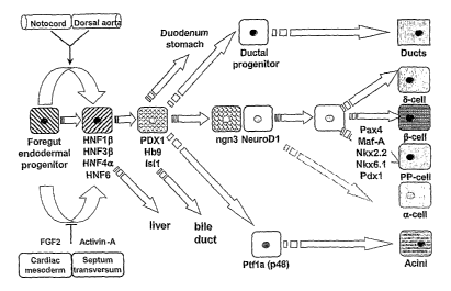
Figure 1 depicts the regulation of endoderm specification, foregut and
midgut en.doderm specification and subsequently pancreas specification by a
complement of specific transcription factors.
Figure 2 depicts the phenotype of low-Oz mouse MAPCs. mMAPCs
were derived and maintained at 5% 02. (A) Some clones have Oct-4 mItNA
expression at levels between 5 and 40% of embryonic stem cells (which is
>1,000 fold higher than in MAPCs isolated under normoxic (20% 02)
14
CA 02625883 2008-04-14
WO 2007/047509 PCT/US2006/040212
conditions). (B) This is confirmed by FACS for Oct-4 protein and by
intracellular staining for Oct-4. Compared to ES cells, MAPCs express Oct-4,
Rex-1, Fbx15, FoxD3, Egsl, Dnmt3l and Ecat7 at ES levels, but not Nanog,
Sox-2, Fgf4, Utfl, Eras, Ecatl and GDF3. Low-02 derived mouse MAPCs are
Scal, Thyl, CD34, CD3 1, MHC-class I and II, CD44 negative, but cKit
positive. Although mouse MAPCs express Oct-4 mRNA at levels similar to
ESCs, they do not form embryoid bodies or teratomas (5x10⁶ MAPCs grafted
under the skin of 5 nude inice). When MAPCs isolated under normoxic
conditions are subsequently switched to 5% 02 conditions, no changes in
transcriptional or cell surface phenotype are seen, suggesting that the
isolation
under low 02 may select for a more primitive cell population and that the
phenotype is not inducible in vitro.
Figure 3A depicts a pancreas differentiation protocol. 3B and 3C depict
various factors for use in differentiation of MAPCs towards a pancreatic fate
and
various transct-iption factors that can be expressed during the
differentiation
process.
Figure 4 depicts the morphological appearance of MAPC-beta-cell
differentiation cultures. By day 151arge patches of epithelioid cells can be
seen
in the adherent layer, surrounded by "stromal" looking cells. By day 18 these
patches start fonning three dimensional very well delineated clusters which
eventually bud off in the culture supernatant (day 21).
Figure 5 depicts the results of Q-RT-PCR evaluation of rat MAPCs
differentiated for 21 days towards endocrine pancreas. Rat MAPCs were plated
on matrigel using the sequential protocol described in Figure 3A. Every 3
days,
cultures were harvested (data for d18, 21 and 24 represent data on non-
attached
clusters only), RNA extracted, and. levels of transcription factors and
hormones
were measured by Q-RT-PCR compared with GAPDH as control, and compared
with levels detected in primary rat pancreas, except for Ngn-3, Nkx2.2 and
Neuro-Dl, where levels were compared with fetal rat RNA. Results shown are
mean +/- SEM for 3 experiments.
Figure 6 depicts immunohistology of clusters. Top panels: Clusters were
harvested on d15 and d21, dissociated with trypsin and stained with anti-Pdx-
1,
c-peptide and glucagon antibodies. Bottom panel: Clusters were harvested on
CA 02625883 2008-04-14
WO 2007/047509 PCT/US2006/040212
d21, and western blot performed with Abs against Pdx-1 andfl-actin. In both
panels, RIN cells serve as control.
Figure 7 depicts c-peptide secretion in vitro in response to 18mM
glucose. Cells were cultured with 3nM glucose, and from d16-24, a daily pulse
for lh of 18nM glucose was added to the cultures, after which the supernatant
was collected and c-peptide production measured by ELISA.
Figure 8 depicts functional K and Ca channels on beta-like cell clusters.
Left, an image of a cluster of cells that were loaded with fura-2 AM and
placed
on an inverted fluorescence microscope for video imaging. The plot on the
right
shows changes in intracellular calcium ion concentration ([Ca2}]i, as
reflected by
fura-2 ratio, in the region marked by a circle in the image. Increases in
([Ca2}];
were evoked by increasing extracellular K ion concentration to 50 mM (from 3
mM), and this increase was inhibited by the L-type calcium channel blocker
nifedipine (50 M).
Figure 9 depicts transplantation of endocrine pancreas differentiated rat
MAPCs in SZO treated nude mice. Blood glucose-levels (mg/dl) on the y axis;
time in days on the x axis.
Figure 10 depicts hematopoietic reconstitution from MAPCs. Six week-
old NOD-SCID mice received 1 million Tg-GFP MAPCs IV following 275cGy
irradiation, and under cover of anti-asialo-GM1 injection (d-1, dl l, 21).
After 16
weeks, the animals were sacrificed. PB, BM and spleen hematopoietic cells were
analyzed by FACS for presence of donor cells, and their lineage
differentiation.
Representative example of 1/21 engrafted mice.
Figure 11 depicts GFP}Insuliii donor islets in GFP MAPC grafted NOD-
SCID mice. A 6 week-old NOD-SCID mouse received 1 million Tg-GFP
MAPCs IV following 275cGy irradiation, and under cover of anti-asialo-GM1
injection (d-1, dl 1, 21). After 12 weeks, the animal was sacrificed. 70% of
the
PB, BM and spleen hematopoietic cells were GFP+. 7% GFY T cells were also
present. The pancreas of the animal was analyzed by anti-GFP-Abs combined
with anti-insulin Abs. Shown is a GFP+ islet and a GFP" islet from the saine
pancreas. (Example of 1 of 2 identical animals.)
16
CA 02625883 2008-04-14
WO 2007/047509 PCT/US2006/040212
Detailed Description of the Invention
Definitions
As used herein, the terms below are defined by the following meanings:
"MAPC" is an acronym for "multipotent adult progenitor cell." It is used
herein to refer to a non-embryonic stem (non-ES), non-germ, non-embryonic
geim (non-EG) cell that can give rise to (differentiate into) cell types of
more
thaii one embryonic lineage. It can form cell lineages of at least two germ
layers
(i.e., endoderm, mesoderm and ectoderm) upon differentiation. The term
"adult," with respect to MAPC is non-restrictive. It refers to a non-
embryoiiic
somatic cell.
"Multipotent" refers to the ability to give rise to cell types of more than
one embryonic lineage. "Multipotent," with respect to MAPC, is non-
restrictive.
MAPCs can form cell lineages of all three primitive germ layers (i.e.,
endoderm,
mesoderm and ectoderm). The term "progenitor" as used in the acronym
"MAPC" does not limit these cells to a particular lineage.
"Expansion" refers to the propagation of cells without differentiation.
"Progenitor cells" are cells produced during differentiation of a stem cell
that have some, but not all, of the characteristics of their tenninally-
differentiated progeny. Defined progenitor cells, such as "pancreatic
progenitor
cells," are committed to a lineage, but not to a specific or terminally-
differentiated cell type.
"Self-renewal" refers to the ability to produce replicate daugliter cells
having differentiation potential that is identical to those from which they
arose.
A similar term used in this context is "proliferation."
"Increased expression" of a marker (e.g., Pdx-1, Ngn3, NeuroD or insulin
1) refers to an increase (in mRNA and/or protein) relative to the parent cell
(a
cell prior to the recited treatment (e.g., contacting with Activin-A) and/or
treatments) on an average per cell basis (for example, if the parent cell does
not
express a marker and the progeny does, there is an increase in expression; or
if
the progeny expresses more of the marker compared to the parent cell there is
also an increase in expression). For example, increased expression of a marker
(e.g., Pdx-1) can be an increase in expression of up to about 1.01 fold, about
1.015 fold, about 1.02 fold, about 1.025 fold, about 1.03 fold, about 1.035
fold,
about 1.04 fold, about 1.045 fold, about 1.05 fold, about 1.055 fold, about
1.06
17
CA 02625883 2008-04-14
WO 2007/047509 PCT/US2006/040212
fold, about 1.065 fold, about 1.07 fold, about 1.075 fold, about 1.08 fold,
about
1.85 fold, about 1.9 fold, about 1.95 fold, about 2 fold (e.g., 2x), about 3
fold,
about 4 fold, about 5 fold, about 6 fold, about 7 fold, about 8 fold, about 9
fold,
about 10 fold, about 15 fold, about 20 fold, about 25 fold, about 30 fold,
about
35 fold, about 40 fold, about 45 fold, about 50 fold, about 55 fold, about 60
fold,
about 65 fold, about 70 fold, about 75 fold, about 80 fold, about 85 fold,
about
90 fold, about 95 fold, about 100 fold, about 150 fold, about 200 fold, about
250
fold, about 300 fold, about 350 fold, about 400 fold, about 450 fold, about
500
fold, about 600 fold, about 700 fold, about 800 fold, about 900 fold, about
1,000
fold, about 2,000 fold, about 3,000 fold, -about 4,000 fold, about 5,000 fold,
about 6,000 fold, about 7,000 fold, about 8,000 fold, about 9,000 fold, about
10,000 fold, about 15,000 fold, about 20,000 fold, about 25,000 fold, about
30,000 fold, about 35,0000 fold, about 40,000 fold, about 50,000 fold, about
60,000 fold, about 65,000 fold, about 70,000 fold, about 75,000 fold, about
80,000 fold, about 85,000 fold, about 90,000 fold, about 100,000 fold or
greater
as compared to the parent cell (on an average per cell basis).
An effective amount of an agent (e.g., Activin-A, an agent that inhibits
SHH, EGF, HGF, nicotinamide, exendin4, GDF11 or betacellulin) is an amount
effective to differentiate the cells as recited, when applied alone or in
combination with one or more other agents.
"Engraft" or "engraftment" refers to the process of cellular contact and
incorporation into an existing tissue or site of interest. In one embodiment,
greater than about 5%, greater than about 10%, greater than about 15%, greater
than about 20%, greater than about 25%, greater than about 30%, greater than
about 35%, greater than about 40%, greater than about 45%, greater than about
50%, greater than about 55%, greater than about 60%, greater than about 65%,
greater than about 70%, greater than about 75%, greater than about 80%,
greater
than about 85%, greater than about 90%, greater than about 95% or about 100%
of administered MAPCs or progeny derived therefrom engraft in the pancreas or
other tissues.
Persistence refers to the ability of cells to resist rejection and remain or
increase in number over time (e.g., days, weeks, months, years) in vivo. Thus,
by persisting, the MAPC or progeny can populate the pancreas or other tissues
or
remain in vivo, such as in barrier devices or other encapsulated forms.
18
CA 02625883 2008-04-14
WO 2007/047509 PCT/US2006/040212
"hnmunologic tolerance" refers to the survival (in amount and/or length
of time) of foreign (e.g., allogeneic or xenogeneic) tissues, organs or cells
in
recipient subjects. This survival is often a result of the inhibition of a
graft
recipient's ability to mount an immune response that would otherwise occur in
response to the introduction of foreign cells. Immune tolerance can encompass
durable immunosuppression of days, weeks, months or years. Included in the
definition of immunologic tolerance is NK-mediated immunologic tolerance.
This term also encompasses instances where the graft is tolerant of the host.
The term "isolated" refers to a cell or cells which are not associated with
one or more cells or one or more cellular components that are associated with
the
cell or cells in vivo. An "enriched population" refers to a relative increase
in
nuinbers of the cell of interest, such as MAPCs, relative to one or more other
cell
types, such as non-MAPC cell types, in vivo or in primary culture.
"Cytokines" refer to cellular factors that induce or enhance cellular
movement, such as homing of MAPCs or other stem cells, progenitor cells or
differentiated cells. Cytokines may also stimulate such cells to divide or
differentiate.
A "subject" or cell source can be a vertebrate, including a mammal, such
as a human. Marnmals include, but are not limited to, humans, farm animals,
sport animals and conlpanion animals. In included in the term "animal" is dog,
cat, fish, gerbil, guinea pig, hamster, horse, rabbit, swine, mouse, monkey
(e.g.,
ape, gorilla, chimpanzee, oraligutan) rat, sheep, goat, cow and bird.
As used herein, "treat," "treating" or "treatment" includes treating,
reversing, preventing, ameliorating, or inhibiting an injury or disease-
related
condition or a symptom of an injury or disease-related condition.
An "effective amount" generally means an amount which provides the
desired effect. For example, an effective dose is an amount sufficient to
effect a
beneficial or desired result, including a clinical result. The dose can be
administered in one or more administrations and can include any preselected
amount of cells. The precise determination of what would be considered an
effective dose may be based on factors individual to each subject, including
size,
age, injury or disease being treated and amount of time since the injury
occurred
or the disease began. One skilled in the art, particularly a physician, would
be
able to determine the number of cells that would constitute an effective dose.
19
CA 02625883 2008-04-14
WO 2007/047509 PCT/US2006/040212
Doses can vary depending on the mode of administration, e.g., local or
systemic;
free or encapsulated. The effect can be engraftment or other clinical
endpoints,
such as reversal or treatment of diabetes. Other effects can include providing
beta cells, recruiting endogenous cells, effecting angiogenesis, and/or
providing
pancreatic progenitors.
"Co-administer" can include sequential, simultaneous and/or separate
administration of two or more agents.
To provide pancreatic cells in a subject, several routes are possible. In
one embodiment MAPCs can be administered and allowed to provide pancreatic
cells in vivo. This can occur, as described herein, by differentiation of the
MAPCs themselves or by other means, such as by recruitment of endogenous
cells. Alternatively, more mature cells can be administered, these cells
having
been differentiated ex vivo from MAPC. Such cells include progeny at all
stages
of differentiation, including pancreatic progenitor cells that can give rise
to
mature pancreatic cell types, committed progenitor cells that cannot form
every
one of those types, and further differentiated types, which can include beta-
cells.
The tenns "comprises", "comprising", and the like can have the meaning
ascribed to them in U.S. Patent Law and can mean "includes", "including" and
the like. As used herein, "including" or "includes" or the like means
including,
without limitation.
MAPCs
MAPCs are non-embryonic (non-ES), non-germ and non-embryonic
germ (non-EG) cells that can differentiate into ectodermal, endodermal and
mesodermal cells types. MAPCs can be positive for telomerase. They can also
be positive for Oct-3A (Oct-3/4). MAPCs can differentiate in vivo where they
can form pancreatic cells, such as beta-cells. Alternatively, MAPCs can be
differentiated ex vivo into progeny cells with pancreatic phenotypes. MAPCs or
their differentiated progeny can be administered to a subject.
Human MAPCs from bone marrow are described in U.S. Patent No.
7,015,037 (PCT/US00/21387 (published as WO 01/11011)) and U.S. Patent
Application Serial No. 10/467,963 (PCT/US02/04652 (published as WO
02/064748)), the contents of which are incorporated herein by reference for
their
description of MAPCs. MAPCs have been identified in other mammals. Murine
MAPCs, for exainple, are also described in U.S. Patent No. 7,015,037 and U.S.
CA 02625883 2008-04-14
WO 2007/047509 PCT/US2006/040212
Patent Application Serial No. 10/467,963, the contents of which are
incorporated
herein by reference for their description of murine MAPCs. Rat MAPCs are also
described in 10/467,963, the contents of which are incorporated herein by
reference for their description of rat MAPCs. Swine MAPCs are described in
Patent Application No. PCT/US2005/038979, the contents of which are
incorporated herein by reference for their description of swine MAPCs.
Cynomologous monkey MAPCs are described in Clavel et al. (2005) the
contents of which are incorporated herein by reference for their description
of
cynomologous monkey MAPCs.
Isolation and Growth
Methods of MAPC isolation for humans and mouse are described in U.S.
Patent No. 7,015,037 (PCT/US00/21387 (published as WO 01/11011)) and for
rat in U.S. Patent Application Serial No. 10/467,963 (PCT/US02/04652
(published as WO 02/064748)), and these methods, along with the
characterization of MAPCs disclosed therein, are incorporated herein by
reference. '
MAPCs were initially isolated from bone marrow, but were subsequently
established from other tissues, including brain and muscle (Jiang, Y., et al.,
2002). Thus, MAPCs can be isolated from multiple sources, including, but not
limited to, bone marrow, placenta, umbilical cord and cord blood, muscle,
brain,
liver, spinal cord, blood or skin. For example, MAPCs can be derived from bone
marrow aspirates, which can be obtained by standard means available to those
of
skill in the art (see, for exainple, Muschler, G.F., et al., 1997; Batinic,
D., et al.,
1990). It is therefore now possible for one of skill in the art to obtain bone
marrow aspirates, brain or liver biopsies and other organs, and isolate the
cells
using positive or negative selection techniques available to those of skill in
the
art, relying upon the genes that are expressed (or not expressed) in these
cells
(e.g., by fiulctional or morphological assays, such as those disclosed in the
above-referenced applications, which have been, incorporated herein by
reference
for teaching such assays).
MAPCs from Human Bone Marrow as Described in U.S. 7,015 037
Bone marrow mononuclear cells were derived from bone marrow
aspirates, which were obtained by standard means available to those of skill
in
the art (see, for exainple, Muschler, G.F. et al. 1997; Batinic, D. et al.
1990).
21
CA 02625883 2008-04-14
WO 2007/047509 PCT/US2006/040212
Multipotent adult stem cells are present within the bone marrow (or other
organs
sucli as liver or brain), but do not express the common leukocyte antigen CD45
or erythroblast specific glycophorin-A (Gly-A). The mixed population of cells
was subjected to a Ficoll Hypaque separation. The cells were then subjected to
negative selection using anti-CD45 and anti-Gly-A antibodies, depleting the
population of CD45+ and Gly-A+ cells, and the remaining approximately 0.1% of
marrow mononuclear cells were then recovered. Cells can also be plated in
fibronectin-coated wells and cultured as described below for 2-4 weeks to
deplete the cell population of CD45+ and Gly-A+ cells.
Alternatively, positive selection can be used to isolate cells via a
combination of cell-specific markers. Both positive and negative selection
techniques are available to those of skill in the art,, and numerous
monoclonal
and polyclonal antibodies suitable for negative selection purposes are also
available in the art (see, for example, Leukocyte Typing V, Schlossman, et
al.,
Eds. (1995) Oxford University Press) and are commercially available from a
number of sources.
Techniques for mammalian cell separation from a mixture of cell
populations have also been described by, for example, Schwartz, et al., in U.
S.
Patent No. 5,759,793 (magnetic separation), Basch et al. 1983 (immunoaffinity
chromatography), and Wysocki and Sato 1978 (fluorescence-activated cell
sorting).
Recovered CD45-/GlyA- cells were plated onto culture dishes coated with
about 5-115 ng/ml (about 7-10 ng/ml can be used) serum fibronectin or other
appropriate matrix coating. Cells were maintained in Dulbecco's Minimal
Essential Medium (DMEM) or other appropriate cell culture medium,
supplemented with about 1-50 ng/ml (about 5-15 ng/ml can be used) platelet-
derived growth factor-BB (PDGF-BB), about 1-50 ng/ml (about 5-15 ng/ml can
be used) epidermal growth factor (EGF), about 1-50 ng/ml (about 5-15 ng/ml
can be used) insulin-like growth factor (IGF), or about 100-10,000 IU (about
1,000 IIJ can be used) LIF, witli about 10-10 to about 10-81VI dexamethasone
or
other appropriate steroid, about 2-10 g.g/ml linoleic acid, and about 0.05-
0.15
M ascorbic acid. Other appropriate media include, for example, MCDB,
MEM, IMDM and RPMI. Cells can either be maintained without serum, in the
22
CA 02625883 2008-04-14
WO 2007/047509 PCT/US2006/040212
presence of about 1-2% fetal calf serum, or, for example, in about 1-2% human
AB serum or autologous seruin.
When re-seeded at about 2x10³ cells/cm2 about every 3 days, >40 cell
doublings were routinely obtained, and some populations underwent >70 cell
doublings. Cell doubling time was about 36-48h for the initial 20-30 cell
doublings. Afterwards cell-doubling time was extended to as much as 60-72h.
Telomere length of MAPCs from 5 donors (age about 2 years to about 55
years) cultured at re-seeding densities of about 2x10³ cells/cmz for about 23-
26
cell doublings was between about 11-13 KB. This was about 3-5 KB longer
than telomere lengtli of blood lymphocytes obtained from the same donors.
Telomere length of cells from 2 donors evaluated after about 23 and about 25
cell doublings, respectively, and again after about 35 cells doublings, was
unchanged. The karyotype of these MAPCs was normal.
Phenotype of Human MAPCs under Conditions Described in U S 7,015,037
hnmunophenotypic analysis by FACS of human MAPCs obtained after
about 22-25 cell doublings showed that the cells do not express CD31, CD34,
CD36, CD38, CD45, CD50, CD62E and -P, HLA-DR, Mucl8, STRO-1, cKit,
Tie/Tek; and express low levels of CD44, HLA-class I and ,62-microglobulin,
and express CD10, CD13, CD49b, CD49e, CDw90, Fikl (N>10).
23
CA 02625883 2008-04-14
WO 2007/047509 PCT/US2006/040212
Once cells underwent >40 doublings in cultures re-seeded at about 2x 10³
cells/cm2, the phenotype became more homogenous and no cell expressed HLA
class-I or CD44 (n=6). When cells were grown at higher confluence, they
expressed high levels of Muc18, CD44, HLA class I and 02-microglobulin,
which is similar to the pheiiotype described for MSC (N=8) (Pittenger, 1999).
Immunohistochemistry showed that human MAPCs grown at about
2x10³ cells/cm2 seeding density express EGF-R, TGF-R1 and -2, BMP-R1A,
PDGF-R1A and -B, and that a small subpopulation (between about 1 and about
10%) of MAPCs stain with anti-SSEA4 antibodies (Kannagi, R 1983).
Using Clontech cDNA arrays the expressed gene profile of human
MAPCs cultured at seeding densities of about 2x10³ cells/cm2 for about 22 and
about 26 cell doublings was detei7nined:
A. MAPCs did not express CD31, CD36, CD62E, CD62P, CD44-H, cKit,
Tie, receptors for ILl, IL3, IL6, IL11, G CSF, GM-CSF, Epo, F1t3-L, or CNTF,
and low levels of HLA-class-I, CD44-E and Muc-18 mRNA.
B. MAPCs expressed mRNA for the cytokines BMP1, BMP5, VEGF, HGF,
KGF, MCP1; the cytokine receptors Flkl, EGF-R, PDGF-R1ca, gp130, LIF-R,
activin-R1 and -R2, TGFR-2, BMP-RlA; the adhesion receptors CD49c,
CD49d, CD29; and CD 10.
C. MAPCs expressed mRNA for hTRT and TRF1; the POU domain
transcription factor Oct-4, sox-2 (required with Oct-4 to maintain
undifferentiated state of ES/EC, Uwanogho D. 1995), sox 11 (neural
development), sox 9 (chondrogenesis) (Lefebvre V. 1998); homeodeomain
transcription factors: Hoxa4 and -a5 (cervical and thoracic skeleton
specification; organogenesis of respiratory tract) (Packer, A.I. 2000), Hox-a9
(myelopoiesis) (Lawrence, H. 1997), D1x4 (specification of forebrain and
peripheral structures of head) (Akimenko, M.A. 1994), MSX1 (embryonic
mesoderm, adult heart and muscle, chondro- and osteogenesis) (Foerst-Potts, L.
1997), PDX1 (pancreas) (Offield, M.F. 1996).
D. Presence of Oct-4, LIF-R, and hTRT mRNA was confinned by RT-PCR.
E. In addition, RT-PCR showed that Rex-1 mRNA and Rox-1 mRNA were
expressed in MAPCs.
MAPCs were also demonstrated to be CD 105 and CD106 negative.
24
CA 02625883 2008-04-14
WO 2007/047509 PCT/US2006/040212
Oct-4, Rex-1 and Rox-1 were expressed in MAPCs derived from human
and murine marrow and from murine liver and brain. Human MAPCs expressed
LIF-R and stained positive with SSEA-4. Finally, Oct-4, LIF-R, Rex-1 and Rox-
1 mRNA levels were found to increase, in human MAPCs cultured beyond 30
cell doublings, which resulted in phenotypically more homogenous cells. In
contrast, MAPCs cultured at high density lost expression of these markers.
This
was associated with senescence before about 40 cell doublings and loss of
differentiation to cells other than chondroblasts, osteoblasts and adipocytes.
Culturing MAPCs as Described in U.S. 7,015,037
MAPCs isolated as described herein can be cultured using methods
disclosed herein and in U.S. 7,015,037, which is incorporated by reference for
these methods.
Briefly, for the culture of MAPCs, culture in low-serum or serum-free
medium was preferred to maintain the cells in the undifferentiated state.
Medium used to culture the cells, as described herein, was supplemented as
described in Table 1. Human MAPCs do not require LIF.
Table 1
Insulin about 10 - 50 g/ml (about 10 g/ml)*
Transferrin about 0 - 10 g/ml (about 5.5 .g/ml)
Selenium about 2 - 10 ng/ml (about 5 ng/ml)
Bovine serum albumin (BSA) about 0.1 - 5 g/ml (about 0.5 g/ml)
Linoleic acid about 2 - 10 g/ml (about 4.7 g/ml)
Dexamethasone about 0.005 = 0.15 4M (abou0.01 M)
L-ascorbic acid 2-phosphate about 0.1 mM
Low-glucose DMEM (DMEM-LG) about 40 - 60% (about 60%)
MCDB-201 about 40 - 60% (about 40%)
Fetal calf serum about 0-2%
Platelet-derived growth about 5 - 15 ng/ml (about 10 ng/ml)
Epidennal growth factor about 5 - 15 ng/ml (about 10 ng/ml)
Insulin like growth factor about 5 - 15 ng/ml (about 10 ng/ml)
Leukemia inhibitory factor about 10-10,0001U (about 1,000 IU)
* Preferred concentrations are shown in parentheses.
Addition of about 10 ng/mL LIF to human MAPCs did not affect short-
term cell growth (same cell doubling time till 25 cell doublings, level of Oct
4
(Oct 3/4) expression). In contrast to what was seen with huinan cells, when
fresh murine marrow mononuclear cells, depleted on day 0 of CD45+ cells, were
plated in MAPC culture, no growth was seen. When murine marrow
mononuclear cells were plated, and cultured cells 14 days later depleted of
CA 02625883 2008-04-14
WO 2007/047509 PCT/US2006/040212
CD45+ cells, cells with the morphology and phenotype similar to that of human
MAPCs appeared. This suggested that factors secreted by hematopoietic cells
were needed to support initial growth of murine MAPCs. When cultured with
PDGF-BB and EFG alone, cell doubling was slow (>6 days) and cultures could
not be maintained beyond about 10 cell doublings. Addition of about 10 ng/mL
LIF significantly eillianced cell growth.
Once established in culture, cells can be frozen and stored as frozen
stocks, using DMEM with about 40% FCS and about 10% DMSO. Other
methods for preparing frozen stocks for cultured cells are also available to
those
of skill in the art.
Thus, MAPCs can be maintained and expanded in culture medium that is
available to the art. Such media include, but are not limited to, Dulbecco's
Modified Eagle's Medium (DMEM), DMEM F 12 medium , Eagle's
Minimum Essential Medium , F-12K medium , Iscove's Modified Dulbecco's
Medium , RPMI-1640 medium . Many media are also available as a low-
glucose formulation, with or without sodium pyruvate.
Also contemplated is supplementation of cell culture medium with
mammalian sera. Sera often contain cellular factors and components that are
necessary for viability and expansion. Examples of sera include fetal bovine
serum (FBS), bovine serum (BS), calf serum (CS), fetal calf serum (FCS),
newborn calf serum (NCS), goat serum (GS), horse serum. (HS), human serum,
. chicken serum, porcine seruin, sheep serum, rabbit serum, serum
replacements,
and bovine embryonic fluid. It is understood that sera can be heat-inactivated
at
about 55-65 C if deeined necessary to inactivate components of the complement
cascade.
Additional supplements can also be used advantageously to supply the
cells with the trace elements for optimal growth and expansion. Such
supplements include insulin, transferrin, sodium selenium and combinations
thereof. These components can be included in a salt solution such as, but not
limited to Hanks' Balanced Salt Solution (HBSS), Earle's Salt Solution ,
antioxidant suppleinents, MCDB-201 supplements, phosphate buffered saline
(PBS), ascorbic acid and ascorbic acid-2-phosphate, as well as additional
amino
acids. Many cell culture media already contain amino acids; however some
26
CA 02625883 2008-04-14
WO 2007/047509 PCT/US2006/040212
require supplementation prior to culturing cells. Such amino acids include,
but
are not limited to, L-alanine, L-arginine, L-aspartic acid, L-asparagine, L-
cysteine, L-cystine, L-glutainic acid, L-glutamine, L-glycine, L-histidine, L-
isoleucine, L-leucine, L-lysine, L-methionine, L-phenylalanine, L-proline, L-
serine, L-threonine, L-tryptophan, L-tyrosine and L-valine. It is well within
the
skill of one in the art to determine the proper concentrations of these
supplements.
Antibiotics are also typically used in cell culture to mitigate bacterial,
mycoplasmal and fungal contamination. Typically, antibiotics or anti-mycotic
compounds used are mixtures of penicillin/streptomycin, but can also include,
but are not limited to, amphotericin (Fungizone ), ampicillin, gentamicin,
bleoinycin, hygromycin, kanamycin, mitomycin, mycophenolic acid, nalidixic
acid, neomycin, nystatin, paromomycin, polymyxin, puromycin, rifampicin,
spectinomycin, tetracycline, tylosin and zeocin. Antibiotic and antimycotic
additives can be of some concern, depending on the type of work being
performed. One possible situation that can arise is an antibiotic-containing
media wherein bacteria are still present in the culture, but the action of the
antibiotic performs a bacteriostatic rather than bacteriocidal mechanism.
Also,
antibiotics can interfere with the metabolism of some cell types.
Hormones can also be advantageously used in cell culture and include,
but are not limited to, D-aldosterone, diethylstilbestrol (DES),
dexamethasone,
(3-estradiol, hydrocortisone, insulin, prolactin, progesterone,
somatostatin/human
growth hormone (HGH), thyrotropin, thyroxine -and L-thyronine.
Lipids and lipid carriers can also be used to supplement cell culture
media, depending on the type of cell and the fate of the differentiated cell.
Such
lipids and carriers can include, but are not limited to cyclodextrin (a, 0,
y),
cholesterol, linoleic acid conjugated to albumin, linoleic acid and oleic acid
conjugated to albumin, unconjugated linoleic acid, linoleic-oleic-arachidonic
acid conjugated to albumin, oleic acid unconjugated and conjugated to albumin,
among others.
Also contemplated is the use of feeder cell layers. Feeder cells are used
to support the growth of fastidious cultured cells, including stem cells.
Feeder
cells are normal cells that have been inactivated by y-irradiation. In
culture, the
feeder layer serves as a basal layer for other cells and supplies cellular
factors
27
CA 02625883 2008-04-14
WO 2007/047509 PCT/US2006/040212
without further growth'or division of their own (Lim, J.W. and Bodnar, A.,
2002). Examples of feeder layer cells are typically human diploid lung cells,
mouse embryonic fibroblasts, Swiss mouse embryonic fibroblasts, but can be
any post-mitotic cell that is capable of supplying cellular components and
factors
that are advantageous in allowing optimal growth, viability and expansion of
stein cells. In many cases, feeder cell layers are not necessary to keep the
ES
cells in an undifferentiated, proliferative state, as leulcemia inhibitory
factor
(LIF) has anti-differentiation properties. Therefore, supplementation with LIF
can be used to maintain MAPC in some species in an undifferentiated state.
Cells in culture can be maintained either in suspension or attached to a
solid support, such as extracellular matrix components and synthetic or
biopolymers. Stem cells often require additional factors that encourage their
attachinent to a solid support, such as type I, type II and type IV collagen,
concanavalin A, chondroitin sulfate, fibronectin, "superfibronectin" and
fibronectin-like polyiners, gelatin, lanlinin, poly-D and poly-L-lysine,
thrombospondin and vitronectin.
The maintenance conditions of stein cells can also contain cellular factors
that allow stem cells, such as MAPCs, to remain in an undifferentiated form.
It
is advantageous under conditions where the cell must remain in an
-undifferentiated state of self-renewal for the medium to contain epidermal
growth factor (EGF), platelet derived growth factor (PDGF), leukemia
inhibitory
factor (LIF; in selected species), and combinations thereof. It is apparent to
those skilled in the art that supplements that allow the cell to self-renew
but not
differentiate should be removed from the culture medium prior to
differentiation.
Stem cell lines and other cells can benefit from co-culturing with another
cell type. Such co-culturing methods arise from the observation that certain
cells
can supply yet-unidentified cellular factors that allow the stem cell to
differentiate into a specific lineage or cell type. These cellular factors can
also
induce expression of cell-surface receptors, some of which can be readily
identified by monoclonal antibodies. Generally, cells for co-culturing are
selected based on the type of lineage one skilled in the art wishes to induce,
and
it is within the capabilities of the skilled artisan to select the appropriate
cells for
co-culture.
28
CA 02625883 2008-04-14
WO 2007/047509 PCT/US2006/040212
Differentiation of MAPCs to Pancreatic Cells
MAPCs and pancreatic progenitor cells differentiated from MAPCs are
useful as a source of pancreatic cells. The maturation, proliferation and
differentiation of MAPCs may be effected through culturing MAPCs with
appropriate factors including, but not limited to, activin-A (or other members
TGF(3 family), BMP4 (or other members of the BMP family), an agent that
inhibits sonic hedgehog activity (including, but not limited to, cyclopamine
and
anti-SHH antibody), EGF or HGF (or other mitogenic proteins), nicotinamide
(and possibly nicotinic acid), exendin (including, but not limited to, exendin
4
and exenatide (a 39-amino acid peptide which closely resembles exendin-4),
GDF11 (or other members of the bone morphogenetic protein/transforming
growth factor beta (BMP/TGFbeta) superfamily), betacellulin, or with stromal
cells or other cells which secrete factors responsible for stem cell
regeneration,
commitment and differentiation.
An agent that inhibits sonic hedgehog (SHH) activity (e.g., signaling)
includes any agent (e.g., a peptide, protein, including antibodies, small
molecule,
drug, chemical, or nucleic acid, such as DNA or RNA) which inhibits the
function or expression of sonic hedgehog (including, but not limited to,
providing signal(s) in the patterning of the early embryo, such as patterning
of
the ventral neural tube, the anterior-posterior limb axis, and the ventral
somites).
Such agents include, but are not limited to, an anti-sonic hedgehog antibody,
cyclopamine (CPA), analogs thereof, such as cyclopamine-4-ene-3-one or other
steroidal alkaloids. As used herein, "inhibit" refers to a reduction (e.g.,
about
5%, about 10%, about 15%, about 20%, about 25%, about 30%, about 35%,
about 40%, about 45%, about 50%, about 55%, about 60%, about 65%, about
70%, about 75%, about 80%, about 85%, about 90%, about 95% or about 100%)
in the activity of sonic hedgehog as compared to the activity of SHH in the
absence of an agent that inhibits SHH activity.
As described in Example 2 herein below, MAPCs were differentiated
into pancreatic progenitor cells and beta-cells in vitro. Briefly, MAPCs were
cultured in medium containing Activin-A (about 0.5 ng/mL to about 200 ng/mL,
such as about 50 ng/mL to about 100 ng/mL), BMP-4 (about 10 ng/mL to about
100 ng/mL, such as about 20 ng/mL to about 30 ng/mL or about 50 nglmL) for
about 3 days, followed by about six days of culture in Activin A, BMP-4 and
29
CA 02625883 2008-04-14
WO 2007/047509 PCT/US2006/040212
cyclopamine (e.g., about 5 to about 50 M, including about 10 M) or an anti-
SHH antibody (about 10 ,ug/mL) for about six days. The cells obtained
therefrom were next cultured in medium containing EGF (e.g., about 5 to about
100 ng/mL, including about 50 ng/mL) or HGF (e.g., about 5 to about 100
ng/mL, including about 50 ng/mL) for about 6 days. The cells obtained
therefrom were then cultured in medium containing nicotinamide (about 5 M to
about 50 M, including about 10 M) or exendin4 (e.g., about 5 nM to about 50
nM, including about 10 nM), GFD11 (e.g., about 10 ng/mL to about 100 ng/mL,
including about 50 ng/mL), and betacellulin (e.g., about 10 ng/mL to about 100
ng/mL, including about 50 ng/mL) for abotit six days.
Additional factors to enhance the initial commitment of MAPCs to
pancreatic endoderm (Pdx-1 positive cells on day 9) can,include factors known
to play a role in endoderm commitment, such as members of the Wnt family,
TGF-0 family, and FGF fainily. Wnt-3 plays a role in endoderm specification.
(Heller et al., 2002), as Wnt3"/- mice do not form endoderm or mesoderm
(Heller
et al., 2002). Pancreatic, but not hepatic, endoderm specification is
regulated by
members of the Wnt family. Compared with initial endoderm specification,
which may depend on Wnt-3, Wnts may inhibit pancreatic endoderm
specification. Dickkopf related protein 1(Dkk-1), a member of the Dkk protein
family of secreted proteins, antagonizes the canonical Wnt pathway by direct
high-affinity binding to the Wnt coreceptor LRP5/6 and inhibiting interaction
of
LRP5/6 with the Wnt-Frizzled complex (Nusse 2001). Thus, addition of Dkk-1
or an inhibitor of 0-catenin (e.g., a GSK3 inhibitor such as GSKSp inhibitor
IX)
(Willert et al., 1998) can increase the frequency of Pdx-1 positive
progenitors
generated from MAPCs.
Nodal also plays a role in endoderm specification, as Nodal-/- mice do not
form endoderm or mesodenn. BMP-4, and other TGF family members, induces
mesoderrn rather than endoderm specification, as BMP-4-/- embryos die early in
gestation without forming any organized mesoderm (Winnier et al., 1995). In
vitro studies in which ES cell differentiation to endoderm is evaluated,
demonstrated that Activin-A at high concentrations specifies cells to endoderm
but not mesoderm. In subsequent steps needed for pancreatic endoderm
specification (Kubo et al., 2004; D'Amour et al., 2005), Activin-A (Maldonado
CA 02625883 2008-04-14
WO 2007/047509 PCT/US2006/040212
et al., 2000) alone or combined with bFGF (Hardikar et al., 2003) inhibits SHH
expression, which prevents pancreatic endodenn specification.
FGF-8 may play a role in initial endoderm specification. Similarly, FGF-
4 has been identified, at least in chicken, to play a role in endodenn
specification
(Wells et al., 2000). The role of FGF-2 in pancreas vs. liver specification is
more complex. As indicated above FGF-2 iiihibits SHH production (Hardikar et
al., 2003; Jung et al., 1999), which should lead to paincreas specification.
Thus,
FGF-8, FGF4, FGF-2 or combination thereof can be used in the method to
differentiate MAPCs (also in combination with other factors mentioned herein).
Pancreas commitment from the ventral as well as dorsal foregut
endodenn is inhibited by sonic hedgehog (SHH) (Hebrok et al., 2000). Activin-
A (Maldonado et al., 2000) and/or FGF-2 (Hardikar et al., 2003) signals from
the
notochord act to repress SHH expression in pre-pancreatic endoderm.
Alternatively, or in addition, SHH can be blocked with cyclopamine, or
specific
anti-SHH antibodies.
Pancreas specification and differentiation is regulated by Notch signaling
at multiple steps (Hardikar et al., 2003). Specifically, Notch signaling
prevents
commitment to pancreas, to endocrine pancreas and maturation of endocrine
Ngn3 pancreatic progenitors. Thus, inhibitors of Notch signaling can be using
in the methods to differentiate MAPCs.
Although soine experiments indicted that the combination of activin-A
and BMP4 was superior to activin-A alone, to induce Pdx-1 expression, BMP4
may be responsible for the mesodermal cells that are also present in culture.
Thus, BMP4 many not be necessary, may be substituted with BMP2 or BMP7,
or may be used at lower concentrations or with BMP-4 for only a few days
followed by Activin-A alone. Additionally, Activin-A combined with Wnt-3 for
the initial three days of differentiation may further enhance endoderm
differentiation. As Wnts may inhibit pancreatic endoderm differentiation,
addition of Wnt-3 followed by Dldc-1 may induce greater numbers of Pdx-1
positive cells by day 9. This can be confirmed by adding a GSK3 inhibitor that
blocks the canonical Wnt pathway. The efficacy of Dkk-1 or GSK3 inhibitor
can be demonstrated by measuring levels of phosphorylated 0-catenin.
Subsequently, the role of different FGFs (FGF-8, FGF-4, and FGF-2), by
addition of graded doses of FGF-8 and/or FGF-2 and graded doses of FGF-4
31
CA 02625883 2008-04-14
WO 2007/047509 PCT/US2006/040212
and/or an inhibitor of FGF-2 (e.g., SU5402) can be determined. Additionally,
inliibiting Notch signaling, for example, by using an inhibitor of 'y-
secretase
(compound E, Calbiochem) can affect pancreatic endoderm (Pdx-1'- cell)
induction. The efficacy of the -y-secretase can be assessed by evaluating
expression of Hes1 and Herp.
In one embodiment, the cells are transfected with a pancreatic
transcription factor, including, but not limited to, Ngn3, NeuroD, Pdx-1,
Pax4,
Ptfla/p48, Pax6, N1x6.1, Nkx2.2 or a combination thereof, for example, by
DNA, RNA or viral transfection or by protein transduction. Expression of these
transcription factors can induce pancreatic differentiation of MAPCs.
Additionally, the endogenous factor can be activated or increased in the cell
by
methods know in the art (e.g., homolgous recombination (e.g., U.S. 5,641,670),
non-homologus recombination (e.g., U.S. 6,602,686; RAGE (Random
Activation of Gene Expression) technology; Athersys, Inc. (Cleveland, Ohio)),
or other endogenous expression techniques available to the art worker (the
above
mentioned patents are incorporated by reference for teaching of methods of
endogenous gene activation). In addition to the factors/genes described
herein,
variants, homologs or orthologs of the factors/genes, which have the same
biological function/acitivty, can be used or assayed for in methods of the
invention. For example, variants, homolog or orthologs of use in the present
invention may be homologous or have sequence identity (nucleotide or amino
acid sequence) with factors/genes involved pancreagenesis, including those
factors/genes provided herein. Assays and programs to determine if a
factor/gene is homolgous is are known in the art.
Methods of identifying and subsequently separating differentiated cells
from their undifferentiated counterparts can be carried out by methods well
known in the art and described herein. Cells that have been induced to
differentiate can be identified by selectively culturing cells under
conditions
whereby differentiated cells outnuinber undifferentiated cells. Similarly,
differentiated cells can be identified by morphological changes and
characteristics that are not present on their undifferentiated counterparts,
such as
cell size, the number of cellular processes, the complexity of intracellular
organelle distribution, and the production of insulin or C-peptide and the
secretion of insulin or C-peptide in response to glucose.
32
CA 02625883 2008-04-14
WO 2007/047509 PCT/US2006/040212
Also contemplated are methods of identifying differentiated cells by their
expression of specific cell-surface marlcers such as cellular receptors and
transmembrane proteins. Monoclonal antibodies against these cell-surface
marlcers can be used to identify differentiated cells. Detection of these
cells can
be achieved through fluorescence activated cell sorting (FACS) and enzyme-
linlced immunosorbent assay (ELISA). From the standpoint of transcriptional
upregulation of specific genes, differentiated cells often display levels of
gene
expression that are different (increased or decreased expression of mRNA or
protein) from undifferentiated cells, such as insulin-1, insulin-2, glucagon,
somatostatin, NeuroDl, Pdx-1, Ngn3, Nkx6.1, Nkx2.2. Reverse-transcription
polymerase chain reaction (RT-PCR) can be used to monitor such changes in
gene expression during differentiation. In addition, whole genome analysis
using microarray technology can be used to identify differentiated cells.
Accordingly, once differentiated cells are identified, they can be
separated from their undifferentiated counterparts, if necessary. The methods
of
identification detailed above also provide methods of separation, such as
FACS,
preferential cell culture methods, ELISA, magnetic beads, and combinations
thereof. A preferred einbodiment of the invention envisions the use of FACS to
identify and separate cells based on cell-surface antigen expression.
Additional Culture Methods
The density at which MAPCs are cultured can vary from about 100
cells/cm2 or about 150 cells/cm2 to about 10,000 cells/cin2, including about
200
cells/cm2 to about 1500 cells/cm2 to about 2,000 cells/cm2. The density can
vary
between species. Additionally, optimal density can vary depending on culture
conditions and source of cells. It is within the skill of the ordinary artisan
to
deternZine the optimal density for a given set of culture conditions and
cells.
Also, in specific embodiments the atmospheric oxygen concentration for
isolating, culturing, expanding and/or differentiation of cells includes
oxygen
concentrations between about 0.1% to about 10% oxygen. In other
embodiments, the atmospheric oxygen concentration includes oxygen
concentrations between about 1% to about 9%. In other enzbodiments, the
atmospheric oxygen concentration includes oxygen concentrations between
about 1.5% to about 8%. In additional embodiments, the atmospheric oxygen
concentrations include oxygen concentrations between about 2% to about 7%
33
CA 02625883 2008-04-14
WO 2007/047509 PCT/US2006/040212
oxygen.
The above ranges are exemplary ranges of atmospheric oxygen
concentrations to be used in the culture of non-ES, non-EG, non-germ cells
that
are Oct3/4 positive and can differentiate into ectodermal, en.dodermal, and
mesodermal cell types and it should be understood that those of skill in the
art
will be able to einploy oxygen concentrations falling within any of these
ranges.
Thus, one of slcill in the art could set the oxygen culture concentrations at
about
0.1%, 0.5%, .5%, 2%, 2.5%, 3%, 3.5%, 4%, 4.5%, 5%, 5.5%, 6%, 6.5%, 7%.,
7.5%, 8%, 8.5%, 9%, 9.5%, 10%, or any other oxygen concentration between
any of these percentages. hi one embodiment, the oxygen concentration is about
2% to about 7%, such about 5%, which more closely approximates physiological
oxygen concentrations (Guyton and Hall, in Textbook of Medical Physiology,
W.B. Saunders Co., Philadelphia, pp. 513-523 (1996)). The oxygen
concentration can be varied within a given range during the culturing period.
The remainder of the atmospheric gases are conventional, inert gases, e.g.,
nitrogen, argon and the like, as well as carbon dioxide. Additionally, the
cells of
the invention are cultured in about 5% to about 6% COz.
Isolating and culturing MAPCs at 5% 02 was shown to result in fewer
cytogenetic abnormalities. Additionally, it resulted in a slight change in the
phenotype of MAPCs. When rodent MAPCs are isolated and maintained at 5%
02, Oct-4 transcript levels approach those of embryonic stem (ES) cells (50-
80%), and >90% of cells express nuclear Oct-4 protein by
immunohistochemistry. 5%-02 derived rodent MAPCs also express Rex-1 at
levels approaching that of ES cells. Although mouse MAPCs expressed Oct-4
mRNA at levels similar to ES cells, they did not form embryoid bodies or
teratomas (5xl06 MAPCs grafted under the skin of 5 nude mice).
MAPCs can also be cultured in the presence of a GSK3 inhibitor (e.g., a
6-bromoindirubin compound, including but not limited to, 6-bromoindirubin-3'-
oxiine (also known as BIO); incorporated herein by reference are U.S.
Provisional Patent Application Nos. 60/703,823 (filed July 29, 2005) and
60/704,169 (filed July 29, 2005) and PCT applications PCT/US2006/029736
(filed July 31, 2006) and PCT/US2006/029547 (filed July 31, 2006) for the
disclosure of culturing cells in the present of a GSK3 inhibitor).
34
CA 02625883 2008-04-14
WO 2007/047509 PCT/US2006/040212
Use of MAPCs and Progeny Therefrom
The pancreatic progenitor or beta-cells of the invention and/or the
MAPCs can be used to repopulate a pancreas by either direct introduction into
the area of dasnage or by systemic administration, which allows the cells to
home to the area of damage. Accordingly, the invention provides methods of
treating a subject in need of pancreatic cells comprising administering to a
subject an effective amount of the pancreatic progenitor cells of the
invention or
MAPCs.
For the purposes described herein, either autologous, allogeneic or
xenogeneic cells can be administered to a patient, either in undifferentiated,
terminally differentiated or in a partially differentiated form, genetically
altered
or unaltered, by direct introduction to a site of interest, e.g., on or around
the
surface of an acceptable matrix, or systemically, in combination with a
pharmaceutically acceptable carrier so as to repair, replace or promote the
growth of existing and/or new pancreatic cells.
Generally, the invention provides methods to treat a pancreatic disorder.
The term "pancreatic disorder" or "pancreatic disease" refers to a state where
pancreatic function is impaired. Examples of "pancreatic disorders" or
"pancreatic diseases" that can be treated with the compositions and methods of
the invention include, but are not limited to, diabetes (including Type 1,
Type 2,
MODY and other genetic causes of diabetes), obesity, pancreatic atresia,
pancreas inflammation, alphal-antitrypsin deficiency, acute, chronic or
hereditary pancreatitis, pancreatic cancer (including endocrine tumors of the
pancreas), pancreas malfunction due to cystic fibrosis or Shwachman Diamond
syndrome, pancreatic insufficiency or pancreatic enzyme deficiency, pancreatic
cysts, hyperinsulinism, pancreatic digestive diseases, genetic disorders of
the
exocrine pancreas and pancreatic injury, including, but not limited to, injury
as a
result of physical trauma (including, but not limited to, surgery), chemical,
radiological, aging, and/or disease.
Administration of MAPCs or Their Differentiated Progeny
1VIAPCs, or their differentiated progeny, can be administered to a subject
by a variety of methods available to the art, including but not limited to
localized
injection, catheter administration, systemic injection, intraperitoneal
injection,
parenteral administration, intra-arterial injection, intravenous injection,
CA 02625883 2008-04-14
WO 2007/047509 PCT/US2006/040212
transvascular injection, intrainuscular injection, surgical injection into a
tissue of
interest (e.g., injection into the pancreas) or via direct application to
tissue
surfaces (e.g., during surgery or on a wound).
MAPCs can be administered either peripherally or locally through the
circulatory system. "Homing" of stem cells would concentrate the implanted
cells in an environment favorable to their growth and function. Pre-treatnlent
of
a patient with cytokine(s) to promote homing is another alternative
contemplated
in tlie methods of the present invention. Certain cytolcines (e.g., cellular
factors
that induce or enhance cellular movement, such as homing of MAPCs or other
stem cells, progenitor cells or differentiated cells) can enhance the
migration of
MAPCs or their progeny. Cytokines include, but are not limited to, stromal
cell
derived factor-1 (SDF-1), stem cell factor (SCF), angiopoietin-1, placenta-
derived-growtli factor (PIGF) and granulocyte-colony stimulating factor (G-
CSF). Cytokines also include any which promote the expression of endothelial
adhesion molecules, such as ICAMs, VCAMs and others, which facilitate the
hoining process:
Viability of newly forming tissues can be enhanced by angiogenesis.
Factors promoting angiogenesis include, but are not limited to, VEGF, aFGF,
angiogenin, angiotensin-1 and -2, betacellulin, bFGF, Factor X and Xa, HB-
EGF, PDGF, angiomodulin, angiotropin, angiopoetin-1, prostaglandin El and
E2, steroids, heparin, 1-butyryl-glycerol and nicotinic amide.
Factors that decrease apoptosis can also promote the formation of new
tissue, such as pancreatic tissues. Factors that decrease apoptosis include
but are
not limited to (3-blockers, angiotensin-converting enzyme inhibitors (ACE
inhibitors), AKT, HIF, carvedilol, angiotensin II type 1 receptor antagonists,
caspase inhibitors, cariporide and eniporide.
Exogenous factors (e.g., cytokines, differentiation factors (e.g., cellular
factors, such as growth factors or angiogenic factors that induce lineage
commitment), angiogenesis factors and anti-apoptosis factors) can be
administered prior to, after or concomitantly with MAPCs or their
differentiated
progeny. For example, a form of concomitant administration would comprise
combining a factor of interest in the MAPC suspension media prior to
administration. Administrations are variable and may include an initial
administration followed by subsequent administrations.
36
CA 02625883 2008-04-14
WO 2007/047509 PCT/US2006/040212
A method to potentially increase cell survival is to incorporate MAPCs
or progeny into a biopolymer or synthetic polymer. Depending on the patient's
condition, the site of injection might prove inhospitable for cell seeding and
growth because of scarring or other impediments. Examples of biopolymer
include, but are not limited to, fibronectin, fibrin, fibrinogen, thrombin,
collagen
and proteoglycans. This can be constructed with or without included
cytolcines,
differentiation factors, angiogenesis factors or anti-apoptosis factors.
Additionally, these can be in suspension. Another alternative is a three-
dimensional gel with cells entrapped within the interstices of the cell
biopolymer
adinixture. Again cytokines, differentiation factors, angiogenesis factors,
anti-
apoptosis factors or a combination thereof can be included within the gel.
These
can be deployed by injection via various routes described herein.
The cells can also be encapsulated with a capsule that is permeable to
nutrients and oxygen while allowing appropriate cellular products (for
example,
insulin in the case of islet cells) to be released into the bloodstream or to
adjacent
tissues. In one embodiment, the capsular material is restrictive enough to
exclude immune cells and antibodies that could reject and destroy the
inzplant.
Such encapsulation can be achieved using, for example, polymers (Chang,
2000). Such polymeric encapsulation systems include, but are not limited to,
alginate (e.g., alginate bead), polysaccharide hydrrogels, chitosan, calcium
or
barium alginate, a layered matrix of alginate and polylysine, a
photopolymerizable poly(ethylene glycol) (PEG) polymer (Novocell, Inc.), a
polyanionic material termed Biodritin (US Patent 6,281,341), polyacrylates, a
photopolymerizable poly(ethylene glycol) polymer, and polymers such as
hydroxyetliyl methacrylate methyl methacrylate.
Another approach to encapsulate cells involves the use of
photolithography techniques adapted from the semiconductor industry to
encapsulate living cells in silicon capsules that have pores only a few
nanometers wide (Desai 2002).
Also, suitable immune-compatible polycations, including but not limited
to, poly-l-lysine (PLL) polycation or poly-l-ornithine or poly(methylene-co-
guanidine) hydrochloride, may be used to encapsulate cells.
Additionally, cells can be encapsulated with biocompatible
seinipermeable membranes to surround encapsulated cells, sometimes within a
37
CA 02625883 2008-04-14
WO 2007/047509 PCT/US2006/040212
capillary device, to create a miniature artificial organ, such as one that
would
include functional pancreas or liver cells (e.g., a liver or pancreatic
artificial
device). This is often called macroencapsulation. The membrane lets glucose,
oxygen, and insulin pass in and out of the blood stream, and preferably keeps
out
the antibodies and T cells of the immune system, which may destroy the cells
(e.g., islets). Such membranes can be used in a perfusion device, a capsule
that
is grafted to an artery where it makes direct contact with the body's
circulating
blood; in this way, the device can draw nutrients from the blood and release
insulin to circulate throughout the body. Another method provides for coating
a
small group of islet cells (macroencapsulation) or individual islet cells
(microencapsulation) and implanting them inside the abdominal cavity. In these
devices nutrients and insulin would be exchanged by way of the body fluids
permeating the tissues in which they are implanted.
The quantity of cells to be administered will vary for the subject being
treated. hi a preferred embodiment, between about 104 to about 108, more
preferably about 105 to about 107 and most preferably, about 3 x 10⁷ stem
cells
and optionally, about 50 to about 500 g/kg per day of a cytokine can be
administered to a human subject. However, the precise determination of what
would be considered an effective dose may be based on factors individual to
each patient, including their size, age, disease or injury, amount of damage,
amount of time since the damage occurred and factors associated with the mode
of delivery (direct injection - lower doses, intravenous - higher doses).
Dosages
can be readily ascertained by those skilled in the art from this disclosure
and the
knowledge in the art.
An issue regarding the use of stem cells or their progeny is the purity of
the enriched or isolated cell population. Bone marrow cells, for example,
comprise mixed populations of cells, which can be purified to a degree
sufficient
to produce a desired effect. Those skilled in the art can readily determine
the
percentage of MAPCs or progeny in a population using various well-known
metliods, such as fluorescence activated cell sorting (FACS). Preferable
ranges
of purity in populations comprising MAPCs, or their differentiated progeny,
are
about 50-55%, about 55-60%, and about 65-70%. More preferably the purity is
about 70-75%, about 75-80%, about 80-85%; and most preferably the purity is
about 85-90%, about 90-95%, and about 95-100%. However, populations with
38
CA 02625883 2008-04-14
WO 2007/047509 PCT/US2006/040212
lower purity can also be useful, such as about 25-30%, about 30-35%, about 35-
40%, about 40-45% and about 45-50%. Purity of MAPCs or their progeny can
be determined according to the gene expression profile within a population.
Dosages can be readily adjusted by those skilled in the art (e.g., a decrease
in
purity may require an increase in dosage).
The skilled artisan can readily determine the amount of cells and optional
additives, vehicles, or carrier in compositions to be administered in methods
of
the invention. Typically, additives (in addition to the active stem cell(s) or
cytokine(s)) are present in an amount of about 0.001 to about 50 wt % solution
in phosphate buffered saline, and the active ingredient is present in the
order of
micrograms to milligrams, such as about 0.0001 to about 5 wt %, preferably
about 0.0001 to about 1 wt %, most preferably about 0.0001 to about 0.05 wt %
or about 0.001 to about 20 wt %, preferably about 0.01 to about 10 wt %, and
most preferably about 0.05 to about 5 wt %. Of course, for any composition to
be administered to an aniinal or human, and for any particular method of
administration, it is preferred to determine therefore: toxicity, such as by
determining the lethal dose (LD) and LD50 in a suitable animal model e.g., a
rodent, such as mouse; and, the dosage of the composition(s), concentration of
components therein and timing of administering the coniposition(s), which
elicit
20- a suitable response. Such determinations do not require undue
experimentation
from the knowledge of the skilled artisan, this disclosure and the documents
cited herein. Additionally, the time for sequential administrations can be
ascertained without undue experimentation.
When adininistering a therapeutic composition of the present invention, it
can generally be formulated in a unit dosage injectable form (solution,
suspension, emulsion). The pharmaceutical formulations suitable for injection
include sterile aqueous solutions and dispersions. The carrier can be a
solverit or
dispersing medium containing, for example, water, saline, phosphate buffered
saline, polyol (for example, glycerol, propylene glycol, liquid polyethylene
glycol, and the like) and suitable mixtures thereof.
Additionally, various additives which enhance the stability, sterility, and
isotonicity of the compositions, including antimicrobial preservatives,
antioxidants, chelating agents and buffers, can be added. Prevention of the
action of microorganisms can be ensured by various antibacterial and
antifungal
39
CA 02625883 2008-04-14
WO 2007/047509 PCT/US2006/040212
agents, for example, parabens, chlorobutanol, phenol, sorbic acid, and the
like.
In many cases, it will be desirable to include isotonic agents, for example,
sugars, sodium chloride, and the like. Prolonged absorption of the injectable
pharmaceutical form can be brought about by the use of agents delaying
absorption, for example, aluminum monostearate and gelatin. According to the
present invention, however, any vehicle, diluent, or additive used should be
compatible with the cells.
Sterile injectable solutions can be prepared by incorporating the cells
utilized in practicing the present invention in the required amount of the
appropriate solvent with various amounts of the other ingredients, as desired.
In one embodiment, MAPCs, or differentiated progeny thereof, can be
administered initially, and thereafter maintained by further administration of
MAPCs or differentiated progeny thereof. For instance, MAPCs can be
administered by one method of injection, and thereafter further administered
by
a different or the same type of method.
It is noted that human subjects are treated generally longer than canines
or other experimental animals, such that treatment has a length proportional
to
the length of the disease process and effectiveness. The doses may be single
doses or multiple doses over a period of several days. Thus, one of skill in
the
art can scale up from animal experiments, e.g., rats, mice, canines and the
like, to
humans, by techniques from this disclosure and documents cited herein and the
lcnowledge in the art, without undue experimentation. The treatinent generally
has a length proportional to the length of the disease process and treatment
effectiveness and the subject being treated.
Examples of compositions coinprising MAPCs, or differentiated progeny
thereof, include liquid preparations for administration, including
suspensions,
and, preparations for direct or intravenous administration (e.g., injectable
administration), such as sterile suspensions or emulsions. Such compositions
may be in admixture with a suitable carrier, diluent, or excipient such as
sterile
water, physiological saline, glucose, dextrose, or the like. The compositions
can
also be lyophilized. The compositions can contain auxiliary substances such as
wetting or emulsifying agents, pH buffering agents, gelling or viscosity
enhancing additives, preservatives, flavoring agents, colors, and the like,
depending upon the route of administration and the preparation desired.
CA 02625883 2008-04-14
WO 2007/047509 PCT/US2006/040212
Standard texts, such as "REMINGTON'S PHARMACEUTICAL SCIENCE,"
17th edition, 1985, incorporated herein by reference, may be consulted to
prepare suitable preparations, without undue experimentation.
Compositions are conveniently provided as liquid preparations, e.g.,
isotonic aqueous solutions, suspensions, emulsions or viscous compositions,
wlzich may be buffered to a selected pH. Liquid preparations are normally
easier
to prepare than gels, other viscous compositions and solid compositions.
Additionally, liquid compositions are somewhat inore convenient to administer,
especially by injection. Viscous compositions, on the other hand, can be
formulated within the appropriate viscosity range to provide longer contact
periods with specific tissues.
The choice of suitable carriers and other additives will depend on the
exact route of administration and the nature of the particular dosage form,
e.g.,
liquid dosage form (e.g., whether the composition is to be formulated into a
solution, a suspension, gel or another liquid form, such as a time release
form or
liquid-filled fonn).
Solutions, suspensions and gels normally contain a major amount of
water (preferably purified, sterilized water) in addition to the cells. Minor
amounts of other ingredients such as pH adjusters (e.g., a base such as NaOH),
emulsifiers or dispersing agents, buffering agents, preservatives, wetting
agents
and jelling agents (e.g., methylcellulose), may also be present. The
compositions can be isotonic, i.e., they can have the same osmotic pressure as
blood and lacrimal fluid.
The desired isotonicity of the compositions of this invention may be
accomplished using sodium chloride, or other pharmaceutically acceptable
agents such as dextrose, boric acid, sodium tartrate, propylene glycol or
other
inorganic or organic solutes. Sodium chloride is preferred particularly for
buffers containing sodium ions.
Viscosity of the compositions, if desired, can be maintained at the
selected level using a pharmaceutically acceptable thickening agent.
Methylcellulose is preferred because it is readily and economically available
and
is easy to work with. Other suitable thickening agents include, for example,
xanthan gum, carboxymethyl cellulose, hydroxypropyl cellulose, carbomer, and
the like. The preferred concentration of the thickener will depend upon the
agent
41
CA 02625883 2008-04-14
WO 2007/047509 PCT/US2006/040212
selected and the desired viscosity. Viscous compositions are normally prepared
fiom solutions by the addition of such thickening agents.
A pharmaceutically acceptable preservative or cell stabilizer can be
employed to increase the life of the compositions. Preferably, if
preservatives
are necessary, it is well within the purview of the skilled artisan to select
coinpositions that will not affect the viability or efficacy of the MAPCs or
progeny as described in the present invention.
Those skilled in the art will recognize that the components of the
compositions should be selected to be chemically inert. This will present no
problem to those skilled in chemical and pharmaceutical principles, or
problems
caa.Z be readily avoided by reference to standard texts or simple experiments
(not
involving undue experimentation), from this disclosure and the documents cited
herein.
Compositions can be administered in dosages and by techniques
available to those skilled in the medical and veterinary arts taking into
consideration such factors as the age, sex, weight and condition of the
particular
patient, and the composition form used for administration (e.g., solid vs.
liquid).
Dosages for humans or other animals can be determined without undue
experimentation by the skilled artisan, from this disclosure, the documents
cited
herein, and the knowledge in the art.
Suitable regimes for initial administration and further doses or for
sequential administrations also are variable, may include an initial
administration
followed by subsequent administrations; but nonetheless, can be ascertained by
the skilled artisan, from this disclosure, the documents cited herein, and the
knowledge in the art.
Approaches for Transplantation to Prevent Immune Rejection
In some embodiments, it may be desired that the MAPCs (or
differentiated progeny) be treated or otherwise altered prior to
transplantation/administration in order to reduce the risk of stimulating host
immunological response against the transplanted cells. Any method known in
the art to reduce the risk of stimulating host immunological response may be
employed. The following provides a few such examples.
1. Universal donor cells: MAPCs can be manipulated to serve as
universal donor cells. Although undifferentiated MAPCs do not express MHC-I
42
CA 02625883 2008-04-14
WO 2007/047509 PCT/US2006/040212
or -1I antigens, some differentiated progeny may express one or both of these
antigens. MAPCs can be modified to serve as universal donor cells by
eliminating MHC-I or MHC-II antigens, and potentially introducing the MHC-
antigens from the prospective recipient so that the cells do not become easy
targets for NK-mediated killing, or become susceptible to unlimited viral
replication or malignant transformation. Elimination of MHC-antigens can be
accomplished, for example, by homologous recombination or by introduction of
point=mutations in the promoter region or by introduction of a point mutation
in
the-initial exon of the antigen to introduce a stop-codon, such as with
chimeroplasts. Transfer of the host MHC-antigen(s) can be achieved by
retroviral, lentiviral, adeno associated virus or other viral transduction or
by
transfection of the target cells with the MHC-antigen cDNAs.
2. Intrauterine transplant to circumvent immune reco gnition: MAPCs can
be used in an intrauterine transplantation setting to correct genetic
abnormalities,
or to introduce cells that will be tolerated by the host prior to immune
system
development. This can be used to make human cells in large quantities in
animals or it can be used to correct human embryo genetic defects by
transplanting cells that make the correct protein or enzyme.
3. Hematopoietic chimerism and tolerance induction
Benefit can be achieved through use of a stem cell, capable of reconstituting
the
immune system, which did not carry risk of graft-versus-host response. The
graft-
versus-host reaction is due to contanlinating T cells inherent in the bone
marrow graft.
Although purification of hematopoietic stem cells from bone marrow is routine,
their
successful engraftment in the patient requires accompaniment by accessory T
cells.
Thus, a critical balance must be achieved between the beneficial engraftment
value of T
cells and the detrimental effect of graft-versus-host response.
MAPCs and ES cells represent a stem cell population which can be delivered
without risk of graft-versus-host reactivity, as they can be expanded free of
hematopoietic cell types, including T cells. This greatly reduces clinical
risk. The
transient elimination of NK cell activity during the acute phase of cell
delivery increases
the frequency of primitive stem cell engraftment and hematopoietic
reconstitution to a
clinically useful threshold without risk of long term imniunosuppression.
As MAPC or ES engraft and contribute to hematopoiesis, the newly formed T
cells undergo thymic and peripheral self versus non-self education consistent
with host
43
CA 02625883 2008-04-14
WO 2007/047509 PCT/US2006/040212
T cells as described above. Co-exposure of newly created naive T cells of
donor and
host origin results in reciprocal depletion of reactive cells; hence tolerance
to T cells
expressing allogeneic antigens derived from a MAPC or ES donor can be
achieved. A
patient can thus be rendered tolerant to the cellular and molecular components
of the
MAPC or ES donor immune system, and would accept a cell, tissue or organ graft
without rejection.
4. Natural Killer (NK1 Cell Function:
Any means, such as an agent, which inliibits NK cell fiulction, including
depleting NK_ cells from a population of cells, may also be administered to
prevent immune rejection, increase engraftment or increase immune tolerance.
Such an agent includes an anti-NK cell antibody, irradiation or any other
method
which can inhibit NK cell function. NK function inhibition is further
described
in PCT Application No. PCT/US2005/015740, filed May 5, 2005, which
application is incorporated herein by reference for teaching methods of
inhibiting NK cells to aid in stem cell persistence in vivo.
In one embodiment of the invention at least one means for inhibiting
NK cell function, including inhibition of NK cell-mediated cytotoxicity, is
administered. NK cell function can be negated by NK depletion using either
genetic (recipients deficient in NK cells) or epigenetic (in vivo
depletion/inactivation with, for exanlple, an anti-NK antibody) means. Any
material capable of inhibiting NK cell function can be used (e.g., multimeric
compounds that bind to P-Selectin Glycoprotein 1 (PSGL-1) on the surface of
T cells or NK cells (U.S. Pat. Pub. No. 2004/0116333) or modulation of SH2-
containing inositol phophatase (SHIP) expression or function (U.S. Pat. Pub.
No. 2002/0165192)). Any means/agent including, but not limited to, chemical
(e.g., a chemical compound, including, but not limited to, a pharmaceutical,
drug, small molecule), protein (e..g., anti-NK cell antibody), peptide,
microorganism, biologic, nucleic acid (including genes coding for recombinant
proteins, or antibodies), or genetic construct (e.g., vectors, such as
expression
vectors, including but not limited to expression vectors which lead to
expression of an antagonist against NK cell activity) can be used to inhibit
NK
cell function.
There are several antibodies available in the art which inhibit NK cell
function, including, but not limited to, anti-human thymocyte globulin (ATG;
44
CA 02625883 2008-04-14
WO 2007/047509 PCT/US2006/040212
U.S. Pat. No. 6,296,846), TM-Bl (anti-IL-2 receptor B chain Ab), anti-asialo-
GM1 (immunogen is the glycolipid GAl ), anti-NK1.1 antibodies or monoclonal
anti-NK-cell antibodies (5E6; Pharmingen, Piscataway, NJ). Additionally,
antibodies directed against, for exainple, a natural cytotoxicity receptor
(NCR),
including, for exainple, NKp46, or an antibodies directed against a leulcocyte-
associated Ig like receptor family, including, for example, LAIR-1, or
antibodies
directed against a member of the lciller cell immunoglobulin-like receptor
(KIR)
fainily, including, for example, KIR2DLl, KIR2DL2 or KR2DL3 are available
to the art worker or can be made by methods available to an art worker and are
useful in the present invention.
Additionally, a means, such as an agent which can cross-link LAIR-1
molecules on NK cells may be used to inhibit NK cell function. Also, ,
irradiation (lethal, sub-lethal, and/or localized irradiation) may be used to
inhibit NK cell function. In one embodiment, the means for inhibiting NK cell
function is an antibody which is reactive with Natural Killer cells.
Additionally, a means for inhibiting NK cell function can include agents that
modulate the immune system, such as those developed for immunosuppression.
It should be noted that any of these means/agents can be used alone or in
combination.
Thus, there is also provided herein a method to increase immunologic
tolerance in a subject to MAPCs and other cells comprising administering a
population of the MAPCs and an effective amount of an agent for inhibiting
Natural Killer cell function to the subject, so that immunologic tolerance to
the
MAPCs increases compared to the method without administration of the
inhibiting agent.
5. Gene Theraby:
MAPCs can be extracted and isolated from the body, grown in culture in
the undifferentiated state or induced to differentiate in culture, and
genetically
altered using a variety of techniques, especially viral transduction. Uptake
and
expression of genetic material is demonstrable, and expression of foreign DNA
is stable throughout development. Retroviral and other vectors for inserting
foreign DNA into stem cells are available to those of skill in the art.
(Mochizuki, H. et al. 1998; Robbins, P. et al. 1997; Bierhuizen, M. et al.
1997;
Douglas, J. et al. 1999; Zhang, G. et al. 1996). Once transduced using a
CA 02625883 2008-04-14
WO 2007/047509 PCT/US2006/040212
retroviral vector, enhanced green fluorescent protein (eGFP) expression
persists
in tenninally differentiated muscle cells, endothelium and c-Kit positive
cells
derived from isolated MAPCs, demonstrating that expression of retroviral
vectors introduced into MAPC persists tliroughout differentiation. Terminal
differentiation was induced from cultures initiated with about 10 eGFP+ cells
previously transduced by retroviral vector and sorted a few weeks into the
initial
MAPC culture period.
Monitorin ogL f Subject After Administration of MAPCs or Progeny Therefrom
Following transplantation, the growth or differentiation of the
administered MAPCs or progeny or the therapeutic effect of the MAPCs or
progeny may be monitored. For example, blood glucose, serum glucose and/or
serum insulin may be monitored.
Following administration, the immunological tolerance of the subject to
the MAPCs or progeny may be tested by various methods known in the art to
assess the subject's immunological tolerance to MAPCs. In cases where the
subject's tolerance of MAPCs is suboptimal (e.g., the subject's immune system
is rejecting the exogenous MAPCs), therapeutic adjunct immunosuppressive
treatment, which is known in the art, of the subject may be performed.
Genetically-Modified MAPCs or Differentiated Pro eny Derived Therefrom
MAPCs or differentiated progeny derived therefrom can be genetically
altered ex vivo, eliminating one of the most significant barriers for gene
therapy.
For example, a subject's bone marrow aspirate is obtained, and from the
aspirate
MAPCs are isolated. The MAPCs are then genetically altered to express one or
more desired gene products (e.g., pancreatic genes, including, but not limited
to,
insulin, glucagon, somatostatin or any of the various genes which code for
digestive enzymes produced by the pancreas). The MAPCs can then be screened
or selected ex vivo to identify those cells which have been successfully
altered,
and these cells can be introduced into the subject or can be differentiated
and
introduced into the subject, either locally or systemically. Alternately,
MAPCs
can be differentiated and then the differentiated cells can be genetically
altered
prior to administration. In either case, the cells provide a stably-
transfected
source of cells that can express a desired gene product. Especially where the
patient's own tissue, such as bone marrow, is the source of the MAPCs, this
46
CA 02625883 2008-04-14
WO 2007/047509 PCT/US2006/040212
method provides an immunologically safe method for producing cells for
transplant.
Methods for Genetically Altering MAPCs or Differentiated Pro eny
Cells isolated by the methods described herein, or their differentiated
progeny, can be genetically modified by introducing DNA or RNA into the cell
by a variety of methods available to those of skill in the art. These methods
are
generally grouped into four major categories: (1) viral transfer, including
the use
of DNA or RNA viral vectors, sucli as retroviruses, including lentiviruses
(Mochizuki, H., et al., 1998; Martin, F., et al. 1999; Robbins, et al. 1997;
Salmons, B. and Gunzburg, W.H., 1993; Sutton, R., et al., 1998; Kafri, T., et
al.,
1999; Dull, T., et al., 1998), Simian virus 40 (SV40), adenovirus (see, for
example, Davidson, B.L., et al., 1993; Wagner, E., et al., 1992; Wold, W.,
Adenovirus Methods and Protocols, Humana Methods in Molecular Medicine
(1998), Blackwell Science, Ltd.; Molin, M., et al., 1998; Douglas, J., et al.,
1999;
Hofnlann, C., et a1., 1999; Schwarzenberger, P., et al., 1997), alpha virus,
including Sindbis virus (U.S. Patent No. 5,843,723; Xiong, C., et al., 1989;
Bredenbeek, P.J., et al., 1993; Frolov, I., et al., 1996), herpes virus
(Laquerre, S.,
et al., 1998) and bovine papillomavirus, for example; (2) chemical transfer,
including calcium phosphate transfection and DEAE dextran transfection
methods; (3) membrane fusion transfer, using DNA-loaded membranous vesicles
such as liposomes (Loeffler, J. and Behr, J., 1993), red blood cell ghosts and
protoplasts, for example; and (4) physical transfer techniques, such as
microinjection, microprojectile J. Wolff in "Gene Therapeutics" (1994) at page
195. (see J. Wolff in "Gene Therapeutics" (1994) at page 195; Joh2iston, S.A.,
et
al., 1993; Williams, R.S., et al., 1991; Yang, N.S., et al., 1990),
electroporation,
nucleofection or direct "naked" DNA transfer.
Cells can be genetically altered by insertion of pre-selected isolated
DNA, by substitution of a segment of the cellular genome with pre-selected
isolated DNA, or by deletion of or inactivation of at least a portion of the
cellular
genome of the cell. Deletion or inactivation of at least a portion of the
cellular
genome can be accomplished by a variety of means, including but not limited to
genetic recombination, by antisense technology (which can include the use of
peptide nucleic acids or PNAs), or by ribozyme technology, for example.
Insertion of one or more pre-selected DNA sequences can be accomplished by
47
CA 02625883 2008-04-14
WO 2007/047509 PCT/US2006/040212
homologous recombination or by viral integration into the host cell genome.
Methods of non-homologous recombination are also known, for example, as
described in U.S. Patent Nos. 6,623,958, 6,602,686, 6,541,221, 6,524,824,
6,524,818, 6,410,266, 6,361,972, the contents of which are specifically
incorporated by reference for their entire disclosure relating to methods of
non-
homologous recombination.
The desired gene sequence can also be incorporated into the cell,
particularly into its nucleus, using a plasmid expression vector and a nuclear
localization sequence. Methods for directing polynucleotides to the nucleus
have been described in the art. For example, signal peptides can be attached
to
plasmid DNA, as described by Sebestyen, et al. (1998), to direct the DNA to
the
nucleus for more efficient expression.
The genetic material can be introduced using promoters that will allow
for the gene of interest to be positively or negatively induced using certain
chemicals/drugs, to be eliminated following administration of a given
drug/chemical, or can be tagged to allow induction by chemicals (including but
not limited to the tamoxifen responsive mutated estrogen receptor) in specific
cell compartments (including, but not limited to, the cell membrane).
Any of transfection or transduction technique can also be applied to
introduce a transcriptional regulatory sequence into MAPCs or progeny to
activate a desired endogenous gene. This can be done by both homologous (e.g.,
U.S. 5,641,670) or non-homologous (e.g., U.S. 6,602,686) recombination.
These patents are incorporated by reference for teaching of methods of
endogenous gene activation.
Successful transfection or transduction of target cells can be
demonstrated using genetic markers, in a technique that is luiown to those of
skill in the art. The green fluorescent protein of Aequorea victoria, for
example,
has been shown to be an effective marker for identifying and tracking
genetically
modified hematopoietic cells (Persons, D., et al., 1998). Alternative
selectable
markers include the (3-Gal gene, the truncated nerve growth factor receptor,
drug
selectable markers (including but not limited to NEO, MTX, hygromycin).
Protein Transduction
Proteins can be transferred directly to cells when they are linked to a
protein transduction domain (PTD), small cationic peptide domains that can
48
CA 02625883 2008-04-14
WO 2007/047509 PCT/US2006/040212
freely and rapidly cross cell membranes. Several PTDs such "as poly-arginine
(poly-arginine-mediated protein transduction) and HIV-derived Tat have been
identified that allow a fused protein to efficiently cross cell membranes. A
distinct advantage of protein transduction is that the transduced proteins are
present in the cells only transiently, a feature which depends on the
intrinsic
turnover of the expressed protein. In addition, intracellular concentration of
the
transduced protein can be controlled by varying the amount of protein added.
Identification of Pancreatic Progenitors from MAPCs
To allow identification of intermediary progenitors from MAPCs as well
as insulin-1 positive cells, MAPC cell lines from transgenic mice can be
engineered to express markers, such as fluorochromes, under the control of a
pancreatic promoter, such as the Pdx-1, Ngn3, Pax4 and Insulin promoters and
different crosses can be generated (e.g., MAPCs from the bone marrow (BM) of
mice with PDXl-GFP, Ngn3-YFP, Pax4-RFP and MIP-GFP, as well as Nkx6.1-
GFP and PDX-1 x Ngn3 mice can be isolated). Clonal populations can be
isolated and tested for their pluripotency, phenotype, cytogenetics, and
differentiation to 0-cell like cells (evaluating expression of Pdxl, Ngn3,
NeuroDl, Pax4, Nkx6.1 and insulin over time). These cell lines will allow one
to follow differentiation as well as select intermediary progenitors from
differentiation cultures.
Examples
The following examples are provided in order to demonstrate and further
illustrate certain embodiments and aspects of the present invention and are
not to
be construed as limiting the scope thereof.
Exainple 1
In Vivo Differentiation of MAPCs to a-Cells
Murine MAPC cell lines were established from eGFP transgenic C57B1/6
Thyl.1 mice bone marrow cells as described in Jiang, Y. et al. (2002). MAPCs
were cultured in 60% DMEM-LG (Gibco BRL), 40% MCDB-201 (Sigma) witli
lx SITE (Sigma), 1X lenolenic acid-bovine serum albumin (LA-BSA) (Sigma),
0.1 inM ascorbic acid 2-phosphate (Sigma), 1X Chemically Defined Lipid
Concentrate (Gibco), 0.05 M Dexamethasone, 0.1 mM beta-mercaptoethanol
(Sigma), 100 U penicillin (Gibco), 1000 U streptomycin (Gibco), 1000 U/mL
49
CA 02625883 2008-04-14
WO 2007/047509 PCT/US2006/040212
LIF (Cheinicon), 10 ng/mL mEGF (Sigma), 10 ng/mL hPDGF-BB (R86D
systems), 2% fetal calf serum (FCS) (Hyclone Laboratories) on humail 10
ng/cm2 fibronectin (Sigma)-coated dish (Nunc) at about 5% CO2 and about 5%
Oz. Plating cell density was about 100 cells/cmz and cells were split every
two
days.
About 0.03-1 x 1065% 02 cultured eGFP C57B1/61ow-02 MAPCs were
transplanted via tail vein injection in 6-8 week old NOD-SCID mice (n=28)
following irradiation at 275cGy. Intraperitoneal injection of anti-asialo-GM1
antibody (Wako) (20 1 of the stock solution diluted in 380 l of PBS lx) was
given on day -1, +10 and +20 to decrease NK activity.
Hematopoietic reconstitution was assessed in peripheral blood (PB) at
periodic intervals after infusion (5-20 weeks), after which animals were
sacrificed. In all animals that were sacrificed, blood, BM, and spleen were
evaluated for presence of eGFP hematopoietic cells, and small bowel, pancreas,
liver, lung, skin, skeletal muscle and brain were harvested to determine
contribution of MAPC-derived cells to non-hematopoietic lineages.
As described in Figure 10, 21/28 animals had signs of MAPC-derived
hematopoiesis. The 7 animals that did not show engraftment were transplanted
with MAPCs with low levels of Oct-4 (<1% mESCs), whereas all other animals
received MAPCs with Oct-4 mRNA levels between 30 and 80% of mESCs.
Analysis of lymphohematopoietic tissues demonstrated multilineage engraftment
(Figure 10). FACS analysis of PB, BM, spleen, and thymus shows multilineage
engraftment, including all myeloid cells, T-cells, 0-cells and NK-cells. eGFP
sorted splenic CD4/CD8 T cells were capable of reacting to Balb/C derived
cells
in an mixed lymphocyte reaction culture and to stimulation by anti-CD3 + anti-
CD28 mAbs (not shown). In addition -1% of the GFP+ cells expressed
Scal+cKit or Thyl+cKit, suggesting generation of hematopoietic stem cells
(HSCs) from MAPC, consistent with the fact that eGFP+ CFU-Mix and BFU-E
could be cultured and the ability of BM from the primary recipients to
reconstitute the hematopoietic system of lethally irradiated secondary
recipients.
Other organs, such as lung, pancreas, heart, liver and small bowel, were
also analyzed for the presence of MAPC-derived progeny. High-level
contribution was seen in the gut, with differentiation into cells with
morphological and phenotypical characteristics of gut epithelium.
Interestingly,
CA 02625883 2008-04-14
WO 2007/047509 PCT/US2006/040212
engraftment is seen in all but the lower 3-4 cells in the crypt, consistent
with
engraftment in the gut stem cell coinpartment. This was seen in 2/12 animals,
both of which had become very ill at about w5-6, but improved by
administration of an antibiotic. These two animals were sacrificed on w13. In
the other 10 animals, no engraftment in the gut epithelium was seen. For other
organs, low-level engraftment, mainly in the mesenchymal compartment was
seen. In the heart, 200-300 GFP+ cardiac Troponin+ cells were detected.
Immunofluorescence for insulin and GFP and immunohistocheinistry for
GFP on pancreas after transplantation of MAPCs was carried out. First,
immunohistochemistry for GFP on pancreas was carried out as follows.
Pancreata were fixed for 24 hours at 4 C in 10% neutral buffered formalin in
PBS. After two washes with PBS, samples were paraffin-embedded. 6 micron
sections were cut and placed on SuperFrost Plus slides. After standard
dewaxing
and rehydration, sections were washed three times for 5 minutes each with
distilled water. Antigen retrieval was done by steaming for 20 minutes in 0.01
M
citrate buffer pH 6.0 (Invitrogen) in a house-hold rice cooker, followed by a
20
minute cool-down period. After a quick rinse in distilled water, sections were
permeabilized for 5 minutes with PBS + 0.05% Tween=20. Endogenous
peroxidase was blocked by sequential 5 minute incubations with 1.8% H202 in
distilled water and 2.5% periodic acid (Sigma) in water, separated by a 5
minute
wash with running tap water. After a 5 minute wash with running tap water,
slides were incubated for 2 minutes with 0.02% sodium borohydride (Sigma) in
distilled water. After a 5-minute wash with running tap water, endogenous
biotin was blocked by sequential 15 minute incubations with avidin and biotin
(Biotin Bloclcing Systenl, DakoCytomation), separated by a 5 minute wash in
PBS + 0.05% Tween-20. After incubation with biotin, sections were washed for
5 minutes with PBS + 0.05% Tween-20. Non-specific binding sites were
blocked by incubation for 30 minutes with 0.4% fish skin gelatin in PBS.
Blocking buffer was removed and primary antibody, diluted in PBS + 0.05%
Tween-20 + 1% BSA was added to the sections and incubated overnight at 4 C.
Rabbit anti-GFP was from Abcam (ab6556) and used at 0.67 g/ml. The
following morning, slides were washed three times for 5 minutes each with PBS
+ 0.05% Tween-20. Biotinylated anti-rabbit F(ab')2 antibody, diluted 1:1500 in
PBS + 0.05% Tween-20 was added to the sections and incubated for 30 minutes.
51
CA 02625883 2008-04-14
WO 2007/047509 PCT/US2006/040212
Slides were washed three times for 5 minutes each with PBS + 0.05% Tween-20.
The Vectastain ABC peroxidase complex (Vector Laboratories), prepared
according to manufacturer's instructions, was added to the sections and
incubated for 30 minutes. Slides were washed three times for 5 minutes each
with PBS + 0.05% Tween-20. Color was developed using DAB+
(DakoCytomation), according to manufacturer's instructions. After standard
hematoxylin counterstaining, dehydration and mounting, pitures were taken
using a Nikon Coolpix 4500 digital camera, mounted on a Zeiss Axioskop 2.
GFP+ islets were observed in two animals in a single staining experiment.
Next, immunofluorescence for insulin and GFP on pancreas after
transplantation of MAPCs was carried out as follows. Pancreata were fixed for
24 hours at 4 C in 10% neutral buffered formalin in PBS. After two washes
with PBS, samples were paraffin-embedded. 6 micron sections were cut and
placed on SuperFrost Plus slides. After standard dewaxing and rehydration,
sections were washed three times for 5 minutes each with distilled water.
Antigen retrieval was done by steaming for 20 minutes in 0.01 M citrate buffer
pH 6.0 (Invitrogen) in a house-hold rice cooker, followed by a 20 minute cool-
down period. After a quick rinse in distilled water, sections were
permeabilized
for 5 minutes with PBS + 0.05% Tween-20. Non-specific binding sites were
blocked by incubation for 30 minutes with 0.4% fish skin gelatin in PBS.
Blocking buffer was removed and primary antibody, diluted in PBS + 0.05%
Tween-20 + 1% BSA was added to the sections and incubated overnight at 4 C.
Guinea pig anti-insulin was from DakoCytomation (A0564) and used at 21.25
g/ml. Rabbit anti-GFP was from Abcam (ab6556) and used at 2 g/ml. The
following morning, slides were washed three times for 5 minutes each with PBS
+ 0.05% Tween-20. Sections were incubated for 30 minutes with Cyanine-2
labeled anti-guinea pig and cyanine-3 labeled anti-rabbit F(ab')2 antibodies
(Jackson Lnmunoresearch), diluted respectively 1:125 and 1:450 in PBS
containing TO-PRO-3-iodide (Invitrogen) at 1:1000. After three washes for 5
minutes 'each in PBS + 0.05% Tween-20, slides were mounted using ProLong
Gold (Invitrogen). Confocal laser scanning pictures were taken on BioRad
Radiance 2100, mounted on a Zeiss Axioskop 2.
In at least two NOD-SCID mice in which undifferentiated GFP+ MAPCs
were grafted, GFP+/insulin+ islets were detected in the pancreas (Figure 11).
52
CA 02625883 2008-04-14
WO 2007/047509 PCT/US2006/040212
Example 2
In Vitro Differentiation of Rodent MAPCs to
.L3-Cell Progenitors and B-Cells
Commitment of MAPCs Towards an Endocrine Pancreas Lineage
Using low-Oa, high Oct-4 (Figure 2; in one embodiment, high Oct-4 is
about 5 to about 25% of that found in rat embryonic stem cell or universal
mRNA, while low Oct-4 is generally about 4 orders of magnitude less)
expressing mouse MAPCs and rat MAPCs (isolated and cultured as described
herein), it was determined that transcription factors known to play a role in
endoderm- pancreas-> endocrine pancreas- 0-cell commitment and
differentiation can be activated in the correct sequence yie~'ding a final
cell
population that expresses insulin-1 mRNA. For example, MAPCs were induced
to express the transcriptional program of,6-cells, with sequential expression
of
the transcription factors (TFs) Hnf3(3, Hnf6, Pdx-1, Ngn3, NeuroDl, Pax4,
Nkx61, as wells as insulin-1, insulin-2, glucagon and somatostatin mRNA when
cultured with Activin-A and BMP4 dO-9, anti-SHH d3-d9, EGF d9-15;
nicotinamide (or exendin 4), 0-cellulin and GDFl 1 d15-21.
The expansion and differentiation media (supplemented with factors
described herein below) for the rodent MAPCs are described in Table 2.
Table 2
Coniponents Expansion fnedia Differentiation naedia
DMEM-LG (Gibco) 300 mL 300 mL
MCDB (Sigma) 200 mL (40%) 200 mL (40%)
FCS (Hyclone) 10 mL (2%) 10 mL (2%)
ITS+i (Sigma) 5 mL 5 mL
L-Ascorbic Acid (Sigma) 5 mL (0.1mM) 5 mL (0.1mM)
Pen/Strep (Gibco) 5 mL 5 mL
Dexamethasone (Sigma) 100 L (0.05 M)
P-Mercaptoethanol (Gibco) 500 L (0.1 mM) 500 L (0.1 mM)
hPDGF (R&D) 500 L (10 ng/mL)
hEGF (Sigina) 500 gL (10 ng/mL)
mLIF (Chemicon) 50 L (1000 U/mL)
Additionally, culture dishes/flasks were coated at room temperature for one
hour
with 10 ng/mL of fibronectin (FN).
53
CA 02625883 2008-04-14
WO 2007/047509 PCT/US2006/040212
Cytokine mediated differentiation
Cytokine mediated differentiation was achieved after a multifactorial
analysis of the effect of different cytokines and extracellular matrix (ECM)
components known to play a role in endoderm, foregut, pancreas and endocrine
pancreas specification and differentiation. Using Q-RT-PCR, it was determined
that undifferentiated rat MAPCs do not express significant levels of
transcripts
for Pdx-1, Ngn3, NeuroDl, Nlcx6.1, Ins-1 and In.s-2 (less than 35 cycles), low
levels of transcripts for Hnfl, Hnf3,(i (between 30 and 35 cycles), and
detectable
levels of transcripts for Nkx2.2 and Glut2 (<30 cycles).
For Q-RT-PCR reactions described herein, the primer sequences are
presented in Tables 3-5 and the following protocols were used: RNA was
extracted from the cells with the aid of the RNeasy Mini Kit (Qiagen; Valencia
CA), followed by a DNase Treatment with DNA-FreeTM (Ambion, Austin, TX).
The RNA was reverse transcribed witll TaqMan (Applied Biosystems, Foster
City, CA; Step 1(Incubation): 25 C, 10 min; Step 2 (RT): 48 C, 30 min; Step 3
(RT Inactivation): 95 C, 10 min). A reaction mixture is presented in Table 6.
54
CA 02625883 2008-04-14
WO 2007/047509 PCT/US2006/040212
rn
=~
cd O
cn
o
F~ o '\ 00 N N f~ O
M M
O 00 O
o
CJ
z
A r N M d ~n ~O l~ 00 Q\ O --N M d
M ~+ --=--~ - + ---~ W ~ (-J
ai
~
a~ U '~ U U U U U U U E ~ E-i
C40D
U L7 H U U H U C'7 Q1 U ~ U C7
U ~ U H ~ H F' U C7 U~
[~-~ U E~-+ ~ U CU7 U [-~+ ~ ~ ~ LU7
~~ w a w -4 w ~ w -4
w w r~ r~ a w~
>
pq gq
E,~,, ~ U U U A Q
CA 02625883 2008-04-14
WO 2007/047509 PCT/US2006/040212
Cd
o C', 4-1
I ~
Cd ,~=
bl~
(D 9:1 y N ' N
=~ '"~ v ~~ ~s~
.~
':3a
q ~ ~ Q)
0 Ai
'..
00 \O O ~ M N O
~0*) c~ N~ N N N
N N
CD~ o~ c:) OI
z
00 ~ N N N N N N N N N N oM
W ~p
H H U ~ U~ L) ~ C) H H ~ C7 ~
H H~ U H f
U U ~ H H U U~ E-' H U U
CU7 H L~7 [-H1 ~ ~ C7 U U LU7 ~ U ~ ~ ~
H H H U C7 L7 ~~ U H C7 U U U U ~
H H C7 ~ CH.7 U [-U~ U v U [-~+ ~ [=U-~ ~ H ~
H C~ H U H U U H~ U E-~ C7 U U H
U U C~.7 U U E==~-+ ~ L'? C7 C7 E-~ ~ E-+ ~ ~
H U E-+ U
~ ~ U ~7 U C7L7 ~H ~U U ~ EH- C-~~
H ~ C7 C7 ~ U H U C~7 H U U
' U U U E~-~ v v H C~~ ~ ~ C~7 ~ v U H
v~~ w a w ~
CIO
CA 02625883 2008-04-14
WO 2007/047509 PCT/US2006/040212
---------------
M M M N
N d' N M M M d
O 01 00
\O ~D N N
00
p 1-4 M-+
C.~ I I I
----- ---------
a~ ~ Q t~ u t~ p
'--'--,--Ol
II ~~ II II II W) w 4-1 w r=, ;.T-,, w
=1
o
~" A ~ N M d V~ ~D l~ 00 01 O -N M d
F'~ M M M M M M M M M ~} ~ e~ ty- d
E"~ W
~
C-v+ ()C7 8 -.!~Uu ~ UH U~ C7 dC-, H
+ C7 ~~
U~ UC~ C7L7 UU ~ C7 UE--~ HU
U U C7 C7 H C7 (7 ~ C7 Q H H U
~~ L~7C~7 ~ C~7(D C7E~-' UH
U C7 U ~ ~"
r,Uj " uU C7U
0 HU U
UH u U8 H( C7~ UH UH
6 U
U ~ ~ ~ u U 5 ~ Uy C-H+ U
HC7 U UU UL7 UH U
03
cn +' SC
C)
CA 02625883 2008-04-14
WO 2007/047509 PCT/US2006/040212
M 00
M O '--i
00
M N N
~ O O O N_ ~ N N
00 00 00 Ln M
00
~00 0~ ~~ ~ ~ ~~ '
- - - - - - - - - - - - - - - - -
E-U+U U~ ~ H HEU- ~~ [-H{U [-~+U C~7C7
E- C7 ~ E- L7 U U H ~ F C7 F,
a~ C7 C7~ ~~U E-'~ ~U ~~G UCJ L)CJ
EU-
U
U L7 L7 ~ U E-+ ~ U
C7H C~7 U~ H~ C7U Uv E~-+~ C~7~
C7 C7 C7 F+ U C7 H U
w
~ ~ C~ C7
CA 02625883 2008-04-14
WO 2007/047509 PCT/US2006/040212
p
S~ N M 0~ pp
U O p ,~ -+ p N
~I ~I p~ p N ~
v pI O 00
di 5 Ip I~ ~
00
00 l~ 0~1 ~ W Vn 00
- - - - - - - - - - - -
Z
A '-+ N M d ~/ )~O l'00 01 -
~ m ~
~
H~ ~ C7 E-
ci U C7 U U U U ~ U (j ~ H EU~ U
U
~ ~ ~ ~ ~ U C~ LU7 ~ U C.~7 ~
au E-~ U C7 U ~ H L7 ~ ~ ~
v~
U~
N
C40 1-4 A M "~ 00
z
CA 02625883 2008-04-14
WO 2007/047509 PCT/US2006/040212
~ N =~ c:rl\ 0
~ cY - ~ cj a c.j ai N ~o N_
N
CV
c'sa
00
00 00
~ O
A ~ ~ h 00 0% O - N M d t1r) ~O l~
~ 00 00 00 00 00 00 00 OC)
U
v E~-~ ~ ~C ~ U C7 ~
~ ~~ UU C7C7
a~ H ~ ~ ~ L7 H~ Ei ~
ai
U U H ~~ U
;4
Ei a~ a~ Q, ai v~ rn ~
CA 02625883 2008-04-14
WO 2007/047509 PCT/US2006/040212
01~
=.. 00
y r-'
c~
~ 00 c~
~
~
H H
'H U~~ C~7U
L7U C7~+ ~
ci~ U V U ~~ C7 ~
U U U ~ U
CID
U U U H
U U U U ~ ~
CA 02625883 2008-04-14
WO 2007/047509 PCT/US2006/040212
Table 6
Co7nponent 100,uL Reaction Final conc.
10x Taqman RT Buffer 10.0 L 1X
25mM Magnesium Chloride 22.0 L 5.5mM
DeoxyNTPx Mixture 20.0 L 500 M/dNTP
Random Hexamers 5.0 L 2.5 M
RNase In.liibitor 2.0 L 0.4 U/ L
MultiScribe Reverse Transcriptase (50U/ L) 2.5 L 1.25 U/ L
RNA (1 g/100 L reaction) X
RNase-free water 38.5 L - X
TOTAL Volume 100 L
Following reverse transcription, Quantitative Real Time PCR (Q-RT-PCR) was
performed as follows: Step 1: 50 C, 2 min (Incubation); Step 2: 95 C, 10 min
(Taq
activation); Step 3: 95 C, 15 sec (Denaturation) followed by 60 C, 1 min
(Extension)
and repeat for 40 cycles; Step 4: 95 C, 15 sec (Dissociation); Step 5: 60 C,
20 sec
(Melting curve); Step 6: 95 C, 15 sec. Table 7 provides a reaction mixture for
Q-RT-
PCR.
Table 7
Conzponent 12,uL Reaction Final conc.
Syber Green (2x) 6.0 L 1X
Reverse Primer (5 M) 0.25 L 200nM
Forward Primer (5 M) 0.25 L 200nM
cDNA (10ng/ L) 3.0 L (30ng/reaction)
RNase-free water 2.5 L
TOTAL Volume 12 L
In an initial series of studies, the effect of cell density (about 104-105
cells/cm), ECM component (fibronectin, collagen, matrigel) and different
concentrations (10-100 ng/mL) of bFGF (10-100 ng/mL), FGF2 (10-100 ng/mL),
Activin A (e.g., about 10 ng/inL to about 100 ng/mL), BMP4 (e.g., about 10
ng/mL to
about 50 ng/mL), retinoic acid (about 10-6M); EGF (e.g., about 10 to about 100
ng/mL), FGF10 (e.g., about 50 to about 150 ng/mL, including about 100 ng/mL),
HGF (e.g., about 10 to about 100 ng/mL), GDF11 (e.g., about 50 to abut 150
ng/mL),
cyclopamine (e.g., about 10 M), anti-SHH antibody (Ab; e.g., about 10 g/mL),
nicotinamide (e.g., 5 M to about 50 M, including about 10 M), exendin 4
(about 5
nM to about 50nM, including about lOnM), betacellulin (e.g., about 50 to abut
150
ng/mL) alone or in combinations of 2 or 3 and in different temporal sequences,
on the
62
CA 02625883 2008-04-14
WO 2007/047509 PCT/US2006/040212
induction of transcription factors, insulin, glucagon and somatostatin
expression
levels after 6, 12 and 18 days using Q-RT-PCR was detennined.
A combination of activin-A, BMP4 and cyclopamine or anti-SHH Ab induced
the highest (>100 fold) increase in Pdx-1 niRNA. The highest increase in Pdx-1
mRNA was seen when cellswere plated on matrigel at about 50,000 cells/cma.
However, when cells were maintained for >6 days, MAPC-derived cells appeared
to
die.
Subsequent addition of factors believed to play a role in further
proliferation
and differentiation from pancreas-committed cells to mature endocrine
pancreas,
including EGF (e.g., about 50 ng/n1L), HGF(e.g., about 50 ng/mL) and FGF10
(e.g.,
about 100 ng/mL), either alone or in combination was tested. These studies
demonstrated that cell survival was significantly better when activin-A, BMP4
and
cyclopamine or anti-SHH Ab were withdrawn after d6 and cells maintained in the
presence of EGF or HGF, but not FGF10. Combining EGF, HGF and/or FGF10 did
not have an additive effect. However, when cultures were maintained with EGF,
cells
again appeared to die beyond day 12. Addition of 10 ,uM nicotinamide (Sigma,
St.
Louis, MO) and 10 nM Exendin4 (Sigma, St. Louis, MO) following day 12
supported
better survival of cells and further differentiation to endocrine pancreas as
levels of
insulin-1 iizRNA increased by an additional 2-4 fold by d18. When GDF11 was
added, a further increase in Ins 1 and Ins2 mRNA levels was seen. Further
optimization was obtained by adjusting the duration of the different steps
along the
differentiation course, as well addition of 0-cellulin (50 ng/mL).
Differentiation in
20% 02 with Exendin4 (or nicotinamide), GDF11 and betacellulin yielded Insulin
1
mRNA at levels between 1 and 3% of pancreas. The schema for differentiating
MAPCs to pancreatic cells is depicted in Figure 3.
With this differentiation schema, differentiation was extended beyond d15-18.
By day 18, clusters of cells budded off the cells attached to the bottom of
the plate
(Figure 4). Expression of transcription factors and hormone mRNAs in the
cultures
and from d18 in the clusters above the "stromal feeder" are summarized in
Figure 5.
These studies demonstrate that there is a consistent early increase in
expression of the Hnf3c~ Hnf6, and HnflamRNA from d3 on. Pdx-1 mR.NA starts to
increase from 0 and expression increased further till day 21. Ngn3 transcripts
were
detected on d3, but increased further by >10,000 fold by day 12-18. Its
downstream
target Neuro-D significantly increased from d6. Significant increases in
Nlax2.2,
63
CA 02625883 2008-04-14
WO 2007/047509 PCT/US2006/040212
Insulin-1 and -2 mRNA were seen from d15. Clusters harvested froin the
supernatant
of the cultures on day 18 and 21 express levels of Insulin-1 mRNA of -l % of
total rat
pancreas, Insulin-2 mRNA of -l % of total rat pancreas, glucagon mRNA (not
shown)
of -0.5% of total rat pancreas. High levels of Ngn-3 and Neuro-D inRNA were
still
detected, this suggests that the cells within the clusters are at different
stages of
differentiation. Unexpectedly, levels of some transcription factors (Ngn3 and
Neuro-
D and to a lesser extent HNF3(3 and HNF6) appeared to drop on day 12 and/or
d18,
after whiclllevels increased again. This may be a reflection of how the
culture is fed:
half media changes are done d3, 6, 12, 18 and 21, whereas 100% of the medium
is
replaced on d9 and d15 at which time the cytokine mix is changed as well. It
is
therefore possible that due to the full media change, cytokines secreted in
the culture
system itself, or the abrupt changeover of cytokines niay play a role.
On day 18, 21 and 24, the attached "stromal" layer of cells were examined for
endodermal and mesodermal character-istics. Significantly levels not different
from
what is detectable in the clusters above the feeder, of Hnf's were detected.
However,
levels of Pdxl, Ngn3, Neuro-D, Insl and Ins2 mRNA in the adherent layer were
1,000-10,000 fold lower than those ineasured in the non-attached clusters. In
addition, there were readily detectable transcripts for a number of
endothelial genes
(Flkl, Fltl, VE-Cadherin and vWF) and smooth muscle (SM22 and aSMA) in the
stromal layer. Presence of endothelium may be beneficial, as there is a
significant
body of evidence that development of pancreas (and liver) depends on presence
of
endothelium (Matsumoto et al., 2001; Lammert et al, 2001).
Aside from endocrine pancreas transcripts, low levels of exocrine pancreas
transcripts, including amylase, were also detected. These levels were 107-108
fold
lower than those in fresh pancreatic tissue (data not shown). Moreover, other
endodermal genes, specifically hepatocyte associated genes including AFP and
albumin, were also expressed in cultures aimed at pancreas differentiation,
indicating
that the differentiation conditions are not 100% specific for pancreas.
The expression of endocrine pancreas markers at the protein level were
evaluated. Immunoliistological examination of the cultured rat MAPCs at -21
days
after culture under pancreas differentiation conditions (Activin-A and BMP4 dO-
9,
anti-SHH d3-d9, EGF d9-15; nicotinamide, ,6-cellulin, exendin4 and/or GDF1 1
d15-
25) was performed. Staining was performed on the clusters of cells that bud
off the
stromal feeder for glucagon, c-peptide and Pdx-1. As shown in Figure 6,
between 10
64
CA 02625883 2008-04-14
WO 2007/047509 PCT/US2006/040212
and 20% of the cells stain positive for Pdx-1, in a typical nuclear pattern;
between l
and 2% of cells in the clusters stain positive for c-peptide, in a typical
granular
pattern, all of which are also Pdx-1 positive; between 5 and 10% of the cells
stain
positive for glucagon, only found in the cytoplasm; and as a comparison, rat
insulinoma (RIN) cells stained for insulin and glucagon.
The effect of performing differentiations at 5% 02 or at 20% 02 was also
tested. It was determined that differentiation in 20% 02 yielded higher Ins 1
levels.
Transcription Factor Transduction and Optionally Cytokine Mediated
Differentiation
Recent publications have demonstrated that exogenous expression of the
pancreatic lineage transcription factor PDX-1 in ES cells (Miyazalci et al,
2004) or
adult tissues (intestinal epithelioid cells; Yoshida et al., 2002) or
exogenous
expression of the secondary transcription factor neurogenin-3 in ES cells
(Doininguez-Bendala et al., 2005) or adult tissues (pancreatic duct cells;
Heremans et
al., 2002) can up-regulate insulin in these cells.
Studies in which rat MAPCs induced down an endodermal pancreas pathway
were transduced with adenoviral vectors expressing murine Pdx-l and murine
Ngn3
cDNA were conducted. The vectors contained the coding sequences of
hemagglutinin-tagged mouse Neurogenin-3 (Ad-Ngn3) or mouse Pancreatic-and-
Duodenal homeobox-1 (Ad-Pdxl) or enhanced Green Fluorescent Protein (Ad-GFP),
all constitutively expressed under control of the CytoMegaloVirus (CMV)
promoter
(Heremans, Y., et al. 2002). hi addition, the adenovirus encoding Ngn3 also
contained the eGFP cDNA downstream of a separate CMV promoter (Ad-Ngn3-
GFP). Recombinant, replication-deficient adenoviruses were amplified following
the
standard protocol as described by He T-C. et al. 1998.
Transductions were done on day0, d6 after initial treatment with activin,
BMP4 and anti-SHH Ab or d12 after initial treatment with activin, BMP4 and
anti-
SHH Ab followed by EGF for 6 days). Adenoviral infection of cultured cells was
carried in 12-well multi-well plates. The cells were washed twice with
Phosphate-
Buffered Saline (PBS) (Cellgro). 1 well of cultured cells was trypsinized and
used to
determine the cell number. For single-infection experiments, i.e., Ad-GFP, Ad-
Pdxl
or Ad-Ngn3-GFP, viruses were diluted to a Multiplicity of Infection (MOI) of
2500
(2500 infectious viral particles per cell) in low glucose Dulbecco's Modified
Eagle
Medium (DMEM), containing 1 g/1 glucose (Cellgro). For double-infection
experiments, i.e., AdPdxl + AdNgn3GFP, both viruses were diluted to an MOI of
CA 02625883 2008-04-14
WO 2007/047509 PCT/US2006/040212
1250. Negative control consisted of DMEM without virus. After the last PBS
wash,
PBS was removed from the cells and 0.35 ml of the viral suspension was added
per
well after which the cells were cultured at 37 C, 7.5% COZ in humidified air
(21 %
02). After 2 hours, the viral suspension was removed, the cells were washed
twice
with PBS and fu.rtlier cultured at 37 C, 7.5% CO2 in humidified air (21% 02)
in 60%
low glucose DMEM, 40% MCDB-201 (Sigma), supplemented with 2% Fetal Calf
Serum (FCS). Following transduction, the cells were maintained in the absence
of
cytokines or with exendin, nicotinamide and/or GDF11 for 3 days.
Followiing transduction with the adenoviral vectors, levels of murine Pdx-1
and Ngn3 mRNA were equal to (PDX-1) or significantly higher (Ngn3) than levels
detected in mature murine pancreas. When cultured cells transduced with both
the
adenoviruses (Ad-Pdxl and Ad-Ngn3, each at MOI 1:1250) were stained with
antibodies against Pdx-1 and NeuroDl, most cells stained positive.
Transduction on dO did not induce any changes in transcripts thought to be
downstream of Pdx-1 and Ngn3, like NeuroDl. Transduction on d6, at which time
low levels of endogenous Pdx-l were present, induced some increase in
expression of
NeuroD l and Ins 1. Transduction of d12 rat MAPCs with Ad-Pdx-1 alone,
increased
expression of NeuroDl by <100-fold, associated with a minimal effect on Insl
and
somatostatin mRNA levels. Transduction of d12 rat MAPCs with Ad-Ngn3 alone
induced a >10,000 in NeuroDl mRNA associated with a 100,000-fold increase in
somatostatin mRNA and minimal increase in insulin-1 and glucagon mRNA.
Transduction of d12 rat MAPCs with a combination of Ad-Pdx-1 and Ad-
Ngn3 resulted in a 10 fold increase in rat Pdx-1 mRNA, a 50 fold increase in
NeuroD 1 rnRNA, 100 fold increase in Pax-4 mRNA associate with a >1 0,000-fold
increase in Insl and 3,000 fold increase in somatostatin mRNA and no
detectable
amylase mRNA. Levels of earlier endoderm TFs, such as Hnf3,l3 and Hnf6 were
not
significantly affected by'adenoviral transductions, and levels of other
endocrine
pancreas TFs such as Pax6 and Nkx6.1 were only minimally affected.
These studies demonstrate that although overexpression of Ngn3 alone
increases levels of NeuroD I and somatostatin, only minimal effects are seen
on
insulin-1 mRNA. However, coinbining Ngn3 and Pdx-1 not only induces NeuroDi
expression, but also insulin-1 expression, consistent with the notion that the
insulin
promoter is maximally activated when NeuroDl and Pdxl, two insulin-1 enhancer
regulatory factors, are both present at high levels.
66
CA 02625883 2008-04-14
WO 2007/047509 PCT/US2006/040212
Immunohistocliemistry for Pdx-1, Neuro-D1 and insulin in cultured cells was
carried out as follows. Cells were washed twice with PBS (Cellgro) and fixed
for 10
minutes with 10% neutral buffered formalin (Sigma) in PBS. After two washes
with
PBS, cells were permeabilized for 15 minutes with PBS + 0.05% Tween-20.
Endogenous peroxidase was blocked by incubation for 30 minutes in 3% H202
(Sigma) in methanol (Sigma). After a 2-minute wash with distilled water,
endogenous biotin was blocked by sequential 15 minute incubations with avidin
and
biotin (Biotin Blocking System, DakoCytomation), separated by a 5 minute wash
in
PBS + 0.05% Tween-20. After incubation with biotin, cells were washed for 5
minutes with PBS + 0.05% Tween-20. Non-specific binding sites were blocked by
incubation for 30 minutes with 0.4% fish skin gelatin (Sigma) in PBS. Blocking
buffer was removed and primary antibody, diluted in PBS + 0.05% Tween-20 + 1%
Bovine Serum Albumin (BSA) (Jackson Immunoresearch) was added to the cells and
incubated overnight at 4 C. Rabbit anti-Pdxl was a gift from C. Wright,
Vanderbilt
University, Nashville and used at 1:2000. Rabbit anti-NeuroDl was from
Chemicon
(AB5686) and used at 0.67 g/ml. Guinea pig anti-insulin was from
DakoCytomation
(A0564) and used at 21.25 g/ml. Rabbit and guinea pig isotype controls were
from
Jackson Iinmunoresearch and used at the same final concentration as the
respective
primary antibodies. The following morning, cells were washed three times for 5
minutes each with PBS + 0.05% Tween-20. Biotinylated anti-rabbit or anti-
guinea pig
F(ab')2 antibody, diluted 1:1500 in PBS + 0.05% Tween-20 was added to the
cells and
incubated for 30 minutes. Cells were washed three times for 5 minutes each
with PBS
+ 0.05% Tween-20. The Vectastain ABC peroxidase complex (Vector Laboratories),
prepared according to manufacturer's instructions, was added to the cells and
incubated for 30 minutes. Cells were washed three times for 5 minutes each
with PBS
+ 0.05% Tween-20. Color was developed using DAB+ (DakoCytomation), according
to manufacturer's instructions. Pictures were taken using a Nikon Coolpix 4500
digital cainera, mounted on a Zeiss Axioskop 2.
Imniunohistochemistry for C-peptide on paraffin-embedded suspension cells
was carried out as follows. Cells were washed twice with PBS and fixed for 10
minutes with 10% neutral buffered formalin in PBS. After two washes with PBS,
cells were entrapped in a 2% Type VII Low Gelling Temperature agarose gel
(Sigma)
in PBS and paraffin-embedded. 6 micron sections were cut and placed on
SuperFrost
Plus slides (Fisher Scientific). After standard dewaxing and rehydration,
staining and
67
CA 02625883 2008-04-14
WO 2007/047509 PCT/US2006/040212
taking of pictures was done as described above for immunohistochemistry for
Pdxl,
Neuro-D 1 and insulin in cultured cells. Rabbit anti-C-peptide (ab 1043) was
obtained
from the Beta Cell Biology Consortium and used at 1:2000.
Insulin and C-peptide were measured by ELISA with the aid of the
Ultrasensitive Rat Insulin ELISA kit (Mercodia) and the Rat C-peptide ELISA
kit
(Walco). For example, Rat Fisher MAPCs differentiated during 6 days with
Activin-
A, BMP4 and Anti-Shh Ab, followed by 6 days with EGF, were infected with Ad-
Pdx-1 and Ad-Ngn3 (both at MOI 1250). Three days after infection, the
supematants
were collected to measure the basal levels of insulin and C-peptide. The cells
were
next washed twice with PBS and incubated with 750 L of 20mM glucose. One hour
later, the level of insulin and C-peptide in the supernatants of these cells
was
measured. RNA samples were also collected from these cells to evaluate the
expression of insulin and other pancreatic markers. Table 8 provides a summary
of
the results regarding C-peptide secretion. Cells generated using sequential
cytokine
addition and transduction with a Pdx-1 and Ngn-3 adenoviral vector secrete C-
peptide
in response to 20 mM glucose.
Table 8
C-Peptide Secretion by MAPCs Differentiated to Endocrine Pancreas
0 mM glucose 20 rnM glucose
D15 (FCS+Ad+3) 0.47 +1- 0.09 0.39 +/- 0.04
D15 (ex/Nic+Ad+3) 0.36 +/- 0.07 0.67 +/- 0.02
D15 (ex/Nic/GDF+Ad+3) 0.48 +/- 0.008 0.83 +/- 0.01
(MAPCs were plated confluently and treated sequentially with Activin, BMP4 and
anti-SHH antibody
(dl-6), then with EGF (d6-12). On day 12, cells were transduced with Ad-PDX-1
and AD-Ngn3, and
maintained in basal medium for 3 days (FCS 2%), or maintained with exendin and
nicotinamide and
with or without GDF 11. Cells were then exposed to 0 mM/rnL glucose or 20
m1V1/mL glucose for 1
liour and media collected to measure C-peptide. Values are ng/L using a rat
specific c-peptide ELISA.)
The data demonstrates that cells generated using sequential cytokine addition
and
transduction with a Pdx-1 and Ngn-3 adenoviral vector secrete C-peptide in
response
to 20 mM glucose. Assuming that 1 finol of C-peptide contains 0.003325 ng of C-
peptide, about 150.97 fmols of C-peptide/well (0.6 mL of media/well) was
detected.
Thus, these studies demonstrate that MAPCs induced to express Tnsl mRNA and
Pdx-
1 mRNA can also secrete insulin in response to glucose, a salient feature of
functional
#-cells.
68
CA 02625883 2008-04-14
WO 2007/047509 PCT/US2006/040212
Non-viral vectors may also be used. For example, cDNAs encoding PDXl
and neurogenin-3 (e.g., human or rat) were ainplified from either RNA. or
genomic
DNAs (Invitrogen). PCR amplification was conducted using standard methods with
primers designed to exclusively amplify the open reading frame sequence of the
gene,
introduce the Kozale sequence (ccaccATG) for enhanced translation initiation,
and
incorporate unique flanking restriction enzyme sites for HindIII (aagctt) and
XhoI
(ctcgag) to facilitate cloning of the eDNA. Table 9 provides the primers for
generating these cDNAs.
Table 9
Primer Primer Sequence RE Site SEQ Source
Name IDNO: Material
hPDX.F atacaaagcttccaccATGAACGGCGAGG HiradlII 95 total RNA,
AGCAGTACTA human
hPDX.R atacactcgagTCATCGTGGTTCCTGCG Xhol 96 Pancreas
GCCGCCGAG
rPDX.F atacaaagcttccaccATGAATAGTGAGG Hifzdlll 97 total RNA,
AGCAGTACTA rat Pancreas
rPDX.R atacactcgagTCACCGGGGTTCCTGCG XhoI 98
GTCGCAGTGGC
hNRG.F atacaaagcttccaccATGACGCCTCAACC HindIII 99 genomic
CTCGGGTGC DNA, human
hNRG.R atacactcgagTCACAGAAAATCTGAG Xhol 100
AAAGCCAGACTG
rNRG.F atacaaagcttccaccATGGCGCCTCATCC HindIII 101 genomic
CTTGGATGC DNA, rat
rNRG.R atacactcgagTCACAAGAAGTCTGAG XhoI 102
AACACCAGGGTG
Subsequently, amplified cDNAs were digested with HindIII and Xhol and
cloned into the complenlentary sites of the commercial expression vector
pcDNA3.l/Hygro(+) (Invitrogen cat. #V870-20). Purified vectors were linearized
with Ahdl or BglII prior to cell transfection. Linearized cDNAs were
transfected into
MAPCs by chemical transfection. Cells were grown in expansion media containing
hygromycin as a selective agent.
Secretion of C-Peptide Ifz Vitro in Response to Glucose in Non-Transduced
Cells
Xnsulin production by MAPC-progeny under the influence of high glucose was
measured. It was deternnined that samples with high level expression of
insulin-I
mRNA also secreted insulin in the media under the influence of 20 mM glucose,
69
CA 02625883 2008-04-14
WO 2007/047509 PCT/US2006/040212
fiirther indicating that MAPCs can differentiate into cells with endocrine
pancreatic
features.
Additionally, C-peptide secretion was measured to assure that the insulin that
was released was not insulin absorbed from the media that contains insulin
(Figure 7).
In this evaluation, 4 cultures were cultured under low glucose (3nM) and from
d 16-
24, a daily pulse for lh of 18nM glucose was added to the cultures, after
which the
supematant was collected and c-peptide production measured. These studies
demonstrated that MAPCs cultured under the conditions described above, secrete
c-
peptide daily from d18 of culttire on, with maximal secretion seen between
days 20
and 22.
Insulin and C-peptide were measured by ELISA (Rajagopal et al., 2003;
Hansson et al., 2004).
Calciuin imaging to assess channel expression
Pancreatic (3-cells secrete insulin in response to elevated glucose levels. (3-
cells are equipped with glucose transporters, ATP-sensitive potassiuin
channels and
voltage-activated (L-type) calcium channels that serve this fimction. Imaging
and
whole cell patch clamp studies can be used to determine if stem cell-derived
pancreas
0-like cells are similarly equipped to respond to elevated extracellular
glucose (from
3-20 mM).
It was determined that MAPC-derived beta-cells express K channels and L-
type voltage-activated calcium channels (calcium imaging experiments were
carried
out with cells loaded with the calcium indicator Fura-2 respond). Figure 8
depicts the
response of one cell. The saine results were obtained in 12 other cells in the
same
cluster, and in two other clusters differentiated in the same manner. The data
suggest
that MAPC-derived beta-cells have K channels that are open under control
conditions.
Increasing extracellular K concentration causes a depolarization which leads
to
opening of voltage activated Ca channels. These channels are known to play a
role in
the response to glucose and resultant insulin secretion in 4-cells.
Functional assessment of 3-cells in vivo
Proof that the,6-cells cells derived in vitro from MAPCs are ftulctionally
equivalent to ,6-cells isolated from adult mice was obtained by transplanting
cells
under the kidney capsule of SZO treated nude mice. Mice were rendered diabetic
by
a single intravenous injection of streptozotocin (200-240 mg/kg) via the tail
vein.
Diabetes was confirmed by two consecutive blood glucose values >400 mg/dl.
CA 02625883 2008-04-14
WO 2007/047509 PCT/US2006/040212
Recipient mice were anesth.etized using 0.015 ml/g body weight Avertin. The
left or
right kidney was exposed tli.rough a flank incision, a capsulotomy was
performed in
the upper pole of the kidney, and a pouch was created by separating the kidney
capsule from the parenchyma with a fine glass probe toward the lower and -
anterolateral aspect of the kidney, and the MAPC-derived cells slowly advanced
into
the pouch. Mice that receive no cells served as negative controls, and mice
grafted
with 300 adult islets were used as positive controls (correction of
hyperglycemia
within 1 day).
As mice will not suivive long term in a diabetic state, insulin pellets
(LinBit,
LinShin, Scarborough (Toronto), Ontario, Canada) were implanted to decrease
blood
glucose levels to around 300mg/dL. Insulin pellets will progressively be
removed
when blood glucose levels are <200mg/dL on 3 consecutive days. Alternatively,
a
renal subcapsuldr islet isograft in the opposite kidney from the one that
received
NIAPC-derived 0-cells will be performed. This will render the animal
normoglycemic. The islet graft will then be removed about 2, 4 or 6 weeks
after
grafting the MAPC-derived 0-cells/ graft.
In one set of transplants, d21 MAPC derived progeny cells were grafted under
the kidney capsule of 8 SZO treated animals. Animals had received SZO >5 days
before the cell transplant. All animals had blood glucose levels of >500
mg/dL. On
day -1, insulin pellets were implanted under the skin (insulin pellets were
implanted
according to the weight of the animal, e.g., 1 pellet for the first 20 grams
and one
more pellet for every 5 grams). On day 0, cells were grafted under the kidney
capsule. In 4 animals, 1 million of the suspended clusters were grafted (-10-
20,000
insulin positive cells). In the other 4 animals, a combination of 1 million of
the
suspended clusters were grafted (-10-20,000 insulin positive cells) + 1
million of the
attached stromal cells (enriched for cells expressing endothelial and smooth
muscle
markers, but low levels of endodermal transcripts and Insulin-1 transcripts).
Animals
were evaluated every 1-2 days for blood glucose levels. After -4 weeks,
insulin
pellets were removed and animals were observed for 6-7 days. Animals that had
glucoses >600mg/dL on 2 consecutive days, were sacrificed. Animals that had
blood
glucoses of --,-400mg/dL were kept for 6 days at which time the kidney with
the graft
was removed. Animals were then maintained for an additional 3-4 days to
valuate
blood glucose levels. The schema is shown in Figure 9.
71
CA 02625883 2008-04-14
WO 2007/047509 PCT/US2006/040212
As shown in Figure 9, 1 animal that received only supernatant cells, remained
mostly normoglycemic (100-200 mg/dL) following removal of the insulin pellets.
Upon removal of the kidney, a surgical problem occurred in that the diaphragm
was
damaged. Despite surgical correction, the animal remained ill, and did not
eat, sucli
that glucose levels post nephrectomy could not be evaluated.
2/4 animals that received a combination of supernatant cells and attached
stromal cells maintained glucoses between 350 and 450 following removal of the
insulin pellets and blood glucoses increased to >600 mg/dL after nephrectomy.
These studies suggest that cells grafted in the kidney of 3/8 animals
contained
cells that secrete insulin, which maintained blood glycemias between 150 and
450.
Bibliog_raphy
Abe, A., et a,l., J. Virol. 1998;72: 6159-6163.
Ahlgren, U., et al., Genes Dev. 1998;12:1763-1768.
Akimenko, M.A., JNeurosci 1994;14:3475-86.
Alter BP, Cancer Genet Cytogenet. 1992;58:206-8; discussion 209.
Alvarez-Dolado M., et al. Nature. 2003;425:968-973.
Ang, S.L., et al., Development 1993;119:1301-1315.
Apelqvist, A., Natuy-e et al., 1999;400:877-881.
Aranguren XL, et al. Keystone Symposium on stem cells. 2005.
Babcook, et al., Proc. Natl. Acad. Sci. (USA). 1996;93: 7843-7848.
Barker JN and Wagner JE. Nature Reviews Cancer. 2003;3:526-532.
Barker JN et al., Blood. 2003;102:1915-1919.
Barker JN et al., Blood. 2004;ePub.
Barnett MJ et al., Blood. 1994;84:724-732.
Basch, et. al., J. Imsnunol. Methods. 1983;56:269.
Batinic, D., et al., Bone Marrow Transplant. 1990;6(2):103-7.
Beattie, G.M., et al., Diabetes. 1996;45(9):1223-8.
Beattie, G.M., et al., Diabetes. 1999;48(5):1013-9.
Ben-Shushan E et a1.,Mol Cell Biol. 1998;18:1866-1878.
Bertrand JY et al., Proc Natl Acad Sci U S A. 2005;102:134-139.
Bhardwaj G et al. Nat Inzmunol. 2001;2:172-180.
Bhatia M et al., Nat Med. 1998;4:1038-1045.
Bhatia R et al., Blood. 1995;85:3636-3645.
Bhatia R et al., Blood. 2003;101:4701-4707.
Bhatia R et al., J Clin Invest. 1994;94:384-391.
Bhushan A, et al. Developfnent. 2001;128:5109-5117.
Bierhuizen, M. et al., Blood. 1997;90(9):3304-3315.
Bird, et al., Science. 1988;242:423-426.
Bittira B, et al. Eu3=.I Cardiothorac Surg. 2003;24:393-398.
Bjorklund LMea. Proc Natl Acad Sci USA. 2002;99:2344-2349.
Bodnar, A.G., et al., Science. 1998;279(5349):349-52.
Boneva, R.S. and T.M. Folks, Ann Med. 2004;36(7):504-17.
Borue X, et al. Ana JPathol. 2004;165:1767-1772.
Bossard, P., and Zaret, K.S. Development 1998;125:4909-4917.
72
CA 02625883 2008-04-14
WO 2007/047509 PCT/US2006/040212
Bredenbeelc, P.J., et al. J. Tjirol. 1993;67:6439-6446.
Brice, G.T., et al., J. Acquir Imrn. Defic Syndr Hum Retrovif=ol. 1998;19:210-
220.
Brownlee, M., Nature. 2001;414(6865):813-20.
Buclcley S et al., Stem Cells. 2004.
Cai Z.H., et al., Artif Organs. 1988;12(5):388-93.
Camargo FD et al., J Clin Invest. 2004;113:1266-1270.
Cardona, K., et al., Nat Med. 2006;12(3):304-6.
Carella AM et al., Blood Rev. 1997;11:154-159.
Cefalu, W.T., Ain JMed. 2002;113 Supp16A:23S-35S.
Cerdan C et al., Blood. 2004;103:2504-2512.
Chambers 1 et al., Cell. 2003;113:643-655.
Chang, Blood Purif. 2000;18:91=96.
Chang, P., et al., Trends in Biotech. 1999;17:78-83.
Chang, T.M., At=tif Organs. 1992;16(1):71-4.
Choi K et al., Biocliem Cell Biol. 1998;76:947-956.
Choi K et al., Developnzent. 1998;125:725-732.
Clackson et al. Nature. 1991;352:624-628.
Clarke, Science. 2000;28 8 :1660-3.
Clavel C, et al., Keystone Symposium on stem cells. 2005.
Clothia et al., J. Mol. Biol. 11985;186:651-66, 1985.
Coligan, et al., Current Protocols in Immunology, (1991 and 1992).
Cras-Meneur, C., et al., Diabetes 2001;50:1571-1579.
Csernus, V.J., et al., Cell Mol Life Sci 1998;54:733-743.
D'Amour KA, et al., Nat Biotech. 2005;23:1432-1441.
Dao MA and Nolta JA. Int JMo1 Med. 1998;1:257-264.
Davidson, B.L., et al., Nature Genetics. 1993;3:219-223.
Deisseroth AB et al., Blood. 1994;83:3068-3076.
Desai, T.A., Exp. Opin. Biol. Tlaer. 2002;2:633-646.
Dominguez-Bendala, J., et al., Diabetes 2005;54:720-726.
Dor, Y., et al., Nature. 2004;429(6987):41-6.
Douglas, J. et al., Hum. Gene Ther. 1999 ;10(6):935-945.
Douglas, J., et al. Natuf e Biotech. 1999;17:470-475.
Drukker M; et al. Proc Natl Acad Sci U S A. 2002;99:9864-9869.
Dull, T., et al., J. Vir ol. 1998;72:8463-8471.
Dyer MA et al., Developmeytt. 2001;128:1717-1730.
Eckfeldt CE et al., PLoS Biology. 2004.
Edlund, H., Nat Rev Genet. 2002;3(7):524-32.
Efrat, S., Ann N YAcad Sci. 1999;875:286-93.
Efrat, S., Ann N YAcad Sci. 2004;1014:88-96.
Efrat, S., Diabetes. 2001;50 Suppl 1:S189-90.
Efrat, S., et al., Proc Natl Acad Sci U S A. 1995;92(8):3576-80.
Embury, J., et al., Diabetes 2001;50:1706-1713.
Faloon P et al., Development. 2000;127:1931-1941.
15 Ferrari, Science. 1998;279:528-30.
Fleischer, N., et al., Diabetes. 1998;47(9):1419-25.
Foerst-Potts, L. Dev Dyn 1997;209:70-84.
Froguel, P. and G. Velho, Trends Endocrinol Metab. 1999;10(4):142-146.
Frolov, I., et al. (Proc. Natl. Acad. Sci. USA. 1996;93:11371-11377.
;0 Gambacorti-Passerini CB et al., Lancet Oncol. 2003;4:75-85.
73
CA 02625883 2008-04-14
WO 2007/047509 PCT/US2006/040212
Gerberding, J.L., Diabetes: Disabling, Deadly, and on the Rise, in CDC At A
Glance. 2006, Centers for Disease Control and Prevention.
Gittes, G.K., et al., Developinent 1996;122:439-447.
Gluclcman E. Stem Cells 1993;11 Suppl 2:180-3.
Good, A.H., et al., Transplant Proc., 1992;24(2):559-62.
Gradwohl, G., et al., Proc Natl Acad Sci U S A. 2000;97:1607-1611.
Grant MB et al., Nat Med. 2002;8:607-612.
Guardiola P, et al., Blood. 2000;95:422-9.
Guinan EC et al., JPediatr 1994;124:144-50.
Gunsilius E et al., Lancet. 2000;355:1688-1691.
Gupta P et al., Blood. 1996;87:3229.
Gupta P et al., Blood. 1998;92:4641-4651.
Gussoni, Nature. 1999;401: 3 90-4.
Gyulkhandanyan. AV, et al., JBiol Chem. 2006;281:9361-9372.
Habener, J.F., ed. Molecular Basis of Pancreas Development and Function.
2001, Kluwer Academic Publishers.
Hansson, M., et al., Diabetes 2004;53:2603-2609.
Hardikar, A.A., et al., Pf oc Natl Acad Sci U.S.A. 2003;100:7117-7122.
Harmon, E.B., et al., Developn2ent 2004;131:6163-6174.
Harraz M et al., Stem Cells. 2001;19:304-312.
Harris RG et al., Science. 2004;305:90-93.
Hart, A., et al. Dev Dyn. 2003;228:185-193.
Harvey K and Dzierzak E. Stein Cells. 2004;22:253-258.
Hayek, A., et a1., Diabetes. 1995;44(12):145S-60.
He T-C et al., Ps oc. Natl. Acad. Sci USA. 1998;95:2509.
Hebrok, M., et al., Development 2000;127:4905-4913.
Helter, R.S., Dev Dyn. 2002;225:260-270.
Heller, R.S., et al., Dev Biol. 2005;286(1):217-24.
Hematti P et al., PLOSBiology. 2004;2:e243.
Hemmati-Brivanlou A et al., Dev Genet. 1995;17:78-89.
Henry GL, et al., Science. 1998;281:91-96.
Hentsch, B., et al., Genes Dev. 1996;10:70-79.
Heremans Y et al., J. Cell Biol. 2002;159:303-12.
Hering, B.J., et al., Nat Med. 2006;12(3):301-3.
Hofinann, C., et al. J: Vir-ol. 1999;73:6930-6936.
Hogan CJ et al., Blood. 1997;90:85-96.
Hollinger et al., Proc. Natl. Acad Sci. USA. 2993;906444-6448 (1993).
Holmes, et al., .I. Inanaunol. 1997;158:2192-2201.
Holyoake TL et al., Exp Heinatol. 1999;27:1418-1427.
Hori and Kim, PLos Biology 2005.
Hori, Y., et al., Proc Natl Acad Sci U.S.A. 2002;99:16105-16110.
Howe CWS, Radde-Stepanick T. Hematopoietic Cell Donor Registries. In:
Thomas ED, Blume, Karl G. and Forman, Stephan J.,.ed. Hematopoietic Cell
Transplantation. Vol. 2. Malden, MA: Blackwell Sciences; 1999:503-512.
Huotari, M.A., et al., Endocrinology 2002;143:4437-4446.
Hurley RW et al., ,I Clin Invest. 1995;96:511-521.
lanus A, et al., J Clizz Invest. 2003;111:843-850.
Jackson, PNAS USA. 1999;96:14482-6.
Jahagirdar, B.N., et al. Exp Henzatol. 2001;29(5):543-56.
Jensen, J., et al., Diabetes 2000;49:163-176.
74
CA 02625883 2008-04-14
WO 2007/047509 PCT/US2006/040212
Jiang Y et al., Blood. 2000;95:846-854.
Jiang Y et al., Proc Natl Acad Sci U S A. 2000;97:10538-10543.
Jiang Y, et al., Exp Henaatol. 2002;30:896-904.
Jiang Y, et al., Nature. 2002;418:41-49.
Jiang Y, et al., Proc NatlAcad Sci U S A. 2003;100 Suppl 1:11854-11860.
Johnston, S.A., et al., Genet. Eng. (NY) 1993;15: 225-236.
Jones et al., Nature. 1986;321:522-525.
Jung, J., et al., Science 1999;284:1998-2003.
Kafri, T., et al., J. Virol. 1999;73:576-584.
Kahan, B.W., Diabetes et al., 2003;52:2016-2024.
Kanai-Azuma, M., et al., Development 2002;129:2367-2379.
Kannagi, R. EMBO J 1983;2:2355-61.
Kantarjian HM et al., Blood. 2003;101::97-100.
Kataoka, K., et al., JBiol Claem. 2002;277:49903-49910.
Kaufinan DS et al., P3=oc Natl Acad Sci USA. 2001;98:10716-10721.
Kaufinan DS, and Thomson JA. JAnat. 2002;200:243-248.
Kawada H, et al. Blood. 2004;104:3581-3587.
Kawaguchi, Y., et al., Nat Genet. 2002;32:128-134.
Keene, C.D., et al., J Cell Science 2003;12:210-213.
Kemahli S et al., Bf JHaematol 1994;87:871-2.
Kogler G et al., JExp Med. 2004;200:123-135.
Kohler & Milstein, Nature. 1975;256:495.
Kohli-Kuniar M et al., Blood. 84:2050-4, 1994.
Kojima, H., et al., Nat Med. 2003;9:504-505.
Korytlcowski, M., et al., Clin Ther. 2005;27 Suppl B:S89-100.
Krause DS, et al. Cell. 2001;105:369-377.
Ku, H.T., et al., Stem Cells 2004;22:1205-1217.
Kubo, A., et al. Developnaent 2004;131:1651-1662. -
Kusadasi N et al., Leukemia. 2002;16:1782-1790.
Kyba M et al., Cell. 2002;109:29-37.
Kyba M et a1., Pyoc Natl Acad Sci USA. 2003;100, Suppl 1:11904-11910.
Lagasse E, et al. Nat Med. 2000;6:1229-1234.
Lammert, E., et al., Science 2001;294:264-267.
Lanier LL. "NK Cell Recognition." Annu Rev Immunol. 2004.
Laquerre, S., et al. J. Virol. 1998;72:9683-9697.
Larrick, et al., Methods: A Companion to Methods in Enzymology. (1991).
Lawrence, H. Blood 1997;89:1922.
Lechner A, et al., Diabetes. 2004;53:616-623.
Lecliner, A. and J.F. Habener, Am JPhysiol Endocrinol Metab.
2003;284(2):E259-66.
Ledermann, H.M., Diabetologia. 1995;38(12):1482.
Lee, S.H., et al., Nat Biotechnol. 2000;18:675-679.
Lefebvre V. Matrix Biol 1998;16:529-40.
Leslie, R.D., et al., J Clin Endocrinol Metab. 2006;91(5):1654-9.
Leung AYH et al., Dev Biol. 2004.
Lewis ID et al., Blood. 2001;97:3441-3449.
Li Z, et al. Dev Biol. 2004;269:252-263.
Lim, J.W. and Bodnar, A., Proteomics. 2002;2(9):1187-1203 (2002).
Liu HJ, Lamming C, C.M. V. Characterization of Human Bone Marrow and
Umbilical Cord Blood Self-Renewing Multi-lineage Hematopoietic Stem Cells.
2003.
CA 02625883 2008-04-14
WO 2007/047509 PCT/US2006/040212
Liu JM: Fanconi's anemia, in Young NS (ed): Bone inarrow failure
syndroines. Philadelphia, W.B.Saunders company, 2000, p 47-68.
Liu, P., et al., Nat Genet. 1999;22:361-366.
Loeffler, J. and Behr, J., Methods in Enzynzology. 1993;217:599-618.
Lumelslcy, N., et al., Science 2001;292:1389-1394.
Maldonado, T.S., et al., J Gastroifitest Surg. 2000;4:269-275.
Marks et al., J. Mol Biol. 1991;222:581-597.
Martin, F., et al., J. Virol. 1999;73:6923-6929.
Mathews V, et al., Diabetes. 2004;53:91-98.
Matsumoto, K., et al., Science 2001;294:559-563.
Matsuoka, T.A., et al., P3 oc Natl Acad Sci (USA) 2004;101:2930-2933.
Matthew, H.W., et al., ASAIO Trans. 1991;37(3):M328-30.
Matzuk, M.M., et al., Nature 1995;374:356-360.
McDowell, N., et al., Curr Biol.1997;7:671-681.
McEntyre, L.D.a.J.R., The GeneticLandscape ofDiabetes. 2004: NLM/NCBI.
McEvoy RC, et al., J Clin Invest. 1984;74:715-722.
Medvinsky A et al., Cell. 1996;86:897.
Meivar-Levy and Ferber, Trends Endocrinol Metab. 2003;14(10):460-6.
Miklcola HK et al., Blood. 2003;101:508-516.
Miller, A.D., and C. Buttimore, Mol. Cell. Biol. 1986;6:2895-2902.
Miralles F, et al., Dev Dyn. 1999;214:116-126.
Miyazaki et al., Diabetes. 2004;53 :1030-7.
Mochizuki, H., et al., J. Virol. 1998;72:8873-8883.
Molin, M., et al. J: Virol. 1998;72:8358-8361.
Montague, W., and Cook, J.R. Biochem .I. 1971;122:115-120.
Morrison et al. Py-oc. Natl. Acad Sci. 1984;81, 6851-6855.
Movassat, J., et al., J Clin Endocrinol Metab. 2002;87:87.
Muguruma, Y., et al., Exp Hematol. 2003;31:1323-1330.
Murtaugh, L.C., et al., Proc Natl Acad Sci U.S.A. 2003;100:14920-14925.
Muschler, G.F., et al. J. Bone Joint Surg. Am. 1997;79(11):1699-709.
Najarian, J.S., et al., TyansplantProc. 1977;9(1):233-6.
Nakano T et al., Science. 1994;265:1098-1101.
Narushima, M., et al., Nat Biotechnol. 2005;23(10):1274-82.
Nichols J et al., Cell. 1998;95:379-391.
Norgaard, G.A., et al., Dev Biol 2003;264:323-338.
Novotny and Haber, Pyoc. Natl. Acad. Sci. USA. 1985;82;4592-4596.
Nusse, R. Nature 2001;411:255-256.
Nyqvist, D., et al., Diabetes. 2005;54(8):2287-93.
Offield, M.F., et al., Development 1996;122:983-995.
Ohara-Imaizumi, M., et al., JBiol Chem. 2002;277:50805-50811.
Oostendorp RA et al., Blood. 2002;99:1183-1189.
Oostendorp RA et al., J Cell Sci. 2002;115:2099-2108.
Otonkoski, T., et al., J Clin Invest. 1993;92:1459-1466.
Pack, et al., Bio/Technology. 1993;11:1271-77.
Packer, A.I., Dev Dyn 2000;17:62-74.
Pelengaris S, et al., Cell. 2002;109:321-334.
Persons, D., et a1., Nature Medicine. 1998;4:1201-1205.
Petersen, H.V., et al., Mol Cell Biol Res Cofnnaun. 2000;3:249-254.
Petersen, Science. 1999;284:1168-1170.
Pipeleers, D. and Z. Ling, Diabetes Metab Rev. 1992;8(3):209-27.
76
CA 02625883 2008-04-14
WO 2007/047509 PCT/US2006/040212
Pittenger, Science 1999;284:143-147.
Potocnik AJ et al., Pr=oc NatlAcad Sci U S A. 1997;94:10295-10300.
Presta, Curr. Op. Struct. Biol. 1992;2:593-596.
Punzel M et al., Blood. 1999;93:3750-3756.
Raclcoff WR et al., Blood. 1996;88:1588-93.
Rajagopal, J., et al. Science 2003;299:363.
Rayat, G.R., et al., JEndocr=inol. 2003;177(1):127-35.
Rebel VI et al., Blood. 1996;87:3500-3507.
Reichmaml et al., Nature. 1988;332:323-329.
Rescan, C., et al., Lab Invest. 2005;85:65-74.
Reya T et al., Nature. 2003;423:409-414.
Reyes M et al., Ann N YAcad Sci. 2001;938:231-233; discussion 233-235.
Reyes, M., et al., Blood 2001;98:2615-2625.
Reyes, M., et al., J Clin Invest. 2002;109:337-346.
Reyes and Verfaillie, Ann N YAcad Sci. 2001;938:231-233; discussion 233-
235.
Rideout WMr et al. Cell. 2002;109:17-27.
Robbins, et al. J. Virol. 1997;71(12):9466-9474:
Rojas, E., et al., FEBSLett.1990;261:265-270.
Rood, P.P. and D.K. Cooper, Ana J Transplant. 2006;6(6):1269-74.
Rosario, L.M., et al., Adv Exp Med Biol. 1986;211:413-425.
Rosenfeld, L., Clin Chem. 2002;48(12):2270-88.
Rosfjord E, Rizzino A. Biochern Biophys Res Conunun. 1997;203:1795-802.
Rowley J. Cancei . 1990;65:2178-2184.
Salmons, B. and Gunzburg, W.H., 1993;4:129-141.
Saltiel, A.R. and C.R. Kahn, Nature. 2001;414(6865):799-806.
Sander, M., et al., Development 2000; 127:5533-5540.
Sander, M., et al., Genes Dev. 1997;11:1662-1673.
Sanvito, F., et al., Developfnent 1994;1994:3451-3462.
Sawai, K., et al., Mol Genet Metab. 1998;64:44-51.
Scagni P et al., Haefnatologica 1988; 83:432-7.
Schuh AC, et al., Pyoc Natl Acad Sci USA. 1999;96:2159-2164.
Schwartz RE, et al. JClin Invest. 2002;109:1291-1302.
Schwartz RE, et al. J Clin Invest. 2002;96:1291-1302.
Schwarzenberger, P., et al., J Virol. 1997;71:8563-8571.
Sebestyen, et al. Nature Biotech. 1998;16:80-85.
Shapiro, A.M., et al., NEngl JMed. 2000;343(4):230-8.
Shen, C.N., et al., Nat Cell Biol. 2000;2:879-887.
Shimozaki et al. Development. 2003;130:2505-12.
Sipione, S., et al. Diabetologia 2004;47:499-508.
Smith, S.B., et al., Mol Cell Bio1.1999;19:8272-8280.
Soria, B., et al., Diabetes 2000;49:157-162.
Sosa-Pineda, B., et al., Nature 1997;386:399-402.
Spangrude G et al,, Science. 1988;241:58.
Sreenan S, et al., Diabetes. 1999;48:989-996.
Stock, P.G. and J.A. Bluestone, Annu Rev Med. 2004;55:133-56.
Sussel, L., et al., Development 1998;125:2213-2221.
Sutherland, D.E., et al., Surg Clin North Am. 1978;58(2):365-82.
Sutton, R., et al., .I. Virol. 1998;72:5781-5788.
Takahashi, J Clin Invest. 2000;105:71-7.
77
CA 02625883 2008-04-14
WO 2007/047509 PCT/US2006/040212
Takahashi, Nat Med. 1999;5:434-8.
Theise, Hepatology. 2000a;31:23 5-40.
Theise, Hepatology. 2000b;32:11-6.
Theunissen K and Verfaillie CM. Exp Hematol. 2004.
Thomas E et al., Ann .Int Med. 1986;104:155-163.
Thomas ED. Sefnin Heinatol. 1999;36:95-103.
Thomson JA et al., Science. 1998;282:1145-1147.
Tian X et al., Exp Hematol. 2004;32:1000-1009.
Tolar J et al., Blood. 2003; ASH Abstract.
Traggiai E et al., Science. 2004;304:104-107.
Trube, G., et al., Pflugers Ai-cli. 1986;407: 493-499.
Tsuchida, K., et al., Mol Cell Endocrinol. 2004;220:59-65.
Uwanogho D. et al., Mech Dev 1995; 49:23-36.
Vaca P, et al., Stem Cells. 2005;[Epub ahead of print].
Van Tendeloo, et al., Blood 2001 et al., 98:49-56.
Van Tendeloo, et al., Gene Ther. 2000;7:1431-1437.
Vaswani, et a1., Annals Allergy, Asthma & Immunol. 1998;81:105-115.
Verfaillie C et al., Blood. 1992;79:1003-1010.
Verfaillie C et al., Blood. 1998;92:1820-1831. 20 Verfaillie C et al., JExp
Med. 1991;174:693-703.
Verfaillie C. Blood. 1992;79:2821-2826.
Verfaillie CM et al., J Clin Invest. 1992;90:1232.
Verfaillie CM, et al., Blood. 1996;87:4770-4779.
Verfaillie, C.M. Trends Cell Biol. 2002;12(11):502-8.
Vodyanik MA et al., Blood. 2005;105:617-626.
Wagers AJ, et al. Science. 2002;297:2256-2259.
Wagner, E., et al., Proc. Natl. Acad. Sci. USA. 1992;89:6099-6103.
Wang L et a1., Imrnunity. 2004;21:31-41.
Wang X et al., Nature. 2003;422:897-901.
Weber, H., et al., Developnaen.t 2000;127:4345-4360.
Wells, J.M., and Melton, D.A. Development 2000;127:1563-1572.
Whitlow, et al., Methods: A Companion to Methods in Enzymology (1991).
Willert K, and Nusse R. Curr 4pin Genet Dev. 1998;8:95.
Willianis, R.S., et al., Proc. Natl. Acad. Sci. USA. 1991;88:2726-2730.
Winnier, G., et al., Genes Dev. 1995;9:2105-2116.
Wold, W., Adenovirus Methods and Protocols, Humana Methods in Molecular
Medicine (1998), Blaclcwell Science, Ltd.
Wu GD, et al. Transplantation. 2003;75:679-685.
Wysoclci and Sato, Proc. Natl. Acad. Sci. (USA). 1978;75:2844.
Xiong, C., et al., Science. 1989;243:1188-1191.
Yahata T et al., Jltnnaunol. 2002;69:204-209.
Yanagi, K., et al., ASAI Trans. 1989;35(3):570-2.
Yang, N.S., et al., Proc. Natl. Acad. Sci. USA. 1990;87:9568-9572.
Yoder M et al., Proc Natl Acad Sci USA. 1997;94:6776.
Yoshida et al., Diabetes. 2002;51:2505-13.
Zambanini, A., et al., Diabetes Res Clin Pract. 1999;46(3):239-46.
Zaret, K.S. Mech Dev. 2000;15:83-88.
Zeng, L., et al., Blood ASHAbstract 2004.
Zhang, G. et al., Biocheni. Biophys. Res. Commun. 1996;227(3):707-711.
Zhao LR, et al. Exp Neurol, 2002;174:11-20.
78
CA 02625883 2008-04-14
WO 2007/047509 PCT/US2006/040212
Zhao R et al., Blood. 1997;90:4687-4698.
Zhao RCH et al., Blood. 2001;97:2406-2412.
Zhao, L., et al., JBiol Cheria. 2005;280:11887-11894.
Zhou, X., et al., Nature 1993;361:543-547.
Zimmet, P., et al., Nature. 2001;414(6865):782-7.
All publications, patents and patent applications are incorporated herein by
reference. Wliile in the foregoing specification this invention has been
described in
relation to certain preferred embodiments thereof, and many details have been
set
forth for purposes of illustration, it will be apparent to those skilled in
the art that the
invention is susceptible to additional embodiments and that certain of the
details
described herein may be varied considerably without departing from the basic
principles of the invention.
79
CA 02625883 2008-04-14
WO 2007/047509 PCT/US2006/040212
SEQUENCE LISTING
<110> Regents of the University of Minnesota
Verfaillie, Catherine
Barajas, Miguel
Heremans, Yves
<120> Differentiation of Non-embryonic Stem Cells to Cells Having a
l0 Pancreatic Phenotype
<130> 2017.016W01
<150> US 60/726,750
15<151> 2005-10-14
<160> 102
<170> FastSEQ for Windows Version 4.0
<210> 1
<211> 20
<212> DNA
<213> Artificial Sequence
<220>
<223> A synthetic primer
<400> 1
30ttaccccaca cacagtccaa 20
<210> 2
<211> 20
<212> DNA
35<213> Artificial Sequence
<220>
<223> A synthetic primer
40<400> 2
ccaaaaacac ggcttcagtt 20
<210> 3
<211> 20
45<212> DNA
<213> Artificial Sequence
CA 02625883 2008-04-14
WO 2007/047509 PCT/US2006/040212
<220>
<223> A synthetic primer
5<400> 3
tagggtgcca tttggagttc 20
<210> 4
<211> 20
10<212> DNA
<213> Artificial Sequence
<220>
<223> A synthetic primer
<400> 4
acattgccca ggtttgtctc 20
<210> 5
20<211> 20
<212> DNA
<213> Artificial Sequence
<220>
25<223> A synthetic primer
<400> 5
cctacctccc attccaggat 20
30<210> 6
<211> 20
<212> DNA
<213> Artificial Sequence
35<220>
<223> A synthetic primer
<400> 6
gcaccctaag cacagctacc 20
<210> 7
<211> 22
<212> DNA
<213> Artificial Sequence
CA 02625883 2008-04-14
WO 2007/047509 PCT/US2006/040212
<220>
<223> A synthetic primer
<400> 7
5ccacttgaga atttcatgag ca 22
<210> 8
<211> 20
<212> DNA
10<213> Artificial Sequence
<220>
<223> A synthetic primer
15<400> 8
tttacatgac ccagcacacc 20
<210> 9
<211> 20
20<212> DNA
<213> Artificial Sequence
<220>
<223> A synthetic primer
<400> 9
aggtggcagc ttgtgaagat 20
<210> 10
30<211> 20
<212> DNA
<213> Artificial Sequence
<220>
35<223> A synthetic primer
<400> 10
gagtcggctc ctatggtgtc 20
40<210> 11
<211> 21
<212> DNA
<213> Artificial Sequence
CA 02625883 2008-04-14
WO 2007/047509 PCT/US2006/040212
<220>
<223> A synthetic primer
<400> 11
5agcccattgt tcattcttgt g 21
<210> 12
<211> 20
<212> DNA
10<213> Artificial Sequence
<220>
<223> A synthetic primer
15<400> 12
agaggaaggg tgtgctctga 20
<210> 13
<211> 22
20<212> DNA
<213> Artificial Sequence
<220>
<223> A synthetic primer
<400> 13
tgagccatta atttttgggt tt 22
<210> 14
30<211> 20
<212> DNA
<213> Artificial Sequence
<220>
35<223> A synthetic primer
<400> 14
agcagtatct gcctgtgcaa 20
40<210> 15
<211> 20
<212> DNA
<213> Artificial Sequence
CA 02625883 2008-04-14
WO 2007/047509 PCT/US2006/040212
<220>
<223> A synthetic primer
<400> 15
5aatgtttcct tgtgcctgct 20
<210> 16
<211> 20
<212> DNA
10<213> Artificial Sequence
<220>
<223> A synthetic primer
15<400> 16
ccaggtccag tgtttcaggt 20
<210> 17
<211> 20
20<212> DNA
<213> Artificial Sequence
<220>
<223> A synthetic primer
<400> 17
caggcatgat gctgagtgac 20
<210> 18
30<211> 20
<212> DNA
<213> Artificial Sequence
<220>
35<223> A synthetic primer
<400> 18
cagggacctc atctttggaa 20
40<210> 19
<211> 20
<212> DNA
<213> Artificial Sequence
CA 02625883 2008-04-14
WO 2007/047509 PCT/US2006/040212
<220>
<223> A synthetic primer
<400> 19
5tcacaatcct gtggatctgg 20
<210> 20
<211> 20
<212> DNA
10<213> Artificial Sequence
<220>
<223> A synthetic primer
15<400> 20
ccctcagtac ctggaccaaa 20
<210> 21
<211> 20
20<212> DNA
<213> Artificial Sequence
<220>
<223> A synthetic primer
<400> 21
ccaaggggaa tcagaactca 20
<210> 22
30<211> 20
<212> DNA
<213> Artificial Sequence
<220>
35<223> A synthetic primer
<400> 22
tggagcaggc ccaaatatag 20
40<210> 23
<211> 20
<212> DNA
<213> Artificial Sequence
CA 02625883 2008-04-14
WO 2007/047509 PCT/US2006/040212
<220>
<223> A synthetic primer
<400> 23
5tgcaacagct ctcacctacg 20
<210> 24
<211> 20
<212> DNA
10<213> Artificial Sequence
<220>
<223> A synthetic primer
15<400> 24
aagatgggca actcaaatgg 20
<210> 25
<211> 20
20<212> DNA
<213> Artificial Sequence
<220>
<223> A synthetic primer
<400> 25
actgagatgc tgggctgtct 20
<210> 26
30<211> 20
<212> DNA
<213> Artificial Sequence
<220>
35<223> A synthetic primer
<400> 26
gacccttcct gacagtcgtc 20
40<210> 27
<211> 20
<212> DNA
<213> Artificial Sequence
CA 02625883 2008-04-14
WO 2007/047509 PCT/US2006/040212
<220>
<223> A synthetic primer
<400> 27
5gagaccttcg tccacctctg 20
<210> 28
<211> 20
<212> DNA
10<213> Artificial Sequence
<220>
<223> A synthetic primer
15<400> 28
cattgacaac ggcgacatac 20
<210> 29
<211> 20
20<212> DNA
<213> Artificial Sequence
<220>
<223> A synthetic primer
<400> 29
ccctttgaac tgccttgtgt 20
<210> 30
30<211> 20
<212> DNA
<213> Artificial Sequence
<220>
35<223> A synthetic primer
<400> 30
tgcattcatt tggattggaa 20
40<210> 31
<211> 20
<212> DNA
<213> Artificial Sequence
CA 02625883 2008-04-14
WO 2007/047509 PCT/US2006/040212
<220>
<223> A synthetic primer
<400> 31
5tcactgaccc ttccatcctc 20
<210> 32
<211> 20
<212> DNA
10<213> Artificial Sequence
<220>
<223> A synthetic primer
75<400> 32
ggaataccga ggctgatgaa 20
<210> 33
<211> 21
20<212> DNA
<213> Artificial Sequence
<220>
<223> A synthetic primer
<400> 33
tggaagaagg agagctcaca g 21
<210> 34
30<211> 20
<212> DNA
<213> Artificial Sequence
<220>
35<223> A synthetic primer
<400> 34
cattcaggtg tgaggcaagg 20
40<210> 35
<211> 20
<212> DNA
<213> Artificial Sequence
CA 02625883 2008-04-14
WO 2007/047509 PCT/US2006/040212
<220>
<223> A synthetic primer
<400> 35
5gatgccttca tgctgggtat 20
<210> 36
<211> 20
<212> DNA
10<213> Artificial Sequence
<220>
<223> A synthetic primer
15<400> 36
taggtgtaac caggggcaag 20
<210> 37
<211> 20
20<212> DNA
<213> Artificial Sequence
<220>
<223> A synthetic primer
<400> 37
ccaatcagct tgggctagag 20
<210> 38
30<211> 20
<212> DNA
<213> Artificial Sequence
<220>
35<223> A synthetic primer
<400> 38
cctgggaaag gtgtcctgta 20
40<210> 39
<211> 20
<212> DNA
<213> Artificial Sequence
CA 02625883 2008-04-14
WO 2007/047509 PCT/US2006/040212
<220>
<223> A synthetic primer
<400> 39
5caaggcaagg gaggtagaca 20
<210> 40
<211> 20
<212> DNA
10<213> Artificial Sequence
<220>
<223> A synthetic primer
15<400> 40
gctcctgatc aacagcatca 20
<210> 41
<211> 20
20<212> DNA
<213> Artificial Sequence
<220>
<223> A synthetic primer
<400> 41
cacaactcgg agatcagcaa 20,
<210> 42
30<211> 20
<212> DNA
<213> Artificial Sequence
<220>
35<223> A synthetic primer
<400> 42
tgtaatccgg gtgttccttc
40<210> 43
<211> 20
<212> DNA
<213> Artificial Sequence
CA 02625883 2008-04-14
WO 2007/047509 PCT/US2006/040212
<220>
<223> A synthetic primer
<400> 43
5tattccagga gcgatccaac 20
<210> 44
<211> 20
<212> DNA
10<213> Artificial Sequence
<220>
<223> A synthetic primer
15<400> 44
ctcgttccag ttgctcacaa 20
<210> 45
<211> 20
20<212> DNA
<213> Artificial Sequence
<220>
<223> A synthetic primer
<400> 45
aggaacatgg ttgccttcag 20
<210> 46
30<211> 20
<212> DNA
<213> Artificial Sequence
<220>
35<223> A synthetic primer
<400> 46
agtgcttgac aaagcccagt 20
40<210> 47
<211> 20
<212> DNA
<213> Artificial Sequence
CA 02625883 2008-04-14
WO 2007/047509 PCT/US2006/040212
<220>
<223> A synthetic primer
<400> 47
5ggaaacattg ggggaacttt 20
<210> 48
<211> 20
<212> DNA
10<213> Artificial Sequence
<220>
<223> A synthetic primer
15<400> 48
gtgtggccca gctatttagg 20
<210> 49
<211> 20
20<212> DNA
<213> Artificial Sequence
<220>
<223> A synthetic primer
<400> 49
tgccactcag aagactgtgg 20
<210> 50
30<211> 20
<212> DNA
<213> Artificial Sequence
<220>
35<223> A synthetic primer
<400> 50
ggatgcaggg atgatgttct 20
40<210> 51
<211> 20
<212> DNA
<213> Artificial Sequence
CA 02625883 2008-04-14
WO 2007/047509 PCT/US2006/040212
<220>
<223> A synthetic primer
<400> 51
Sttttgtgcag tggttgatga 20
<210> 52
<211> 20
<212> DNA
10<213> Artificial Sequence
<220>
<223> A synthetic primer
15<400> 52
cagcatgcct ctcaaattca 20
<210> 53
<211> 20
20<212> DNA
<213> Artificial Sequence
<220>
<223> A synthetic primer
<400> 53
gtggagccca gttgttgact 20
<210> 54
30<211> 20
<212> DNA
<213> Artificial Sequence
<220>
35<223> A synthetic primer
<400> 54
ggctcatcac cttcttcagg 20
40<210> 55
<211> 20
<212> DNA
<213> Artificial Sequence
CA 02625883 2008-04-14
WO 2007/047509 PCT/US2006/040212
<220>
<223> A synthetic primer
<400> 55
Saccctcacca gcatgtcttc 20
<210> 56
<211> 20
<212> DNA
10<213> Artificial Sequence
<220>
<223> A synthetic primer
15<400> 56
gtcaggtcgc tggacttctc 20
<210> 57
<211> 20
20<212> DNA
<213> Artificial Sequence
<220>
<223> A synthetic primer
<400> 57
cctgatgcaa gaacacatgg 20
<210> 58
30<211> 20
<212> DNA
<213> Artificial Sequence
<220>
35<223> A synthetic primer
<400> 58
tgatggctgt ggagtctcag 20
40<210> 59
<211> 20
<212> DNA
<213> Artificial Sequence
CA 02625883 2008-04-14
WO 2007/047509 PCT/US2006/040212
<220>
<223> A synthetic primer
<400> 59
5ctgtgaaact cccccaggta 20
<210> 60
<211> 20
<212> DNA
10<213> Artificial Sequence
<220>
<223> A synthetic primer
15<400> 60
tcatcccgca taagtgtgaa 20
<210> 61
<211> 20
20<212> DNA
<213> Artificia7. Sequence
<220>
<223> A synthetic primer
<400> 61
cacctttgtg gtcctcacct 20
<210> 62
30<211> 20
<212> DNA
<213> Artificial Sequence
<220>
35<223> A synthetic primer
<400> 62
gacgggactt gggtgtgtag 20
40<210> 63
<211> 20
<212> DNA
<213> Artificial Sequence
CA 02625883 2008-04-14
WO 2007/047509 PCT/US2006/040212
<220>
<223> A synthetic primer
<400> 63
5gaagtggagg acccacaagt 20
<210> 64
<211> 20
<212> DNA
10<213> Artificial Sequence
<220>
<223> A synthetic primer
15<400> 64
cagtgccaag gtctgaaggt 20
<210> 65
<211> 20
20<212> DNA
<213> Artificial Sequence
<220>
<223> A synthetic primer
<400> 65
gggacgggaa aacctactgt 20
<210> 66
30<211> 20
<212> DNA
<213> Artificial Sequence
<220>
35<223> A synthetic primer
<400> 66
cacgaagtcg ttcttgctga 20
40<210> 67
<211> 20
<212> DNA
<213> Artificial Sequence
CA 02625883 2008-04-14
WO 2007/047509 PCT/US2006/040212
<220>
<223> A synthetic primer
<400> 67
5cccaaagcaa acaaccactt 20
<210> 68
<211> 20
<212> DNA
10<213> Artificial Sequence
<220>
<223> A synthetic primer
15<400> 68
gtaccccatc ctcctggaat 20
<210> 69
<211> 20
20<212> DNA
<213> Artificial Sequence
<220>
<223> A synthetic primer
<400> 69
gagtgggtgg gcgtactcta 20
<210> 70
30<211> 20
<212> DNA
<213> Artificial Sequence
<220>
35<223> A synthetic primer
<400> 70
ttggaactga gcacttcgtg 20
40<210> 71
<211> 20
<212> DNA
<213> Artificial Sequence 45
CA 02625883 2008-04-14
WO 2007/047509 PCT/US2006/040212
<220>
<223> A synthetic primer
<400> 71
5acttggcagg accagagaga 20
<210> 72
<211> 20
<212> DNA
10<213> Artificial Sequence
<220>
<223> A synthetic primer
15<400> 72
ggaaccagac cttgacctga 20
<210> 73
<211> 20
20<212> DNA
<213> Artificial Sequence
<220>
<223> A synthetic primer
<400> 73
aggcccagaa ggtcatcatc 20
<210> 74
30<211> 20
<212> DNA
<213> Artificial Sequence
<220>
35<223> A synthetic primer
<400> 74
gaggagggag accgtagtcc 20
40<210> 75
<211> 20
<212> DNA
<213> Artificial Sequence
CA 02625883 2008-04-14
WO 2007/047509 PCT/US2006/040212
<220>
<223> A synthetic primer
<400> 75
5aggacaaggc tcccagtgta 20
<210> 76
<211> 20
<212> DNA
10<213> Artificial Sequence
<220>
<223> A synthetic primer
15<400> 76
taggaagagc tggagccaaa 20
<210> 77
<211> 20
20<212> DNA
<213> Artificial Sequence
<220>
<223> A synthetic primer
<400> 77
tcccagggat ctgagaattg 20
<210> 78
30<211> 20
<212> DNA
<213> Artificial Sequence
<220>
35<223> A synthetic primer
<400> 78
cacaacggtt.tgaaatgacg 20
40<210> 79
<211> 20
<212> DNA
<213> Artificial Sequence
CA 02625883 2008-04-14
WO 2007/047509 PCT/US2006/040212
<220>
<223> A synthetic primer
<400> 79
Stctgcctctg ggactctttc 20
<210> 80
<211> 20
<212> DNA
10<213> Artificial Sequence
<220>
<223> A synthetic primer
15<400> 80
gggaccgctc aagtttgtaa 20
<210> 81
<211> 20
20<212> DNA
<213> Artificial Sequence
<220>
<223> A synthetic primer
<400> 81
ccacccaagt ggataggaga 20
<210> 82
30<211> 21
<212> DNA
<213> Artificial Sequence
<220>
35<223> A synthetic primer
<400> 82
cagcagaagg taggtgtctg g 21
40<210> 83
<211> 20
<212> DNA
<213> Artificial Sequence
CA 02625883 2008-04-14
WO 2007/047509 PCT/US2006/040212
<220>
<223> A synthetic primer
<400> 83
5cccagactcc gtcagtttct 20
<210> 84
<211> 20
<212> DNA
10<213> Artificial Sequence
<220>
<223> A synthetic primer
15<400> 84
gttgggctca gacagcagtt 20
<210> 85
<211> 20
20<212> DNA
<213> Artificial Sequence
<220>
<223> A synthetic primer
<400> 85
tgatcgtgtg ttgccatttt 20
<210> 86
30<211> 20
<212> DNA
<213> Artificial Sequence
<220>
35<223> A synthetic primer
<400> 86
aacaataccg aagggcacag 20
40<210> 87
<211> 20
<212> DNA
<213> Artificial Sequence
CA 02625883 2008-04-14
WO 2007/047509 PCT/US2006/040212
<220>
<223> A synthetic primer
<400> 87
5atccacattc ggaacaggac 20
<210> 88
<211> 20
<212> DNA
10<213> Artificial Sequence
<220>
<223> A synthetic primer
15<400> 88
caaggttccg gtgatcttgt 20
<210> 89
<211> 20
20<212> DNA
<213> Artificial Sequence
<220>
<223> A synthetic primer
<400> 89
ccaagctcag cacacaaaaa 20
<210> 90
30<211> 20
<212> DNA
<213> Artificial Sequence
<220>
35<223> A synthetic primer
<400> 90
ccaaccactc tgggaactgt 20
40<210> 91
<211> 21
<212> DNA
<213> Artificial Sequence
CA 02625883 2008-04-14
WO 2007/047509 PCT/US2006/040212
<220>
<223> A synthetic primer
<400> 91
5cccaccggat ggctaggtat t - 21
<210> 92
<211> 21
<212> DNA
10<213> Artificial Sequence
<220>
<223> A synthetic primer
15<400> 92
gaggcggatc tgtttgaggt t 21
<210> 93
<211> 20
20<212> DNA
<213> Artificial Sequence
<220>
<223> A synthetic primer
<400> 93
ggccaacgaa ttggattcta 20
<210> 94
30<2i1> 20
<212> DNA
<213> Artificial Sequence
<220>
35<223> A synthetic primer
<400> 94
gtttactggc accacgtcct 20
40<210> 95
<211> 39
<212> DNA
<213> Artificial Sequence
CA 02625883 2008-04-14
WO 2007/047509 PCT/US2006/040212
<220>
<223> A synthetic primer
<400> 95
5atacaaagct tccaccatga acggcgagga gcagtacta 39
<210> 96
<211> 37
<212> DNA
10<213> Artificial Sequence
<220>
<223> A synthetic primer
15<400> 96
atacactcga gtcatcgtgg ttcctgcggc cgccgag 37
<210> 97
<211> 39
20<212> DNA
<213> Artificial Sequence
<220>
<223> A synthetic primer
<400> 97
atacaaagct tccaccatga atagtgagga gcagtacta 39
<210> 98
30<211> 39
<212> DNA
<213> Artificial Sequence
<220>
35<223> A synthetic primer
<400> 98
atacactcga gtcaccgggg ttcctgcggt cgcagtggc 39
40<210> 99
<211> 39
<212> DNA
<213> Artificial Sequence
CA 02625883 2008-04-14
WO 2007/047509 PCT/US2006/040212
<220>
<223> A synthetic primer
<400> 99
5atacaaagct tccaccatga cgcctcaacc ctcgggtgc 39
<210> 100
<211> 39
<212> DNA
10<213> Artificial Sequence
<220>
<223> A synthetic primer
15<400> 100
atacactcga gtcacagaaa atctgagaaa gccagactg 39
<210> 101
<211> 39
20<212> DNA
<213> Artificial Sequence
<220>
<223> A synthetic primer
<400> 101
atacaaagct tccaccatgg cgcctcatcc cttggatgc 39
<210> 102
30<211> 39
<212> DNA
<213> Artificial Sequence
<220>
35<223> A synthetic primer
<400> 102
atacactcga gtcacaagaa gtctgagaac accagggtg 39