Note: Descriptions are shown in the official language in which they were submitted.
CA 02626039 2008-04-15
WO 2007/049076
PCT/GB2006/050356
- 1 -
Maintaining Disinfection of Medical Equipment
This invention relates to a method for maintaining the disinfection of medical
equipment immediately following the equipment being disinfected, and to
apparatus for
use in such a method.
The term "disinfection" is used herein in preference to the term "sterility"
since the
latter implies the complete absence of pathogenic organisms, which in practice
is rarely,
if ever, achievable. It is to be appreciated however that the ultimate aim of
disinfecting
medical equipment is indeed to get as close to absolute sterility as is
practicable.
The present invention has been developed in connection with the processing and
storage of flexible medical endoscopes, and therefore will be described herein
with
particular emphasis on this application. It is envisaged however, that the
method of the
present invention may be applied to the processing and storage of
substantially all types
of medical, surgical, dental and veterinary equipment, apparatus, and
instruments.
After use in a surgical procedure, articles of medical equipment such as
endoscopes, are usually subjected to a rigorous cleaning and disinfecting
procedure,
before being stored in a disinfected environment. An example of a suitable
storage
environment is disclosed in the applicant's own publication no. GB 2,381,521
A, which
describes a deep-dished tray having a liner with a protective cover for
isolating an
endoscope (or other medical equipment) stored therein, from the surrounding
atmosphere.
When stored in such a way, the degree of disinfection of the endoscope can be
maintained at an acceptable level for a finite period ¨ usually about 3 hours.
This is due
to the multiplication of residual pathogens which may remain on the endoscope
after
disinfection, or which may be present in the atmosphere. If the endoscope is
not used
in a further surgical procedure within this time, then further cleaning and
disinfection
("processing") will be necessary prior to its next use. Frequent and repeated
processing
is undesirable, since it reduces the availability of the endoscope for
surgical procedures,
whilst increasing the operating costs, due to the need for cleaning and
disinfectant
materials and the operation of cleaning equipment. Furthermore, repeated
processing
reduces the lifetime of the endoscope due to wear and tear.
Previous attempts to extend the viable storage time of endoscopes between
surgical procedures, include the use of storage cabinets, which may
accommodate
several endoscopes. Air is continuously circulated through the cabinets,
usually passed
through filters and silica gel, and the stored endoscopes may also be
irradiated with
. CA 02626039 2013-03-08
2
ultra-violet light. A disadvantage of such a system is that storing several
endoscopes
together increases the risk of cross-contamination.
Additionally, the disinfected
environment will be disturbed whenever the cabinet is opened to insert or
remove an
endoscope, so that all endoscopes stored within the cabinet are exposed to the
ambient
¨ and whatever biological contaminants are contained therein ¨ every time a
single
endoscope is inserted or removed. Furthermore, the use of UV light can lead to
degradation of rubber and plastics components of the endoscopes.
The present invention seeks to address these issues by providing a method by
which the viable inter-procedural disinfected storage time of endoscopes, and
other
medical equipment, may be extended from the current UK standard of 3 hours, to
perhaps more than 500 hours. The method of the present invention is cost
effective
and causes no deterioration in the condition of the endoscope. The method of
the
present invention may be used independently in conjunction with any suitable
apparatus, however it is believed that it will be particularly effective when
used in
combination with the apparatus described in the applicant's publication No. GB
2,381,521 A.
According to the present invention, there is provided a method for
maintaining the disinfection of medical equipment following processing
thereof,
comprising placing the disinfected equipment in a sealed chamber, and
subsequently performing the following steps:
(A) reducing the pressure within the sealed chamber by means of a
mechanical, electrical or manual suction device to cause evaporation
of residual moisture;
(B) removing atmospheric oxygen from the sealed chamber by means of
a gas scavenger;
(C) charging the sealed chamber with a disinfectant gas or vapour;
and subsequently maintaining a biostatic environment within the sealed
chamber;
wherein the sealed chamber comprises a pouch and/or a reusable tray, and
wherein the pouch and/or the reusable tray is provided with a valve for
connection to a disinfection maintenance station comprising a mechanical,
electrical or manual suction device, for the performance of step (A), and a
vessel
or generator for disinfectant gas or vapour, for the performance of step (C).
. CA 02626039 2013-03-08
2a
The term "sealed" as used herein with reference to the chamber in which the
processed medical equipment is placed, should be construed as meaning that the
chamber is isolated from the ambient by the provision of a substantially gas-
tight seal.
However, since certain aspects of the method of the present invention concern
the
delivery and removal of gases and vapours to and from the chamber, it should
be
appreciated that total hermetic sealing of the chamber is not intended.
CA 02626039 2008-04-16
0a1119Ci:41//01260/ ,DESCPAMD B2006059356
- 3 -
Steps (A) and (6) of the method of the present invention may be performed
either
sequentially, or simultaneously, and the method may be performed either with
or without
the following additional step:
(C) charging the sealed chamber with a disinfectant gas or vapour.
The reduction of pressure in step (A) is achieved by a manual, mechanical or
electrical suction device. The reduced pressure provides a benefit to the
system in that
it facilitates the vaporisation of any residual moisture which may be present
on the
medical equipment or in the intemal channels thereof. This water vapour,
together with
atmospheric water vapour, may then be removed from the sealed chamber by use
of a
standard desiccant such as silica gel.
By removing water vapour from the sealed chamber, it is possible to control
the
population of anaerobic micro-organisms, since water acts as a solvent for
many
nutrients required by such micro-organisms.
By removing oxygen from the sealed chamber in step (B) using oxygen
scavengers, aerobic micro-organisms present within the environment will be
deprived of
an essential ingredient required for their survival, and their ability to
multiply will be
inhibited. In theory, if all oxygen were removed from the environment, then
multiplication of aerobic pathogens would decrease to zero, and the population
would
remain static.
The removal of atmospheric oxygen from the sealed environment results in a
further decrease in the chamber's pressure, so long as the volume of the
chamber
remains constant, and it is therefore preferable that the chamber has a rigid
construction. The further reduction in pressure is due to the elimination of
the partial
pressure exerted by the removed gas ¨ thus if all atmospheric oxygen were
removed
from the sealed chamber, then the total pressure would decrease by
approximately
20%.
A further advantage of the method of the present invention, is that the
absence of
oxygen and moisture in the chamber inhibits corrosion of the medical
equipment, thus
prolonging its usable lifetime.
Preferably, other gases may also be removed from the sealed chamber in step
(B) by means of appropriate gas scavengers or "getters". In particular, gases
such as
carbon dioxide, hydrogen sulphide, sulphur dioxide, hydrogen chloride and
ammonia,
which are produced by micro-organisms, may be removed. These gases are
produced
by certain species of micro-organism, and subsequently act as nutrients for
other
species. Their removal from the sealed chamber serves to break the
microbiological
food chain, thus leading to a decrease in the pathogen population.
Additionally, many
A2s,,
AMEN DE D SH EET
P4M4?9947:
CA 02626039 2008-04-16
b
TfintN: 11/P9/2007 DEsCpArop
!GB2006050855t
- 4 -
of these gases are corrosive, and their removal thus prolongs the life of the
stored
medical equipment.
Suitable materials for use as oxygen scavengers include finely-divided iron
powders, such as those sold under the trademark ATCO. Activated carbon pads,
sometimes described as activated charcoal, may be used to umop up" the
biologically
produced gases such as hydrogen sulphide.
Due to the reduced pressure within the sealed chamber, the disinfectant gas or
vapour introduced in step (C) permeates through the intemal channels etc. of
the
processed medical equipment. A sterile gas such as dry nitrogen gas may be
used.
However, it is generally preferred that the principal disinfectant agent in
step (C) is
vapour phase hydrogen peroxide (VPHP).
The hydrogen peroxide vapour may be introduced into the sealed chamber from
a storage vessel via a metering system, with the input of VPHP being monitored
and
controlled by a micro-processor control unit in communication with said
metering
system.
Alternatively, the hydrogen peroxide vapour may be generated in situ by a VPHP
generator, in communication with the sealed chamber. The generator is
preferably
adapted to produce droplets or an atomised spray of hydrogen peroxide vapour
from an
aqueous solution of at least 35% hydrogen peroxide, by weight.
Step (C) is optionally followed by an additional step (D), in which the
pressure
within the sealed chamber is reduced again to enable removal of the hydrogen
peroxide
vapour. The environment within the sealed chamber is then maintained in a
biostatic
condition by maintaining the reduced pressure and/or re-introducing a charge
of dry
sterile nitrogen gas.
The maintenance of the reduced pressure within the chamber during storage
subsequent to steps (A), (B), (C) and (D), if present, is preferably achieved
by
maintaining communication between the sealed chamber and a mechanical or
electrical
suction device. In the event of a power failure during storage, the reduced
pressure will
be at least partially retained by the action of the gas scavengers, thus
ensuring that the
efficacy of the system is not compromised.
The sealed chamber itself is preferably provided with an oxygen indicator, to
provide a visual indication ¨ such as a colour change ¨ to inform a user as to
the
condition of the environment within the chamber i.e. whether the integrity of
the sealed
chamber has been compromised. Similarly, indicators could be used to show%the
AMENDED SHEET
,4
CA 02626039 2008-04-15
WO 2007/049076
PCT/GB2006/050356
- 5 -
chamber has been compromised. Similarly, indicators could be used to show the
moisture level, and levels of other gases desired to be controlled.
As stated above, it is believed that the method of the present invention
particularly lends itself to use in conjunction with the apparatus described
in the
applicant's publication no. GB 2,381,521 A.
Therefore, in a preferred embodiment of the present invention, the sealed
chamber comprises a re-usable tray having a downwardly-dished, inner
compartment
defined by a generally planar base, and surrounding walls upstanding
therefrom, said
tray being further provided with a single-use, disposable tray-liner formed of
a flexibly
1 o deformable, sheet material such that in use the tray-liner is able to
conform itself
substantially to the contours of the underlying tray, and a protective cover
formed of
substantially inflexible material which in use can be detachably secured
across the top
of the inner compartment, thereby to provide a substantially gas tight seal.
The provision of a disposable liner in the tray enables a high level of
cleanliness
to be maintained. The liner is supplied in a sterile or near-sterile
condition, and is
discarded and replaced with a like liner after each use, thus removing the
need for the
tray to be disinfected between each use.
Clearly, it will be necessary to ensure that a gas-tight seal is provided
between
the protective cover and the tray, to create a sealed environment within the
lined tray
compartment, and that the cover is relatively inflexible so as to ensure that
it does not
sag into the tray as a result of the pressure loss. To achieve this, the
protective cover
preferably is, or further comprises, a rigid lid having tapered edges adapted
to engage
with complementary tapered edges provided on the walls of the tray.
The gas scavengers and the desiccant may conveniently be present in sachets
placed within the liner. Where additional scavengers for gases other than
oxygen are
also employed, these may either be provided separately, or alternatively may
be
combined in a single unit with the oxygen scavenger sachet. It is envisaged
that the
scavengers will be supplied in a vacuum-sealed sachet, which could be
activated by the
removal of a tear-off strip to expose the scavengers to the environment within
the
chamber.
In a further variation of the method of the present invention, the entire
assembly
of the lined tray containing the processed medical equipment and activated
scavenger
sachets, is placed inside an oxygen-impermeable pouch, which is then sealed by
means of a zip or other gas-tight sealing method to create a sealed
environment.
CA 02626039 2008-04-15
WO 2007/049076
PCT/GB2006/050356
- 6 -
It is envisaged that each of the inflexible protective cover and the oxygen-
impermeable pouch may be used independently of the other, or alternatively the
two
may be used in combination. Indeed the tray and the oxygen-impermeable pouch
may
be used either independently of the other, or in combination. Where the pouch
is used
in the absence of the tray, the pouch itself forms the sealed chamber for the
medical
equipment.
The oxygen-impermeable pouch is preferably equipped with a valve adapted for
connection to a mechanical or electrical suction device capable of removing
some or
substantially all of the air from within said pouch in step (A). A like valve
is preferably
1 o
also incorporated into the tray ¨ and may be located either in a wall of the
tray, or
incorporated into the protective cover. Where both the tray and the pouch are
used, the
respective valves are arranged so as to enable communication therebetween.
The valve in the pouch and/or the tray may be further adapted for connection
to a
vessel or generator for the disinfectant gas or vapour, for the performance of
step (C).
Alternatively, separate ports in the tray and/or the pouch may be provided to
enable the
ingress of the disinfectant gas or vapour. To ensure that the volume of the
pouch
remains generally constant during the pressure reducing steps (A) and (B) and
the gas
charging step (C), the pouch may desirably be formed with a substantially
inflexible
construction.
In one embodiment of the method of the present invention, the valve is adapted
for connection to a disinfection maintenance station comprising both a manual,
electrical
or mechanical suction device for the performance of step (A) and a vessel or
generator
for the disinfectant gas or vapour for the performance of step (C), combined
within the
same unit.
In a further variation of the present invention, multiple sealed chambers are
provided within a rack or cabinet, to enable the disinfection of a plurality
of articles of
medical equipment to be maintained simultaneously, and independently of one
another.
This ensures that removal of a selected article of medical equipment from its
sealed
chamber does not compromise the disinfected condition of other articles of
medical
equipment also housed in like chambers within the rack or cabinet.
Preferably, the rack or cabinet comprises a plurality of disinfection
maintenance
stations, each comprising a port adapted to engage with the valve of the tray
and/or the
pouch, said port enabling connection of the sealed chamber to a mechanical,
electrical
or manual suction device for the performance of step (A). Most preferably,
said port
CA 02626039 2008-04-15
WO 2007/049076
PCT/GB2006/050356
- 7 -
further enables connection of the sealed chamber to a vessel or generator for
the
disinfectant gas or vapour, for the performance of step (C).
The apparatus as hereinbefore described, for use in the method of the present
invention, constitutes a further aspect of the present invention.
In order that the present invention may be more clearly understood, a
preferred
embodiment thereof will now be described in detail, though only by way of
example,
with reference to the accompanying drawings, in which:
Figure 1 is a partly cut-away side view showing a processed medical endoscope,
placed in a lined tray ready to be treated according to the method of the
present
invention;
Figure 2 is a cross-sectional view of the tray of Figure 1, showing the liner
in
more detail, including the provision of gas scavenger sachets;
Figure 3 is a partly cut-away side view showing the tray of Figures 1 and 2
sealed for the performance of the oxygen scavenging step of the method of the
present
invention;
Figure 4 is a partly cut-away side view showing the tray of Figures 1 to 3,
further
sealed in an oxygen-impermeable pouch for the performance of the oxygen
scavenging
step of the method of the present invention;
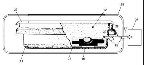
Figure 5 is a partly cut-away side view showing the tray and pouch assembly of
Figure 4 docked with a disinfection maintenance station for the performance of
the
pressure reducing step of the method of the present invention;
Figure 6 is a partly cut-away side view showing the tray and pouch assembly of
Figure 4 docked with a disinfection maintenance station for the performance of
the gas
charging step of the method of the present invention; and
Figures 1 to 6 together form a sequence illustrating a preferred embodiment of
the method of the present invention, and the apparatus for use in such a
method.
Referring first to Figure 1, there is shown a medical endoscope, generally
indicated 10, which is in a disinfected or "processed" state, having been
subjected to
rigorous cleaning and disinfecting, following use in a surgical procedure. On
emerging
from processing, the disinfected endoscope 10 is placed into a re-usable rigid
tray 11
having a downwardly-dished, inner compartment, generally indicated 12, defined
by a
generally planar base 13, and surrounding walls 14 upstanding therefrom. One
wall 14
of the tray 11 has a valve 15 formed therein, for connection to a disinfection
CA 02626039 2008-04-15
WO 2007/049076
PCT/GB2006/050356
- 8 -
maintenance station, as will be discussed in more detail below with reference
to Figures
4 and 5.
As is best seen in Figure 2, the tray 11 is provided with a single-use,
disposable
liner 16, formed of a flexibly deformable, sheet material which enables the
liner 16 to
conform itself substantially to the contours of the base 13 and walls 14 of
the underlying
tray 11. The liner 16 has a flap 17 which is adapted to be extended across the
inner
compartment 12 of the tray 11, and secured to an opposed wall 14 of the tray
11 by
means of an adhesive strip 18. Alternatively, the liner 16 may be provided
with a
separate cover (not shown) having an elasticated rim. The liner 16 is also
provided with
an aperture 19 to enable the valve 15 to communicate with the inner
compartment 12
within the liner 16.
Scavenger sachets 21 are located within the liner 16, either being placed
therein
along with the processed endoscope 10, or being integrally formed within the
liner 16.
The sachets 21 contain oxygen scavengers, further scavengers or "getters" for
other
gases, and silica gel desiccant. The scavenger sachets 21 are activated by
removing a
tear-off strip (not shown) to expose the scavengers to the atmosphere within
the inner
compartment 12 of the tray 11, before the compartment 12 is sealed by closing
the flap
17.
Referring now to Figure 3, the endoscope 10 and scavenger sachets 21 are now
isolated from the ambient, within the inner compartment 12 of the tray 11. In
order to
ensure a substantially air-tight seal, such that the inner compartment 12
forms a sealed
chamber as hereinbefore described in the method of the present invention, a
rigid
protective cover 22 is then placed over the tray 11. The cover 22 has tapered
edges 23
to provide a substantially air-tight seal by co-operating with complementary
tapers (not
shown) provided on the upper edges of the walls 14 of the tray 11. The cover
22 is
further provided with a viewing window 24 in order that the condition of the
endoscope
10 within the compartment 12 may be monitored. The viewing window 24 may be
provided with an oxygen level indicator (not shown), to provide a visual
indication of the
condition of the compartment 12, for example by means of a colour change.
The scavengers 21 within the sealed compartment 12 act to decrease the
oxygen levels within the compartment, thus inhibiting the multiplication of
aerobic micro-
organisms, and leading to a decrease in their population. The oxygen decrease
also
leads to reduction in the gas pressure within the sealed compartment 12, as a
result of
the air-tight seal provided by the liner flap 17 and the rigid cover 22. The
reduced
CA 02626039 2008-04-16
Pnntcl "1 1 /09/2007;:bE QF,3Al\AP'
';;GE)2005G8$6
- 9 -
along with water vapour naturally present in the air within the compartment
12. The
removal of water prevents access to nutrients dissolved therein, thus
inhibiting the
multiplication of both aerobic and anaerobic micro-organisms.
As shown in Figure 4, the sealing of the compartment 12 from the ambient may
s be further enhanced by placing the entire assembly of endoscope 10, tray
11, liner 16,
sachets 21 and cover 22 into an oxygen-impermeable, substantially inflexible
pouch 25.
The pouch 25 is provided with a valve 15 and a viewing window 24, which are
arranged
so as to be aligned respectively with the valve 15 in the tray 11 and the
viewing window
24 in the tray cover 22. The valve 15 in the tray 11 and/or the pouch 25 may
be
is connected to a suction device (not shown) to evacuate air from within
the pouch 25,
thus causing a further reduction in the gas pressure within the compartment
12, as
described above. Although described here as separate method steps, the
pressure
reducing step and the oxygen scavenging step will In practice be carried out
virtually
simultaneously.
15 In preferred embodiments of the method of the present invention, the
valves 15
of the tray 11 and the pouch 25 are connected to a disinfection maintenance
station, as
will now be described in more detail with reference to Figures 5 and 6. The
processed
endoscope 10 within the sealed compartment 12 is treated by performing: an
oxygen
scavenging step, as described above with reference to Figures 2 to 3; a step
of
20 reducing the pressure within the sealed compartment 12 by connecting the
valve 15 of
the tray 11 and/or the pouch 25 to a mechanical, electrical or manual suction
device
(not shown), as described above with reference to Figure 4; and a step of
charging the
sealed compartment 12 with a disinfectant gas or vapour such as dry nitrogen
gas, or
vapour phase hydrogen peroxide, by introducing said gas or vapour through the
valve
25 15 of the tray 11 and/or the pouch 25.
As noted above, although described here as separate method steps, the
pressure reducing step and the oxygen scavenging step will in practice be
carried out
virtually simultaneously.
To facilitate the performance of the method of the present invention, the
30 combined tray 11 and pouch 25 assembly is adapted to be docked with a
disinfection
maintenance station 26, which is schematically represented in Figures 5 and 6.
The
disinfection maintenance station 26 comprises both a mechanical, electrical or
manual
suction device for evacuating the sealed compartment 12 in the pressure
reducing step,
as indicated by arrows a in Figure 5; and a vessel or generator. for charging
the
AMENDED SHEET
iti4/061200T:
CA 02626039 2008-04-15
WO 2007/049076
PCT/GB2006/050356
- 10 -
suction device for evacuating the sealed compartment 12 in the pressure
reducing step,
as indicated by arrows a in Figure 5; and a vessel or generator for charging
the
compartment 12 with the disinfectant gas or vapour in the gas charging step,
as
indicated by arrows b in Figure 6.
The docking of the respective valves 15 of the tray 11 and/or pouch 25 with
the
disinfection maintenance station 26 can be achieved in a variety of ways,
depending on
the particular embodiment of the method of the present invention being
performed, and
the precise configuration of the apparatus being utilised in that method. In
the preferred
embodiment illustrated in Figures 5 and 6, the respective valves 15 of the
tray 11 and
the pouch 25 are both docked with a single port 27 of the disinfection
maintenance
station 26, said port 27 being used for both the removal a of air from the
sealed
compartment 12, and the charging b of the compartment 12 with the disinfectant
gas or
vapour. The port 27 may be provided with apertures 28 along its length so as
to permit
the ingress a and egress b of gas and/or vapour into and out of the pouch 25
as well as
into and out of the sealed compartment 12 at its end 29.
A typical sequence for the performance of the preferred embodiment of the
method of the present invention, will now be described, with reference to
Figures 5 and
6:
After docking the tray 11 and pouch 25 assembly with the disinfection
maintenance station 26, the sealed compartment 12 is connected to a mechanical
suction device within the station 26 so as to evacuate air therefrom, as
indicated by
arrows a in Figure 5. Following this, the sealed compartment 12 is connected
to a
generator within the disinfection maintenance station 26 for the production of
vapour
phase hydrogen peroxide (VPHP), and the compartment 12 is charged with VPHP,
as
indicated by arrows b in Figure 6. The reduced pressure in the compartment 12
causes
the VPHP to permeate through the channels of the endoscope 10.
After charging the compartment 12 for a pre-determined length of time, the
compartment 12 is again connected to the suction device within the station 26
to
remove VPHP from the compartment 12 and from the channels of the endoscope 10,
as
indicated by arrows a in Figure 5. The compartment 12 is then connected to a
dry
sterile nitrogen gas vessel within the station 26, and the compartment is
charged with
dry sterile nitrogen gas, as indicated by arrows b in Figure 6.
Once the above cycle is complete, the endoscope 10 remains stored within the
sealed compartment 12 until required in a surgical procedure. This storage may
be
CA 02626039 2008-04-15
WO 2007/049076
PCT/GB2006/050356
- 11 -
carried out with the tray 11 remaining docked on the station 26, and either
under a
charge of dry sterile nitrogen gas, as shown in Figure 6, or under reduced
pressure, as
shown in Figure 5. Alternatively, these two options may be combined, or
periodically
alternated between. A further alternative is to withdraw the tray 11 from the
station 26,
but with the compartment 12 remaining sealed, as shown in Figure 4, and
retaining the
sterile nitrogen gas charge and/or the reduced pressure therein.