Note: Descriptions are shown in the official language in which they were submitted.
CA 02626302 2008-04-17
WO 2007/050211
PCT/US2006/036802
EXTRACORPOREAL FLUID CIRCUIT
BACKGROUND
[0001] This invention relates to extracorporeal liquid circuits.
Hemodialysis
removes toxic substances and metabolic wastes from the bloodstream using an
extracorporeal circuit with components designed to perform ultrafiltration and
diffusion
on the blood. Before the blood is returned to the body, air bubbles are
removed from the
blood to prevent embolisms.
[0002] Referring to Fig. 1, a typical extracorporeal circuit 100 includes
tubing
through which the blood flows and components for filtering and performing
dialysis on
the blood. Blood flows from a patient 105 through arterial tubing 110. Blood
drips into a
drip chamber 115 where a sensor 125 in communication with air in the drip
chamber 115
determines the pressure of the blood flow on the arterial side of the circuit
100. A pump
120 forces the blood to continue along the path through the circuit 100. A
dialyzer 130
separates waste products from the blood.
[0003] After passing through the dialyzer 130, the blood flows through
venous
tubing 140 into a drip chamber 150. The drip chamber 150 can function as an
air trap.
Free gases in the blood may be able to escape into the drip chamber 150 before
theblood
continues to the patient. A sensor 170 is in communication with air in the
drip chamber
through tube 165. The sensor 170 can determine the pressure on the venous side
of the
circuit 100.
[0004] Heparin or drugs 160 can be added to the blood in the drip chamber
150.
When blood is exposed to oxygen, the blood begins to clot. Even with the
addition of
heparin to the blood to prevent clots, some clotting may still occur. The drip
chamber
150 includes a filter for preventing any clots from exiting the drip chamber
150 and
entering the patient 105. The blood continues from the drip chamber through
venous
tubing 180 and through a bubble detector 175 before returning to the patient
105.
SUMMARY
[0005] An airless drip chamber is described that prevents blood in an
extracorporeal blood circuit from being exposed to air. The airless chamber
can be a
stand-alone item, or incorporated into an integrated fluid circuit that also
includes
channels for directing blood to and from the drip chamber and pockets at which
the
pressure through the circuit can be measured. The integrated fluid circuit is
a
1
CA 02626302 2008-04-17
WO 2007/050211
PCT/US2006/036802
one-time-use disposable component that plugs into a hemodialysis machine or
other
extracorporeal fluid line.
[0006] In general, in one aspect, the invention is directed to an
apparatus for
removing air from a bodily liquid in extracorporeal circuitry. The apparatus
has a vertical
chamber having a bottom region and a top region and a pair of fluid entry and
exit ports at
or near the bottom region. A microporous filter is at or near the top region.
Fluid passes
through the vertical chamber from the entry port to the exit port so as to
fill the vertical
chamber with liquid while removing air from the chamber.
[0007] Implementations of the invention can include one or more of the
following
features. The microporous filter can include a hydrophobic material. The
microporous
filter can fotm at least a portion of a top surface of the vertical chamber.
The fluid entry
and exit ports can be in a bottom surface of the vertical chamber. The chamber
can have
a dam between the entry port and the exit port. The vertical chamber has a
height
sufficient to maintain an interface between a first liquid and a second liquid
in the vertical
chamber when the first and second liquids are miscible and the second liquid
is flowing
through the vertical chamber. The vertical chamber can have a bottom region
that is
wider than the top region. A clot filter can be positioned so that fluid
passes through the
clot filter prior to passing through the exit port. The microporous filter can
fit within a
housing connected to the top of the chamber. The bottom surface of the chamber
can be
sufficiently wide to accommodate an entry tube and an exit tube wherein the
entry tube is
connected to the entry port and the exit tube is connected to the exit port.
[0008] In another implementation, the invention can be directed to a
method of
removing air from a bodily liquid in extracorporeal circuitry. A first liquid
is passed
through an entry port into a bottom region of a vertical chamber, filling the
chamber so
that substantially no air remains in the chamber. A second liquid is passed
through the
entry port, thereby forcing a portion of the first liquid out of an exit port
from the bottom
region of the vertical chamber. A liquid-liquid interface forms between the
first and
second liquids. Gas bubbles in the second liquid are forced out of a top
region of the
vertical chamber through a microporous filter.
[0009] Implementations of the invention can include one or more of the
following
features. The first and second liquids can be miscible and the vertical
chamber can be
long enough to prevent the first and second liquids from mixing, because the
first liquid
stagnates. The method can include passing the second liquid through an exit
port after air
2
CA 02626302 2008-04-17
60412-3951
in the second liquid has escaped, thereby passing a
substantially air-free second liquid through the exit port
for delivery to a patient. The first liquid can be saline.
The second liquid can be blood. The second liquid can be
forced over a dam and out an exit port. The method can
include passing the second liquid through a clot filter and
an exit port.
[0010] In yet another implementation, the invention can
be directed to an integrated fluid circuit component adapted
to removably seat in a bodily liquid purification machine.
The component includes a rigid body and a flexible backing.
The rigid body has a substantially flat main portion and a
plurality of recessed portions extending from the flat main
portion. The flexible backing covers at least one of the
recessed portions. One of the recessed portions forms a
vertical chamber. The vertical chamber has a microporous
filter at or near a top region. One recessed portion forms
a channel that is in fluid communication with a bottom
region of the vertical chamber. Another recessed portion
forms another channel in fluid communication with the bottom
region of the vertical chamber.
[0011] Implementations of the invention can include one
or more of the following features. The flexible backing can
be sealed to the flat main portion of the rigid body, at
least partially enclosing the recessed portions. One
recessed portion can overlap one of the channels, where the
recessed portion is wider than the channel. The channels
can extend to the edge of the rigid body. The channels can
be in fluid communication with tubes. The vertical chamber
can include a clot filter adjacent to the second channel.
The microporous filter can include a hydrophobic material.
The vertical chamber can have a height sufficient to
3
CA 02626302 2013-03-19
60412-3951
maintain an interface between a first liquid and a second liquid in the
vertical chamber when
the first and second liquids are miscible and the first liquid is stagnant as
the second liquid
flows through the vertical chamber. The component can include injection sites,
such as
neoprene gaskets. The component can include a rigid backing that backs at
least the channels.
According to one aspect of the present invention, there is provided an
apparatus for removing air from a bodily liquid in extracorporeal circuitry,
comprising: a
vertical chamber having a bottom region and a top region and a pair of fluid
entry and exit
ports at or near the bottom region the bottom region being flared such that
the bottom region
is wider than the top region; and a microporous filter at or near the top
region, whereby liquid
is passed through the vertical chamber from the entry port to the exit port so
as to fill the
vertical chamber with the liquid while removing air from the chamber, and the
vertical
chamber has a height sufficient to allow a first liquid in the top region of
the vertical chamber
to remain stagnant as a portion of a second liquid in the bottom region of the
vertical chamber
flows through the vertical chamber from the fluid entry port to the fluid exit
port such that the
first liquid remains in the top region of the vertical chamber and the second
liquid which is
miscible with the first liquid remains in the bottom region of the vertical
chamber as the
portion of the second liquid flows through the vertical chamber from the fluid
entry port to the
fluid exit port, and the first liquid inhibits direct contact between the
second liquid and the
microporous filter.
According to another aspect of the present invention, there is provided a
method of removing air from a bodily liquid in extracorporeal circuitry,
comprising: passing a
first liquid through an entry port into a bottom region of a vertical chamber,
filling the
chamber so that substantially no air remains in the chamber; passing a second
liquid through
the entry port into the bottom region of the vertical chamber, thereby forcing
a portion of the
first liquid out of an exit port from the bottom region of the vertical
chamber and forming a
liquid-liquid interface between the first and second liquids within the
vertical chamber; and
passing a portion of the second liquid out of the exit port from the bottom
region of the
vertical chamber while maintaining the liquid-liquid interface.
3a
CA 02626302 2013-03-19
60412-3951
[0012] The invention can be implemented to realize one or more of the
following
advantages. Air may be prevented from contacting blood in the chamber.
Preventing air from
getting in the chamber may reduce the likelihood of clots forming in the
blood. A
hydrophobic microporous filter at the top of the chamber can allow any free
gas or air that
enters the chamber to escape. The chamber has a sufficient height so that a
first liquid, such
as saline, is located near a top of the chamber and blood is located near the
bottom of the
chamber and little mixing of the two liquids occurs. The length of the chamber
is
3b
CA 02626302 2008-04-17
WO 2007/050211
PCT/US2006/036802
sufficient to allow the saline to stagnate and prevents mixing of the liquids,
thereby
preventing the blood from contacting the microporous filter. The saline can
prevent most
of the proteins in the blood from contacting the microporous filter. If
protein accumulates
on the microporous filter, the filter will wet and the filter's hydrophobic
properties may
be inhibited. That is, if the microporous filter wets, the filter may allow
blood to escape
from the chamber. Also, the filter can become inefficient at allowing air to
pass through
if protein collects on it. A dam in the chamber between the entry and exit
ports diverts air
bubbles toward the microporous filter. The microbubbles in the blood may then
escape
through the microporous filter rather than passing through the exit port. The
system is
free of a blood-air interface, which reduces the need for anti-coagulants.
Reducing clot
formation, reducing the need for anti-coagulant and reducing gas in the blood
are
desirable as safety measures for the patient undergoing hemodialysis.
[0013] Placing the components, such as a pocket for taking pressure
measurements, channels for fluid flow and the airless chamber into a single
integrated
fluid circuit can eliminate multiple separate components. Fewer components are
easier
for an operator to work with and can reduce the risk of operator error. The
integrated
fluid circuit can have a rigid side that maintains the integrity of the
components and a
flexible side that allows for transducers to take measurements, such as
pressure or
temperature measurements. The flexible side can be sealed to the rigid side,
potentially
eliminating the need to construct a machine in which the integrated fluid
circuit must be
tightly held to the machine to seal the rigid and flexible sides together.
Alternatively, the
integrated fluid circuit can have two rigid sides with membranes only at
critical locations.
[0014] The details of one or more embodiments of the invention are set
forth in
the accompanying drawings and the description below. Other features, objects,
and
advantages of the invention will be apparent from the description and
drawings, and from
the claims.
DESCRIPTION OF DRAWINGS
[0015] Fig. 1 is a schematic of a conventional hemodialysis system.
[0016] Fig. 2 is a schematic cross-sectional view of an airless chamber.
[0017] Fig. 2A is a schematic top view of the airless chamber.
[0018] Fig. 2B is a schematic bottom view of the airless chamber.
[0019] Fig. 2C is a schematic perspective view of the airless chamber.
4
CA 02626302 2008-04-17
WO 2007/050211
PCT/US2006/036802
[0020] Fig. 2D, 2E and 2F are each a schematic cross-sectional view of an
airless
chamber.
[0021] Fig. 3 is a schematic of an airless chamber in an extracorporeal
circuit.
[0022] Fig. 4 is a flow diagram for using the airless chamber in an
extracorporeal
circuit.
[0023] Fig. 4A is a schematic of the blood flow path through an airless
chamber.
[0024] Fig. 5 is a plan view of an integrated extracorporeal circuit.
[0025] Fig. 5A is a cross sectional view of the integrated extracorporeal
circuit of
Fig. 5.
[0026] Fig. 6 is a perspective view of the integrated extracorporeal
circuit.
[0027] Fig. 7 is a perspective view of a bloodline guide for holding a
tubing
assembly, which is configured to retain the airless chamber shown in Fig. 3.
[0028] Like reference symbols in the various drawings indicate like
elements.
DETAILED DESCRIPTION
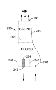
[0029] Referring to Figs. 2, 2A, 2B and 2C, in some implementations, an
airless
chamber 230 is provided as a component of an extracorporeal fluid circuit. The
chamber
230 is substantially hollow for filling with a liquid. The chamber 230 can be
used for
removing gas from blood, but can also be used with a number of other fluids,
such as
bodily fluids, including plasma. The chamber 230 has a bottom region 234 and a
top
region 236, where the bottom and top are relative to the chamber's orientation
during use.
An entry port 240 and an exit port 242 are in the bottom region 234 of the
chamber 230.
In some implementations, the ports 240, 242 are located in a bottom surface of
the
chamber 230. In other implementations, as shown in Fig. 2F, at least one of
the ports 240,
242 is located in a side surface of the chamber 230. In one implementation, a
dam 248 is
between the ports 240, 242. The dam 248 extends at least part way from one
side wall to
an opposite side wall. In one implementation, the dam 268 contacts each side
wall so
that all fluid entering entry port 240 flows over the top of the dam 248
before flowing out
the exit port 242. In one implementation, a clot filter 254 is positioned
adjacent to the
exit port 242. Fluid flows through the clot filter 254 prior to flowing out of
the exit port
242. In one implementation, the clot filter 245 has a porosity of between
about 50-500
microns.
[0030] The ports 240, 242 are holes in the chamber which can be in fluid
communication with tubular shaped extensions. The extensions are able to be
connected
CA 02626302 2008-04-17
WO 2007/050211
PCT/US2006/036802
to tubes, such as by pressure fitting or bonding. The extensions can be
integrally formed
with the chamber or subsequently attached to the chamber, such as by bonding
or
welding.
[0031] At the top region 236 of the chamber 230 is a microporous filter
260. The
microporous filter 260 allows gas to vent from the chamber 230. Pores in the
microporous filter 260 are small enough to keep foreign particles and
organisms from
entering the chamber 230 from the outside air. In one implementation, the
filter 260
includes a hydrophobic material. A hydrophobic microporous filter keeps liquid
from
leaking out of the chamber 230 when the chamber 230 is substantially fined
with liquid.
A suitable filter has a pore size equal to or less than 0.45 microns, such as
about 0.22
microns. The filter may be formed of polytetrafluoroethylene (PTFE) or any
other
suitable material.
[0032] When the chamber 230 is filled with blood, preventing the protein
in the
blood from accumulating on the filter 260 can maintain the hydrophobic
characteristic of
the filter 260. Whole blood can be kept from the filter by providing a barrier
between the
blood and the filter 260, such as a liquid barrier 268, as described further
below. The
height of the chamber 230 is sufficient to maintain this barrier 268 and
prevents the liquid
above the barrier 268 from substantially mixing with liquid below the barrier.
[0033] The shape of the chamber is approximately elongate. In some
implementations, such as those shown in Fig. 2 and 2D, the bottom region 234
of the
chamber 230, 230' is wider than the top region 236, such that the chamber 230,
230' has a
quasi-conical shape or a flare at the bottom. In some implementations, such as
those
shown in Fig. 2E, the top and bottom dimensions of the chamber 230" are
approximately
equal so that the chamber 230" has a rectangular or cylindrical shape. The
bottom region
234 can also be narrower than the top region 236. If the ports 240, 242 are in
the bottom
surface of the chamber, the bottom surface has a sufficiently large dimension
to
accommodate the ports 240, 242 as well as any tubes coupled to the ports for
directing
fluid into and out of the chamber. For example, if the tubing has an outer
diameter of
6.25 mm, the bottom surface is at least 12.5 mm wide. The exact dimensions of
the
chamber 230 are unimportant as long as the liquid barrier 268 is maintained,
although the
chamber 230 can be at least about two inches in height, preferably three to
four inches.
[0034] The chamber is formed of a material \suitable for medical devices,
that is, a
medical grade material. Plastics, such as polyvinylchloride, polycarbonate,
polyolefins,
6
CA 02626302 2008-04-17
WO 2007/050211
PCT/US2006/036802
polypropylene, polyethylene or other suitable medical grade plastic can be
used because
of their ease of manufacturing, ready availability and disposable nature. The
chamber is
formed, such as by molding, for example, extruding, blow molding or injection
molding.
The chamber can be formed of a transparent or clear material so that the
liquid flowing
through the chamber can be observed. The microporous filter at the top of
chamber can
be connected to the chamber in a number of ways. In one implementation, the
filter fits
into a cap-type housing and the housing is screwed or welded onto the top of
the chamber.
In another implementation, the microporous filter is adhesively attached to
the chamber,
such as with an epoxy. In yet another implementation, the microporous filter
is co-
molded during the injection molding process.
[0035] Referring to Figs. 3 and 4, the airless chamber 230 is in line in
the
extracorporeal fluid circuit of a system for fluid filtration and air removal.
A first fluid
that is compatible with the fluid to be filtered (the second fluid) is
introduced into the
system to prime the system (step 404). In hemodialysis, the first fluid is a
blood
compatible solution, such as saline. The saline flows through an arterial
channel 330 to
an arterial pressure sensor 336. The arterial pressure sensor 336 includes a
transducer so
that the pressure of the fluid flowing through the circuit 324 on the arterial
side can be
monitored. The saline then flows through a portion of the channel that abuts a
pump 120,
such as a peristaltic pump. The pump 120 forces the saline through the system
324. In
some implementations, the pressure sensor 336 is after the pump 120.
Alternatively, a
arterial pressure sensor can be both before and after the pump 120. The saline
then flows
to the dialyzer 130 and then to a venous pressure sensor 360.
[0036] Next, the saline, or the first fluid, flows through the entry port
of the
chamber 230 and fills the chamber (step 412). To fill the chamber completely,
venous
channel 368 can be clamped to create a positive pressure once the saline is
introduced
into the chamber. Air is forced out the top of the chamber and through the
microporous
filter as saline fills the chamber. The saline contacts the filter and the
chamber is
substantially free of air once the chamber is completely filled. However, the
saline does
not exit through the filter, because the filter is hydrophobic. After the
venous channel 368
is unclamped, the saline exits through the exit port of the chamber and out
the venous
channel 368.
[0037] The second liquid, such as a bodily fluid, for example, blood, is
then
introduced into the system (step 418). The blood follows the same route as the
saline and,
7
CA 02626302 2008-04-17
WO 2007/050211
PCT/US2006/036802
for the most part, pushes the saline through the circuit. When the blood
enters the
chamber 230, the blood forces the saline at the bottom of the chamber through
the exit
port (step 422). However, the blood does not displace all of the saline within
the chamber
230. Because of the height of the chamber 230, the blood enters the chamber
230 and
only traverses part of the height of the chamber before flowing back down
along flow
path 274 to the exit port (as shown in the airless chamber formed of
transparent material
in Fig. 4A). An interface 268 between the saline and the blood delineates the
furthest
extent of most of the blood within the chamber 230. The interface 268 between
the blood
and saline can visually be observed and stretches across the entire width of
the chamber.
Because blood and saline are not immiscible, there is some amount of mixing
between the
two fluids around the interface 268.
[0038] The saline keeps the blood from contacting the filter. However, a
percentage of blood can be present in the saline without hindering the
operation of the
system. That is, the saline need not be completely free from blood for the
airless chamber
to both allow gas to vent from the system and retain the liquid in the system.
The solution
that is mostly saline substantially protects the filter from becoming coated
with protein.
If the chamber is sufficiently elongated, the blood does not mix with the
saline at the top
portion of the chamber because the saline remains relatively stagnant as the
blood flows
through the chamber.
[0039] Any unbound gas, or air, that is in the blood, such as air that is
introduced
by the dialyzer or air that comes out of solution from the blood, rises as
tiny air bubbles
within the blood and saline until the air eventually vents out through the
microporous
filter (step 430). With a dam 248 inside of the chamber 230, the blood travels
up and over
the dam rather than straight across the bottom of the chamber out the exit
port. By
directing the flow of blood upwards, the blood with air is not able to flow in
and directly
back out of the chamber without flowing upwards to at least a height greater
then the
height of the dam. The surface area of the dam and the inner walls of the
chamber
enables air, including microbubbles, to separate from the blood and exit the
fluid circuit
through the microporous filter.
[0040] Throughout the circuit, the blood flows without there being a
substantial
air-blood interface. Although the blood does not come into contact with air
and thus
clotting is less likely to occur, the blood can pass through an optional
filter in the chamber
for added safety. In some implementations, after exiting the chamber, the
blood passes by
8
CA 02626302 2008-04-17
WO 2007/050211
PCT/US2006/036802
or through one or more sensors, such as temperature or air detecting sensors
as an
additional safety measure.
[0041] In one implementation, the airless chamber and one or more other
components can be incorporated into an integrated fluid circuit. The
integrated fluid
circuit has the components described above, such as the airless chamber,
formed together
in one assembly or integrated molding rather than discrete separate or modular
devices.
The integrated fluid circuit is adapted to removably seat into a machine, such
as a blood
purification machine, like a hemodialysis machine. The integrated fluid
circuit is similar
to a cassette or cartridge, where an operator merely snaps the integrated
fluid circuit into
the machine and after just a few additional connections, begins operation.
[0042] Referring to Fig. 5, the integrated fluid circuit 512 has a rigid
body 518
and a flexible backing (not shown). The rigid body has a substantially flat
surface 520
with one or more concave (when viewed from the backside) portions or recessed
portions
protruding from a front surface of the body 518. The flexible backing can be
applied so
that the backing covers only the recessed portions or so that the backing
covers more than
just the recessed portions, up to all of the back surface of the rigid body.
[0043] The integrated fluid circuit has a recessed portion that serves as
the airless
chamber 526. As with the chamber described above, the airless chamber 526
includes a
microporous filter 528 at a top region and optionally includes a dam 560 and a
clot filter
568. A first channel 534 in rigid body 518 leads from an edge of the rigid
body 518 to a
bottom region of the airless chamber 526. Over one portion of the channel 534,
a venous
recess or pocket 548 is foimed. The flexible backing backs the venous pocket
548. The
venous pocket 548 is sized so that a transducer in the machine can measure the
venous
fluid pressure through the flexible backing. A second channel 578 extends from
the outlet
of the airless chamber 526 to an edge of the rigid body 518. The first and
second
channels extend to the same or different edges of the rigid body 518. The
first channel
534 and second channel 578 are in fluid communication with the airless chamber
526.
[0044] In some implementations, a third channel 584 is formed in the
rigid body
518. The third channel 584 is not in fluid communication with the first or
second
channels when the integrated fluid circuit is not in the machine or connected
to a dialyzet
In some implementations, an arterial pocket 588 is formed along the third
channel 584.
The arterial fluid pressure can be measured through the flexible backing of
the arterial
9
CA 02626302 2008-04-17
WO 2007/050211
PCT/US2006/036802
pocket 588. One end of the third channel 584 extends to one edge of the rigid
body 518
and the other end extends to the same or a different edge, as shown in Fig. 5.
[0045] Optionally, a fourth channel 592 extends across the rigid body
518. A
post-pump arterial pocket 562 overlaps the fourth channel 592. In some
implementations,
additional recesses and channels are formed in the rigid body.
[0046] In some implementations, tubes 594a, 594b, 594c, 594d and 594e are
connected to the rigid body 518, such as at the locations where the first,
second, third and
fourth channels extend to the edges. The tubes are connected to the rigid body
using
techniques known in the art. In some embodiments, the tubes fit into a pre-
formed
grooves in the rigid body 518. The tubes can be pressure fitted into the
grooves. In other
implementations, the tubes are clipped onto the rigid body 518. Optionally, at
the end of
the tubes 594a, 594b, 594c and 594e are fasteners for connecting the tubes to
components
of the machine, such as the dialyzer or to a patient. Tube 594d wraps around a
peristaltic
pump in the machine. Tubes 594a and 594e connect to a dialyzer. Tubes 594b and
594c
connect to a patient.
[0047] Each of the recesses can protrude from the flat surface 520 to
approximately the same distance. Alternatively, some of the recesses, such as
the
channels, may be shallower than other recesses, such as the airless chamber
526.
Referring to Fig. 5A, a cross section of the integrated circuit 512 shows an
outline of the
pocket 548, channel 534 and part of chamber 526. The rigid body 520 can have
an
overall thickness of less than about 2 mm, such as less than about 1 mm.
Flexible
membrane 564 covers the back of the rigid body 520.
[0048] In some implementations, instead of one or more of the channels
being
founed in the rigid body 518, a tube is connected directly to a feature in the
rigid body.
For example, instead of forming second channel 578, tube 594b can be connected
directly
to the airless chamber 526.
[0049] In some implementations, the integrated circuit 512 has two rigid
sides.
The first rigid side is as described above. The second rigid side is
substantially flat with
openings located adjacent to the pockets formed in the first side. The
openings are
covered with a flexible membrane.
[0050] In some implementations, the integrated circuit 512 has posts that
extend
from one or more sides of the circuit. The posts can mate with recesses in the
machine,
ensuring correct registration of the integrated circuit 512 with components,
such as
CA 02626302 2008-04-17
WO 2007/050211
PCT/US2006/036802
sensors, in the machine. In some implementations, the integrated circuit 512
has latches,
clips or other such device for registering the integrated circuit 512 with the
machine and
locking the integrated circuit 512 in place.
[00511 The machine can have a mechanism that holds the integrated circuit
in
place. The mechanism can comprise a door, a locking device or a suction device
for
holding the integrated circuit in tight contact with the machine. When the
integrated
circuit is seated in the machine, pressure transducers interface with the
flexible backing to
directly measure the fluid pressure at each of the corresponding locations.
Holding the
integrated circuit in contact with the machine allows the pressure transducers
to sense
flow through the circuit. Once the integrated fluid circuit is plugged into
the machine and
connected with the machine's components, an operator uses the integrated fluid
circuit in
a manner similar to the method of using the circuit chamber 230 described
above.
[0052] As with the airless chamber 230, the rigid body 518 is constructed
of a
medical grade material. The flexible backing is constructed from a polymer
that is
flexible and suitable for medical use, such as an elastomer, including silicon
elastomers.
Other suitable materials include, high and low density poly ethylene, high and
low
density poly propylene, separately co-extruded mono layers or multiple layers
of
polyamides, nylons, silicones or other materials commonly known in the art for
flexible
applications. The backing is attached to the back of the rigid body 518, such
as by laser,
ultrasonic or RF welding or with an adhesive. In some implementations, the
backing is
attached so that the edge of each recess is sealed to the backing.
Alternatively, the
backing is attached only at the edge of the rigid body. If the backing does
not seal the
recesses from the flat portions, the machine into which the integrated fluid
circuit seats is
constructed to apply sufficient pressure to keep the fluid flowing through the
circuit from
leaking out of the recesses and between the backing and the flat surface 520.
In the back
of the rigid portion 518, ridges can be formed which surround the recesses.
The ridges
can aid in sealing the flexible membrane to the flat portion 518 when pressure
is applied
to the circuit.
[0053] In some implementations, injection sites 598 are formed at one or
more of
the recesses. The injection sites 598 can be used to inject drugs or solutions
into the fluid.
Suitable injection sites 598 are formed of neoprene gaskets into which a
needle can be
introduced and removed so that the gaskets do not leak or weep after the
needle is
removed.
11
CA 02626302 2008-04-17
WO 2007/050211
PCT/US2006/036802
[0054] Fig. 6 shows a perspective view of the integrated fluid circuit
512. As in
Fig. 5, the flexible membrane has been removed from the integrated fluid
circuit 512 to
show the recesses.
[0055] Referring to Fig. 7, a bloodline guide 700 is configured to hold a
tubing
assembly in proper alignment with respect to the components of the machine.
The tubing
assembly include the airless chamber 230 and tubes, such as those shown in
Fig. 3, and in
some embodiments, includes components for allowing for detecting pressure
within the
guide. The bloodline guide 700 includes a rigid body 718 with a flat surface
720. A
recess 726 is formed in the rigid body 718 that is configured to fit the
airless chamber
230. Recesses 734, 778, 784 and 792 are sized for retaining tubing. Recesses
748, 762
and 788 are sized for retaining a component attached to or part of the tube
where pressure
can be monitored. Embodiments of the bloodline guide 700 include one or more
of the
recesses 726, 748, 734, 762, 778, 784, 788 and 792. In some implementations,
the
recesses 748, 762 and 788 are holes made in the rigid body 718.
[0056] In some implementations, the bloodline guide 700 includes a cover
(not
shown). The cover is a flat layer, such as a flat piece of plastic. The cover
can be
separate from the guide and can be temporarily attached to the bloodline
guide, such as
with clips. In some implementations, the cover extends from one side of the
bloodline
guide 700 with a hinge for bending the cover over the back of the bloodline
guide 700,
thereby covering the recesses. In some implementations, the cover has holes
that align
with the recesses 748, 762, 788. The holes can be open or covered with a
membrane.
The holes allow for a sensing device, such as a pressure transducer, to detect
the pressure
of liquid in the component in the recess.
[0057] In some implementations, the bloodline guide 700 includes clips,
such as
at an edge of the bloodline guide 700, for holding the tubes in the correct
placement.
Similar to the integrated circuit 500, the bloodline guide 700 can include
posts for
properly aligning the guide 500 with the machine. The bloodline guide 500 can
be
transparent, so that a user can see that the components and tubes in the guide
are properly
aligned when the guide is loaded into the machine.
[0058] Once the assembly including the airless chamber and tubes are
placed in
the bloodline guide 700, the bloodlines guide 700 is loaded into the machine.
In some
implementations, the bloodline guide 700 includes posts, latches or other
mechanism for
ensuring proper registration with the machine.
12
CA 02626302 2008-04-17
WO 2007/050211
PCT/US2006/036802
[0059] Using the airless chambers described herein in an extracorporeal
blood
circuit prevents air from contacting blood flowing through the circuit.
Preventing air in
the chamber can reduce the likelihood of forming clots in the blood. In the
event that
there is air in the blood before the blood exits the chamber, a hydrophobic
microporous
filter at the top of the chamber allows air that enters the chamber to escape.
The filter is a
part of or connected directly to the airless chamber. This allows the air to
easily escape
from the liquid filled chamber. Thus, lines need not be connected to the top
of the
chamber for withdrawing air from the circuit. Eliminating an air-blood
interface
increases the safety of the treatment for the patient. When clots form in the
blood, the
patient can be serious injured. Blood clots can cause thrombus, embolism,
heart attack or
stroke. Air bubbles in the blood also can injure the patient, such as by
causing an air
embolism. If the patient's blood never contacts air while flowing through the
extra-
corporeal circuit, no air will get into the blood, preventing air embolisms
and blood clots
caused by the treatment. Because the likelihood of clots is lessened, the
amount of
anticoagulant that is added to the blood can be decreased. Fewer additives in
the blood
during treatment are preferred because of the benefit to the patient's health.
[0060] The chamber is first filled with saline before being filled with
blood. The
chamber has a sufficient height so that after the saline and blood are
introduced into the
chamber, the saline is located near the top of the chamber and the blood is
located near
the bottom, and little mixing of the two liquids occurs. The saline prevents
most of the
proteins in the blood from contacting the filter at the top of the chamber. If
protein
accumulates on the filter, the filter's hydrophobic properties can be
inhibited, that is, the
filter can wet, allowing liquid to leak from inside the chamber to outside the
chamber.
Also, if protein collects on the filter, the filter becomes inefficient at
allowing air to pass
through. Thus, a sufficiently long chamber allows the saline to stagnate at
the top,
preventing protein from contacting the filter.
[0061] A dam in the chamber between the entry and exit ports may provide
a
surface for microbubbles to accumulate. The microbubbles in the blood may then
escape
through the chamber rather than passing through the exit port. Reducing clot
formation
and reducing gas in the blood is safer for the patient undergoing
hemoclialysis. The dam
also forces the liquids up into the chamber so that the liquids, and any gases
traveling
with the liquids, are not immediately pushed out of the chamber before the gas
can escape
out to the top of the chamber.
13
CA 02626302 2008-04-17
WO 2007/050211
PCT/US2006/036802
[0062] Placing components, such as a pocket for taking pressure
measurements,
channels for fluid flow and the airless chamber, into a single integrated
fluid circuit
eliminates multiples separate components. Fewer components are easier for an
operator
to work with and reduce the risk of operator error. The integrated fluid
circuit has a rigid
side that maintains the integrity of the components, and flexible portions
that allow for
taking measurements, such as pressure or temperature measurements. Further,
the
pockets in the integrated circuit eliminate the need for pressure sensing
lines in fluid
communication with the top of the chamber.
[0063] A number of embodiments of the invention have been described.
Nevertheless, it will be understood that various modifications may be made
without
departing from the spirit and scope of the invention. For example, the
components
described herein can be used with other fluids, such as plasma. Additionally,
fluids other
than saline can be used to prime the system. Accordingly, other embodiments
are within
the scope of the following claims.
14