Note: Descriptions are shown in the official language in which they were submitted.
CA 02626363 2008-04-15
WO 2006/044790 PCT/US2005/037241
COMPOSITIONS AND METHODS
FOR PROLONGING SURVIVAL OF PLATELETS
FIELD OF THE INVENTION
The inventions relate to compositions and methods for reducing the clearance
of
transfused platelets from circulation in a mammal, and prolonging the
biological activity
and survival of the transfused platelets.
BACKGROUND OF THE INVENTION
Platelets are anucleate bone marrow-derived blood cells that protect injured
mammals from blood loss by adhering to sites of vascular injury and by
promoting the
formation of plasma fibrin clots. Humans depleted of circulating platelets by
bone
marrow failure suffer from life threatening spontaneous bleeding, and less
severe
deficiencies of platelets contribute to bleeding complications following
trauma or
surgery.
A reduction in the number of circulating platelets to below -70,000 per L
reportedly results in a prolongation of a standardized cutaneous bleeding time
test, and
the bleeding interval prolongs, extrapolating to near infinity as the platelet
count falls to
zero. Patients with platelet counts of less than 20,000 per L are thought to
be highly
susceptible to spontaneous hemorrhage from mucosal surfaces, especially when
the
thrombocytopenia is caused by bone marrow failure and when the affected
patients are
ravaged with sepsis or other insults. The platelet deficiencies associated
with bone
marrow disorders such as aplastic anemia, acute and chronic leukemias,
metastatic cancer
but especially resulting from cancer treatment with ionizing radiation and
chemotherapy
represent a major public health problem. Thrombocytopenia associated with
major
surgery, injury and sepsis also eventuates in administration of significant
nuinbers of
platelet transfusions.
CA 02626363 2008-04-15
WO 2006/044790 PCT/US2005/037241
A major advance in medical care half a century ago was the development of
platelet transfusions to correct such platelet deficiencies, and over 9
million platelet
transfusions took place in the United States alone in 1999 (Jacobs et al.,
2001). Platelets,
however, unlike all other transplantable tissues, do not tolerate
refrigeration, because they
disappear rapidly from the circulation of recipients if subjected to even very
short periods
of chilling, and the cooling effect that shortens platelet survival is
irreversible (Becker et
al., 1973; Berger et al., 1998).
The resulting need to keep these cells at room temperature prior to
transfusion has
imposed a unique set of costly and complex logistical requirements for
platelet storage.
Because platelets are actively metabolic at room temperature, they require
constant
agitation in porous containers to allow for release of evolved CO2 to prevent
the toxic
consequences of metabolic acidosis. Room temperature storage conditions result
in
macromolecular degradation and reduced hemostatic functions of platelets, a
set of
defects known as "the storage lesion" (Chemoff and Snyder, 1992). But the
major
problem with room-temperature storage, leading to its short (5-day)
limitation, is the
higher risk of bacterial infection. Bacterial contamination of blood
components is
currently the most frequent infectious complication of blood component use,
exceeding
by far that of viral agents (Engelfriet et al., 2000). In the USA, 3000-4500
cases yearly
of bacterial sepsis occur because of bacterially containinated blood
components
(Yomtovian et al., 1993).
The mechanism underlying the unique irreversible cold intolerance of platelets
has been a mystery as has its physiological significance. Circulating
platelets are
smooth-surfaced discs that convert to complex shapes as they react to vascular
injury.
Over 40 years ago investigators noted that discoid platelets also change shape
at
refrigeration temperatures (Zucker and Borrelli, 1954). Subsequent evidence
that a
discoid shape was the best predictor of viability for platelets stored at room
temperature
(Schlichter and Harker, 1976) led to the conclusion that the cold-induced
shape change
per se was responsible for the rapid clearance of chilled platelets.
Presumably
2
CA 02626363 2008-04-15
WO 2006/044790 PCT/US2005/037241
irregularly-shaped platelets deformed by cooling became entrapped in the
microcirculation.
Based on studies linking signaling to the mechanisms leading to platelet shape
changes induced by ligands Hartwig et al., 1995 predicted that chilling, by
inhibiting
calcium extrusion, could elevate calcium levels to a degree consistent with
the activation
of the protein gelsolin, which severs actin filaments and caps barbed ends of
actin
filaments. They also reasoned that a membrane lipid phase transition at low
temperatures
would cluster phosphoinositides. Phosphoinositide clustering uncaps actin
filament
barbed ends (Janmey and Stossel, 1989) to create nucleation sites for filament
elongation.
They produced experimental evidence for both mechanisms, documenting gelsolin
activation, actin filament barbed end uncapping, and actin assembly in cooled
platelets
(Hoffmeister et al., 2001; Winolcur and Hartwig, 1995). Others had reported
spectroscopic changes in chilled platelets consistent with a membrane phase
transition
(Tablin et al., 1996). This information suggested a method for preserving the
discoid
shape of chilled platelets, using a cell-permeable calciuin chelator to
inhibit the calciuin
rise and cytochalasin B to prevent barbed end actin assembly. Although
addition of these
agents retained platelets in a discoid shape at 4 C (Winokur and Hartwig,
1995), such
platelets also clear rapidly from the circulation. Therefore, the problem of
the rapid
clearance of chilled platelets remains, and methods of increasing circulation
time as well
as storage time for platelets are needed.
SUMMARY OF THE INVENTION
The present invention provides glycan modified platelets having a reduced
incidence of platelet clearance following transplant and methods for reducing
platelet
clearance observed in a heterologous platelet transplant recipient. Also
provided are
compositions and methods for the preservation and storage of platelets, such
as
mammalian platelets, particularly human platelets. The invention also provides
methods
for making a pharmaceutical composition containing the modified platelets and
for
3
CA 02626363 2008-04-15
WO 2006/044790 PCT/US2005/037241
administering the pharmaceutical composition to a maminal to mediate
hemostasis,
particularly a cytopenic mammal.
It has now been discovered that cooling of 1luman platelets causes clustering
of
the von Willebrand factor (vWf) receptor complex a subunit (GPlba) coinplexes
on the
platelet surface. The clustering of GPlba complexes on the platelet surface
elicits
recognition by macrophage complement type three receptors (aM(32, CR3) in
vitro and in
vivo. CR3 receptors recognize N-linked sugars with terminal (3G1cNAc on the
surface of
platelets, which have formed GPlba complexes, and phagocytose the platelets,
clearing
them from the circulation and resulting in a concomitant loss of hemostatic
function.
Applicants have discovered that treatment of platelets with an effective
amount of
a glycan modifying agent such as N-acetylneuraminic acid (sialic acid), or
certain
nucleotide-sugar molecules, such as CMP-sialic acid or UDP-galactose leads to
sialylation or glycation of the exposed (3G1cNAc residues on GPlba, with the
effect of
ameliorating or substantially reducing storage lesion defects in the treated
platelets.
Effective amounts of a glycan modifying agent range from about 1 micromolar to
about
10 millimolar, about 1 micromolar to about 1 millimolar, and most preferably
about 200
micromolar to about 600 micromolar of the glycan modifying agent. This has the
functional effect of reducing storage lesion defects, reducing platelet
clearance in a
mammal following transfusion, blocking platelet phagocytosis, increasing
platelet
circulation time, and increasing both platelet storage time and tolerance for
temperature
changes in samples collected for transfusion. Additionally, platelets removed
from a
mammal for autologous or heterologous transplantation may be stored cold for
extended
periods, i.e., at 4 degrees C for 24 hours, 2 days, 3 days, 5 days, 7 days, 12
days or 20
days or more, without significant loss of hemostatic function following
transplantation.
Cold storage provides an advantage that it inhibits the growth of
contaminating
microorganisms in the platelet preparation, important as platelets are
typically given to
cancer patients and other immunocompromised patients. Room temperature stored-
4
CA 02626363 2008-04-15
WO 2006/044790 PCT/US2005/037241
treated platelets also demonstrate ameliorated or substantially reduced
storage lesion
defects over an extended period of time relative to untreated platelets. The
treated
platelets retain their biological functionality for longer periods of time
than untreated
platelets and are suitable for autologous or heterologous transplantation, at
least one day,
three days, five days, or even seven days or more following collection.
According to one aspect of the invention, methods for increasing the
circulation
time of a population of platelets is provided. The method comprises contacting
an
isolated population of platelets with at least one glycan modifying agent in
an amount
effective to aineliorate, substantially, or partially reduce storage lesions,
maintain or
1o improve biological functionality and reduce the clearance of the population
of treated
platelets, when transfused into a mammal. In some embodiments, the glycan
modifying
agent is selected from the group consisting UDP-galactose and UDP-galactose
precursors. In some preferred embodiments, the glycan modifying agent is UDP-
galactose. In other preferred embodiments, the glycans modifying agent is CMP-
sialic
acid. In other preferred embodiment, two glycan modifying agents are used,
including
UDP-galactose and CMP-sialic acid.
In some embodiments, the method furtlzer coinprises adding an enzyme that
catalyzes the modification of a glycan moiety on the platelet. One example of
an enzyme
that catalyzes the modification of the glycan moiety is galactosyl
transferase, particularly
a beta-1-4- galactosyl transferase. Another example of an enzyme that
catalyzes the
modification of a glycan moiety is a sialyl transferase, which adds sialic
acid to the
terminal galactose on the glycan moiety of the platelet.
In one of the prefelTed embodiments, the glycan modifying agent is UDP-
galactose and the enzyme that catalyzes the modification of the glycan moiety
is
galactosyl transferase. In certain aspects, the glycan modifying agent further
includes a
second chemical moiety, which is added to the glycan on the platelet in a
directed
manner. An exainple of this second chemical moiety is polyethylene glycol
(PEG),
which when coupled to the glycan modifying agent such as UDP-galactose as UDP-
5
CA 02626363 2008-04-15
WO 2006/044790 PCT/US2005/037241
galactose-PEG, in the presence of an enzyme such as galactosyl transferase,
will catalyze
the addition of PEG to the platelet at the terminus of the glycan moiety. Thus
in certain
einbodiments, the invention provides for compositions and methods for the
targeted
addition of compounds to the sugars and proteins of cells.
In some embodiments, the method for increasing the circulation time of a
population of platelets further comprises chilling the population of platelets
prior to,
concurrently with, or after contacting the platelets witll the at least one
glycan modifying
agent.
In some embodiments, the population of platelets retains substantially normal
hemostatic activity.
In some einbodiments, the step of contacting the population of platelets with
at
least one glycan modifying agent is performed in a platelet bag.
In some embodiments, the circulation time is increased by at least about 10%,
15%, 20%, 25%, 30%, 40%, 50%, 60%, 75%, 100%, 150%, 200%, 500% or more.
According to another aspect of the invention, a method for increasing the
storage
time of platelets is provided. The method comprises contacting an isolated
population of
platelets with an amount of at least one glycan modifying agent effective to
reduce the
clearance of the population of platelets, and storing the population of
platelets. Effective
amounts of a glycan modifying agent range fiom about 1 micromolar to about
1200
micromolar, and most preferably about 200 micromolar to about 600 micromolar
of the
glycan modifying agent. In certain aspects the platelet preparation is stored
at cold
temperatures, i.e., frozen or refrigerated.
In some embodiments, the glycan modifying agent is selected from the group
consisting of: a sugar, a monosaccharide sugar, a nucleotide sugar, sialic
acid, sialic acid
precursors, CMP-sialic acid, UDP-galactose, and UDP-galactose precursors. In
some
einbodiments, the glycan modifying agent is preferably UDP-galactose.
In some embodiments, the method further coinprises adding an effective amount
of an enzyme that catalyzes the addition of the glycan modifying agent to a
glycan on the
6
CA 02626363 2008-04-15
WO 2006/044790 PCT/US2005/037241
surface of the platelets. In one of the preferred embodiments, the glycan
modifying agent
is UDP-galactose and the enzyme that catalyzes the addition of the glycan
modifying
agent to a glycan on the surface of the platelets is galactosyl transferase,
preferably a
beta-1-4- galactosyl transferase. In anotlier preferred embodiment, the glycan
modifying
agent is CMP-sialic acid and the enzyme that catalyzes the addition of the
glycan
modifying agent to a glycan on the surface of the platelets is sialyl
transferase.
In some embodiments, the method further comprises chilling the population of
platelets prior to, concurrently with, or after contacting the platelets with
the at least one
glycan modifying agent. In other embodiments, the chilled platelets are warmed
slowly,
1o e.g., 0.5, 1, 2, 3, 4, or 5 degrees C per hour. In a currently preferred
embodiment, the
method includes slow warming and concurrent glycation of the platelet
population.
In some embodiments, the population of platelets retains substantially normal
hemostatic activity when transplanted in a mammal. Prior to transplantation
the glycan
modifying agent is preferably diluted or reduced to concentrations of about 50
micromolar or less. Thus, in other embodiments, the glycans added to the
platelet
preparation during storage are maintained at high concentration, e.g., 100-
10000
micromolar, and are reduced prior to transplatation.
In certain embodiments, the step of contacting the population of platelets
with at
least one glycan modifying agent is performed during collection of whole blood
or
collection of the platelets. In certain embodiments, the glycan modifying
agent is
introduced into a platelet bag prior to, concurrently with, or after
collection of the
platelets.
The platelets are capable of being stored at reduced teinperatures, for
example,
frozen, or chilled, and can be stored for extended periods of time, such as at
least about 3
days, at least about 5 days, at least about 7 days, at least about 10 days, at
least about 14
days, at least about 21 days, or at least about 28 days.
In various other embodiments, the treated platelets are stored at room
temperature. Treatment with glycan modifying agents preserves the platelet
population,
7
CA 02626363 2008-04-15
WO 2006/044790 PCT/US2005/037241
i.e., improves the hemostatic function of the platelet population following
transplantation
into a marnmal, and reduces the incidence of storage lesions in room
temperature stored
platelets, when compared to untreated platelet samples over a period of time
following
treatment. Treated platelet samples stored at room temperature are thus
suitable for
autologous or heterologous transplantation for extended periods of time, such
as at least
about 3 days, at least about 5 days, at least about 7 days, at least about 10
days, at least
about 14 days, at least about 21 days, or at least about 28 days.
According to another aspect of the invention, a modified platelet is provided.
The
modified platelet comprises a plurality of modified glycan molecules on the
surface of
the platelet. The modified glycan molecules include sialic acid additions to
the terminal
sugar residues, or galactosylation of the terminal sugar residues, or both
sialylation and
glycation of the terminal sugar residues. In various preferred embodiments,
the added
nucleotide sugar is CMP-sialic acid, or UDP-galactose, or both.
In some embodiments, the terminal glycan molecules so modified, are GPlba
molecules. The modified platelets thus comprise glycan structures with
terminal GPlba
molecules, that following treatment have terminal galactose or sialic acid
attached to the
GPlba molecules. The added sugar may be a natural sugar or may be a non-
natural
sugar. Examples of added sugars include but are not limited to: nucleotide
sugars such as
UDP-galactose and UDP-galactose precursors. In one of the preferred
embodiments, the
added nucleotide sugar is CMP-sialic acid or UDP-galactose.
In another aspect, the invention provides a platelet composition comprising a
plurality of modified platelets. In some embodiments, the platelet
coinposition further
comprises a storage medium. In some embodiments, the platelet composition
further
comprises a pharmaceutically acceptable carrier.
According to yet another aspect of the invention, a method for making a
pharinaceutical composition for administration to a mainmal is provided. The
method
comprises the steps of:
8
CA 02626363 2008-04-15
WO 2006/044790 PCT/US2005/037241
(a) contacting a population of platelets contained in a pharmaceutically-
acceptable
carrier with at least one glycan modifying agent to form a treated platelet
preparation,
(b) storing the treated platelet preparation, and
(c) warming the treated platelet preparation.
In some embodiments, the step of warming the treated platelet preparation is
performed by warming the platelets to 37 C. Warming can occur gradually or by
stepwise temperature increases. It is preferable to warm a cold stored and
treated platelet
population by slow addition of heat, and with continuous gentle agitation such
as is
common with the rewarming of blood products. A blood warming device is
disclosed at
1o WO/2004/098675 and is suitable for rewarining a treated platelet population
from cold
storage conditions.
In some einbodiments, the step of contacting a population of platelets
contained in
a pharmaceutically-acceptable carrier with at least one glycan modifying agent
comprises
contacting the platelets with at least one glycan modifying agent, alone or in
the presence
of an enzyme that catalyzes the modification of a glycan moiety. The glycan
modifying
agent is preferably added at concentrations of about 1 micromolar to about
1200
micromolar, and most preferably about 200 micromolar to about 600 micromolar.
In
some embodiments, the method further comprises reducing the concentration of,
or
removing or neutralizing the glycan modifying agent or the enzyme in the
platelet
preparation. Methods of reducing the concentration of, removing or
neutralizing the
glycan modifying agent or enzyme include, for example, washing the platelet
preparation
or dilution of the platelet preparation. The glycan modifying agent is
preferably diluted
to about 50 micromolar or less prior to transplantation of the platelets into
a human
subject.
Examples of glycan modifying agents are listed above. In one of the preferred
embodiments, the glycan modifying agent is CMP-sialic acid or UDP-galactose.
In some
embodiments, the method further comprises adding an exogenous enzyme that
catalyzes
9
CA 02626363 2008-04-15
WO 2006/044790 PCT/US2005/037241
the addition of the glycan modifying agent to a glycan moiety, such as a beta-
1-4
galactosyl transferase.
In one of the preferred einbodiments, the glycan modifying agent is LIDP-
galactose and the enzyme is galactosyl transferase.
In some embodiments, the population of platelets demonstrate substantially
normal hemostatic activity, preferably after transplantation into a mammal.
In certain embodiments, the step of contacting the population of platelets
with at
least one glycan modifying agent is perforined during the collection process
on whole
blood or fractionated blood, such as on platelets in a platelet bag.
In some embodiments, the platelet preparation is stored at a temperature of
less
than about 15 C, preferably less than 10 C, and more preferably less than 5 C.
In some
other embodiments, the platelet preparation is stored at room temperature. In
other
embodiments, the platelets are frozen, e.g., 0 C, -20 C, or -80 C or cooler.
According to yet another aspect of the invention, a method for mediating
hemostasis in a mammal is provided. The method comprises administering a
plurality of
modified platelets or a modified platelet composition to the mammal. The
platelets are
modified with the glycan modifying agent prior to administration, such as
during
collection, prior to storing, after storage and during warming, or immediately
prior to
transplantation.
According to still yet another aspect of the invention, a storage composition
for
preserving platelets is provided. The composition comprises at least one
glycan
modifying agent, added to the platelets in an amount sufficient to modify
platelets
glycans, thereby increase the storage time and/or the circulation time of
platelets added to
the storage composition by reducing platelet clearance.
In some embodiments the composition further comprises an enzyme that catalyzes
the modification of a glycan moiety. The enzyme may be exogenously added. A
beta-l-
4 galatosyl transferase or a sialyl transferase, or both, exemplify preferred
enzymes for
catalyzing the modification of the glycan moieties on the platelets.
CA 02626363 2008-04-15
WO 2006/044790 PCT/US2005/037241
According to another aspect of the invention, a container for collecting (and
optionally processing) platelets is provided. The container comprises at least
one glycan
modifying agent in an amount sufficient to modify glycans of platelets
contained therein.
The container is preferably a platelet bag, or other blood collection device.
In some embodiments, the container further coinprises an enzyine that
catalyzes
the modification of a glycan moiety with the glycan modifying agent, such as a
beta-1-4
galatosyl transferase or a sialyl transferase.
In some embodiments the container furtller comprises a plurality of platelets
or
plasma comprising a plurality of platelets.
In some embodiments, the glycan modifying agent is present at a concentration
higher than it is found in naturally occurring platelets or in serum. In
certain aspects
these concentrations are 1 micromolar to 1200 micromolar, and most preferably
about
200 micromolar to about 600 micromolar. In other embodiments, the beta-1-4
galatosyl
transferase or a sialyl transferase is at a concentration higher than it is
found in naturally
occurring platelets or in serum, such as concentrations that would be observed
if the
enzyme were added exogenously to the platelets.
According to still yet another aspect of the invention, a device for
collecting and
processing platelets is provided. The device comprises: a container for
collecting
platelets; at least one satellite container in fluid communication with said
container; and
at least one glycan modifying agent in the satellite container. The container
optionally
includes an enzyme such as a beta-1-4 galatosyl transferase or a sialyl
transferase.
In some embodiments, the glycan modifying agent in the satellite container is
present in sufficient amounts to preserve the platelets in the container, for
example from
concentrations of about 1 micromolar to about 1200 nzicromolar.
In some embodiments, the glycan modifying agent in the satellite container is
prevented from flowing into the container by a breakable seal.
In other aspects, the invention includes a kit having a sterile container
capable of
receiving and containing a population of platelets, the container
substantially closed to
11
CA 02626363 2008-04-15
WO 2006/044790 PCT/US2005/037241
the enviroiunent, and a sterile quantity of a glycan modifying agent
sufficient to modify a
volume of platelets collected and stored in the container, the kit further
includes suitable
packaging materials and instructions for use. Glycan modifying agents in the
kit include
CMP-sialic acid, UDP-galactose, or sialic acid. The container is suitable for
cold-storage
of platelets.
The invention also includes, in certain aspects, a method of modifying a
glycoprotein comprising, obtaining a plurality of platelets having GPlba
molecules, and
contacting the platelets with a glycan modifying agent, wherein the glycan
modifying
agent galactosylates or sialylates the terminus of a GPlba molecule on the
platelets.
The invention further includes a method of modifying a blood constituent
comprising, obtaining a sample of blood having platelets, and contacting at
least the
platelets with a glycan modifying agent, wherein the glycan modifying agent
galactosylates or sialylates the terminus of a GPlba molecule on the
platelets.
In other aspects, the invention includes a method of reducing pathogen growth
in
a blood sample comprising, obtaining a sample of blood having platelets,
contacting at
least the platelets with a glycan modifying agent, wherein the glycan
modifying agent
galactosylates or sialylates the teiminus of a GPlba molecule on the
platelets, and
storing the blood sample having modified platelets at a temperature of about 2
degrees C
to about 18 degrees C for at least three days, thereby reducing pathogen
growth in the
blood sample.
These and other aspects of the invention, as well as various advantages and
utilities, will be more apparent in reference to the following detailed
description of the
invention. Each of the limitations of the invention can encompass various
embodiments
of the invention. It is therefore, anticipated that each of the limitation
involving any one
element or combination of elements can be included in each aspect of the
invention.
BRIEF DESCRIPTION OF THE DRAWINGS
12
CA 02626363 2008-04-15
WO 2006/044790 PCT/US2005/037241
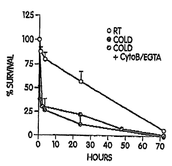
FIG. lA shows circulation time in mice of room temperature platelets and of
platelets chilled and rewarmed in the presence or absence of EGTA-AM and
Cytochalasin B. The curves depict the survival of 5-chloromethylfluorescein
diacetate
(CMFDA) labeled, room temperature (RT) platelets, platelets chilled at ice-
bath
temperature (Cold) and rewarmed to room temperature before injection and
chilled and
rewarmed platelets treated with EGTA-AM and cytochalasin B (Cold + CytoB/EGTA)
to
preserve their discoid shape. Each curve represents the mean SD of 6 mice.
Identical
clearance patterns were observed with 111Indium-labeled platelets.
FIG. 1B shows that chilled platelets aggregate normally in vitro. Washed,
chilled-rewarmed (Cold) or room temperature (RT) wild type platelets were
stimulated
by the addition of the indicated agonists at 37 C and liglit transmission was
recorded on a
standard aggregometer. Aggregation responses of chilled platelets treated with
EGTA-
AM and cytochalasin B were identical to untreated chilled platelets.
FIG. 1 C shows that cold induced clearance occurs predominantly in the liver
of
mice. The liver is the primary clearance organ of chilled platelets,
containing 60-90% of
injected platelets. In contrast, RT platelets are cleared more slowly in the
spleen.
111 Indium labeled platelets were injected into syngeneic mice and tissues
were harvested
at 0.5, 1 and 24 hours. Data are expressed per gram of tissue. Each bar
depicts the mean
values of 4 animals analyzed SD.
FIG. 1D shows that chilled platelets co-localize witli hepatic sinusoidal
macrophages (Kupffer cells). This representative confocal-micrograph shows the
hepatic
distribution of CMFDA-labeled, chilled-rewarmed platelets (green) after 1 hour
of
transfusion, which preferentially accumulate in periportal and midzonal fields
of liver
lobules. Kupffer cells were visualized after injection of nile red-labeled
spheres. The
merged micrograph that shows co-localization of chilled platelets and
macrophages in
yellow. The lobule organization is indicated (CV: central vein; PV: portal
vein, bar: 100
M).
13
CA 02626363 2008-04-15
WO 2006/044790 PCT/US2005/037241
FIG. 2 shows that chilled platelets circulate normally in CR3-deficient mice,
but
not in complement 3 (C3) or vWf deficient mice. CMFDA-labeled chilled-rewarmed
(Cold) and room temperature (RT) wild type platelets were transfused into six
each of
syngeneic wild type (WT), CR3-deficient (A), vWf-deficient (B) and C3-
deficient (C)
recipient mice and their survival times determined. Chilled platelets
circulate in CR3-
deficient animals with the same kinetics as room-temperature platelets, but
are cleared
rapidly from the circulation of C3- or vWf-deficient mice. Data are mean SD
for 6
mice.
FIG. 3 shows that chilled platelets adhere tightly to CR3-expressing mouse
macrophages in vivo. FIG. 3A - Chilled-rewarmed TRITC-labeled platelets (left
panel)
adllere with a 3-4 x higher frequency to liver sinusoids than room temperature
CMFDA-
labeled platelets (right panel). The intravital fluorescence micrographs were
obtained 30
min after the infusion of the platelets. FIG. 3B - Chilled-rewarmed (Cold,
open bars) and
room temperature platelets (RT, filled bars) adhere to sinusoidal regions with
high
macrophage density (midzonal) with similar distributions in wild type mice.
Fig. 3C -
Chilled-rewarmed platelets adhere 3-4 x more than room temperature platelets
to
macrophages in the wild type liver (open bars). In contrast, chilled-rewarmed
or room
teinperature platelets have identical adherence to macrophages in CR3-
deficient mice
(filled bars). 9 experiments with wild type mice and 4 experiments with CR3-
deficient
mice are shown (mean SEM, * P < 0.05: ** P < 0.01).
FIG. 4 shows that GPlba mediates chilled platelet clearance, aggregates in the
cold, but binds activated vWf normally on chilled platelets. Fig. 4A - CMFDA-
labeled
platelets enzymatically cleared of the GPlba extracellular domain (left panel,
inset, filled
area) or control platelets were kept at room temperature (left panel) or
chilled-rewarmed
(right panel) infused into syngeneic wild type mice, and platelet survivals
were
determined. Each survival curve represents the mean values ~ SD for 6 mice.
Fig. 4 B -
Chilled, or RT platelet rich plasma was treated with (shaded area) or without
(open area)
botrocetin. vWf bound was detected using FITC labeled anti-vWf antibody. Fig.
4C - The
14
CA 02626363 2008-04-15
WO 2006/044790 PCT/US2005/037241
vWf receptor redistributes from linear arrays (RT) into aggregates (Chilled)
on the
surface of chilled murine platelets. Fixed, chilled-rewarmed, or room
temperature
platelets (RT) were incubated with monoclonal rat anti-mouse GPlba antibodies
followed by 10 nm colloidal gold particles coated with goat anti-rat IgG. The
bars are
100 nm. Inset: low magnification of platelets.
FIG. 5 shows GPlba-CR3 interaction mediates phagocytosis of chilled human
platelets in vitro. FIGS. 5A and 5B show a representative assay result of THP-
1 cells
incubated with room temperature (RT) ( Fig. 5A) or chilled-rewarmed (Cold)
platelets
(Fig. 5B). CM-Orange-labeled platelets associated with macrophages shift in
orange
fluorescence up the y axis. The mean percentage of the CM-Orange positive
native
macrophages incubated with platelets kept at room temperature was normalized
to 1.
Chilling of platelets increases this shift from -4% to 20%. The platelets are
predominantly ingested, because they do not dual label with the FITC-
conjugated mAb to
CD61. Fig. 5C Undifferentiated (open bars) THP-1 cells express -50% less CR3,
and
ingest half as many chilled-rewarmed platelets. Differentiation (filled bars)
of CR3
expression however, had no significant effect on the uptake of RT platelets.
Treatment of
human platelets with the snake venom metalloprotease, mocarhagin (Moc), which
removes the N-terminus of GPlba from the surface of human platelets (inset;
control:
solid line, mocarhagin treated platelets: shaded area), reduced phagocytosis
of chilled
platelets by -98%. Data shown are means SD of 5 experiments.
FIG. 6 shows circulating, chilled platelets have hemostatic function in CR3
deficient mice. Normal in vivo function of room temperature (RT) platelets
transfused
into wild type mice (Fig. 6A and 6B) and of chilled (Cold) platelets
transfused into CR3
deficient mice (Fig. 6C and 6D), as determined by their equivalent presence in
platelet
aggregates emerging from the wound 24 hrs after infusion of autologous CMFDA
labeled
platelets. Peripheral blood (Fig. 6A and 6C) and the blood emerging from the
wound
(shed blood, Fig. 6B and 6D) were analyzed by whole blood flow cytometry.
Platelets
CA 02626363 2008-04-15
WO 2006/044790 PCT/US2005/037241
were identified by forward light scatter characteristics and binding of the PE-
conjugated
anti-GPlba mAb (pOp4). The infused platelets (dots) were identified by their
CMFDA
fluorescence and the non-infused platelets (contour lines) by their lack of
CMFDA
fluorescence. In the peripheral whole blood samples, analysis regions were
plotted
around the GPlba-positive particles to include 95% of the population on the
forward
scatter axis (region 1) and the 5% of particles appearing above this forward
light scatter
threshold were defined as aggregates (region 2). The percentages refer to the
number of
aggregates formed by CMFDA-positive platelets. This shown result is
representative of
4 experiments. Fig. 6E shows ex vivo fanction of CM-Orange, room temperature
(RT)
platelets transfused into wild type mice and CM-Orange, chilled-rewarmed
(Cold)
platelets transfused into CR3 deficient mice, as determined by exposure of P-
selectin and
fibrinogen binding following thrombin (1 U/ml) activation of blood drawn from
the mice
after 24 hours post infusion. CM-Orange labeled platelets have a circulation
half-life
time comparable to that of CMFDA labeled platelets (not shown). Transfused
platelets
were identified by their CM-Orange fluorescence (filled bars). Non-transfused
(non-
labeled) analyzed platelets are represented as open bars. Results are
expressed as the
percentage of cells present in the P-selectin and fibrinogen positive regions
(region 2).
Data are mean + SD for 4 mice.
FIG. 7 is a schematic depicting two platelet clearance pathways. Platelets
traverse
central and peripheral circulations, undergoing reversible priming at lower
temperatures
at the body surface. Repeated priming leads to irreversible GPlb-IX-V (vWfR)
receptor
complex reconfiguration and clearance by complement receptor type 3 (CR3)
bearing
hepatic macrophages. Platelets are also cleared after they participate in
microvascular
coagulation.
FIG. 8 shows the effect of monosaccharides on phagocytosis of chilled
platelets.
FIG. 9 shows the dot plots of binding of WGA lectin to room temperature
platelets or chilled platelets.
16
CA 02626363 2008-04-15
WO 2006/044790 PCT/US2005/037241
FIG. 10 shows the analysis of various FITC labeled lectins bound to room
temperature or chilled platelets.
FIG. 11A shows the summary of FITC-WGA binding to the surface of room
teinperature or chilled platelets obtained'by flow cytometry before and after
(3-
hexosaminidase treatment.
FIG. 11B shows that GP1ba removal from the platelet surface reduced FITC-
WGA binding to chilled platelets.
FIG. 12 shows that galactose transfer onto platelet oligosaccharides reduces
chilled platelet (Cold) phagocytosis, but does not affect the phagocytosis of
room
temperature (RT) platelets.
FIG. 13 shows the survival of chilled, galactosylated murine platelets
relative to
untreated platelets.
FIG. 14 shows that platelets containing galactose transferases on their
surface
transfer galactose without the addition of external transferases as judged by
WGA
binding (Fig 14A) and in vitro phagocytosis results for human platelets (Fig
14B). Fig.
14C shows that of UDP-galactose with or without Galactose transferase (Ga1T)
on
survival of murine platelets. UDP-galactose with or without Ga1T was added to
murine
platelets before chilling for 30 min at 37 C. The platelets were chilled for 2
hours in an
ice bath and then transfused (108 platelets/mouse) into mice and their
survival
2o determined.
FIG. 15 shows the time course of 14C-labeled UDP-galactose incorporation into
human platelets.
FIG. 16 shows galactosylation of platelets in four platelet concentrate
samples at
different concentrations of UDP-galactose.
FIG. 17 shows the complement receptor mediates phagocytosis and clearance of
chilled platelets.
17
CA 02626363 2008-04-15
WO 2006/044790 PCT/US2005/037241
FIG. 18 shows the GPlba subunit of platelet von Willebrand factor receptor
binds
the I-domain of aM of aM/(32 integrin.
FIG. 19 shows that chilled platelets circulate and function normally in aM
knockout mice.
FIG. 20 illustrates vWf receptor inactivation.
FIG. 21 shows that aM/(32 recognizes the outer tip of GPlba and mediates
clearance of chilled platelets, thus demonstrating that GPlba has coagulant
(vWf
binding) and non-coagulant (clearance) functions.
FIG. 22 illustrates the primary structure of aM (CD11b).
FIG. 23 shows that aM has a lectin affinity site.
FIG. 24 shows that the lectin domain of macrophage aM/(32 receptors recognizes
(3G1cNAc residues on clustered GPlba.
FIG. 25 shows that a soluble aM-lectin domain inhibits chilled human platelet
phagocytosis by macrophages.
FIG. 26 shows the construction of CHO cells expressing aMaX chimeric
proteins.
FIG. 27 illustrates a phagocytic assay for altered platelet surface induced by
chilling.
FIG. 28 shows that the aM-lectin domain mediates chilled human platelet
phagocytosis.
FIG. 29 shows that macrophage aM/(32 receptors recognize PG1cNAc residues on
clustered GPlba receptors of chilled platelets.
FIG. 30 illustrates the galactosylation of platelets through GPlba.
FIG. 31 shows expression of (34GalT1 on the platelet surface.
FIG. 32 illustrates that galatosylated chilled murine platelets can circulate
in vivo.
FIG. 33 illustrates that galatosylated chilled murine platelets can function
normally in murine models.
18
CA 02626363 2008-04-15
WO 2006/044790 PCT/US2005/037241
FIG. 34 shows that human platelet concentrates can be galactosylated, which
preserves platelet function.
FIG. 35 illustrates a metllod for galactosylation of human platelet
concentrates.
FIG. 36 shows surface galactose on platelet concentrates is stable.
FIG. 37 shows that galactosylation iiihibits phagocytosis by THP-1 macrophages
of human chilled platelets.
FIG. 38 shows that platelet counts and pH remain unchanged in refrigerated
platelet concentrates.
FIG. 39 shows the effects of refrigeration and galatosylation on retention of
platelet responses to agonists during storage of concentrates.
FIG. 40 shows the effect of storage conditions on shape change (spreading) and
clumping of platelets in concentrates.
FIG. 41 illustrates an embodiment of the invention wherein a bioprocess for
collecting, treating and storing platelets is described. Platelets are derived
from a variety
of blood sources, including IRDP - Individual Random Donor Platelets, PRDP -
Pooled
Random Donor Platelets and SDP - Single Donor Platelets. The container having
the
glycan modifying agent, e.g., a solution of UDP-Gal and/or CMP-NeuAc is
sterile
docked to the bag containing the platelets. A sterile dock is also referred to
as a sterile
connection device (SCD) or a total containment device (TCD). The sterile dock
permits
connection of two pieces of conduit while maintaining sterility of the system.
The
glycans modifying agent is mixed with the platelets and then the modified
platelets are
transferred to a non-breathable bag through a leukocyte filter. Glass wool or
affinity
separation methods for removing leukocyte fractions from whole blood are known
in the
art, and provide examples of means for filtering the leukocytes from the
platelets.
FIG. 42 illustrates a nonlimiting embodiment 2 of the invention wherein a
bioprocess for collecting, treating and storing platelets is described.
FIG. 43 illustrates a nonlimiting embodiment 3 of the invention wherein a
bioprocess for collecting, treating and storing platelets is described.
19
CA 02626363 2008-04-15
WO 2006/044790 PCT/US2005/037241
FIG. 44 illustrates a nonlimiting embodiment 4 of the invention wherein a
bioprocess for collecting, treating and storing platelets is described.
FIG. 45 illustrates a nonlimiting embodiment 5 of the invention wherein a
bioprocess for collecting, treating and storing platelets is described.
FIG. 46 illustrates a nonlimiting embodiment 6 of the inverition wherein a
bioprocess for collecting, treating and storing platelets is described.
FIG. 47 illustrates a nonlimiting embodiment 7 of the invention wherein a
bioprocess for collecting, treating and storing platelets is described.
FIG. 48 illustrates a nonlimiting einbodiment 8 of the iuzvention wherein a
1o bioprocess for collecting, treating and storing platelets is described.
FIG. 49 illustrates a nonlimiting embodiment 9 of the invention wherein a
bioprocess for collecting, treating and storing platelets is described.
FIG. 50 illustrates a nonlimiting embodiment 10 of the invention wherein a
bioprocess for collecting, treating and storing platelets is described.
FIG. 51 illustrates a nonlimiting embodiment 11 of the invention wherein a
bioprocess for collecting, treating and storing platelets is described.
FIG. 52 illustrates a nonlimiting embodiment 12 of the invention wherein a
bioprocess for collecting, treating and storing platelets is described.
FIG. 53 illustrates a nonlimiting embodiment 13 of the invention wherein a
2o bioprocess for collecting, treating and storing platelets is described.
FIG. 54 illustrates a nonlimiting embodiment 14 of the invention wherein a
bioprocess for collecting, treating and storing platelets is described.
FIG. 55 illustrates a nonlimiting embodiment 15 of the invention wherein a
bioprocess for collecting, treating and storing platelets is described.
FIG. 56 illustrates a nonlimiting embodiment 16 of the invention wherein a
bioprocess for collecting, treating and storing platelets is described.
FIG. 57 illustrates a nonlimiting embodiment 17 of the invention wherein a
bioprocess for collecting, treating and storing platelets is described.
CA 02626363 2008-04-15
WO 2006/044790 PCT/US2005/037241
FIG. 58 illustrates a nonlimiting embodiment 18 of the invention wherein a
bioprocess for collecting, treating and storing platelets is described.
FIG. 59 illustrates a nonlimiting embodiment 19 of the invention wherein a
bioprocess for collecting, treating and storing platelets is described.
FIG. 60 illustrates a nonlimiting embodiment 20 of the invention wherein a
bioprocess for collecting, treating and storing platelets is described.
FIG. 61 illustrates a nonlimiting embodiment 21 of the invention wherein a
bioprocess for collecting, treating and storing platelets is described.
FIG. 62 illustrates a nonlimiting embodiment 22 of the invention wherein a
bioprocess for collecting, treating and storing platelets is described.
FIG. 63 illustrates a nonlimiting embodiment 23 of the invention wherein a
bioprocess for collecting, treating and storing platelets is described.
FIG. 64 illustrates a nonlimiting embodiment 24 of the invention wherein a
bioprocess for collecting, treating and storing platelets is described.
FIG. 65 illustrates a nonlimiting embodiment 25 of the invention wherein a
bioprocess for collecting, treating and storing platelets is described.
FIG. 66 illustrates a nonlimiting einbodiment 26 of the invention wherein a
bioprocess for collecting, treating and storing platelets is described.
FIG. 67 illustrates that platelets contain an endogenous intra-cellular and
extra-
cellular sialyltransferase.
FIG. 68 illustrates the endogenous platelet sialyltransferase activity
catalyzes the
elongation of exposed P-galactose on platelets by the sole addition of the
donor substrate
CMP-sialic acid.
FIG. 69 illustrates that platelets with reduced sialic acid are rapidly
cleared in
vivo as demonstrated by the clearance of ST3GalIV -/- platelets in wt mice.
FIG. 70 illustrates that glycosylation improves the circulation of non-chilled
platelets.
21
CA 02626363 2008-04-15
WO 2006/044790 PCT/US2005/037241
DETAILED DESCRIPTION OF THE INVENTION
The invention provides a population of modified platelets that have enhanced
circulation properties and that retain substantially normal in vivo hemostatic
activity.
Hemostatic activity refers broadly to the ability of a population of platelets
to mediate
bleeding cessation. Various assays are available for determining platelet
hemostatic
activity (Bennett, J. S. and Shattil, S. J., 1990, "Platelet function,"
Hematology, Williams,
W. J., et al., Eds. McGraw Hill, pp 1233-12250). However, demonstration of
"hemostasis" or "hemostatic activity" ultimately requires a demonstration that
platelets
infused into a thrombocytopenic or thrombopathic (i.e., non-functional
platelets) animal
or human circulate and stop natural or experimentally-induced bleeding.
Short of such a demonstration, laboratories use in vitro tests as surrogates
for
determining hemostatic activity. These tests, which include assays of
aggregation,
secretion, platelet morphology and metabolic changes, measure a wide variety
of platelet
functional responses to activation. It is generally accepted in the art that
the in vitro tests
are reasonably indicative of hemostatic function in vivo.
Substantially nomlal hemostatic activity refers to an amount of hemostatic
activity seen in the modified platelets, that is functionally equivalent to or
substantially
similar to the hemostatic activity of untreated platelets in vivo, in a
healthy (non-
thrombocytopenic or non-throinbopathic mainmal) or functionally equivalent to
or
substantially similar to the hemostatic activity of a freshly isolated
population of platelets
in vitro.
The instant invention provides metllods for reduced teinperature storage of
platelets which increases the storage time of the platelets, as well as
methods for reducing
clearance of or increasing circulation time of a population of platelets in a
mammal. Also
provided are platelet compositions methods and compositions for the
preservation of
platelets witli preserved hemostatic activity as well as methods for making a
pharmaceutical composition containing the preserved platelets and for
administering the
22
CA 02626363 2008-04-15
WO 2006/044790 PCT/US2005/037241
pharmaceutical composition to a mammal to mediate hemostasis. Also provided
are kits
for treating a platelet preparation for storage, and containers for storing
the same.
In one aspect of the invention, the method for increasing circulation time of
an
isolated population of platelets involves contacting an isolated population of
platelets
with at least one glycan modifying agent in an ainount effective to reduce the
clearance
of the population of platelets. As used herein, a population of platelets
refers to a sample
having one or more platelets. A population of platelets includes a platelet
concentrate.
The term "isolated" means separated from its native environment and present in
sufficient
quantity to permit its identification or use. As used herein with respect to a
population of
platelets, isolated means removed or cleared from the blood circulation of a
mammal.
The circulation time of a population of platelets is defined as the time when
one-half of
the platelets in that population are no longer circulating in a mammal after
transplantation
into that mammal. As used herein, "clearance" means removal of the modified
platelets
from the blood circulation of a mammal (such as but not limited to by
macrophage
phagocytosis). As used herein, clearance of a population of platelets refers
to the
removal of a population of platelets from a unit volume of blood or serum per
unit of
time. Reducing the clearance of a population of platelets refers to
preventing, delaying,
or reducing the clearance of the population of platelets. Reducing clearance
of platelets
also may mean reducing the rate of platelet clearance.
A glycan modifying agent refers to an agent that modifies glycan residues on
the
platelet. As used herein, a "glycan" or "glycan residue" is a polysaccharide
moiety on
surface of the platelet, exemplified by the GP lba polysaccharide. A
"terminal" glycan or
glycan residue is the glycan at the distal terminus of the polysaccharide,
which typically
is attached to polypeptides on the platelet surface. Preferably, the glycan
modifying
agent alters GPlba on the surface of the platelet.
The glycan modifying agents suitable for use as described herein, includes
monosaccharides such as arabinose, fructose, fucose, galactose, mannose,
ribose,
gluconic acid, galactosamine, glucosamine, N-acetylgalactosamine, muramic
acid, sialic
23
CA 02626363 2008-04-15
WO 2006/044790 PCT/US2005/037241
acid (N-acetylneuraminic acid), and nucleotide sugars such as cytidine
monophospho-N-
acetylneuraminic acid (CMP-sialic acid), uridine diphosphate galactose (UDP-
galactose)
and UDP-galactose precursors such as UDP-glucose. In some preferred
embodiments,
the glycan modifying agent is UDP-galactose or CMP-sialic acid.
UDP-galactose is an intermediate in galactose metabolism, formed by the enzyme
UDP-glucose-a-D-galactose- 1 -phosphate uridylyltransferase which catalyzes
the release
of glucose-l-phosphate from UDP-glucose in exchange for galactose-1-phosphate
to
make UDP-galactose. UDP-galactose and sialic acid are widely available from
several
cominercial suppliers such as Sigma. In addition, methods for syntllesis and
production
of UDP-galactose are well known in the art and described in the literature
(see for
example, Liu et al, ChemBioChem 3, 348-355, 2002; Heidlas et al, J. Org. Chem.
57,
152-157; Butler et al, Nat. Biotechnol. 8, 281-284, 2000; Koizumi et al,
Carbohydr. Res.
316, 179-183, 1999; Endo et al, Appl. Microbiol., Biotechnol. 53, 257-261,
2000). UDP-
galactose precursors are molecules, compounds, or interinediate compounds that
may be
converted (e.g., enzymatically or biochemically) to UDP-galactose. One non-
limiting
example of a UDP-galactose precursor is UDP-glucose. In certain embodiments,
an
enzyme that converts a UDP-galactose precursor to UDP-galactose is added to a
reaction
mixture (e.g. in a platelet container).
An effective amount of a glycan modifying agent is that amount of the glycan
modifying agent that alters a sufficient number of glycan residues on the
surface of
platelets, that when introduced to a population of platelets, increases
circulation time
and/or reduces the clearance of the population of platelets in a mammal
following
transplantation of the platelets into the mammal. An effective amount of a
glycan
modifying agent is a concentration from about 1 micromolar to about 1200
micromolar,
preferably from about 10 micromolar to about 1000 micromolar, more preferably
from
about 100 micromolar to about 750 micromolar, and most preferably from about
200
micromolar to about 600 micromolar.
24
CA 02626363 2008-04-15
WO 2006/044790 PCT/US2005/037241
Modification of platelets with glycan modifying agents can be preformed as
follows. The population of platelets is incubated with the selected glycan
modifying
agent (concentrations of 1-1200 M) for at least 1, 2, 5, 10, 20, 40, 60, 120,
180, 240, or
300 min. at 22 C - 37 C. Multiple glycan modifying agents (i.e., two, three
four or
more) may be used simultaneously or sequentially. In some embodiments 0.1-500
inU/ml galactose transferase or sialyl transferase is added to the population
of platelets.
Galactose transfer can be monitored functionally using FITC-WGA (wheat germ
agglutinin) binding. The goal of the glycan modification reaction is to reduce
WGA
binding to resting room temperature WGA binding-levels. Galactose transfer can
be
quantified using 14C-UDP-galactose. Non-radioactive UDP-galactose is mixed
with 14C-
UDP-galactose to obtain appropriate galactose transfer. Platelets are
extensively washed,
and the incorporated radioactivity measured using a y-counter. The measured
cpm
permits calculation of the incorporated galactose. Similar tecliniques are
applicable to
monitoring sialic acid transfer.
Reducing the clearance of a platelet encoinpasses reducing clearance of
platelets
after storage at room temperature, or after chilling, as well as "cold-induced
platelet
activation". Cold-induced platelet activation is a term having a particular
meaning to one
of ordinary skill in the art. Cold-induced platelet activation may manifest by
changes in
platelet morphology, some of which are similar to the changes that result
following
platelet activation by, for example, contact with glass. The structural
changes indicative
of cold-induced platelet activation are most easily identified using
techniques such as
light or electron microscopy. On a molecular level, cold-induced platelet
activation
results in actin bundle formation and a subsequent increase in the
concentration of
intracellular calcium. Actin-bundle formation is detected using, for example,
electron
microscopy. An increase in intracellular calcium concentration is determined,
for
example, by einploying fluorescent intracellular calcium chelators. Many of
the above-
described chelators for inhibiting actin filament severing are also useful for
deterinining
CA 02626363 2008-04-15
WO 2006/044790 PCT/US2005/037241
the concentration of intracellular calcium (Tsien, R., 1980, supra.).
Accordingly, various
techniques are available to determine whether or not platelets have
experienced cold-
induced activation.
The effect of galactose or sialic acid addition to the glycan moieties on
platelets,
resulting in diminished clearance of modified platelets, can be measured for
example
using either an in vitro system employing differentiated THP-1 cells or murine
macrophages, isolated from the peritoneal cavity after thioglycolate injection
stinlulation.
The rate of clearance of modified platelets compared to unmodified platelets
is
deterinined. To test clearance rates, the modified platelets are fed to the
macrophages
and ingestion of the platelets by the macrophages is monitored. Reduced
ingestion of
modified platelets relative to unmodified platelets (twofold or greater)
indicates
successful modification of the glycan moiety for the purposes described
herein.
In accordance with the invention, the population of modified platelets can be
chilled without the deleterious effects (cold-induced platelet activation)
usually
experienced on chilling of untreated platelets. The population of modified
platelets can
be chilled prior to, concurrently with, or after contacting the platelets with
the at least one
glycan modifying agent. The selective modification of glycan moieties reduces
clearance, following chilling (also if not chilled), thus permitting longer-
term storage
than is presently possible. As used herein, chilling refers to lowering the
temperature of
the population of platelets to a temperature that is less than about 37 C. In
some
embodiments, the platelets are chilled to a temperature that is less than
about 15 C. In
some preferred embodiments, the platelets are chilled to a temperature ranging
from
between about 0 C to about 4 C. Chilling also encompasses freezing the
platelet
preparation, i.e., to temperatures less than 0 C, -20 C, -50 C, and -80 C
or cooler.
Process for the cryopreservation of cells are well known in the art.
In some einbodiments, the population of platelets is stored chilled for at
least 3
days. In some embodiments, the population of platelets is stored chilled for
at least 5, 7,
10, 14, 21, and 28 days or longer.
26
CA 02626363 2008-04-15
WO 2006/044790 PCT/US2005/037241
In some embodiments of the invention, the circulation time of the population
of
platelets is increased by at least about 10%. In some other embodiments, the
circulation
time of the population of platelets is increased by at least about 25%. In yet
some other
embodiments, the circulation time of the population of platelets is increased
by at least
about 50% to about 100%. In still yet otller embodiments, the circulation time
of the
population of platelets is increased by about 150% or greater.
The invention also embraces a method for increasing the storage time of
platelets.
As used herein the storage time of platelets is defined as the time that
platelets can be
stored without substantial loss of platelet function or hemostatic activity
such as the loss
of the ability to circulate or increased platelet clearance.
The platelets are collected from peripheral blood by standard techniques known
to
those of ordinary skill in the art, for example by isolation from whole blood
or by
apheresis processes. In some embodiments, the platelets are contained in a
pharmaceutically-acceptable carrier prior to treatment with a glycan modifying
agent.
According to another aspect of the invention, a modified platelet or a
population
of modified platelets is provided. The modified platelet comprises a plurality
of modified
glycan molecules on the surface of the platelet. In some embodiments, the
modified
glycan moieties are GPlba molecules. The invention also encompasses a platelet
composition in a storage mediuin. In some embodiments the storage medium
comprises
a pharmaceutically acceptable carrier.
The term "pharmaceutically acceptable" means a non-toxic material that does
not
interfere with the effectiveness of the biological activity of the platelets
and that is a non-
toxic material that is compatible with a biological system such as a cell,
cell culture,
tissue, or organism. Pharmaceutically acceptable carriers include diluents,
fillers, salts,
buffers, stabilizers, solubilizers, and other materials which are well known
in the art, for
example, a buffer that stabilizes the platelet preparation to a pH of 7.4, the
physiological
pH of blood, is a pharmaceutically acceptable composition suitable for use
with the
present invention.
27
CA 02626363 2008-04-15
WO 2006/044790 PCT/US2005/037241
The invention further einbraces a method for making a pharmaceutical
composition for administration to a mammal. The method comprises preparing the
above-described platelet preparation, and warming the platelet preparation. In
some
embodiments, the method comprises neutralizing, removing or diluting the
glycan
modifying agent(s) and/or the enzyme(s) that catalyze the modification of the
glycan
moiety, and placing the modified platelet preparation in a phamlaceutically
acceptable
carrier. In a preferred einbodiment, the chilled platelets are warined to room
temperature
(about 22 C) prior to neutralization or dilution. In some embodiments, the
platelets are
contained in a pharmaceutically acceptable carrier prior to contact witli the
glycan
modifying agent(s) with or without the enzyme(s) that catalyze the
modification of the
glycan moiety and it is not necessary to place the platelet preparation in a
pharmaceutically acceptable carrier following neutralization or dilution.
As used herein, the terms "neutralize" or "neutralization" refer to a process
by
which the glycan modifying agent(s) and/or the enzyme(s) that catalyze the
modification
of the glycan moiety are rendered substantially incapable of glycan
modification of the
glycan residues on the platelets, or their concentration in the platelet
solution is lowered
to levels that are not harmful to a mammal, for example, less that 50
micromolar of the
glycan modifying agent. In some embodiments, the chilled platelets are
neutralized by
dilution, e.g., with a suspension of red blood cells. Alternatively, the
treated platelets can
be infused into the recipient, which is equivalent to dilution into a red
blood cell
suspension. This method of neutralization advantageously maintains a closed
system and
minimizes damage to the platelets. In a preferred embodiment of glycan
modifying
agents, no neutralization is required.
An alternative method to reduce toxicity is by inserting a filter in the
infusion
line, the filter containing, e.g. activated charcoal or an immobilized
antibody, to remove
the glycan modifying agent(s) and/or the enzyme(s) that catalyze the
modification of the
glycan moiety.
28
CA 02626363 2008-04-15
WO 2006/044790 PCT/US2005/037241
Either or both of the glycan modifying agent(s) and the enzyme(s) that
catalyze
the modification of the glycan moiety also may be removed or substantially
diluted by
washing the modified platelets in accordance with standard clinical cell
washing
techniques.
The invention further provides a method for mediating hemostasis in a mammal.
The method includes administering the above-described pharmaceutical
preparation to
the mammal. Administration of the modified platelets may be in accordance with
standard methods known in the art. According to one embodiment, a human
patient is
transfused witli red blood cells before, after or during administration of the
modified
platelets. The red blood cell transfusion serves to dilute the administered,
modified
platelets, thereby neutralizing the glycan modifying agent(s) and the
enzyme(s) that
catalyze the modification of the glycan moiety.
The dosage regimen for mediating hemostasis using the modified platelets is
selected in accordance with a variety of factors, including the type, age,
weight, sex and
medical condition of the subject, the severity of the disease, the route and
frequency of
adininistration. An ordinarily skilled physician or clinician can readily
determine and
prescribe the effective amount of modified platelets required to mediate
hemostasis.
The dosage regimen can be determined, for example, by following the response
to
the treatment in terms clinical signs and laboratory tests. Examples of such
clinical signs
and laboratory tests are well known in the art and are described, see,
Harrison's
Principles of Internal Medicine, 15th Ed., Fauci AS et al., eds., McGraw-Hill,
New York,
2001.
Also within the scope of the invention are storage compositions and
pharmaceutical compositions for mediating hemostasis. In one embodiment, the
compositions comprise a pharmaceutically-acceptable carrier, a plurality of
modified
platelets, a plurality of glycan modifying agent(s) and optionally the
enzyme(s) that
catalyze the modification of the glycan moiety. The glycan modifying agent(s)
and the
enzyme(s) that catalyze the modification of the glycan moiety are present in
the
29
CA 02626363 2008-04-15
WO 2006/044790 PCT/US2005/037241
composition in sufficient amounts so as to reduce platelet clearance.
Preferably, glycan
modifying agent(s) (and optionally the enzyme(s) that catalyze the
modification of the
glycan moiety) are present in amounts whereby after chilling and
neutralization, the
platelets maintain substantially normal hemostatic activity. The amounts of
glycan
modifying agent(s) (and optionally the enzyme(s) that catalyze the
modification of the
glycan moiety) which reduce platelet clearance can be selected by exposing a
preparation
of platelets to increasing amounts of these agents, exposing the treated
platelets to a
chilling temperature and determining (e.g., by microscopy) whether or not cold-
induced
platelet activation has occurred. Preferably, the amounts of glycan modifying
agent(s)
and the enzyme(s) that catalyze the modification of the glycan moiety can be
determined
functionally by exposing the platelets to varying amounts of glycan modifying
agent(s)
and the enzyme(s) that catalyze the modification of the glycan moiety,
chilling the
platelets as described herein, warming the treated (chilled) platelets,
optionally
neutralizing the platelets and testing the platelets in a hemostatic activity
assay to
determine whether the treated platelets have maintained substantially normal
hemostatic
activity.
For example, to determine the optimal concentrations and conditions for
preventing cold-induced activation of platelets by modifying them with a
glycan
modifying agent(s) (and optionally the enzyme(s) that catalyze the
modification of the
glycan moiety), increasing amounts of these agents are contacted with the
platelets prior
to exposing the platelets to a chilling temperature. The optimal
concentrations of the
glycan modifying agent(s) and the enzyme(s) that catalyze the modification of
the glycan
moiety are the minimal effective concentrations that preserve intact platelet
function as
determined by in vitro tests (e.g., observing morphological changes in
response to glass,
thrombin, cryopreservation temperatures; ADP-induced aggregation) followed by
in vivo
tests indicative of hemostatic function (e.g., recovery, survival and
shortening of bleeding
time in a thrombocytopenic animal or recovery and survival of 51Cr-labeled
platelets in
human subjects).
CA 02626363 2008-04-15
WO 2006/044790 PCT/US2005/037241
According to yet another aspect of the invention, a composition for addition
to
platelets to reduce platelet clearance or to increase platelet storage time is
provided. The
composition includes one or more glycan modifying agents. In certain
embodiments, the
composition also includes an enzyme(s) that catalyze the modification of the
glycan
moiety. The glycan modifying agent and the enzyme(s) that catalyzes the
modification of
the glycan moiety are present in the composition in amounts that prevent cold-
induced
platelet activation.
The invention also embraces a storage composition for preserving platelets.
The
storage composition comprises at least one glycan modifying agent in an amount
sufficient to reduce platelet clearance. In some embodiments the storage
composition
further coinprises an enzyme that catalyzes the modification of a glycan
moiety on the
platelet. The glycan modifying agent is added to the population of platelets
that are
preferably kept between about room temperature and 37 C. In some embodiments,
following treatment, the population of platelets is cooled to about 4 C. In
some
embodiments, the platelets are collected into a platelet pack, bag, or
container according
to standard methods known to one of skill in the art. Typically, blood from a
donor is
drawn into a primary container which may be joined to at least one satellite
container, all
of which containers are connected and sterilized before use. In some
embodiments, the
satellite container is connected to the container for collecting platelets by
a breakable
seal. In some embodiments, the primary container furtlzer comprises plasma
containing a
plurality of platelets.
In some embodiments, the platelets are concentrated (e.g. by centrifugation)
and
the plasma and red blood cells are drawn off into separate satellite bags (to
avoid
modification of these clinically valuable fractions) prior to adding the
glycan modifying
agent with or without the enzyme that catalyzes the modification of a glycan
moiety on
the platelet. Platelet concentration prior to treatment also may minimize the
amounts of
glycan modifying agents required for reducing the platelet clearance, thereby
minimizing
the amounts of these agents that are eventually infused into the patient.
31
CA 02626363 2008-04-15
WO 2006/044790 PCT/US2005/037241
In one embodiment, the glycan modifying agent(s) are contacted with the
platelets
in a closed system, e.g. a sterile, sealed platelet pack, so as to avoid
microbial
contamination. Typically, a venipuncture conduit is the only opening in the
pack during
platelet procurement or transfusion. Accordingly, to maintain a closed system
during
treatment of the platelets with the glycan modifying agent(s), the agent(s) is
placed in a
relatively small, sterile container which is attached to the platelet pack by
a sterile
connection tube (see e.g., U.S. Pat. No. 4,412,835, the contents of which are
incorporated
herein by reference). The connection tube may be reversibly sealed, or have a
breakable
seal, as will be known to those of skill in the art. After the platelets are
concentrated, e.g.
by allowing the platelets to settle and squeezing the plasma out of the
primary pack and
into a second bag according to standard practice, the seal to the container(s)
including the
glycan modifying agent(s) is opened and the agents are introduced into the
platelet pack.
In one einbodiment, the glycan modifying agents are contained in separate
containers
having separate resealable connection tubes to permit the sequential addition
of the
glycan modifying agents to the platelet concentrate.
Following contact with the glycan modifying agent(s), the treated platelets
are
chilled. In contrast to platelets stored at, for example, 22 C, platelets
stored at
cryopreservation temperatures have substantially reduced metabolic activity.
Thus,
platelets stored at 4 C are metabolically less active and therefore do not
generate large
amounts of COZ coinpared with platelets stored at, for example, 22 C.
(Slichter, S. J.,
1981, Vox Sang 40 (Suppl 1), pp 72-86, Clinical Testing and Laboratory-
Clinical
correlations.). Dissolution of CO2 in the platelet matrix results in a
reduction in pH and a
concomitant reduction in platelet viability (Slichter, S., 1981, supra.). This
can be
resolved by adding buffers to the platelet population, the buffers selected to
keep the
platelet population at or near the physiological pH of blood. Likewise,
conventional
platelet packs are formed of materials that are designed and constructed of a
sufficiently
permeable material to maximize gas transport into and out of the pack (02 in
and COZ
out). The prior art limitations in platelet pack design and construction are
obviated by the
32
CA 02626363 2008-04-15
WO 2006/044790 PCT/US2005/037241
instant invention, wliich permits storage of platelets at cryopreservation
temperatures,
thereby substantially reducing platelet metabolism and diminishing the amount
of CO2
generated by the platelets during storage. Accordingly, the invention further
provides
platelet containers that are substantially non-permeable to COZ and/or 02,
wllich
containers are useful particularly for cold storage of platelets. In both the
gas perineable
and non-gas permeable embodiments, the invention provides for a blood storage
container having therein, a quantity of a glycan modifying agent sufficient to
substantially modify the carbohydrates of the platelets introduced therein,
such that the
platelets become capable of cold storage and subsequent in vivo circulation.
The present invention also provides for kits that are used for platelet
collection,
processing and storage, further including suitable packaging materials and
instructions
for using the kit contents. It is preferred that all reagents and supplies in
the kit be sterile,
in accordance with standard medical practices involving the handling and
storage of
blood and blood products. Methods for sterilizing the kit contents are known
in the art,
for example, ethylene gas, iiTadiation and the like. In certain embodiments,
the kit may
include venipuncture supplies and/or blood collection supplies, for example a
needle set,
solution for sterilizing the skin of a platelet donor, and a blood collection
bag or
container. Preferably the container is "closed", i.e., substantially sealed
from the
environment. Such closed blood collection containers are well known in the
art, and
provide a means of preventing microbial contamination of the platelet
preparation
contained therein. Other einbodiments include kits containing supplies for
blood
collection and platelet apheresis. The kits may further include a quantity of
the glycan
modifying agent, sufficient to modify the voluine of platelets collected and
stored in the
container. In certain embodiments, the kit includes reagents for modifying the
terminal
glycan of platelets with a second or third chemical moiety, for example to
PEGylate
collected platelets. In other embodiments, the kit includes a blood collection
system
having a blood storage container wherein the glycan modifying agent is
provided within
the container in an amount sufficient to treat the volume of blood or
platelets held by the
33
CA 02626363 2008-04-15
WO 2006/044790 PCT/US2005/037241
container. The quantity of glycan modifying agent will depend on the volume of
the
container. It is preferred the glycan modifying agent be provided as a sterile
non-
pyogenic solution, but it may also be supplied as a lyophilized powder. For
example, a
blood bag is provided having a capacity of 250 ml. Contained in the blood bag
is a
quantity of UDP-Gal such that when 250 ml of blood is added, the final
concentration of
the UDP-Gal is approximately 200 micromolar. Other embodiments contain
different
concentrations of glycan modifying agents, for example but not limited to
quantities
resulting in final concentrations of 10 micromolar to 10 millimolar, and
preferably 100
micromolar to 1 millimolar of the glycan modifying agents. Other embodiments
use
1o combinations of glycan modifying agents, e.g., to effect sialyiation or
galactosylation of
N-linked glycoproteins on blood products introduced into the container.
The invention will be more fully understood by reference to the following
examples. These examples, however, are merely intended to illustrate the
einbodiments
of the invention and are not to be construed to limit the scope of the
invention.
EXAMPLES
Example 1
Introduction
Modest cooling primes platelets for activation, but refrigeration causes shape
changes and rapid clearance, compromising storage of platelets for therapeutic
transfusions. We found that shape change inhibition does not normalize cold-
induced
clearance. We also found that cooling platelets rearranges the surface
configuration of
the von Willebrand factor (vWf) receptor complex a subunit (GPlba) such that
it
becomes targeted for recognition by complement receptor 3 receptors (CR3)
predominantly expressed on liver macrophages, leading to platelet phagocytosis
and
clearance. GPlb a removal prolongs survival of unchilled platelets. Chilled
platelets
bind vWf and function normally in vitro and ex vivo after transfusion into CR3-
deficient
34
CA 02626363 2008-04-15
WO 2006/044790 PCT/US2005/037241
mice. Cooled platelets, however, are not "activated" like platelets exposed to
thrombin or
ADP, and their vWf-receptor complex reacts norinally witli activated vWf.
As the temperature falls below 37 C platelets become more susceptible to
activation by thrombotic stimuli, a phenomenon known as "priming" (Faraday and
Rosenfeld, 1998; Hoffmeister et al., 2001). Priining may be an adaptation to
limit
bleeding at lower temperatures of body surfaces where most injuries occur. We
propose
that the hepatic clearance system's purpose is to remove repeatedly primed
platelets, and
that conformational changes in GPlba that promote this clearance do not affect
GPlba's
hemostatically iinportant binding to vWf. Therefore, selective modification of
GPlba
may accommodate cold storage of platelets for transfusion.
Materials and Methods
We obtained fluorescein isothiocyanate (FITC)-conjugated annexin V,
pliycoerythrin (PE)-conjugated anti-human CD1lb/Mac-1 monoclonal antibodies
(mAb),
FITC-conjugated anti-mouse and anti-human IgM niAb, FITC-conjugated anti-mouse
and anti-human CD62P-FITC mAb from Pharmingen (San Diego, CA); FITC-conjugated
rat anti-mouse anti-human IgG mAb from Santa Cruz Biotechnology, Inc. ( Santa
Cruz,
CA); FITC-conjugated anti-lluman CD61 mAbs (clone BL-E6) from Accurate
Scientific
Corp. (Westbuiy, NY); FITC-conjugated anti-human GPlba mAb (clone SZ2) from
Immunotech (Marseille, France); and FITC-conjugated polyclonal rabbit anti-vWf
antibody from DAKOCytomation (Glostrup, Denmark). We purchased EGTA-
acetoxymethylester (AM), Oregon Green coupled fibrinogen from human plasma,
Ce1lTrackerTM Orange CMTMR; Ce1lTracker Green CMFDA, Nile-red (535/575)
coupled and carboxylate-modified 1 m microspheres/F1uoSpheres from Molecular
Probes, Inc. (Eugene, OR) and 111Indium from NEN Life Science Products
(Boston,
MA). We purchased Cytochalasin B, dimethyl sulfoxide (DMSO), trisodium
isothiocyanate (TRITC), human thrombin, prostaglandin El (PGE1), phorbol ester
12-
tetradecanoylphorbol-13 acetate (PMA), A23187 ionophore from Sigma (St. Louis,
MO);
CA 02626363 2008-04-15
WO 2006/044790 PCT/US2005/037241
botrocetin from Centerchein Inc. (Norwalk, CT); and O-sialoglycoprotein-
endopeptidase
from Cerladane (Hornby, Canada). HBSS containing Ca2+ and Mg2+, pH 6.4; RPMI
1640; 0.05% Trypsin-EDTA (0.53 mM) in HBSS without Ca2+ and Mg2+; and other
supplements (penicillin, streptomycin and fetal bovine serum) were from GIBCO
Invitrogen Corp. (Grand Island, NY). TGF-(31 from Oncogene Research Products
(Cambridge, MA); 1,25-(OH)2 vitamin D3 from Calbiochem (San Diego, CA); and
Adenosine-5'-Diphosphate (ADP) were from USB (Cleveland, OH). Avertin (2,2,2-
tribromoethanol) was purchased from Fluka Chemie (Steinheim, Germany).
Collagen
related peptide (CRP) was synthesized at the Tufts Core Facility, Physiology
Dept.
(Boston, MA) and cross-linked as previously described (Morton et al., 1995).
Mocarhagin, a snake venom metalloprotease, was provided by Dr. M. Berndt,
Baker
Medical Research Institute, Melbourne Victoria 318 1, Australia. Additional
unconjugated anti mouse GPlba mAbs and a PE-conjugated anti-mouse GPlba mAb
pOp4 were provided by Dr. B. Nieswandt (Witten/Herdecke University, Wuppertal,
Germany). We obtained THP-1 cells from the American Type Culture Collection
(Manassas, VA).
Animals
For assays of clearance and survival studies, we used age-, strain- and sex-
matched C57BL/6 and C57BL/6 x 129/sv wild type mice obtained from Jackson
Laboratory (Bar Harbor, ME). C57BL/6 x 129/sv mice deficient in complement
coinponent C3 (Wessels et al., 1995) were provided by Dr. M. C. Carroll
(Center for
Blood Research and Department of Pediatrics, Harvard Medical School, Boston,
MA).
C57BL/6 mice deficient in CR3 (Coxon et al., 1996) were provided by Dr. T
Mayadas
and C57BL/6 mice deficient in vWf (Denis et al., 1998) were provided by Dr. D.
Wagner. Mice were maintained and treated as approved by Harvard Medical Area
Standing Committee on Animals according to NIH standards as set forth in The
Guide for
the Care and Use of Laboratory Animals.
Human platelets
36
CA 02626363 2008-04-15
WO 2006/044790 PCT/US2005/037241
Blood was drawn from consenting normal human volunteers (approval was
obtained from the Institutional Review Boards of both Brigham and Women's
Hospital
and the Center for Blood Research (Harvard Medical School)) by venipuncture
into 0.1
volume of Aster-Jandl citrate-based anticoagulant (Hartwig and DeSisto, 1991)
and
platelet rich plasma (PRP) was prepared by centrifugation of the
anticoagulated blood at
300 x g for 20 min at room temperature. Platelets were separated from plasma
proteins
by gel-filtration at room temperature through a small Sepharose 2B column
(Hoffmeister
et al., 2001). Platelets used in the in vitro phagocytosis assay described
below were
labeled with 1.8 M Ce1lTrackerTM Orange CMTMR (CM-Orange) for 20 rnin at 37 C
(Brown et al., 2000), and unincorporated dye was removed by centrifugation
(850 x g, 5
min.) with 5 volumes of washing buffer containing 140 mM NaCI, 5 mM KC1, 12 mM
trisodium citrate, 10 mM glucose, and 12.5 mM sucrose, 1 g/ml PGEI, pH 6.0
(buffer
A). Platelets were resuspended at 3 x 10⁸/ml in a solution containing 140 mM
NaCI, 3
mM KC1, 0.5 mM MgC12, 5 inM NaHCO3, 10 mM glucose and 10 mM Hepes, pH 7.4
(buffer B).
The N-terminus of GPlba was enzymatically removed from the surface of chilled
or room temperature maintained and labeled platelets in buffer B, also
containing 1 mM
Ca2+ and 10 g/ml of the snake venom metalloprotease mocarhagin (Ward et al.,
1996).
After the enzymatic digestion, the platelets were washed by centrifugation
with 5x
volume of buffer A and routinely checked by microscopy for aggregates. GPlba-N-
terminus removal was monitored by incubating platelet suspensions with 5 g/ml
of
FITC-conjugated anti-human GPlba (SZ2) mAb for 10 min at room temperature and
followed by immediate flow cytometry analysis on a FACScalibur Flow Cytometer
(Becton Dickinson Biosciences, San Jose, CA). Platelets were gated by
forward/side
scatter characteristics and 50,000 events acquired.
Murine platelets
37
CA 02626363 2008-04-15
WO 2006/044790 PCT/US2005/037241
Mice were anesthetized with 3.75 mg/g (2.5%) of Avertin, and 1 ml blood was
obtained from the retroorbital eye plexus into 0.1 volume of Aster-Jandl
anticoagulant.
PRP was prepared by centrifugation of anticoagulated blood at 300 x g for 8
min at room
temperature. Platelets were separated from plasma proteins by centrifugation
at 1200 x g
for 5 min and washed two times by centrifugation (1200 x g for 5 min) using 5
x volumes
of washing buffer (buffer A). This procedure is meant by subsequent use of the
term
"washed". Platelets were resuspended at a concentration of 1 x 10⁹/ml in a
solution
containing 140 mM NaCI, 3 mM KCI, 0.5 mM MgC12, 5 mM NaHCO3, 10 mM glucose
and 10 mM Hepes, pH 7.4 (buffer B). Platelet count was determined using a
Bright Line
Hemocytometer (Hausser Scientific, Horsham, PA) under a phase-contrast
microscope at
400 x magnification. Some radioactive platelet clearance studies were
performed with
111Indium, and we labeled mouse platelets using a method described for primate
platelets
(Kotze et al., 1985). Platelets were resuspended at a concentration of 2 x
109/ml in 0.9%
NaCI, pH 6.5 (adjusted with 0. 1 M sodium citrate), followed by the addition
of 500 Ci
111Indium chloride for 30 min at 37 C and washed as described above and
suspended in
buffer B at a concentration of 1 x 10⁹/ml.
For intravital microscopy or other platelet survival experiments, washed
platelets
were labeled either with 2.5 M Ce1lTracker Green CMFDA (5-chloromethyl
fluorescein
diacetate) (CMFDA) for 20 min at 37 C (Baker et al., 1997) or with 0.15 M
TRITC for
20 min at 37 C in buffer B also containing 0.001% DMSO, 20 mM HEPES.
Unincorporated dye was removed by centrifugation as described above, and
platelets
were suspended at a concentration of 1 x 10⁹/ml in buffer B.
The N-terminus of GPlba was enzymatically removed from the surface of chilled
or room temperature labeled platelets with 100 g/ml O-sialoglycoprotein
endopeptidase
in buffer B containing 1mM Ca2} for 20 min at 37 C (Bergmeier et al., 2001).
After
enzymatic digestion, platelets were washed by centrifugation and checked by
light
microscopy for aggregates. Enzymatic removal of the GP1ba-N-terminus removal
was
38
CA 02626363 2008-04-15
WO 2006/044790 PCT/US2005/037241
monitored by incubating the platelet suspensions with 5 g/ml of PE-conjugated
anti-
mouse GP lba mAb pOp4 for 10 inin at room temperature, and bound PE analyzed
by
flow cytometry.
To inhibit cold-induced platelet shape changes, 109/inl platelets in buffer B
were
loaded with 2 M EGTA-AM followed by 2 M cytochalasin B as previously
described
(Winokur and Hartwig, 1995), labeled with 2.5 M CMFDA for 30 min at 37 C and
then
chilled or maintained at room temperature. The platelets were subjected to
standard
washing and suspended at a concentration of 1 x 10⁹/ml in buffer B before
injection into
mice.
Platelet tenzpef=ature protocols
To study the effects of temperature on platelet survival or function,
unlabeled,
radioactively labeled, or fluorescently-labeled mouse or huinan platelets were
incubated
for 2 hours at room temperature (25-27 C) or else at ice bath temperatures and
then
rewarmed for 15 minutes at 37 C before transfusion into mice or in vitro
analysis.
Platelets subjected to these treatments are designated cooled or chilled (or
chilled,
rewanned) and room temperature platelets respectively.
Murine platelet recovery, survival and fate
CMFDA labeled chilled or room temperature murine platelets (10$) were injected
into syngeneic mice via the lateral tail vein using a 27-gauge needle. For
recovery and
survival determination, blood samples were collected iinmediately (< 2 min)
and 0.5, 2,
24, 48, 72 hours after transfusion into 0.1 volume of Aster-Jandl
anticoagulant. Whole
blood analysis using flow cytometly was performed and the percentage of CMFDA
positive platelets determined by gating on all platelets according to their
forward and side
scatter characteristics (Baker et al., 1997). 50,000 events were collected in
each sample.
CMFDA positive platelets measured at a time < 2 min was set as 100%. The input
of
transfused platelets per mouse was - 2.5 - 3% of the whole platelet
population.
39
CA 02626363 2008-04-15
WO 2006/044790 PCT/US2005/037241
To evaluate the fate of platelets, tissues (heart, lung, liver, spleen,
muscle, and
femur) were harvested at 0.5, 1 and 24 hours after the injection of 108
chilled or room
temperature 111lndiuin labeled platelets into mice. The organ-weight and their
radioactivity were determined using a Wallac 1470 Wizard automatic gamma
counter
(Wallac Inc., Gaithetsburg, MD). The data were expressed as gainma count per
gram
organ. For recovery and survival determination of radioactive platelets, blood
samples
were collected immediately (< 2 min) and 0.5 and hours after transfusion into
0.1 volume
of Aster-Jandl anticoagulant and their gamma counts determined (Kotze et al.,
1985).
Platelet Aggregation
Conventional tests were performed and monitored in a Bio/Data aggregometer
(Horsham, PA). Samples of 0.3-m1 murine washed and stirred platelets were
exposed to
1U/ml thrombin, 10 M ADP, or 3 g/ml CRP at 37 C. Light transmission was
recorded
over 3 min.
Activated VWf binding
Platelet rich plasma was treated with or without 2 U/ml botrocetin for 5 min
at
37 C (Bergmeier et al., 2001). Bound vWf was detected by flow cytometry using
FITC
conjugated polyconal rabbit anti-vWf antibody.
Suiface labeling ofplatelet GPl b a
Resting mouse platelets maintained at room temperature or chilled 2 hrs were
diluted to a concentration of 2 x 10⁶/ml in phosphate buffered saline (PBS)
containing
0.05% glutaraldehyde. Platelet solutions (200 l) were placed on a polylysine-
coated
glass coverslip contained in wells of 96-well plate, and the platelets were
adhered to each
coverslip by centrifugation at 1,500 x. g for 5 min at room temperature. The
supernatant
fluid was then removed, and platelets bound to the coverslip were fixed
wit110.5%
glutaraldehyde in PBS for 10 min. The fixative was removed, unreacted
aldehydes
quenched with a solution containing 0.1% sodium borohydride in PBS followed by
washing with PBS containing 10% BSA. GPlba on the platelet surface was labeled
with
CA 02626363 2008-04-15
WO 2006/044790 PCT/US2005/037241
a mixture of three rat anti-mouse GPlba monoclonal antibodies, each at 10
g/ml
(Bergmeier et al., 2000) for 1 hr followed by 10 nm gold coated with goat anti-
rat IgG.
The coverslips were extensively washed with PBS, post-fixed with 1%
glutaraldehyde,
washed again with distilled water, rapidly frozen, freeze-dried, and rotary
coated with 1.2
nm of platinum followed by 4 mn of carbon without rotation in a Cressington
CFE-60
(Cressington, Watford, UK). Platelets were viewed at 100 kV in a JEOL 1200-EX
electron microscope (Hartwig et al., 1996; Kovacsovics and Hartwig, 1996)
In vitro pha'gocytic assay
Monocytic THP- 1 cells were cultured for 7 days in RPMI 1640 cell culture
media
supplemented with 10% fetal bovine serum, 25 mM Hepes, 2 mM glutamine and
differentiated using 1 ng/ml TGFP and 50 nM 1,25-(OH)2 vitamin D3 for 24
hours,
which is accompanied by increased expression of CR3 (Simon et al., 2000). CR3
expression was monitored by flow cytometry using a PE-conjugated anti-human
CD 11 b/Mac-1 mAb. Undifferentiated or differentiated THP- 1 cells (2 x
106/ml) were
plated onto 24-well plates and allowed to adhere for 45 minutes at 37 C. The
adllerent
undifferentiated or differentiated macrophages were activated by the addition
of 15 ng/ml
PMA for 15 inin. CM-range-labeled, chilled or room temperature platelets (107
/well),
previously subjected to different treatments were added to the
undifferentiated or
differentiated phagocytes in Ca2+ - and MgZ+-containing HBSS and incubated for
30 min
at 37 C. Following the incubation period, the phagocyte monolayer was washed
with
HBSS for 3 times, and adherent platelets were removed by treatment with 0.05%
trypsin/0.53 mM EDTA in HBSS at 37 C for 5 min followed by 5 mM EDTA at 4 C to
detach the macrophages for flow cytometric analysis of adhesion or ingestion
of platelets
(Brown et al., 2000). Human CM-Orange-labeled, chilled or room temperature
platelets
all expressed the same ainount of the platelet specific marker CD61 as freshly
isolated
unlabeled platelets (not shown). CM-Orange-labeled platelets incubated with
macrophages were resolved from the phagocytes according to their forward and
side
scatter properties. The macrophages were gated, 10,000 events acquired for
each sample,
41
CA 02626363 2008-04-15
WO 2006/044790 PCT/US2005/037241
and data analyzed with CELLQuest software (Becton Dickenson). CM-Orange-
labeled
platelets that associate with the phagocyte population have a shift in orange
fluorescence
(Fig. 6a and Fig. 6b, ingested, y axis). These platelets were ingested rather
than merely
adherent, because they failed to dual label with the FITC-conjugated mAb to
CD61.
Imfnunolabeling and f ow cytoinetry of platelets
Washed murine or human platelets (2 x 10⁶) were analyzed for surface
expression
of CD62P, CD61, or surface bound IgM and IgG after chilling or room
temperature
storage by staining with fluorophore-conjugated Abs (5 g/ml) for 10 min at 37
C.
Phosphatidylserine exposure by chilled or room temperature platelets was
determined by
resuspending 5 l of platelets in 400 l of HBSS containing 10 mM Ca2+ with 10
g/ml
of FITC-conjugated annexin-V. As a positive control for PS exposure, platelet
suspensions were stimulated with 1 M A23187. Fibrinogen binding was
determined by
the addition of Oregon Green-fibrinogen for 20 inin at room temperature. All
platelet
samples were analyzed immediately by flow cytometry. Platelets were gated by
forward
and side scatter characteristics.
Intravital microscopy experim.ents
Animal preparation, technical and experimental aspects of the intravital video
microscopy setup have been described (von Andrian, 1996). Six to eight week-
old mice
of both sexes were anesthetized by intraperitoneal injection of a mixture of
Xylazine and
Ketamin. The right jugular vein was catheterized with PE-10 polyethylene
tubing. The
lower surface of the left liver lobe was surgically prepared and covered by a
glass cover
slip for further in vivo microscopy as described (McCuskey, 1986). 10$ chilled
platelets
and room temperature platelets labeled with CMFDA and TRITC respectively were
mixed 1:1 and administered intravenously. The circulation of labeled platelets
in liver
sinusoids was followed by video triggered stroboscopic epi-illumination. Ten
video
scenes were recorded from 3 centrilobular zones at each indicated time point.
The ratio
42
CA 02626363 2008-04-15
WO 2006/044790 PCT/US2005/037241
of cooled (CMFDA)/RT (TRITC) adherent platelets in the identical visualized
field was
calculated. Confocal microscopy was performed using a Radiance 2000 MP
confocal-
multiphoton imaging system connected to an Olympus BX 50 WJ upright microscope
(Biorad, Hercules, CA), using a 10 x water immersion objective. Images were
captured
and analyzed with Laser Sharp 2000 software (Biorad) (von Andrian, 2002).
Platelet aggregation in shed blood
We used a flow cytometric method to analyze aggregate formation by platelets
in
whole blood emerging from a wound as described for primates (Michelson et al.,
1994).
We injected 108 CMFDA labeled room temperature murine platelets into syngeneic
wild
type mice and 108 CMFDA labeled, chilled platelets into CR3-deficient mice.
Twenty-
four hours after the platelet infusion, a standard bleeding time assay was
performed,
severing a 3-mm segment of a mouse tail (Denis et al., 1998). The amputated
tail was
immersed in 100 10.9% isotonic saline at 37 C. The emerging blood was
collected for
2 min., and 0.1 volume of Aster-Jandl anticoagulant added and followed
immediately
with 1% paraformaldehyde (final concentration). Peripheral blood was obtained
by
retroorbital eye plexus bleeding in parallel as described above
and'immediately fixed
with 1% paraforinaldehyde (final concentration). To analyze the number of
aggregates in
vivo by flow cytometry, the shed blood emerging from the bleeding time wound,
as well
as a peripheral whole blood sample, were diluted and labeled with PE-
conjugated anti-
murine GPlba mAb pOp4 (5 g/ml, 10 min.). Platelets were discriminated from
red
cells and white cells by gating according to their forward scatter
characteristics and
GP1ba positivity. A histograin of log forward light scatter (reflecting
platelet size)
versus GPlba binding was then generated. In the peripheral whole blood
samples,
analysis regions were plotted around the GP1ba-positive particles to include
95% of the
population on the forward scatter axis (region 1) and the 5% of particles
appearing above
this forward light scatter threshold (region 2). Identical regions were used
for the shed
43
CA 02626363 2008-04-15
WO 2006/044790 PCT/US2005/037241
blood samp~es. The nurr~ber of platelet aggregates in shed blood as a
percentage of the
number of single p$telets As calculated fro~n the following formula: [(nuriber
of
partic eslffi reg,ioq ~ o~~shad b~ood)y -=(nt~~nber=of pai~ticlesii~ fegio~12
ofperipheral
~ Q .
blo~ofd)] ('nu~rrbor of partickes in ragibn 1 io~f shed ljloo~d) T = 100 /d: -
'Tk~e= ml'used platelets
were identified by their CMFDA labeling and discriminated from the CMFDA
negative
non-infused platelets.
Flow cyton2etr=ic analysis of naurine platelet fibrinogen binding and P-
selectin exposure
of circulating platelets
Room temperature CM-Orange-labeled room temperature platelets (108) were
injected into wild type mice and CM-Orange-chilled labeled platelets (108)
into CR3
deficient mice. Twenty-four hours after platelet infusion the mice were bled
and the
platelets isolated. Resting or thrombin activated (1 U/ml, 5 min) platelet
suspensions (2 x
108) were diluted in PBS and either stained with FITC-conjugated anti-mouse P-
selectin
mAb or with 50 ghnl Oregon Green-conjugated fibrinogen for 20 min at room
teinperature. Platelet samples were analyzed immediately by flow cytometry.
Transfused
and non-transfused platelets were gated by their forward scatter and CM-Orange
fluorescence characteristics. P-selectin expression and fibrinogen binding
were measured
for each CM-Orange positive and negative population before and after
stimulation with
thrombin.
Statistics
The intravital microscopy data are expressed as means SEM. Groups were
compared using the nonpaired t test. P values < 0.05 were considered
significant. All
other data are presented as the mean SD.
Results
44
CA 02626363 2008-04-15
WO 2006/044790 PCT/US2005/037241
The clearance of chilled platelets occurs predominantly in the liver and is
independent of
platelet shape.
Mouse platelets kept at room temperature (RT) and infused into syngeneic mice
disappear at fairly constant rate over time for about 80 hours (Fig. 1A). In
contrast,
approximately two-thirds of mouse platelets chilled at ice-batli temperature
and
rewarmed (Cold) before injection rapidly disappear from the circulation as
observed
previously in humans and mice (Becker et al., 1973; Berger et al., 1998).
Chilled and
rewarmed platelets treated with the cell-permeable calcium chelator EGTA-AM
and the
actin filament barbed end capping agent cytochalasin B (Cold + CytoB/EGTA) to
preserve their discoid shape (Winokur and Hartwig, 1995), left the circulation
as rapidly
as chilled, untreated platelets despite the fact that these platelets were
fully functional as
determined by thrombin-, ADP- or collagen related peptide- (CRP) induced
aggregation
in vitro (Fig.1B). The recoveries of infused platelets immediately following
transfusion
were 50-70%, and the kinetics of platelet disappearance were indistinguishable
whether
we used 111Indium or CMFDA to label platelets. The relative survival rates of
room
temperature and chilled mouse platelets resemble the values reported
previously for
identically treated mouse (Berger et al., 1998) and human platelets (Becker et
al., 1973).
Fig.1C shows that the organ destinations of room temperature and chilled mouse
platelets differ. Whereas room-temperature platelets primarily end up in the
spleen, the
liver is the major residence of chilled platelets removed from the
circulation. A greater
fraction of radionuclide detected in the kidneys of animals receiving
111Indium-labeled
chilled compared with room-teinperature platelets at 24 hours may reflect a
more rapid
degradation of chilled platelets and delivery of free radionuclide to the
urinary system.
One hour after injection the organ distribution of platelets labeled with
CMFDA was
comparable to that of platelets labeled with 111Indium. In both cases, 60-90 %
of the
labeled chilled platelet population deposited in the liver, - 20 % in the
spleen and - 15%
in the lung. In contrast, a quarter of the infused room temperature platelets
distributed
equally among the liver, spleen and lung.
CA 02626363 2008-04-15
WO 2006/044790 PCT/US2005/037241
Chilled platelets co-localize with livey- naacf=ophages (Kupffer cells).
The clearance of chilled platelets by the liver and the evidence for platelet
degradation is consistent with recognition and ingestion of chilled platelets
by Kupffer
cells, the major phagocytic scavenger cells of the liver. Fig.1D shows the
location of
phagocytotic Kupffer cells and adherent chilled CMFDA-labeled platelets in a
representative confocal micrograph of a mouse liver section 1 hour after
transfusion.
Sinusoidal macrophages were visualized by the injection of 1 m carboxyl
modified
polystyrene microspheres marked with Nile-red. Co-localization of transfused
platelets
and macrophages is indicated in yellow in the merged micrograph of botli
fluorescence
emissions. The chilled platelets localize with Nile-red-labeled cells
preferentially in the
periportal and midzonal domains of liver acini, sites rich in sinusoidal
macrophages
(Bioulac-Sage et al., 1996; MacPhee et al., 1992).
CR3-deficient inice do not rapidly clear chilled platelets.
CR3 (aM(32 integrin; CD11b/CD18; Mac-1) is a major mediator of antibody
independent clearance by hepatic macrophages. Fig. 2a shows that chilled
platelets
circulate in CR3-deficient animals with the same kinetics as room-temperature
platelets,
although the clearance of both platelet populations is shorter in the CR3-
deficient mouse
compared to that in wild-type mice (Fig. la). The reason for the slightly
faster platelet
removal rate by CR3-deficient mice compared to wild-type mice is unclear.
Chilled and
rewarmed platelets also clear rapidly from complement factor 3 C3-deficient
mice (Fig.
2c), missing a major opsonin that promotes phagocytosis and clearance via CR3
and from
von Willebrand factor (vWf) deficient mice (Denis et al., 1998) (Fig. 2b).
Clailled platelets adhere tightly to Kupffer cells in vivo.
Platelet adhesion to wild-type liver sinusoids was further investigated by
intravital
microscopy, and the ratio between chilled and room temperature stored adherent
platelets
46
CA 02626363 2008-04-15
WO 2006/044790 PCT/US2005/037241
infused together was determined. Figure 3 shows that both chilled and room
temperature
platelets attach to sinusoidal regions with high Kupffer cell density (Fig. 3a
and 3b), but
that 2.5 to 4-times more chilled platelets attach to Kupffer cells in the wild-
type mouse
than room-temperature platelets (Fig. 3c). In contrast, the number of
platelets adhering to
Kupffer cells in CR3-deficient mice was independent of chilling or room
temperature
exposure (Fig. 3c).
Clailled platelets lacking the N-terminal d main of GPI ba cif=culate
normally.
Because GPlba, a component of the GP1b-IX-V receptor complex for vWf, can
1o bind CR3 under certain conditions in vitro (Simon et al., 2000), we
investigated GP1ba
as a possible counter receptor on chilled platelets for CR3. The 0-
sialoglycoprotein
endopeptidase cleaves the 45-kDa N-terminal extracellular domain of the murine
platelet
GP1ba, leaving other platelet receptors such as (a11b(33, a2a1, GPVI/FcRy-
chain and the
protease-activated receptors intact (Bergmeier et al., 2001). Hence, we
stripped this
portion of the extracellular domain of GPlba from mouse platelets with 0-
sialoglycoprotein endopeptidase (Fig. 4A inset) and examined their survival in
mice
following room temperature or cold incubation. Fig. 4A shows that chilled
platelets no
longer exhibit rapid clearance after cleavage of GPlba. In addition, GPlba
depleted
room temperature-treated platelets have sliglitly elongated survival times (-
5-10 %)
when compared to the GPlba-containing room-temperature controls.
Chilling does not affect binding of activated vWf to the platelet vWf-receptor
but induces
clustering of GPI b a on the platelet suf face.
Fig. 4B shows that botrocetin-activated vWf binds GPlba equally well on room
temperature as on cold platelets, although chilling of platelets leads to
changes in the
distribution of GPlba on the inurine platelet surface. GPlba molecules,
identified by
immunogold labeled monoclonal murine anti-GPlba antibodies, form linear
aggregates
47
CA 02626363 2008-04-15
WO 2006/044790 PCT/US2005/037241
on the smooth surface of resting discoid platelets at room temperature (Fig.
4C, RT).
This arrangement is consistent with information about the architecture of the
resting
blood platelet. The cytoplasmic domain of GPlba binds long filaments curving
with the
plane of the platelet membrane through the intennediacy of filamin A molecules
(Hartwig and DeSisto, 1991). After chilling (Fig. 4C, Chilled) many GPlba
molecules
organize as clusters over the platelet membrane deformed by internal actin
rearrangements (Hoffmeister et al., 2001; Winokur and Hartwig, 1995).
Recognition ofplatelet GPI ba by CR3-mediates phagocytosis of chilled human
platelets
in vitro.
Differentiation of human monocytoid THP-1 cells using TGF-(31 and 1,25-(OH)2
Vitamin D3 increases expression of CR3 by - 2-fold (Simon et al., 1996).
Chilling
resulted in 3-fold increase of platelet phagocytosis by undifferentiated THP-1
cells and a
- 5-fold increase by differentiated THP-1 cells (Fig. 5B and 5c), consistent
with
mediation of platelet uptake by CR3. In contrast, the differentiation of THP-
1 cells had
no significant effect on the uptake of room temperature stored platelets (Fig.
5A and 5c).
To determine if GPlba is the counter receptor for CR3-mediated phagocytosis on
chilled
human platelets, we used the snake venom metalloprotease mocarhagin, to remove
the
extracellular domain of GPlba (Ward et al., 1996). Removal of human GPlba from
the
surface of human platelets with mocarhagin reduced their phagocytosis after
chilling by -
98% (Fig. 5C).
Exclusion of other mediators of cold-induced platelet clearance
Table 1 shows results of experiments that examined whether cooling affected
the
expression of platelet receptors other than GPlba or their interaction with
ligands. These
experiments revealed no detectable effects on the expression of P-selectin,
aIIb(33-integrin
density or on aiib(33 fibrinogen binding, a marker of a4Ib(33 activation.
Chilling also did
48
CA 02626363 2008-04-15
WO 2006/044790 PCT/US2005/037241
not increase phosphatidylserine (PS) exposure, an indicator of apoptosis, nor
did it
change platelet binding of IgG or IgM immunoglobulins.
Table 1. Effect of chilling on binding of various antibodies or ligands to
platelet
receptors.
Binding ratio 4 C : 22 C
Platelet receptor (ligand) Human platelets Murine platelets
P-Selectin (anti-CD62P inAb) 1.01 10.06 1.02 0.03
Platelet associated IgGs 1.05 0.14 1.06 0.03
Platelet associated IgMs 0.93 10.10 1.01 :L 0.02
Phosphatidylserine (annexin V) 0.95 0.09 1.04 ~ 0.02
aIIb(33 (anti-CD61 mAb) 1.03 :h 0.05 1.04 ~ 0.10
alIb(33 (fibrinogen) 1.05 =L 0.10 1.06 ~ 0.06
The binding of fluorescently labeled antibodies or ligands against various
receptors on chilled-rewarmed or room temperature human and murine platelets
was
measured by flow cytometry. The data are expressed as the ratio between the
mean
flurophore bound to the surface of chilled versus room teinperature platelets
(mean ::L SD,
n=3-4).
Circulating chilled platelets have hemostatic function in CR3-deficient mice.
Despite their rapid clearance in wild type mice, CM-Orange or CMFDA labeled
chilled platelets were functional 24 h after infusion into CR3-deficient mice,
as
determined by three independent methods. First, chilled platelets incorporate
into platelet
aggregates in shed blood emerging from a standardized tail vein bleeding wound
(Fig 6).
CMFDA-positive room temperature platelets transfused into wild type mice (Fig.
6b) and
CNIFDA-positive chilled platelets transfused into CR3-deficient mice (Fig. 6d)
formed
aggregates in shed blood to the same extent as CMFDA-negative platelets of the
recipient
mouse. Second, as detemlined by platelet surface exposure of the fibrinogen-
binding site
49
CA 02626363 2008-04-15
WO 2006/044790 PCT/US2005/037241
on aIIb(33 24 hours after transfusion of CM-Orange-labeled chilled and
rewanned platelets
into CR3 deficient mice following ex vivo stimulation by thrombin. Third, CM-
Orange
platelets chilled and rewarmed were fully capable of upregulation of P-
selectin in
response to thrombin activation (Fig. 6e).
Discussion
Cold-induced platelet shape change alone does not lead to platelet clearance
in vivo
Cooling rapidly induces extensive platelet shape changes mediated by
intracellular cytoskeletal rearrangements (Hoffineister et al., 2001; White
and Krivit,
1967; Winokur and Hartwig, 1995). These alterations are partially but not
completely
reversible by rewarming, and rewarmed platelets are more spherical than
discoid. The
idea that preservation of platelet discoid shape is a major requirement for
platelet survival
has been a dogma, despite evidence that transfused murine and baboon platelets
activated
ex vivo by thrombin circulate normally with extensive shape changes (Berger et
al., 1998;
Michelson et al, 1996). Here we have shown that chilling leads to specific
changes in the
platelet surface that mediate their removal independently of shape change, and
that the
shape change per se does not lead to rapid platelet clearance. Chilled and
rewarined
platelets, preseived as discs with pharmacological agents, clear with the same
speed as
untreated chilled platelets, and misshapen chilled and rewarmed platelets
circulate like
room temperature maintained platelets in CR3-deficient mice. The small size of
platelets.
may allow them to remain in the circulation, escaping entrapment despite these
extensive
shape deformities.
Receptors mediating clearance of chilled platelets: CR3 and GP1 ba
The norinal platelet life span in humans is approximately 7 days (Aas, 1958;
Ware
et 2000). The incorporation of platelets into small blood clots engendered by
continuous
mechanical stresses undoubtedly contributes to platelet clearance, because
massive
clotting reactions, such as occur during disseminated intravascular
coagulation, cause
thrombocytopenia (Seligsohn, 1995). The fate of platelets in such clotting
reactions
differs from that of infused ex vivo-activated platelets such as in the
experiments of
CA 02626363 2008-04-15
WO 2006/044790 PCT/US2005/037241
Michelson et al (Michelson et al., 1996) and Berger et al (Berger et al.,
1998), because in
vivo platelet stimulation occurs on injured vessel walls, and the activated
platelets rapidly
sequester at these sites.
Isoantibodies and autoantibodies accelerate the phagocytic removal of
platelets by
Fc-receptor-bearing macrophages in individuals sensitized by immunologically
incoinpatible platelets or in patients with autoiminune thrombocytopenia, but
otherwise
little information exists regarding mechanisms of platelet clearance. We
showed,
however, that the quantities of IgG or IgM bound to chilled or room-
temperature human
platelets are identical, iinplying that binding of platelet-associated
antibodies to Fc-
receptors does not mediate the clearance of cooled platelets. We also
demonstrated that
chilling of platelets does not induce detectable phosphatidylserine (PS)
exposure on the
platelet surface in vitro militating against PS exposure and the involvement
of scavenger
receptors in the clearance of chilled platelets.
Although many publications have referred to effects of cold on platelets as
"activation", aside from cytoskeletally-mediated shape changes, chilled
platelets do not
resemble platelets activated by stimuli such as thrombin or ADP. Norinal
activation
markedly increases surface P-selectin expression, a consequence of secretion
from
intracellular granules (Bennan et al., 1986). Chilling of platelets does not
lead to up-
regulation of P-selectin (Table 1), but the clearance of chilled platelets
isolated from
wild-type or P-selectin-deficient mice is equally rapid (Berger et al., 1998).
Activation
also increases the amount of aIIb(33-integrin and its avidity for fibrinogen
(Shattil, 1999),
but cooling does not have these effects (Table 1). The normal survival of
thrombin-
activated platelets is consistent with our findings.
We have shown that CR3 on liver macrophages is primarily responsible for the
recognition and clearance of cooled platelets. The predominant role of CR3
bearing
macrophages in the liver in clearance of chilled platelets despite abundant
CR3-
expressing macrophages in the spleen is consistent with the principally
hepatic clearance
of IgM-coated erythrocytes (Yan et al., 2000) and may reflect blood filtration
properties
51
CA 02626363 2008-04-15
WO 2006/044790 PCT/US2005/037241
of the liver that favor binding and ingestion by macrophage CR3. The
extracellular
domain of GPlba binds avidly to CR3, and under shear stress in vitro supports
the
rolling and firm adhesion of THP-1 cells (Simon et al., 2000). Cleavage of the
extracellular domain of murine GPlba results in normal survival of chilled
platelets
transfused into mice. GP 1ba depletion of human chilled platelets greatly
reduces
phagocytosis of the treated platelets by macrophage-like cells in vitro. We
propose,
therefore, that GPlba is the co-receptor for liver macrophage CR3 on chilled
platelets
leading to platelet clearance by phagocytosis.
The normal clearance of cold platelets lacking the N-terminal portion of GP1ba
rules out the many other CR3-binding partners, including molecules expressed
on platelet
surfaces as candidates for mediating chilled platelet clearance. These ligand
candidates
include ICAM-2, fibrinogen bound to the platelet integrin alIb(33, iC3b, P-
selectin,
glucosaminoglycans, and high molecular weight kininogen. We excluded
deposition of
the opsonic C3b fragment iC3b as a mechanism for chilled platelet clearance
using mice
deficient in complement factor 3, and the expression level of alIb(33 and
fibrinogen
binding are also unchanged after chilling of platelets.
Binding to activated vWf and cold-induced binding to CR3 appear to be separate
functions of GPI ba
GP lba on the surface of the resting discoid platelet exists in linear arrays
(Fig 5)
in a complex with GPlba, GP1X and V, attached to the submembrane actin
cytoskeleton
by filamin-A and Filamin B (Stossel et al., 2001). Its role in hemostasis is
to bind the
activated form of vWf at sites of vascular injury. GPlba binding to activated
vWf is
constitutive and requires no active contribution from the platelet, since
activated vWf
binds equally well to GP lba on resting or on stimulated platelets.
Stimulation of
platelets in suspension by thrombin and other agonists causes GPlba to
redistribute in
part froin the platelet surface into an internal membrane network, the open
canalicular
system, but does not lead to platelet clearance in vivo (Berger et al., 1998;
Michelson et
52
CA 02626363 2008-04-15
WO 2006/044790 PCT/US2005/037241
al., 1996) or to phagocytosis in vitro (unpublished observations). Cooling of
platelets
however, causes GPlba clustering rather than internalization. This clustering
is
independent of barbed end actin assembly, because it occurs in the presence of
cytochalasin B.
Despite cold's promoting recognition of platelet GPlba by CR3, it has no
effect
on interaction between GPlba and activated vWf in vitro, and chilled platelets
transfused
into vWf-deficient mice disappear as rapidly as in wild-type mice. The
separability of
GPlba's interaction with vWf and CR3 suggests that selective modification of
GPlba.
might inhibit cold-induced platelet clearance without impairment of GPlba's
hemostatically important reactivity with vWf. Since all tests of platelet
function of
cooled platelets in vitro and after infusion into CR3-deficient mice yielded
normal
results, suitably modified platelets would predictably be hemostatically
effective.
Physiological importance of cold-induced platelet clearance.
Although gross platelet shape changes become obvious only at temperatures
below 15 C, accurate biochemical analyses show that cytoskeletal alterations
and
increased responsiveness to thrombin are detectable as the temperature falls
below 37 C
(F,araday and Rosenfeld, 1998; Hoffmeister et al., 2001; Tablin et al., 1996).
We refer to
those changes as "priming" because of the many functional differences that
remain
between cold-exposed and thrombin- or ADP-stimulated platelets. Since platelet
activation is potentially lethal in coronary and cerebral blood vessels
subjected to core
body temperatures, we have proposed that platelets are thermosensors, designed
to be
relatively inactive at the core body temperature of the central circulation
but to become
primed for activation at the lower temperatures of external body surfaces,
sites most
susceptible to bleeding throughout evolutionary history (Hoffmeister et al.,
2001). The
findings reported here suggest that irreversible changes in GPlba are the
reason for the
clearance of cooled platelets. Rather than allowing chilled platelets to
circulate, the
organism clears low temperature-primed platelets by phagocytosis.
53
CA 02626363 2008-04-15
WO 2006/044790 PCT/US2005/037241
A system involving at least two clearance pathways, one for removal of locally
activated platelets and another for taking out excessively primed platelets
(Fig. 7), can
possibly explain why chilled platelets circulate and function normally in CR3-
deficient
mice and have a slightly prolonged circulation following removal of GP1ba. We
propose that some primed platelets enter microvascular clots on a stochastic
basis.
Others are susceptible to repeated exposure to body surface temperature, and
this
repetitive priming eventually renders these platelets recognizable by CR3-
bearing liver
macrophages. Platelets primed by chilling are capable of normal heinostatic
function in
CR3-deficient mice, and coagulation contributes to their clearance. However,
the slightly
shorter survival time of autologous platelets in CR3-deficient mice examined
is probably
not ascribable to increased clearance of normally primed platelets in
microvascular clots,
because the clearance rate of refrigerated platelets was indistinguishable
from that of
platelets kept at room temperature.
References for Background of the invention and Exanaple 1
Aas, K. A. Gardener, F.H. (1958). Survival of blood platelets with chromium51.
J
Clin. Invest. 37, 1257-1268.
Baker, G., Sullam, P. and Levin, J. (1997). A simple, fluorescent method to
internally label platelets suitable for physiological measurements. Am. J.
Hem. 56, 17-25.
Becker, G., Tuccelli, M., Kunicki, T., Chalos, M. and Aster, R. (1973).
Studies of
platelet concentrates stored at 22 C and 4 C. Transfusion. 13, 61-68.
Berger, G., Hartwell, D. and Wagner, D. (1998). P-selectin and platelet
clearance.
Blood. 92, 4446-4452.
Bergmeier, W., Bouvard, D., Eble, J., Mokhatari-Nejad, R., Schulte, V.,
Zirngibl,
H., Brakebusch, C., Fdssler, R. and Nieswandt, R. (2001). Rhodocytin
(aggretin)
activates platelets lacking aII(31 integrin, glycoprotein VI, and the ligand-
binding domain
of glycoprotein Iba. 2001. 276, 25121-25126.
54
CA 02626363 2008-04-15
WO 2006/044790 PCT/US2005/037241
Bergmeier, W., Rackebrandt, K., Schroder, W., Zirngibl, H. and Nieswandt, B.
(2000). Structural and functional characterization of the mouse von Willebrand
factor
receptor GPlb-IX with novel monoclonal antibodies. Blood. 95, 886-983.
Berman, C., Yeo, E., Wencel-Drake, J., Furie, B., Ginsberg, M. and Furie B.
(1986). A platelet alpha granule membrane protein that is associated with the
plasma
membrane after activation. Characterization and subcellular localization of
platelet
activation-dependent granule-external membrane protein. J Clin Invest. 78, 130-
137.
Bioulac-Sage, P., Kuiper, J., Van Berkel, T. J. C. and Balabaud, C. (1996).
Lymphocyte and macrophage populations in the liver. Hepatogastroenterology.
43, 4-14.
Brown, S., Clarke, 14, Magowan, L. and Sanderson, H. (2000). Constitutive
death
of platelets leading to scavenger receptor-mediated phagocytosis. A caspase
independent
program. J. Biol. Chem. 275, 5987-5995.
Chernoff, A. and Snyder, In. (1992). The cellular and molecular basis of the
platelet storage lesion: A symposium summery. Transfusion. 32, 386-390.
Coxon, A., Rieu, P., Barkalow, F. J., Askari, S., Sharpe, A. H., Von Andrian,
U.
H., Amout, M. A. and Mayadas, T.N. (1996). A novel role for the (32 integrin
CD 1 lb/CD 18 in neutrophil apoptosis: a homeostatic mechanism in
inflammation.
Immunity. 5, 653-666.
Denis, C., Methia, N., Frenette, P., Rayburn, H., Ullman-Cullere, M., Hynes,
R.
2o and Wagner, P. (1998). A mouse model of severe von Willebrand disease:
defects in
hemostasis and throinbosis. Proc Natl Acad Sci U S A. 95, 9524-9529.
Engelfriet, C., Reesink, H. and Blajchman, M. (2000). Bacterial contamination
of
blood components. Vox Sang. 78, 59-67.
Faraday, N. and Rosenfeld, B. (1998). Ibz vitro hypothermia enhances platelet
GP1Ib-IIIa activation and P-selectin expression. Anesthesiology. 88, 1579-
1585.
Hartwig, J., Bokoch, G., Carpenter, C., Janmey, P., Taylor, L., Toker, A. and
Stossel, T. (1995). Thrombin receptor ligation and activated Rac uncap actin
filament
CA 02626363 2008-04-15
WO 2006/044790 PCT/US2005/037241
barbed ends through phosphoinositide synthesis in permeabilized human
platelets. Cell.
82, 643-653.
Hartwig, J. and DeSisto, M. (1991). The cytoskeleton of the resting lluman
blood
platelet: Structure of the membrane skeleton and its attachment to actin
filaments. J. Cell
Biol. 112, 407-425.
Hartwig, J., Kung, S., Kovacsovics, T., Janmey, P., Cantley, L., Stossel, T.
and
Toker, A. (1996). D3 phosphoinositides and outside-in integrin signaling by
GPHb/IIIa
mediate platelet actin assembly and filopodial extension induced by phorbol 12-
myristate
13-acetate. J. Biol. Chem. 271, 32986-32993.
Hoffmeister, K., Falet, H., Toker, A., Barkalow, K., Stossel, T. and Hartwig,
J.
(2001). Mechanisms of Cold-induced Platelet Actin Assembly. J Biol Chem.
276,24751-
24759.
Jacobs, M., Palavecino, E. and Yomtovian, R. (2001). Don't bug me: the problem
of bacterial contamination of blood components-challenges and solutions.
Transfusion.
41, 1331-1334.
Janmey, P. and Stossel, T. (1989). Gelsolin-polyphosphoinositide interaction.
Full
expression of gelsolin-iiihibiting function by polyphosphoinositides in
vesicular form and
inactivation by dilution, aggregation, or masking of the inositol head group.
J. Biol.
Chem. 264, 4825-483 1.
Kotze, H. F., Lotter, M.G., Badenhorst, P. N. and Heyns, A. du P. (1985).
Kinetics if In-I 11-Platelets in the Baboon: I. Isolation and labeling of a
viable and
representative platelet population. Throinbosis and Hemostasis. 53, 404-407.
Kovacsovics, T. and Hartwig, J. (1996). Thrombin-induced GP1b-IX
centralization on the platelet surface requires actin assembly and myosin H
activation.
Blood. 87, 618-629.
MacPhee, P. J., Schmid, E. and Groom, A, (1992). Evidence for Kupffer cell
migration along liver sinusoides, from high-resolution in vivo microscopy. Am.
J.
Physiol. 263, 17-23.
56
CA 02626363 2008-04-15
WO 2006/044790 PCT/US2005/037241
McCuskey, R. S. (1986). Microscopic methods for studying the microvasculature
of internal organs. Physical Techniques in Biology and Medicine Microvascular
Technology, edited by C. H. Barker, and W. F. Nastuk. Orlando, FL: Academic.
247-264.
Michelson, A., Barnard, M., Hechtman, H., MacGregor, H, Connolly, W,
Loscalzo, J. and Valeri, C. (1996). In vivo tracking of platelets: circulating
degranulated
platelets rapidly lose surface P-selectin but continue to circulate and
function. Proc. Natl.
Acad. Sci., U.S.A. 93, 11877-11882.
Michelson, A., MacGregor, H., Barnard, M., Kestin, A., Rolirer, M. and Valeri,
C.
(1994). Reversible inhibition of human platelet activation by hyperthermia in
vivo and in
vitf o. Thromb. haemost. 71, 633-640.
Morton, L., Hargreaves, P., Farndale, R., Young, R. and Barnes, M. (1995).
Integrin - a2(31-independent activation of platelets by simple collagen-like
peptides:
collagen tertiary (triple-helical) and quaternary (polymeric) structures are
sufficient alone
for a2(31-independent platelet reactivity. Biochem J. 306, 337-344.
Schlichter, S. and Harker, L. (1976). Preparation and storage of platelet
concentrates II. Storage variables influencing platelet viability and
function. Brit J
Haemat. 34, 403-419.
Sehgsohn, U. (1995). Disseminated intravascular coagulation. Blood: Principles
and Practice of Hematology. R.I. Handin, S.E. Lux, T.P. Stossel, ed.
(Philadelphia, J.B.
Lippincott Company) pp 1289-1317.
Shattil, S. (1999). Signaling througli platelet integrin aIib(33: inside-out,
outside-in,
and sideways. Thromb Haemost. 82, 318-325.
Simon, D., Chen, Z., Xu, H., Li, C., Dong, J.-f., McIntire, L., Ballantyne,
C.,
Zhang, L., Furman, M., Berndt, M. and Lopez, J. (2000). Platelet glycoprotein
iba is a
counterreceptor for the leukocyte integrin Mac- 1(CD11b/CD18). J Exp Med. 192,
193-
204.
57
CA 02626363 2008-04-15
WO 2006/044790 PCT/US2005/037241
Simon, D. I., Rao, N. K., Xu, Y., Wei, 0., Majdic, E., Ronne, L., Kobzik, L.
and
Chapman, H. A. (1996). Mac-1 (CD11b/CD18) and the urokinase receptor (CD87)
form
a functional unit on monocytic cells. Blood. 88, 3185-94.
Stossel, T., Condeelis, J., Cooley, L., Hartwig, J., Noegel, A., Schleicher,
M. and
Shapiro, S. (2001). Filamins as integrators of cell mechanics and signalling.
Nat Rev Mol
Cell Biol. 2, 138-145.
Tablin, F., Oliver, A., Walker, N., Crowe, L. and Crowe, J. (1996). Membrane
phase transition of intact human platelets: correlation with cold-induced
activation. J.
Cell. Pliys. 168, 305-313.
Von Andrian, U. (2002). Immunology. T cell activation in six dimensions.
Science. 296, 1815-1817.
Von Andrian, U. H. (1996). Intravital microscopy of the peripheral lymph node
microcirculation in mice. Microcirculation. 3, 287-300.
Ward, C., Andrews, R., Smith, A. and Bemdt, M. (1996). Mocarhagin, a novel
cobra venom metalloproteinase, cleaves the platelet von Willebrandt factor
receptor
glycoprotein Iba.
Identification of the sulfated tyrosine/anionic sequence Tyr-276-Glu-282 of
glycoprotein Iba as a binding site for von Willebrandt factor and a-thrombin.
Biochemistry. 28, 8326-8336.
Ware, J., Russell, S. and Ruggeri, Z. (2000). Generation and rescue of a
murine
model of platelet dysfunction: the Bernard-Soulier syndrome. Proc Natl Acad
Sci, USA.
97, 2803-2808.
Wessels, M. R., Butko, P., Ma, M., H.B., W, Lage, A. and Cauoll, M. C. (1995).
Studies of group B streptococcal infection in mice deficient in complement
component
C3 or C4 demonstrate an essential role for complement in both innate and
acquired
immunity. Proc. Natl. Acad. Sci. USA. 92,11490-11494.
White, J. and Krivit, W. (1967). An ultrastructural basis for the shape
changes
induced in platelets by chilling. Blood. 30, 625-635.
58
CA 02626363 2008-04-15
WO 2006/044790 PCT/US2005/037241
Vinokur, R. and Hartwig, J. (1995). Mechanism of shape change in chilled human
platelets. Blood. 85, 1796-1804.
Yan, J., Vetvicka, V., Xia, Y., Hanikyrova, M., Mayadas, T.N., Ross, G.D.
(2000). Critical role of Kupffer cell CR3 (CD1 lb/CD18) in the clearance of
IgM-
opsonized erythrocytes or soluble P-glucan. Immunopharmacology. 46, 39-54.
Yomtovian, R., Lazarus, H., Goodnough, L., Hirschler, N., Morrissey, A. and
Jacobs, M.R (1993). A prospective microbiologic surveillazce program to detect
and
prevent the transfitsion of bacterially contaminated platelets. Transfusion.
33, 902-909.
Zucker, M. and Borrelli, J. (1954). Reversible alteration in platelet
morphology
produced by anticoagulants and by cold. Blood. 28, 524-534.
Example 2
Implication of the aM,82 (CR3) lectin domain in chilled platelet phagocytosis.
aM(32 (CR3) has a cation-independent sugar-binding lectin site, located "C-T"
to
its I-domain (Thornton et al, J. Immonol. 156, 1235-1246, 1996), which binds
to
mannans, glucans and N-Acetyl-D-glucosamine (G1cNAc). Since CD 16b/aM(32
membrane complexes are disrupted by (3-glucan, N-Acetyl-D-galactosamine
(Ga1NAc),
and methyl-a-mamnoside, but not by other sugars, it is believed that this
interaction
occurs at the lectin site of the aM(32 integrin (CR3) (Petty et al, J. Leukoc.
Biol. 54, 492-
494, 1993; Sehgal et al, J. linmunol. 150, 4571-4580, 1993).
The lectin site of aM(32 integrin has a broad sugar specificity (Ross, R.
Critical
Reviews in Immunology 20, 197-222, 2000). Although sugar binding to lectins is
usually
of low affinity, clustering can cause a more robust interaction by increasing
avidity. The
clustering of GPlba following cooling, as shown by electron microscopy,
suggests such
a mechanism. The most common hexosamines of animal cells are D-glucosamine and
D-
galactosamine, mostly occurring in structural carbohydrates as G1cNAc and
Ga1NAc,
suggesting that the aM(32 integrin lectin domain might also bind to mammalian
59
CA 02626363 2008-04-15
WO 2006/044790 PCT/US2005/037241
glycoproteins containing carbohydrates that are not covered by sialic acid.
The soluble
form of GPlba, glycocalicin, has a carbohydrate content of 60% comprising N-
as well
as 0-glycosidically linked carbohydrate chains (Tsuji et al, J. Biol.Chem.
258, 6335-
6339, 1983). Glycocalicin contains 4 potential N-glycosylation sites (Lopez,
et al, Proc.
Natl. Acad. Sci., USA 84, 5615-5619,1987). The 45 kDa region contains two
sites that
are N-glycosylated (Titani et al, Proc Natl Acad Sci 16, 5610-5614, 1987). In
normal
mammalian cells, four common core structures of O-glycan can be synthesized.
All of
them may be elongated, sialylated, fucosylated and sulfated to form functional
carbohydrate structures. The N-linked carbohydrate chains of GPlba are of the
complex-type and di-, tri- and tetra- antennary structures (Tsuji et al, J.
Biol.Chem. 258,
6335-6339, 1983). They are sialylated Ga1NAc type structures with an a(1-6)-
linked
fucose residue at the Asn-bound G1cNAc unit. There is a stiuctural similarity
of Asn-
linked sugar chains with the Ser/Thr-linked: i.e., their position is of a
common Gal-
G1cNAc sequence. Results suggested that removal of sialic acid and galactose
has no
influence on the binding of vWf to glycocalicin, but partial removal of G1cNac
resulted in
the inhibition of vWf binding (Korrel et al, FEBSLett 15, 321-326, 1988). A
more recent
study proposed that the carbohydrate patterns are involved in maintaining an
appropriate
functional conformation of the receptor, without participating directly in the
binding of
vWf (Moshfegh et al, Biochena. Biophys. Res. Communic. 249, 903-909, 1998).
A role of sugars in the interaction between chilled platelets and macrophages
has
the important consequence that covalent modification, removal or masking of
oligosaccharide residues could prevent this interaction. We hypothesized that
if such
prevention does not impair normal platelet function, we may be able to modify
platelets
and enable cold platelet storage. Here, we show evidence that favor this
hypothesis: 1)
Saccharides inhibited phagocytosis of chilled platelets by macrophages in
vitro, and the
specific sugars that are effective implicated (3-glucans as the relevant
targets. Low
concentrations of (3-G1cNAc were surprisingly effective inhibitors, consistent
with the
CA 02626363 2008-04-15
WO 2006/044790 PCT/US2005/037241
idea that interference with a relatively small number of clustered sugars may
be sufficient
to inhibit phagocytosis. Addition of sugars at concentrations that maximally
inhibited
phagocytosis of chilled platelets has no effect on normal GPlba function (vWf-
binding);
2) A(3-G1cNAc-specific lectin, but not other lectins, bound avidly to chilled
platelets; 3)
Removal of GPlba or (3-G1cNAc residues from platelet surfaces prevented this
binding
(since (3-G1cNAc removal exposed mannose residues, it did not prevent
phagocytosis by
macrophages which have mannose receptors); 4) Blocking of exposed (3-Glucans
on
chilled platelets by enzymatic addition of galactose markedly inhibited
phagocytosis of
chilled platelets by macrophages in vitro and extended the circulation times
of chilled
platelets in normal animals.
Effect of monosaccharides on phagocytosis of chilled platelets.
To analyze the effects of monosaccharides on platelet phagocytosis, phagocytes
(differentiated monocytic cell line THP- 1) were incubated in monosaccharide
solutions at
various concentrations, and the chilled or room temperature platelets were
added. Values
in the Figures are means SD of 3-5 experiments comparing percentages of
orange-
positive monocytes containing ingested platelets incubated with RT or chilled
platelets).
While 100 mM D-glucose inhibited chilled platelet phagocytosis by 65.5% (P <
0.01),
100 mM D-galactose did not significantly inhibit chilled platelet phagocytosis
(n=3) (Fig.
8A). The D-glucose a-anomer (a-glucoside) did not have an inhibitory effect on
chilled
platelet phagocytosis, although 100 mM inhibited by 90.2% (Fig. 8B) In
contrast, (3-
glucoside inhibited phagocytosis in a dose-dependent manner (Fig. 8B).
Incubation of
the phagocytes with 100 mM (3-glucoside inhibited phagocytosis by 80 % (p <
0.05) and
200 mM by 97 % (P < 0.05), therefore we concluded that the (3-anomer is
preferred. D-
mannose and its a- and (3-anomers (methyl-a-D-mannopyranoside (Fig. 8C) and
methyl-
(3-D-mannopyranoside (Fig. 8C) had no inhibitory effect on chilled or RT
platelet
phagocytosis. Incubation of phagocytes using 25 to 200 mM G1cNAc (N-acetyl-D-
61
CA 02626363 2008-04-15
WO 2006/044790 PCT/US2005/037241
glucosamine) significantly inhibited chilled platelet phagocytosis. Incubation
with 25
mM G1cNac was sufficient to inhibit the phagocytosis of chilled platelets by
86 % (P <
0.05) (Fig. 8D), wliereas 10gM of the (3-anomer of G1cNAc inhibited the
phagocytosis of
chilled platelets by 80% (p<0.01) (Fig. 8D). None of the monosaccharides had
an
inhibitory effect on RT platelet phagocytosis. Table 2 summarizes the
inhibitory effects
of monosaccharides at the indicated concentrations on chilled platelet
phagocytosis (**P
< 0.01, *P < 0.05). None of the monosaccharides inhibited thrombin or
ristocetin induced
human platelet aggregation or induced a-granule secretion as measured by P-
selectin
exposure.
Table 2. Inhibitofy effects of monosaccharides on chilled platelet
phagocytosis
Monosaccharides % inhibition ha oc tosis mM
D-(+ - lucose 65.5 100
D- + - alactose -- 100
Methyl-a-D- 90.2* 100
glucopyranoside
Methyl-B-D- 80.2* 100
gludopyranoside 97.1 * 200
D- + -mannose -- 100
Methyl-a-D- -- 100
mannopyranoside
Methyl-B-D- -- 100
mannopyranoside
13-G1cNac 80.9* 0.01
G1cNac 86.3* 25
83.9* 100
83.1* 200
Binding of various lectins to room temperature platelets or chilled platelets.
(3-G1cNAc strongly inhibited chilled human platelet phagocytosis in vitro at
M
concentrations, indicating that G1cNac is exposed after incubation of
platelets in the cold.
We then investigated whether wheat germ agglutinin (WGA), a lectin with
specificity
towards the terminal sugar (G1cNAc), binds more effectively to chilled
platelets than to
room temperature platelets. Washed, chilled or room temperature platelets were
62
CA 02626363 2008-04-15
WO 2006/044790 PCT/US2005/037241
incubated with 2 g/ml of FITC coupled WGA or FITC coupled succinyl-WGA for 30
min at room temperature and analyzed by flow cytometry. Figs. 9A and 9B show
the dot
plots after incubation with FITC-WGA of room temperature (RT) or chilled
(Cold)
human platelets. WGA induces platelet aggregation and release of serotonin or
ADP at
concentrations between 25-50 g/ml WGA (Greenberg and Jainieson, Biochem.
Biophys.
Acta 345, 231-242, 1974). Incubation with 2 gg/ml WGA induced no significant
aggregation of RT-platelets (Fig. 9A, RT w/WGA), but incubation of chilled
platelets
with 2gg/ml WGA induced massive aggregation (Fig. 9B, Cold/w WGA). Fig. 9C
shows
the analysis of FITC-WGA fluorescence binding to chilled or room temperature
platelets.
To verify that the increase of fluorescence binding is not aggregation
related, we used
succinyl-WGA (S-WGA), a dimeric derivate of the lectin that does not induce
platelet
aggregation (Rendu and Lebret, Tlaromb Res 36, 447-456, 1984). Figs. 9D and 9E
show
that succinyl-WGA (S-WGA) did not induce aggregation of room temperature or
chilled
platelets, but resulted the same increase in WGA binding to chilled platelets
versus room
temperature platelets (Fig. 9F). The enhanced binding of S-WGA after chilling
of
platelets cannot be reversed by warming of chilled platelets to 37 C.
Exposed (3-G1cNAc residues serve as substrate for a(31,4glactosyltransferase
enzyme
that catalyses the linkage Ga1p-1G1cNAc(31-R. In support of this prediction,
masking of
(3-G1cNAc residues by enzymatic galactosylation inhibited S-WGA binding to
cold
platelets, phagocytosis of chilled platelets by THP-1 cells, and the rapid
clearance of
chilled platelets after transfusion into mice. The enzymatic galactosylation,
achieved
with bovine (31,4galactosyltransferase and its donor substrate UDP-Gal,
decreased S-
WGA binding to chilled human platelets to levels equivalent to room
teinperature
platelets. Conversely, the binding of the galactose-specific RCA I lectin
increased by - 2
fold after galactosylation. UDP-Glucose and UDP alone had no effect on S-WGA
or
RCA I binding to chilled or room temperature human platelets.
63
CA 02626363 2008-04-15
WO 2006/044790 PCT/US2005/037241
We found that the enzymatic galactosylation of human and mouse platelets is
efficient without addition of exogenous 0 1,4galactosyltransferase. The
addition alone of
the donor substrate UDP-Gal reduces S-WGA binding and increases RCA I binding
to
chilled platelets, inhibits phagocytosis of chilled platelets by THP1 cells in
vitro, and
prolongs the circulation of chilled platelets in mice. An explanation for this
unexpected
finding is that platelets reportedly slowly release endogenous
galactosyltransferase
activity. A least one forin of (31,4galactosyltransferases, (34Ga1 T1, is
present in human
plasma, on washed human platelets and in the supernatant fluids of washed
platelets.
Galactosyltransferases may associate specifically with the platelet surface.
Alternatively,
the activity may be plasma-derived and leak out of the platelet's open
canalicular system.
In either case, modification of platelet glycans responsible for cold-mediated
platelet
clearance is possible by simple addition of the sugar-nucleotide donor
substrate, UDP-
Gal.
Importantly, both chilled and non-chilled platelets show the same increase in
RCA I binding after galactosylation, implying that (3-G1cNAc residues are
exposed on the
platelet surface independent of temperature. However chilling is a requirement
for
recognition of (3-G1cNAc residues by S-WGA and by the aM(32 integrin. We have
demonstrated that chilling of platelets induces an irreversible clustering of
GPlb.
Generally lectin binding is of low affinity and multivalent interactions with
high density
of carbohydrate ligands increases binding avidity. Possibly the local
densities of exposed
(3-G1cNAc on the surface of non-chilled platelets are too low for recognition,
but cold-
induced clustering of GPlba provides the necessary density for binding to S-
WGA or the
aM(32 integrin lectin domain. We confirmed by S-WGA and RCA-I binding flow
cytometry that UDP-Gal transfers galactose onto murine platelets in the
presence or
absence of added galactosyl transferase and documented that chilled,
galactosylated
murine platelets circulate and initially survive significantly better than
untreated room
temperature platelets.
64
CA 02626363 2008-04-15
WO 2006/044790 PCT/US2005/037241
Although the earliest recoveries (< 2min) did not differ between transfused
RT,
chilled and chilled, galactosylated platelets, galactosylation abolished an
initial platelet
loss of about 20% consistently observed with RT platelets.
Galactosylation of murine and human platelets did not impair their
functionality
in vitro as measured by aggregation and P-selectin exposure induced by
collagen related
peptide (CRP) or thrombin at concentrations ranging from maximally effective
to three
orders of magnitude lower. Iinportantly, the aggregation responses of
unmodified and
galactosylated chilled human platelets to a range of ristocetin
concentrations, a test of the
interaction between GPlb and activated VWF, were indistinguishable or slightly
better.
The attachment points for N-linked glycans on GPlba are outside of the binding
pocket
for VWF. Moreover, mutant GPlba molecules lacking N-linked glycans bind VFW
tightly.
Using FITC labeled lectins with specificities towards (3-galactose (R.
conamunis
lectin/RCA), 2-3 sialic acid (Maackia aznaurensis lectin/MAA) or 2-6 sialic
acid
(Sambucus Nigra bark lectinISNA), we could not detect increased binding after
chilling
of platelets by flow cytometry (Fig. 10), showing that exposure after chilling
of platelets
is restricted to G1cNAc.
We localized the exposed (3-G1cNAc residues mediating aMP2 legtin domain
recognition of GPlba N-glycans. The extracellular domain of GPlba contains 60%
of
total platelet carbohydrate content in the form of N- and 0-glycosidically
linked
carbohydrate chain. Accordingly, binding of peroxidase-labeled WGA to GPlba is
easily detectable in displays of total platelet proteins resolved by SDS-PAGE,
demonstrating that GP1ba contains the bulk of the P-G1cNAc-residues on
platelets, and
binding of WGA to GPlba is observable in GPlba immunoprecipitates. UDP-Gal
with
or without added galactosyltransferase diminishes S-WGA binding to GPlba,
whereas
RCA I binding to GPlba increases. These findings indicate that galactosylation
CA 02626363 2008-04-15
WO 2006/044790 PCT/US2005/037241
specifically covers exposed (3-G1cNAc residues on GPlba. Removal of the N-
terminal
282 residues of GPlba from human platelet surfaces using the snake venom
protease
mocarhagin, which inhibited phagocytosis of human platelets by THP-1 cells in
vitro,
reduces S-WGA binding to chilled platelets nearly equivalent to S-WGA room
temperature binding levels. WGA binds predominantly to the N-terminus of
GPlba released by mocarhagin into Oplatelet supernatant fluids as a
polypeptide band of
45 kDa recognizable by the monoclonal antibody SZ2 specific for that domain.
The
glycans of this domain are N-linked. A small portion of GPlba remains intact
after
mocarhagin treatment, possibly because the open canalicular system of the
platelet
sequesters it. Peroxidase-conjugated WGA weakly recognizes the residual
platelet
associated GPlba C-terminus after mocarhagin cleavage, identifiable with
monoclonal
antibody WM23.
The cold-induced increase in binding of human platelets to aM(32 integrin and
to S-
WGA occurs rapidly (within minutes). The enhanced binding of S-WGA to chilled
platelets remained stable for up to 12 days of refrigerated storage in
autologous plasma.
RCA I binding remained equivalent to room temperature levels under the same
conditions. Galactosylation doubled the binding of RCA I lectin to platelets
and reduced
S-WGA binding to baseline RT levels. The increase in RCA I and decrease in S-
WGA
binding were identical whether galactosylation proceeded or followed storage
of the
platelets in autologous plasma for up to 12 days. These findings indicate that
galactosylation of platelets to inhibit lectin binding is possible before or
after
refrigeration and that the glycan modification is stable during storage for up
to 12 days.
Platelets stored at room temperature rapidly lose responsiveness to
aggregating agents;
this loss does not occur with refrigeration. Accordingly, refrigerated
platelets with or
without galactosylation, before or after storage, retained aggregation
responsiveness to
thrombin for up to 12 days of cold storage.
66
CA 02626363 2008-04-15
WO 2006/044790 PCT/US2005/037241
Effects of 8-hexosaminidase (/3-Hex) and nzocarhagin (MOC) on FITC-WGA lectin
binding to chilled versus room temperature stored platelets.
The enzyme 0-hexosaminidase catalyzes the hydrolysis of terminal (3-D-N-
acetylglucosamine (G1cNAc) and galactosamine (Ga1NAc) residues from
oligosaccharides. To analyze whether removal of G1cNAc residues reduces the
binding
of WGA to the platelet surface, chilled and room temperature washed human
platelets
were treated with 100 U/ml (3-Hex for 30 min at 37 C. Fig 11A shows the
summary of
FITC-WGA binding to the surface of room temperature or chilled platelets
obtained by
flow cytometry before and after (3-hexosaminidase treatment. FITC-WGA binding
to
chilled platelets was reduced by 85% after removal of G1cNac (n = 3). We also
checked
whether, as expected, removal of GPlba from the platelet surface leads to
reduced
WGA-binding after platelet chilling. GP1ba was removed from the platelet
surface using
the snake venom mocarhagin (MOC), as described previously (Ward et al,
Biochemistry
28, 8326-8336,1996). Fig 11B shows that GPlba removal from the platelet
surface
reduced FITC-WGA binding to chilled platelets by 75% and had little influence
on
WGA-binding to GPlba-depleted room temperature platelets (n = 3). These
results
indicate that WGA binds mostly to oligosaccharides on GPlba after chilling of
human
platelets, and it is very tempting to speculate that the Mac-1 lectin site
also recognizes
these exposed sugars on GPlba leading to phagocytosis.
Maskirzg of human platelet GIcNAc residues by galactose-transfer greatly
reduces their
phagocytosis after chilling in vitro and dramatically increases their survival
in mice.
To achieve galactose transfer onto platelets, isolated human platelets were
incubated
with 200 M UDP-galactose and 15 mU/ml galactose transferase for 30 min at 37
C,
followed by chilling or maintenance at room temperature for 2 h.
Galactosylation
reduced FITC-WGA binding almost to resting room temperature levels. Platelets
were
fed to the monocytes and platelet phagocytosis was analyzed as described
above. Fig 12
67
CA 02626363 2008-04-15
WO 2006/044790 PCT/US2005/037241
shows that galactose transfer onto platelet oligosaccharides reduces greatly
chilled
platelet (Cold) phagocytosis, but does not affect the phagocytosis of room
temperature
(RT) platelets (n = 3). These results show that in vitro the phagocytosis of
chilled
platelets can be reduced through coverage of exposed G1cNAc residues. We
tested
whether this approach could be extended to animals and used to increase the
circulation
time of chilled platelets. Murine platelets were isolated and stained with
CMFDA. Using
the same approach of galactose transfer described for human platelets above,
wild type
murine platelets were galactosylated and chilled, or not, for 2 hours. 108
Platelets were
transfused into wild type mice and their survival determined. Fig 13 shows the
survival
of these chilled, galactosylated murine platelets relative to untreated
platelets. Both
platelets kept at room temperature (RT) and the galactosylated chilled
platelets (Cold +
Ga1T) had almost identical survival times, whereas chilled untreated platelets
(Cold) were
cleared rapidly as expected. We believe galactosylated chilled platelets will
circulate in
humans.
We noted that our control reaction, in which galactose transferase was heat-
inactivated also resulted in glycan modification of platelets as occurred in
the
experimental reaction with active galactose transferase, as judged by WGA
binding (Fig.
14A), in vityo phagocytosis results in human platelets (Fig. 14B), and
survival of murine
platelets (Fig. 14C). Therefore, we conclude that platelets contain galactose
transferase
activity on their surface, which is capable of directing glycan modification
using only
UDP-galactose without the addition of any exogenous galactose transferase.
Thus,
glycan modification of platelets can be achieved simply by incubation with UDP-
galactose.
UDP-galactose incor=porate into hufnan platelets in a tifne dependent inatter.
In another set of experiments we have shown that 14C-labeled UDP-galactose
incorporates into human platelets in a time dependent manner in the presence
or absence
of the enzyme galactosyl transferase. Fig. 15 shows the time course of 14 C-
labeled UDP-
68
CA 02626363 2008-04-15
WO 2006/044790 PCT/US2005/037241
galactose incorporation into washed human platelets. Huinan platelets were
incubated
with 14C-labeled UDP-galactose for different time intervals in the absence of
galactosyl
transferase. The platelets were then washed and the 14C radioactivity
associated with
platelets measured.
Example 3
Enzymatic modification of platelet f3-glycans inhibit phagocytosis of cooled
platelets by
naacrophages in vitro and accommodate norinal circulation in vivo.
Our preliminaiy experiments have demonstrated the enzymatic covering of
G1cNAc residues on GPlba using galactose-transfer (glycan modification) onto
chilled
human platelet surfaces greatly reduced their in vitro phagocytosis. One
interpretation of
these findings is that GPlba structure is altered on the surface of chilled
human and
murine platelets. This causes the exposure or clustering of G1cNAc, which is
recognized
by the lectin binding domain of aM(321eading to platelet removal. (3-GlcNAc
exposure
can be measured by WGA binding and possibly by binding of recombinant aM(32
lectin
domain peptides. Resting human platelets bind WGA, which increases greatly
after
chilling. We propose that galactose transfer (glycan modification) will
prevent GPlba's
interaction with aM(32 -lectin but not with vWf. This modification (galactose
transfer
onto platelet surface) leads to normal survival of chilled platelets in WT
mice as shown
by our preliminary experiments.
Example 4
This example shows that the aM(321ectin site mimics WGA and sugar
modifications prevent the engagement of the recombinant lectin site with
chilled
platelets. Dr. T. Springer (Corbi, et al., J. Biol Clzem. 263, 12403-12411,
1988) provided
the human aM cDNA and several anti-aM antibodies. The smallest r-huaM
construct
exhibiting lectin activity that has been reported includes its C-T and a
portion of its
divalent cation binding region (residues 400-1098) (Xia et al, Jlmmunol 162,
7285-7293,
1999). The construct is 6xHis-tagged for ease of purification. We first
determined if the
69
CA 02626363 2008-04-15
WO 2006/044790 PCT/US2005/037241
recombinant lectin domain can be used as a coinpetitive iiihibitor of chilled
platelet
ingestion in the phagocytic assay. Competition proved that the aM lectin site
mediates
binding to the platelet surface and initiates phagocytosis. As controls, a
construct lacking
the lectin-binding region of aM was used and the recombinant protein was
denatured.
Lectin binding domain functions as a specific inhibitor of chilled platelet
ingestion. We
made a aM construct that include GFP and express and labeled the aM-lectin
binding
site with FITC and used it to label the surface of chilled platelets by flow
cytometry.
Platelets were labeled with CMFDA. We found that chilled platelets bind more
efficiently to the aM lectin side of aM(32 integrin compared to room
temperature
platelets. The lectin side and whole aM-construct (Mac-1) was expressed in Sf9
insect
cells.
The platelet sugar chains are modified to inhibit the platelet-oligosaccharide
interaction witli the r-huaM -lectin site. The efficiency of sugar
modifications is also
monitored by inhibition of the binding of fluorescent-lectin domain binding to
platelets
by flow cytometry.
The recovery and circulation times of room temperature, chilled and chilled-
modified platelets are compared to establish that galactose transfer onto
chilled murine
platelets results in longer circulating platelets. Room temperature, chilled
and chilled-
modified platelets are stained with CMFDA, and 108 platelets transfused into
wild type
mice as described above. The mice are bled immediately (< 2 min.), 30 min, 1
h, 2, 24,
48 and 72 hours after transfusion. The blood obtained is analyzed using flow
cytometry.
The percentage of fluorescent labeled platelets within the gated platelet
population
measured immediately after injection is set as 100 %. The recovery of
fluorescently
labeled platelets obtained at the various time points is calculated
accordingly.
Example 5
This example demonstrates that chilled, unmodified and chilled, galactosylated
(modified) platelets have hemostatic function ifz vitf o and ifz vivo. Chilled
platelets are
CA 02626363 2008-04-15
WO 2006/044790 PCT/US2005/037241
not "activated" in the sense of agonist-stimulated platelets. Patients
undergoing surgery
under hypo-thermic conditions may develop thrombocytopenia or show severe
hemostatic post-operative iinpairments. It is believed that under these
hypothermic
conditions, platelets might lose their functionality. However, when patients
undergo
hypotherinic surgery, the whole organism is exposed to hypothermia leading
therefore to
changes in multiple tissues. Adhesion of non-chilled platelets to hepatic
sinusoidal
endothelial cells is a major mechanism of cold preservation injury (Takeda, et
al.
Transplantation 27, 820-828, 1999). Therefore, it is likely that it is the
interaction
between cold hepatic endothelium and platelets, not platelet chilling per se,
that leads to
deleterious consequences under hypothermic conditions of surgery or trans-
plantation of
cold preserved organs (Upadhya et al, Transplantation 73, 1764-1770, 2002).
Two
approaches showed that chilled platelets have hemostatic function. In one
approach, the
circulation of chilled platelets in aM(32-deficient mice facilitates studies
of platelet
function after cooling. In the other approach, the function of modified
chilled and
(presumably) circulating platelets was tested.
Human and murine unmodified and modified (galactosylated) chilled platelets
were tested for functionality, including in vitro aggregation to agonists, P-
selectin
exposure and fibrinogen binding.
aM(32 deficient or WT mice are transfused with murine chilled/RT platelets
modified or not, and allowed to circulate for 30 min., 2 and 24 hours. We
determine if
chilled platelets contribute to clotting reactions caused by tail vein
bleeding and if these
platelets bind agents such as fibrinogen after activation. We also determine
how chilled
platelets, modified or not, contribute to clotting on ferric chloride injured
and exteriorized
mouse mesenteries, an in vivo thrombus-formation model that we developed. This
method detects the nuinber of platelets adherent to injured vessels and has
documented
impaired platelet vessel wall interactions of platelets lacking glycoprotein V
or (33-
71
CA 02626363 2008-04-15
WO 2006/044790 PCT/US2005/037241
integrin function (Ni et al,. Blood 98, 368-373 2001; Andre, et al. Nat Med 8,
247-252,
2002). Last, we determine the storage parameters of the modified platelets.
In vitro platelet function is compared using aggregation with thrombin and ADP
and botrocetin induced vWf-binding to murine platelets. Murine and human
chilled
platelets modified (galactosylated) or unmodified platelets are normalized to
a platelet
concentration of 0.3 x 10⁹/mm3, and aggregation induced using the various
agonists
according to standard protocols (Bergmeier, et al. 2001 276, 25121-25126,
2001). To
study vWf-binding we activate murine vWf using botrocetin and analyze the
binding of
fluorescently labeled vWf to chilled platelets modified or not in PRP
(Bergmeier, et al.
lo 2001 276, 25121-25126, 2001). To evaluate whether degranulation of
platelets occurs
during modification, we also measure P-selectin exposure of chilled murine and
human
platelets modified or not using fluorescent labeled anti-P-selectin antibodies
by flow
cytometry (Michelson et al., Proc. Natl. Acad. Sci., USA 93, 11877-11882,
1996).
109 CMFDA-labeled platelets are transfased into mice, first verifying that
these
platelets are functional in vitro. We determine whether chilled platelets
contribute to
aggregation by transfusing chilled or room temperature CMFDA-labeled platelets
into
aM(32 deficient mice. At 30 min., 2 hours and twenty-four hours after the
infusion of
platelets, a standard tail vein bleeding test is perforined (Denis, et al.
Proc Natl Acad Sci
USA 95, 9524-9529, 1998). The emerging blood is fixed immediately in 1%
formaldehyde and platelet aggregation is determined by whole blood flow
cytometry.
Platelet aggregates appear as bigger sized particles in the dot plot analysis.
To verify that
the transfused platelets do not aggregate in the normal circulation we also
bleed the mice
through the retroorbital eye plexus into an anticoagulant. Platelets do not
form
aggregates under these bleeding conditions. The emerging blood is fixed
immediately
and platelets are analyzed by flow cytometry in whole blood as described
above.
Platelets are identified through binding of a phycoerythrin-conjugated aIib(33
specific
monoclonal antibody. The infused platelets in the blood sample are identified
by their
CMFDA-fluorescence. Non-infused platelets are identified by their lack of
CMFDA
72
CA 02626363 2008-04-15
WO 2006/044790 PCT/US2005/037241
fluorescence (Michelson, et al, Proc. Natl. Acad. Sci., U.S.A. 93, 11877-
11882, 1996).
The same set of tests is performed with CMFDA modified (galactosylated)
chilled
platelets transfusing these platelets into aM(32 and WT. This experiment tests
aggregation of chilled platelets modified or not in shed blood.
109 CM-orange labeled unmodified chilled or room temperature platelets are
transfused into aM(32 deficient mice to verify that these platelets are
functional in vitro.
At 30 min., 2 h and twenty-four hours after the infusion of CM-orange labeled
platelets,
PRP is isolated as described and analyzed by flow cytometry. P-selectin
exposure is
measured using an anti FITC-conjugated anti P-selectin antibody (Berger, et
al, Blood 92,
4446-4452, 1998). Non-infused platelets are identified by their lack of CM-
orange
fluorescence. The infused platelets in the blood sample are identified by
their CM-orange
fluorescence. CM-orange and P-selectin positive platelets appear as double
positive
fluorescently (CM-orange/FITC) stained platelets. To verify that chilled
platelets still
expose P-selectin after thrombin activation, PRP is activated througli the
addition of
thrombin (1 U/ml, 2 min at 37 C) and P-selectin exposure is measured as
described. To
analyze the binding of fibrinogen to aIIb(33, isolated platelets are activated
through the
addition of thrombin (1U/ml, 2 min, 37 C) and Oregon-green coupled fibrinogen
(20
g/ml) added for 20 min at 37 C (Heilmann, et al, Cytometry 17, 287-293, 1994).
The
samples are analyzed immediately by flow cytometry. The infused platelets in
the PRP
sample are identified by their CM-orange fluorescence. CM-orange and Oregon-
green
positive platelets appear as double positive fluorescently stained (CM-
orange/Oregon
green) platelets. The same sets of experiments are performed with CM-orange
labeled
modified (galactosylated) chilled platelets transfused into aM(32 deficient
and WT mice.
Example 6
In Vivo Thrombosis Model
First, we show the delivery of RT and unmodified chilled platelets to injured
endotheliuin of aM(32 deficient mice using double fluorescently labeled
platelets. The
73
CA 02626363 2008-04-15
WO 2006/044790 PCT/US2005/037241
resting blood vessel is monitored for 4 min., then ferric chloride (30 l of a
250-mM
solution) (Sigma, St Louis, MO) is applied on top of the arteriole by
superfusion, and
video recording resumed for another 10 min. Centerline erythrocyte velocity
(Vrbc) is
measured before filming and 10 min after ferric chloride injury. The shear
rate is
calculated on the basis of Poiseuille's law for a Newtonian fluid (Denis, et
al, Proc Natl
Acad Sci USA 95, 9524-9529, 1998). These experiments show if chilled platelets
have
normal hemostatic function. We repeat these experiments in WT mice comparing
RT
and galactosylated chilled platelets using two different, fluorescently
labeled platelet
populations injected into the saine mouse and analyze the thrombus formation
and
incorporation of both platelet populations.
We then compare in vitro platelet functions and survival and in vivo
hemostatic
activity of chilled and modified chilled murine platelets stored for 1, 5, 7
and 14 days
under refrigeration as described above. We compare the recovery and
circulation times
of these stored chilled and modified chilled platelets and prove that: 1) the
modification
through galactose transfer onto chilled niurine platelets is stable after the
long term
refrigeration; and 2) that these platelets function norinally. Survival
experiments are
performed as described above. We use WGA binding, to verify that G1cNAc
residues
remain covered by galactose after the longer storage time points. As an
ultimate test that
these modified, stored platelets are functionally intact and contribute to
hemostasis, we
transfuse them into total-body-irradiated mice (Hoyer, et al, Oncology 49, 166-
172,
1992). To obtain the sufficient numbers of platelets, we inject mice with
commercially
available murine thrombopoietin for seven days to increase their platelet
count (Lok, et
al. Nature 369, 565-558, 1994). Isolated platelets are modified using the
optimized
galactose transfer protocol, stored under refrigeration, transfused, and tail
vein bleeding
times measured. Since unmodified chilled platelets do not persist in the
circulation, a
comparison of modified cooled platelets with room temperature stored platelets
is not
necessary at this point. The murine platelets are stored under refrigeration
in standard
test tubes. If a comparison with room temperature stored murine platelets is
necessary
74
CA 02626363 2008-04-15
WO 2006/044790 PCT/US2005/037241
we switch to primate platelets. Rather than engineer special down-scale, gas-
permeable
storage containers to accommodate mouse platelets, such comparisons are more
appropriate for primates (including humans) for which room temperature storage
bags
have been designed.
Example 7
Galactosylation ofplatelets in a platelet concentrate.
Four different platelet concentrates were treated with increasing
concentrations of
UDP galactose: 400 M, 600 M, and 800 M. Future experiments will use between
10
M and 5000 M UDP galactose. RCA binding ratio measurements showed a dose
dependent increase in galactosylation in the four samples tested. (Fig. 16).
Our results
provide evidence that galactosylation is possible in platelet concentrates.
It should be understood that the preceding is merely a detailed description of
certain preferred embodiments. It tllerefore should be apparent to those
skilled in the art
that various modifications and equivalents can be made without departing from
the spirit
and scope of the invention. It is intended to encompass all such modifications
witliin the
scope of the appended claims. All references, patents and patent publications
that are
recited in this application are hereby incorporated by reference herein in
their entirety.
Example 8
Evaluation of the In vivo Suf-vival of UDP-Galactose Treated Platelets Stored
in the Cold
The technology for galactosylating hunian platelets with the use of the
activated
carbohydrate substrate UDP-galactose may allow large-scale human platelet
storage
under refrigeration (4 C). Untreated platelets stored at 4 C are rapidly
cleared from the
circulation. In contrast, untreated platelets stored at room temperature
survive for - 5-7
days following transfusion. The present study is intended to demonstrate that
the
galactosylated modified human platelets circulate in vivo when infused
autologously into
individuals.
The reason for the removal of chilled platelets from the circulation has
recently
been defined. Cooling of platelets causes clustering of the platelet GPIb/V/IX
complex
CA 02626363 2008-04-15
WO 2006/044790 PCT/US2005/037241
on the platelet surface. The aM(32 integrin receptor (CR3, Mac-1) present on
hepatic
macrophages recognizes clustered GPIba molecules, and platelets are ingested
by the
macrophages. The a M(32 integrin receptor contains a carbohydrate binding
domain
(lectin domain) that is critical for the recognition of exposed (3-N-
acetylglucosamine
((3G1cNAc) residues on the platelet surface by macrophages. Covering of
exposed
(3G1cNAc residues by enzymatic galactosylation prevents recognition and
phagocytosis of
chilled platelets. This has been extensively demonstrated in a mouse model,
where
chilled and galactosylated murine platelets have survival superior to that of
room
temperature stored platelets. In vitro studies using human platelets indicate
that
galactosylated platelets stored at 4 C are likely also to circulate when
transfused into
humans.
To determine and demonstrate that galactosylated modified lluman platelets
survive and circulate in vivo when infused autologously into individuals. This
will be
determined by comparing the survival rates of radiolabeled refrigerated (2 -8
C) platelets
with or without galactosylation to radiolabeled non-galactosylated platelets
stored at
room temperature (22 zL2 C) and in the cold (Stored for 36 to 48 hrs).
The following describes a Phase I study in which in vivo recovery and half-
life of
autologously-infused galactosylated platelets in normal, healthy volunteer
group subjects
is determined.
Six (6) healthy donors will donate a unit of apheresis platelets. The
collected
apheresis product will be divided into two bags. One bag will have the
platelets treated
witli UDP-galactose and stored under refrigeration for 36-48 hours. The other
platelet
bag will either be stored under refrigeration or as per current FDA guidelines
at room
temperature for 36-48 hours. The two bags of platelets will each be
radiolabeled with a
different radioactive isotope, 51Chromium or 111lndium and 5-10 mL of labeled
platelets
will be injected in the healthy volunteers. Blood samples will be drawn before
and at 2
hours after the transfusion and then on days 1, 2, 3, 5, 7 and 10 after
reinfusion, and the
post-transfusion recovery and survival of the platelets will be determined.
76
CA 02626363 2008-04-15
WO 2006/044790 PCT/US2005/037241
The experimental material injected in the healthy volunteers will be 5-10 mL
aliquots of platelets that have been taken from the study subjects, with or
without
modification by galactosylation and either stored at room temperature (22+2 C)
or stored
in the cold (4 2 C).
Upon confirmation of eligibility and enrollment in the study, healthy donors
will
be recruited to donate a unit of platelets on the Haemonetics MCS+ apheresis
machine.
This machine draws whole blood from a donor's arm, centrifuges the blood to
separate
the platelets from the plasma and the red cells, collects the platelets with a
small amount
of plasma and returns most of the plasma and the red cells back to the donor.
The
collected platelets and plasma will be divided into two bags. Each bag will be
weighed
and the platelet count determined on the day of collection, day 1 and day of
infusion.
After collection the platelets will be rested for 1 hour. After the resting
period one
platelet bag will be treated with a naturally occurring sugar substance, UDP-
galactose.
This bag will be incubated for 1 hour at 37 C and stored under refrigeration.
The other
platelet bag will likewise be incubated for 1 hour at 37 C and stored under
refrigeration
or as per current FDA guidelines at room temperature. On Day 1 following
collection a
sample from each bag will be sent to a microbiology lab for culture.
. The platelet culture results will be recorded along with the results of a
gram stain
sample that will be sent to the lab on the day of reinfusion. If either report
is positive the
platelet units will not be reinfused. The two bags of platelets will each be
radiolabeled
with a different radioactive isotope, 51Chromium or 111Indium. Blood samples
will be
drawn before and at 2 hours after and then on days 1, 2, 3, 5, 7 and 10 after
the
reinfusion. The blood samples will be analyzed for radioactivity to determine
the post-
transfusion recovery and survival of the platelets. Since the two units of
platelets have
been tagged with different radioactive isotopes, we will be able to
distinguish between
the platelets that were subjected to the UDP Galactose and those that are
untreated.
UDP-galactose (Uridine-5'-diphosphogalactose) is a natural sugar compound
found in the human body. It is used in this study as a donor for the addition
of galactose
77
CA 02626363 2008-04-15
WO 2006/044790 PCT/US2005/037241
to the surface of the human platelets to be transfused. The UDP-galactose was
manufactured by Roche Diagnostics GmbH and is over 97% pure. It contains trace
quantities of by-products of the manufacturing process. It was formulated and
filled into
syringes by a licensed filling facility, and tested for sterility and
pyrogenicity.
Blood samples taken from each study subject will be tested for platelet count
and
anti-platelet antibodies before and at two weeks and three months after the
platelet
infusion.
Between 5 and 10 mL of platelets radiolabeled with the two different
radioactive
isotope, 51Chromium or 111Indium, will be injected at day 0. Blood sainples
will be
drawn before and at 2 hours and on days 1, 2, 3, 5, 7 and 10 after reinfusion.
During each reinfusion, the subject will be carefully monitored for adverse
reactions, most usually fever, chills, dyspnea, urticaria or pain (infusion
site, chest pain or
other), or significant changes in vital signs. In addition, each subject will
be queried
during the follow up period visits up to tliree months after the infusion to
obtain
information on any occurrence of adverse events during that time. Non-modified
and
modified platelets will be characterized by a nuinber of in vitro analyses
including but not
limited to: pH, p02, pCO2, bicarbonate, hypotonic shock response, morphology,
extent
of shape change, ATP levels, glucose, 02 consumption, p-Selectin, and Annexin
V
binding.
References: Incorporated herein in their entirety.
1. Becker, Tucecelli et al. G. Transfusion 13, 61 (1973).
2. Hoffineister, Felbinger et al. Cell 10, 87 (2003).
3. Valeri, Ragno et al. Transfusion 44(6):865-70 (2004).
4. Murphy S, Oski FA et al N Engl J Med. 1969 16;281(16):857-62
5. Dumont, VandenBroeke et al. Transfus Med Rev.13(1):31-42 (1999).
6. Michelson, Adelman et al. J Clin Invest. 81(6):1734-40 (1988).
7. Ribeiro, Swann et al. Thromb Res. 66(6):619-27 (1992).
8. Jaremo, Rubach-Dahlberg et al. Thromb Res. 69(5):467-77 (1993).
78
CA 02626363 2008-04-15
WO 2006/044790 PCT/US2005/037241
9. Hoffmeister, Josefsson et al. Science Sep 12;301(5639):1531-4 (2003).
10. J Pediatr Gastroent Nutr 13:26 0- 266 (1991).
11. J Pediatr Gastroent Nutr 19:100-108 (1994).
12. Mizoguchi, Ono et al., Eur J Pediatr 159: 851-853 (2000).
13. Lancet 346:1073-1074 (1995).
14. Acta Medica Scandinav Suppl 177:1-125 (1947).
15. Lazarowski, Shea et al. Mol Pharmacol 63: 1190-1197 (2003).
16. Josefsson et al J Biol Chem. 2005 Mar 1; [Epub ahead of print]
17. Puget Sound Blood Center SOP, "Radiolabeling Fresh Platelets with
I11lndium Oxine or 51Chromium ", Rev. 01-12-05
18. Puget Sound Blood Center SOP, "Radiolabeling Stored Apheresis Platelets
with 51 Chromium ", Rev. 01-12-05
19. Puget Sound Blood Center SOP, "Radiolabeling Stored Apheresis Platelets
with 111Indium Oxine", Rev. 01-12-05
Example 8
Demonstration of enzymatic transfer of sialic acids fron2 CMP-sialic acid to
exposed ~-
galactose on platelet glycoconjugates catalyzed by endogenous platelet
sialyltransferase
activity
This example provides evidence that human platelets contain endogenous
sialyltransferase activity, which can catalyze transfer of sialic acid from
CMP-sialic acid
to exogenous high molecular weight substrates with exposed (3-galactose
residues as well
as to endogenous glycoconjugates in platelets. The enzymatic modification can
be
achieved without addition of exogenous sialyltransferase and by simple
addition of the
donor substrate CMP-sialic acid alone.
Initial studies demonstrated the presence of sialyltransferase activity in
detergent
lysates of platelets as well as on the surface of intact non-lysed platelets.
Sialyltransferase activity was estimated by in vitro measurement of transfer
of sialic acid
79
CA 02626363 2008-04-15
WO 2006/044790 PCT/US2005/037241
from the donor substrate CMP-[14C]sialic acid to the large and non-permeable
glycoprotein acceptor substrate asialofetuin.
We tested for the presence of a sialyltransferase activity in both platelet
extracts
and on the surface of intact non-lysed platelets. The sialyltransferase
activity was
estimated in vitro by the measurement of the transfer of sialic acid from the
carbohydrate
donor substrate CMP-sialic acid to the large glycoprotein acceptor substrate
asialofetuin.
The measurement of the total ainount of sialyltransferase activity was
performed using a
platelet detergent lysate as enzyme source, while surface located
sialyltransferase activity
was measured using non-lysed platelets. Briefly, platelets collected by
apheresis were
separated from plasma by centrifugation at 1200 x g for 5 min and washed twice
in a
solution of 140 mM NaCI, 5 mM KCL, 12 mM trisodium citrate, 10 inM glucose,
prostaglandin E and 12.5 mM sucrose, pH 6Ø Washed platelets were resuspended
at a
concentration of 5 x 10⁸/ml in 140 mM NaCl, 3 inM KCI, 0.5 mM MgCl2, 5 mM
NaHCO3, 10 mM Hepes, pH 7.4. Platelet lysis was made by lysis of 5X10e9
platelets in
lysis buffer (25 mM HEPES-KOH (pH 7.4), 10 mM MgC12, 1% Triton X-100 (Sigma),
and 1 tablet of EDTA-free protease inhibitor cocktail (Roche). Activity in
platelet lysis
were analyzed by standard enzyme assay performed in 100 l reaction mixtures
containing 25 mM HEPES-KOH (pH 7.4), 10 mM MgC12, 0.25% Triton X-100 (Sigma),
and 250 M CMP-[14C]-sialic acid (14,000 cpm/hlmol) (Amershain), and varying
concentration of the acceptor substrate asialofetuin (0-3 mg/mL) (Sigma). 2-10
L of
platelet lysis was used as enzyme source. The total reaction mixture was
incubated for 1
hour at 37 C. The amount of CMP-[14C]-activity incorporated in asialofetuin
was
evaluated after acid precipitation by filtration througli Whatman GF/C glass
fiber filters
(Schwientek et al, 1998, P4GA1T4). As seen both lysed platelets (Panel A) and
intact
platelets (Panel B) catalyze the incorporation of sialic acid in the acceptor
substrate
asialofetuin in a concentration dependant manner.
CA 02626363 2008-04-15
WO 2006/044790 PCT/US2005/037241
As shown in FIG. 67. both platelet extracts and intact platelets catalyze the
transfer of 14C-labeled sialic acid into the acceptor substrate asialofetuin
in a
concentration dependent manner. This demonstrates that sialyltransferase
activity is
found in platelets and that this activity is available on intact platelets to
large exogenous
acceptor substrates, such as asialofetuin, which can not penetrate the
platelet meinbrane.
The results indicate that at least some of the detected sialyltransferase
activity in platelets
is associated with the cell membrane and that it is functional on the surface
of the intact
platelet.
With the surprising finding that platelets contain active sialyltransferase
activity at
the surface membrane, we next tested if this activity could act on endogenous
membrane
glycoproteins potentially expressing incomplete sialylated glycans. Transfer
of sialic
acids to endogenous glycoproteins by platelet sialyltransferase activity was
tested in two
ways. Platelet lysates were used to test capacity of the total
sialyltransferase activity in
platelets to transfer to the total glycoproteins found in platelets. Intact
platelets
suspended in buffer (140 mM NaCI, 3 inM KCI, 0.5 mM MgC12, 5 mM NaHCO3, 10 mM
Hepes, pH 7.4) were used to assess the capacity of surface exposed
sialyltransferase
activity to transfer to platelet membrane glycoproteins. The experiments were
designed
also to determine if prior galactosylation of exposed (3G1cNAc residues would
be
required to form the appropriate galactose terminating glycans that serve as
substrates for
the identified sialyltransferase activity. Previously, it was demonstrated
that platelets,
especially after cooling, expressed significant amounts of (3G1cNAc
(Hoffmeister et al,
Science 2003). Thus, it was possible to use three different glycan
modification strategies:
addition of 1) IJDP-[14C]-galactose, 2) UDP- [14C]-galactose and CMP-[14 C]-
sialic acid,
and 3) CMP-[14C]-sialic acid alone. The incorporation of radioactive sugar
nucleotides
were monitored by SDS-PAGE chromatography followed by autoradiography.
The incorporation of radioactive carbohydrate sialic acid in endogenous
platelet
acceptor proteins was evaluated by incubating either detergent lysed platelets
or non-
lysed platelets with CMP-[14C]-sialic. The incorporation 14C-sialic acid was
monitored by
81
CA 02626363 2008-04-15
WO 2006/044790 PCT/US2005/037241
SDS-PAGE chromatography of the glycosylation mixture followed by
autoradiography.
Briefly, human apheresis platelets were washed and resuspended in resuspension
buffer
(40 mM NaC1, 3 mM KC1, 0.5 mM MgC12, 5 mM NaHCO3, 10 mM Hepes, pH 7.4) and
split in two fractions. One fraction was incubated with addition of CMP-[14C]-
sialic acid
at 37 C for 60 minutes. The other fraction was lysed (as described above in
FIG. 67) and
incubated in glycosylation buffer (as described above in FIG. 67) and CMP-
[14C]-sialic
acid for 60 minutes. The incubation products were dissolved in Laemlli buffer
and
subjected to SDS-PAGE, transferred to PVDF membrane (Millipore. Bedford. MA.
USA) followed by autoradiograph (Autoradiography film. Denville Inc.). As seen
14C
1o labeled sialic acid was incorporated in surface proteins on intact
platelets. Incubation
with CMP-[14C]-sialic acid alone or in combination with UDP-[14C]-galactose
yielded
similar degree of incorporation, indicating that mainly galactose is exposed
on the surface
of platelets. In platelet lysates we found a clear additive effect on
incorporation of
radioactive sugars with the incubation witlz both UDP-[14C]-galactose and CMP-
[14C]-
sialic acid. This indicates that intracellular platelet proteins have both
exposed galactose
and G1cNAc.
As shown in FIG. 68, platelet lysates showed incoiporation of radioactive
sugars
into a number of glycoproteins in the presence of any of the sugar nucleotide
combinations tested. This demonstrates that platelet detergent lysates contain
glycoproteins with sufficient exposure of (3G1cNAc as well as [iGal to serve
as acceptor
substrates for galactosyltransferase and sialyltransferase activities.
Importantly, the
combined reactions with both UDP-Gal and CMP-sialic acid resulted in higher
levels of
incorporation than CMP-sialic acid alone, suggesting that galactosylation
increased the
quantity or perhaps the quality of acceptors. Surprisingly, incubation of
intact platelets
with the sugar nucleotide combinations resulted in a different pattern. Under
the
conditions used, no or only very faint incorporation of 14C-Gal was observed.
In striking
contrast, addition of CMP-sialic acid resulted in high level of incorporation
of radioactive
sialic acid into platelet membrane glycoproteins (FIG. 68, panel B).
Interestingly, the
82
CA 02626363 2008-04-15
WO 2006/044790 PCT/US2005/037241
incorporation was mainly into a protein witli the approximated weight of 135
kD with
less intense banding of approximately 130 kD. Detectable incorporation of
radioactive
sugars in non-lysed platelets were only found with the addition of either CMP-
sialic acid
or CMP-sialic acid and UDP-galactose. No incorporation was seen with non-lysed
platelets incubated with UDP- [C 14] -galactose alone. These results
surprisingly suggest
that human platelet membrane glycoproteins express significant quantities of
unsialylated
(3galactose terminating oligosaccharide chains, while the quantity of (3G1cNAc
terminated
oligosaccharide chains are minor in comparison.
In conclusion, this example provides experimental evidence that demonstrate
the
existence of sialyltransferase activity in human platelets capable of
sialylation of the
exposed (3galactose residues on the surface of platelets after the sole
addition of CMP-
sialic acid to isolated buffered platelets preparations.
Example 9
Platelets with reduced surface sialic acid are rapidly cleared in vivo.
This example demonstrates that a reduction of surface sialic acid and hence an
exposure of galactose, results in an increased removal of platelets from the
circulation
following autologous or heterologous transplantation into a mammal.
Sialyltransferases
are a family of 18 enzymes that catalyze the transfer of sialic acid to
various glycans in
either a2-3, a2-6 or a2-8 linkages. The majority of sialic acids attached to
plasma
components are a2-3 linked, synthesized by one of six different ST3Ga1
transferases
(ST3Ga1 I-VI). Studies of mice deficient in different sialyltransferases have
suggested
that ST3Ga1-IV is the most important modulator of platelet function and
haemostasis
(see, Ellies, LG, et al., PNAS 99: 10042-10047). Ellies et al. demonstrates
that the lack
of 2,3Sialyltransferase IV in mice leads to low platelet numbers and that
platelets from
the KO-mice lack 2,3Sialic Acid linked to Gal(34G1cNAc-R. The low platelet
number was
suggested to be the results of inhibition of platelet formation or/and
decreased platelet
83
CA 02626363 2008-04-15
WO 2006/044790 PCT/US2005/037241
survival. The authors suggest that the main reason for the low platelet number
is
increased uptake of platelets by the asialoglycoprotein receptor. This is
suggested by the
fact that the administration of the competitive inhibitor protein asialofetuin
corrects the
platelet count. Although this is a strong indicator of the proposed mechanism,
Ellies et
al. do not show that the KO platelets (with decreased sialylation) have
decreased survival,
which is illustrated by the present example. In addition, it is appreciated by
certain
embodiments of the invention, that re-sialylation of the KO-platelets rescues
their
survival.
The a2,3sialyltransferase IV catalyzes the transfer of sialic acid from CMP-
sialic
acid to type 2 chains (Gal(34G1cNAc(33-R) on complex type N-linked glycans.
Mice
lacking a2,3sialyltransferase IV have a reduced number of platelets. However,
it has not
been known if the low platelet count in the knock-out mice is due to low
platelet
production or increased clearance. We hypothesized that the increased amount
of
galactose on the surface of the platelets from the ST3Ga1-IV knock out mice
resulted in
the recognition by asialo-glycoprotein receptors leading to increased
clearance. Before
testing this hypothesis by mouse transfusion experiments we confirmed previous
findings
that platelets from the knock out mice have increased amount of galactose
present on
their surface. This was done by labeling the platelets with a FITCH labeled
carbohydrate
binding protein ECA as demonstrated in FIG. 69, panel A. We then tested if the
increased presentation of galactose resulted in decreased survival of the
transfused
platelets.
Transfusion studies were performed to determine in vivo clearance of ST3Ga1IV -
/- platelets in wt mice. The life span of the ST3GalIV-/- platelets (open
squares) was
found to be significantly reduced compared to the life span of wild type and
heterozygote
platelets (black squares). Platelets obtained from ST3Ga11V -/- mice were
labeled with
CMFDA and transfused into the retro-orbital venous plexus of wt mice. Blood
was
collected at different time points, and platelet survival followed by flow
cytometry. Mice
were anesthetized by intra peritonal injection of 2.5 % Avertin (Fluka Chemie,
Steinham,
84
CA 02626363 2008-04-15
WO 2006/044790 PCT/US2005/037241
Germany) and blood obtained by retro-orbital eye bleeding into 0.1 volume of
Aster-
Jandyl anticoagulant (as discussed in Example 8). Whole blood was centrifuged
at 300 x
g for 8 minutes and platelet rich plasma (PRP) isolated. Platelets were
separated from
plasma by centrifugation at 1200 x g for 5 min and washed twice in a solution
of 140 mM
NaCl, 5 mM KCL, 12 mM trisodium citrate, 10 inM glucose, and 12.5 mM sucrose,
pH
6Ø Washed platelets were resuspended at a concentration of 5 x 10$/ml in 140
mM
NaC1, 3 inM KCI, 0.5 mM MgC12, 5 mM NaHCO3, 10 mM Hepes, pH 7.4. Platelets
were labeled with CMFDA (diluted 1:100 in DMSO) for 15 min at 37 C. 300 gl of
labeled platelets were transfused into the retro-orbital venous plexus of wild
type mice.
Blood was collected from time zero to 48 hours, and platelet survival followed
by flow
cytometry. Blood from heterozygous and wild type mice were examined in
parallel for
comparison. Lectin labeling: Mice were anesthetized by intra peritonal
injection of 2.5 %
Avertin (Fluka Chemie, Steinham, Gerinany) and blood obtained by retro-orbital
eyebleeding into 0.1 volume of Aster-Jandyl anticoagulant. Platelets were
washed as
described above. Washed platelets were resuspended at a concentration of 1 x
106/ml in
140 mM NaCI, 3 mM KC1, 0.5 mM MgC1Z, 5 mM NaHCO3, 10 mM Hepes, pH 7.4 and
incubated with the FITCH conjugated carbohydrate binding protein RCA-1 at a
concentration 0.1 g/mL for 20 minutes at RT. The labeling was followed by
flow
cytometry.
FIG. 69, panel B demonstrates that platelets from the ST3Ga1T-IV knock out
mice
have decreased survival time when transfused into wild type animals compared
to control
platelets. This demonstrates that reduction of a2,3 sialic acid is essential
for the
protection of the circulating platelets from clearance. The data further
underscores the
potential importance of sialic acid in the protection of underlying galactose
residues from
recognition and phagocytosis mediated by the asialoglycoprotein-receptor.
CA 02626363 2008-04-15
WO 2006/044790 PCT/US2005/037241
Example 10
Sialylation impf oves the survival of non-chilled mouse platelets
This example demonstrates that glycosylation of mouse platelets with UDP-
galactose and CMP-sialic acid results in increased survival, and decreased
storage
lesions, when the platelet preparation is maintained at room temperature
(approximately
18 degrees C to 25 degrees C). It has previously been demonstrated that the
Von
Willenbrandt Receptor (VWF) complex is recognized by the aMP2-integrin on
hepatic
macrophages, which through its lectin domain, binds exposed (3N-
acetlglucosmaine
((3G1cNAc) residues on tlle GPilba subunit of the VWF receptor. Covering of
the
exposed G1cNAc by galactosylation prevents recognition and clearance of
chilled
platelets. Furthermore, it is known that platelets lose surface sialic acid
over time, either
in circulation or when stored. Without being restricted to theory, this could
arise from an
exchange of glycans on the membrane surface, as well as in part due to the
fluid nature of
the membrane. This loss of sialic acid leads to unmasking of penultimate
galactose that
could be recognized by asialoreceptors. In order to test if re-galactosylation
and re-
sialylation of non-chilled platelets would increase platelet survival, we
performed
transfusion experiments comparing the survival of glycosylated and non-
glycosylated
platelets. As seen in FIG. 70, a larger fraction of sialylated and
galactosylated platelets
(closed squares) can be recovered at the different time-points as compared
with untreated
control (open squares). demonstrating that glycosylation increases the
survival of
heterologously transfused non-chilled glycan modified platelets relative to
untreated
platelets.
Mice were anesthetized by intra peritonal injection of 2.5 % Avertin (Fluka
Chemie, Steinham, Germany) and blood obtained by retro-orbital eyebleeding
into 0.1
volume of Aster-Jandyl anticoagulant (85 mM sodium citrate, 69 mM citric acid,
20
mg/ml glucose, pH = 4.6). Whole blood was centrifuged at 300 x g for 8 minutes
and
platelet rich plasma (PRP) isolated. Platelets were glycosylated by incubation
at 37 C for
60 minutes with 1.2 mM of UDP-galactose and CMP-sialic acid added directly to
the
86
CA 02626363 2008-04-15
WO 2006/044790 PCT/US2005/037241
PRP. Following incubation the platelets were separated from plasma by
centrifugation at
1200 x g for 5 min and washed twice in a solution of 140 mM NaCI, 5 mM KCL, 12
mM
trisodium citrate, 10 mM glucose, and 12.5 mM sucrose, pH 6Ø Washed
platelets were
resuspended at a concentration of 5 x 10⁸/ml in 140 mM NaC1, 3 mM KCI, 0.5 mM
MgC12, 5 mM NaHCO3, 10 mM Hepes, pH 7.4 and incubated with CMFDA (diluted
1:100 in DMSO) for 15 min at 37 C. 300 l of labeled platelets were transfused
into the
retro-orbital venous plexus of wild type mice. Blood was collected from time
zero to 48
hours, and platelet survival determined by flow cytometry. Blood from
heterozygote and
wild type mice were examined in parallel for comparison. As seen, a larger
fraction of
platelets incubated with CMP-sialic acid and UDP-galactose circulate at the
different
time-points as compared witll untreated control, demonstrating that glycan
modification,
e.g., glycosylation/sialylation increases the survival of non-chilled
platelets when
transfused into wild type animals compared to control platelets.
87