Note: Descriptions are shown in the official language in which they were submitted.
CA 02626540 2008-04-18
WO 2007/050546 PCT/US2006/041369
DEVICES AND METHODS FOR TREATING MITRAL VALVE REGURGITATION
CROSS-REFERENCE TO RELATED APPLICATION
[001] This application claims the benefit of priority from U.S. Provisional
Patent
Application Serial No. 60/730,410, filed October 26, 2005, the disclosure of
which is
incorporated by reference herein in its entirety.
BACKGROUND OF THE INVENTION
Field of the Invention
[002] The present invention relates to a method and system for treating the
luminal
system of a patient. Particularly, the present invention is directed to a
method and system for
treating mitral valve regurgitation.
Description of Related Art
[003] Mitral regurgitation, or leakage, is the backflow of blood from the left
ventricle into the left atrium due to an imperfect closure of the mitral
valve. Leakage often
occurs when a gap is created between the anterior leaflet and posterior
leaflet of the mitral
valve. A variety of methods and systems are known in the art for treating
mitral valve
regurgitation. Of such devices, many are directed to open surgical techniques
as well as
complex endoscopic techniques that can be difficult to perform.
[004] In general, a relatively significant gap may exist between the anterior
leaflet
and posterior leaflet of the mitral valve for a variety of different reasons.
For example, a gap
may exist due to congenital malformations, because of ischemic disease, or
because a heart
has been damaged by a previous heart attack. A gap may also be created when
congestive
heart failure, e.g., cardiomyopathy, or some other type of distress causes a
heart to be
enlarged. When a heart is enlarged, the walls of the heart, e.g., wall of a
left ventricle, may
stretch or dilate, causing the posterior leaflet of the mitral valve to
stretch. It should be
appreciated that anterior leaflet of the mitral valve generally does not
stretch. Accordingly, a
gap can be created between the leaflets of the mitral valve when the walls of
the left ventricle
stretch. Hence, due to the existence of the gap, the mitral valve is unable to
close properly,
and may begin to leak. Leakage through the mitral valve generally causes a
heart to operate
less efficiently, as the heart must work harder to maintain a proper amount of
blood flow
therethrough.
-1-
CA 02626540 2008-04-18
WO 2007/050546 PCT/US2006/041369
[005] Treatments used to correct for mitral valve leakage are typically highly
invasive, open-heart surgical procedures. Ventricular assist devices such as
artificial hearts
may be implanted in a patient whose own heart is failing. The implantation of
a ventricular
assist device is often expensive, and a patient with a ventricular assist
device must be placed
on extended anti-coagulant therapy. As will be appreciated by those skilled in
the art, anti-
coagulant therapy reduces the risk of blood clots being formed, as for
example, within the
ventricular assist device. While reducing the risks of blood clots associated
with the
ventricular assist device is desirable, anti-coagulant therapies may increase
the risk of
uncontrollable bleeding in a patient, e.g., as a result of a fall, which is
not desirable.
[006] Open-heart surgical procedures which are intended to correct for mitral
valve
leakage, specifically, involve the iinplantation of replacement valves. Valves
from animals,
e.g., pigs, may be used to replace a mitral valve in a human. While the use of
a pig valve
may relatively successfully replace a mitral valve, such valves generally wear
out, thereby
requiring additional open surgery at a later date. Mechanical valves, which
are less likely to
wear out, may also be used to replace a leaking mitral valve. However, when a
mechanical
valve is implanted, there is an increased risk of thromboembolism, and a
patient is generally
required to undergo extended anti-coagulant therapies.
[007] One open-heart surgical procedure that is particularly successful in
correcting
for mitral valve leakage and, in addition, mitral regurgitation, is an
annuloplasty procedure.
During an annuloplasty procedure, an annuloplasty ring may be implanted on the
mitral valve
to cause the size of a stretched mitral valve to be reduced to a relatively
normal size. An
annuloplasty ring is shaped approximately like the contour of a normal mitral
valve. That is,
an annuloplasty ring is shaped substantially like the letter "D." Typically,
annuloplasty rings
may be formed from a rod or tube of biocompatible material, e.g., plastic,
that has a
DACRON mesh covering.
[008] In order for an annuloplasty ring to be implanted, a surgeon surgically
attaches
the annuloplasty ring to the mitral valve on the atrial side of the mitral
valve. Conventional
methods for installing such a ring require open-heart surgery which involve
opening a
patient's sternum and placing the patient on a heart bypass machine. The
annuloplasty ring is
sewn to the posterior leaflet and the anterior leaflet of a top portion of the
mitral valve. In
sewing the annuloplasty ring onto the mitral valve, a surgeon generally
alternately acquires a
relatively large amount of tissue from mitral tissue, e.g., a one-eighth inch
bite of tissue,
using a needle and thread, followed by a smaller bite from the annuloplasty
ring. Once a
-2-
CA 02626540 2008-04-18
WO 2007/050546 PCT/US2006/041369
thread has loosely coupled the annuloplasty ring to the mitral valve tissue,
the annuloplasty
ring is slid onto the mitral valve such that tissue that was previously
stretched out, e.g., due to
an enlarged heart, is effectively pulled in using tension applied by the
annuloplasty ring and
the thread which binds the annuloplasty ring to the mitral valve tissue. As a
result, the gap
between the anterior leaflet and the posterior leaflet may be substantially
closed off. After
the mitral valve is shaped by the annuloplasty ring, the anterior and
posterior leaflets of the
mitral valve will reform to create a new contact line and will enable the
mitral valve to appear
and to function as a normal mitral valve.
[009] Once implanted, tissue generally grows over the annuloplasty ring, and a
line
of contact between the annuloplasty ring and the mitral valve will essentially
enable the
mitral valve to appear and function as a normal mitral valve. Although a
patient who
receives the annuloplasty ring may be subjected to anti-coagulant therapies,
the therapies are
not extensive, as a patient is only subjected to the therapies for a matter of
weeks, e.g., until
tissue grows over the annuloplasty ring.
[0010] A second surgical procedure which is generally effective in reducing
mitral
valve leakage involves placing an edge-to-edge suture in the mitral valve.
Such a surgical
procedure, e.g., an Alfieri stitch procedure or a bow-tie repair procedure,
will be described.
An edge-to-edge stitch is used to stitch together an area at approximately the
center of a gap
defined between the anterior and posterior leaflets of the mitral valve. Once
the stitch is in
place, the stitch is pulled in to form a suture which holds anterior leaflet
against the posterior
leaflet, as shown. By reducing the size of the gap between the anterior
leaflet and the
posterior leaflet, the amount of leakage through the mitral valve may be
substantially
reduced.
[0011] Although the placement of an edge-to-edge stitch is generally
successful in
reducing the amount of mitral valve leakage through the gap between the
leaflets of the mitral
valve, this technique is conventionally made through open-heart surgery. In
addition, the use
of the edge-to-edge stitch is generally not suitable for a patient with an
enlarged, dilated
heart, as blood pressure causes the heart to dilate outward, and may put a
relatively large
ainount of stress on the edge-to-edge stitch.
[0012] While invasive surgical procedures have proven to be effective in the
treatment of mitral valve leakage, invasive surgical procedures often have
significant
drawbacks. Any time a patient undergoes open-heart surgery, there is a risk of
infection.
-3-
CA 02626540 2008-04-18
WO 2007/050546 PCT/US2006/041369
Opening the sternum and using a cardiopuhnonary bypass machine has also been
shown to
result in a significant incidence of both short and long term neurological
deficits.
[0013] Thus, there still remains a continued need in the art for a minimally
invasive
technique for treating mitral valve regurgitation that permits a surgeon
greater control over
tuning the performance of the mitral valve and that minimizes trauma to the
patient. The
present invention provides a solution for these problems, as described herein.
SUMMARY OF THE INVENTION
[0014] The purpose and advantages of the present invention will be set forth
in and
apparent from the description that follows, as well as will be learned by
practice of the
invention. Additional advantages of the invention will be realized and
attained by the
methods and systems particularly pointed out in the written description and
claims hereof, as
well as from the appended drawings.
[0015] To achieve these and other advantages and in accordance with the
purpose of
the invention, as embodied herein and broadly described, the invention
includes a medical
device. The medical device includes a tissue plicator adapted and configured
to form a
plication of tissue proximate a target region of a patient. The medical device
further includes
a retainer applicator operatively associated with the tissue plicator. The
retainer applicator is
adapted and configured to apply a retainer to the plication to maintain the
plication after the
medical device is removed from the patient.
[0016] In accordance with a further aspect, the tissue plicator may plicate
tissue by
mechanically clamping the tissue. The tissue plicator may include forceps
adapted to
mechanically grasp the tissue. If desired, the forceps may include a plurality
of teeth for
gripping tissue. Additionally or alternatively, the tissue plicator may
plicate the tissue at least
in part by applying suction thereto. In accordance with this aspect, a sheath
can be provided
defining a lumen. The tissue plicator may plicate the tissue by drawing the
tissue into the
lumen. A mechanical plicator can be disposed in the lumen to assist plication,
if desired.
The mechanical plicator can be adapted to expand the sheath in a radial
direction when
grasping tissue. The entirety or portions of the tissue plicator may be formed
at least in part
of radiopaque material, among other materials.
[0017] In accordance with another aspect of the invention, the retainer
applicator can
be adapted and configured to deliver the retainer along the tissue plicator to
the target region.
In accordance with this aspect, the retainer applicator can adapted and
configured to deliver
-4-
CA 02626540 2008-04-18
WO 2007/050546 PCT/US2006/041369
the retainer along the outside of the tissue plicator to the target region.
Alternatively, if
desired, the tissue plicator can define a lumen therethrough and the retainer
applicator can be
adapted and configured to deliver the retainer along the lumen defined by the
tissue plicator
to the target region.
[0018] In accordance with still another aspect, a plicator actuator can be
provided
operably coupled to the tissue plicator. The plicator actuator is configured
and adapted to
adjust the tissue plicator from a first configuration wherein the tissue
plicator is disengaged
from the target area to an second configuration wherein the tissue plicator is
engaged with the
target area. Additionally or alternatively, an applicator actuator can be
operably coupled to
the retainer applicator, wherein the actuator is configured and adapted to
affix the retainer
onto the plication of tissue.
[0019] In accordance with another aspect of the invention, a medical device is
provided having a tissue plicator adapted and configured to form a plication
of tissue in
endocardial muscular tissue proximate the mitral valve of a patient. The
medical device can
include a retainer applicator operatively associated with the tissue plicator,
wherein the
retainer applicator is adapted and configured to apply a retainer to the
plication to maintain
the plication after the medical device is removed from the patient.
[0020] In accordance with still another aspect of the invention, a medical
device is
provided having a retainer applicator adapted and configured to apply a
retainer to a plication
of tissue by rotating the retainer about a longitudinal axis defined by the
retainer applicator to
maintain the plication after the medical device is removed from the patient.
[0021] In further accordance with the invention, a system is provided. The
system
includes a inner catheter as described herein. The system further includes a
retainer
configured and adapted to maintain the plication of tissue after the device is
removed from
the patient.
[0022] In accordance with a further aspect of the system, an outer catheter
can also be
provided defining a first lumen, wherein the inner catheter is disposed in the
first lumen. The
outer catheter can further define a second lumen parallel to the first lumen.
The second
lumen can be connected to a source of beneficial agent, and the system can be
adapted and
configured to selectively deliver the beneficial agent to the target region.
The beneficial
agent can be chosen from the group consisting of contrast agents, medicaments,
viral vectors,
and genetic material, among others. Moreover, a stiffening wire can be
disposed in the
-5-
CA 02626540 2008-04-18
WO 2007/050546 PCT/US2006/041369
second lumen. The stiffening wire can be movably disposed in the second lumen
or can be
stationary, if desired. The stiffening wire can have a varying stiffness along
its length.
[0023] In accordance with still a further aspect of the system, the retainer
can include
a main body portion having a proximal end and a distal end, wherein the
proximal end of the
main body portion can define a mating portion for mating with the applicator.
The retainer
can be further provided with a distal end including a first prong adapted and
configured to
pass through tissue of a patient's vascular system. The retainer can include a
second prong
attached to the main body portion. The second prong can be deformable from an
open
position for capturing a tissue plication between the first prong and second
prong to a closed
position for maintaining a tissue plication by the applicator. The mating
portion of the
retainer can define a loop adapted and configured to receive a portion of the
applicator.
Moreover, the retainer can include a third prong attached to the main body
portion, wherein
the second prong and third prong are generally parallel to the main body
portion in the open
position.
[0024] In accordance with another embodiment, the retainer can be
substantially ring
shaped. In accordance with this aspect, the retainer can be deformed from an
open position to
a closed position for capturing and maintaining a tissue plication. If
desired, the retainer can
adapted and configured to be folded by the applicator about the tissue
plication. Additionally
or alternatively, the retainer can be helically shaped and rotated about a
longitudinal axis
defined by the medical device to introduce the retainer into the target
region. The retainer
can be made from a variety of materials, including, for example, shape memory
materials,
radiopaque materials, resorbable materials, polymeric materials, echogenic
materials and/or
fluoroscopically visible materials. The retainer can also include one or more
barbs for
anchoring the retainer into the patient.
[0025] In further accordance with the invention, a method is provided. The
method
includes the steps of providing a inner catheter having a distal portion for
creating a plication
in tissue, introducing the inner catheter into a lumenal system of a patient,
and advancing the
distal portion to a target region to be plicated. The method further includes
the steps of
temporarily plicating tissue proximate the target region to form a first
plication, applying a
first retainer to the first plication and removing the inner catheter from the
patient.
[0026] In further accordance with the invention, the target region can be
proximate
the mitral valve of a patient. The first plication can be formed, for example,
on the
ventricular wall or the atrial wall. The target region can be proximate the
posterior leaflet of
-6-
CA 02626540 2008-04-18
WO 2007/050546 PCT/US2006/041369
the mitral valve. The retainer may be introduced into the patient by sliding
it over the inner
catheter.
[0027] In accordance with another aspect, the method can further include the
step of
observing the circulation through the patient's heart after plicating the
tissue but before
applying the retainer to determine if mitral regurgitation has been decreased
by plicating the
tissue. If desired, the plication can be released and a new plication can be
formed in order to
improve the regurgitation of the mitral valve. To assist in this procedure,
the circulation of
the patient can be observed, for example by using a fluoroscopic technique or
ultrasonographic technique, among others. The inner catheter can be introduced
into a
patient through a lumen of an outer catheter.
[0028] In accordance with another aspect, the mitral valve of the patient can
define a
perimeter and a plication can be formed in the cardiac tissue to reduce the
perimeter of the
mitral valve. Additional plications proximate the mitral valve and radially
displaced from the
first plication can be formed, if desired. These plications can be held in
place by additional
retainers to further reduce the perimeter of the mitral valve.
[0029] In accordance with yet another aspect, a method is provided including
the
steps of providing a inner catheter having a distal portion for creating a
plication in tissue,
introducing the inner catheter into a lumenal system of a patient, advancing
the distal portion
proximate an endocardial location and engaging endocardial tissue to form a
plication
therein. The endocardial tissue can be proximate a mitral valve of a patient
for purposes of
treating mitral valve regurgitation, for example. In accordance with one
embodiment, the
endocardial tissue is muscular tissue. The plication can be formed temporarily
by a device
such as a tissue forceps and/or can be formed by applying a retainer to the
endocardial tissue.
The plication can be formed on the ventricular and/or atrial walls.
[0030] The invention provides still another alternate method, including the
steps of
providing a inner catheter having a distal portion for creating a plication in
tissue, introducing
the inner catheter into a lumenal system of a patient, advancing the distal
portion proximate
an interior surface of the lumenal system, engaging endocardial tissue to form
a plication
therein and attaching a retainer to maintain the plication by rotating the
retainer about a
longitudinal axis defined by the inner catheter.
[0031] It is to be understood that both the foregoing general description and
the
following detailed description are exemplary and are intended to provide
further explanation
of the invention claimed.
-7-
CA 02626540 2008-04-18
WO 2007/050546 PCT/US2006/041369
[0032] The accompanying drawings, which are incorporated in and constitute
part of
this specification, are included to illustrate and provide a further
understanding of the method
and system of the invention. Together with the description, the drawings serve
to explain the
principles of the invention.
BRIEF DESCRIPTION OF THE DRAWINGS
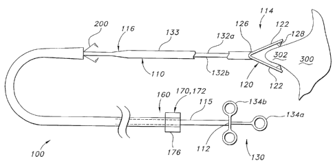
[0033] Figs. 1(a)-1(e) are schematic views of portions of first and second
representative embodiments of a system in accordance with the present
invention.
[0034] Fig. 2 is a schematic view of a portion of a third representative
embodiment of
a system in accordance with the present invention.
[0035] Fig. 3 is a schematic view of a portion of a fourth representative
embodiment
of a system in accordance with the present invention.
[0036] Fig. 4 is a schematic view of a portion of the embodiment of Fig. 1(a).
[0037] Fig. 5 is a schematic view of a portion of a fifth representative
embodiment of
a system in accordance with the present invention.
[0038] Fig. 6 is a cross-sectional view of a portion of the embodiment of Fig.
1(a).
[0039] Figs. 7(a)-7(e) are schematic views of different embodiments of
retainers
made in accordance with the present invention.
[0040] Figs 8(a)-8(b) are partial schematic views of a sixth representative
embodiment of a system made in accordance with the present invention.
[0041] Fig. 9 is a schematic representation illustrating a method in
accordance with
the present invention.
[0042] Figs. 10(a)-10(b) are schematic representations illustrating a method
in
accordance with the present invention.
DETAILED DESCRIPTION OF THE PREFERRED EMBODIMENT
[0043] Reference will now be made in detail to the present preferred
embodiments of
the invention, an example of which is illustrated in the accompanying
drawings. The method
and corresponding steps of the invention will be described in conjunction with
the detailed
description of the system.
[0044] The devices and methods presented herein may be used for treating the
luminal system of a patient. The present invention is particularly well suited
for treating
valve regurgitation, such as mitral valve regurgitation. In accordance with
the invention, a
-8-
CA 02626540 2008-04-18
WO 2007/050546 PCT/US2006/041369
medical device is provided including a tissue plicator adapted and configured
to form a
plication of tissue proximate a target region of a patient.
[0045] For purpose of explanation and illustration, and not limitation, a
partial view
of an exemplary embodiment of the medical device in accordance with the
invention is
shown in Fig. 1(a) and is designated generally by reference character 100.
Other
embodiments of a medical device in accordance with the invention, or aspects
thereof, are
provided in Figs. 2 - 10, as will be described.
[0046] In accordance with the invention, the medical device includes a tissue
plicator
adapted and configured to form a plicatiori of tissue proximate a target
region of a patient.
[0047] For purposes of illustration and not limitation, as embodied herein and
as
depicted in Fig. 1(a), medical device 100 is provided with a tissue plicator
110. As depicted
in Fig. 1(a), tissue plicator 110 includes a proximal end 112, a distal end
114 and includes an
elongate body 116. In the embodiment of Fig. 1(a), tissue plicator plicates
tissue 300 by
mechanically clamping tissue 300 using forceps 120. Forceps 120 include first
and second
jaws 122 that are adapted to open and close about a hinge 126 to mechanically
grasp tissue
300 to form a plication 302. Hinge 126 can be an actual hinge with a pivot, or
can be a living
hinge made from spring like material that is biased to cause the jaws to
either open or close.
If desired, forceps 120 may include a plurality of teeth 128 for gripping
tissue.
[0048] As depicted in Fig. 1(a), a plicator actuator 130 is provided. Actuator
130 is
operably coupled to proximal end 112 of plicator 110. Plicator actuator 130 is
configured
and adapted to adjust the tissue plicator 110 from a first configuration
wherein the tissue
plicator is disengaged from the target area, wherein jaws 122 are open, to an
second
configuration wherein jaws 122 of tissue plicator are engaged with the target
area. If hinge
126 is a living hinge, actuator can be configured and adapted to oppose the
bias of hinge 126.
That is, actuator 130 can be adapted to cause jaws 122 to splay apart or come
together, as
desired.
[0049] Actuator can take on a variety of forms. For example, and as depicted
in Fig.
1(a), actuator 130 includes a plurality of linkages 132a, 132b operably
coupled to a handle
134 having portions 134a and 134b. As portion 134a is moved with respect to
134b, jaws-
122 can be caused to move toward or away from one another. The handle 134 can
take on a
variety of forms. While a two piece push-pull handle 134 is depicted, it is
also possible to
use other actuators as are known in the art, such as threaded rotating
actuators similar to those
-9-
CA 02626540 2008-04-18
WO 2007/050546 PCT/US2006/041369
for retractable sheaths as described in U.S. Patent No. 6,488,694 to Lau and
U.S. Patent No.
5,906,619 to Olson, the specifications of which are incorporated herein by
reference.
[0050] Linkages 132a, 132b can take on a variety of forms that permit relative
movement. For example, as disclosed in Fig. 1(a), linkages 132a, 132b can be
disposed
within a sheath 133 that prevents splaying of linkages 132. By way of further
example,
linkages 132 can be formed concentrically as disclosed in Fig. 2, whereby
outer linkage 132a
is sleeve shaped and has a distal end 132d that slides along inner linkage
132b over jaws 122
to cause jaws 122 to grip tissue. In addition, other types of actuators are
possible, including
hydraulically, pneumatically and electromagnetic actuators.
[0051] Plicator 110 can grasp tissue 300 to form a plication 302 in a variety
of ways.
In addition or instead of mechanically grasping the tissue with forceps 120,
as depicted in
Fig. 3, tissue plicator 110 may also plicate the tissue at least in part by
applying suction
thereto. In accordance with this aspect, a suction sheath 140 can be provided
having a
proximal end 142 and a distal end 144 and defining a lumen 146 therethrough.
Proximal end
142 of lumen 146 can be placed in fluid communication with a suction source
150. When the
suction source 150 is activated, the tissue plicator 110 may plicate the
tissue at least in part by
drawing the tissue 300 into the lumen under suction from suction source 150.
If desired,
forceps 120 or similar structure can be disposed within lumen 146 to grasp
tissue that has
been drawn into lumen 146 under suction. Forceps 120 can initially be provided
in a
collapsed state when introducing medical device 100 into a patient, and can
then expand to
cause sheath 140 to expand in a radial direction. This facilitates formation
of a larger
plication 302 of tissue 300.
[0052] Tissue plicator 110 can be made from a variety of materials. Tissue
plicator
110 should be made of materials that are sufficiently flexible to traverse the
lumenal system
of a patient to access the heart. Suitable materials include, for example,
surgical grades of
stainless steel, nitinol, other alloys, plastic, polymer materials and the
like. It is also possible
to make at least first and second jaws 122 of forceps 120 at least in part
from radiopaque
materials that are visible under fluoroscopy, such as platinum gold, barium or
iridium, for
example. Forceps 120 can also be made from less expensive surgical steel, and
plated with
radiopaque materials. Similarly, marker bands 121 made from radiopaque
material can also
be provided as depicted in Fig. 1(a). By way of further example, materials
visible under
ultrasound imaging can also be used, such as materials including
microparticles, materials
having altered surface texture, materials including microbubbles, and the
like. Moreover, if
-10-
CA 02626540 2008-04-18
WO 2007/050546 PCT/US2006/041369
magnetic resonance imaging is used, medical device 100 can be formed from
materials that
are not sensitive to high magnetic fields, such as composite materials
including carbon fiber
and the like.
[0053] In further accordance with the invention, the medical device of the
present
invention includes a retainer applicator for applying a retainer to the
plication to maintain the
plication after the medical device is removed from the patient.
j00541 For purposes of illustration and not limitation, as embodied herein and
as
depicted in Fig. 1(a), medical device 100 includes retainer applicator 160.
Retainer
applicator 160 is preferably operatively associated with the tissue plicator
110, but can be
introduced separately, if desired. The retainer applicator 160 is adapted and
configured to
apply a retainer 200, discussed in detail below, to the plication 302 to
maintain the plication
302 after the medical device 100 is removed from the patient.
[0055] The retainer applicator 160 can be adapted and configured to deliver
the
retainer 200 along the tissue plicator 110 to the target region. In accordance
with this aspect,
the retainer applicator 160 can adapted and configured to deliver the retainer
along the
outside 115 of the tissue plicator 110 in monorail fashion to the target
region. Alternatively,
as depicted in Figs. 1(b)-I(e), the tissue plicator 110 can define a lumen 118
therethrough and
the retainer applicator 160 can be adapted and configured to deliver the
retainer through the
lumen 118 defined by the tissue plicator 110 to the target region T.
[0056] As depicted in Figs. 1(a) and 4, retainer applicator 160 includes an
applicator
actuator 170 that can be operably coupled to the retainer applicator 160,
wherein the actuator
is configured and adapted to affix the retainer 200 onto the plication 302 of
tissue 300. As
depicted in Fig. 4, applicator actuator 170 includes an advancement mechanism
172 for
advancing a retainer to the target region T. Handle 176 can also be provided
for actuating the
advancement mechanism 172.
[0057] As depicted in Fig. 1(a), advancement mechanism 172 can be provided in
the
form of a pusher tube that advances retainer 200 along the outside 115 of
tissue plicator or
through lumen 118 of plicator I 10 as depicted in Fig. 1(b). Advancement
mechanism 172
could also be provided as a hydraulic piston actuated by a plunger 179 as
depicted in Fig. 5 to
advance retainer 200 along the outside 115 of plicator 110, among other
possible
embodiments as disclosed herein. Advancement mechanism 172 could also be a
combination
of a push-pull arrangement to position the retainer proximate the target area,
combined with a
-11-
CA 02626540 2008-04-18
WO 2007/050546 PCT/US2006/041369
threaded fine adjustment to precisely set the retainer over the plication
without compromising
the tissue by cutting through it with the retainer 200.
[0058] If desired, an engagement mechanism 174 for engaging the retainer 200
with
the tissue plication 302 can also be provided. Handle 178 can be provided for
actuating the
engagement mechanism 174. Engagement mechanism 174 can also take on a variety
of
forms. For example, and as depicted in Fig. 4, engagement mechanism 174 can
include a
plurality ofjaws 175 for clamping down on retainer 200 to cause it to engage
plication 302.
Jaws 175 can be actuated by advancing, for example, a tubular member 177 with
respect to
advancement mechanism over jaws 175 causing them to compress retainer 200 and
anchor it
into plication 302. By way of further example and as shown in Fig. 7(e),
engagement
mechanism 174 can be configured to rotate retainer 200 about a longitudinal
axis X defined
by medical device 100 to affect engagement between retainer 200 and plication
302 by
moving retainer 200 through a helical path. Engagement mechanism can be
provided in the
form of a tubular member that rotates about axis X that is configured to
engage helical
member 200 in a variety of ways, such as a threaded connection, force-fit, or
by having an
end 200a of member 200 engage a hole 174a in the periphery of engagement
mechanism 174
as depicted in Fig. 7(e).
[0059] The system described herein also preferably includes an outer catheter
190
(such as a guiding catheter) to facilitate delivery of medical device 100 in
combination with
retainer 200 to the target region T of a patient. For purposes of illustration
only and as
depicted in Fig. 6, outer catheter 190 includes a proximal end 192, a distal
end 194 and
defines a lumen 196 therethrough. Medical device 100 can be disposed within
lumen 196 of
outer catheter 190 and act as an inner catheter of the system.
[0060] Outer catheter 190 can be made from a variety of materials, including
multilayer polymeric extrusions, such as those described in U.S. Patent No.
6,464,683 to
Samuelson or U.S. Patent No. 5,538,510 to Fontirroche, the disclosure of each
being
incorporated by reference herein in its entirety. Other structures are also
possible, including
single or multilayer tubes reinforced by braiding, such as metallic braiding
material.
[0061] As depicted in Fig. 6, outer catheter 190 can further define a second
lumen
198 parallel to the first lumen 196. The second lumen 198 can be connected to
a source 220
of beneficial agent 222, and the system can be adapted and configured to
selectively deliver
the beneficial agent 222 to target region T through the second lumen 198 for
example, by
actuating a plunger 224. The beneficial agent 222 can be chosen from the group
consisting of
-12-
CA 02626540 2008-04-18
WO 2007/050546 PCT/US2006/041369
contrast agents, medicainents, viral vectors, and genetic material. Other
beneficial agents can
also be delivered in this manner, including polymer materials, cells in
polymeric matrices,
nanoparticles, and the like.
[0062] Additionally or alternatively, a stiffening wire 230 can be disposed in
the
second lumen 198 to impart desired stiffness characteristics to outer catheter
190. The
stiffening wire 230 can be movably disposed in the second lumen or can be
stationary, if
desired. Stiffening wire 230 is provided with a proximal region 232, a medial
region 234 and
a distal region 236. Stiffening wire 230 can have a varying stiffness along
its length. For
example, it may be desired to have a stiffening wire with a comparatively
stiff proximal
region 232 to provide rigidity to the outer catheter 190, and progressively
less stiff medial and distal regions 234, 236. Depending on the application at
hand, it may be more beneficial
to have a stiffening wire with a medial region 234 or distal region 236 that
is stiffer than the
proximal region 232. Stiffening wire 230 can be made from a variety of
materials, including
stainless steel, nitinol, various suitable plastics and other alloys.
Stiffening wire 230 can also
be coated with a lubricious coating to facilitate movement within lumen 198 as
described
below.
[0063] Any surface of various components of the system described herein (e.g.,
medical device 100, outer catheter 190) or portions thereof can be provided
with one or more
suitable lubricious coatings to facilitate procedures by reduction of
frictional forces. Such
coatings can include, for example, hydrophobic materials such as
PolyTetraFluoroEthylene
("PTFE") or silicone oil, or hydrophilic coatings such as Polyvinyl
Pyrrolidone ("PVP").
Other coatings are also possible, including, echogenic materials, radiopaque
materials and
hydrogels, for example.
[0064] In another aspect, as disclosed herein, the system of the invention
also can
include a retainer for maintaining a plication of tissue.
[0065] For purposes of illustration, and not limitation, as depicted in Fig.
7(a),
retainer 200 is provided. Retainer 200 includes a proximal portion 202 having
a proximal
end 204, a distal end 206 and a body 205, wherein the proximal end 204 of the
main body
portion can define a mating portion 208 for mating with the applicator 160.
The retainer 200
can be further provided with a distal portion 210 including a first prong 212
adapted and
configured to pass through tissue of a patient's vascular system. The retainer
can include a
second prong 214 attached to the main body portion 202. The second prong 214
can be
deformable from an open position for capturing a tissue plication between the
first prong and
-13-
CA 02626540 2008-04-18
WO 2007/050546 PCT/US2006/041369
second prong to a closed position for maintaining a tissue plication by the
applicator, as
depicted in Fig. 7(b). The mating portion 208 of the retainer 200 can define a
loop adapted
and configured to receive a portion of the applicator as depicted in Fig.
7(a).
[0066] Retainer 200 can take on a variety of forms. For example, loop 208
could be
omitted and applicator can be configured and adapted to mate with prongs 212
and 214.
Loop 208 can also be directly attached to prongs 212, 214 by eliminating body
205.
Moreover, the retainer 200 can additional prongs such as third prong 216
attached to the main
body portion 200, wherein the second prong 214 and third prong 216 are
generally parallel to
the main body portion 202 in the open position.
[0067] By way of further example and as depicted in Figs. 7(c), retainer 200
can be
substantially ring shaped. If desired, the retainer can adapted and configured
to be folded
about hinge portions 201 by jaws 175 of an applicator 160 about the tissue
plication 302 as
depicted in Fig. 4. By way of further example, as depicted in Fig. 7d,
retainer 200 can be
helically shaped and rotated about a longitudinal axis defined by the medical
device to
introduce the retainer into the target region. Retainer can be provided with
one or more barbs
203 to prevent retainer from backing out from tissue 300, as well as one or
more tabs 213 to
allow for later removal, if desired.
[0068] Retainer 200 can be made from a variety of materials, including, for
example,
shape memory materials, radiopaque materials, resorbable materials, polymeric
materials,
echogenic materials and/or fluoroscopically visible materials. If made from
shape memory
material, retainer can be configured to clamp down on plication 302 when it
reaches body
temperature. For example, the retainer as disclosed in Fig. 7d can be made
from shape
memory material and trained so that it is an elongate spiral as depicted in
7(e) that
compresses longitudinally into a ring shape when its temperature increases as
depicted in
Figure 7d.
[0069] Other variations of the system herein are also possible. For example, a
tissue
plicator 110 including any desired number of jaws 122 can be used. For
example, it is
possible to use more than two jaws 122 as disclosed in Fig. 8(a). In the
embodiment of Fig.
8(a), four jaws 122 are used to make up forceps 120. Lower jaws 122a and 122b
can be
moved relative to upper jaws 122c and 122d. In addition, jaws 122a, 122b can
be moved
laterally with respect to jaws 122c, 122d respectively to facilitate delivery
of a retainer 200.
[0070] In the embodiment of Fig. 8(a), retainer 200 is initially provided in
two
separate portions. First portion 207 is trapped between lower jaws 122a, 122b,
and second
-14-
CA 02626540 2008-04-18
WO 2007/050546 PCT/US2006/041369
portion 209 is trapped between upper jaws 112c, 122d. Plicator 110 is advanced
to a target
location T as depicted in Fig. 8(b). The sets ofjaws 122 are then brought
together to form a
plication. As this occurs, first portion 207 and second portion 209 are caused
to mate. If the
formation of plication 302 has created a beneficial result (such as reduce
mitral
regurgitation), the lower jaws 122a and 122b can be separated from one another
and upper
jaws 112c and 122d can be separated from one another to release retainer, and
maintain
plication 302. Mechanical actuators (not shown) to cause desired movement
ofjaws 112(a-d)
can be designed to create the desired movement.
[0071] In accordance with another aspect of the invention, a method of for
treating
the lumenal system of a patient is provided.
[0072] For purposes of illustration and not limitation, as embodied herein,
the method
includes the steps of providing a inner catheter, such as medical device 100,
having a distal
portion for creating a plication in tissue such as distal portion 112 of
plicator 110.
[0073] The method further includes introducing the inner catheter into a
lumenal
system of a patient, and advancing the distal portion to a target region to be
plicated. By way
of example, in accordance with one aspect, the method preferably begins with
creating an
access into the lumenal system of a patient, such as through the femoral
artery. A valved
adaptor such as a trocar (not shown) is placed into the opening in order to
avoid loss of blood.
Next, a guidewire 250 can be introduced through the trocar and advanced to the
target region
T of a patient. The target region can be the mitral valve 310 of a patient,
but can be other
locations in the lumenal system of the patient, as is desired. The mitral
valve 310 can be
accessed from the atrial side or the ventricular side, as is desired.
[0074] Preferably, as depicted in Fig. 9 and Figs. 1(b)-1(e), an outer
catheter 190 is
next introduced into the patient over the guidewire. Distal end 194 of outer
catheter 190 is
positioned proximate target region T of a patient, such as proximate the
mitral valve 310.
The procedure is preferably done under visualization of the target region,
such as by under
fluoroscopy, ultrasound or magnetic resonance imaging.
[0075] Next, the guidewire 250 can be withdrawn and medical device 100 is
introduced into the lumenal system of a patient through lumen 196 of outer
catheter 190 as
depicted in Figs. 1(b)-1(e). Distal end 112 of plicator 110 is moved distally
through lumen
196 of outer catheter until jaws 122 are positioned proximate target region T.
Jaws 122 are
then moved into an open position using plicator actuator 130. Jaws are further
advanced
against tissue 300 of target region T such that teeth 128, if provided, bite
into tissue 300.
-15-
CA 02626540 2008-04-18
WO 2007/050546 PCT/US2006/041369
Actuator 130 is then actuated, causing jaws 122 to close and pull on tissue
300 to form a
plication 302, as depicted in Fig. 1(c).
[0076] Plication 302 of tissue is preferably formed by pinching tissue along a
circumferential direction outside the mitral annulus, proximate the posterior
leaflet 304 of
mitral valve, as depicted in Figs. 10(a)-10(b). While plication 302 can be
formed in fibrous
tissue near the annulus, plication is preferably formed in the muscular tissue
of the wall 312
of the ventricle 314 or wa11316 of the atrium 318. The aim of forming
plication 302 is to
reduce the effective perimeter 320 of mitral valve by pinching it together. If
successful, this
will ideally cause the edges 304a, 306a of posterior leaflet 304 and anterior
leaflet 306 of
mitral valve to realign, thereby reducing mitral valve regurgitation. As can
be seen, in Fig.
10(a), the perimeter 319 of the mitral valve is reduced as compared to Fig.
10(b), after the
procedure.
[0077] Once plication 302 is formed, if desired, it is possible to view the
effect that
formation of plication 302 has had on alignment of leaflets 304, 306. Under
fluoroscopy,
regurgitation of mitral valve 310 can be viewed during the procedure to
determine if forming
plication 302 has had a beneficial result. If the result has not been
beneficial, plication 302
can be released without permanently altering the tissue. A new plication can
then be formed
in a different location in an attempt to reduce mitral valve regurgitation. As
can be seen, this
technique of forming a temporary plication can provide a significant advantage
over more
invasive procedures since the latter usually require stopping the heart.
However, if the result
has reduced regurgitation to some extent, the plication 302 can be maintained
by applying a
retainer 200 to the plication. The retainer 200 can be delivered in any
manner, such as
described herein.
[0078] In certain circumstances, it is also possible to form the plication by
using the
retainer 200 itself in a single step without first forming a temporary
plication. In accordance
with this aspect, a medical device 100 is provided having a tissue plicator
110 that is adapted
and configured to form a plication of tissue in endocardial muscular tissue
proximate the
mitral valve of a patient. Tissue plicator can also perform the function of
delivering a
retainer 200 to a target region T in a single step without forming a plication
of tissue 302
prior to delivering retainer 200.
[0079] Additional plications 302 proximate the mitral valve 310 radially
displaced
from one another can be formed, if desired. These plications 302 can be held
in place by
additional retainers 200 to further reduce the perimeter of the mitral valve
310.
-16-
CA 02626540 2008-04-18
WO 2007/050546 PCT/US2006/041369
[0080] The methods and systems of the present invention, as described above
and
shown in the drawings, provide for a medical device and method for treating
mitral valve
regurgitation with superior properties including, for example, greater ease of
use and
effectiveness. It will be apparent to those skilled in the art that various
modifications and
variations can be made in the device and method of the present invention
without departing
from the spirit or scope of the invention. Thus, it is intended that the
present invention
include modifications and variations that are within the scope of the appended
claims and
their equivalents.
-17-