Note: Descriptions are shown in the official language in which they were submitted.
CA 02626546 2008-04-18
WO 2007/047853 PCT/US2006/040909
METHODS AND SYSTEMS FOR IMPROVING NEURAL
FUNCTIONING, INCLUDING COGNITIVE FUNCTIONING AND
NEGLECT DISORDERS
TECHNICAL FIELD
[0001] The present invention is directed generally toward methods and systems
for improving neural functioning, including cognitive functioning. In
particular
embodiments, the methods and systems can be used to address neglect disorders.
BACKGROUND
[0002] A wide variety of mental and physical processes are known to be
controlled or influenced by neural activity in particular regions of the
brain. In some
areas of the brain, such as in the sensory or motor cortices, the organization
of the
brain resembles a map of the human body; this is referred to as the
"somatotopic
organization of the brain." There are several other areas of the brain that
appear to
have distinct functions that are located in specific regions of the brain in
most
individuals. For example, areas of the occipital lobes relate to vision,
regions of the
left inferior frontal lobes relate to language in the majority of people, and
regions of
the cerebral cortex appear to be consistently involved with conscious
awareness,
memory, and intellect. This type of location-specific functional organization
of the
brain, in which discrete locations of the brain are statistically likely to
control
particular mental or physical functions in normal individuals, is herein
referred to as
the "functional organization of the brain."
[0003] Many problems or abnormalities with body functions can be caused by
damage, disease and/or disorders of the brain. A stroke, for example, is one
very
common condition that damages the brain. Strokes are generally caused by
emboli
(e.g., obstruction of a vessel), hemorrhages (e.g., rupture of a vessel), or
thrombi
(e.g., clotting) in the vascular system of a specific region of the cortex,
which in turn
generally causes a loss or impairment of a neural function (e.g., neural
functions
related to face muscles, limbs, speech, etc.). Stroke patients are typically
treated
using physical therapy to rehabilitate the loss of function of a limb or
another
CA 02626546 2008-04-18
WO 2007/047853 PCT/US2006/040909
affected body part. For most patients, little can be done to improve the
function of
the affected limb beyond the recovery that occurs naturally without
intervention.
[0004] One existing physical therapy technique for treating stroke patients
constrains or restrains the use of a working body part of the patient to force
the
patient to use the affected body part. For example, the loss of use of a limb
is
treated by restraining the other limb. Although this type of physical therapy
has
shown some experimental efficacy, it is expensive, time-consuming and little-
used.
Stroke patients can also be treated using physical therapy plus adjunctive
therapies.
For example, some types of drugs, including amphetamines, increase the
activation
of neurons in general. These drugs also appear to enhance neural networks.
However, these drugs may have limited efficacy because their mechanisms of
action
are very non-selective and they cannot be delivered in high concentrations
directly at
the site where they are needed. Still another approach is to apply electrical
stimulation to the brain to promote the recovery of functionality lost as a
result of a
stroke. While this approach has been generally effective, it has not
adequately
addressed all stroke symptoms.
[0005] One common syndrome following a stroke is neglect. Neglect is a
cognitive defect that causes patients to lose cognizance of portions of their
surroundings and/or themselves. Most frequently, neglect results from damage
to
the right (i.e., non-language) hemisphere of the brain, and affects the
contralesional
side of the patient and/or the patient's perception of his or her
contralesional
surroundings. For example, patients demonstrating neglect may fail to be aware
of
objects (including their own body parts) or people in the left half of the
space around
them. Patients suffering from negiect may,fail to spontaneously move their
eyes to
the left, even though such movements are possible for the patient during
formal
testing. Patients may examine only half of a page presented before them, may
be
unable to bisect a line at its middle, may copy only half of a drawing
positioned
before them, may fail to groom the left side of their faces or heads, and/or
may
exhibit other such symptoms.
[0006] In many cases, the patient may be unaware of the fact that he or she
exhibits the foregoing symptoms (i.e., if they are unaware of their paretic
left arm
they may deny any problem). Accordingly, treating neglect is often difficult
because
the patient is not motivated by the physically manifested reminders of the
condition,
33734-8070W0/LEGAL11632232.1 -2-
CA 02626546 2008-04-18
WO 2007/047853 PCT/US2006/040909
though such reminders would appear to be continual and obvious to an observer.
Therefore, there is a need to develop more effective and efficient treatments
for
rehabilitating stroke patients and patients that have other types of brain
damage
and/or can otherwise benefit from an improvement in cognitive functioning.
BRIEF DESCRIPTION OF THE DRAWINGS
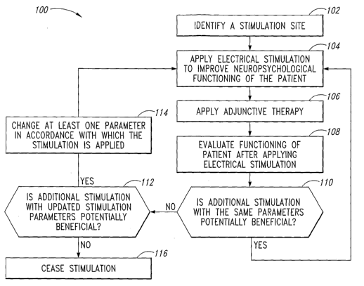
[0007] Figure 1 is a flow diagram illustrating a process for improving
neuropsychological functioning of a patient in accordance with an embodiment
of the
invention.
[0008] Figure 2 is a left-side view of a patient's brain, identifying
potential
stimulation sites in accordance with embodiments of the invention.
[0009] Figure 3 is a top view of a patient's brain illustrating further
potential
target stimulation sites in accordance with embodiments of the invention.
[0010] Figure 4 is a partially schematic, isometric illustration of a magnetic
resonance chamber in which a patient may be evaluated in accordance with an
embodiment of the invention.
[0011] Figure 5 illustrates a patient wearing a peripheral stimulation device
that
may be used in combination with evaluation devices in accordance with further
embodiments of the invention.
[0012] Figure 6 illustrates a patient wearing a network of electrodes
positioned
to detect brain activity in accordance with further embodiments of the
invention.
[0013] Figure 7 illustrates an electrical stimulation device implanted in a
patient
in accordance with an embodiment of the invention.
[0014] Figure 8 illustrates an electrical device operatively coupled to an
external
controller in accordance with another embodiment of the invention.
[0015] Figure 9 is a schematic illustration of a pulse system configured in
accordance with an embodiment of the invention.
[0016] Figured 10 is an isometric illustration of a device that carries
electrodes
in accordance with another embodiment of the invention.
33734-8070W0/LEGAL11632232.1 -3-
CA 02626546 2008-04-18
WO 2007/047853 PCT/US2006/040909
[0017] Figure 11 is a partially schematic, side elevation view of an electrode
configured to deliver electromagnetic stimulation to a subcortical region in
accordance with an embodiment of the invention.
DETAILED DESCRIPTION
A. Introduction
[0018] The present invention is directed generally toward methods and systems
for improving neural functioning, including cognitive functioning. A method in
a
particular aspect of the invention is directed to treating a patient by
applying
electrical stimulation beneath the patient's skull to improve
neuropsychological
functioning of the patient. After applying the electrical stimulation, the
process can
further include evaiuating the functioning of the patient. Based at least in
part on the
results of the evaluation, the method can still further include changing
and/or
maintaining at least one parameter in accordance with which the electrical
stimulation is applied, and/or ceasing to apply the electrical stimulation.
[0019] In further particular embodiments, the method can include selecting at
least one type of cognitive functioning and, based at least in part on the
selected
type of cognitive functioning, selecting a target neural population to which
the
electrical stimulation is directed. The electrical stimulation can be applied
at or
beneath the patient's cortex and in at least some embodiments, can be applied
to
the parietal lobe of the brain. Electrical stimulation can be provided to
improve the
patient's memory, effectuate a lasting change in the patient's cognitive
functioning,
and/or be applied to a patient having a perceptual disorder. In other
embodiments,
electrical stimulation can be provided to a patient having generally normal
cognitive
functioning. In still further embodiments, electrical stimulation can be
provided to
improve a neuropsychiatric functioning of the patient.
[0020] In yet another embodiment, a method for treating a patient having a
neglect disorder can include applying electromagnetic stimulation to the
patient's
brain to at least partially reduce the effects of the neglect disorder. The
method can
further inciude determining a severity of the neglect disorder by
administering a
neglect test to the patient after applying the electromagnetic stimulation.
Based at
least in part on the results of the neglect test, the method can further
include
33734-8070W0/LEGAL11632232.1 -4-
CA 02626546 2008-04-18
WO 2007/047853 PCT/US2006/040909
changing at least one parameter in accordance with which the electromagnetic
stimulation is applied, or ceasing to apply the electromagnetic stimulation,
or both.
B. Methods for Improving a Patient's Functioning
[0021] Figure 1 is a flow diagram illustrating a method 100 for improving a
patient's neuropsychological functioning in accordance with an embodiment of
the
invention. Further details regarding the processes identified in Figure 1 are
described below with reference to Figures 2-11. Beginning with Figure 1,
process
portion 102 includes identifying a stimulation site. The stimulation site is
typically
located at the patient's central nervous system, and in many instances, is
located at
the patient's brain. In process portion 104, electrical stimulation is applied
to the
patient's central nervous system (e.g., beneath the patient's skull) to
improve the
neuropsychological functioning of the patient. In particular embodiments, the
electrical stimulation can enhance the patient's naturally occurring efforts
to recruit
neural cells to take over functions performed by damaged cells (e.g., based on
neuroplasticity). The electrical stimulation can be applied in association
with an
adjunctive therapy, as indicated by process portion 106. The adjunctive
therapy can
be selected based at least in part upon the particular symptoms the patient
exhibits,
so as to at least partially address those symptoms.
[0022] Process portion 108 can include evaluating the functioning of the
patient
after the electrical stimulation has been applied. Based at least in part on
the results
of the evaluation, process portion 110 can include determining whether
additional
stimulation with the same stimulation parameters is potentially beneficial. If
so, then
the process returns to process portion 104. If not, then in process portion
112, it can
be determined whether additional stimulation with different parameters may be
potentially beneficial. If so, then in process portion 114 at least one of the
stimulation parameters can be changed, and the process can return to process
portion 104 for application of additional electrical stimulation to the
patient. If not,
then in process portion 116, the electrical stimulation ceases.
C. Identifying a Stimulation Site
[0023] Figure 2 is a side illustration of the brain 120 illustrating the four
major
brain lobes, e.g., the parietal lobe 121, the frontal lobe 122, the occipital
lobe 124
33734-8070W0/LEGAL11632232.1 -5-
CA 02626546 2008-04-18
WO 2007/047853 PCT/US2006/040909
(which includes the visual cortex 123), and the temporal lobe 125. In many
cases,
patients suffering from neglect may benefit from stimulation at the parietal
lobe 121,
and/or the frontal lobe 122. In other embodiments, cognitive functioning
and/or
neuropsychological functioning can be improved by stimulation at the occipital
lobe
124 and/or the temporal lobe 125. Accordingly, the practitioner can select a
stimulation site that is consistent with the patient's condition.
[0024] Figure 3 is a top view of the brain 120 illustrating particular aspects
of
the parietal lobe 121, including the superior parietal lobule 126, the
inferior parietal
lobule 127, and the intraparietal suicus 128. Figure 3 also illustrates a
target neural
population 131 located at the superior parietal lobule 126 of the patient's
right brain
hemisphere 129. As described above, many patients suffering from neglect
suffer
from neglect of the left side of their bodies or fields of view, and
accordingly, may
benefit from the stimulation of the right hemisphere. In other embodiments,
the
patient's cognitive and/or other functioning may be improved by stimulating
the left
hemisphere 130. Depending upon embodiment details and/or the nature or extent
of a patient's neurologic dysfunction, the patient may benefit from neural
stimulation
directed toward one or more target neural populations, which may reside in one
or
both brain hemispheres. Further details regarding the particular sites
selected for
stimulation are described below.
[0025] In particular embodiments, one or more target neural population 131 can
be selected based on past experience with patients presenting with similar
symptoms. For example, if over the course of time, it is determined that
stimulating
the superior parietal lobe 126 is particularly effective for treating one or
more types
of neglect, electrical stimulation can be applied at this location in patients
exhibiting
the corresponding symptom(s). In other embodiments, selecting a set of target
neural populations 131 can be performed on a patient-specific (e.g., patient-
by-
patient) basis. For example, the particular portion of the brain that benefits
from
electrical stimulation may vary from patient to patient, even for patients
presenting
with similar or identical symptoms. In such cases, techniques can be used to
identify the areas of the brain well suited for electrical stimulation for
each individual
patient. In many instances, this process can include (a) providing a stimulus
that
causes the patient to exhibit a problematic symptom, and then (b)
simultaneously
identifying areas of the brain that are either active, or are inactive, but
should be
33734-8070W0/LEGAL11632232.1 -6-
CA 02626546 2008-04-18
WO 2007/047853 PCT/US2006/040909
active. Accordingly, identifying target stimulation areas can include (a)
identifying
lesioned or other damaged areas, (b) identifying areas adjacent or proximate
to the
damaged areas, and/or (c) identifying other areas expected to assume, at least
in
part, the functions of a damaged area, or otherwise improve the functionality
of the
patient. Figures 4-6 illustrate representative techniques for performing such
identification tasks.
[0026] Figure 4 illustrates a magnetic resonance system 140 having a patient
platform 141 for carrying the patient while a practitioner identifies one or
more
electrical stimulation sites. If the stimulation site is to be located based
on previous
data for similarly situated patients, the magnetic resonance system 140 can be
used
to provide magnetic resonance imaging (MRI) data that are in turn used to
locate
target brain areas relative to patient-specific features (e.g., anatomical
features or
fiducials). In other embodiments, the system 140 can provide functional MRI
(fMRI)
results. For example, the patient can be placed in the system 140 and asked to
perform a task that causes the patient to exhibit the problematic symptom. The
data
obtained while the patient is in system 140 can then be used to identify where
active
and/or inactive brain regions are located, which can in turn provide
information for
identifying the electrical stimulation sites. The data can be in the form of
human-
readable images, and/or computer-readable output.
[0027] Because the system 140 tends to be loud and confined, it may be
difficult to provide the peripheral stimulus and/or gauge the patient's
response to the
peripheral stimulus while the patient is in the system chamber. In some
instances,
the stimulus can include asking the patient a question (via a headset, speaker
system or other peripheral stimulation device), and the patient can respond
verbally
via a microphone system. In other instances, for example, when the stimulus is
of a
more complex visual nature, the patient may be outfitted with another type of
peripheral stimulation device. Referring now to Figure 5, such a peripheral
stimulation device 142 can include virtual reality goggles placed on the
patient 144
before the patient is placed within the chamber 140. The patient can view a
visually-
based test (e.g., a bells cancellation test or a matrix reasoning test) via
the
peripheral stimulation device 142, and can provide a response by voice, or by
pressing a hand-held key, moving a joystick, or by another suitable method
(e.g.,
through a choice or selection made by an eye movement recognized by an ocular
33734-8070W0/LEGAL11632232.1 -7-
CA 02626546 2008-04-18
WO 2007/047853 PCT/US2006/040909
monitoring/tracking device incorporated into a headset or virtual reality
goggles).
The peripheral stimulation device 142 and any device used to transmit the
patient's
response can be compatible with the system 140. For example, these devices can
be operated by fiber optic links and/or can otherwise be compatible with the
strong
magnetic fields associated with the system 140.
[0028] In other embodiments, other techniques, such as EEG techniques, can
be used to identify the activity in the patient's brain while the patient
responds to a
stimulus. Figure 6 illustrates the patient 144 wearing an electrode or sensor
net 143
(e.g., a geodesic sensor net manufactured by Electrical Goedesics, Inc., of
Eugene,
Oregon) that includes a network of receptor electrodes positioned over the
patient's
scalp. When the patient is provided with an external or peripheral stimulus
(e.g., a
cognitive or other type of test) the patient's brain generates electrical
signals in
response to the stimulus, and these signals can be identified by the electrode
net
143. The waveform, spatial, and/or temporal characteristics of the signals (or
the
absence of signals) can be used to identify one or more target neural
populations.
The patient's performance on the test (e.g., how accurately the patient
answers
particular questions, or identifies particular objects) can be separately
tracked to
identify the severity of the patient's symptoms and/or the pace of the
patient's
progress during the course of treatment.
[0029] In other embodiments, other techniques can be used to locate areas of
the brain at which electrical stimulation may provide a benefit. Such
techniques can
include magnetic resonance spectroscopy (MRS) techniques (which can identify
the
presence and relative levels of particular neurochemical species) to identify
neurotransmitter imbalances or states associated with neuropsychiatric and/or
other
disorders, PET techniques, optical tomography techniques, and/or other
techniques.
In any of these embodiments, various techniques can be used to identify areas
(e.g.,
neuroplastic areas) that can take over functions for other brain areas,
improve on an
existing level of functioning, and/or otherwise provide a benefit to the
patient, as a
direct or indirect result of electrical stimulation.
D. Applying Electrical Stimulation
[0030] Once the electrical stimulation site or sites have been identified, an
electrical stimulation device may be positioned at a location to provide
electrical
33734-8070W0/LEGAL11632232.1 -8-
CA 02626546 2008-04-18
WO 2007/047853 PCT/US2006/040909
stimulation to the selected sites. Figures 7-11 illustrate representative
devices for
accomplishing this function. Figure 7 is a schematic illustration of a
neurostimulation
system 700 implanted in the patient 144 to provide stimulation in accordance
with
several embodiments of the invention. The system 700 can include an electrode
device 701 carrying one or more electrodes 750. The electrode device 701 can
be
positioned in the skull 732 of the patient 144, with the electrodes 750
positioned to
stimulate target areas of the brain 120. For example, the electrodes 750 can
be
positioned just outside the dura mater 733 (which surrounds the brain 120) to
stimulate cortical tissue. In another embodiment described later with
reference to
Figure 11, an electrode can penetrate the dura mater 733 to stimulate
subcortical
tissues. In still further embodiments, the electrodes 750 can penetrate the
dura
mater 733 but not the underlying pia mater 734, and can accordingly provide
stimulation signals through the pia mater 734.
[0031] The electrode device 701 can be coupled to a pulse system 710 with a
communication link 703. The communication link 703 can include one or more
leads, depending (for example) upon the number of electrodes 750 carried by
the
electrode device 701. The pulse system 710 can direct electrical signals to
the
electrode device 701 to stimulate target neural tissues.
[0032] The pulse system 710 can be implanted at a subclavicular location, as
shown in Figure 7. In particular embodiments, the pulse system 710 (and/or
other
implanted components of the system 700) can include titanium and/or other
materials that can be exposed to magnetic fields generated by magnetic
resonance
systems (e.g., the system shown in Figure 4) without harming the patient. The
pulse
system 710 can also be controlled internally via pre-programmed instructions
that
allow the pulse system 710 to operate autonomously after implantation. In
other
embodiments, the pulse system 710 can be implanted at other locations, and at
least some aspects of the pulse system 710 can be controlled externally. For
example, Figure 8 illustrates an embodiment of the system 700 in which the
pulse
system 710 is positioned on the external surface of the skull 732, beneath the
scalp
735. The pulse system 710 can be controlled internally and/or via an external
controller 715.
[0033] Figure 9 schematically illustrates a representative example of a pulse
system 710 suitable for use in the neural stimulation system 700 described
above.
33734-8070W0/LEGAL11632232.1 -9-
CA 02626546 2008-04-18
WO 2007/047853 PCT/US2006/040909
The pulse system 710 generally includes a housing 711 carrying a power supply
712, an integrated controller 713, a pulse generator 716, and a pulse
transmitter
717. The power supply 712 can be a primary battery, such as a rechargeable
battery or other suitable device for storing electrical energy. In other
embodiments,
the power supply 712 can be an RF transducer or a magnetic transducer that
receives broadcast energy emitted from an external power source and that
converts
the broadcast energy into power for the electrical components of the pulse
system
710.
[0034] In one embodiment, the integrated controller 713 can include a
processor, a memory, and a programmable computer medium. The integrated
controller 713, for example, can be a microcomputer, and the programmable
computer medium can include software loaded into the memory of the computer,
and/or hardware that performs the requisite control functions. In another
embodiment identified by dashed lines in Figure 9, the integrated controller
713 can
include an integrated RF or magnetic controller 714 that communicates with the
external controller 715 via an RF or magnetic link. In such an embodiment,
many of
the functions performed by the integrated controller 713 may be resident on
the
external controller 715 and the integrated portion 714 of the integrated
controller 713
may include a wireless communication system.
[0035] The integrated controller 713 is operatively coupled to, and provides
control signals to, the pulse generator 716, which may include a plurality of
channels
that send appropriate electrical pulses to the pulse transmitter 717. The
pulse
generator 716 may have multiple channels, with at least one channel associated
with a particular one of the electrodes 750 described above. The pulse
generator
716 sends appropriate electrical pulses to the pulse transmitter 717, which is
coupled to a plurality of the electrodes 750 (Figure 1). In one embodiment,
each of
these electrodes 750 is configured to be physically connected to a separate
lead,
allowing each electrode 750 to communicate with the pulse generator 716 via a
dedicated channel. Suitable components for the power supply 712, the
integrated
controller 713, the external controller 715, the pulse generator 716, and the
pulse
transmitter 717 are known to persons skilled in the art of implantable medical
devices.
33734-8070W0/LEGAL11632232.1 -10-
CA 02626546 2008-04-18
WO 2007/047853 PCT/US2006/040909
.,.. 1~ ,. ._ .. ...... ..... .....
[0036] The pulse system 710 can be programmed and operated to adjust a
wide variety of stimulation parameters, for example, which electrodes are
active and
inactive, whether electrical stimulation is provided in a unipolar or bipolar
manner,
and/or how the stimulation signals are varied. In particular embodiments, the
pulse
system 710 can be used to control the polarity, frequency, duty cycle,
amplitude,
and/or spatial and/or temporal qualities of the stimulation. The stimulation
can be
varied to match naturally occurring burst patterns (e.g., theta burst
stimulation),
and/or the stimulation can be varied in a predetermined, pseudorandom, and/or
aperiodic manner at one or more times and/or locations. Various systems and/or
procedures for providing and/or varying neural stimulation in manners that may
be
relevant to particular embodiments of the invention are described in detail in
U.S.
Application No. 11/182,713, entitled "Systems and Methods for Enhancing or
Affecting Neural Stimulation, Efficiency and/or Efficacy, filed on July 15,
2005, which
is incorporated herein by reference in its entirety.
E. Adjunctive Therapies
[0037] A given treatment regimen may also include, in addition to electrical
stimulation, one or more adjunctive or synergistic therapies to facilitate
enhanced
symptomatic relief and/or at least partial recovery from neurological
dysfunctions.
An adjunctive or synergistic therapy may include a behavioral therapy, such as
a
physical therapy activity, a movement and/or balance exercise, an activity of
daily
living (ADL), a vision exercise, a reading exercise, a speech task, a memory
or
concentration task, a visualization or imagination exercise, an auditory
activity, an
olfactory activity, a relaxation activity, and/or another type of behavior,
task or
activity. When a patient is being treated for neglect, the patient may
undertake tasks
that specifically engage a transition from a perceived region into a neglected
region.
For example, therapy may include applying stimulation while the patient tracks
a light
from a portion of the right extrapersonal space to the left extrapersonal
space. In
another embodiment, the patient may track a somatic simulation from right to
left
relative to his or her body, or drag a block from right to left to hit a
target (e.g., on a
display device). Further examples of representative adjunctive therapies are
disclosed by Tripathi et al. in a paper entitled, "Rehabilitation of patients
with
hemispatial neglect using visual-haptic feedback in virtual reality
environment,"
(International Conference on Human-Computer Interaction HCII, 2005),
incorporated
33734-8070W0/LEGAL11632232.1 -1 1-
CA 02626546 2008-04-18
WO 2007/047853 PCT/US2006/040909
herein by reference. In other embodiments, the adjunctive therapy can include
the
introduction of a drug or other chemical substance into the patient's body.
The
adjunctive therapy can be provided before, during and/or after the electrical
stimulation during a given treatment session. When the adjunctive therapy is
provided before or after the electrical stimulation, the temporal spacing
between the
electrical stimulation and the adjunctive therapy can be selected to provide a
desired
effect. In any of these embodiments, the relative timing between the
electrical
stimulation portion of the treatment regimen and the adjunctive therapy
portion of the
treatment regimen can be controlled and/or altered during the course of the
treatment regimen.
[0033] The particular adjunctive therapy selected can depend upon the
symptoms the particular patient exhibits. For example, if the patient exhibits
spatial
neglect, the selected adjunctive therapy may be different than if the patient
exhibits
another cognitive defect (e.g., memory loss). In some instances, the
adjunctive
therapy can be similar or at least partially similar to an evaluation
technique that may
be performed to gauge the severity level of the patient's dysfunction. For
example, if
a patient performs a bells cancellation test as an evaluation technique for
determining the severity of a spatial neglect dysfunction, the patient may
engage in a
similar or identical exercise as part of an adjunctive therapy. In any of
these
embodiments, it is believed that the adjunctive therapy can improve on and/or
make
more permanent the results obtained from applying electrical stimulation
alone.
[0039] In another particular example, a patient suffering from neglect can
have
electrical stimulation applied at the cortex (e.g., at the right parietal
lobe) and
possibly other central nervous system locations (a) while stimulating the
neglected
parts of the body, or (b) while the patient tries to use or move those body
parts,
and/or (c) while a practitioner passively moves those body parts. The cortical
stimuiation can be performed independently of, simultaneously with, or in a
temporally sequenced manner (e.g., based upon an estimated or measured neural
signaling latency) with sensory stimulation and/or peripheral stimulation
(e.g.,
Functional Electrical Stimulation (FES)) to strengthen neural signaling input
to
healthy or surviving brain tissue. For visual neglect, the cortical
stimulation can be
applied to surviving areas around and/or associated with (e.g., having neural
33734-8070W0/LEGAL11632232.1 -12-
CA 02626546 2008-04-18
WO 2007/047853 PCT/US2006/040909
projections into) the occipital visual cortex, while the sensory stimulation
can be
provided visually.
F. Evaluating the Functioning of the Patient
[0040] At periodic intervals during the course of a treatment regimen, the
patient's level of functioning can be evaluated. In some instances, for
example, if
the adjunctive therapy applied to the patient includes an evaluative test, the
evaluation can be conducted during each therapy session by tracking patient
performance on the test. In other embodiments, the evaluation can be provided
on
a less frequent basis and/or via other techniques.
[0041] As described above, one method for performing an evaiuation is to
administer a symptom-specific type of test. Such a test can include a bells
cancellation test for neglect or other tests for other specific symptoms,
including
other cognitive deficits such as memory deficits. In many of these tests, the
evaluation includes, and is based at least in part on, an active motor
response by the
patient. For example, if the patient is instructed to draw an object, identify
objects,
or respond verbally to a query, the response includes a motor response as well
as a
cognitive response. The nature of the test can be focused on the cognitive
response
and, to the extent the patient has motor deficits in addition to cognitive
deficits, the
test results can be segregated into cognitive-based results and motor-based
results
so that each can be tracked independently.
[0042] In other embodiments, the patient's functioning can be evaluated by
evaluating a physiologic function that corresponds to a neuropsychological
functioning level of the patient. Such an evaluation can be based on changes
in
neurotransmitter levels (e.g., using MRS), or changes in cerebral blood flow
or other
parameters that correlate with neural functioning. For example, a cognitively
dysfunctional patient may exhibit a relatively small change in cerebral blood
flow (at
the appropriate brain location) when engaging in a cognitive task, while a
more fully
functioning patient may exhibit a larger change in cerebral blood flow.
Accordingly,
identifying a difference between cerebral blood flow at one or more times, or
the
difference between a small change in cerebral blood flow and a large change in
cerebral blood flow can indicate an improvement in cognitive functioning. In
other
embodiments, other physiological changes (e.g., changes in neuronal signals)
or
33734-8070W0/LEGAL11632232.1 -13-
CA 02626546 2008-04-18
WO 2007/047853 PCT/US2006/040909
differences in changes can provide similar information. Further details
regarding
such techniques are described in the following copending patent applications,
filed
on October 19, 2005 and incorporated herein by reference: U.S. Application No.
11/254,240, titled "Methods and Systems for Establishing Parameters for Neural
Stimulation" (Attorney Docket No. 33734.8079US) and U.S. Application No.
60/728,650, titled "Neural Stimulation and Optical Monitoring Systems and
Method"
(Attorney Docket No. 33734.8084US).
[0043] The type of evaluation technique selected for a given patient may
depend at least in part on the nature of the electrical stimulation device
implanted in
the patient. For example, in some cases, magnetic resonance techniques such as
fMRI can be used to identify and/or evaluate neural changes associated with
the
patient's level of functioning. If the patient is to undergo evaluation while
in a
magnetic resonance chamber, the practitioner first establishes that the
electrical
stimulation device is compatible with such techniques, and does not create
unwanted electromagnetic or thermal effects in the patient's brain. If it is
expected
that the patient will undergo exposure to strong magnetic fields, the
practitioner may
elect to implant stimulation devices (e.g., a magnetic resonance compatible
IPG,
and/or one or more microstimulators such as BIONST"' (Advanced Bionics
Corporation, Sylmar, California)) that are compatible with magnetic fields
found in
magnetic resonance environments. In other embodiments, other techniques can be
used to evaluate the patient's functionality level without subjecting the
patient to
strong magnetic fields. For example, functional optical imaging, and/or EEG
using
an electrode or sensor net similar to that described above with reference to
Figure 6
can be used to evaluate the patient's improvement, neurofunctional condition
or
change, or performance.
G. Changing Test Parameters at Least in Part on the Basis of the Evaluation
[0044] In some instances, the results of the foregoing evaluation can have a
direct or indirect effect on the selection of parameters for electrically
stimulating the
patient. For example, if the evaluation indicates that the patient's
performance is
improving at an expected rate, the stimulation parameters need not be changed.
If
the evaluation indicates that the patient's progress has leveled off, one or
more
stimulation parameters may be changed to further increase patient functioning.
If,
33734-8070W0/LEGAL11632232.1 -14-
CA 02626546 2008-04-18
WO 2007/047853 PCT/US2006/040909
,. -u... .. - - _ _
after multiple parameter changes, no further change in patient functioning
results,
the electrical stimulation program can be interrupted for a given time period
(e.g., a
number of weeks over which neural consoiidation may occur), or halted.
[0045] Any of a wide variety of stimulation parameters can be changed to
expand upon and/or solidify the functional gains experienced by the patient.
Such
parameters can include the polarity of the electrical stimulation (e.g.,
anodal or
cathodal), the manner in which the stimulation is applied (e.g., bipolar or
monopolar),
the location of the stimulation, and/or the waveform of the stimulation. For
example,
the current, voltage, frequency, pulse width, interpulse interval and/or other
waveform-related functions can be changed to improve patient gains.
Representative ranges for these parameters include: pulse widths from 50-300
p/sec, frequencies from 1-200 Hz, current from 2-10mA, voltage from 2-15V and
interpulse intervals from 1-1000 msec. Also, to reduce any effect that neural
adaptation and/or habituation may have on clinical benefit, random variations
in
parameters may be programmed into the pulse delivery system. The location at
which the stimulation signals are provided may be changed by activating
different
electrodes on a particular electrode device, (e.g., using a device generally
similar to
the one described below with reference to Figure 10). In other embodiments,
additional electrode devices may be implanted within the patient's skull to
effectively
change the stimulation location.
H. Electronic Devices in Accordance With Further Embodiments
[0046] Stimulation can be provided to the patient using devices in addition to
or
in lieu of those described above. For example, Figure 10 is a top, partially
hidden
isometric view of an embodiment of an electrode device 1001 configured to
carry
muitiple cortical electrodes 1050. The electrodes 1050 can be carried by a
flexible
support member 1004 (located within the patient's skull) to place each
electrode
1050 at a stimulation site of the patient when the support member 1004 is
implanted
within the patient's skull. Electrical signals can be transmitted to the
electrodes 1050
via leads carried in a communication link 1003. The communication link 1003
can
include a cable 1002 that is connected to the pulse system 710 (Figure 7) via
a
connector 1008, and is protected with a protective sleeve 1007. Coupling
apertures
or holes 1057 can facilitate temporary attachment of the electrode device 1001
to
33734-8070W0/LEGAL11632232.1 -15-
CA 02626546 2008-04-18
WO 2007/047853 PCT/US2006/040909
LL q.un u ,- ..... ...- ----- ----.
the dura mater at, or at least proximate to, a stimulation site. The
electrodes 1050
can be biased cathodally and/or anodally, as described above. In an embodiment
shown in Figure 10, the electrode device 1001 can include six electrodes 1050
arranged in a 2x3 electrode array (i.e., two rows of three electrodes each),
and in
other embodiments, the electrode device 1001 can include more or fewer
electrodes
1050 arranged in symmetrical or asymmetrical arrays. The particular
arrangement
of electrodes 1050 can be selected based on the region of the patient's brain
that is
to be stimulated, and/or the patient's condition.
[0047] Figure 11 illustrates an electrode device 1101 that may be configured
to
apply electrical stimulation signals to a cortical region 1136 or a
subcortical region
1137 of the brain 120 in accordance with further embodiments of the invention.
The
electrode device 1101 can include an electrode 1150 having a head and a
threaded
shaft that extends through a pilot hole in the patient's skull 732. If the
electrode
1150 is intended for cortical stimulation, it can extend through the skull 732
to
contact the dura mater 733 or the pia mater 734. If the electrode 1050 is to
be used
for subcortical stimulation, it can include an elongate conductive member 1154
that
extends downwardly through the cortical region 1136 into the subcortical
region
1137. Most of the length of the elongate conductive member 1154 can be
insulated,
with just a tip 1155 exposed to provide electrical stimulation in only the
subcortical
region 1137. Subcortical stimulation may be appropriate in at least in some
instances, for example, when the brain structures such as the basal ganglia
are to
be stimulated. In other embodiments, other deep brain structures (e.g., the
amygdala or the hippocampus) can be stimulated using a subcortical electrode.
If
the hippocampus is to be stimulated, stimulation may be provided to the
perihippocampal cortex using a subdurally implanted electrode, that need not
penetrate through brain structures other than the dura.
[0048] Further details of electrode devices that may be suitable for
electromagnetic stimulation in accordance with other embodiments of the
invention
are described in the following pending U.S. Patent Applications, all of which
are
incorporated herein by reference: 10/891,834, filed July 15, 2004; 10/418,796,
filed
April 18, 2003; and 09/802,898, filed March 8, 2001. Further devices and
related
methods for providing neural stimulation and adjunctive therapy are described
in a
copending U.S. Application No. 11/255,187, titled "Systems and Methods for
Patient
33734-8070W0/LEGAL11632232.1 -16-
CA 02626546 2008-04-18
WO 2007/047853 PCT/US2006/040909
Interactive Neural Stimulation and/or Chemical Substance Delivery," (Attorney
Docket No. 33734.8082US) filed on October 19, 2005 and incorporated herein by
reference.
[0049] In still further embodiments, other techniques may be used to provide
stimulation to the patient's brain. Such techniques can include
electromagnetic
techniques in addition to purely electrical techniques. In particular, such
techniques
can include transcranial magnetic stimulation techniques, which do not require
that
an electrode be implanted beneath the patient's skull. In still further
embodiments,
other techniques, which also may not require an implant, can be used. Such
additior.al techniques can include transcranial direct current stimulation.
[0050] One feature of several embodiments of the methods and devices
described above is that they can be used to improve the neuropsychological
functioning of a patient. For example, by selecting a set of stimulation site
based on
historic data and/or the characteristics of a specific patient, and then
providing
electrical stimulation at one or more stimulation sites, possibly in
association or
conjunction with one or more adjunctive therapies (in which a type of
adjunctive
therapy selected may correspond to a stimulation site under consideration), a
long-
lasting change in the patient's neuropsychological functioning can be
achieved. The
long-lasting change can last for many weeks, months, or years, while the
application
of the treatment may be provided over a significantly shorter period of time
(e.g.,
over a single period or temporally separated periods of about three weeks,
about six
weeks, or about two to eight weeks).
[0051] Another feature of embodiments of the methods and devices described
above is that they can include updating the parameters with which stimulation
is
applied to the patient, based on an evaluation of the patient. The evaluation
can
include a test (e.g., a cognitive test), or another suitable evaluation of the
patient's
level of functioning. Accordingly, a given therapy program can be changed
dynamically to account for individual patient performance.
[0052] The foregoing techniques can be applied to patients having a wide
variety of neuropsychological dysfunctions. Such dysfunctions include neglect
dysfunctions, which can in turn include unilateral neglect (e.g., sensory
neglect,
motor neglect, representational neglect, personal neglect, or spatial
neglect). In
33734-8070W0/LEGAL11632232.1 -17-
CA 02626546 2008-04-18
WO 2007/047853 PCT/US2006/040909
other embodiments, other perceptual and/or cognitive disorders can be treated
using
these techniques. Such disorders can relate to the patient's vision (e.g.,
visual field
cut, cortical blindness, or central achromatopsia), or loss of tactile and/or
other
sensations (hemianesthesia, Balint's syndrome, sensory extinction, and
others).
These disorders may arise in connection with a stroke, other brain lesion, or
other
brain trauma.
[0053] Any of the foregoing techniques can be used to treat a patient having a
neurological dysfunction (e.g., a neglect dysfunction, and/or another
cognitive
dysfunction). In other embodiments, the foregoing techniques can be applied to
patients functioning at normal levels or above normal levels to further
improve
patient functioning. In still further embodiments, techniques generally
similar to the
foregoing techniques can be used to address neuropsychiatric disorders,
including
but not limited to depression or post-traumatic stress disorder. In these
embodiments, the methods used to identify stimulation sites and track patient
progress may be selected to focus on neuropsychiatric indicators. Such methods
can include identifying cortical areas, subcortical areas, and/or associated
neural
projections that may exhibit and/or influence (e.g., as a result of neural
stimulation)
neurotransmitter levels which, as described above, can be identified using MRS
techniques.
[0054] In still further embodiments, techniques generally similar to those
described above can be used to treat other disorders or functional deficits.
For
example, such techniques can be used to treat learning disabilities and/or
dyslexia.
In other instances, the disorders described above may result from conditions
other
than those described above. For example, while neglect is often associated
with
stroke patients, it may also result from plaque formations (associated with
Alzheimer's disease) or neurodepletion (associated with Parkinson's disease).
[0055] From the foregoing, it will be appreciated that specific embodiments of
the invention have been described herein for purposes of illustration, but
that various
modifications may be made without deviating from the invention. For example,
certain aspects of the methods described above may be automated or partially
automated, and may be implemented on computer systems and/or via computer-
readable media. In particular embodiments, aspects of the stimulation site
selection
procedure and/or the evaluation procedure can be automated in such a fashion.
33734-8070W0/LEGAL11632232.1 -18-
CA 02626546 2008-04-18
WO 2007/047853 PCT/US2006/040909
Aspects of the invention described in the context of particular embodiments
may be
combined or eliminated in other embodiments. For example, in some cases a
treatment regimen can proceed without an adjunctive therapy. Although
advantages
associated with certain embodiments of the invention have been described in
the
context of those embodiments, other embodiments may also exhibit such
advantages. Additionally, none of the foregoing embodiments need necessarily
exhibit such advantages to fall within the scope of the invention.
Accordingly, the
invention is not limited except as by the appended claims.
33734-8070W0/LEGAL11632232.1 -19-