Note: Descriptions are shown in the official language in which they were submitted.
CA 02626686 2008-04-18
=
File:27672-CA-599-PCT 1
DESCRIPTION
APPARATUS FOR MONITORING THROMBUS FORMATION AND METHOD OF
MONITORING THROMBUS FORMATION
Technical Field
[0001] The present invention relates to a method of monitoring efficacy of an
antithrombotic
drug administered to a patient or the like, and specifically, to an apparatus
for and a method of
comprehensively evaluating blood coagulation and platelet thrombus formation
under a
bloodstream-equivalent environment with a whole blood or plasma containing
platelets.
Background Art
[0002] For example, the atherothrombosis such as the myocardial infarction
causes serious
thrombus formation such that an atheromatous plaque is broken at an
arteriosclerosis site,
platelets are adhered on collagen including tissue factor exposed to the
bloodstream. Further,
the platelet aggregation, the activation of a blood coagulation system, and
the like complexly
occur resulting in serious obstructive thrombus. Heart disease such as
myocardial infarction,
is a serious disease and is the second leading cause of overall deaths in
Japan.
However, thrombus formation proceeds only in an atherosclerotic region in the
myocardial infarction, and a thrombotic tendency in the whole body is not
extremely
proceeded. In-vitro examinations are unsuitable for evaluating the thrombotic
tendency in
such a thrombosis and the monitoring of the antithrombotic effect in the
antithrombotic
therapy. Thus, it is important to make comprehensive evaluations on
coagulation and
platelets (adhesion and agglutination) in the presence of the bloodstream.
[0003] Heretofore, the blood coagulability has been evaluated by determining
activated
partial thromboplastin time (APTT), thromboplastin time (PT) using the plasma.
The APTT
mainly reflects intrinsic coagulation and the PT mainly reflects extrinsic
coagulation. The
examination of blood platelets is carried out by using platelet-rich plasma
and adding a
platelet-activating substance such as ADP or collagen to thereby evaluate the
aggregating
property of platelets from a change in transmittance thereof or the like. In
addition, the
coagulation time of the whole blood can be determined with the whole blood
clotting time,
the whole blood clotting time after calcium re-addition, and the like.
CA 02626686 2008-04-18
File:27672-CA-599-PCT 2
Further, an examination system using the whole blood employs thromboelastogrm,
which monitors the activations of clotting factors, the platelet
agglutination, and the like.
However, thrombus grows under a blood flow in vivo. In contrast, the above
examination method or the like is determined in-vitro that is in the closed
state. Thus, the
status of in-vivo thrombus growth cannot be observed.
[0004] As proposals for solving the above problems, Patent Document 1 and Non-
patent
Documents 2 and 3 disclose the method including bringing the blood provided
with an
antithrombotic drug to be evaluated to pass on a collagen cell and monitoring
the adhesion or
agglutination of the platelets by fluorescently-labeling the platelets with a
confocal
microscope.
However, in the invention described in the document the observation is carried
out
under the presence of an anticoagulation drug. Thus, the fact that a thrombus
which is
caused by the adhesion or agglutination of platelets induced by the blood
coagulation system
is not formed or decreased property to form thrombus is evaluated by
monitoring a
morphological change in platelet. Thus, the evaluation does not reflect the
platelet activation
interlocking with coagulation system. Therefore, such an invention is
favorable for the
evaluation of the efficacy of an antiplatelet drug but is unable to monitor a
thrombus itself and
the whole process of thrombus formation. In addition, a fluorescence
microscope is
expensive, so it can be hardly used for general examination.
Further, in Patent Document 2, the fluidity of the anticoagulated blood is
determined
by passing the blood through a fine-comb-like silicon cell. Likewise, the
process of Patent
Document 2 also uses the anticoagulated blood, so the influence of a
coagulation system
cannot be determined. In addition, the viscosity of blood in the process has
large individual
variations and in diurnal variations, so it is difficult to reflect drug
therapy using the system.
The platelet is activated by the coagulation system, and the coagulation
system is
promoted by activated platelets. Therefore, the efficacy of an antithrombotic
drug cannot be
observed in the anticoagulated blood, because activation of platelet is also
suppressed by the
anticoagulation treatment. In addition, non-anticoagulated blood can not used
in an
examination, because it promptly forms clot.
Patent Document 1: JP 2004-251630 A
Patent Document 2: JP 2006-145345 A
Non-patent Document 1: Blood. 1990;75:390-398
CA 02626686 2008-04-18
File:27672-CA-599-PCT 3
Non-patent Document 2: Blood. 1999:Aug 1:94(3):968-75
Disclosure of the Invention
[0005] The present invention has been made in consideration of the above
circumstances
and intends to provide an apparatus and method of comprehensively evaluating
the thrombus
formation due to the blood coagulation and platelet under a bloodstream-
equivalent
environment with a whole blood or plasma containing platelets (in the
specification of the
present invention, they may be inclusively referred to as "blood"), when
monitoring the
efficacy of an antithrombotic drug administered to a patient or the like.
Means for solving the problems
[0006] To solve the above-mentioned problems, the present invention provides
an apparatus
for monitoring thrombus formation, which monitors thrombus formation by
flowing
anticoagulated blood through a channel that simulates a blood vessel while
releasing an
anticoagulation treatment or promoting a blood coagulation, comprising: a
thrombus
formation chamber in at least a part of which a thrombus inducing material
that induces
thrombus formation is provided; an inlet tube which is connected to the
thrombus formation
chamber and through which blood is flown into the thrombus formation chamber;
and a drug
tube which is connected to the inlet tube and through which a drug that
promotes blood
coagulation (hereinafter may be referred to as "coagulation promotion") or a
drug that releases
the anticoagulation treatment is supplied. In the present invention, the term
"monitoring"
means not only visual evaluation of thrombus formation with eyes, an imaging,
but also
evaluation of the degree of thrombus formation in numerical terms by pressure
determination
or the like.
Further, the present invention provides an apparatus for monitoring thrombus
formation which comprises a thrombus formation chamber in at least a part of
which a
thrombus inducing material that induces thrombus formation is provided; an
inlet tube which
is connected to the thrombus formation chamber and through which blood is
flown into the
thrombus formation chamber; and a thrombus formation inhibitor inlet tube
which is
connected to the thrombus formation chamber and mixes a thrombus formation
inhibitor with
the blood after passing through the thrombus formation chamber.
In this case, the apparatus for monitoring thrombus formation is preferably
formed
CA 02626686 2008-04-18
File:27672-CA-599-PCT 4
on a substrate.
The apparatus for monitoring thrombus formation of the present invention
preferably
further comprises a pump for pressurizing the inlet tube and/or the drug tube
or a pump for
aspirating a discharge tube which is connected to the thrombus formation
chamber and
provided for discharging the blood from the thrombus formation chamber.
The apparatus for monitoring thrombus formation of the present invention
preferably
further include a pressure-measuring apparatus and a camera for taking images
of a thrombus
formation chamber.
Further, the thrombus inducing material preferably comprises collagen.
More preferably, the thrombus inducing material further comprises a tissue
factor
(tissue thromboplastin).
[0007] Further, the present invention provides a method of monitoring thrombus
formation,
comprising: flowing anticoagulated blood into a thrombus formation chamber, in
at least a
part of which a thrombus inducing material inducing thrombus formation is
provided, while
releasing an anticoagulation treatment or promoting a blood coagulation to
thereby monitor
thrombus formation. In the present invention, the term "flowing the blood
while releasing
the anticoagulation treatment or promoting blood coagulation" may be a state
where an
anticoagulation-releasing reaction or a coagulation-promoting reaction in the
channel is being
occurred, and includes a state where an drug that releases an anticoagulation
releasing agent,
or a coagulation promoting agent is flown while mixing with the blood in the
channel or a
state where the anticoagulation releasing agent or the coagulation promoting
agent is
promptly flown after mixing with the blood.
In the method of monitoring thrombus formation of the present invention, it is
preferable that the anticoagulation treatment is a treatment with a calcium
chelator such as
citric acid and the anticoagulation treatment is released with a free calcium
donor.
In the method of monitoring the thrombus formation of the present invention,
it is
preferable that the anticoagulation treatment is a treatment with a thrombin
aptamer and the
anticoagulation treatment is released with the antisense DNA of the thrombin
aptamer.
Here, in the method of monitoring thrombus formation of the present invention,
it is
preferable to monitor the thrombus formation by flowing anticoagulated blood
into a
thrombus formation chamber, while promoting blood coagulation without
releasing the
anticoagulation treatment. In this case, a means for promoting the blood
coagulation is
CA 02626686 2013-06-12
76536-32
preferably the addition of tissue thromboplastin.
In addition, in the method of monitoring thrombus formation of the present
invention,
the blood which has been anticoagulated with one kind or more kinds of
anticoagulation
agents is preferably released form the anticoagulation treatment with at least
one kind of
anticoagulation treatment releasing agent that corresponds to the
anticoagulation treatment
agent used. Here, it is preferable that the anticoagulation treatment agents
are a contact
phase factor inhibitor and a calcium ehelator, and the anticoagulation
treatment releasing
agent is a free calcium donor. Further, it is also preferable that the
anticoagulation treatment
agents are a contact phase factor inhibitor and heparin, and the
anticoagulation treatment
releasing agent is heparinase. Further, it is also preferable that the
anticoagulation treatment
agents are an inhibitor for a contact phase factor such as a blood coagulation
XII factor,
kallikrein, or the like and a thrombin aptamer, and the anticoagulation
treatment releasing
agent is the antisense DNA of the thrombin aptamer. The inhibitor for the
blood coagulation
XII factor is preferably a maize-derived trypsin inhibitor.
In the method of monitoring thrombus formation of the present invention, it is
preferable to determine the pressure at the time of inflow and/or outflow of
the blood in the
thrombus formation chamber.
In the method of monitoring thrombus formation of the present invention, the
thrombus inducing material preferably comprises collagen and a tissue factor.
Effects of the Invention
[0008] According to the apparatus for monitoring thrombus formation of the
present invention, the apparatus comprises: a thrombus formation chamber in at
least a part of
which a thrombus inducing material that induces thrombus formation is
provided; an inlet
tube which is connected to the thrombus formation chamber and through which
blood is
flown into the thrombus formation chamber; and a drug tube which is connected
to the inlet
tube and through which a drug that releases the anticoagulation treatment or a
drug that
promotes blood coagulation is supplied. Therefore, the anticoagulated blood,
which is
treated in order to prevent the blood collected after administering the
antithrombotic drug to a
patient from coagulating in the channel extending to the thrombus formation
chamber, can be
monitored by intentionally forming a thrombus in a thrombus formation chamber.
Thus, the
efficacy of an antithrombotic drug can be specifically monitored in the
environment similar to
CA 02626686 2013-06-12
76536-32
6
the inside of the human body. In addition, an anticoagulation treatment agent
can be used at
the time of blood sampling. Therefore, there is an advantage in that samples
after the blood
sampling can be stored for a certain period of time and the examination time
can be randomly
selected.
According to the apparatus for monitoring thrombus formation of the
present invention, the apparatus comprises: a thrombus formation chamber in at
least a part of
which a thrombus inducing material that induces thrombus formation is
provided; an inlet
tube which is connected to the thrombus formation chamber and through which
blood is
flown into the thrombus formation chamber; and an inlet for a thrombus
formation inhibitor,
which is connected to the thrombus formation chamber and mixes the thrombus
formation
inhibitor with the blood after passing through the thrombus formation chamber.
Therefore,
the thrombus observation can be carried out in a manner as described above. In
addition, the
blood coagulation does not proceed downstream in the thrombus formation
chamber,
therefore, influence on the pressure determination can be prevented and more
delicate
pressure changes can be monitored.
[0009] A small amount of blood can be monitored when the apparatus for
monitoring
thrombus formation of the present invention is formed on a substrate. In
addition, the
apparatus is provided with a pump for pressurizing the inlet tube and/or the
drug tube or a
pump for aspirating a discharge tube which is connected to the thrombus
formation chamber
and provided for discharging the blood from the thrombus formation chamber.
Therefore,
the blood and the drug for promoting blood coagulation can be stably flown for
a
predetermined period of time at predetermined pressure or predetermined flow
rate.
If the apparatus for monitoring thrombus formation of the present invention
comprises a pressure-measuring apparatus, the degree of thrombus formation can
be
converted into numbers, so a quantitative evaluation can be performed.
The apparatus can be easily set when the thrombus inducing material comprises
collagen. Thrombus formation can be efficiently induced when the thrombus
inducing
material further comprises a tissue factor such as tissue thromboplastin
together with
collagen.
Further, if the apparatus for monitoring thrombus formation of the present
invention
comprises a camera for taking an image of the thrombus formation chamber, the
appearance
of the thrombus formation can be observed as an image and the image can be
then stored.
CA 02626686 2013-06-12
.76536-.32
7
[0010] Further, according to the method of monitoring thrombus formation of
the
present invention, the method comprises: monitoring thrombus formation by
flowing
anticoagulated blood through a thrombus formation chamber in at least a part
of which a
thrombus inducing material that induces thrombus formation is provided, while
releasing the
anticoagulation treatment or promoting blood coagulation. Thus, the thrombus
formation on
the thrombus inducing material can be monitored by flowing the anticoagulated
blood, which
is obtained by anticoagulating the blood collected after administering the
antithrombotic drug
to a patient to prevent coagulation, while promoting blood coagulation.
Therefore, the
efficacy of an antithrombotic drug can be specifically monitored in the
environment similar to
the inside of the human body. In addition, an anticoagulation agent can be
used for blood
sampling. Therefore, there is an advantage in that samples after the blood
sampling can be
stored for a certain period of time and the examination time can be randomly
selected. If the
anticoagulation treatment is a treatment with a calcium chelator such as
citric acid and the
anticoagulation treatment is released by a free calcium donor, the reagent can
be easily
obtained and thus it is preferable. If the anticoagulation treatment is a
treatment with a
thrombin aptamer and the anticoagulation treatment is released by the
antisense DNA of the
thrombin aptamer, it is possible to carry out the examination while reflecting
the physiological
calcium ion concentration of the blood.
[0011] In addition, according to the method of monitoring thrombus formation
of the present invention, it is able to monitor thrombus formation by flowing
anticoagulated
blood into the thrombus formation chamber without performing the operation of
releasing the
anticoagulation treatment, while prompting blood coagulation. Therefore, the
thrombus
formation can be observed with a small amount of the blood and the burden of
the subject can
be reduced. In this case, further, the drug tube is not always required, so
the apparatus for
monitoring thrombus formation can be simplified. Here, if tissue
thromboplastin is used as a
blood coagulation promoting agent, the blood coagulation can be promoted by
activating the
coagulation system in an alternative pathway that avoids the XII-factor
activation and the
kallikrein activation to thereby monitor thrombus formation in the thrombus
formation
chamber.
[0012] Further, the thrombus formation can be monitored with a simple
operation by
releasing the anticoagulated blood obtained using one kind or more kinds of
anticoagulation
treatment agents with at least one kind of anticoagulation treatment releasing
agent
CA 02626686 2008-04-18
File:27672-CA-599-PCT 8
corresponding to the anticoagulation treatment agent used. In this case, when
an
anticoagulation treatment is carried out with a contact phase factor inhibitor
and a calcium
chelator, and the anticoagulation treatment is released with a free calcium
donor, or when an
anticoagulation treatment is carried out with a contact phase factor
inhibitor, and heparin and
the anticoagulation treatment is released with heparinase, the anticoagulation
treatment
exerting an effect at the time of monitoring thrombus formation is an
anticoagulation
treatment with the contact phase factor inhibitor, therefore monitoring of
thrombus formation
can be performed under more physiological conditions, particularly while
reflecting a divalent
metal ion associated with thrombosis, such as calcium or magnesium. In this
case, when the
anticoagulation treatment is carried out with an inhibitor of a contact phase
such as a blood
coagulation XII factor or kallikrein and a thrombin aptamer, and then the
anticoagulation
treatment is released by the antisense DNA of the thrombin inhibition aptamer,
the blood can
be stored over a prolonged period. Further, the anticoagulation treatment can
be efficiently
carried out when a maize-derived trypsin inhibitor is used as the inhibitor of
the blood
coagulation XII factor.
[0013] If the method of monitoring thrombus formation of the present invention
performs
the measurement of pressure at the time of inflow and/or outflow of the blood
in the thrombus
formation chamber, the degree of thrombus formation can be converted into
numbers, so a
quantitative evaluation can be easily performed by an extremely simple
apparatus.
Further,if the thrombus inducing material comprises collagen and the tissue
factor,
the apparatus can be easily set and the thrombus formation can be effectively
induced.
Brief Description of the Drawings
[0014] FIG. 1 is a schematic diagram illustrating an apparatus for monitoring
thrombus
formation according to a first embodiment of the present invention.
FIG. 2 (a) is a schematic diagram illustrating installation conditions of a
thrombus
inducing material 15 of Examples 3, 5, and 6 according to the first embodiment
of the present
invention, and FIG. 2 (b) is a schematic diagram illustrating results of
thrombus formation of
Examples 3, 5, and 6 according to the first embodiment of the present
invention.
FIG. 3 (a) is a schematic diagram illustrating installation conditions of the
thrombus
inducing material 15 of Example 4 of the first embodiment of the present
invention, and FIG.
3 (b) is a schematic diagram illustrating results of thrombus formation of
Example 4
CA 02626686 2008-04-18
File:27672-CA-599-PCT 9
according to the first embodiment of the present invention.
FIG. 4 is a schematic diagram illustrating a main part of an apparatus for
monitoring
thrombus formation (main body of a microchip) according to a second embodiment
of the
present invention.
FIG. 5 is a schematic diagram illustrating a main part of the apparatus for
monitoring
thrombus formation (cover of a microchip) according to the second embodiment
of the
present invention.
FIG. 6 is a schematic diagram illustrating the position of thrombus formation
of
Example 7 according to the second embodiment of the present invention.
FIG. 7 is a schematic diagram illustrating a main part of an apparatus for
monitoring
thrombus formation of another example (main body of a microchip) according to
the second
embodiment of the present invention.
FIG. 8 is a schematic diagram illustrating a main part of the apparatus for
monitoring
thrombus formation (cover of a microchip) of another example according to the
second
embodiment of the present invention.
FIGS. 9 are schematic diagrams illustrating main parts of an apparatus for
monitoring thrombus formation of another example according to a third
embodiment of the
present invention, where FIG. 9 (A) illustrates a completion drawing, FIG 9
(B) illustrates a
main body of a microchip, and (C) illustrates a cover of the microchip.
FIG. 10 is a schematic diagram illustrating a thrombus-monitoring system
systemized
using the apparatus for monitoring thrombus formation of the present
invention.
FIG. 11(A) is a result obtained by analyzing a thromboelastogram waveform of
coagulation by adding 10 RM of each of aptamers for exosite I and exosite II
to the blood, and
after storing the blood at room temperature for 15 minutes, adding 40 ttM of
each of antisense
DNAs of both aptamer. FIG. 11(B) illustrates the thromboelastogram waveform of
the blood
just after blood sampling.
FIGS. 12 are schematic diagrams illustrating main parts of an apparatus for
monitoring thrombus formation according to a fourth embodiment of the present
invention,
where FIG. 12 (A) illustrates a completion drawing, FIG. 12 (B) illustrates a
cover of a
microchip, and FIG. 12 (C) illustrates a substrate of the microchip.
FIG. 13 is a graph showing results of pressure measurements of Example 17
(control),
Example 18 (heparin), and Example 19 (Reopro).
CA 02626686 2008-04-18
File:27672-CA-599-PCT 10
FIG. 14 is a graph showing results of pressure measurements with addition of 0
(control), 0.2, 0.5, and 1 unit/ml of heparin.
FIG. 15 is a graph showing results of pressure measurements with addition of 0
(control), 2, 5, and 10 ylg/nal of heparin.
Description of Reference Numerals
[0015] 1 apparatus for monitoring thrombus formation,
10, 110, 311, 411 thrombus formation chamber,
11, 111 inlet tube,
12, 112, 312 drug tube,
13, 313, 413 discharge tube,
14, 114 constriction portion,
15, 115, 315 thrombus inducing material,
16 generated thrombus,
20, 21, 320, 420 syringe,
30, 31, 330, 331, 414, 423 =-= pump,
40, 41, 340 pressure sensor,
100 substrate (main body of microchip),
200 substrate (cover of microchip),
100A, 100B, 100C connection part,
100D circuit to be pressure gauge,
100E regulation valve,
300, 400 microchip (apparatus for monitoring thrombus formation),
306 pump control unit,
307 computer,
308 fluorescent stereoscopic microscope with CCD camera,
317 pressure inlet tube,
322, 422 blood caoagulation inhibitor inlet,
412 connection tube,
430 CCD camera,
431 lens,
432 illumination optical source,
CA 02626686 2008-04-18
File:27672-CA-599-PCT 11
433 rail for moving camera,
A, B thrombus-monitoring system
Best Mode for Carrying out the Invention
[0016] Hereinafter, the present invention will be described with reference to
the attached
drawings in accordance the best mode.
FIG. 1 is a schematic diagram illustrating a first embodiment of the apparatus
for
monitoring thrombus formation of the present invention.
[0017] As shown in FIG. 1, the apparatus 1 for monitoring thrombus formation
of this
embodiment comprises a thrombus formation chamber 10; an inlet tube 11 which
is connected
to the thrombus formation chamber and through which blood is flown into the
thrombus
formation chamber; and a drug tube 12 which is connected to the inlet tube and
through which
a drug that releases the anticoagulation treatment or a drug that promotes
blood coagulation is
supplied.
[0018] The thrombus formation chamber 10 is in the form of a substantially
cylindrical
shape provided with a thrombus-inducing material that induces thrombus
formation in a part
of the inside thereof and can be produced from a transparent glass, a
thermoplastic resin, or
the like. Examples of the thrombus inducing material include collagen, vWF
(von
Willebrand factor), previously-prepared thrombus, and fibrous substrates of
silk, cotton, or the
like. These materials may be used solely or in combination of two or more
thereof. Among
them, collagen is particularly preferable because it can be easily obtained,
easily handled, and
provided as a model similar to the actual blood vessel. The collagen may
comprise a tissue
factor. The thrombus inducing material of collagen or vWF is preferably in a
state of being
coated inside of the thrombus formation chamber 10 to prevent the thrombus
inducing
material from outflowing with the blood flow. Coating can be easily performed,
for example,
as described in JP 05-260950 A or Blood. 1995 Apr 1; 85(7): 1826-35, by
dissolving collagen
in an acidic solution and dipping therein a substrate having hydrophillicity
such as glass or
polystyrene, followed by washing and drying to coat the surface of the
material.
[0019] Further, it is preferable that the thrombus inducing material of a
fibrous material or a
previously-prepared thrombus may be in a state of being fixed in the inside of
the thrombus
formation chamber 10. Further, by impregnating a hygroscopic thin fibrous
material such as
cotton, nonwoven fabric, or fabric cloth with collagen, and drying them, a
thrombus inducing
CA 02626686 2008-04-18
File:27672-CA-599-PCT 12
material with a higher thrombus inducing ability can be obtained. In addition,
substrate may
be dipped in a collagen solution containing tissue thromboplastin and then
dried to further
enhance the thrombus-inducing ability thereof.
The thrombus inducing material can be selected depending on the inner diameter
of
the thrombus formation chamber 10 and the monitoring object. When
atherothrombosis such
as myocardial infarction is provided for a model, it is preferable to contain
collagen solely or
contain both collagen and tissue thromboplastin. In addition, it is more
preferable that a
constriction portion may be formed on the channel in the thrombus formation
chamber to
provide the thrombus formation chamber with a shearing stress. Further, in the
case of a
thrombus examination of cardiac cerebral infarction of cardiac origin or the
like, in which a
thrombus may be transferred from another portion with blood flow and adhered
to occlude the
blood vessel of another portion, it is preferable that a small amount of
thrombus is previously
adhered to the thrombus formation chamber 10 and provided as a thrombus
inducing material,
followed by monitoring the growth of thrombus formed thereon. In the case of a
thrombosis
examination of the blood capillary, the inside of the channel in the thrombus
formation
chamber may be divided into a plurality of channels each having a narrowed
width or
thickness of 10 to 30 gm. If the thrombus formation chamber 10 has a
constriction portion
with 100 gm or less in width or thickness, the channel may be occluded with a
small amount
of thrombus formed in such a constriction portion. Therefore, there is no need
of using an
additional thrombus inducing material, so the thrombus formation can be
monitored by a
blood coagulation promoting agent or an anticoagulation releasing agent.
Therefore, the
present invention includes this constriction portion as a thrombus inducing
material.
[0020] The thrombus inducing material may be only coating with collagen or
vWF. The
thrombus formation chamber 10 at the coating portion may be preferably
constricted and
provided for a constriction portion 14, so high shearing stress-induced
platelet aggregation
can be monitored. In the case of coating with collagen as a thrombus inducing
material, it is
preferable that at least the substrate at a portion to be provided for a base
is made of flat glass
or plastic to obtain excellent adhesiveness. In addition, a portion on which
the thrombus
formation chamber 10 or a portion where the thrombus inducing material of the
thrombus
formation chamber 10 to be formed may be constructed as a detachable cassette.
This case is
preferable because the resulting thrombus can be easily washed or observed, or
the thrombus
inducing material can be easily exchanged with new one. In this case, the
cassette may form
CA 02626686 2008-04-18
File:27672-CA-599-PCT 13
a liquid-tight connection with a silicon rubber 0-ring or the like.
Preferably, the end of the
cassette opposite to the end thereof connecting to the inlet tube 11 of the
thrombus formation
chamber 10 may be connected to a discharge tube 13 that permits discharge of
the blood.
Preferably, the discharge tube 13 may be divided with a cheese and the tip
thereof may be
then provided with a pressure gauge 41 such as a diaphragm-type one. On the
other hand,
the tip of the discharge tube 13 is preferably connected to a storage
container (not shown).
[0021] The inlet tube 11 connected to the thrombus formation chamber 10 can be
made
using a transparent glass, a thermoplastic resin, or the like. The end of the
inlet tube 11,
which is opposite to the other end thereof connecting to the thrombus
formation chamber 10,
is connected to a syringe 20 that supplies blood. The syringe 20 is connected
to a pump 30
and a pressing means (not shown) so that the plunger of the syringe 20 is
pressed at
predetermined pressure. The pump may be a general pump commercially available.
Alternatively, the pump may be a syringe pump constructed by extruding the
syringe with air
at a constant pressure, or inverting the syringe so that the plunger is on the
top side, and
placing a weight on the plunger.
The inlet tube 11 may be preferably divided by a cheese and the end thereof is
preferably provided with a pressure gauge 40 such as a diaphragm-type one at a
part of the
inlet tube 11 near the thrombus formation chamber 10.
[0022] The blood in the syringe 20 is subjected to an anticoagulation
treatment. Examples
of the anticoagulation treatment agent used in the anticoagulation treatment
include sodium
citrate or potassium citrate, sodium oxalate or potassium oxalate, Acid
Citrate Dextrose
(ACD), and ethylenediaminetetraacetate (EDTA). Such an anticoagulation
treatment agent
may be used in the form of powder, a lyophilized product, or a solution such
as an aqueous
solution. Among these anticoagulation agents, general 3.2% sodium citrate is
preferable
because it is easily obtainable. In this case, one volume of the
anticoagulation treatment
agent is preferably mixed with 9 volumes of blood.
[0023] In general, the whole blood or plasma without an anticoagulation
treatment agent is
coagulated within several minutes. The coagulation can be reduced or
eliminated by the
addition of a calcium chelator such as citrate. In particular, it has been
reported that citrate
can inhibit the agglutination and functions of prothrombinase and exogenous
and intrinsic
tenase.
The citrate-treated blood can be stored in liquid form for a predetermined
period of
CA 02626686 2008-04-18
File:27672-CA-599-PCT 14
time (for example, from several hours to several days) and processed into
blood preparations
such as a product of cytapheresis, platelet-rich plasma, and platelet-poor
plasma. The
citrate-containing plasma can be stored at about -70 C or lower for a
prolonged period (from
several months to several years). In the present invention, the whole blood
and the plasma
can be also used and, in this case, calcium or the like may be preferably
added again.
However, in general, the whole blood or plasma added with calcium again is
spontaneously coagulated due to contact activation in any of most storage
containers. In this
case, the contact activation may occur within about 2 to 4 minutes. On this
account, in the
present invention, the blood newly prepared by a citrate treatment after blood
sampling, the
blood prepared by a citrate treatment after being frozen for storage and then
defrosted, or
platelet-containing plasma may be added with an anticoagulation releasing
agent such as
calcium, just after monitoring thrombus.
[0024] Other anticoagulation agents may include heparin, hirudin, hirulog
(peptide of
hirudin C-terminal region), aprotinin, antithrombin antibody, thrombin
aptamer,
maize-derived trypsin inhibitor (1977, J. Biol. Chem 252, 8105). These
materials inhibit
blood coagulation by inhibiting a coagulation cascade as a result of
inhibiting a blood
coagulation factor, so they will be sometimes referred to as "coagulation-
factor inhibitors" in
the specification of the present invention.
The blood for monitoring can be sampled by any method such as a method in
which
coagulation-factor inhibitor is previously placed in a syringe or a vacuum
blood-collecting
vessel and the blood is then collected, or a method in which a coagulation-
factor inhibitor is
quickly added to the blood just after the blood sampling, to thereby obtain
anticoagulated
blood.
Further, the blood is collected in a vacuum blood-collecting vessel containing
a
coagulation-factor inhibitor such as heparin, and then heparinase and an
anticoagulation
treatment agent suitable for the monitoring object are added to degrade
heparin so that hepatin
is replaced by an anticoagulation treatment agent suitable for the monitoring
object. In
addition, the blood is collected in a vacuum blood-collecting vessel
containing citric acid and
then added with calcium chloride and a coagulation-factor inhibitor suitable
for the
monitoring object, such as a maize-derived trypsin inhibitor, or a thrombin
aptamer.
Therefore, the anticoagulated blood can be collected depending on the
monitoring object.
[0025] The drug tube 12 connected to the thrombus formation chamber 10 can be
made
CA 02626686 2008-04-18
File:27672-CA-599-PCT 15
using a transparent glass, a thermoplastic resin, or the like. The end of the
drug tube 12,
which is opposite to the other side thereof connecting to the thrombus
formation chamber 10,
is connected to a syringe 21 for supplying a drug releasing the
anticoagulation treatment or a
drug promoting blood coagulation. The syringe 21 may be connected to a pump 31
and a
pressing means (not shown) so that the plunger of the syringe 21 can be
pressed at
predetermined pressure. The pump may be a general pump commercially available.
Alternatively, the pump may be a syringe pump constructed by extruding the
syringe with air
at a predetermined pressure, or inverting the syringe so that the plunger is
on the top side, and
placing a weight on a plunger. The drug tube 12 is filled with an
anticoagulation releasing
agent or a coagulation promoting agent, as described later.
[0026] For carrying out monitoring the thrombus with the apparatus for
monitoring
thrombus formation of the present invention, for example, the syringe 20 is
filled with the
whole blood or platelet plasma subjected to an anticoagulation treatment with
a sodium citrate
treatment (solution A). The syringe 21 is filled with a drug that releases the
anticoagulation
treatment, such as a calcium chloride solution (solution B). The solution A
and the solution
B are supplied into the inlet tube 11 by the pumps 30 and 31, respectively, so
the solution B
can reach to a concentration of 5 to 20 mmol at which the coagulation cascade
of the solution
A can be initiated. Subsequently, the solution A and the solution B are mixed
in the inlet tube
11, so that the mixture is flown into the thrombus formation chamber 10.
Further, for
example, collagen or the like capable of inducing thrombus formation is
previously applied on
a part of the inside of the thrombus formation chamber 10 to form a thrombus
inducing
material. The thrombus formation chamber may be made using, for example, a
transparent
plastic tube. Thrombus formation can be easily monitored by an apparatus for
monitoring
the blood passing through such a transparently visible thrombus formation
chamber 10.
[0027] The monitoring of thrombus formation can be evaluated with visual
observation by
flowing the blood for a predetermined period of time through a cell (thrombus
formation
chamber 10) treated with collagen and then removing the blood therefrom. When
the pump
for feeding the solution A and the solution B is air-driving, the solution A
and the solution B
may be fed at constant pressure. Therefore, thrombus formation on the collagen
can be
monitored by a decrease in flow rate of the blood discharged from the
discharge tube 13.
Alternatively, when a pressure gauge is mounted on the parts near the thrombus
formation
chamber 10 of the inlet tube 11 and the discharge tube 13, thrombus formation
on the collagen
CA 02626686 2008-04-18
File:27672-CA-599-PCT 16
can be monitored by observing a change in inner pressure of the thrombus
formation chamber
10. Alternatively, it can be observed under microscope by providing the
thrombus formation
chamber 10 with a thickness of 500 um or less. In particular, thrombus with
platelet-rich
plasma can be easily observed because it is highly visible and the thrombus
formation thereof
can be also observed by the naked eyes. Further, it is also possible to
fluorescently label the
blood platelets and monitor the fluorescence thereof with a fluorescence
microscope by the
method described in Patent Document I.
[0028] Examples of the drug for releasing the anticoagulation treatment by a
chelating agent
such as citric acid include: calcium halides such as calcium chloride, calcium
bromide, and
calcium iodide; inorganic calcium salts such as calcium phosphate, calcium
sulfate, calcium
nitrate, and calcium bicarbonate; and calcium salts of organic acids such as
formic acid, acetic
acid, propionic acid, butyric acid, alginic acid, lactic acid, gluconic acid,
glyceric acid, and
glycerophosphoric acid, which are calcium compounds provided as free calcium
donors.
[0029] The anticoagulation releasing agent can be selected and used depending
on the
coagulation-factor inhibitor when an anticoagulation treatment is carried out
with a
coagulation-factor inhibitor (anticoagulation treatment agent). For example,
as an
anticoagulation releasing agent when the anticoagulation treatment with
heparin is carried out,
protamine, heparinase or antiheparin antibody can be used. As an
anticoagulation releasing
agent when anticoagulation treatments are carried out with hirudin, hirulog,
and aprotinin,
anticoagulation releasing agent such as antihirudin antibody, antihirulog
antibody, and
antiaprotinin antibody, respectively, can be used.
Examples of the anticoagulation releasing agent when an anticoagulation
treatment is
carried out using antithrombin antibody as a coagulation-factor inhibitor
include: a
completely-inactivated thrombin such as a PPACK thrombin; the degradation
fragment of
thrombin, and a synthetic polypeptide containing an antibody-recognition
epitope of
thrombin.
The antibodies used in the anticoagulation treatment or the release of
anticoagulation
preferably include antibodies from which Fc domains are removed by papainase
or the like to
minimize the effects thereof on the complement system or antibodies such as
chicken egg
antibodies without the ability of activating the human complement system.
[0030] When a thrombin aptamer (Blood. 1993 Jun15; 81(12): 3271-6 or J Mol
Biol. 1997
Oct 10; 272(5): 688-98.), which is a single-strand oligo DNA, is used as an
anticoagulation
CA 02626686 2008-04-18
File:27672-CA-599-PCT 17
treatment agent, a substance that binds to the thrombin aptamer and inhibits
the functions
thereof, such as antisense DNA or antisense RNA, can be used as an
anticoagulation releasing
agent. When two kinds of thrombin aptamers, one recognizing exosite I and the
other
recognizing exosite II, are used in combination, an extremely higher effect of
the
anticoagulation treatment can be obtained in comparison with the case in which
each of them
is solely used. The antisense DNA used in this case may be one against part of
the thrombin
aptamer as long as it substantially inactivates the antithrombin function of
the thrombin
aptamer.
In addition, a fact that the thrombin aptamer and the antisense DNA thereof
are
effective as an anticoagulation treatment agent and an anticoagulation
releasing agent,
respectively, will be described with reference to reference examples as
described below.
[0031] The coagulation system cannot be activated within several hours when an
anticoagulation treatment is carried out with heparin. Thus, the blood can be
stored for a
long period of time in monitoring thrombus formation. However, for example,
there is a case
where the anticoagulation treatment is not suitable for examination of the
blood collected
from a patient subjected to heparin administration.
[0032] On the other hand, hirudin, hirulog, and antithrombin antibody are
inhibitors for
inhibiting the conversion of fibrinogen to fibrin by inhibiting thrombin which
acts on the final
stage of the coagulation system. However, a contact phase factor (such as pre-
kallikrein or
XII factor) of the coagulation cascade is gradually activated even in the case
of completely
inhibiting thrombin, so the activation of the upstream of the coagulation
cascade can occur.
Therefore, there is a case that it is not suitable for a prolonged storage of
the blood.
[0033] Also, aprotinin inhibits the activity of kallikrein in the contact
phase to delay the
intrinsic blood coagulation cascade. However, the coagulation cascade is
gradually activated
even under the inhibition of kallikrein activity, so the blood is coagulated
after several hours
even in the presence of aprotinin.
Therefore, in the monitoring of thrombus formation of the blood after about
several
tens of minutes, it is possible to monitor the thrombus formation by
anticoagulation treatment
with the inhibitior of thrombin and/or aprotinin. However, it may be
unpreferable in the case
of requiring a long time to start the monitoring or requiring a long time for
the monitoring.
When the thrombus formation is monitored after one hour or more from the blood
sampling, an anticoagulation treatment may be carried out using a thrombin
aptamer in
CA 02626686 2008-04-18
File:27672-CA-599-PCT 18
combination with a maize-derived trypsin inhibitor, hirudin, and aprotinin.
Alternatively, a
small amount of heparin, for example, less than 1 unit/ml, which is a level
that does not have
a great influence on the coagulation time, may be used in combination with an
inhibitor of
contact phase factor or a thrombin inhibitor.
An extremely higher effect of the anticoagulation treatment can be obtained
when a
thrombin aptamer is used in combination with two kinds of aptamers for exosite
I and exosite
II. Further, the antisense DNAs of the respective aptamers can release the
anticoagulation
treatment, immediately.
[0034] Also, an inhibitor of contact phase factor, such as a maize-derived
trypsin inhibitor
(XII factor inhibitor) or aprotinin (kallikrein inhibitor), and an inhibitor
of thrombin, such as a
thrombin aptamer, are added to a sample (blood) in advance and then added with
the antisense
DNA of the thrombin aptamer (thrombin aptamer inhibitor) or the like. Thus,
the function of
the thrombin inhibitor alone is inhibited to release the functional inhibition
of thrombin,
followed by quickly flowing the sample into the thrombus formation chamber. As
a result,
for example, an extrinsic coagulation cascade can be activated by a tissue
factor (tissue
thromboplastin) or the like, so the blood coagulation is promoted and the
physiological
thrombus formation can be monitored.
Alternatively, anticoagulation treatments may be carried out with both the
maize-derived trypsin inhibitor and the anticoagulation treatment agent which
are capable of
releasing an anticoagulation treatment, such as a chelator (citric acid),
heparin, or the like; and
then a free calcium donor such as calcium chloride, or heparinase may be
allowed to release
the corresponding anticoagulation treatments, followed by allowing the blood
to be flown into
a thrombus forming chamber to promote blood coagulation by activation of the
extrinsic
coagulation cascade.
In this case, a thrombus inducing material may preferably contain an
appropriate
amount of a tissue factor (tissue thromboplastin). Particularly preferably,
the thrombus
inducing material may be coated with a mixture prepared by mixing collagen
with tissue
thromboplastin and then drying because thrombus formation is promoted only in
the thrombus
formation chamber. When a thrombus inducing material coated with a collagen
solution
containing a tissue factor (tissue thromboplastin) as an atheromatous thrombus
model is used,
monitoring that reflects a pathological mechanism can be carried out.
In addition, the thrombus formation can be monitored by adding the free
calcium
CA 02626686 2008-04-18
File:27672-CA-599-PCT 19
donor and the trypsin inhibitor to the blood subjected to the anticoagulation
treatment with the
calcium chelator such as citric acid, and quickly flowing the blood into the
thrombus
formation chamber, or by adding heparinase and trypsin inhibitor to the blood
subjected to the
anticoagulation treatment with heparin, and then quickly flowing the blood
into the thrombus
formation chamber. The method as described above, in which thrombus is
monitored by
mixing the anticoagulated blood obtained using one kind or more kinds of
anticoagulation
treatment agents with at least one anticoagulation treatment releasing agent
corresponding to
the anticoagulation agent used and then quickly flowing the blood into the
thrombus
formation chamber, may employ an apparatus for monitoring thrombus formation
as
illustrated in FIG. 9 as described below.
[0035] The above-mentioned thrombin inhibitors, such as hirudin and thrombin
aptamers,
synthesized low molecular inhibitors of contact phase factor, and
anticoagulation treatment
agents of protein are expensive compared with any of anticoagulation treatment
agents such
as citric acid and EDTA, which form chelates with calcium and the like.
However, they do
not change the concentrations of divalent metal ions such as calcium,
magnesium, and zinc,
so thrombus formation with the original concentrations of these ions in the
patient's blood can
be reflected to the monitoring. Therefore, the monitoring that more reflects
the clinical
condition of the patient becomes possible.
[0036] Blood coagulation can be suppressed by the inhibition of a contact
phase factor such
as activated factor XII or kallikrein. However, it has been reported that the
activation of Xlla
and kallikrein does not contribute very much to the actual physiological
thrombus or
hemostasis. Actually, there is no finding of hemorrhage or the like even in a
patient with
congenital deficiency of factor XII, pre-kallikrein, or the like at all.
Particularly, in
atherothrombosis such as myocardial infarction, it is widely known that the
coagulation
system is activated by a tissue factor due to plaque caused by the
arteriosclerosis. Factor XII
can be activated mainly by thrombin on the activated platelet. Therefore, in
performing an
anticoagulation treatment, when an inhibitor for inhibiting the activation of
factor XII or
prekallikrein or an inhibitor for inhibiting activated factor XII or
kallikrein is used as an
anticoagulation agent, there is no need to add the anticoagulation releasing
agent. By adding
any substance that activates extrinsic coagulation, such as a tissue factor
(e.g., tissue
thromboplastin), or a thrombus inducing material to the blood instead of the
anticoagulation
treatment agent or by adding the substance to the thrombus inducing material,
the coagulation
CA 02626686 2008-04-18
File:27672-CA-599-PCT 20
system is activated in an alternative pathway that avoids the XII-factor
activation and the
kallikrein activation to promote blood coagulation, so thrombus can be
monitored in the
thrombus formation chamber.
[0037] As anticoagulation treatment agents, which can be used in monitoring
the thrombus
formation after promoting blood coagulation by the addition of a substance
that activates
extrinsic coagulation, such as a tissue factor without adding the
anticoagulation releasing
agent, synthesized low molecular inhibitors such as the Kallikrein inhibitor
PKSI-527
(Thromb Res 57: 889, 1990) and D-Phe-Arg-CK which is the activated factor XII
inhibitor
(Cal Biochem, Co., Ltd.) are exemplified. In addition, the protein inhibitors
include
antibodies against contact-phase protease in the coagulation cascade, such as
anti-kallikrein
antibody, anti-prekallikrein antibody, anti-coagulation factor XII antibody,
anti-activated
coagulation factor XII antibody, and maize-derived trypsin inhibitor.
For monitoring the thrombus formation after promoting blood coagulation by the
addition of a substance that activates extrinsic coagulation without the
addition of the
anticoagulation releasing agent, particularly from the viewpoints of
availability and
anticoagulation ability, it is preferable that the blood is once subjected to
an anticoagulation
treatment with a combination of citric acid and maize-derived trypsin
inhibitor or a
combination of thrombin aptamer and maize-derived trypsin inhibitor, and the
blood is stored
thereafter, because the degree of freedom of monitoring operation can be
heightened. Further,
just before thrombus monitoring, an anticoagulation treatment with a maize-
derived trypsin
inhibitor is maintained while another anticoagulation treatment is partially
released by a free
calcium donor or the antisense DNA of a thrombin aptamer. In this case, as a
model of
atheromatous thrombus, it is preferable to activate extrinsic coagulation by
using a thrombus
inducing material coated with collagen or collagen containing tissue
thromboplastin to
promote blood coagulation.
[0038] Containers used in the thrombus monitoring, such as a syringe for
supplying the
blood and a vacuum blood-collecting vessel, are preferably coated with heparin
or with a
material having antithrombus ability and blood compatibility, such as
polyvinyl lactoamide
(PVLA) or poly-2-methoxyethyl acrylate (PMEA).
[0039] In an apparatus for monitoring thrombus formation according to another
embodiment
of the present invention, a tube and a thrombus formation chamber may be
integrated with
each other on a substrate by a fine channel such as a microchip.
CA 02626686 2008-04-18
File:27672-CA-599-PCT 21
FIG. 4 is a plane view of a main part of an apparatus for monitoring thrombus
formation according to a second aspect of the present invention. In FIG. 4, a
substrate 100 is
grooved and a circuit is formed thereon. This embodiment is configured as
follows. On a
small substrate, a thrombus formation chamber as a main part of the apparatus
for monitoring
thrombus formation, an inlet tube which is connected to the thrombus formation
chamber and
through which blood is flown into the thrombus formation chamber, and a drug
tube which is
connected to the inlet tube and through which an anticoagulation releasing
agent or a drug
promoting blood coagulation is introduced into the thrombus formation chamber
are
integrated with each other and provided as a microchip circuit. In this
embodiment, parts
other than the main part, which are integrated on the substrate are the same
as those of the
first embodiment.
In FIG. 4, a substrate 100 is a main body of a microchip and the materials
thereof
may be any of metal, glass, plastic, silicone, and the like. In the light of
observing a
thrombus or the like, a transparent material is preferable. In the light of
forming a circuit, a
plastic material is preferable. Therefore, a transparent plastic material is
particularly
preferable. When it is made of silicon resin, such as polydimethyl siloxane
(PDMS), the
adhesiveness thereof is excellent. Thus, the circuit can be formed by contact
bonding with a
cover without adhesive or the like. When a substrate made of polystyrene is
used, the
channel can be easily coated with PVLA and subjected to an antithrombus
treatment. Further,
PMEA may also allow a simple, effective antithrombus treatment (see Reference
Example 4).
[0040] FIG. 5 is a plane view of a substrate to be provided as a cover of the
microchip which
is overlapped and bonded to the substrate 100 of FIG. 4. A substrate 200 of
FIG. 5 is a
transparent slide glass or a plate or a sheet that are formed of plastic or
the like. A substrate
200 is provided as a cover, and is overlapped and bonded to the substrate 100,
so the circuit of
the substrate can be formed of a thrombus formation chamber 110, an inlet tube
111, and a
drug tube 112.
[0041] For providing the inner surface of the thrombus formation chamber 110
with a
thrombus inducing material 115, for example, collagen or the like may be
applied on the
predetermined setting position of the substrate 200. An apparatus for
monitoring thrombus
formation having a thrombus formation chamber can be obtained when the
substrate 100 and
the substrate 200 are bonded to each other by joining or fitting while
directing the
collagen-applied surface of the substrate 200 inward. Because of simple
application,
CA 02626686 2008-04-18
File:27672-CA-599-PCT 22
collagen may be preferably applied to the flat substrate 200 having no groove.
In addition,
tissue thromboplastin is preferably applied after mixing with collagen,
because a thrombus
inducing material having a higher thrombus-inducing ability can be obtained.
Further, for
easily thrombus formation with applied collagen, the glass having the collagen-
applied
portion may be subjected to the additional treatment, such as replacing the
glass with a frosted
glass or the like so that the surface area can be increased.
[0042] A connection part 100A, the connection part 100B, and the connection
part 100C of
the substrate 100 are connected to tubes (not shown) that form parts of the
inlet tube, the drug
tube, and the discharge tube, respectively. Thus, the connection part 100A and
the
connection part 100B are each connected to pumps (not shown) through the
tubes. The
anticoagulated blood is injected from the connection part 100A, and the
anticoagulation
releasing agent corresponding to the anticoagulation treatment agent is
injected from the
connection part 100B.
According to this embodiment, thrombus formation can be monitored with an
extremely small amount of blood, thus the burden of the subject can be made
small. In
addition, platelets attached on the substrate 200 from the substrate 200 side
can be monitored
by labeling the platelets with a fluorescence reagent, such as mepacrine.
[0043] Further, the circuit 100D has a sealable end in which air is blocked.
Thus, the
circuit 100D is provided as a pressure gauge. The end of the circuit 100D may
be preferably
provided with a regulation valve 100E for relieving pressure or increasing
sensitivity when
the circuit 100D is provided as a pressure gauge. The regulation valve 100E
can be closed
by a cap or an adhesion tape and the volume thereof can be changed with
packing such as
resin depending on the required sensitivity. The circuit 100D is formed narrow
so as to
prevent the blood from mixing with air and prevent air from escaping. A
thickness of the
circuit 100D depends on the material of the substrate 100, but is about 0.1 mm
to 0.5 mm. If
the inner pressure of the inlet tube 111 of the apparatus for monitoring
thrombus formation is
increased by thrombus formation, the air in the circuit 100D is compressed by
the blood, so
the anticoagualted blood can be introduced into the circuit 100D by just that
much. The
inner pressure can be monitored by movement of the blood in the circuit 100D
at any time.
[0044] The adhesion of platelets to collagen, the activation of the
coagulation system on the
activated platelets, and the accumulation of activated platelets by the
platelet agglutination
can be monitored by the apparatus for monitoring thrombus formation and/or the
method of
CA 02626686 2008-04-18
File:27672-CA-599-PCT 23
monitoring thrombus formation of the present invention, respectively.
Further, for reproducing the thrombus formation of atherothrombosis,
constriction
portions 14 and 114 are formed on the thrombus formation chambers 10 and 110,
respectively.
Therefore, the monitoring, which also reflects a high shearing-stress creating
platelet
agglutination, can be monitored.
[0045] FIGS. 9 are schematic diagrams representing the main part of an
apparatus for
monitoring thrombus formation according to a third embodiment of the present
invention.
FIGS. 9 describe a blood coagulation inhibitor inlet tube 322 and a pressure
inlet tube 317,
which mixes the blood coagulation inhibitor with the blood located downstream
of the
thrombus formation chamber. In this embodiment, the drug tube is not provided
upper
stream of the thrombus formation chamber.
According to the third embodiment, the blood collected after the addition of
anticoagulation treatment agents (e.g., citric acid and maize-derived trypsin
inhibitor) is
further added with an anticoagulation releasing agent (e.g., free calcium
donor) just before the
measurement and then introduced into a syringe 320. Subsequently, a liquid for
pressure-filling the blood, such as mineral oil, is pressed into the syringe
from a pressure inlet
tube 317 connected to the syringe 320, thereby extruding the blood to a
microchip 300.
An increase in pressure exerted on the sample syringe 320 shows an occluded
state
of the channel with thrombus formation, so the monitoring of thrombus
formation with a
change in pressure is particularly suitable for the monitoring a model of
strong atheromatous
thrombus in which an obstructive thrombus is formed. The thrombus inducing
material may
be, but not specifically limited to, one prepared from collagen and a tissue
factor. For
example, such a thrombus inducing material is effective to the monitoring of
thrombus when
blood coagulation is promoted by activating extrinsic coagulation.
At the time of pressure measurement, for a more correct measurement of
pressure in
the channel with thrombus formation, as shown in FIGS. 9, a blood coagulation
inhibitor is
introduced by the blood coagulation inhibitor inlet tube 322 and mixed with
the blood located
downstream of the thrombus formation chamber through a channel formed in the
microchip
300. Thus, the blood in the channel subsequent to the thrombus formation
chamber can be
prevented from thrombus formation. Therefore, such a configuration is
preferable because a
change in pressure in the channel, which is due to constriction or closure of
the channel with
thrombus formation in the thrombus formation chamber, can be specifically,
exactly
CA 02626686 2008-04-18
File:27672-CA-599-PCT 24
measured.
Such a blood coagulation inhibitor may be preferably, but not specifically
limited to,
the anticoagulation treatment agent used in the present invention. In
consideration of cost
effectiveness and handleability, it is preferable to suitably select any of
those that prevent
blood coagulation by albuminoidal deformation, including an alkali or acidic
solution, alcohol,
urea, and SDSs.
Here, in the embodiment illustrated in FIGS. 9, the apparatus for monitoring
thrombus formation of the present invention comprises a suction pump provided
on a
discharge tube 313 instead of a pressure inlet tube 317, so the apparatus can
determine the
degree of thrombus formation on the basis of a change in suction pressure
(negative pressure).
Such a configuration may lead to a more efficient measurement while preventing
the
occurrence of a situation that a liquid for pressure-feeding of mineral oil or
the like are mixed
with the blood in a contact region and the mixture is fed, when the amount of
the blood is
small in the case of using a pump that indirectly extrudes the blood via a
liquid separated in a
layer as described later.
Further, there is no need of using a closed container such as a syringe as a
container
(sample tank) for supplying the blood into the apparatus for monitoring
thrombus formation
of the present invention. Thus, the container may have no cover and may be
opened to the
air. As a result, the apparatus for monitoring thrombus formation can be
simplified. Further,
in the apparatus for monitoring thrombus formation of the present invention,
the inner
pressure of the cannel is negative. Thus, when the apparatus for monitoring
thrombus
formation of the present invention is prepared from a microchip, for example,
even in the case
of an incomplete adhesion between the main body of the microchip and the cover
of the
microchip, there is no leak of the blood at all. In some cases, it may be
directly used as an
apparatus for monitoring thrombus formation only by fitting the main body of
the microchip
and the cover of the microchip together as long as the fitting is accurate.
[0046] FIG. 10 illustrates a further systematized thrombus-monitoring system
A.
The thrombus-monitoring system A of FIG. 10 fills a micro-feeding pump 330
with a
liquid having a density smaller than the blood. Then, the liquid is pressed
into a sample
syringe 320 in which the blood is placed by a pressure inlet tube 317 and then
layered on the
blood, thereby extruding the blood into a microchip 300. The extruded blood is
mixed with
anticoagulation releasing agent injected from a drug tube 312, followed by
reaching to a
CA 02626686 2008-04-18
File:27672-CA-599-PCT 25
thrombus formation chamber 311. A liquid for pumping the blood in a micro-
feeding pump
330 may be any of oils and fats, such as liquid paraffin, mineral oil, and
silicone oil, and a
normal saline solution. In this way, it becomes possible to prevent both the
micro-feeding
pump 330 and a pressure sensor 340 from being polluted with the blood by
indirectly
extruding the blood.
Further, such a pump for indirectly extruding the blood by the liquid being
separated
in a layer with respect to the blood can be employed in the apparatus for
monitoring thrombus
formation according to any of the embodiments of the present invention.
Further, the pressure sensor 340 can determine the pressure applied on the
sample
syringe 320 by the micro-feeding pump 330.
Further, in this thrombus-monitoring system A, the state of thrombus formation
can
be recognized in more detail by an image analysis. In particular, in the case
of labeling
platelets and white blood cells with quinacrine and then monitoring the
adhesion and the
agglutination thereof to collagen, luminance per unit area due to the
fluorescence color
development is monitored by an image analysis. Thus, the results of the
monitoring can be
evaluated and stored as data. The image analysis can be carried out by
processing an image
captured by a fluorescent stereoscopic microscope 308 with a CCD camera by a
computer 307
and representing the image on a display.
Therefore, in the thrombus-monitoring system A, it is possible to determine a
comprehensive state of thrombus formation from the results of the image
analysis and a
change in pressure applied on the sample syringe 320.
Further, such a thrombus-monitoring system can be systematized using the
apparatus
for monitoring thrombus formation of any of the embodiments of the present
invention.
[0047] FIGS. 12 are schematic diagrams illustrating an apparatus for
monitoring thrombus
formation of a fourth embodiment of the present invention. The apparatus for
monitoring
thrombus formation is provided with a camera for the observation of thrombus
formation.
In the apparatus for monitoring thrombus formation, for example, the blood
which is
anticoagulated with citric acid and a maize-derived trypsin inhibitor is added
with an
anticoagulation treatment releasing agent (e.g., free calcium donor) and
quickly filled in a
syringe 420. The syringe 420 is aspirated by a pump 414 connected to a
discharge tube 413
to allow the blood to pass through a thrombus formation chamber 411, thereby
forming a
thrombus. The degree of thrombus formation can be determined by the change in
the suction
CA 02626686 2008-04-18
File:27672-CA-599-PCT 26
pressure (negative pressure).
Also, for preventing the blood after passing through the thrombus formation
chamber
from coagulating to thereby clogging a discharge tube 413, or from affecting
on a pressure
measurement, a thrombus formation inhibitor inlet tube 422 mixes a thrombus
formation
inhibitor with the blood after passing through the thrombus formation chamber.
Further, a camera 430 (e.g., CCD camera) is arranged under the microchip 400
to
take an image of the thrombus formation chamber, so the space above the
microchip can be
effectively used. A rail 433 is able to move the camera 430 back and forth and
around under
the thrombus foramtion chamber. By using the camera 430 which has a function
of storing
its position quantified as the X-axis and the Y-axis, it can take images while
regularly moving
around a plurality of specific points. As configured above, it is possible to
monitor the
process of thrombus formation with time over a wide area of the thrombus
formation chamber
411 even the magnification of the camera 430 is set to a high level. Further,
an optical source
432 (e.g., LED) is preferably located around the camera 430 as the optical
source 432 can be
simultaneously moved while keeping its positional relationship with the camera
430. It is
preferable that the optical source 432 is able to irradiate the light at a
wavelength that can
excite a specific fluorescent substance together with white light depending on
a fluorescent
material to be provided as an imaging target, thereby allowing the excitation
of various
fluorescent materials.
[0048] Examples
Hereinafter, the present invention will be described in detail with specific
examples.
However, the present invention is not limited to these examples.
Example 1
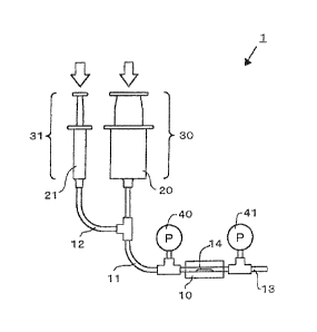
An apparatus for monitoring thrombus formation illustrated in FIG. 1 is used
to fill a
syringe 20 with 50 ml of a citrate-treated blood (solution A) in which 9 parts
by volume of the
blood immediately after the blood sample is mixed with 1 part by volume of
3.2% sodium
citrate, and to fill a syringe 21 with 10 ml of 0.2M CaC12 (solution B). The
syringes 20 and
21 are connected to transparent nylon tubes (inlet tube 11 and drug tube 12)
with an inner
diameter of 3 mm. Both tubes are joined together at a T-shaped joint (cheese)
and then
connected to a polycarbonate thrombus formation chamber 10 of 3 mm in inner
diameter and
1 cm in length through a single nylon tube (inlet tube 11) of 3 mm in inner
diameter and 3 cm
in length. The thrombus formation chamber 10 itself is constructed as a
removable cassette.
CA 02626686 2008-04-18
File:27672-CA-599-PCT 27
The joint portion between the cassette and the nylon tube (inlet tube 11) is
made liquid-tight
via an 0-ring made of silicon rubber. A glass member is fixed on the inside of
the cassette
by an epoxy-based adhesive, thereby forming a constriction portion 14. The
constriction
portion 14 is designed so that the most narrowed site of the constriction
portion 14 may have
an inner diameter (the maximum gap between the constriction portion 14 and the
inner wall)
of 1.5 mm. In addition, the thrombus formation chamber 10 is connected liquid-
tight with a
tube as a discharge tube 13 having the same diameter and formed of the same
material
through an 0-ring made of silicon rubber, and thus a thrombus monitoring
apparatus 1 as
illustrated in FIG. 1 is manufactured. Note that flange-type pressure gauges
40 and 41 are
mounted on parts of the inlet tube 11 and the discharge tube 13 near the
thrombus formation
chamber 10 via joints (cheeses), respectively. In addition, the glass material
of the
constriction portion on the inner surface of the thrombus formation chamber 10
is prepared
such that collagen is coated as a thrombus inducing material 15 on the glass
constriction
portion on the inside by immersing in a 0.1 N acetic acid solution containing
1% insoluble
collagen type I (manufactured by Wako Pure Chemical Industries, Ltd.) and then
drying. The
syringes 20 and 21 are inverted so that plunger are on the top side and
weights are then placed
on the plungers so as to allow the solution A and the solution B to be flown
at 5m1/min and at
0.5 ml/min, respctively, to be syringe pumps.
[0049] When the solution A and the solution B are flown for 10 minutes, a
difference
between the pressure gauges 40 and 41 of the inlet tube 11 and the discharge
tube 13 is
emerged after several minutes and such a difference is then increased with
time.
Simultaneously, it is confirmed that the blood discharged from the discharge
tube 13 is also
gradually decreased. When the flow of all solutions is completed, a
physiological saline
solution is flown into the apparatus for monitoring thrombus formation 1 to
wash the
thrombus formation chamber 10. Thus, the formation of thrombus can be found in
the
thrombus formation chamber 10 by visual observation.
[0050] Example 2
An apparatus for monitoring thrombus formation 1 is prepared and thrombus
formation is then monitored in a manner similar to Example 1, except that the
solution A of
Example I is further added with an unfractionated heparin (prepared from pig
mucous) at a
concentration of 1 mg/ml.
In this case, there is no pressure difference and no decrease in blood flow
occurred,
CA 02626686 2008-04-18
File:27672-CA-599-PCT 28
and thrombus cannot be found in the thrombus formation chamber 10 by visual
observation
after washing with a physiological saline solution.
[0051] Example 3
An apparatus for monitoring thrombus formation 1 was prepared in a manner
similar
to Example 1 with the exceptions that: a glass tube of 3 mm in diameter and 5
cm in length
was provided as a cassette type thrombus formation chamber 10; the syringes 20
and 21 were
each connected to syringe pumps driven by electric motors; and, instead of the
constriction
portion 14 and the collagen coated as a thrombus inducing material 15 on the
constriction
portion 14, as shown in FIG. 2(a), silk of 1.5 cm in length immersed in
collagen of Example 1
and air dried at 4 C were attached by an adhesive as a thrombus inducing
material 15 on the
inner surface of the thrombus formation chamber 10 which is 5 mm inside from
the
connection part of the glass tube and the inlet tube 11.
[0052] After flowing the solution A in the syringe 20 at 3 ml/min and the
solution B in the
syringe 21 at 0.3 ml/min for 15 minutes, the thrombus formation chamber 10 was
detached
and the inside thereof was then washed with a physiological saline solution.
As a result,
visual observation confirmed that thrombus 16 with a length of about 1 cm and
a thickness of
about 1 mm in the form of a comet shape as shown in FIG. 2(b), which seems to
be formed
from the downstream side of the silk as a starting point and extend to the
downstream was
attached on the inner surface of the glass tube. Here, it is considered that
the influence of
blood flow may cause the thrombus in a comet shape.
The widest part of the thrombus had a width of about 3 mm.
[0053] Example 4
An apparatus for monitoring thrombus formation 1 was prepared in a manner
similar
to Example 3 with the exception that, instead of silk, as shown in FIG. 3(a),
50 ul of the blood
without an anticoagulation treatment was dropped on the inner surface of a
glass tube by a
Pasteur pipette and then left standing at room temperature for 15 minutes,
thereby obtaining a
disk-shaped thrombus with a diameter of about 2 mm as a thrombus inducing
material 15.
[0054] In a manner similar to Example 3, after flowing the solution A and the
solution B, the
thrombus formation chamber 10 was removed and then the inside thereof was
washed with a
physiological saline solution. As a result, visual observation confirmed that
thrombus 16
with a length of about 1 cm and a thickness of about 1 mm in the form of a
comet shape as
shown in FIG. 3(b) extending toward the downstream while wrapping around
thrombus as a
CA 02626686 2008-04-18
File:27672-CA-599-PCT 29
thrombus inducing material 15 was attached on the inner surface of the glass
tube. Here, it is
considered that the influence of blood flow may cause the generated thrombus
in a comet
shape.
The widest part of the thrombus had a width of about 3 mm.
[0055] Example 5
An apparatus for monitoring thrombus formation 1 was prepared and thrombus
formation was then monitored in a manner similar to Example 3 with the
exception that the
solution A was further added with Argatroban (registered mark, manufactured by
Daiichi
Pharmaceutical Co., Ltd.) as an an anticoagulation treatment agent at a
concentration of 0.1
mg/ml with respect to the solution A of Example 3.
As a result, visual observation did not confirm the presence of thrombus in a
glass
tube after washing with the physiological saline solution.
[0056] Example 6
An apparatus 1 for monitoring thrombus formation is prepared and thrombus
formation is then monitored in a manner similar to Example 3 with the
exceptions that the
anticoagulated blood prepared by the addition of hirudin at a concentration of
1 Rg/m1 with
respect to the whole blood is used as the solution A and antihirudin
polyclonal antibody
(manufactured by COSMO BIO CO., LTD.) dissolved at a concentration of 1 mg/ml
in a
physiological saline solution with respect to the whole blood is used as the
solution B.
In this case, almost the same thrombus formation is confirmed as that of
Example 3.
[0057] Example 7
There are prepared 10 ml of the whole blood (solution A) which was subjected
to an
anticoagulation treatment by adding to the blood immediately after the blood
sampling 10
jig/m1 of aprotinin and 1 Wm' of recombinant hirudin (manufactured by Wako
Pure
Chemical Industries, Ltd.); and 1 ml of a solution (solution B) which was
prepared by adding
an antihirudin polyclonal antibody (manufactured by COSMO BIO CO., LTD.) from
which a
Fe domain was removed by a papain-immobilized resin to 1000-fold diluted
physiological
saline s solution of 1 vial/ml of thromboplastin reagent (manufactured by
Sysmex
Corporation) as a drug for promoting blood coagulation.
For manufacturing an apparatus for monitoring thrombus formation, a substrate
100
shown in FIG. 4, which is made of polydimethyl siloxane with a width of 40 mm,
a length of
70 mm, and a thickness of 1.5 mm, and a substrate 200 shown in FIG. 5, which
has a
CA 02626686 2008-04-18
File:27672-CA-599-PCT 30
thickness of 1 mm and is made of glass with the same dimensions, are used.
[0058] A circuit containing a thrombus formation chamber 110 is formed by
cutting a
groove with a depth of 0.5 mm on the surface of a substrate 100 with a pattern
shown in FIG.
4. The depth of the part of the groove to be provided as an inlet tube 111
is 1 mm. The
length of the circuit 100D is 30 mm. A through hole of 1 mm in diameter is
formed at the
end of the groove and provided as a regulation value 100 E which is closed by
an operable
and closable cap. The thrombus formation chamber 110 has a length of 30 mm and
a width
of 2.5 mm at the wider portion and a constriction portion 114 having a width
of 0.5 mm.
Then, a through hole with a diameter of 1 mm is provided as each of the
connection parts
100A and 100B. In addition, a through hole with a diameter of 2.5 mm is
provided as a
connection part 100C.
[0059] Collagen in the form of a band with a width of 10 mm is applied as a
thrombus
inducing material 115 to the substrate 200 at a position covering the
constriction portion 114
of the substrate 100 as shown in FIG. 5. The application of collagen is
carried out such that
the rectangular masking tape corresponding to the constriction portion 114 of
the substrate
200 is attached and a silicone-based stripping agent SRX-211 (Dow Corning
Toray Co., Ltd.)
is then coated thereon. Subsequently, the masking tape is peeled off. Then, a
solution of
collagen type 1 (Wako Pure Chemical Industries, Ltd.) dissolved in 0.1N acetic
acid is
dropped so as to be 1% thereof on the glass region free of the stripping agent
and then left
standing for 1 hour at 25 C. After that, collagen on the stripping agent layer
is washed out
with purified water, and collagen is only appllid to a rectangular glass
region corresponding to
the constriction portion 114, where the stripping agent is not applied.
Subsequently, the collagen-applied surface of the substrate 200 and the
circuit-formed surface of the substrate 100 are bonded together face-to-face.
[0060] Connection parts 100A and 100B are connected to silicone tubes having
an inner
diameter of 1mm from the back surface (circuit-free side) of the substrate 100
and then
connected to a 10-ml syringe filled with the solution A and a 1-ml syringe
filled with the
solution B, respectively. These syringes are each attached to syringe pumps,
respectively.
Note that another connection part 100C is connected to a silicon tube having
an inner
diameter of 2.5 mm, which is provided as a discharge tube.
[0061] The solution A is injected from the connection part 100A at a flow rate
of 0.3 ml/min.
The solution B is injected from the connection part 100B at a flow rate of
0.03 ml/min.
CA 02626686 2008-04-18
File:27672-CA-599-PCT 31
The solution A and the solution B are allowed to run from the connection part
100A
and the connection part 100B for 5 minutes, respectively. A physiological
saline solution is
injected from the connection part 100A to wash out the blood. As a result,
thrombus
formation is confirmed mainly on the region a and the region b of FIG. 6 on
the
collagen-treated surface of the substrate 200.
[0062] Example 8
An apparatus for monitoring thrombus formation is prepared and thrombus
formation
is monitored in a manner similar to Example 7 with the following exceptions:
solution A is
prepared such that the blood immediately after blood sampling is added with
anti-factor XII
polyclonal antibody (manufactured by COSMO BIO CO., LTD.), from which a Fe
domain is
cut off by papain, at a concentration of 0.3 mg/ml and unfractionated heparin
(pig origin,
Wako Pure Chemical Industries, Ltd.) at a unit of 0.3; solution B is prepared
by diluting a
thromboplastin reagent (manufactured by Daiichi Pure Chemicals Co., Ltd.) 50
times; the
application of collagen is carried out by pretreating the collagen-applied
region of a
transparent polystyrene substrate 200 (1 mm in thickness) shown in FIG. 8 with
the dropping
of a solution prepared by dissolving potassium permanganate in concentrated
sulfuric acid at
a concentration of 2g/l, subjecting to reaction at 25 C for 10 minutes
followed by quickly
washing with purified water, collagen is applied by droppping a solution in
which collagen
type 1 (Wako Pure Chemical Industries, Ltd.) dissolved in 0.1N acetic acid is
dissolved at a
concentration of 1% on the pretreated region, and leaving standing at 25 C for
1 hour
followed by washing out with purified water; and a substrate 100 without a
constriction
portion is used for the thrombus formation chamber 110 shown in FIG. 7. Note
that the
connection part 100C of the substrate 100 is provided for a through hole of 1
mm in diameter
and connected to a silicone tube of 1 mm in inner diameter as a discharge
tube.
[0063] After flowing the solution A and the solution B for 10 minutes, a
physiological saline
solution is injected from the connection part 100A to wash out the blood,
thereby confirming
the attachment of red thrombus on the channel part of the collagen-applied
region of the
substrate 200.
[0064] Example 9
An apparatus for monitoring thrombus formation was prepared and thrombus
formation was monitored in a manner similar to Example 7 with the exceptions
that:
a solution A was prepared such that the blood immediately after the blood
sampling
CA 02626686 2008-04-18
File:27672-CA-599-PCT 32
was subjected to an anticoagulation treatment by the addition of aprotinin at
a concentration
of 0.05 mg/ml and recombinant hirudin(manufactured by Wako Pure Chemical
Industries,
Ltd.) at a concentration of 1 Rg/m1 and then added with quinacrine (Sigma Co.,
Ltd.); and
the connection part 100B is closed by a cap and only the solution A is then
injected
from the connection part 100A at a flow rate of 1 ml/min.
[0065] When observed with a fluorescence stereoscopic microscope focused on
the
collagen-applied surface from the substrate 200 side, a fluorescence color
development of
green quinacrine was confirmed on the portion of the collagen-applied surface
and such a
region spreads out in as macular region with time. After 10 minutes, the green
fluorescence
color development which may be due to thrombocytic adhesion was confirmed with
the most
of the collagen-applied surface.
[0066] Example 10
An apparatus for monitoring thrombus formation was prepared and thrombus
formation was monitored in a manner similar to Example 9, except that solution
A was
prepared without the addition of quinalin.
The blood was injected from the connection part 100A at a flow rate of 1
ml/min.
When the tube of the connection part 100C was pinched to close the tube for 2
seconds during
the injection, the blood was moved up instantly to 20 mm from the branching
point in the
circuit 100D accompanied by an increase in inner pressure. Even after opening
the
connection part 100C, the blood does not move from the 20-mm point, so the
evidence of an
increase in pressure could be confirmed.
[0067] Example 11
An apparatus for monitoring thrombus formation is prepared and thrombus
formation
is monitored in a manner similar to Example 8 with the exceptions that: the
antithrombin
polyclonal antibody (COSMO BIO CO., LTD.) from which a Fc domain is removed by
a
papain is added so as to be in a concentration of 30 ig/m1 instead of heparin
of the solution A
of Example 8; and the solution B of Example 8 is further added with PPACK
thrombin in a
concentration of 5 mg/ml.
After flowing the solution A and the solution B for 15 minutes, a
physiological saline
solution is injected from the connection part 100A to wash out the blood. As a
result, it is
confirmed that red thrombus is attached on a channel portion of the collagen-
treated region of
the substrate 200.
CA 02626686 2008-04-18
File:27672-CA-599-PCT 33
[0068] Example 12
An apparatus for monitoring thrombus formation was prepared and thrombus
formation was monitored in a manner similar to Example 7 with the exception
that:
solution A was prepared such that 50 ml of the blood immediately after the
blood
sampling, which was added with 0.5 units/m1 of heparin and 10 pg/m1 of
aprotinin, was
centrifuged at 800 rpm and platelet-rich plasma (PRP) was prepared from the
supernatant,
thereby obtaining the solution A; and
a solution in which 1 vial/ml of a thromboplastin reagent (Sysmex Corporation)
was
diluted 30 times with a physiological saline solution was used as solution B.
A thrombus formation of the constriction portion 114 was monitored for 15
minutes
using a stereoscopic microscope. A plurality of platelet clots attached on
around the
constriction portion 114 was confirmed. It was also monitored that the number
of the clots
increased while the sizes thereof also increased, with the platelet clots
repeating attaching and
detaching.
[0069] Example 13
Thrombus formation was monitored by running solution A and solution B in a
manner similar to Example 7 with the following exceptions:
solution A was prepared by adding 500 ttg of erythrocyte collected from the
centrifuged sediment to 20 ml of PRP of the solution A of Example 12;
a solution in which 1 vial/ml of a thromboplastin reagent (Sysmex Corporation)
diluted 30 times with a physiological saline solution was used as solution B;
a slide glass was prepared by coating the whole surface of the substrate 200
with a
silicone-based stripping agent SRX-211 (Dow Corning Toray Co., Ltd.); and
approximately 1 pi of the whole blood was attached on near the widening end
point
of a widening portion successively extending to 2.5 mm in width, which is
adjacent to
upstream of the constriction portion 114 of the thrombus formation chamber 110
on the
substrate 100, then the whole blood was left standing at room temperature for
10 minutes to
cause coagulation thereof, and a preliminary thrombus was formed to be used as
a thrombus
inducing material.
[0070] The thrombus-attached region was monitored by a stereoscopic microscope
for 15
minutes. The formation of a new red thrombus, which was located downstream
from the
preliminary thrombus as an origin, was confirmed after about 5 minutes,
gradually extended
CA 02626686 2008-04-18
File:27672-CA-599-PCT 34
downstream up to 15 minutes, and the width thereof extended to 2.5 mm. It was
confirmed
that the blood flows along the formed thrombus or flows through the gaps
between the formed
thrombus.
[0071] Example 14
An apparatus for monitoring thrombus formation was prepared and thrombus
formation was monitored in a manner similar to Example 7 with the following
exceptions:
50 ml of blood immediately after the blood sampling added with an exosite I
thrombin aptamer (GGTTGGTGTGGTTGG: SEQ ID NO. 1) at a final concentration of 5
RM
and a maize-derived trypsin inhibitor (COSMO BIO CO., LTD.) at a final
concentration of 30
jig/m1 was centrifuged at 800 rpm for 10 minutes, and platelet-rich plasma
(PRP) was formed
from the supernatant and provided as the solution A;
antisense DNA (CCAACCACACCAACC: SEQ ID NO. 2) was dissolved in a
physiological saline solution so as to be 1501.1M in concentration and
provided as the solution
B; and
at the time of mounting a thrombus inhibitor 115, a solution, which was
prepared by
mixing the collagen solution of Example 7 with a solution prepared by
dissolving 1 vial of
thromboplastin reagent (Sysmex Corporation) in 1 ml of purified water and then
dialyzed at a
ratio of 5 : 1, was dropped onto a silicone-unapplied region and dried with
air at 4 C.
A thrombus formation of the constriction portion 114 was monitored for 5
minutes
using a stereoscopic microscope. A plurality of platelet clots attached around
the constriction
portion 114 was confirmed. It was also monitored that the number of the clots
increased
while the sizes thereof also increased, with the platelet clots repeating
attaching and
detaching.
[0072] Example 15
An apparatus for monitoring thrombus formation was prepared and thrombus
formation was monitored in a manner similar to Example 14 with the following
exceptions:
after preparing PRP by the same procedure as that of Example 14, antisense DNA
(CCAACCACACCAACC: SEQ ID NO. 2) was added so as to be 15 ItM in concentration
immediately before the measurement, thereby releasing the anti-thrombin
treatment and
provided as the solution A;
the injection opening of the connection part 100B is closed in a manner
similar to
Example 9;
CA 02626686 2008-04-18
File:27672-CA-599-PCT 35
collagen type I (collagen reagent for coating a cell culture dish with
collagen, Type
I-A stock solution, Cellmatrix Co., Ltd.) was used instead of the collagen
solution of Example
14; and
only the solution A was flown at a flow rate of 0.2 ml/min. for 5 minutes.
A thrombus formation of the constriction portion 114 was monitored using a
stereoscopic microscope. A plurality of platelet clots attached around the
constriction portion
114 was confirmed. It was also monitored that the number of the clots
increased while the
sizes thereof increased, with the platelet clots repeating attaching and
detaching.
[0073] Example 16
An apparatus for monitoring thrombus formation was prepared in a manner
similar to
Example 7 with the following exceptions:
a groove of 0.1 mm in depth was formed on the surface of the substrate by
cutting in
a pattern shown in FIG. 7, while leaving partition walls such that convex rows
each having a
length of 1 mm and a width of 40 m were made stand on the whole surface of
the bottom
portion around the center of the thrombus formation chamber 110 with intervals
of 40)tm
there between;
the injection opening of the connection part 100B was closed in a manner
similar to
Example 9; and
the substrate 200 was a simple slide glass without a surface treatment. The
gap
(channel) between the partition walls simulates a blood capillary.
The whole blood immediately after the blood sampling was subjected to an
anticoagulation treatment by the addition of a maize-derived trypsin inhibitor
with a final
concentration of 30 g/m1 and an exosite I thrombin aptamer with a final
concentration of 10
M. Immediately before monitoring, antisense DNA of exosite I thrombin
aptamer was
added so as to be 30 M in concentration, followed by injection from 100A at a
flow rate of
0.05 ml/min for 5 minutes.
The thrombus formation in the gap between the partition walls was monitored
using
a stereoscopic microscope. As a result, the channels were closed in sequence
during the
monitoring for 5 minutes. It was monitored that about half of the channels
were closed by
thrombus formation.
[00741 Example 17
A syringe-type solution-feeding system shown in FIGS. 9 was used. A microchip
CA 02626686 2008-04-18
File:27672-CA-599-PCT 36
300, where the substrate and the cover of the microchip shown in FIG. 9(B) and
FIG. 9(C),
respectively, made of the same materials as those of Example 7 were pressure-
bonded, was
used. The microchip 300 comprises a blood coagulation inhibitor inlet tube 322
and a
discharge tube 313 which were connected to the substrate 100 as in the case
with the
discharge tube of Example 7. However, the grooves of the microchip 300 of FIG.
9 are all
formed of 200 gm in depth. Also, a channel was formed such that the blood
could be
introduced into the channel from the sample syringe 320, while the channel had
a width of I
mm and a length of 40 mm and was connected to a thrombus formation chamber 311
composed of narrow channel having a width of 200 gm and a length of 10 mm and
the blood
coagulation inhibitor inlet tube 322. A solution was prepared such that 1 vial
of the Sysmex
Corporation PT reagent (tissue thromboplastin) was dissolved in 1 ml of
purified water and
then dialyzed in purified water, the dialyzed product in purified water was
mixed with
collagen type I (manufactured by Nitta Gelatin Inc.) at a ratio of 1 : 1. The
solution was
applied to the position of the thrombus inducing material 315 as shown in FIG.
9. After that,
the applied portion was dried at 4 C in air and thrombus formation chamber 311
was covered,
which was used as a thrombus inducing material 315. The blood was collected
after the
addition of an anticoagulation treatment agent such that the final
concentrations of the
respective components in the syringe were 25gg/m1 for maize-derived trypsin
inhibitor, 10
gM for exosite I thrombin aptamer, 10 M for exosite II thrombin aptamer
(5'-AGTCCGTGGTAGGGCAGGTTGGGGTGACT-3': SEQ ID NO. 3). Immediately
before monitoring, the blood thus collected was added with antisense DNAs with
respect to
thrombin aptamers of exosite I and exosite II so that each of them would reach
to a
concentration of 40 M. Then, a syringe (1 ml syringe, manufactured by Terumo
Corporation) was filled with the blood and provided as the sample syringe 320
shown in FIG.
9. The My Flow (manufactured by Arbiotec Co., Ltd), which is a micro-feeding
pump
capable of monitoring the inner pressure of a liquid-feeding pump, was
connected to a
pressure inlet tube 317 shown in FIG. 9. Mineral oil was pressed into the
microchip 300
from the top of the syringe 320 and the blood was pushed out into the
microchip 300 at a flow
rate of 50 gl/min, while the pressure exerted on the sample syringe 320 was
monitored.
As a blood coagulation inhibitor, 1 M of Tris-HCI (pH 10) was flown from the
blood
coagulation inhibitor inlet tube 322 at a flow rate of 50 gl/min. Pressure
began to rise 6
minutes after starting the pressure measurement.
CA 02626686 2008-04-18
File:27672-CA-599-PCT 37
The pressure, repeating the changes thereof up and down due to the movement of
thrombus ("control" shown in Fig. 13), rose up to 80 kPa.
[0075] Example 18
The blood sampling was carried out with the addition of an anticoagulation
treatment
agent in manner similar to Example 17. Subsequently, the same procedure as
that of
Example 17 was carried out, except that unfractionated heparin was added at a
concentration
of 1 unit/ml. The measurement of pressure was then performed. The pressure
began to rise
13 minutes after starting the measurement. The pressure rose up to about 10
kPa ("Heparin"
shown in FIG. 13).
[0076] Example 19
The blood sampling was carried out with the addition of an anticoagulation
treatment
agent in manner similar to Example 17. Subsequently, the same procedure as
that of
Example 17 was carried out, except that ReoPro (LILLY Co., Ltd.) was added at
a
concentration of 50 g/ml. The measurement of pressure was then performed. An
increase
in pressure could not be confirmed in the measurement for 18 minutes ("ReoPro"
shown in
FIG. 13).
[0077] Example 20
The blood sampling was carried out with the addition of 3.2% citric acid so
that the
ratio of citric acid and the blood be 1 : 9. Further, the blood was added with
a maize-derived
trypsin inhibitor at a concentration of 25 Kg/m1 to carry out an
anticoagulation reaction.
Immediately after adding calcium chloride to the anticoagulated blood so that
it should reach
15 mM with respect to the anticoagulated blood at the time of monitoring
thrombus formation,
the blood was added to a syringe (1-ml syringe, manufactured by Terumo
Corporation),
followed by inflowing the blood into the same microchip as that of Example 17
at a flow rate
of 40 ttl/min. Subsequently, a change in pressure was measured using the same
system as
that of Example 17. The pressure began to rise 15 minutes after starting the
pressure
measurement and it rose up to about 60 kPa.
[0078] Example 21
The blood sampling was carried out with the addition of an anticoagulation
treatment
agent in manner similar to Example 20. Subsequently, the same procedure as
that of
Example 20 was carried out, except that heparin was added at concentrations of
0.2 units/ml,
0.5 units/ml, and 1 unit/ml, respectively. The measurement of pressure was
then performed.
CA 02626686 2008-04-18
File:27672-CA-599-PCT 38
The results are shown in FIG. 14.
[0079] Example 22
The blood sampling was carried out with the addition of an anticoagulation
treatment
agent in manner similar to Example 20. Subsequently, the same procedure as
that of
Example 20 was carried out, except that ReoPro (LILLY Co., Ltd.) was added at
a
concentration of 2 lig/ml. The measurement of pressure was then performed. The
pressure
began to rise 20 minutes after starting the pressure measurement and it rose
up to about 15
kPa after 25 minutes (FIG. 15).
[0080] Example 23
A syringe-type solution-feeding system shown in FIG. 12 was used. In addition,
a
microchip 400, where the substrate 200 as a cover of the microchip and the
substrate 100 as a
body of the microchip shown in FIG. 12(13) and FIG. 12(C) were pressure-bonded
with each
other, was used. A discharge tube 413 made of a Teflon (registered trademark)
tube was
filled with mineral oil and one end thereof was connected to the microchip 400
through a
connection tube 412 made of silicon rubber and the other end thereof was
connected to a
suction pump 414 in which a pressure sensor was installed. An inlet tube
forming a channel
of the microchip 400 was connected to a syringe 420 through the connection
tube 412 made
of silicone rubber. One end of the blood coagulation inhibitor inlet tube 422
made of Teflon
(registered trademark) was connected to a liquid-feeding pump 423 and the
other end thereof
was connected to the terminal end of the thrombus formation chamber through
the connection
tube 412 made of silicone rubber. In this way, the microchip 400 was connected
to the
discharge tube 413, the syringe 420, and the blood coagulation inhibitor inlet
tube 422, and
thus an apparatus B for monitoring thrombus formation was produced. A CCD
camera 430
provided with an illumination optical source 432 (LED) which was mounted on a
rail 433 and
movable was set under the thrombus formation chamber.
All of the grooves in the microchips had a depth of 120 p.m.
The channel in which the blood was introduced had a width of 800 pm and a
length
of 7 mm. The narrowed channel had a width of 200 pm and a length of 10 mm.
A solution was prepared such that 1 vial of PT reagent (tissue thromboplastin,
Sysmex Corporation) was dissolved in 1 ml of purified water and then dialyzed
in purified
water, the dialyzed product in purified water was mixed with collagen type I
(manufactured
by Nitta Gelatin Inc.) at a ratio of 1 : 1. The solution was then applied to
the region of the
CA 02626686 2008-04-18
File:27672-CA-599-PCT 39
substrate 200 of the microchip facing the channel of the constriction portion
on the surface to
be pressure-bonded with the substrate 100 of the microchip. After that, the
applied portion
was vacuum dried at 4 C and the substrate 100 and the substrate 200 were then
pressure-bonded with each other, thereby providing a microchip 400 having a
thrombus
formation chamber 411.
The blood sampling was carried out using a vacuum blood-collecting vessel in
which
sodium citrate provided as an anticoagulation agent was enclosed. Immediately
before
introducing into the syringe 420, the blood was added with calcium chloride at
a final
concentration of 12.5 mM and a maize-derived trypsin inhibitor at a final
concentration of 25
1-Lgirni=
The My Flow (manufactured by Arbiotec Co., Ltd), which is a micro-feeding pump
capable of monitoring the inner pressure of a liquid-feeding pump, was
connected to a
discharge tube 413 as a pump 414. Thus, the suction was performed at a rate of
15 [11/min
while the inner pressure of the channel was monitored.
As a blood coagulation inhibitor, 1 M of Tris-HCI (pH 10) was flown from the
blood
coagulation inhibitor inlet tube 422 at a flow rate of 8 [tl/min. White
thrombus formation
was mainly observed after 4 minutes from the start of the pressure
measurement, while a
decrease in inner pressure was confirmed. In addition, it was confirmed that
the inner
pressure decreased 30 kPa after 10 minutes from the start.
Further, thrombus formation was observed by taking images while the camera 430
was moved around the thrombus-forming region in the channel. The position
where the
thrombus formation was observed was memorized, and the camera 430 took images
while
regularly moving around a plurality of specific points. As a result, growth
and destruction of
thrombus with time was recorded with respect to a plurality of large thrombi.
[0081] Hereinafter, reference experiments will be performed as described below
to show the
effectiveness of thrombin aptamers and the antisense DNAs thereof as
anticoagulation
treatment agents and anticoagulation releasing agents, respectively.
[Reference Experiment 1]
The blood immediately after blood sampling, the blood prepared by adding a
volume
of one tenth of 300 ii.g/m1 of a maize-derived trypsin inhibitor to the first
blood, and the blood
prepared by adding a thrombin aptamer that recognizes exosite Ito the second
blood so as to
be 5 [IM in concentration were provided. Then, for these three kinds of the
blood, the time
CA 02626686 2008-04-18
File:27672-CA-599-PCT 40
taken for coagulation were measured in Eppendorf tubes (made of
polypropylene). The
sample was reversed up side down every one minute and the flowability thereof
was
confirmed. The blood free of additive showed a coagulation time of 9 minutes.
In contrast,
the blood added with the trypsin inhibitor showed a coagulation time of 22
minutes. Further,
the blood added with the thrombin aptamer showed a coagulation time of 62
minutes.
Further, when 5 jiM of a thrombin aptamer that recognizes exosite I and 5 iaM
of a
thrombin aptamer that recognizes exosite II were used in combination, blood
coagulation was
not confirmed even after 3 hours even in the case of the blood free of the
trypsin inhibitor.
Exosite I recognition thrombin aptamer: 5'-GGTTGGTGTGGTTGG-3' SEQ ID NO. 1,
Exosite II recognition thrombin aptamer:
5'-AGTCCGTGGTAGGGCAGGTTGGGGTGACT-3' SEQ ID NO. 3.
[0082] [Reference Experiment 2]
The measurement of APTT was carried out on 200 1.tM of plasma with respect to
each of sample A added with 10 M of physiological saline solution, sample B
added with 10
jiM of 500- M exosite I recognizing thrombin aptamer, and sample C added with
10 [1.1 of a
solution containing 500- M exosite I recognizing thrombin aptamer and 1500- M
exosite I
recognizing thrombin aptamer antisense DNA. The sample A was 44 seconds, the
sample B
was 2 minutes or more, and the sample C was 45 seconds.
[0083] [Reference Experiment 3]
FIG. 11(A) is an analysis of a waveform of coagulation with thromboelastogram
by
storing the blood added with 10 jil of aptamers to exosite I and exosite II at
room temperature
for 15 minutes, and then by adding 40 1AM of antisense DNAs of both aptamers.
FIG. 11(B)
is a thromboelastogram waveform of the blood just after blood sampling.
As is evident from the results of FIG. 11(A) and FIG. 11(B), it is found that
the
addition of two kinds of thrombin aptamers allow the blood to be stored and
the antisense
DNA can release the anticoagulation treatment thereof.
From the results of Reference Experiments I to 3, it is evident that the
combination
of the maize-derived trypsin inhibitor and the thrombin aptamer can
effectively inhibit the
coagulation of the whole blood and that the anticoagulation effect of the
thrombin aptamer
can be inactivated by the antisense DNA to the thrombin aptamer.
[0084] [Reference Experiment 4]
Hereinafter, the efficiencies of PMEA coating to the storage of blood and
platelet
CA 02626686 2008-04-18
File:27672-CA-599-PCT 41
adhesion on the substrate will be described.
(1) 1% PMEA-containing methanol solution prepared according to JP 04-152952 A
was applied to a 2.5-ml acryl resin container (inner dimensions: 1 cm x 1 cm x
4.5 cm) and
then dried at 90 C for 10 minutes. Subsequently, 760 ul of the blood subjected
to an
anticoagulation treatment with 2% citric acid was added to each of a non-
coated container and
a PMEA-coated container, followed by aspirating with a pipette every 5 minutes
to confirm
blood coagulation.
Consequently, the non-coated container showed an apparent increase in
viscosity at
30 minutes and coagulation at 60 minutes. In contrast, the PMEA-coated
container showed
an apparent increase in viscosity at 35 minutes, but the coagulation was
prolonged to 90
minutes.
[0085] (2) The above PMEA was flatly coated on a transparent acryl plate. It
was used
instead of the collagen-coated plate of Example 9 (FIG. 8) and an adhesion
experiment of
quinacrine-labeled platelets and leucocytes in the blood was carried out in a
manner similar to
Example 9.
As a result, apparent adhesion or agglutination of platelets and leucocytes
was
observed after 10 minutes from beginning of flowing the blood. In contrast,
adhesion or
agglutination of platelets and leucocytes on the PMEA-coated acryl plate was
not observed
even after 30 minutes.
From the above results, in the apparatus for monitoring thrombus formation of
the
present invention, by coating the blood storage syringes, the tube, the
substrate, and the like
with PMEA, thrombus formation can be supressed in a place other than the
thrombus
monitoring chamber. Therefore, it suggested that thrombus monitoring specific
to the
thrombus monitoring chamber can be attained.
[0086] In the above description, the present invention has been described with
reference to
the preferred embodiments. However, the present invention is not limited to
the examples
and embodiments described above, and various modifications may be applied as
long as not
departing from the gist of the present invention. For example, the pressure of
the pump may
be set to be high and the inner diameter of the discharge tube may be set to
be small. In this
case, back pressure can be generated in the inlet tube and the discharge tube,
so thrombus
formation can be monitored while controlling the flow volume of blood under
the load of
inner pressure corresponding to the blood pressure. Further, the flow rate of
blood at this
CA 02626686 2008-04-18
File:27672-CA-599-PCT 42
time can be freely controlled by means of pump pressure and the degree of
throttling the
discharge tube.
Industrial Applicability
[0087] The apparatus for monitoring thrombus formation and the method of
monitoring
thrombus formation of the present invention can be favorably used in
comprehensive
evaluation of blood coagulation and platelet thrombus formation under a
bloodstream-equivalent environment using the whole blood or the plasma
containing platelets,
for evaluating the efficacy of an antithrombotic drug applied to a patient or
the like.
CA 02626686 2013-06-12
%
1
SEQUENCE LISTING
<110> FUJIMORI KOGYO CO., LTD.
<120> APPARATUS FOR MONITORING THROMBUS FORMATION AND METHOD OF MONITORING
THROMBUS FORMATION
<130> OP-C6262-PCT
<140> PCT/JP2006/320789
<141> 2006-10-18
<150> JP2005-302557
<151> 2005-10-18
<150> JP2005-308065
<151> 2005-10-24
<150> JP2005-334594
<151> 2005-11-18
<150> JP2005-358448
<151> 2005-12-13
<150> 3P2006-36148
<151> 2006-02-14
<150> JP2006-234270
<151> 2006-08-30
<160> 3
<170> PatentIn version 3.3
<210> 1
<211> 15
<212> DNA
<213> Artificial sequence
<220>
<223> exosite I thrombin aptamer
<400> 1
ggttggtgtg gttgg 15
<210> 2
<211> 15
<212> DNA
<213> Artificial sequence
<220>
<223> antisense DNA
CA 02626686 2013-06-12
2
<400> 2
ccaaccacac caacc 15
<210> 3
<211> 29
<212> DNA
<213> Artificial sequence
<220>
<223> exosite I thrombin aptamer
<400> 3
agtccgtggt agggcaggtt ggggtgact 29