Note: Descriptions are shown in the official language in which they were submitted.
CA 02627166 2008-04-24
WO 2007/053364 PCT/US2006/041402
-1-
ARTHROPLASTY REVISION DEVICE AND METHOD
RELATED APPLICATION
This application is a continuation of and claims priority to U.S. Application
No. 11/263,603, filed on October 31, 2005. The entire teachings of the above
application is incorporated herein by reference.
BACKGROUND OF THE INVENTION
Arthroplasty is becoming significantly more prevalent as a surgical
procedure to treat injury and disease. Of particular importance is the use of
artificial
discs to replace vertebral discs as functioning artificial joints.
Instrumentation employed to conduct surgical techniques that implant
artificial discs still are of limited variety and generally do not permit
modification
subsequent to completion of the procedure without radical reconstruction and a
significant likelihood of additional tissue damage. Artificial discs typically
include
two endplates and a core between endplates. The core pernlits movement of the
endplates relative to each other, thereby simulating the function of the
intervertebral
disc that it replaces. Artificial discs can be implanted as complete
assemblies, or,
alternatively, endplates of an artificial disc can be inserted first, followed
by
placement of a core between the endplates. As in any surgical implantation,
the
initial placement may not be optimal. In such an instance, the surgeon
typically is
left with the option of leaving the implant in a sub-optimal position or
removing it,
and replacing the implant in a more optimal position. During the process,
further
traumatization of the surrounding tissue can occur. Therefore, a need exists
for a
device and a method that'significantly eliminates or reduces the above-
referenced
problems.
CA 02627166 2008-04-24
WO 2007/053364 PCT/US2006/041402
-2-
SUMMARY OF THE INVENTION
The invention is directed to a surgical instrument and a method for
revising/removing an artificial disc or removing/replacing a core of an
artificial disc.
In one embodiment, a surgical instrument of the invention includes a pair of
tips, each tip having a pair of tines and a stop defining a proximal end of
each tine.
In one embodiment, the stop of each tip extends between the tines of the tip.
The
tines of each tip also can essentially match the tines of the other tip. In
one
embodiment, the tines of each tip have a flat surface, and the flat surface of
the tines
of each tip are parallel. Alternatively, the tines of each tip can have
surfaces that
complement surfaces of the tines of the other tip. In one such embodiment,
each tip
includes a base portion, wherein the tines of each tip extend from the base
portion.
Also, the base portion of each tip can include a surface, at least a portion
of which
complements at least a portion of a surface of a base portion of the other
tip. In one
embodiment, the complementary surfaces of the base portion are continuous with
the complementary surface of at least one tine of each tip. The continuous
complementing surfaces of the tips can partition the remaining portion of the
base of
each tip when the complementary surfaces of the tips are in contact with each
other.
In one embodiment, the surgical instrument includes a forceps portion. In
one embodiment, the forceps portion is a double-action forceps. In another
embodiment, the forceps portion is a parallel-action forceps. The tips can be
releasable from the forceps portion. In one embodiment, at least one of the
tips is
releasable by activation of a spring-loaded clip that releasably couples the
tip to the
forceps portion. In a specific einbodiment, a major axis of the tines extends
at an
oblique angle to a major axis of the forceps portion. The base of at least one
of the
tips can define a chamfered recess having a major axis essentially parallel to
a major
axis of the tines of the tip. In a specific embodiment, botli tips can define
a
chamfered recess, wherein the chamfered recesses are opposed to each other
when
the tips are coupled to the forceps portion. In a particular embodiment, the
continuous step of at least one tip is chainfered.
A method of revising a position of an artificial disc or of implanting a core
of
an artificial disc includes abutting the stop of at least one tip against an
outer surface
of an implanted endplate of the artificial disc, whereby tines of the tip can
support
CA 02627166 2008-04-24
WO 2007/053364 PCT/US2006/041402
-3-
the artificial disc. The tip is then separated from another, opposing tip,
whereby
opposing implanted endplates, each of which is supported by a pair of tines of
a tip,
are separated, thereby distracting vertebrae between which the endplates are
implanted. The core between the endplates can then be removed and replaced by
one that is more appropriately sized (e.g., height of the core), or the core
can be
removed so that the endplates can be removed and easily repositioned (revised)
or
replaced. In a specific embodiment, the stops of each pair of tips abuts each
of a
pair of opposing implanted endplates. In one embodiment, the tips are abutted
against the endplates simultaneously. The tips can be abutted against the
endplates
while the tips are in a nested position. In one embodiment, the tips are
separated
from each other by actuating nonparallel-action forceps to which the tips are
attached or of which they are a component. In another embodiment, the tips are
separated from each other by actuating a parallel-action forceps to wliich the
tips are
attached or of which they are a component. The method can further include the
step
of releasing the forceps, whereby the endplates each rest against the core.
The present invention has many advantages. For example, the apparatus and
method of the invention permit revision or implantation of a core of an
artificial disc
without disturbing seating of implanted endplates of the artificial disc.
Accordingly,
the surgeon can conduct any necessary iterative procedure that may be required
to
optimally place a core between implanted endplates of an artificial disc.
Further,
implanted endplates can be distracted with minimal movement, thereby also
minimizing trauma to adjacent tissue. Also, abutting stops of the tines of
each tip
against an endplate enables the apparatus to be freely manipulated by the
surgeon
without significant risk of injury by incidental contact of the tines, such as
by
contact of the tines to nerve tissue.
BRIEF DESCRIPTION OF THE DRAWINGS
FIG. 1 A is a perspective view of a pair of tips of a surgical instrument of
the
invention.
FIG. 1B is an end view of the pair of tips shown in FIG. 1A.
FIG. 1C is an opposing end view of the pair of tips shown in FIG. lA:
FIG. 2A is a perspective view of FIGS. 1A - 1C in a nearly-nested position.
CA 02627166 2008-04-24
WO 2007/053364 PCT/US2006/041402
-4-
FIG. 2B is an end view of the pair of tips in the nearly-nested position shown
in FIG. 2A.
FIG. 2C is a detail of the end view of FIG. 2B, showing the tines of the pair
of tips nearly nested.
FIG. 2D is an opposing end view of the pair of tips shown in FIG. 2A in the
nearly-nested position.
FIG. 2E is an alternative embodiment of the surgical instrument of the
invention wherein the pair of tips abut each other at flat surfaces.
FIG. 3A is a perspective view of the pair of tips of FIGS. 1A - 1 C and 2A -
2D in combination with a parallel-action forceps in a retraction position and
of the
relation of the pair of tips to the parallel-action forceps upon assembly.
FIG. 3B is a side view of the embodiment shown in FIG. 3A.
FIG. 3C is a perspective view of the embodiment of FIGS. 3A and 3B in a
distracted position.
FIG. 3D is a side view of the embodiment of FIG. 3C in the distracted
position.
FIG. 3E is a plan view of the embodiment of FIG. 3C.
FIG. 3F is an end view of the embodiment of FIG. 3C.
FIG. 4 is a perspective view of the pair of tips of FIGS. lA - 1C and 2A - 2D
in combination with a nonparallel-action forceps in a retraction position.
FIG. 5A is a perspective view of the invention in a retracted position where
tips are non-modular components of parallel action forceps.
FIG. 5B is a side view of the embodiment shown in FIG. 5A.
FIG. 5C is an end view of the embodiment shown in FIG. 5A.
FIG. 5D is a perspective view of the embodiment of FIG. 5A in a distracted
position.
FIG. 5E is a side view of the embodiment of FIG. 5D.
FIG. 5F is an end view of the embodiment of FIG. 5D.
DETAILED DESCRIPTION OF THE INVENTION
The foregoing and other objects, features and advantages of the invention
will be apparent from the following more particular description of preferred
CA 02627166 2008-04-24
WO 2007/053364 PCT/US2006/041402
-5-
embodiments of the invention, as illustrated in the accompanying drawings in
which
like reference characters refer to the same parts throughout the different
views. The
drawings are not necessarily to scale, emphasis instead being placed upon
illustrating the principles of the invention.
The invention generally is directed to a surgical instrument and method for
revising the position of, or implanting a core between, implanted endplates of
an
artificial disc. FIGS. 1A, 1B and 1C represent perspective and opposing end
views
of pair of tips 10 of the surgical instrument of the invention in a distracted
position.
Tip 12 and opposing tip 14 include pairs of tines 16, 18 and 20, 22,
respectively.
Tines 16, 18 of tip 12 and tines 20, 22 of opposing tip 14 are each defined by
stops
24, 26 and stops 28, 30, respectively.
Tip 12 and opposing tip 14 include base 32 and base 34, respectively. As
shown in FIG. 1A, stops 24, 26 of tip 12 are continuous along base 32. In
corresponding manner, stops 28, 30 define a continuous surface along base 34
of
opposing tip 14. Tines 16, 18 of tip 12 and tines 20, 22 of opposing tip 14,
along
with a portion of base 32 and base 34, defme complementary surfaces 36, 38 of
tip
12, and complementary surfaces 40, 42 of opposing tip 14. Specifically,
complementary surface 36 nests with complementary surface 40 and complementary
surface 38 nests with complementary surface 42. As shown in FIG. 1B, tip 12
and
opposing tip 14 also include chamfered surfaces 43, 44, at base 32 and base
34,
respectively. As shown in FIG. 1 C, tips 12, 14 also include recessed portions
46,
48, which define chamfered recesses 50, 52, respectively. Chamfered recesses
50,
52 oppose each other when complementary surfaces 36, 40 and 38, 42 are nested.
Chamfered recesses 50, 52 each include a major axis that is essentially
parallel to a
plane extending through at least one tine of a respective tip. Chamfered
recesses 50,
52 are intended to allow access to space between tines during core
removal/replacement.
Modular connectors 54, 56 extend from base 32 and base 34, respectively. A
major axis of each of modular connectors 54, 56 extends through a major axis
of at
least one tine and a respective tip at an oblique angle. Preferably, the
oblique angle
is in a range of between about 1 degree and about 20 degrees. In a
particularly
CA 02627166 2008-04-24
WO 2007/053364 PCT/US2006/041402
-6-
preferred embodiment, the oblique angle is 15 degrees. In the alternative, a
major
axis of the tines is parallel to the major axis of the forceps, or distraction
instrument.
FIGS. 2A, 2B and 2D represent perspective and opposing end views of the
surgical instrument of the invention shown in FIGS. 1 A - 1 C in a nearly-
nested or
nearly-reduced position. Tip 12 and opposing tip 14 are nearly-nested,
because, as
can be seen in FIG. 2C, which is a detail of FIG. 2B, coinplementary surfaces
38, 42
of tines 18, 22, respectively, are not in contact, but are in close proximate
relation to
each other. Upon contact, tip 12 and opposing tip 14 would be in a nested
position.
It is to be understood, however, that as an alternative to complementary
surfaces,
tines of opposing tips can abut without being complementary. In one
embodiment,
the tines of opposing tips can abut in a retracted position at continuous flat
surfaces
of the tines, as shown in FIG. 2E (in a distracted position).
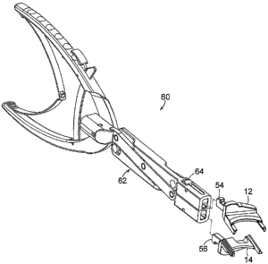
FIGS. 3A and 3B represent, respectively, perspective and side views of
surgical instrument 60 of the invention that includes parallel-action forceps
62 in
combination with tip 12 and opposing tip 14 of FIGS. 1A - 1C and FIGS. 2A -
2D.
Parallel-action forceps 62 can be any suitable parallel-action forceps, such
as is
described in U.S. 5,122,130, issued to Keller on June 16, 1992, the entire
teachings
of which are incorporated herein by reference. Tips 12 and 14 can be modular,
wllereby they are releasable from another component of a surgical instrument.
As
shown in FIGS. 3A and 3B, tip 12 and opposing tip 14 are compatible for
coupling
with parallel-action forceps 62 at modular connectors 54, 56. Modular
connectors
link with the parallel-action forceps with spring-loaded clips 64, 66,
respectively. It
is to be understood, however, that any suitable coupling mechanism could be
employed, such as described in U.S. Serial Number 10/616,506, filed July 8,
2003,
and U.S. Serial Number 10/959,598, filed October 6, 2004, the entire teachings
of
both of which are incorporated herein by reference. FIGS. 3A and 3B represent
surgical instrument 60 in a reduced position. FIGS. 3C, 3D, 3E and 3F
represent
surgical instrument 60 in a distracted position, with tip 12 and opposing tip
14
assembled with parallel-action forceps 62. Alternatively, tip 12 and opposing
tip 14
can be a component of or suitably connected such as by a modular connection as
described, for example, above, to a nonparallel-action forceps as opposed to a
parallel-action forceps. A representative example of a nonparallel-action
forceps is
CA 02627166 2008-04-24
WO 2007/053364 PCT/US2006/041402
-7-
shown in FIG. 4, wherein surgical instrument 70 includes nonparallel-action
forceps
72 coupled to tip 12 and opposing tip 14. In another embodiment, tip 12 and
tip 14
can be non-modular components of parallel action or non-parallel action
forceps.
FIG. 5A is a perspective view of tip 12 and tip 14 as components of parallel
action
forceps 80 arranged as a non-modular embodiment in a retracted position. FIGS.
5B and 5C are side and end views, respectively, of the non-modular embodiment
of
FIG. 5A. FIGS. 5D, 5E and 5F are perspective, side and end views,
respectively, of
the embodiment of FIG. 5A in a distracted position.
In a method of the invention, the size of a core of an artificial disc is
revised,
or the core of an artificial disc is implanted, by abutting the stop or stops
of at least
one tip of the invention against an interior surface of an implanted endplate
of an
artificial disc, whereby tines of the tip can support the artificial disc. The
tip is
separated from another opposing tip, whereby opposing implanted endplates,
each of
which is supported by pairs of tines of a tip, are separated, thereby
distracting
vertebrae between which the endplates are implanted. Preferably, the stops and
the
tines of each tip comport with each endplate, whereby the force of distraction
of the
vertebrae is born, at least substantially, if not entirely, by the endplates,
rather than
by the force of direct contact between the vertebrae and the tines. Upon
sufficient
distraction of the vertebrae, the core between the artificial disc can be
revised or the
core can be removed, implanted, or both, between the endplates of the
artificial disc.
Preferably, the tips are abutted against the endplates while the tips are in a
nested
position. Actuation of the nonparallel-action forceps or parallel-action
forceps
moves the forceps from a reduced position, such as in shown in FIGS. 3A and
3B, in
the case of parallel-action forceps, to a distracted position, such as is
shown in FIGS.
3C through 3F, thereby distracting vertebrae adjacent to implanted endplates
of an
artificial disc. In another embodiment of the method of the invention, the
core can
be removed, followed by removal of the pair of tips from between the
endplates, and
revision (repositioning), removal and/or replacement of the endplates of the
artificial
disc. The endplates can then be distracted again by use of the pair of tips,
as
described above, and a core, of either the same or a different size can be
implanted
between the endplates. The pair of tips is then removed by releasing the
forceps,
and the operation is completed..
CA 02627166 2008-04-24
WO 2007/053364 PCT/US2006/041402
-8-
While this invention has been particularly shown and described with
reference to various embodiments thereof, it will be understood by those
skilled in
the art that various changes in form and details may be made therein without
departing from the scope of the invention encompassed by the appended claims.
