Note: Descriptions are shown in the official language in which they were submitted.
CA 02627248 2008-04-24
WO 2007/057902 1 PCT/IL2006/001329
ABLATING APPARATUS PARTICULARLY USEFUL FOR REMOVAL OF
DENTAL PERIAPICAL LESIONS
FIELD AND BACKGROUND OF THE INVENTION
The present invention relates to ablating apparatus for removing selected
tissues of a person's body. The invention is particularly useful for removing
dental
periapical lesions, and is therefore described below with respect to this
application,
but it will be appreciated that the invention could be used also for removing
otlier
types of tissue, such as bone tissue, and the like.
The term "ablating" devices is used in its broadest respect, to include any
form
of tissue removal, e.g. by resection, cutting, grinding, filing, etc. Dental
periapical
lesions are lesions encompassing or surrounding the tip of the root of a
tooth.
A tooth is composed of a crown and one or more roots which anclior the tooth
in a jawbone. The crown, made of enamel and dentin, surrounds a pulp chamber
which contains the pulp and extends to the root canal or canals. The root
canal opens
at the tip of the root (apex) through an opening termed "apical foramen". A
deep
cavity, a cracked filling, or a cracked tooth can lead to pulp infection or
injury. This
in turn can lead to pulp inflammation and infection which may spread to the
root
canal, often causing sensitivity to hot or cold foods and pain, among other
problems.
If not treated at this stage the pulp may then become necrotic and infected.
Bacteria
that exit from the root canal through apical foramen may spread into adjacent
or
remote tissues. To prevent that, the host mounts an inflammatory response
around the
apical foramen which results in local bone destruction. The lesion thus formed
is
commonly termed a "periapical lesion".
Periapical lesions may also develop when a previous root canal treatment (as
detailed below) was unsuccessful in adequately performing its main task of
elimination of bacteria or when prior root canal filling and/or coronal
restorations are
leaking, thus allowing bacteria to re-contaminate the root canal.
Treatment involves removing the diseased, injured or necrotic pulp, or
contaminated root canal filling material, cleaning shaping and disinfection of
the pulp
chamber and root canals, followed by their sealing with a root canal filling
which is
followed by filling or restoring the crown. Typically, an opening into the
pulp
chamber is made, generally through the crown and dentine, and the pulp or
CA 02627248 2008-04-24
WO 2007/057902 2 PCT/IL2006/001329
necrotic/infected tissues, or the infected root canal filling material is
removed using
an endodontic file. The pulp chamber and root canals are then cleaned, shaped
and
sealed.
To prevent and/or irradicate infection, an antiseptic, such as calcium
hydroxide may be applied to the pulp chamber and root canals before sealing
and
retained there for a period of about two weeks to disinfect them. The crown
opening
can be temporarily filled, e.g., with fRM, GC Fuji 9, or Ketamolar, to protect
the tooth
in order to prevent re-infection of the root canals until the next dental
visit, and
possibly in order to restore the chewing surface.
Following removal of the temporary filling and antiseptic medication, the pulp
chamber and root canals are cleaned and filled with a root canal filling. A
permanent
filling, such as amalgam, conventional composite or a crown, are then used to
restore
the chewing surface of the tooth.
Alternatively, after cleaning and reshaping the root canals and applying
medication, the root canals can be filled with a root filling inaterial, such
as, Gutta
Percha or a paste, to an apical point of the root canal. The pulp chamber can
then be
filled with a temporary filling or a sealing layer. At the next dental visit,
the
temporary filling, as well as some of the root canal filling are removed, and
a post
(also referred to as a dowel) is positioned in the pulp chamber and root canal
and
cemented in place using a dental cement, for example, composite cement, zinc-
phosphate cement, or another cement or sealer.
The post may be formed from a metal, such as a dental alloy, from quartz,
reinforced carbon fibers, or from another suitable material. The post can be
rigid or
flexible to some extent. Where two or more root canals are being treated, one
or more
posts can be used.
The post can be prefabricated and shaped during the procedure. Alternatively,
a mold of one of the root canals and remaining tooth and pulp chamber may be
taken
in the dental clinic and sent to a dental laboratory, to enable a metal cast
post to be
tailor-made based on the mold.
Generally, the above described treatment procedure is effected by an
endodontist who removes the diseased pulp and cleans and seals the pulp
chamber
and root canals, a prosthodontist who fills or restores the crown, and a
dental
technician who prepares the restored crown based on a mold prepared by the
CA 02627248 2008-04-24
WO 2007/057902 3 PCT/IL2006/001329
prosthodontist. Nevertheless, all the above procedures may be, and are
commonly
carried ouf, by a dentist who is a general practitioner.
Root canal infection can also lead to formation of lesions (e.g. abscess,
granuloma, or radicular cyst) around the root apex (periapical). Periapical
lesions are
typically treated according to the procedure described above. While such
treatment is
generally successful and results in healing of the periapical lesion, in cases
where the
root canal treatment fails, where it cannot be accessed, or where it is
desired to
accelerate healing, an apicoectomy surgical procedure is generally used.
Apicoectomy is a procedure in which the root tip is surgically accessed
directly through the gums and the jaw bone. The granulation tissue of the
periapical
lesion is removed, and the root tip is resected, cleaned and sealed through
any one of
- several approaches.
Although widely practiced, apicoectomy is an invasive surgical procedure and
as such it is commonly accoinpanied by postoperative pain, swelling and
complications. In addition, it carries a risk of infection and injury to
nerves, soft
tissue, bone and adjacent teeth. Furthermore, some teeth are less accessible
or
inaccessible surgically (e.g. palatal roots of upper molar), and as such, this
procedure
cannot be utilized in some periapical lesions. Finally, this procedure
oftentimes
results in aesthetic problems such as scarring and recession of gums around
restored
crown and bridgework.
As indicated earlier, while the invention is particularly useful in apparatus
for
removing dental periapical lesions, the invention may also be used in
resection
devices for removing other types of tissue.
Many different types of resection devices are known for removing tissue from
a human body. Resection devices are increasingly used in minimally invasive
laparoscopic or endoscopic procedures, since they allow selective separation
and
removal of tissue through small body openings in a very precise manner.
Typically, different procedures require different resection devices, each
adapted for resection of specific tissue at a specific location. Some devices
need to be
repeatedly inserted aiid removed from the body in order to resect and remove
tissue,
while others incorporate or employ tissue collection mechanisms such as
aspiration
mechanisms.
CA 02627248 2008-04-24
WO 2007/057902 4 PCT/IL2006/001329
Manual resection devices typically employ manually operated scissor-like
cutting heads disposed on elongated meinbers which terminate in levers for
operating
the cutting head from outside the body.
Powered tissue resection devices are typically used in, for example,
arthroscopic procedures performed on knee or shoulder joints. Powered tissue -
resection devices used in such arthroscopic procedures are designed as
elongated,
hollow inner tubular member situated to cyclically move (e.g. rotate) within
an
elongated outer tubular member. The inner member is provided with a cutting
device
at its distal end, and the outer tubular member is provided with a window or
other
opening enabling the cutting device of the inner member to resect desired
tissue
presented through the outer window. During arthroscopic procedures, the joint
is
expanded with a fluid medium in order to provide distension and also to
enhance
visualization of joint tissue. The resected tissue remains suspended in the
fluid, and a
vacuum is applied to aspirate the resected tissue from the joint. Since such
aspiration
necessarily removes ambient fluid as well, continual fluid flow through the
joint is
required to maintain a cleati, debris-free field of view.
Numerous examples of resection or ablating devices are known in the art; see
for example, U.S. Patents 5,456,689; 5,779,662; 6,632,223; 6,632,227,
6,540,747 and
6,746,451. Such devices have been used in various surgical procedures, as
described
for example in the above-cited US Patents 6,540,747 and 6,746,451. However,
they
have not heretofore been used for removing dental periapical lesions, insofar
as we
are aware, and therefore have not been designed for use in removing dental
periapical
lesions according to the present invention.
OBJECTS AND BRIEF SUMMARY OF THE PRESENT INVENTION
One object of the present invention is to provide apparatus particularly
useful
for removing dental periapical lesions without cutting through the gums and
the
jawbone, according to the typical treatments used at the present time. Another
object
of the invention is to provide an ablating device which is particularly useful
for
removing dental periapical lesions, but which may also be used for removing
other
forms of tissue, e.g., for harvesting bone tissue in the treatment or
prevention of bone
fracture, promoting joint fusion, enhancing implant fixation, removal of
diseased
tissue, etc.
CA 02627248 2008-04-24
WO 2007/057902 5 PCT/IL2006/001329
According to one aspect of the present invention, there is provided apparatus
for removing a dental periapical lesion at an apex of a root of a tooth,
comprising: a
rotary ablating device sized and constructed (a) for introduction via an
opening
through the tooth into the root canal; (b) for movement therethrough to
protrude
through the apical foramen into contact with the dental periapical lesion; and
(c) for
rotation while in contact with the dental periapical lesion in order to remove
the lesion
by ablation.
The use of such apparatus for removing dental periapical lesions provides a
number of important advantages over the existing removal procedure involving
cutting through the gums and the jawbone of the patient. Thus, it reduces the
possibility of postoperative pain, swelling and complications normally
accompanying
the existing procedures. In addition, it reduces the risk of infection and
injury to
nerves, soft tissue, bone and adjacent teeth as compared to the existing
procedures.
Moreover, it can be utilized virtually for all teeth, and reduces the
possibility of
esthetic problems;such as scarring and recession of gums, in the existing
procedures.
A iiumber of embodiments of the invention are described below for purposes
of example. In some described embodiments the ablating device comprises a
sleeve
sized and constructed for introduction via the opening through the tooth into
the root
canal and for movement therethrough to the apex of the root canal; and a
filament
within the sleeve, of a length to protrude from the apex such as to define a
curved
protruding end to be brought into contact with the dental periapical lesion
for ablation
thereof by rotation of the filament.
According to further features in these described embodiments, the apparatus
further comprises a suction device for drawing out debris produced by ablation
of the
dental periapical lesion. The filament may be hollow, in which case the
suction
device removes the debris via the hollow filament. Alternatively, the filament
may be
of smaller outer diameter than the inner diameter of the sleeve so as to
define a space
between the filament and sleeve, whereupon the suction device removes the
debris via
the latter space.
The curved protruding end of the filament may be of a polymeric material or
of a metal. Preferably, the apparatus includes at least two such ablating
devices, one
including a filament of a metal capable of roughly ablating upon rotation of
the
filament for mincing the lesion. The other includes a filament of a polytneric
material
capable of further mincing the periapical lesion tissues to finer particles by
ablation
CA 02627248 2008-04-24
WO 2007/057902 6 PCT/IL2006/001329
after the first ablating device has been used, so that the particles may be
removed via
the apical foramen.
According to further features, the filament may include a radio-opaque marker
to allow for X-ray location thereof. Preferably, the curved protruding end of
the
filament constitutes 5-20% of the filament length. When the filament is of a
polymer,
it is preferably made of a biodegradable material.
Another embodiment of the invention is described wherein the ablating device
comprises a sleeve having a proximal end and a distal end. The sleeve is sized
and
constructed for introduction via the cavity in the tooth into the root canal
and for
movement therethrough to protrude its distal one end through the apex of the
root
canal. The ablating device further includes a filament within the sleeve
secured at its
distal end to the distal end of the sleeve. The sleeve is formed with a
plurality of slits
at its distal end, which slits extend generally axially with respect to the
longitudinal
axis of the sleeve. The proximal end of the sleeve is displaceable with
respect to the
filament towards the distal end of the sleeve to force the distal end of the
sleeve to be
bowed outwardly along the slits, to thereby define a plurality of outwardly-
bowed
ablating surfaces effective to remove the dental periapical lesion upon
rotation of the
sleeve.
According to further features in this described embodiment, the proximal end
of the sleeve is formed with an axially-extending slot, and the proximal end
of the
filament is formed with a pin received in the latter slot for guiding the
displacement of
the sleeve with respect to the filament to produce the outwardly-bowed
ablating
surfaces. Preferably, the slits extend angularly with respect to the
longitudinal axis of
the sleeve such that the produced outwardly-bowed surfaces of the sleeve
extend
angularly with respect to the longitudinal axis of the sleeve.
As will be described more particularly below, such an ablating device can also
be used for resecting other tissue, e.g. for harvesting bone tissue and the
like.
Further features and advantages of the invention will be apparent from the
description below.
BRIEF DESCRIPTION OF THE DRAWINGS
The invention is herein described, by way of example only, with reference to
the accompanying drawings, wherein:
CA 02627248 2008-04-24
WO 2007/057902 7 PCT/IL2006/001329
Figs. 1 and 2 illustrate two forms of ablating devices constructed in
accordance with the present invention;
Figs. 3a-3m illustrate various stages in one procedure involving the use of
the
ablating device of Fig. 1 for removing a dental periapical lesion;
Fig. 4a illustrates a modification in the metal filament ablating device of
Fig. 1;
Figs. 4b and 4c are side elevational views, and Fig. 4d is a top plan view, of
the ablating device of Fig. 4a;
Fig. 5 illustrates another polymer-filament ablating device constructed in
accordance with the present invention;
Figs. 6a-6d are views, corresponding to those of Figs. 4a-4d, illustrating
another metal-filament ablating device constructed in accordance with the
invention;
Fig. 7 illustrates the manner in which the ablating device of Figs. 6a-6d is
used for removing a dental periapical lesion;
Fig. 8 illustrates a protective cover used in one step of another procedure as
illustrated in Figs. 10a-1 Ok;
Fig. 9 illustrates the manner in which the protective cover of Fig. 8 is used
in
the procedure of Figs. l0a-l Ok;
Figs. 10a-1 Ok illustrate various stages in another procedure involving the
use
of both ablating devices for removing a dental periapical lesion;
Figs. 11 a and 11 b illustrate another construction of ablating device in
accordance with the present invention in the initial and operative conditions
of the
ablating device;
Fig. 12 illustrates apparatus including the ablating device of Figs. 11 a and
11 b;
and Fig. 13 illustrates the apparatus of Fig. 12 used in harvesting bone
tissue
from a liip bone or the like.
It is to be understood that the foregoing drawings, and the description below,
are provided primarily for purposes of facilitating understanding the
conceptual
aspects of the invention and possible embodiments thereof, including what is
presently considered to be a preferred embodiment. In the interest of clarity
and
brevity, no attempt is made to provide more details than necessary to enable
one
skilled in the art, using routine skill and design, to understand and practice
the
described invention. It is to be further understood that the embodiments
described are
CA 02627248 2008-04-24
WO 2007/057902 8 PCT/IL2006/001329
for purposes of example only, and that the invention is capable of being
embodied in
otlier forms and applications than described herein.
DESCRIPTION OF PREFERRED EMBODIMENTS
As indicated earlier, the present invention provides apparatus particularly
useful for removing dental periapical lesions at an apex of a root of a tooth:
For this
purpose, the apparatus provides a rotatable ablating device sized and
constructed for
(a) introduction tlirough a cavity in the tooth into the root canal; (b)
movement
therethrough to protrude tlirough the apical foranien into contact with the
dental
periapical lesion; and (c) rotation while in contact with the dental
periapical lesion in
order to mince the lesion by ablation so that the particles may be removed via
the
apical foramen.
While the invention is particularly useful for removing dental periapical
lesions, it can also be used in a wide range of laparoscopic procedures, as
well as less
invasive subcutaneous and endoscopic procedures. The terms "laparoscopic" and
"endoscopic" are interchangeably used herein to refer to surgical procedures
performed through small, natural or artificially created openings or portals
in the body
(e.g. arthroscopic, endoscopic, laparoscopic, hysteroscopic, thoracoscopic).
The
apparatus of the present invention may be used in such procedures in
conjunction with
a catnera or other imaging devices (e.g. X-ray, MRI, ultrasound) which enables
the
physician to view the work site during the procedure.
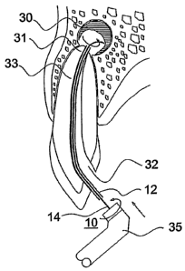
Fig. 1 illustrates one form of rotatable ablating device particularly useful
in
apparatus constructed in accordance with the present invention for removing
dental
periapical lesions. The ablating device 10 illustrated in Fig. 1 includes a
sleeve 12
sized and constructed for introduction via a cavity in the tooth (e.g., a
cavity drilled
through the crown of the tooth) into the tooth root canal, and for movement
therethrough to the apex of the root canal, as will be described more
particularly
below. Sleeve 12 includes a proximal end 12a and a distal end 12b. The latter
end is
to be located at the apex of the root canal having the dental periapical
lesion to be
removed.
The ablating device illustrated in Fig. 1 further includes a filament 14, also
having a proximal end 14a and a distal end 14b. As showii in Fig. 1, distal
end 14b of
filament 14 protrudes outwardly of distal end 12b of sleeve 12. Its protruding
end is
formed with a curvature, curving away from the longitudinal axis of the
filament and
CA 02627248 2008-04-24
WO 2007/057902 9 PCT/IL2006/001329
of the sleeve. As will be described more particularly below, the protruding
outwardly-curved end 14b of filament 14 is brought into contact with the
deiital
periapical lesion to be removed such that rotation of the, filament ablates
the dental
periapical lesion.
The proximal end 14a of filament 14 is fixed to a shank 16 whicll may have an
annular recess 18 to facilitate coupling the filament to a rotary drive, or be
coupled
using friction. In the ablating device illustrated in Fig. 1, filament 14 is
rotatable and
axially-displaceable with respect to sleeve 12.
Sleeve 12 is fabricated from a polymer, such Nylon, Pebax or Teflon, or a
metal, such as stainless steel or a super elastic alloy, such as superelastic
NiTinolTM.
Preferably, it has a length of about 12-40 mm, an external diameter of about
0.25-
0.9 mm, and an internal diameter of about 0.20-0.80 mm.
It will be appreciated that although sleeve 12 is illustrated as having a
single
lumen, a configuration having two or more separate lumens may also be used.
Such a
multi-lumen sleeve configuration can be used for aspiration, drug delivery, or
fiber
optic imaging. The sleeve may also have scales for measuring the depth of
penetration, and an anchoring mechanism (e.g. screw tip, oxidized section) for
anchoring sleeve 12 to a tissue (e.g. bone).
Filament 14 may also be fabricated from a polymer, such as Poly-p-
dioxanone, polylactyc acid or polyglycolic acid, or an alloy such as shape
memory
alloy NitinolTM. It preferably has a length of about 25-50 mm, and an external
diameter of about 0.25-0.80 mm. Filament 14 can be solid or hollow; if hollow,
an
internal diameter of about 0.1-0.7 mm is preferred. Filament 14 may be
fabricated
from a radio-opaque material, but if not, at least one radio-opaque marker can
be
added to the filament at equal intervals to -allow for X-ray location.
The outwardly-curved end portion 14b of filament 14 is typically 5-20% of
the filament length. It may be fabricated from the same material as the
remainder of
the filament, or from a different material (e.g. different hardness,
elasticity, etc).
Since end portion 14b is mechanically stressed by the rotary motion and by
contact
with body tissue, if fabricated from a polymer it is preferably fabricated
from a
biocompatible or bioresorbable polymer such that any fragments resulting from
its
disintegration are resorbed by the body.
End portion 14b can be fabricated in a round, square, tr-iangular, flat, star
or
any other cross-sectional shape suitable for tissue resection or grinding.
This end
CA 02627248 2008-04-24
WO 2007/057902 10 PCT/IL2006/001329
portion is preferably designed to angle or form a predetermined shape where
protruding from the sleeve distal end 12b when positioned within the body.
This can
be achieved by fabricating filament 14, or portion 14b thereof, from a shape
memory
polymer or alloy (e.g. NitinolTM) wliich is straight at room temperature and
angles to
produce a curved portion 14b when placed under temperatures higher than its
transformation temperature (e.g. body temperature). If it is a superelastic
alloy of
Nitinol, it can be forced to a straight shape by the sleeve, when inserted
into it.
As indicated earlier, filament 14 in the ablating device illustrated in Fig. 1
is
both rotatably and axially displaceable witli respect to sleeve 12. Fig. 2
illustrates an
ablating device, therein generally designated 20, also including a sleeve 22
enclosing
a filament 24, with the distal end 24b of the filament projecting from the
distal end
22b of the sleeve. In this case, however, both the filament 24 and the sleeve
22 are
secured to adaptor 26, such that both the sleeve and filament rotate together
with the
adaptor. In fabricating such an ablating device, the filament 24 may be passed
through the sleeve 22 until the distal end 24b of the filament projects
through the
distal end 22b of the sleeve to produce the desired curved end portion of the
filament,
and then the adaptor 26 may be crimped to bind the sleeve and filament to the
adaptor, such that the sleeve rotates with the filament.
The Fig. 2 construction is particularly useful where both the filament and the
sleeve are made of a polymer. The constructions and dimensions of the
protruding
end 24b of the filament may be such that it assumes the curved configuration
(shown
in broken lines in Fig. 2) by centrifugal force upon the rotation of the
filament.
Figs. 3 a-3m illustrate one manner of using the ablating device 10 of Fig.
1(or
20 of Fig. 2) for the removal of a dental periapical lesion, schematically
illustrated at
30 in those figures, located at the apex 31a of a canal 32 formed in a tooth
root 33.
Following a standard pulp chamber access and pulp removal, or removal of
infected root canal filling material from a prior failing treatment, the root
canal is
cleansed using files and liquid to remove all traces of pulp debris, bacteria
or root
canal filling material and the like. The apical foramen of root canal 32 is
then
reshaped and enlarged, using a file 34 to an ISO size of 40-120 (0.4-1.2 mm),
preferably size 60 (0.6 mm), as shown in Figs. 3a, 3b.
Following reshaping of the apical end of the root canal 32, the ablating
device
10 of Fig. 1 is then utilized for lesion removal. Sleeve 12 is first inserted
into the
reshaped root canal 32 to a working length (end of apex 31a), and filament 14
is then
CA 02627248 2008-04-24
WO 2007/057902 11 PCT/IL2006/001329
inserted through sleeve and into lesion 30, such that distal end portion 14b
of the
filament protrudes from the distal end of sleeve 12 (Figs. 3c, 3d).
When utilized for apical lesion removal, sleeve 12 and filament 14 can be
fabricated from a polymer or a metal (e.g. polymers such as nylon, PGA, PLA,
or
metal alloys such as NitinolTM). Filament 14 may have any desired cross
sectional
shape _(e.g., round, elliptical, flat, star-like, etc). If round, it
preferably has a typical
cross sectional diameter of 0.1-0.5 mm and a length of 20-40 mm. Filament 14
can
be solid or hollow and selected of any suitable Shore hardness (typically
Shore
hardness range A 10-90). A hollow configuration is preferred in cases where
provision of medication, such as a local anesthetic or a rinsing fluid, is
required,
although such rinsing or medication provision, as well as suction, can also be
effected
through a lumen in sleeve 12, or through a space formed between sleeve 12 and
filament 14.
The ablating device 10 is then connected to an electrical or pneumatic drill
head (dental handpiece) 35 (Fig. 3e), e.g. KAVO GentleSilence 8000, KAVO
intramatic E or Morita triautozx. Filament 14 is rotated within sleeve 12,
first at a low
speed (several hundred rpm) to enable initial ablation of granulation tissue
surrounding the root apex 31 a (Fig. 3 e). The rotational speed of filament 14
is then
gradually increased (up to 50,000 rpm), and both filament and sleeve are
advanced
(Figs. 3e-3h) in the direction of the lesion with an in-and-out motion, to
enable three
dimensional fine grinding of the tissues of the surrounding lesion 30.
Throughout the procedure, a liquid such as water or saline solution may be
utilized to wash the ground tissue, to assist in grinding, and to prevent
overheating.
Rinsing and suction can be conducted through filament 14, if hollow:
alternatively
filament 14 can be periodically removed, and rinsing/suction can be conducted
tlirough the sleeve. As a still further alternative, rinsing/suction can be
conducted
through a space between sleeve 12 and filament 14.
To enable three ditnensional grinding and coinplete removal of lesion 30, the
ablating device utilizes a filament 14 which angles when protruding through
its sleeve
12. Such angling can be controlled by the amount of filament protruding from
the
sleeve and by the rotational speed used. Alternatively, the filament, or at
least its end
portion, can be made of a material (e.g., NitinolTM) which is capable of
angling,
and/or of forming a shape such as a hook or loop when the end portion
protrudes from
sleeve 12.
CA 02627248 2008-04-24
WO 2007/057902 12 PCT/IL2006/001329
The root's apical portion 31a (Fig. 3h) can also be resected or ablated by
using
a filament 14 having a blade-like end portion 14b which curves back to form a
hook
once it protrudes from sleeve 12. Rotating this blade against apical portion
31 will
grind it off and tlius remove side canals which are a potential source of
infection.
Such root apex resection tends to improve healing and to reduce the chances of
re-
infection.
During or following the above-described ablation procedure, an X-ray
procedure can be used, by the addition of a radio-opaque guide positioned on
filament
14 or injected therethrough, to provide the dentist with information regarding
the size
of the periapical lesion and the extent of its removal. It can also provide a
reference
point for monitoring the healing phase.
In any case, once lesion 30 and surrounding tissue are removed, the ablation
device is removed, the lesion space and root canal are thoroughly rinsed and
the root
canal 32 is sealed (e.g. by using gutta percha and cement), and the crown is
restored.
The procedure may be carried out as a one-visit procedure or as a multiple-
visit one.
In case of a one-visit procedure all the above steps may be carried out. In
case of a
multi-visit procedure the initial stage of cleaning, shaping and disinfection
of the
infected root canal or removal of prior root canal filling, may be carried out
in the first
visit, followed by placement of a medicament (e.g. an antiseptic or
inflammatory
response modifier) in the root canal to be retained there until the second
visit, when
the periapical ablation procedure will be carried out, followed by a root
canal filling.
As another alternative, after lesion 30 and surrounding tissue have been
removed, various substances may be injected into the periapical space 36 (Fig.
3h)
through the sleeve 12 or hollow filament 14, in order to disinfect the region
and
accelerate bone growth/regeneration.
In this example, after lesion 30 with its tissue has been removed, a drill 37
(Fig. 3i), formed with a step or shoulder 37a is utilized to create a step or
shoulder
shown in Fig. 3j at 38 approximately 1 mm from the tip. This reshaping is
effected
such that the canal preferably tapers in a stepwise fashion towards the root
apex 31.
A prefabricated plug 40 having a shoulder 41a (Figs. 3k-3m) is then
positioned via a guide 42 against shoulder 38. Plug 40 can be composed of
mineral
trioxide aggregate (MTA), Titanium, NitinolTM, gutta percha, composite
material,
girconium, or any combination thereof and may be cemented therein, as shown at
43
(Fig. 31). Following plug positioning and its permanent cementation, guide 42
may be
CA 02627248 2008-04-24
WO 2007/057902 13 PCT/IL2006/001329
detached from plug 40 (Fig. 3m), and the root canal 32 is then obturated via
conventional methods.
The above-described procedure illustrates the use of a single ablating device,
such as 10 of Fig. 1 or 20 of Fig. 2, for removing a dental periapical lesion
at the apex
of a root of a tooth. Figs. 4a-l Ok illustrate the use of two such ablating
devices in a
two-step procedure for removing a dental periapical lesion at the apex of a
root of a
tooth, or for otlier applications involving removing or resecting tissue
enclosed within
a harder tissue, typically a diseased/infected/inflamed bone tissue enclosed
within a
healthy bone tissue, without damaging the surrounding tissue.
Such a procedure is performed in two consecutive steps: the first step
utilizes
an ablating device, such as shown at 50 in Figs. 4a-4d, including a Nitinol
superelastic sleeve or sheath 52 enclosing a sliape-memory or superelastic
Nitinol
filament 54; and the second step utilizes an ablating device, as shown at 60
in Fig. 5,
including a superelastic Nitinol sleeve or sheath 62 enclosing a filament 64
of an
elastic biocompatible or bioresorbable polymer, such as poly-dioxanone,
polyglycolic
acid or polyactyc acid.
In ablating device 50 (Figs. 4a-4d) used in the first step, the shape memory
Nitinol filament 54 is fixed to the shank 56 connectable to the rotary drive
(e.g., 35,
Fig. 3e), whereas the superelastic Nitinol sleeve 52 is freely mounted on
filament 54
for axial and rotatable movement with respect thereto. The shape memory
Nitinol
filament 54 has a transformation temperature slightly lower than body
temperature
(typically 25 C). When filament 54 is extended out of the constricting sleeve
52 and
exposed to body temperature, its distal end assumes a predetermined shape
comprising two arcs 54a, 54b which lie on planes orthogonal, or at an angle to
each
other and to the longitudinal axis of sleeve 52. alternatively, the filameiit
may be
constructed of a high elasticity or super elasticity material such as super
elastic
Nitinol TM, which is constricted at a straight shape by the sleeve, and
accepts its pre-
determined shape when release from the sleeve. Filament 54 is preferably of
circular
cross-section, with a blunt end facing a relatively sharp outer edge. The arcs
have a
radius of between 0.5-6 mm for various sizes of lesions.
In the first step, the sleeve 52 and the projecting end of the filament 54 are
rotated at low to medium speeds, of up to 1000 rpm (typically 30-1000 rpm).
This
assures that while the projecting end of the filament is extended into the
inflamed soft
tissue, the sharp edge is pushed forward to allow easy penetration. However,
when
CA 02627248 2008-04-24
WO 2007/057902 14 PCT/IL2006/001329
the filament is fully extended and rotated clockwise, the distal bend 54b
presents a
blunt edge which is deflected from the hard bone tissue, thereby assuring that
the
healthy bone tissue is not damaged during the rotation. Ablating device of
Figs. 4a-
4d is used in the first step to remove the inflamed tissue and/or to grind or
mince the
periapical lesion, before utilizing the ablating device 60, including the
polymer
filament 64, to be inserted for use in the second step in which the lesion is
removed.
In ablating device 60 used in the second step of the treatment, both the
polymer filament 64, and its sleeve 62, are attached to the adapter 66 so that
both
rotate together. In this case, ablating device 60 is rotated at a higher
speed, over
1,000 rpm (typically 14,000-50,000 rpm). At such speed, the centrifugal forces
acting on filament 64 cause it to deflect sideways. Since the polymer filament
64 is
relatively soft, it cannot penetrate the inflamed tissue. However, after the
tissue has
been initially ground by ablating device 50 (Figs. 4a-4d) utilizing the
Nitinol filament
54, the tissue is soft and fragmented enough to allow the penetration of
filament 64 of
ablating device 60 when the filament is rotated at high speed. Filament 64
thus
minces the already ground tissue to very fine particles that may be washed and
suctioned out through the apical foramen, as described above. Filament 64 is
biocompatible or bioresorbable, which ensures that when the filament wears and
tears
as a result of brushing against the hard bone tissue, the resulting filament
particles
will be resorbed by the body in a matter of a few weeks.
Figs. 6a-6d illustrate an ablating device, generally designated 50', of
basically
the same construction as ablating device 50 of Figs. 4a-4d, and therefore
corresponding parts are identified by the same reference numerals. In ablating
device
50' of Figs. 6a-6d, however, the Nitinol filament 54 has a third curved
section 54c at
its distal end, which is of a retrograde configuration, i.e., bent back
towards its
proximal end. Such a retrograde section of the filament allows reaching parts
of the
region that surround the tooth apex and which may otherwise be inaccessible to
the
ablator, as shown in Fig. 7.
As will be described more particularly below, ablating 50 (or 50'), including
the Nitinol filament 54, is used in the first step. When used in the first
step, its sleeve
52 is fixed by an adhesive to the tootli and stabilized, before the Nitinol
filament 54 is
rotated by its adaptor 56. To prevent the adhesive from entering the root
canal, a
protective cover is used, such as shown at 70 in Fig. 8. Such a protective
cover may
be made of thin aluminum foil to be placed over the crown of the tooth (71,
Fig. 9) to
CA 02627248 2008-04-24
WO 2007/057902 15 PCT/IL2006/001329
be treated, after an opening has been formed through the crown to provide
access to
the root canal. The ablating device 50 (or 50'), with the Nitinol filament 54
completely retracted within the sleeve 52, is passed through opening 72 in the
protective cover 70 into the root canal of the tooth, and is moved through the
root
canal to its position at the apex of the root canal. A glob of adliesive 74 is
then applied
over the protective cover 70 and the sleeve (Fig. 9), such that the adhesive
flows
between the tabs 73, and thereby binds the protective cover and the sleeve to
the
tooth. Such an arrangement has been found to firmly hold the sleeve 52 of the
ablating device to the tooth, allowing the filanient 54 to be advanced through
the
sleeve into contact with the periapical lesion to be removed, without clogging
the root
canal by the adhesive.
Figs. l0a-l Ok illustrate an example of a procedure that may be used,
utilizing
the metal-filament ablating device 50 of Figs. 4a-4d (or 50', of Figs. 6a-6d),
and the
polymer-filament ablating device 60 of Fig. 5, for removing a dental
periapical lesion
in accordance with the present invention. The protective cover 70, described
above
with respect to Figs. 8 and 9, is used in the first step of this procedure
with the metal-
filament ablating device 50 (or 50') to fix the outer sleeve 52 to the tooth,
before
deploying the metal filament 54.
1. The root canal 32 of the treated tooth is endodontically prepared by a
No. 45K file 78, to a working length 0.5 mm short of the apical foramen 31.
This
may preferably be done using a rotary LightSpeed file No. 45. (Fig. l Ob)
Patency
should be established using a No. 25K to 30K file 79 (Fig. l Oc). the
resulting shape of
the apical foramen is stepwise shoulder 38 (Fig. lOd)
2. After rinsing and drying the root canal, ablating device 50 (or 50'), with
its
Nitinol working filament 54 still contained and hidden within the Nitinol
sleeve 52, is
inserted to the working length (Fig. l0e).
3. The sleeve is fixed to the tooth and stabilized by placing a protective
cover
70 (Fig. 8) over the tooth 71 (Fig. 9), to cover the opening previously formed
through
its crown leading to the root canal to be treated, and applying a glob of
adhesive 74
over the outer surface of the protective cover and the sleeve. A viscous
adhesive,
such as glass ionomer composite, is used such that it assumes a semi-spherical
shape,
having a thickness of 1-2 mm at its center, and flows by surface tension in
spaces
between the radiating tabs 73. The adhesive used may be a settable dental
adhesive,
e.g., settable by ultraviolet light (Fig. 10e). As indicated earlier, such an
arrangement
CA 02627248 2008-04-24
WO 2007/057902 16 PCT/IL2006/001329
fixes the sheath of the ablating device to the tootli without danger of
clogging the root
canal with the adhesive.
4. The Nitinol filament 54 is then attached to the speed-controlled contra-
angle handpiece 75.
5. While liolding the handpiece gently, the user pushes the Nitinol filament
54 through the stabilized sleeve 52 and through the apical foramen into the
periapical
lesion 30 (Fig. 10f). When the distal curved ends 52a, 52b of Nitinol filament
52 are
out of the sleeve, the filament is easily moved back and forth, allowing the
operator to
know it has emerged from its sleeve.
6. The filament 54 is rotated at a speed of 200-300 rpm while the filament is
moved with in and out movements of 1-2 mm, for 30-60 seconds. The extent of
the
in and out movements can be judged from the distance between the coronal end
of the
sleeve and the handpiece. A rubber stopper placed on the rotating part may
help this
judgment.
7. The filament is retracted through the sleeve, and the coronal fixation is
then gently removed by breaking off the adhesive, and removing the protective
cover
from the tooth and the ablating device 50 out of the root canal (Fig. l Og).
8. The root canal may then be rinsed with saline solution or distilled water
using a small diameter (30-gauge or thinner) needle, inserted through the
apex, such
that some of the debris is flushed out with the back-flow.
9. Ablating device 60 (Figs. 5a-5d) is then measured and its polymer
filament 64 is cut to the proper length. Its curved protruding end 64a should
be 1-
3 mm longer than the estimated diameter of the treated periapical lesion 30.
10. Ablating device 60 is then attaclied to the handpiece and gently inserted
into the root canal, until its metal sleeve 62 reaches the apical stop, wliile
its polymer
filament 64 slides through the apical foramen and into the roughly minced
periapical
lesion 36a (Fig. lOh).
11. Ablating device 60 is then rotated at 15,000-50,000 rpm, for 20-60
seconds, with slight in and out motion, and then taken out of the root canal.
12. The finely minced content of the periapical crypt 36b is then rinsed out
with copious amounts of normal saline solution or distilled water, using a 30-
32 G
needle 76 attached to a syringe 80 (Fig. l0i).
13. The root canal is then dried, using paper points (Fig. 10j), followed by
root
canal obturation 32a (Fig. lOj).
CA 02627248 2008-04-24
WO 2007/057902 17 PCT/IL2006/001329
14. Within several months (2-6), the bone around the bony crypt grows into
the empty space 3 6e, resulting in full recovery (Fig. 10k).
Figs. 11 a and 11b illustrate another construction of ablating device in
accordance with the present invention. The ablating device illustrated in
Figs. 11 a
and 11b, and tlierein generally designated 80, also includes a sleeve 82
having a
proximal end 82a and a distal end 82b, and a filament 84 within the sleeve and
also
having a proximal end 84a and a distal end 84b. In this case, however, the
distal end
84b of filament 84 is secured to the distal end 82b of the sleeve 82, as shown
at 85. In
addition, the distal end. of sleeve 82 is formed with a plurality of slits 86
extending
generally axially, and preferably slightly angularly, with respect to the
longitudinal
axis of the sleeve (Fig. 11 a). The proximal end 82a of the sleeve is
displaceable
towards its distal end 82b and the distal end 84b of the filament fixed
thereto. This
forces the distal end 82b of the sleeve to be bowed outwardly along the slits
86 to
thereby define a plurality of outwardly-bowed strips or surfaces 87 effective,
upon
rotation of the sleeve, to ablate a substance with which the ablating surfaces
87 are in
contact (Fig. 11 b).
In addition, the proximal end 82a of sleeve 82 is formed with a longitudinally-
extending slot 88, and the proximal end 84a of filament 84 is formed with a
pin 89
received in slot 88 for guiding the displacement of the sleeve with respect to
the
filament to produce the outwardly-bowed ablating surfaces 87.
Ablating device 80 illustrated in Figs. 1 la and 11b can also be constructed,
as
described above, for removing a dental periapical lesion at ail apex of a root
canal in a
tooth. Thus, sleeve 82 may be constructed such that, in its original condition
illustrated in Fig. 11, it may be introduced via an opening through the tooth,
into the
root canal and moved therethrough, and through the apex of the root canal,
into
contact with the dental periapical lesion. Sleeve 82 may then be displaced
towards its
distal end fixed at 85 to filament 84, to thereby force the distal end of the
sleeve to be
bowed outwardly along the slits 86, and to define the plurality of outwardly-
bowed
strips or surfaces 87, shown in Fig. 11b, effective to ablate the dental
periapical lesion
when the sleeve is rotated.
Fig. 12 more particularly illustrates the overall apparatus using ablating
device
80 for removing tissue, e.g. a dental periapical lesion, in the manner
described above.
Thus, as shown in Fig. 12, the overall apparatus includes a rotary drive unit
95
which is coupled to filament 84 to rotate the filament, and thereby also to
rotate sleeve
CA 02627248 2008-04-24
WO 2007/057902 18 PCT/IL2006/001329
82 via a coupling device, rotatably mounted within a fixture 91, coupling
these two
elements. The illustrated apparatus further includes an outer sleeve 92
rotatably
receiving the rotatable elements 82, 84 of the ablating device 80 so as to
serve as a
guide or hand grip for the ablating device.
As shown in Fig. 12, the apparatus further includes an aspirator 93 or other
suction device coupled to the ablating device 80 via fixture 91 for drawing-
out the
debris and/or for rinsing the ablated region. Fixture 91 may also be provided
wit11 a
handle 94 to facilitate holding and manipulating the ablating device. In this
case, both
the sleeve 82 and filament 84 are connected to the rotary drive 95. The
outwardly-
bowed distal end of the sleeve define the ablating surfaces 87 wllich ablate
the tissue.
The apparatus illustrated in Fig. 12 can also be used in bone harvesting and
collection procedures, e.g., for harvesting bone tissue from a hip bone as
illustrated in
Fig. 13. Prior to harvesting, an operator inserts a 2 mm guide wire (Synthes
292.65)
into the iliac crest 3 cm lateral to the ASIS, and drills over the guide wire
with a
4.5 cannulated drill (Synthes 310.69) to a depth of 1 cm.
The operator then inserts sleeve 92 which serves as a working channel. In this
configuration of the ablator device 80, sleeve 92 has an outer diameter of 4.5
min, a
screw tip with a positive stop, and an inner cannulated trocar having an outer
diameter
of 3.2 mm and an inner diameter of 2 mm.
Sleeve 92 is secured to hip bone 96 via the screw tip, and the trocar is
removed. Sleeve 82 of the ablator device 80 is then inserted through sleeve 92
and
connected to drill head 95.
In this configuration of the ablator device, sleeve 82 is designed as a semi-
flexible shaft having a cutting portion (i.e., the ablating elements 87 of
sleeve 82)
which will not penetrate the thin cortical bone but will mince spongy bone
material.
The rotational speed (RPM), and also the configuration of the ablating
elements 87, are selected such that thin cortex is not damaged, and the
temperature of
minced tissues does not rise above 42 C. This ensures that cells and bony
trabeculi of
the harvested bone material do not suffer any thermal or mechanical damage. A
typical cutting speed is preferable, is selected from a range of 500 to 800
rpm.
The ablating elements 87 are preferably configured such that during cutting,
the generated bone and tissue fragments are evacuated from the site of
cutting. For
example, in the configuration of Figs. 1 ia, I lb, the reverse spirals of the
ablating
elements 87 facilitate bone and tissue fragment evacuation.
CA 02627248 2008-04-24
WO 2007/057902 19 PCT/IL2006/001329
Collection of bone/tissue material (paste) can be effected through hollow
sleeve 92. The bone paste collected can be stored in a sterile container
attached to the
aspirator.
While the invention has been described above with respect to several preferred
embodiments, it will be appreciated that these are set forth merely for
purposes of
example, and that many other variations, modif cations and applications of the
invention may be made.