Note: Descriptions are shown in the official language in which they were submitted.
DEMANDES OU BREVETS VOLUMINEUX
LA PRESENTE PARTIE I)E CETTE DEMANDE OU CE BREVETS
COMPREND PLUS D'UN TOME.
CECI EST LE TOME DE _2
NOTE: Pour les tomes additionels, veillez contacter le Bureau Canadien des
Brevets.
JUMBO APPLICATIONS / PATENTS
THIS SECTION OF THE APPLICATION / PATENT CONTAINS MORE
THAN ONE VOLUME.
THIS IS VOLUME 1 OF 2
NOTE: For additional volumes please contact the Canadian Patent Office.
CA 02627427 2008-04-25
WO 2007/053447 PCT/US2006/042027
1
COMPOSITIONS AND METHODS FOR THE TREATMENT AND PREVENTION OF
FIBROTIC, INFLAMMATORY AND NEOVASCULARIZATION CONDITIONS
RELATED APPLICATIONS
This application claims priority to, the benefit of, and incorporates by
reference for all
purposes the following patent-related documents, each in its entirety: U.S.
provisional patent
application Ser. No. XX/XXX,XXX [attorney docket no LPT-3010-PV, entitled
"Compositions
and Methods for Binding Sphingosine-l-Phosphate"], and U.S. provisional patent
application
Ser. No. XX/XXX,XXX [attorney docket no LPT-3020-PV, entitled "Humanized
Antibodies to
Sphingosine-l-Phosphate in the Treatment of Ocular Disorders"], both filed
concurrently with
the instant application; U.S. provisional patent application Ser. No.
XX/XXX,XXX [attorney
docket no LPT-3100-PV2], filed 12 August, 2006, and U.S. patent application
Ser. No.
11/261,935, filed 28 October, 2005, of which this application is a
continuation-in-part.
TECHNICAL FIELD
The present invention relates to methods of treatments for ocular disorders
using
immune-derived moieties which are reactive against bioactive lipid molecules
that play role in
human and/or animal disease as signaling molecules. One particular class of
signaling bioactive
lipids considered in accordance with the invention is lysolipids. Particularly
preferred signaling
lysolipids are sphingosine-1-phosphate (S 1P) and the various lysophosphatidic
acids (LPAs).
Antibodies against signaling lipids, and derivatives and variants thereof, can
be used in the
treatment and/or prevention of ocular diseases or disorders through the
delivery of
pharmaceutical compositions that contain such antibodies, alone or in
combination with other
therapeutic agents and/or treatments.
BACKGROUND OF THE INVENTION
I. Introduction
The following description includes information that may be useful in
understanding the
present invention. It is not an admission that any such information is prior
art, or relevant, to the
presently claimed inventions, or that any publication specifically or
implicitly referenced is prior
art or even particularly relevant to the presently claimed invention.
CA 02627427 2008-04-25
WO 2007/053447 PCT/US2006/042027
2
II. Back rg ound
The present invention relates to methods of decreasing or attenuating aberrant
neovascularization, angiogenesis, aberrant fibrogenesis, fibrosis and
scarring, and inflammation
and immune responses. These processes, separately or together are involved in
many diseases
and conditions. These diseases or conditions may be systemic or may be
relatively localized, for
example to the skin or to the eye.
A. Ocular diseases and conditions
Pathologic or aberrant angiogenesis/neovascularization, aberrant remodeling,
fibrosis and
scarring and inflammation occur in association with retinal and ocular
ischemic diseases such as
age-related macular degeneration (AMD), diabetic retinopathy (DR) and in
retinopathy of
prematurity (ROP) and other developmental disorders [Eichler et al. (2006),
Curr Pharm Des, vol
12: 2645-60] as well as being a result of infections and mechanical injury to
the eye [Ciulla et al.
(2001), Curr Opin Ophthalmol, vol 12: 442-9 and Dart et al (2003), Eye, vol
17: 886-92].
Pathologic ocular angiogenesis is a leading cause of blindness in a variety of
clinical
conditions. Choroidal neovascularization (CNV) occurs in a number of ocular
diseases, the most
prevalent of which is the exudative or "wet" form of AMD. As a result of an
increasingly aged
population, AMD is a modern day epidemic and the leading cause of blindness in
the western
world in patients over age 60. Despite the epidemic of vision loss caused by
AMD, only a few
therapies, mostly anti-VEGF based, can slow the progression of AMD and even
fewer can
reverse vision loss [Bylsma and Guymer (2005), Clin Exp Optom,. vol 88: 322-
34, Gryziewicz
(2005), Adv Drug Deliv Rev, vo157: 2092-8 and Liu and Regillo (2004), Curr
Opin Ophthalmol,
vol 15: 221-6.]. Therefore, discovering new treatments for pathologic
neovascularization is
extremely important.
AMD is used here solely for illustrative purposes in describing ocular
conditions relating
to aberrant angiogenesis/neovascularization, aberrant remodeling, fibrosis and
scarring, and
inflammation, which conditions are found in other ocular diseases and
disorders as disclosed and
claimed herein. AMD involves age-related pathologic changes [Tezel, Bora and
Kaplan (2004),
Trends Mol Med, vol 10: 417-20 and Zarbin (2004), Arch Ophthalmol, 122: 598-
614]. Multiple
theories exist but, the exact etiology and pathogenesis of AMD are still not
well understood.
Aging is associated with cumulative oxidative injury, thickening of Bruch's
membrane and
drusen formation. Oxidative stress results in injury to retinal pigment
epithelial (RPE) cells and,
CA 02627427 2008-04-25
WO 2007/053447 PCT/US2006/042027
3
in some cases, the choriocapillaris [Zarbin (2004), Arch Ophthalmol, vol 122:
598-614 and
Gorin et al. (1999), Mol Vis,. vol 5: 29]. Injury to RPE likely elicits a
chronic inflammatory
response within Bruchs membrane and the choroid [Johnson et al. (2000), Exp
Eye Res,. vo170:
441-9]. This injury and inflammation fosters and potentates retinal damage by
stimulating CNV
and atrophy [Zarbin (2004), Arch Ophthalmol, vol 122: 598-614 and Witmer et
al. (2003), Prog
Retin Eye Res, vo122: 1-29]. CNV results in defective and leaky blood vessels
(BV) that are
likely to be recognized as a wound [Kent and Sheridan (2003), Mol Vis, vol 9:
747-55]. Wound
healing arises from the choroid and invades the subretinal space through
Bruchs membrane and
the RPE. Wound healing responses are characterized by a typical early
inflammation response, a
prominent angiogenic response and tissue formation followed by end-stage
maturation of all
involved elements. Wound remodeling may irreversibly compromise photoreceptors
and RPEs
thereby, justifying the need to treat CNV with more than anti-angiogenic
therapies [La Cour,
Kiilgaard and Nissen (2002), Drugs Aging, vol 19: 101-33.12].
Alterations in the normal retinal and sub-retinal architecture as a result of
CNV related
fibrosis, edema and inflammation individually or cumulatively, leads to AMD
related visual loss
[Tezel and Kaplan (2004), Trends Mol Med, vol 10: 417-20 and Ambati et al.
(2003), Surv
Ophthalmol, vo148: 257-93]. The multiple cellular and cytokine interactions
which are
associated with exudative AMD greatly complicate the search for effective
treatments. While
CNV and edema are manageable in part by anti-VEGF therapeutics, potential
treatments to
mitigate scar formation and inflammation have not been adequately addressed
[Bylsma and
Guymer (2005), Clin Exp Optom, vo188: 322-34 and Pauleikhoff (2005), Retina,
vol 25: 1065-
84]. As long as the neovascular complex remains intact, as appears to be the
case in patients
treated with anti-VEGF agents, the potential for subretinal fibrosis and
future vision loss persists.
Anti-VEGF-A therapies represent a recent, significant advance in the treatment
of
exudative AMD. However, the phase III VISION Trial with PEGAPTANIB, a high
affinity
aptamer which selectively inhibits the 165 isoform of VEGF-A, demonstrated
that the average
patient continues to lose vision and only a small percent gained vision
[Gragoudas et al. (2004),
N Engl J Med, vo1351: 2805-16]. Inhibition of all isoforms of VEGF-A (pan-VEGF
inhibition)
with the antibody fragment RANIBIZUMAB yielded much more impressive results
[Brown et
al. N Eng Med,2006 355:1432-44, Rosenfeld et al. N Eng J Med 2006355:1419-31].
The 2 year
MARINA trial and the 1 year ANCHOR trial demonstrated that approximately 40%
of patients
achieve some visual gain. Although these results represent a major advance in
our ability to treat
exudative AMD, they also demonstrate that 60% of patients do not have visual
improvement.
CA 02627427 2008-04-25
WO 2007/053447 PCT/US2006/042027
4
Furthermore, these patients had to meet strictly defined inclusion and
exclusion criteria. The
results in a larger patient population may be less robust.
There is still a well defined need to develop further therapeutic agents that
target other
steps in the development of CNV and the processes that ultimately lead to
photoreceptor
destruction. First, the growth of choroidal BVs involves an orchestrated
interaction among many
mediators, not just VEGF, offering an opportunity to modulate or inhibit the
entire process
[Gragoudas et al. (2004), N Engl J Med, vo1351: 2805-16]. Second, exudative
AMD is
comprised of vascular and extravascular components. The vascular component
involves vascular
endothelial cells (EC), EC precursors and pericytes. The extravascular
component, which
volumetrically appears to be the largest component, is composed of
inflammatory, glial and
retinal pigment epithelium (RPE) cells and fibroblasts. Tissue damage can
result from either
component. These other aspects of the pathologic process are not addressed by
current anti-
VEGF treatments. Targeting additional elements of the angiogenic cascade
associated with
AMD could provide a more effective and synergistic approach to therapy [Spaide
RF (2006),
Am J Ophthalmol, vol 141: 149-156].
1. Inflammation in ocular disease
There is increasing evidence that inflammation, specifically macrophages and
the
complement system [Klein et al. (2005), Science, vol 308: 385-9 and Hageman et
al.(2005), Proc
Natl Acad Sci U S A, vol 102: 7227-32] play an important role in the
pathogenesis of exudative
AMD. Histopathology of surgically excised choroidal neovascular membranes
demonstrates that
macrophages are almost universally present [Grossniklaus, et al.(1994),
Ophthalmology, vol
101: 1099-111 and Grossniklaus et al. (2002), Mol Vis, vo18: 119-26]. There is
mounting
evidence that macrophages may play an active role in mediating CNV formation
and propagation
[Grossniklaus et al. (2003), Mol Vis, vo18: 119-26; Espinosa-Heidmann, et al.
(2003), Invest
Ophthalmol Vis Sci, vo144: 3586-92; Oh et al. (1999), Invest Ophthalmol Vis
Sci, vo140:
1891-8; Cousins et al. (2004), Arch Ophthalmol, vol 122: 1013-8; Forrester
(2003), Nat Med,
vo19: 1350-1 and Tsutsumi et al. (2003), J Leukoc Biol, vol 74: 25-32] by
multiple effects
which include secretion of enzymes that can damage cells and degrade Bruchs
membrane as well
as release pro-angiogenic cytokines [Otani et al. (1999), Ophthalmol Vis Sci,
vo140: 1912-20
and Amin, Puklin and Frank (1994), Invest Ophthalmol Vis Sci, vo135: 3178-88]
At the site of
injury, macrophages exhibit micro-morphological signs of activation, such as
degranulation [Oh
et al. (1999), Invest Ophthalmol Vis Sci, vol 40: 1891-8 and Trautmann et al.
(2000), J Pathol,
CA 02627427 2008-04-25
WO 2007/053447 PCT/US2006/042027
vol 190: 100-6]. Thus it is believed that a molecule which limited macrophage
infiltration into to
the choroidal neovascular complex may help limit CNV formation.
2. Choroidal neovascularization and blood vessel maturation in ocular disease
Angiogenesis is an essential component of normal wound healing as it delivers
oxygen
and nutrients to inflammatory cells and assists in debris removal [Lingen
(2001), Arch Pathol
Lab Med, vol 125: 67-71]. Progressive angiogenesis is composed of two distinct
processes:
Stage I: Migration of vascular ECs, in response to nearby stimuli, to the tips
of the capillaries
where they proliferate and form luminal structures; and Stage II: Pruning of
the vessel network
and optimization of the vasculature [Guo et al. (2003), Am J Pathol, vol 162:
1083-93].
Stage I: Neovascularization. Angiogenesis most often aids wound healing.
However,
new vessels when uncontrolled, are commonly defective and promote leakage,
hemorrhaging
and inflammation. Diminishing dysfunctional and leaky BVs, by targeting pro-
angiogenic GFs,
has demonstrated some ability to slow the progression of AMD [Pauleikhoff
(2005), Retina, vol
25: 1065-84.14 and van Wijngaarden, Coster and Williams (2005), JAMA, vol 293:
1509-13].
Stage II: Blood vessel maturation and drug desensitization. Pan-VEGF
inhibition
appears to exert its beneficial effect mostly via an anti-permeability action
resulting in resolution
of intra- and sub-retinal edema, as the actual CNV lesion does not markedly
involute
[Presentation. at Angiogenesis 2006 Meeting. 2006. Bascom Palmer Eye Institute
Miami,
Florida]. The lack of marked CNV involution may in part be a result of
maturation of the newly
formed vessels due to pericyte coverage. Pericytes play a critical role in the
development and
maintenance of vascular tissue. The presence of pericytes seems to confer a
resistance to anti-
VEGF agents and compromise their ability to inhibit angiogenesis [Bergers and
Song (2005),
Neuro-oncol, vol 7: 452-64; Yamagishi and Imaizumi (2005), Int J Tissue React,
vol 27: 125-35;
Armulik, Abramsson and Betsholtz (2005), Circ Res, vol 97: 512-23; Ishibashi
et al. (1995),
Arch Ophthalmol, vol 113: 227-31]. An agent which has an inhibitory effect on
pericyte
recruitment would likely disrupt vascular channel assembly and the maturation
of the choroidal
neovascular channels thereby perpetuating their sensitivity to anti-angiogenic
agents.
Remodeling of the vascular network involves adjustments in BV density to meet
nutritional needs [Gariano and Gardner (2005), Nature, 438: 960-6]. Periods of
BV immaturity
corresponds to a period in which new vessels are functioning but have not yet
acquired a pericyte
coating [Benjamin, Hemo and Keshet (1998), Development, 125: 1591-8 and
Gerhardt and
CA 02627427 2008-04-25
WO 2007/053447 PCT/US2006/042027
6
Betsholtz (2003), Cell Tissue Res, 2003. 314: 15-23]. This delay is essential
in providing a
window of plasticity for the fine tuning of the developing vasculature
according to the nutritional
needs of the retina or choroid.
The bioactive lipid sphingosine-l-phosphate (SIP), VEGF, PDGF, angiopoietins
(Ang)
and other growth factors (GF) augment blood vessel growth and recruit smooth
muscle cells
(SMC) and pericytes to naive vessels which promote the remodeling of emerging
vessels
[Allende and Proia (2002), Biochim Biophys Acta, vo1582: 222-7; Gariano and
Gardner (2005),
Nature, vo1438: 960-6; Grosskreutz et al. (1999), Microvasc Res, vol 58: 128-
36; Nishishita,
and Lin (2004), J Cell Biochem, vo191: 584-93 and Erber et al. (2004), FASEB
J, vol 18: 338-
40.32]. Pericytes, most likely generated by in situ differentiation of
mesenchymal precursors at
the time of EC sprouting or from the migration and de-differentiation of
arterial smooth muscle
cells, intimately associate and ensheath ECs resulting in overall vascular
maturity and survival
[Benjamin, Hemo and Keshet (1998), Development, vol 125: 1591-8]. Recent
studies have
demonstrated that S 1P, and the S 1P 1 receptor, are involved in cell-surface
trafficking and
activation of the cell-cell adhesion molecule N-cadherin [Paik et al. (2004),
Genes Dev, vol 18:
2392-403]. N-cadherin is essential for interactions between EC, pericytes and
mural cells which
promote the development of a stable vascular bed [Gerhardt and Betsholtz
(2003), Cell Tissue
Res, vol 314: 15-23]. Global deletion of the S1P1 gene results in aberrant
mural cell
ensheathment of nascent BVs required for BV stabilization during embryonic
development
[Allende and Proia (2002), Biochim Biophys Acta, vol 1582: 222-7]. Local
injection of siRNA
to S 1P1 suppresses vascular stabilization in tumor xenograft models [Chae et
al. (2004), J Clin
Invest, vol 114: 1082-9]. Transgenic mouse studies have demonstrated that VEGF
and PDGF-B
promote the maturation and stabilization of new BVs [Guo et al. (2003), Am J
Pathol, 162:
1083-93 and Gariano and Gardner (2005), Nature, vo1438: 960-6.50]. VEGF up-
regulates Ang-1
(mRNA and protein) [Asahara et al. (1998), Circ Res, vol 83: 233-40]. Ang-1
plays a major role
in recruiting and sustaining peri-endothelial support by pericytes [Asahara et
al. (1998), Circ
Res, vol 83: 233-40]. Intraocular injection of VEGF accelerated pericyte
coverage of the EC
plexus [Benjamin, Hemo and Keshet (1998), Development, vol 125: 1591-8]. PDGF-
B deficient
mouse embryos lack micro-vascular pericytes, which leads to edema, micro-
aneurisms and lethal
hemorrhages [Lindahl et al. (1997), Science, vo1277: 242-5]. Murine pre-natal
studies have
demonstrated that additional signals are required for complete VEGF- and PDGF-
stimulation of
vascular bed maturation. Based upon the trans-activation of S 1P noted above,
this factor could
be S 1P [Erber et al. (2004), FASEB J, vol 18: 338-40]. Vessel stabilization
and maturation is
associated with a loss of plasticity and the absence of regression to VEGF and
other GF
CA 02627427 2008-04-25
WO 2007/053447 PCT/US2006/042027
7
withdrawal and resistance to anti-angiogenic therapies [Erber et al. (2004),
FASEB J, vol 18:
338-40 and Hughes. and Chan-Ling (2004), Invest Ophthalmol Vis Sci, vo145:
2795-8061.
Resistance of BVs to angiogenic inhibitors is conferred by pericytes that
initially stabilize
matured vessels and those that are recruited to immature vessels upon therapy
[Erber et al.
(2004), FASEB J, vol 18: 338-40]. After ensheathment of the immature ECs, the
pericytes
express compensatory survival factors (Ang-1 and PDGF-B) that protect ECs from
pro-apoptotic
agents.
3. Edema and vascular permeability
CNV membranes are composed of fenestrated vascular ECs that tend to leak their
intravascular contents into the surrounding space resulting in subretinal
hemorrhage, exudates
and fluid accumulation [Gerhardt and Betsholtz (2003), Cell Tissue Res, vol
14: 15-23]. For
many years the CNV tissue itself, and more recently intra-retinal
neovascularization, have been
implicated as being responsible for the decrease in visual acuity associated
with AMD. It is now
thought however, that macular edema caused by an increase in vascular
permeability (VP) and
subsequent breakdown of the blood retinal barrier (BRB), plays a major role in
vision loss
associated with AMD and other ocular diseases [Hughes and Chan-Ling (2004),
Invest
Ophthalmol Vis Sci, vo145: 2795-806; Felinski and Antonetti (2005), Curr Eye
Res, vol 30:
949-57; Joussen et al. (2003), FASEB J, vol 17: 76-8 and Strom et al. (2005),
Invest Ophthalmol
Vis Sci, vol 46: 3855-8].
4. Fibrosis, fibrogenesis and scar formation
The formation of subretinal fibrosis leads to irreversible damage to the
photoreceptors
and permanent vision loss. As long as the neovascular complex remains intact,
as appears to be
the case in patients treated with anti-VEGF agents, the potential for
subretinal fibrosis and future
vision loss persists. In an update of the PRONTO study of RANIBIZUMAB, it was
discovered
that those patients who lost vision did so as, a result of either subretinal
fibrosis or a RPE tear
[Presentation. at Angiogenesis 2006 Meeting. 2006. Bascom Palmer Eye Institute
Miami,
Florida.]. An agent that could diminish the degree of fibroblast infiltration
and collagen
deposition would likely be of value.
Fibroblasts, particularly myofibroblasts, are key cellular elements in scar
formation in
response to cellular injury and inflammation [Tomasek et al. (2002), Nat Rev
Mol Cell Biol, vol
3: 349-63 and Virag and Murry (2003), Am J Pathol, vol 163: 2433-40]. Collagen
gene
CA 02627427 2008-04-25
WO 2007/053447 PCT/US2006/042027
8
expression by myofibroblasts is a hallmark of remodeling and necessary for
scar formation [Sun
and Weber (2000), Cardiovasc Res, vo146: 250-6 and Sun and Weber (1996), J Mol
Cell
Cardiol, vo128: 851-8]. S 1P promotes wound healing by activating fibroblast
migration and
proliferation while increasing collagen production [Sun et al. (1994), J Biol
Chem, vo1269:
165 12-7]. S 1P produced locally by damaged cells could be responsible for the
maladaptive
wound healing associated with remodeling and scar formation. Thus it is
believed that S 1P
inhibitors are useful in diseases or conditions characterized, at least in
part, by aberrant
fibrogenesis or fibrosis. Herein, "fibrogenesis" is defined as excessive
activity or number of
fibroblasts, and "fibrosis" is defined as excessive activity or number of
fibroblasts that leads to
excessive or inappropriate collagen production and scarring, destruction of
the physiological
tissue structure and/or inappropriate contraction of the matrix leading to
such pathologies as
retinal detachment or other processes leading to impairment of organ function.
B. Other diseases or conditions
The role of bioactive signaling lipids such as S 1P and LPA is not limited to
ocular
diseases and conditions. Because of the involvement of biolipid signaling in
many processes,
including neovascularization, angiogenesis, aberrant fibrogenesis, fibrosis
and scarring, and
inflammation and immune responses, it is believed that antibody-based
inhibitors of these
bioactive lipids will be helpful in a variety of diseases and conditions
associated with one or
more of these processes. Such diseases and conditions may be systemic (e.g.,
systemic
scleroderma) or localized to one or more specific body parts or organs (e.g.,
skin, lung, or eye).
C. Bioactive si ng aling lipids
Lipids and their derivatives are now recognized as important targets for
medical research,
not as just simple structural elements in cell membranes or as a source of
energy for (3-oxidation,
glycolysis or other metabolic processes. In particular, certain bioactive
lipids function as
signaling mediators important in animal and human disease. Although most of
the lipids of the
plasma membrane play an exclusively structural role, a small proportion of
them are involved in
relaying extracellular stimuli into cells. "Lipid signaling" refers to any of
a number of cellular
signal transduction pathways that use cell membrane lipids as second
messengers, as well as
referring to direct interaction of a lipid signaling molecule with its own
specific receptor. Lipid
signaling pathways are activated by a variety of extracellular stimuli,
ranging from growth
factors to inflammatory cytokines, and regulate cell fate decisions such as
apoptosis,
differentiation and proliferation. Research into bioactive lipid signaling is
an area of intense
CA 02627427 2008-04-25
WO 2007/053447 PCT/US2006/042027
9
scientific investigation as more and more bioactive lipids are identified and
their actions
characterized.
Examples of bioactive lipids include the eicosanoids (including the
cannabinoids,
leukotrienes, prostaglandins, lipoxins, epoxyeicosatrienoic acids, and
isoeicosanoids), non-
eicosanoid cannabinoid mediators, phospholipids and their derivatives such as
phosphatidic acid
(PA) and phosphatidylglycerol (PG), platelet activating factor (PAF) and
cardiolipins as well as
lysophospholipids such as lysophosphatidyl choline (LPC) and various
lysophosphatidic acids
(LPA). Bioactive signaling lipid mediators also include the sphingolipids such
as
sphingomyelin, ceramide, ceramide- 1 -phosphate, sphingosine,
sphingosylphosphoryl choline,
sphinganine, sphinganine-l-phosphate (Dihydro-S 1P) and sphingosine- 1 -
phosphate.
Sphingolipids and their derivatives represent a group of extracellular and
intracellular signaling
molecules with pleiotropic effects on important cellular processes. Other
examples of bioactive
signaling lipids include phosphatidylserine (PS), phosphatidylinositol (PI),
phosphatidylethanolamine (PEA), diacylglyceride (DG), sulfatides,
gangliosides, and
cerebrosides.
D. Lysolipids
Lysophospholipids (LPLs), also known as lysolipids, are low molecular weight
(typically
less than about 500 dalton) lipids that contain a single hydrocarbon backbone
and a polar head
group containing a phosphate group. Some lysolipids are bioactive signaling
lipids. Two
particular examples of medically important bioactive lysolipids are LPA
(glycerol backbone) and
S IP (sphingoid backbone). The structures of selected LPAs, S 1P, and dihydro
S IP are presented
below.
CA 02627427 2008-04-25
WO 2007/053447 PCT/US2006/042027
1 o 0 0 0 ? ?
~ j~o ~o o,j~o Flo F~'j~o i põj~o Hd o
HO H HO ~..~ Ho H HO H HO H H H
'OH 'CH CH 'CH 'O-I ~i iiiiii
LPA (20:4) LPA (16:0) LPA (18:2) LPA ('18:1) IpA (y8:0) Si P D7hydo-S1 P
LPA is not a single molecular entity but a collection of endogenous structural
variants
with fatty acids of varied lengths and degrees of saturation (Fujiwara et al
(2005), J Biol Chem,
vol. 280: 35038-35050). The structural backbone of the LPAs is derived from
glycerol-based
phospholipids such as phosphatidylcholine (PC) or phosphatidic acid (PA). In
the case of
lysosphingolipids such as S1P, the fatty acid of the ceramide backbone is
missing. The structural
backbone of S1P, dihydro SlP (DHS1P), and sphingosylphosphorylcholine (SPC) is
based on
sphingosine, which is derived from sphingomyelin.
LPA and SlP regulate various cellular signaling pathways by binding to the
same class of
multiple transmembrane domain G protein-coupled (GPCR) receptors (Chun J,
Rosen H (2006),
Current Pharm Des, vol. 12: 161-171 and Moolenaar WH (1999), Experimental Cell
Research,
vol. 253: 230-238). The S 1P receptors are designated as S 1P1, S 1P2, S 1P3,
S 1P4 and S 1P5
(formerly EDG-1, EDG-5/AGR16, EDG-3, EDG-6 and EDG-8) and the LPA receptors
designated as LPAI, LPA2, LPA3 (formerly, EDG-2, EDG-4, and EDG-7). A fourth
LPA
receptor of this family has been identified for LPA (LPA4), and other putative
receptors for these
lysophospholipids have also been reported.
E. Sphinaosine-l-phosphate
S 1P is a mediator of cell proliferation and protects from apoptosis through
the activation
of survival pathways (Maceyka et al. (2002), BBA, vol 1585): 192-201 and
Spiegel S. et al.
CA 02627427 2008-04-25
WO 2007/053447 PCT/US2006/042027
11
(2003), Nature Reviews Molecular Cell Biology, vol 4: 397-407). It has been
proposed that the
balance between ceramide/sphingosine (CER/SPH) levels and S1P provides a
rheostat
mechanism that decides whether a cell is directed into the death pathway or is
protected from
apoptosis. The key regulatory enzyme of the rheostat mechanism is sphingosine
kinase (SPHK)
whose role is to convert the death-promoting bioactive signaling lipids
(CER/SPH) into the
growth-promoting S 1P. S 1P has two fates: S 1P can be degraded by S 1P lyase,
an enzyme that
cleaves S1P to phosphoethanolamine and hexadecanal, or, less common,
hydrolyzed by S1P
phosphatase to SPH. S 1P is abundantly generated and stored in platelets,
which contain high
levels of SPHK and lacks the enzymes for S1P degradation. When platelets are
activated, SIP is
secreted. In addition, other cell types, for example, mast cells, are also
believed to be capable of
secreting S iP. Once secreted, S IP is thought to be bound at high
concentrations on carrier
proteins such as serum albumin and lipoproteins. S 1P is found in high
concentrations in plasma,
with concentrations in the range of 0.5 - 5 uM having been reported. Though
primarily
extracellular, intracellular actions of SIP have also been suggested (see, eg,
Spiegel S, Kolesnick
R (2002), Leukemia, vol. 16: 1596-602; Suomalainen, et al (2005), Am J Pathol,
vol. 166: 773-
81).
Widespread expression of the cell surface SIP receptors allows S 1P to
influence a
diverse spectrum of cellular responses, including proliferation, adhesion,
contraction, motility,
morphogenesis, differentiation, and survival. This spectrum of response
appears to depend upon
the overlapping or distinct expression patterns of the S IP receptors within
the cell and tissue
systems. In addition, crosstalk between S1P and growth factor signaling
pathways, including
platelet-derived growth factor (PDGF), vascular endothelial growth factor
(VEGF), transforming
growth factor beta (TGF(3) and basic fibroblastic growth factor (bFGF), have
recently been
demonstrated (see, e.g., Baudhuin, et al (2004), FASEB J, vol. 18: 341-3).
Because regulation of
various cellular processes involving S 1P has particular impact on neuronal
signaling, vascular
tone, wound healing, immune cell trafficking, reproduction, and cardiovascular
function, among
others, it is believed that alterations of endogenous levels of S1P within
these systems can have
detrimental effects, eliciting several pathophysiologic conditions, including
cancer, heart failure,
ocular disease and infectious and autoimmune diseases. We propose that a
potentially effective
strategy for treating CNV associated with AMD is to reduce the biologically
available
extracellular levels of S 1P. The applicants have developed a murine
monoclonal antibody
(SPHINGOMABTM, anti-S1P mAb) that is specific for S1P. SPHINGOMAB represents
the first
successfully created monoclonal antibody against a bioactive signaling
sphingolipid target.
SPHINGOMAB acts as a molecular sponge to selectively absorb S1P from the
extracellular
CA 02627427 2008-04-25
WO 2007/053447 PCT/US2006/042027
12
fluid, lowering the effective concentration of S 1P. It selectively binds and
neutralizes S 1P with
picomolar affinity in biologic matrices. We propose that SPHINGOMAB would
deprive
fibroblasts, pericytes, and endothelial, inflammatory and immune cells in the
eye of important
growth and survival factors thus targeting the multiple maladaptive steps of
ANM resulting in
the loss of photoreceptors and visual acuity. A therapeutic that
simultaneously targets multiple
components of the choroidal neovascular response has the potential to be a
more potent
therapeutic than "single-target" therapeutics.
As used herein, "sphingosine-l-phosphate"or "S1P" refers to sphingosine-l-
phosphate
[sphingene- 1 -phosphate; D-erythro-sphingosine- 1 -phosphate; sphing-4-enine-
1 -phosphate;
(E,2S,3R)-2-amino-3-hydroxy-octadec-4-enoxy]phosphonic acid; CAS
26993-30-61 and its variants, S1P and DHS1P (dihydro sphingosine-l-phosphate
[sphinganine- 1 -phosphate; [(2S,3R)-2-amino-3-hydroxy-octadecoxy]phosphonic
acid; D-
Erythro-dihydro-D-sphingosine-l-phosphate;CAS 19794-97-9] and
sphingosylphosphorylcholine. "Variants" of S1P and LPA, as used herein,
includes analogs and
derivatives of S 1P and LPA, respectively, which function similarly, or might
be expected to
function similarly, to the parent molecule.
Growing evidence suggests that S 1P could contribute to both the early and
late stages of
maladaptive retinal remodeling associated with exudative A1VID. S 1P has a
pronounced non-
VEGF dependent pro-angiogenic effect. S 1P also stimulates migration,
proliferation and survival
of multiple cell types, including fibroblasts, EC, pericytes and inflammatory
cells-the same
cells that participate in the multiple maladaptive processes of exudative AMD.
S 1P is linked to
the production and activation of VEGF, bFGF, PDGF and other growth factors
(GFs) implicated
in the pathogenesis of exudative AMD. Finally, S 1P may modulate the
maturation of naive
vasculature, a process leading to a loss of sensitivity to anti-angiogenic
agents. Inhibiting the
action of S 1P could be an effective therapeutic treatment for exudative ANM
that may offer
significant advantages over exclusively anti-VEGF approaches or may act
synergistically with
them to address the complex processes and multiple steps that ultimately lead
to AMD
associated visual loss.
There is growing evidence that S 1P is an important mediator of inflammatory
events
[Olivera and Rivera (2005), J Immunol,. vol 174: 1153-8]. Activated platelets,
neutrophils,
macrophages and mast cells serve as rich sources of S1P after coagulation and
inflammatory
events [Yatomi et al. (2000) Blood, vo196: 3431-8]. Because these cells are
important
CA 02627427 2008-04-25
WO 2007/053447 PCT/US2006/042027
13
components in the inflammation response and tissue loss, S 1P may regulate
these events via
control of inflammatory cell function [Tezel (2004), Trends Mol Med, vol 10:
417-20]. S1P
released from mast cells is responsible for many of the responses in
experimental animal models
of inflammation [Jolly et al. (2004), J Exp Med,.vol 199: 959-70 and Jolly et
al. (2005), Blood,
vol 105: 4736-42]. Neutralizing S 1P with SPHINGOMAB could provide an
effective, novel
means of limiting the deleterious inflammatory response that exacerbates
ocular tissue damage
of CNV associated with AMD.
Several lines of evidence suggest that S 1P, and S 1P's complement of
receptors, may play
a major regulatory role in the angiogenic process [Allende and Proia 2002),
Biochim Biophys
Acta,. vol 1582: 222-7; Spiegel (1993), J. Lipid Med,.vol 8: 169-175 and
Argraves et al. (2004),
J Biol Chem, vol 279: 50580-90]. First, S1P stimulates DNA synthesis and
chemotactic motility
of local and bone marrow-derived vascular EC to sites of vascularization,
while inducing
differentiation of multicellular structures consistent with early BV formation
[Lee et al. (1999),
Biochem Biophys Res Commun, vol 264: 743-325 and Annabi, et al (2003), Exp
Hematology,.
vol 31: 640-649]. Second, S 1P stimulates the formation and maintenance of
vascular EC
assembly and integrity by activating both S 1P1 and S 1P3, and S 1P-induced EC
adherent junction
assembly [Paik et al. (2004), Genes Dev, vol 18: 2392-403 and Lee et al.
(1999), Cell, vo199:
301-12]. Antisense oligonucleotides against these S1P receptors diminish S1P-
induced vascular
EC assembly and cell barrier integrity [English, et al. (1999), J Hematother
Stem Cell Res, vol 8:
627-34 and Lee et al. (2001), Mol Cell, vol 8: 693-704]. Third, capillary tube
formation induced
by S1P has been demonstrated to be a more potent pro-angiogenic stimulus than
bFGF or VEGF
[Wang et al. (1999), J. Biol. Chem., vol 274: 35343-50 and Lee et al. (1999),
Biochem Biophys
Res Commun, vol 264: 743-325]. Finally, it has been shown that S1P elicits a
synergic effect
with VEGF, EGF, PDGF, bFGF and IL-8 to promote the development of vascular
networks in
vivo [Wang et al. (1999), J Biol. Chem., vol 274: 35343-50]. S1P trans-
activates EGF and
VEGF2 receptors [Tanimoto, Jin and Berk (2002), J Biol Chem, vol 277: 42997-
3001] and
VEGF up-regulates S 1P receptors [Igarashi et al. (2003), Proc Natl Acad Sci U
S A, vol 100:
10664-9]. Treatment of vascular ECs with VEGF markedly induces the up-
regulation of S1P1
expression and enhances S 1P-mediated signaling pathways leading to the
activation of the
endothelial isoform of nitric oxide synthase (eNOS) [Lee et al. (2001), Mol
Cell, vol 8: 693-704
and Tanimoto, Jin and Berk (2002), J Biol Chem, vol 277: 42997-3001 and
Igarashi and Michel
(2001), J Biol Chem, vol 276: 36281-8]. eNOS activity plays a crucial role in
different cellular
responses and essential vascular functions, including inhibition of apoptosis,
inhibition of
platelet aggregation and angiogenesis [Kwon et al. (2001), J Biol Chem, vol
276: 10627-33;
CA 02627427 2008-04-25
WO 2007/053447 PCT/US2006/042027
14
Huang (2003), Curr Hypertens Rep, vol 5: 473-80; Dantas, Igarashi Michel
(2003), Am J Physiol
Heart Circ Physiol, vol 284: H2045-52; Rkitake et al. (2002), Arterioscler
Thromb Vasc Biol,
vo122: 08-114 and Kimura and Esumi (2003), Acta Biochim Pol, vo150: 49-59].
Vascular
structures resulting from the exposure to both bFGF and S 1P were more
differentiated that those
obtained from the exposure to bFGF alone suggesting that S 1P may be required
for the full
activity of bFGF and VEGF [English et al. (2000), FASEB J, vol 14: 2255-65.].
Thus, SPHINGOMAB may mitigate aberrant BV growth by neutralizing synergistic
pro-
angiogenic GFs and possibly S 1P produced in excess during metabolic stress
from inflammatory
cells associated with CNV. SPHINGOMAB not only inhibits S 1P-induced EC
migration/infiltration and BV formation, but it also neutralizes bFGF and VEGF-
induced
vascularization through its effect on S 1P. SPHINGOMAB has a potential
advantage over
"single-target" therapeutics because of its ability to neutralize S 1P, which
results in
neutralization of multiple GFs via the pleiotropic effects of S 1P.
Direct neutralization of S 1P and an indirect neutralization of VEGF and PDGF-
B by
SPHINGOMAB could prevent pericyte recruitment, BV maturation and slow the
development of
resistance to anti-angiogenic drugs. Targeting pericytes, in the effort to
extended or increase
vulnerability to anti-angiogenic agents, represents an attractive long-term
approach in treating
patients presenting with active CNV lesions and could promote involution of
vascular complexes
[Erber et al. (2004), FASEB J, vol 18: 338-40].
S 1P aids in the organization of actin into cortical rings and strengthens
both intracellular
and cell-matrix adherence [McVerry and Garcia (2005), Cell Signal, vol 17: 131-
9 and McVerry
and Garcia (2004), J Cell Biochem, vol 92: 1075-85]. These structural changes
correlate with
decreased vascular permeability [Hla (2004), Semin Cell Dev Biol, vol 15: 513-
20]. It has been
demonstrated that blocking the function of S 1P increased vascular
permeability in kidneys, the
pulmonary system and tumors [LaMontagne et al. (2006), Cancer Res, vo166: 221-
3 1; Sanchez
et al. (2003), J Biol Chem, vol 278: 47281-90 and Awad et al. (2006), Am J
Physiol Renal
Physiol, vol 290: F1516-24]. Little is known however, about the permeability
effects of S1P in
different organ systems such as the brain and eye. Conduit ECs in the brain,
and likely the eye,
form tighter, less permeable barriers to fluid and solute than pulmonary
artery ECs [Schnitzer et
al. (1994), Biochem Biophys Res Commun, vol 199: 11-19] and most likely than
kidney and
tumors as well. Differential barrier functions have been attributed to a
significantly greater
population of focal adhesion complexes [Schnitzer et al. (1994), Biochem
Biophys Res
CA 02627427 2008-04-25
WO 2007/053447 PCT/US2006/042027
Commun, vol 199: 11-19]. In light of these differences, S1P-induced
alterations in ocular
vascular permeability may be less influential.
VEGF and PDGF can compromise blood-retinal barrier (BRB) integrity:
SPHINGOMAB's ability to neutralize S 1P trans-activation of VEGF and PDGF
could prove
effective in mitigating macular edema associated with AMD [Sanchez et al.
(2003), J Biol
Chem, vol 278: 47281-90; Saishin et al. (2003), J Cell Physiol, vol 195: 241-8
and Vinores et al.
(2000), Gen Pharmacol, vol 35: 233-9]. Transgenic mice overexpressing VEGF
demonstrate a
BRB breakdown occurring in the area of CNV similar to that seen in AMD and
diabetic
retinopathies [Vinores et al. (2000), Adv Exp Med Biol, vol 476: 129-38].
Inhibitors of PDGF
receptor kinase decreased leakage caused by prostaglandin-induced breakdown of
the BRB
[Lindahl et al. (1997), Science, vol 277: 242-5]. Finally, SPHINGOMAB
mitigates the effects of
bFGF and VEGF in vivo as assayed in a murine Matrigel plug model as described
in the
examples of this application.
S 1P and fibroblast proliferation and protection from cell death: Fibroblasts
respond to
S 1P treatment by an increase in DNA synthesis; fibroblasts transfected with
Sphingosine Kinase
1(sphKl) exhibit increased cellular proliferation [Hammer et al. (2004), J
Cell Biochem, vol 91:
840-5 1]. Similar to the effects of S 1P on several other fibroblast types
(Swiss 3T3, lung and
cardiac), S 1P may stimulate ocular fibroblast proliferation (and subsequent
differentiation).
Fibroblasts are directly protected from apoptosis by addition of S 1P, and
apoptosis is enhanced
by inhibitors of sphKl [Olivera et al. (1999), J Cell Biol, vol 147: 545-58].
S 1P blocks
cytochrome C release and subsequent caspase activation [Olivera et al. (1999),
J Cell Biol, vol
147: 545-58 and Kang et al. (2004), Cell Death Differ, vol 11: 1287-98]. It is
established that
sphKl upregulates Akt, thereby regulating Bcl-2 family members [Limaye et al.
(2005), Blood,
vol 105: 3169-77] and protecting fibroblasts from apoptosis.
S 1P and fibroblast migration: S 1P activates signaling systems including Rho,
resulting in
the assembly of contractile actin filaments controlled by-Rho/Rac/Cdc42
system, and leading to
substantial effects on cellular migration [Radeff-Huang et al. (2004), J Cell
Biochem,. Vo192:
949-66]. The activation of Rho and Rho GTPases by S1P may be responsible for
the migration
of ocular fibroblasts into the wound and thereby contribute to fibrosis.
S 1P and fibroblast collagen expression_ S 1P promotes the differentiation of
quiescent
fibroblasts to active myofibroblasts which exhibit enhanced collagen
expression during scar
formation [Urata et al. (2005), Kobe J Med Sci, vol 51: 17-27]. Concurrent
with the proliferation
CA 02627427 2008-04-25
WO 2007/053447 PCT/US2006/042027
16
and migration of fibroblasts into the scarring zone, myofibroblasts deposit a
temporary granular
network consisting primarily of osteopontin and fibronectin [Sun and Weber
(2000), Cardiovasc
Res, vol 46: 250-6]. As remodeling proceeds, the temporary matrix is absorbed
and a collagen
network established [Sun and Weber (2000), Cardiovasc Res, vo146: 250-6]. We
have
demonstrated that S 1P promotes collagen production by myofibroblasts. TGF(3,
a well-lcnown
fibrotic mediator, has been shown to up-regulate several pro-fibrotic
proteins, convert fibroblasts
to myofibroblasts and stimulate inflammatory protein expression possibly
through the action of
S1P [Squires et al. (2005), J Mol Cell Cardiol, vol 39: 699-707 and Butt,
Laurent and Bishop
(1995), Eur J Cell Biol, vo168: 330-51. Up-regulation of TIlVIP1, a signaling
molecule implicated
in TGF(3-stimulated differentiation of fibroblasts to myofibroblasts, is
blocked by siRNA against
sphKl [Yamanaka et al J Biol Chem. 2004 Dec 24;279(52):53994-4001. ,
suggesting that
SPHINGOMAB could mitigate the profibrotic effects of TGF(3 as well as
mitigating the
fibrogenic effects of S 1P itself. Minimizing maladaptive scar formation by
neutralization of S 1P
could be beneficial and prevent irreversible losses in visual acuity by
limiting the extent of sub-
retinal fibrosis and subsequent photoreceptor damage.
F. L so~ phosphatic acids (LPA)
LPA have long been known as precursors of phospholipid biosynthesis in both
eukaryotic
and prokaryotic cells, but LPA have emerged only recently as signaling
molecules that are
rapidly produced and released by activated cells, notably platelets, to
influence target cells by
acting on specific cell-surface receptor (see, eg, Moolenaar et al. (2004),
BioEssays, vol. 26:
870-881 and van Leewen et al. (2003), Biochem Soc Trans, vol 31: 1209-1212).
Besides being
synthesized and processed to more complex phospholipids in the endoplasmic
reticulum, LPA
can be generated through the hydrolysis of pre-existing phospholipids
following cell activation;
for example, the sn-2 position is commonly missing a fatty acid residue due to
de-acylation,
leaving only the sn-3 hydroxyl esterified to a fatty acid. Moreover, a key
enzyme in the
production of LPA, autotaxin (lysoPLD/NPP2), may be the product of an
oncogene, as many
tumor types up-regulate autotaxin (Brindley (2004), J Cell Biochem, vol. 92:
900-12). The
concentrations of LPA in human plasma and serum have been reported, including
determinations
made using sensitive and specific LC/MS procedures (Baker et al. (2001), Anal
Biochem, vol
292: 287-295). For example, in freshly prepared human serum allowed to sit at
25 C for one
hour, LPA concentrations have been estimated to be approximately 1.2 M, with
the LPA
analogs 16:0, 18:1, 18:2, and 20:4 being the predominant species. Similarly,
in freshly prepared
CA 02627427 2008-04-25
WO 2007/053447 PCT/US2006/042027
17
human plasma allowed to sit at 25 C for one hour, LPA concentrations have been
estimated to be
approximately 0.7 gM, with 18:1 and 18:2 LPA being the predominant species.
LPA influence a wide range of biological responses, including induction of
cell
proliferation, stimulation of cell migration and neurite retraction, gap
junction closure, and even
slime mold chemotaxis (Goetzl. et al. (2002), Scientific World Journal, vo12:
324-338). The
body of knowledge about the biology of LPA continues to grow as more and more
cellular
systems are tested for LPA responsiveness. For instance, it is now known that,
in addition to
stimulating cell growth and proliferation, LPA promote cellular tension and
cell-surface
fibronectin binding, which are important events in wound repair and
regeneration (Moolenaar et
al. (2004), BioEssays, vol. 26: 870-881). Recently, anti-apoptotic activity
has also been ascribed
to LPA, and it has recently been reported that peroxisome proliferation
receptor gamma is a
receptor/target for LPA (Simon et al. (2005), J Biol Chem, vol 280: 14656-
14662).
Recently, the applicants have developed several monoclonal antibodies against
the LPAs.
Like the anti-S 1P antibody, the anti-LPA antibodies can neutralize various
LPAs and mitigate
their biologic and pharmacologic action. For application to ocular disease and
conditions, the
anti-LPA antibodies would be expected to act on the following processes for
therapeutic benefit.
CNV and BV maturation: Autotaxin, a secreted lysophospholipase D responsible
for
producing LPAs, is essential for blood vessel formation during development
[van Meeteren et al.
(2006), Mol Cell Biol, vo126: 5015-22]. In addition, unsaturated LPAs were
identified as major
contributors to the induction of vascular smooth muscle cell dedifferentiation
[Hayashi et al.
(2001), Circ Res, vo189: 251-8].
Edema and vascular permeability: LPA induces plasma exudation and histamine
release
in mice [Hashimoto et al. (2006), J Pharmacol Sci, vol 100: 82-7].
Inflammation: LPA acts as inflammatory mediator in human corneal epithelial
cells
[Zhang et al (2006), Am J Physiol, June 7]. LPA participates in comeal wound
healing [Liliom K.
et al (1998), Am. J. Physiol, vo1274: C1065-C1074] and stimulates the release
of ROS in lens
tissue [Rao et al. (2004), Molecular Visions, vol 10: 112-121]. LPA can also
re-activate HSV-1
in rabbit cornea [Martin et al. (1999), Molecular Visions, vo15: 36-42}.
Fibrosis and scar formation: LPA inhibits TGF(3-mediated stimulation of type I
collagen
mRNA stability via an ERK-dependent pathway in dermal fibroblasts [Sato et al.
(2004), Matrix
Biol, vo123: 353-61]. Moreover, LPA have some direct fibrogenic effects by
stimulating
CA 02627427 2008-04-25
WO 2007/053447 PCT/US2006/042027
18
collagen gene expression and proliferation of fibroblasts [ Chen, et al.
(2006) FEBS Lett.
580(19):4737-45.
3. Definitions.
Before describing the instant invention in detail, several terms used in the
context of the
present invention will be defined. In addition to these terms, others are
defined elsewhere in the
specification, as necessary. Unless otherwise expressly defined herein, terms
of art used in this
specification will have their art-recognized meanings.
An "immune-derived moiety" refers to any polyclonal or monoclonal antibody or
antibody fragment, variant, or derivative.
An "anti-S 1P antibody" or an "immune-derived moiety reactive against S 1P"
refers to
any antibody or antibody-derived molecule that binds S 1P.
An "anti-LPA antibody" or an "immune-derived moiety reactive against LPA"
refers to
any antibody or antibody-derived molecule that binds to all or one or more of
the LPAs.
A "bioactive lipid" refers to a lipid signaling molecule. In general, a
bioactive lipid does
not reside in a biological membrane when it exerts its signaling effects,
which is to say that while
such a lipid species may exist at some point in a biological membrane (for
example, a cell
membrane, a membrane of a cell organelle, etc.), when associated with a
biological membrane it
is not a "bioactive lipid" but is instead a "structural lipid" molecule.
Bioactive lipids are
distinguished from structural lipids (e.g., membrane-bound phospholipids) in
that they mediate
extracellular and/or intracellular signaling and thus are involved in
controlling the function of
many types of cells by modulating differentiation, migration, proliferation,
secretion, survival,
and other processes. In vivo, bioactive lipids can be found in extracellular
fluids, where they can
be complexed with other molecules, for example serum proteins such as albumin
and
lipoproteins, or in "free" form, i.e., not complexed with another molecule
species. As
extracellular mediators, some bioactive lipids alter cell signaling by
activating membrane-bound
ion channels or G-protein coupled receptors that, in turn, activate complex
signaling systems that
result in changes in cell function or survival. As intracellular mediators,
bioactive lipids can
exert their actions by directly interacting with intracellular components such
as enzymes and ion
channels. Representative examples of bioactive lipids include LPA and S 1P.
CA 02627427 2008-04-25
WO 2007/053447 PCT/US2006/042027
19
The term "therapeutic agent" means an agent to mitigate angiogenesis and/or
neovascularization, e.g., CNV and BV maturation; edema, vascular permeability
and fibrosis,
fibrogenesis and scarring associated with, or part of the underlying pathology
of, ocular diseases
and conditions.
The term "combination therapy" refers to a therapeutic regimen that involves
the
provision of at least two distinct therapies to achieve an indicated
therapeutic effect. For
example, a combination therapy may involve the administration of two or more
chemically
distinct active ingredients, for example, an anti-LPA antibody and an anti-S
IP antibody.
Alternatively, a combination therapy may involve the administration of an
immune-derived
moiety reactive against a bioactive lipid and the administration of one or
more other
chemotherapeutic agents. Combination therapy may, alternatively, involve
administration of an
anti-lipid antibody together with the delivery of another treatment, such as
radiation therapy
and/or surgery. Further, a combination therapy may involve administration of
an anti-lipid
antibody together with one or more other biological agents (e.g.,anti-VEGF,
TGF(3, PDGF, or
bFGF agent), chemotherapeutic agents and another treatment such as radiation
and/or surgery.
In the context of combination therapy using two or more chemically distinct
active ingredients, it
is understood that the active ingredients may be administered as part of the
same composition or
as different compositions. When administered as separate compositions, the
compositions
comprising the different active ingredients may be administered at the same or
different times,
by the same or different routes, using the same of different dosing regimens,
all as the particular
context requires and as determined by the attending physician. Similarly, when
one or more
anti-lipid antibody species, for example, an anti-LPA antibody, alone or in
conjunction with one
or more chemotherapeutic agents are combined with, for example, radiation
and/or surgery, the
drug(s) may be delivered before or after surgery or radiation treatment.
"Monotherapy" refers to a treatment regimen based on the delivery of one
therapeutically
effective compound, whether administered as a single dose or several doses
over time.
A "patentable" composition, process, machine, or article of manufacture
according to the
invention means that the subject matter satisfies all statutory requirements
for patentability at the
time the analysis is performed. For example, with regard to novelty, non-
obviousness, or the
like, if later investigation reveals that one or more claims encompass one or
more embodiments
that would negate novelty, non-obviousness, etc., the claim(s), being limited
by definition to
"patentable" embodiments, specifically exclude the unpatentable embodiment(s).
Also, the
claims appended hereto are to be interpreted both to provide the broadest
reasonable scope, as
CA 02627427 2008-04-25
WO 2007/053447 PCT/US2006/042027
well as to preserve their validity. Furthermore, the claims are to be
interpreted in a way that (1)
preserves their validity and (2) provides the broadest reasonable
interpretation under the
circumstances, if one or more of the statutory requirements for patentability
are amended or if
the standards change for assessing whether a particular statutory requirement
for patentability is
satisfied from the time this application is filed or issues as a patent to a
time the validity of one
or more of the appended claims is questioned.
The term "pharmaceutically acceptable salt" refers to salts which retain the
biological
effectiveness and properties of the agents and compounds of this invention and
which are not
biologically or otherwise undesirable. In many cases, the agents and compounds
of this
invention are capable of forming acid and/or base salts by virtue of the
presence of charged
groups, for example, charged amino and/or carboxyl groups or groups similar
thereto.
Pharmaceutically acceptable acid addition salts may be prepared from inorganic
and organic
acids, while pharmaceutically acceptable base addition salts can be prepared
from inorganic and
organic bases. For a review of pharmaceutically acceptable salts (see Berge,
et al. (1977) J.
Pharm. Sci., vol. 66, 1-19).
The terms "separated," "purified," "isolated," and the like mean that one or
more
components of a sample contained in a sample-holding vessel are or have been
physically
removed from, or diluted in the presence of, one or more other sample
components present in the
vessel. Sample components that may be removed or diluted during a separating
or purifying step
include, chemical reaction products, unreacted chemicals, proteins,
carbohydrates, lipids, and
unbound molecules.
The term "species" is used herein in various contexts, e.g., a particular
species of
chemotherapeutic agent. In each context, the term refers to a population of
molecules,
chemically indistinguishable from each other, of the sort referred in the
particular context.
"Specifically associate" and "specific association" and the like refer to a
specific, non-
random interaction between two molecules, which interaction depends on the
presence of
structural, hydrophobic/hydrophilic, and/or electrostatic features that allow
appropriate chemical
or molecular interactions between the molecules.
Herein, "stable" refers to an interaction between two molecules (eg, binding
of an anti-
LPA or anti-S 1P antibody to its target bioactive lipid) that is sufficiently
strong such that the
molecules can be maintained for the desired purpose or manipulation.
CA 02627427 2008-04-25
WO 2007/053447 PCT/US2006/042027
21
A "subject" or "patient" refers to an animal in which treatment can be
effected by
molecules of the invention. The animal may have, be at risk for, or be
believed to have or be at
risk for a disease or condition that can be treated by compositions and/or
methods of the present
invention. Animals that can be treated in accordance with the invention
include vertebrates, with
mammals such as bovine, canine, equine, feline, ovine, porcine, and primate
(including humans
and non-human primates) animals being particularly preferred examples.
A "therapeutically effective amount" (or "effective amount") refers to an
amount of an
active ingredient, e.g., an agent according to the invention, sufficient to
effect treatment when
administered to a subject or patient. Accordingly, what constitutes a
therapeutically effective
amount of a composition according to the invention may be readily determined
by one of
ordinary skill in the art. In the context of ocular therapy, a
"therapeutically effective amount" is
one that produces an objectively measured change in one or more parameters
associated with
treatment of the ocular disease or condition including an increase or decrease
in the expression of
one or more genes correlated with the ocular disease or condition, induction
of apoptosis or other
cell death pathways, clinical improvement in symptoms, a decrease in aberrant
neovascularization or in inflammation, etc. Of course, the therapeutically
effective amount will
vary depending upon the particular subject and condition being treated, the
weight and age of the
subject, the severity of the disease condition, the particular compound
chosen, the dosing
regimen to be followed, timing of administration, the manner of administration
and the like, all
of which can readily be determined by one of ordinary skill in the art. It
will be appreciated that
in the context of combination therapy, what constitutes a therapeutically
effective amount of a
particular active ingredient may differ from what constitutes a
therapeutically effective amount
of the active ingredient when administered as a monotherapy (ie., a
therapeutic regimen that
employs only one chemical entity as the active ingredient).
The term "treatment" or "treating" of a disease or disorder includes
preventing or
protecting against the disease or disorder (that is, causing the clinical
symptoms not to develop);
inhibiting the disease or disorder (i.e., arresting or suppressing the
development of clinical
symptoms; and/or relieving the disease or disorder (i.e., causing the
regression of clinical
symptoms). As will be appreciated, it is not always possible to distinguish
between "preventing"
and "suppressing" a disease or disorder since the ultimate inductive event or
events may be
unknown or latent. Accordingly, the term "prophylaxis" will be understood to
constitute a type
of "treatment" that encompasses both "preventing" and "suppressing." The term
"treatment"
thus includes "prophylaxis".
CA 02627427 2008-04-25
WO 2007/053447 PCT/US2006/042027
22
The term "therapeutic regimen" means any treatment of a disease or disorder
using
chemotherapeutic drugs, radiation therapy, surgery, gene therapy, DNA vaccines
and therapy,
antisense-based therapies including siRNA therapy, anti-angiogenic therapy,
immunotherapy,
bone marrow transplants, aptamers and other biologics such as antibodies and
antibody variants,
receptor decoys and other protein-based therapeutics.
SUMMARY OF THE INVENTION
In accordance with the present invention, methods are provided for treating
ocular
diseases or conditions through administration of a pharmaceutical composition
comprising an
immune-derived moiety (e.g, an antibody) reactive against a bioactive lipid,
in order to decrease
the effective concentration so that the bioactive lipid is inhibited in whole
or in part from
eliciting its undesired effects. In some embodiments, the immune-derived
moiety is a
monoclonal antibody or fragment, variant or derivative thereof. In some
embodiments, the
immune-derived moiety is reactive against a lysolipid, such as S 1P or LPA.
Methods are also
provided for decreasing or preventing aberrant fibrogenesis, fibrosis or
scarring; inflammation;
or aberrant neovascularization; modulating surgical and traumatic wound
healing responses of
the eye; or for attenuating an ocular immune response. Further provided are
methods for
decreasing the effective ocular concentration or activity of bioactive lipid.
Also provided are
methods of treating scleroderma using an immune-derived moiety reactive
against a bioactive
lipid, such as the lysolipids S 1P or LPA. Representative bioactive lipids
include sphingolipids
and variants thereof such as sphingosine-1-phosphate (S1P), sphingosine,
sphingosylphosphorylcholine, dihydrosphingosine. Other bioactive lysolipids
include
lysophosphatidic acids (LPAs) and variants thereof.
Another aspect of the invention concerns pharmaceutical or veterinary
compositions,
including those for ocular administration, that comprise a carrier and an
isolated immune-derived
moiety, for example, a monoclonal antibody or antibody fragment, variant, or
derivative, reactive
against a bioactive lipid. Preferred carriers include those that are
pharmaceutically acceptable,
particularly when the composition is intended for therapeutic use in humans.
For non-human
therapeutic applications (e.g., in the treatment of companion animals,
livestock, fish, or poultry),
veterinarily acceptable carriers may be employed.
Exemplary routes of administration of an immune-derived moiety according to
the
invention, preferably as part of a therapeutic composition, include systemic
administration,
parenteral administration (e.g., via injection via an intravenous,
intramuscular, or subcutaneous
CA 02627427 2008-04-25
WO 2007/053447 PCT/US2006/042027
23
route), transdermal, intradermal or transmucosal delivery, intraocular or
periocular injection,
mucosal or topical administration or by inhalation.
These and other aspects and embodiments of the invention are discussed in
greater detail
in the sections that follow.
BRIEF DESCRIPTION OF THE DRAWINGS
This patent application contains at least one figure executed in color. Copies
of this
patent application with color drawing(s) will be provided upon request and
payment of the
necessary fee.
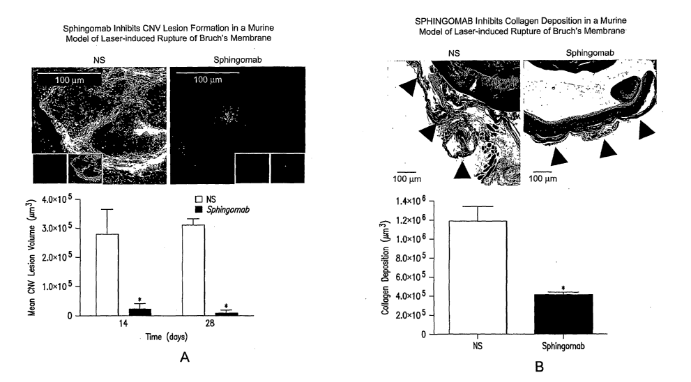
Figure 1: SPHINGOMAB reduced CNV and scar formation in ocular lesions. Mice
were treated with SPHINGOMAB or an isotype-matched non-specific mAb. CNV
lesions were
induced by laser rupture of Bruchs membrane. Shown are graphs and
representative images of
lesions from each treatment group stained with rhodamine-conjugated R.
communis agglutinin I
for vascularization (A) or Masson's Trichrome for collagen scar formation (B).
Figure la shows
that SPHINGOMAB dramatically attenuates choroidal neovascularization 14 and 28
days after
laser-induced rupture of Bruch's membrane.Figure lb shows that SPHIlNGOMAB
significantly
reduces fibrosis associated with CNV lesion formation 28 days after laser-
induced rupture of
Bruchs's membrane.
Figure 2: S 1P promotes neovascularization through induction of HUVECs tube
formation and migration and is reduced by SPHIlNGOMAB. Panel A: Micrographs of
HUVECs
seeded on Matrigel and incubated for 6 hrs to evaluate tube formation. Panel
B: HUVECs were
treated with 1 M S 1P SPHINGOMAB (1 g/ml) for 6 hrs in a Matrigel invasion
chamber. The
number of cells that migrated to the Matrigel membrane were counted in 5
independent fields.
Figure 3. SPHINGOMAB neutralizes S 1P-, VEGF- and bFGF-induced
neovascularization. A: Representative FITC-stained BVs from sections of
Matrigel plugs GFs.
B: S 1P stimulates EC infiltration. C: Quantification of relative fluorescence
from Matrigel plugs
stimulated with VEGF or bFGF as an indicator of neovascularization. S 1P, VEGF
and bFGF's
effects were inhibited when mice were systemically treated with 1 or 25mg/kg
of
SPHINGOMAB.
Figure 4. SPHIIVGOMAB neutralized S1P-stimulated scar formation. Fibroblasts
were
serum-starved and then treated with 0, 0.1, 0.5 or l M S1P +/- 11tg/mL
SPHINGOMAB for 12-
CA 02627427 2008-04-25
WO 2007/053447 PCT/US2006/042027
24
24 hrs. S 1P stimulated Swiss 3T3 fibroblast proliferation as measured by 3H-
thymidine
incorporation (A), murine cardiac fibroblast migration in a scratch assay (B),
collagen gene
expression (relative fluorescence) in isolated cardiac fibroblasts from
transgenic mice expressing
collagen-GFP (C) and WI-38 cell differentiation into myofibroblasts as
measured by decreased
cellular proliferation and increased a-SMA expression (D); SPHINGOMAB
neutralized each of
S 1P's effects. SPHINGOMAB reduced perivascular fibrosis in vivo in a murine
model of a
permanent myocardial infarction (E).
Figure 5. S 1P promotes transformation of ocular epithelial cells and
fibroblasts into
contractile, scar tissue-producing myofibroblasts. The effects of S 1P on
myofibroblast
transformation of several human ocular cell lines were examined. S 1P was
found to stimulate
production of a-Smooth muscle actin (a-SMA; a myofibroblast marker) in human
retinal
pigmented epithelial cells (Figure 5A) and human conjunctiva fibroblasts
(Figure 5B). These
data demonstrate for the first time, that S 1 P is among the factors that
promote transformation of
ocular epithelial cells and fibroblasts into contractile, scar tissue-
producing myofibroblasts. The
effects of S1P on expression of plasminogen activator inhibitor (PAI-1) in
human conjunctiva
fibroblasts were also examined. Increased PAI-1 expression correlates with a
decrease in the
proteolytic degradation of connective tissue and is upregulated in association
with several
fibrotic diseases that involve increased scarring. As shown in Figure 5C, S 1P
stimulates the
PAT-1 expression in a dose-dependent manner.
Figure 6. SPHINGOMAB reduced immune-cell wound infiltration in vivo. Mice were
subjected to MI, treated with saline or 25mg/kg SPHINGOMAB 48 hrs after
surgery and then
sacrificed on day 4. SPHINGOMAB reduced macrophage (A) and mast cell (B)
infiltration into
the wound. Data are represented as fold decrease of saline treated values.
Figure 7. SPHINGOMAB is highly specific for S 1P. A graph based on competitive
ELISA demonstrates SPHINGOMAB's specificity for S 1P compared to other
bioactive lipids.
SPHIlVGOMAB demonstrated no cross-reactivity to sphingosine (SPH), the
immediate
metabolic precursor of S 1P or lysophosphatidic acid (LPA), an important
extracellular signaling
molecule that is structurally and functionally similar to S 1P. SPHINGOMAB did
not recognize
other structurally similar lipids and metabolites, including ceramide- 1 -
phosphate (C1P),
dihydrosphingosine (DH-SPH), phosphatidyl serine (PS), phosphatidyl
ethanolamine (PE), or
sphingomyelin (SM). SPHINGOMAB did cross react with dihydrosphingosine-1-
phosphate
(DH-S1P) and, to a lesser extent, sphingosylphoryl choline (SPC). The affinity
(Kd) of
CA 02627427 2008-04-25
WO 2007/053447 PCT/US2006/042027
SPHINGOMAB for S 1P is <100pM, much higher than most therapeutic antibodies,
particularly
other molecular sponges.
DETAILED DESCRIPTION OF THE INVENTION
1. Compounds
The term "immune-derived moiety," which includes antibodies (Ab) or
immunoglobulins
(Ig), refers to any form of a peptide, polypeptide derived from, modeled after
or encoded by, an
immunoglobulin gene, or a fragment of such peptide or polypeptide that is
capable of binding an
antigen or epitope [see, eg, Immunobiology, 5th Edition, Janeway, Travers,
Walport,
Shlomchiked. (editors), Garland Publishing (2001)]. In the present invention,
the antigen is a
bioactive lipid molecule. Antibody molecules or immunoglobulins are large
glycoprotein
molecules with a molecular weight of approximately 150 kDa, usually composed
of two
different kinds of polypeptide chain. One polypeptide chain, termed the
"heavy" chain (H) is
approximately 50 kDa. The other polypeptide, termed the "light" chain (L), is
approximately 25
kDa. Each immunoglobulin molecule usually consists of two heavy chains and two
light chains.
The two heavy chains are linked to each other by disulfide bonds, the number
of which varies
between the heavy chains of different immunoglobulin isotypes. Each light
chain is linked to a
heavy chain by one covalent disulfide bond. In any given naturally occurring
antibody molecule,
the two heavy chains and the two light chains are identical, harboring two
identical antigen-
binding sites, and are thus said to be divalent, i.e., having the capacity to
bind simultaneously to
two identical molecules.
The "light" chains of antibody molecules from any vertebrate species can be
assigned to
one of two clearly distinct types, kappa (k) and lambda (1), based on the
amino acid sequences of
their constant domains. The ratio of the two types of light chain varies from
species to species.
As a way of example, the average k to 1 ratio is 20:1 in mice, whereas in
humans it is 2:1 and in
cattle it is 1:20.
The "heavy" chains of antibody molecules from any vertebrate species can be
assigned to
one of five clearly distinct types, called isotypes, based on the amino acid
sequences of their
constant domains. Some isotypes have several subtypes. The five major classes
of
immunoglobulin are immunoglobulin M(IgM), immunoglobulin D (IgD),
immunoglobulin G
(IgG), immunoglobulin A (IgA), and immunoglobulin E(IgE). IgG is the most
abundant isotype
and has several subclasses (IgGl, 2, 3, and 4 in humans). The Fc fragment and
hinge regions
CA 02627427 2008-04-25
WO 2007/053447 PCT/US2006/042027
26
differ in antibodies of different isotypes, thus determining their functional
properties. However,
the overall organization of the domains is similar in all isotypes.
The term "variable region" refers to the N-terminal portion of the antibody
molecule or a
fragment thereof. In general, each of the four chains has a variable (V)
region in its amino
teiminal portion, which contributes to the antigen-binding site, and a
constant (C) region, which
determines the isotype. The light chains are bound to the heavy chains by many
noncovalent
interactions and by disulfide bonds and the V regions of the heavy and light
chains pair in each
arm of antibody molecule to generate two identical antigen-binding sites. Some
amino acid
residues are believed to form an interface between the light- and heavy-chain
variable domains
[see Kabat et al. (1991), Sequences of Proteins of Immunological Interest,
Fifth Edition,
National Institute of Health, Bethesda, Md. and Clothia et al. (1985), J. Mol.
Biol, vol 186:
651].
Of note, variability is not uniformly distributed throughout the variable
domains of
antibodies, but is concentrated in three segments called "complementarity-
determining regions"
(CDRs) or "hypervariable regions" both in the light-chain and the heavy-chain
variable domains.
The more highly conserved portions of variable domains are called the
"framework region"
(FR). The variable domains of native heavy and light chains each comprise four
FR regions
connected by three CDRs. The CDRs in each chain are held together in close
proximity by the
FR regions and, with the CDRs from the other chains, form the antigen-binding
site of antibodies
[see Kabat et al. (1991), Sequences of Proteins of Immunological Interest,
Fifth Edition,
National Institute of Health, Bethesda, Md.]. Collectively, the 6 CDRs
contribute to the binding
properties of the antibody molecule for the antigen. However, even a single
variable domain (or
half of an Fv, comprising only three CDRs specific for an antigen) has the
ability to recognize
and bind antigen [see Pluckthun (1994), in The Pharmacology of Monoclonal
Antibodies, vol.
113, Rosenburg and Moore eds., Springer-Verlag, New York, pp. 269-315].
The term "constant domain" refers to the C-terminal region of an antibody
heavy or light
chain. Generally, the constant domains are not directly involved in the
binding properties of an
antibody molecule to an antigen, but exhibit various effector functions, such
as participation of
the antibody in antibody-dependent cellular toxicity. Here, "effector
functions" refer to the
different physiological effects of antibodies (e.g., opsonization, cell lysis,
mast cell, basophil and
eosinophil degranulation, and other processes) mediated by the recruitment of
immune cells by
the molecular interaction between the Fc domain and proteins of the immune
system. The
isotype of the heavy chain determines the functional properties of the
antibody. Their distinctive
CA 02627427 2008-04-25
WO 2007/053447 PCT/US2006/042027
27
functional properties are conferred by the carboxy-terminal portions of the
heavy chains, where
they are not associated with light chains.
As used herein, "antibody fragment" refers to a portion of an intact antibody
that includes
the antigen binding site or variable regions of an intact antibody, wherein
the portion can be free
of the constant heavy chain domains (e.g., CH2, CH3, and CH4) of the Fc region
of the intact
antibody. Alternatively, portions of the constant heavy chain domains (e.g.,
CH2, CH3, and
CH4) can be included in the "antibody fragment". Examples of antibody
fragments are those
that retain antigen-binding and include Fab, Fab', F(ab')2, Fd, and Fv
fragments; diabodies;
triabodies; single-chain antibody molecules (sc-Fv); minibodies, nanobodies,
and multispecific
antibodies formed from antibody fragments. By way of example, a Fab fragment
also contains
the constant domain of a light chain and the first constant domain (CH1) of a
heavy chain.
The term "variant" refers to an amino acid sequence which differs from the
native amino
acid sequence of an antibody by at least one amino acid residue or
modification. A native or
parent or wild-type amino acid sequence refers to the amino acid sequence of
an antibody found
in nature. "Variant" of the antibody molecule includes, but is not limited to,
changes within a
variable region or a constant region of a light chain andlor a heavy chain,
including the
hypervariable or CDR region, the Fc region, the Fab region, the CHl domain,
the CH2 domain,
the CH3 domain, and the hinge region.
The term "specific" refers to the selective binding of an antibody to its
target epitope.
Antibody molecules can be tested for specificity of binding by comparing
binding of the
antibody to the desired antigen to binding of the antibody to unrelated
antigen or analogue
antigen or antigen mixture under a given set of conditions. Preferably, an
antibody according to
the invention will lack significant binding to unrelated antigens, or even
analogs of the target
antigen. Here, the term "antigen" refers to a molecule that is recognized and
bound by an
antibody molecule or immune-derived moiety that binds to the antigen. The
specific portion of
an antigen that is bound by an antibody is termed the "epitope." A'-'hapten"
refers to a small
molecule that can, under most circumstances, elicit an immune response (i.e.,
act as an antigen)
only when attached to a carrier molecule, for example, a protein, polyethylene
glycol (PEG),
colloidal gold, silicone beads, and the like. The carrier may be one that also
does not elicit an
immune response by itself.
The term "antibody" is used in the broadest sense, and encompasses monoclonal,
polyclonal, multispecific (e.g., bispecific, wherein each arm of the antibody
is reactive with a
CA 02627427 2008-04-25
WO 2007/053447 PCT/US2006/042027
28
different epitope or the same or different antigen), minibody,
heteroconjugate, diabody, triabody,
chimeric, and synthetic antibodies, as well as antibody fragments that
specifically bind an
antigen with a desired binding property and/or biological activity.
The term "monoclonal antibody" (mAb) refers to an antibody, or population of
like
antibodies, obtained from a population of substantially homogeneous
antibodies, and is not to be
construed as requiring production of the antibody by any particular method.
For example,
monoclonal antibodies can be made by the hybridoma method first described by
Kohler and
Milstein (1975), Nature, vol 256: 495-497, or by recombinant DNA methods.
The term "chimeric" antibody (or immunoglobulin) refers to a molecule
comprising a
heavy and/or light chain which is identical with or homologous to
corresponding sequences in
antibodies derived from a particular species or belonging to a particular
antibody class or
subclass, while the remainder of the chain(s) is identical with or homologous
to corresponding
sequences in antibodies derived from another species or belonging to another
antibody class or
subclass, as well as fragments of such antibodies, so long as they exhibit the
desired biological
activity [Cabilly et al. (1984), infra; Morrison et al., Proc. Natl. Acad.
Sci. U.S.A. 81:6851].
The term "humanized antibody" refers to forms of antibodies that contain
sequences from
non-human (eg, murine) antibodies as well as human antibodies. A humanized
antibody can
include conservative amino acid substitutions or non-natural residues from the
same or different
species that do not significantly alter its binding and/or biologic activity.
Such antibodies are
chimeric antibodies that contain minimal sequence derived from non-human
immunoglobulins.
For the most part, humanized antibodies are human immunoglobulins (recipient
antibody) in
which residues from a complementary-determining region (CDR) of the recipient
are replaced by
residues from a CDR of a non-human species (donor antibody) such as mouse,
rat, camel,
bovine, goat, or rabbit having the desired properties. Furthermore, humanized
antibodies can
comprise residues that are found neither in the recipient antibody nor in the
imported CDR or
framework sequences. These modifications are made to further refine and
maximize antibody
performance. Thus, in general, a humanized antibody will comprise all of at
least one, and in
one aspect two, variable domains, in which all or all of the hypervariable
loops correspond to
those of a non-human immunoglobulin and all or substantially all of the FR
regions are those of
a human immunoglobulin sequence. The humanized antibody optionally also will
comprise at
least a portion of an immunoglobulin constant region (Fc), or that of a human
immunoglobulin.
See, e.g., Cabilly et al., U.S. Pat. No. 4,816,567; Cabilly et al., European
Patent No. 0,125,023
B1; Boss et al., U.S. Pat. No. 4,816,397; Boss et al., European Patent No.
0,120,694 B1;
CA 02627427 2008-04-25
WO 2007/053447 PCT/US2006/042027
29
Neuberger, M. S. et al., WO 86/01533; Neuberger, M. S. et al., European Patent
No. 0,194,276
B1; Winter, U.S. Pat. No. 5,225,539; Winter, European Patent No. 0,239,400 B1;
Padlan, E. A.
et al., European Patent Application No. 0,519,596 Al; Queen et al. (1989)
Proc. Nat'l Acad. Sci.
USA, vol 86:10029-10033).
The term "bispecific antibody" can refer to an antibody, or a monoclonal
antibody,
having binding properties for at least two different epitopes. In one
embodiment, the epitopes
are from the same antigen. In another embodiment, the epitopes are from two
different antigens.
Methods for making bispecific antibodies are known in the art. For example,
bispecific
antibodies can be produced recombinantly using the co-expression of two
immunoglobulin
heavy chain/light chain pairs. Alternatively, bispecific antibodies can be
prepared using
chemical linkage. Bispecific antibodies include bispecific antibody fragments.
The term "heteroconjugate antibody" can refer to two covalently joined
antibodies. Such
antibodies can be prepared using known methods in synthetic protein chemistry,
including using
crosslinking agents. As used herein, the term "conjugate" refers to molecules
formed by the
covalent attachment of one or more antibody fragment(s) or binding moieties to
one or more
polymer molecule(s).
The term "biologically active" refers to an antibody or antibody fragment that
is capable
of binding the desired epitope and in some way exerting a biologic effect.
Biological effects
include, but are not limited to, the modulation of a growth signal, the
modulation of an anti-
apoptotic signal, the modulation of an apoptotic signal, the modulation of the
effector function
cascade, and modulation of other ligand interactions.
The term "recombinant DNA" refers to nucleic acids and gene products expressed
therefrom that have been engineered, created, or modified by man.
"Recombinant" polypeptides
or proteins are polypeptides or proteins produced by recombinant DNA
techniques, for example,
from cells transformed by an exogenous DNA construct encoding the desired
polypeptide or
protein. "Synthetic" polypeptides or proteins are those prepared by chemical
synthesis.
The term "expression cassette" refers to a nucleotide molecule capable of
affecting
expression of a structural gene (i.e., a protein coding sequence, such as an
antibody of the
invention) in a host compatible with such sequences. Expression cassettes
include at least a
promoter operably linked with the polypeptide-coding sequence, and,
optionally, with other
sequences, e.g., transcription termination signals. Additional regulatory
elements necessary or
CA 02627427 2008-04-25
WO 2007/053447 PCT/US2006/042027
helpful in effecting expression may also be used, e.g., enhancers. Thus,
expression cassettes
include plasmids, expression vectors, recombinant viruses, any form of
recombinant "naked
DNA" vector, and the like.
2. Applications
The invention is drawn to compositions and methods for treating or preventing
ocular
diseases and conditions, using one or more therapeutic agents that alter the
activity or
concentration of one or more undesired bioactive lipids, or precursors or
metabolites thereof.
The therapeutic methods and compositions of the invention act by changing the
effective
concentration, i.e., the absolute, relative, effective and/or available
concentration and/or
activities, of certain undesired bioactive lipids. Lowering the effective
concentration of the
bioactive lipid may be said to "neutralize" the target lipid or its undesired
effects, including
downstream effects. Here, "undesired" refers to a bioactive lipid that is
unwanted due to its
involvement in a disease process, for example, as a signaling molecule, or to
an unwanted
amount of a bioactive lipid which contributes to disease when present in
excess.
Without wishing to be bound by any particular theory, it is believed that
inappropriate
concentrations of lipids such as S 1P and /or LPA, and/or their metabolites or
downstream
effectors, may cause or contribute to the development of various ocular
diseases and disorders.
As such, the compositions and methods can be used to treat these ocular
diseases and disorders,
particularly by decreasing the effective in vivo concentration of a particular
target lipid, for
example, S 1P and /or LPA. In particular, it is believed that the compositions
and methods of the
invention are useful in treating ocular diseases characterized, at least in
part, by aberrant
neovascularization, angiogenesis, fibrogenesis, fibrosis, scarring,
inflammation, and immune
response.
Examples of several classes of ocular diseases that may be treated in
accordance with the
invention are described below. It will be appreciated that many disease and
conditions are
characterized, at least in part, by multiple pathological processes (for
example, both pathological
neovascularization and scarring) and that the classifications provided herein
are for descriptive
convenience and do not limit the invention.
CA 02627427 2008-04-25
WO 2007/053447 PCT/US2006/042027
31
Ischen2ic Retinopatlzies associated with ap tlaologaic neovascularization and
diseases
characterized by epiretinal and or subretinal rnenabrane fornaation
Ischemic retinopathies (IR) are a diverse group of disorders characterized by
a
compromised retinal blood flow. Examples of IR include diabetic retinopathy
(DR), retinopathy
of prematurity (ROP), sickle cell retinopathy and retinal venous occlusive
disease. All of these
disorders can be associated with a VEGF driven proliferation of pathological
retinal
neovascularization which can ultimately lead to intraocular hemorrhaging, epi-
retinal membrane
formation and tractional retinal detachment. Idiopathic epi-retinal membranes
(ERMs), also
called macular pucker or cellophane retinopathy, can cause a reduction in
vision secondary to
distortion of the retinal architecture. These membranes sometimes recur
despite surgical removal
and are sometimes associated with retinal ischemia. VEGF and its receptors are
localized to
ERMs. The presence of VEGF in membranes associated with proliferative diabetic
retinopathy,
proliferative vitreoretinopathy and macular pucker further suggests that this
cytokine plays an
important role in angiogenesis in ischemic retinal disease and in membrane
growth in
proliferative vitreoretinal disorders. In addition VEGF receptors, VEGFRI and
VEGFR2 are also
identified on cells in ERMs. These data show that VEGF may be an autocrine
and/or paracrine
stimulator that may contribute to the progression of vascular and avascular
ERMs. PDGF and its
receptors [Robbins et al. (1994), Invest Ophthalmol Vis Sci; vol 35: 3649-
3663] has been
described in eyes with proliferative retinal diseases [Cassidy et al. (1998),
Br J Ophthamol; vol
82: 181-85 and Freyberger et al. (2000), Exp Clin Endocrinol Diabetes, vol
108: 106-109]. These
findings suggest that PDGF ligands and receptors are widespread in
proliferative retinal
membranes of different origin and suggest that autocrine and paracrine
stimulation with PDGF
may be involved in ERM pathogenesis. Transforming growth factor-(3 (TGF-0) is
involved in the
formation of ERMs [Pournaras et al. (1998), Klin Monatsbl Augenheilkd, vol
212: 356-358] as
demonstrated by TGF staining and immunoreactivity. In addition, TGF-0 receptor
II is expressed
in myofibroblasts of ERM of diabetic and PVR membranes. These results suggest
that TGF-(3,
produced in multiple cell types in retina and ERMs, is an attractive target
for the treatment of
PVR, diabetic and secondary ERMs. Interleukin-6 (IL-6) has been reported to be
increased in
human vitreous in proliferative diabetic retinopathy (PDR) [La Heij et al.
(2002), Am J Ophthal,
134: 367-375] and in one study 100% of the diabetic ERMs studied expressed IL-
6 protein
[Yamamoto et al. (2001) Am J Ophthal, vol 132: 369-377].
Exogenous administration of basic fibroblastic growth factor (bFGF) has been
shown to
induce endothelial proliferation and VEGF expression [Stavri et al. (1995),
Circulation, vol 92:
CA 02627427 2008-04-25
WO 2007/053447 PCT/US2006/042027
32
11-14]. Consistent with these observations, bFGF concentration is increased in
vitreous samples
from patients with PDR [Sivalingam et al. (1990), Arch Ophthalmol, vol 108:
869-872 and
Boulton et al. (1997), Br J Ophthalmol, vol 81: 228-233]. bFGF is also
involved in the formation
of ERMs [Hueber et al. (1996), Int. Ophthalmol, vo120: 345-350] demonstrated
bFGF in 8 out
of 10 PDR membranes studied. Moreover, these workers found positive staining
for the
corresponding receptor, FGFRl. Immunoreactivity for bFGF has also been
demonstrated in
nonvascular idiopathic ERMs. These results implicate bFGF in the formation of
both vascular
and avascular ERMs. [Harada et al. (2006), Prog in Retinal and Eye Res, vol
25; 149-164].
Elevated bFGF has also been detected in the serum of patients with ROP
(Becerril et al. (2005),
Ophthalmology, vol 112, 2238].
Given the known pleotropic effects of S 1P and its interactions with VEGF,
bFGF, PDGF,
TGF-(3 and IL-6, it is believed that an agent that binds, antagonizes,
inhibits the effects or the
production of S 1P will be effective at suppressing pathologic retinal
neovascularization in
ischemic retinopathies and posterior segment diseases characterized by
vascular or avascular
ERM formation. Other ocular conditions characterized, at least in part, by
aberrant
neovascularization or angiogenesis include age-related macular degeneration,
corneal graft
rejection, neovascular glaucoma, contact lens overwear, infections of the
cornea, including
herpes simplex, herpes zoster and protozoan infection, pterygium, infectious
uveitis, chronic
retinal detachment, laser injury, sickle cell retinopathy, venous occlusive
disease, choroidal
neovascularization, retinal angiomatous proliferation, and idiopathic
polypoidal choroidal
vasculopathy.
Proliferative vitreoretinopathy (PVR)
PVR is observed after spontaneous rhegmatogenous retinal detachment and after
traumatic retinal detachment. It is a major cause of failed retinal detachment
surgery. It is
characterized by the growth and contraction of cellular membranes on both
sides of the retina, on
the posterior vitreous surface and the vitreous base. This excessive scar
tissue development in the
eye may lead to the development of tractional retinal detachment, and
therefore treatments
directed at the prevention or inhibition of proliferative vitreoretinopathy
(PVR) are a logical
principle of management of retinal detachment. Histopathologically PVR is
characterized by
excessive collagen production, contraction and cellular proliferation
[Michels, Retinal
Detachment 2nd Edition. Wilkinsin CP, Rice TA Eds, Complicated types of
retinal detachment,
pp 641-771, Mosby St Louis 1997]. Cellular types identified in PVR membranes
include mainly
retinal pigmented epithelial cells, fibroblasts, macrophages and vascular
endothelial cells [Jerdan
CA 02627427 2008-04-25
WO 2007/053447 PCT/US2006/042027
33
JA et al. (1989), Ophthalmology, vol 96: 801-10 and Vidinova et al. (2005),
Klin Monatsbl
Augenheilkd; vo1222:568-571]. The pathophysiology of this excessive scarring
reaction appears
to be mediated by a number of cytokines including platelet derived growth
factor (PDGF),
transforming growth factor (TGF) beta, basic fibroblastic growth factor
(bFGF), interleukin -6
(IL)-6 and interleukin-8 (IL)-8 [Nagineni et al. (2005), J Cell Physiol, vol
203: 35-43; La Heij et
al (2002), Am J Ophthalmol, 134: 367-75; Planck et al. (1992), Curr Eye Res;
vol 11: 1031-9;
Canataroglu et al. (2005) Ocul Immunol Inflamm; vol 13: 375-81 and Andrews et
al. (1999)
Ophthalmol Vis Sci; vol 40: 2683-9]. Inhibition of these cytokines may help
prevent the
development of PVR if given in a timely fashion or limit its severity [Akiyama
et al (2006), J
Cell Physiol, vol 207:407-12 and Zheng Y et al (2003), Jpn J Ophthalmolm, vol
47:158-65].
Sphingosine -1-Phosphate (S1P) is a bioactive lysolipid with pleotrophic
effects. It is pro-
angiogenic, pro inflammatory (stimulates the recruitment of macrophages and
mast cells) and
pro-fibrotic (stimulates scar formation). S 1P generally stimulates cells to
proliferate and migrate
and is anti-apoptotic. S 1P achieves these biologically diverse functions
through its interactions
with numerous cytokines and growth factors. Inhibition of S1P via a monoclonal
antibody
(SPHINGOMAB) has been demonstrated to block the functions of vascular
endothelial growth
factor (VEGF), bFGF, IL-6 and IL-8 [Visentin B et al. (2006), Cancer Cell,
vo19: 1-14].
Binding of S 1P to the S 1P1 receptor can also increase PDGF production;
therefore an agent that
binds S 1P would also be expected to diminish PDGF production [Milstien and
Spiegel (2006),
Cancer Cell, vol 9:148-150]. As shown in the Examples below, it has now been
demonstrated
that in vitro S 1P transforms human RPE cells into a myofibroblast-like
phenotype similar to the
type seen in PVR. Given the pathophysiology that ultimately results in the
excessive scarring
seen in PVR and the known effects of S IP on these same key mediators, it is
believed that an
agent that binds, antagonizes, or inhibits the effects or the production of
S1P will be effective at
eliminating or minimizing the development of PVR and its severely damaging
effects on the eye.
Uveitis
Uveitis is an inflammatory disorder of the uveal tract of the eye. It can
affect the front
(anterior) or back (posterior) of the eye or both. It can be idiopathic or
infectious in etiology and
can be vision-threatening. Idiopathic uveitis has been associated with
increased CD4+ expression
in the anterior chamber. [Calder et al. (1999), Invest Ophthalmol Vis Sci, vol
40: 2019-24]. Data
also suggests a pathologic role of the T lymphocyte and its chemoattractant IP-
10 in the
pathogenesis of uveitis [Abu El-Asrar (2004), Am J Ophthalmol, vol 138: 401-
11]: Other
chemokines in acute anterior uveitis include macrophage inflammatory proteins,
monocyte
CA 02627427 2008-04-25
WO 2007/053447 PCT/US2006/042027
34
chemoattractant protein-1 and IL-8. These cytokines probably play a critical
role in leukocyte
recruitment in acute anterior uveitis. [Verma et al. (1997), Curr Eye Res; vol
16; 1202-8]. Given
the profound and pleiotropic effects of the S 1P signaling cascade, it is
believed that
SPHIlITGOMAB and other immune moieties that reduce the effective concentration
of bioactive
lipid would serve as an effective method of reducing or modulating the
intraocular inflammation
associated with uveitis.
Re
Lractive surgery
The corneal wound healing response is of particular relevance for refractive
surgical
procedures since it is a major determinant of safety and efficacy. These
procedures are
performed for the treatment of myopia, hyperopia and astigmatism. Laser in
situ keratomileusis
(LASIK) and photorefractive keratectomy (PRK) are the most common refractive
procedures
however others have been developed in an attempt to overcome complications.
These
complications include overcorrection, undercorrection, regression and stromal
opacification
among others. A number of common complications are related to the healing
response and have
their roots in the biologic response to surgery. One of the greatest
challenges in corneal biology
is to promote tissue repair via regeneration rather than fibrosis. It is
believed that the choice
between regeneration and fibrosis lies in the control of fibroblast
activation. [Stramer et al
(2003), Invest Ophthalmol Vis Sci; vo144: 4237-4246 and Fini (1999) Prog Retin
Eye Res, vol
18: 529-551]. Cells called myofibroblasts may appear in the subepithelial
stroma 1-2 weeks after
surgery or injury. Myofibroblasts are presumably derived from keratocytes
under the influence
of TGF-(3 [Jester et al (2003) Exp Eye Res, vo177: 581-592]. Corneal haze and
stromal scarring
are characterized by reduced corneal transparency and may be associated with
fibroblast and
myofibroblast generation. In situ and in vitro studies have suggested that TGF-
(3 and PDGF are
important in stimulating myofibroblast differentiation [Folger et al. (2001),
Invest Ophthalmol
Vis Sci; 42: 2534-2541]. Haze can be noted in the central interface after
LASIK under certain
circumstances. These include diffuse lamellar keratitis, donut-shaped flaps,
and retention of
epithelial debris at the interface. It is likely that each of these is
associated with increased access
of TGF-(3 from epithelial cells to the activated keratocytes. [Netto et al.
(2005), Cornea, vo124:
509-522]. Regression is most likely due to heightened epithelial-stromal wound
healing
interactions such as increased production of epithelium modulating growth
factors by corneal
fibroblasts and or myofibroblasts [Netto et al. (2005), Cornea, vo124: 509-
522]. Inhibition of
TGF-0 binding to receptors with topical anti-TGF-0 antibody has been shown to
reduce haze
induced by PRK [Jester et al. (1997), Cornea, vol 16: 177-187]. Given the
known effects of anti-
CA 02627427 2008-04-25
WO 2007/053447 PCT/US2006/042027
bioactive lipid antibody on the fibrotic process and TGF-(3, we believe that
it may aid in treating
some of the complications of refractive surgery such as haze, stromal scarring
and regression.
Modulation of Glauconia Filtration Surgery
Glaucoma is classically thought of a disease whereby elevated intraocular
pressure causes
damage to the optic nerve and ultimately compromises the visual field and or
the visual acuity.
Other forms of glaucoma exist where optic nerve damage can occur in the
setting of normal
pressure or so called "normal tension glaucoma". For many patients medications
are able to
control their disease, but for others glaucoma filtration surgery is needed
whereby a fistula is
surgically created in the eye to allow fluid to drain. This can be
accomplished via
trabeculectomy, the implantation of a medical device or other methods of
surgical intervention.
Glaucoma filtration surgery fails due to a wound healing process characterized
by the
proliferation of fibroblasts and ultimately scarring. Anti-metabolites such as
5-fluorouracil and
mitomycin C can reduce subsequent scarring; however, even with the use of
these drugs long
term follow up shows that surgical failure is still a serious clinical
problem. [Mutsch and Grehn
(2000), Graefes Arch Clin Exp Ophthalmol; vol 238: 884-91 and Fontana et al.
(2006),
Ophthalmology, vol 113: 930-936]. Studies of human Tenon's capsule fibroblasts
demonstrate
that they have the capacity to synthesize bFGF and PDGF and TGF-(3 and that
these growth
factors are implicated in the tissue repair process after glaucoma filtration
surgery that
contributes to the failure of the procedure. [Trpathi et al. (1996), Exp Eye
Res, vol 63: 339-46].
Additional studies have also implicated these growth factors in the post
filtration wound
response [Denk et al. (2003), Curr Eye Res; vol 27: 35-44] concluded that
different isoforms of
PDGF are major stimulators of proliferation of Tenon's capsule fibroblasts
after glaucoma
filtration surgery while TGF-(3 is essential for the transformation of Tenon's
capsule fibroblasts
into myofibroblasts. We have demonstrated that S 1P is present in human
Tenon's
capsule/conjunctival fibroblasts and that S1P is strongly expressed in the
wound healing
response. S1P also stimulates the profibrotic function of multiple fibroblast
cell types and the
transformation into the myofibroblast phenotype and collagen production. Given
the specific
pleotropic effects of S 1P and its known interactions with bFGF, PDGF and TGF-
beta, it is
believed that an agent that binds, antagonizes, inhibits the effects or the
production of S 1P, or
perhaps other bioactive lipids such as LPA, will be effective at modulating
the wound healing
and/or fibrotic response that leads to failure of glaucoma surgery and will be
an effective
therapeutic method of enhancing successful surgical outcomes. It is envisioned
that the agent
could be administered, e.g., via intravitreal or subconjunctival injection or
topically.
CA 02627427 2008-04-25
WO 2007/053447 PCT/US2006/042027
36
Conzeal transplantation
Corneal transplantation (penetrating keratoplasty (PK)) is the most successful
tissue
transplantation procedure in humans. Yet of the 47,000 corneal transplants
performed annually
in the United States, corneal allograft rejection is still the leading cause
of comeal graft failure.
[Ing JJ et al. (1998), Ophthalmology, vol 105: 1855-1865]. Currently, we do
not sufficiently
have the ability to avert allograft rejection although immunosuppression and
immunomodulation
may be a promising approach. Recently it has been discovered that CD4(+) T
cells function as
directly as effector cells and not helper cells in the rejection of corneal
allografts. [Hegde S et al.
(2005), Transplantation, vo179: 23-31]. Murine studies have shown increased
numbers of
neutrophils, macrophage and mast cells in the stroma of corneas undergoing
rejection.
Macrophages were the main infiltrating cell type followed by T-cells, mast
cells and neutrophils.
The early chemokine expression in high risk corneal transplantation was the
mouse homologue
of IL-8 (macrophage inflammatory protein-2) and monocyte chemotactic protein-1
(MCP-1)
[Yamagami S et al. (2005), Mol Vis, vol 11, 632-40].
FTY720 (FTY) is a novel immunosuppressive drug that acts by altering
lymphocyte
trafficking; resulting in peripheral blood lymphopenia and increased
lymphocyte counts in lymph
nodes. FTY mediates its immune-modulating effects by binding to some of the
SIP receptors
expressed on lymphocytes. [Bohler T et al. (2005), Transplantation, vo179: 492-
5]. The drug is
administered orally and a single oral dose reduced peripheral lymphocyte
counts by 30-70%.
FTY reduced T-cell subset, CD4(+) cells more than CD8(+) cells. [Bohler et al.
(2004), Nephrol
Dial Transplant, vol 19: 702-13]. FTY treated mice showed a significant
prolongation of
orthotopic corneal-graft survival when administered orally. [Zhang et al.
(2003), Transplantation,
vo176: 1511-3]. FTY oral treatment also significantly delayed rejection and
decreased its
severity in a rat-to-mouse model of comeal xenotransplantation [Sedlakova et
al. (2005),
Transplantation, vol 79, 297-303]. Given the known pathogenesis of allograft
rejection combined
with the data suggesting that modulating the effects of the S1P signaling can
improve corneal
graft survival, it is believed that immune moieties that decrease the
effective concentration of
bioactive lipids, e.g., SPHINGOMAB, will also be useful in treatment of
immunologic
conditions such as allograft rejection, for example by attenuating the immune
response, and thus
will likely improve corneal graft survival after PK. The drug may also have
the added advantage
that in addition to systemic administration, local administration, e.g., via
topical periocular or
intraocular delivery, may be possible.
CA 02627427 2008-04-25
WO 2007/053447 PCT/US2006/042027
37
Other ocular diseases with an inflammatory or immune component include chronic
vitritis, infections, including herpes simplex, herpes zoster and protozoan
infections, and ocular
histoplasmosis.
Anterior Segment Diseases Characterized by Scarring
Treatment with an antibody targeted to bioactive lipid also is believed to
benefit several
conditions characterized by scarring of the anterior portion of the eye. These
include the
following:
Trauma
The cornea, as the most anterior structure of the eye, is exposed to various
hazards
ranging from airborne debris to blunt trauma that can result in mechanical
trauma. The cornea
and anterior surface of the eye can also be exposed to other forms of trauma
from surgery, and
chemical, such as acid and alkali, injuries. The results of these types of
injuries can be
devastating often leading to corneal and conjunctival scarring symblephera
formation. In
addition corneal neovascularization may ensue. Neutrophils accumulate, their
release of
leukotrienes, and the presence of interleukin-1 and interleukin-6, serves to
recruit successive
waves of inflammatory cells [Sotozono et al. (1997), Curr Eye Res, vol 19: 670-
676] infiltrate
the cornea and release proteolytic enzymes which lead to further damage and
break down of
corneal tissue and a corneal melt. In addition comeal and conjunctival
fibroblasts become
activated and invade and leading to collagen deposition and fibrosis. The
undesirable effects of
excessive inflammation and scarring are promoted by TGF-0. [Saika S et al.
(2006), Am J Pathol
vol 168, 1848-60]. This process leads to loss of corneal transparency and
impaired vision.
Reduced inflammation, including decreased neutrophil infiltrates and reduced
fibrosis resulted in
faster and more complete healing in a murine model of alkali burned corneas [
Ueno et al.
(2005), Ophthalmol Vis Sci, vo146: 4097-106].
Ocular Cicatricial Peniphigoid (OCP)
OCP is a chronic cicatrizing (scar-forming) autoimmune disease that primarily
affects the
conjunctiva. The disease is invariably progressive and the prognosis is quite
poor. In its final
stages conjunctival scarring and the associated keratopathy lead to bilateral
blindness.
Histologically the conjunctiva shows submucosal scarring and chronic
inflammation in which
mast cell participation is surprisingly great. [Yao L et al. (2003), Ocul
Immunol Inflamm, vol 11:
211-222]. Autoantigens lead to the formation of autoantibodies. The binding of
the autoantibody
CA 02627427 2008-04-25
WO 2007/053447 PCT/US2006/042027
38
to the autoantigen sets in motion a complex series of events with infiltration
of T lymphocytes
where CD4 (helper) cells far outnumber CD8 (suppressor) cells. Macrophage and
mast cell
infiltration also ensue as well as the release of proinflammatory and
profibrotic cytokines.
Cytokine induced conjunctival fibroblast proliferation and activation results,
with resultant
subepithelial fibrosis (see examples hereinbelow). Studies have shown a role
of TGF-0 and IL-1
in conjunctival fibrosis in patients with OCP [Razzaque MS et al. (2004),
Invest Ophthalmol Vis
Sci, vo145: 1174-81].
Stevens Johnson Syndrome (SJS) and Toxic E idennal Necrolysis (TEN)
SJS and TEN are life-threatening adverse reactions to medications. The ocular
sequelae
of these two related conditions can be severe and involve pathologic changes
of the bulbar and
palpebral conjunctiva, eyelids and cornea. Drugs and infections are the most
common
precipitating factors. Chronic eye findings include scarring, symblepharon
formation, and
cicatrisation of the conjunctiva as a result of the initial inflammatory
process. This leads to
entropion formation, trichiasis and instability of the tear film. Breakdown of
the ocular surface
leads to corneal scarring, neovascularization, and in severe cases
keratinization. As in OCP
subepithelial fibrosis of the conjunctiva occurs. A vigorous autoimmune
lymphocyte response to
a di-ug or infection is believed to play a role in development of SJS/TEN.
[Harilaos et al. (2005),
Erythema Multiforme, Stevens Johnson Syndrome, and Toxic Epidermal Necrolysis,
in Cornea
2"a edition. Krachmer, Mannis, Holland eds.Elesevier Mosby Philadelphia]. The
infiltrating cell
population in SJS includes macrophages, CD4 positive T cells, and CD8 positive
T cells. This
cell population is similar to those seen in chemical injury. [Kawasaki et al.
(2000), J Ophthalmol,
vol 84: 1191-3].
Ptery iuna
Clinically a pterygium appears as a fleshy, vascular mass that occurs in the
interpalpebral
fissure. The body of the pterygium is a fleshy fibrovascular mass. Active
pterygium are
characterized by marked vascular engorgement and progressive growth. They are
firmly
adherent to the globe. In advanced cases the pterygium encroaches onto the
cornea and may
cause visual loss secondary to loss of corneal transparency within the visual
axis or irregular
astigmatism. Symptomatically, patients may experience foreign body sensation,
tearing and
blurred vision. Histopathology demonstrates hyalinization of the subepithelial
connective tissue
of the substantia propria, increased number of fibroblasts and increased mast
cells. [Butrus et al.
(1995), Am J Ophthalmol, vol 119: 236-237]. Management of pterygium remains
problematic.
CA 02627427 2008-04-25
WO 2007/053447 PCT/US2006/042027
39
Surgical excision is often performed however recurrence rates are high. (Y.rag
et al. (1992), Acta
Ophthalmol, vo170: 530]. In order to help lower the recurrence rate of
pterygium, various
pharmacologic adjuvants have been employed such as Mitomycin-C and
daunorubicin. Although
these may be helpful, long term data are limited and they can be associated
with scleral thinning
and corneal melt. Dougherty et al. and Lee et al. [Dougherty et al. (1996),
Cornea, vol 15: 537-
540 and Lee et al. (2001), Cornea, vol 20: 238-42] were the first to
demonstrate that VEGF may
play an important role in the development of pterygium and to identify VEGF
and nitric oxide in
the epithelium of pterygium. These workers hypothesized that these as well as
other cytokines
are responsible for the fibrovascular ingrowth characteristic of pterygium.
The presence of basic
FGF and TGF-beta 1 in both primary and recurrent pterygium has been
demonstrated [Kira et al.
(1998), Graefes Arch Clin Exp Ophthalmol, vo1236: 702-8] and published
morphometric and
immunohistochemical evidence further supports the notion that angiogenesis may
play a role in
the formation of pterygium [Marcovich et al (2002), Curr Eye Res, vol 25:17-
22]. Other studies
have implicated IL-6 and IL-8 as well as VEGF as mediators that may be
relevant to pterygium
development [Di Girolamo et al. (2006), Invest Ophthalmol Vis Sci, vo147: 2430-
7]. An
effective agent against pterygium formation and growth may diminish the need
for surgical
intervention or reduce recurrence rates.
Other ocular diseases and conditions with a fibrogenesis, fibrosis or scarring
component
include AMD, diabetic retinopathy, retinopathy of prematurity, sickle cell
retinopathy, ischemic
retinopathy, retinal venous occlusive disease and contact lens overwear.
In summary, excessive scarring is an underlying component of the
pathophysiology of
many ocular and non-ocular diseases and conditions. Bioactive lipids like S 1P
and LPAs play a
role in this process and an antibody-related treatment to diminish the
concentrations of these
agents will likely lead to therapeutic benefit to patients receiving the
treatment. In one
embodiment, inhibitors of bioactive lipids, particularly monoclonal antibodies
directed against
S 1P and/or LPA, are believed to be useful in modulating surgical and
traumatic wound healing
responses.
Anti-S IP and anti-LPA antibodies for the treatment of scleroderma
The compositions and methods of the invention will be useful in treating
disorders and
diseases characterized, at least in part, by aberrant neovascularization,
angiogenesis,
fibrogenesis, fibrosis, scarring, inflammation, and immune response. One such
disease is
scleroderma, which is also referred to as systemic sclerosis.
CA 02627427 2008-04-25
WO 2007/053447 PCT/US2006/042027
Scleroderma is an autoimmune disease that causes scarring or thickening of the
skin, and
sometimes involves other areas of the body, including the lungs, heart, and/or
kidneys.
Scleroderma is characterized by the formation of scar tissue (fibrosis) in the
skin and organs of
the body, which can lead to thickening and firmness of involved areas, with
consequent
reduction in function. Today, about 300,000 Americans have scleroderma,
according to the
Scleroderma Foundation. One-third or less of those affected have widespread
disease, while the
remaining two-thirds primarily have skin symptoms. When the disease affects
the lungs and
causing scarring, breathing can become restricted because the lungs can no
longer expand as they
should. To measure breathing capability, doctors use a device that assesses
forced vital capacity
(FVC). In people with an FVC of less than 50 percent of the expected reading,
the 10-year
mortality rate from scleroderma-related lung disease is about 42 percent. One
reason the
mortality rate is so high is that no effective treatment is currently
available.
As described in the examples of this application, existing evidence indicates
that S1P and
LPA are pro-fibrotic growth factors that can contribute to fibroblast
activation, proliferation, and
the resulting increased fibroblast activity associated with maladaptive
scarring and remodeling.
Moreover, potential roles for S 1P and LPA in activity of skin and other types
of fibroblasts have
been demonstrated. For example, it has been shown that LPA stimulates the
migration of murine
skin fibroblasts (Hama, et al., J Biol Chem. 2004 Apr 23;279(17):17634-9), and
human skin
fibroblasts express several S1P receptor subtypes (Zhang, et al., Blood. 1999
May 1;93(9):2984-
90). In addition to the many direct effects of S 1P on fibroblast activity, S
1P also may have many
potential indirect effects on fibroblast activity. For example, S 1P may
facilitate the action of
other well-known pro-fibrotic factors, such as TGF-(3 and platelet derived
growth factor (PDGF).
TGF-0 is one of the most widely studied and recognized contributors to
fibrosis (Desmouliere, et
al., J Cell Biol 122: 103-111, 1993). TGF-0 upregulates SphKl expression and
activity leading
to increased expression of tissue inhibitors of metalloproteinases 1(TIMP-1),
a protein that
inhibits ECM degradation (Yamanaka, et al., J Biol Chenz 279: 53994-54001,
2004). Increased
expression of TIlYIP-1 is linked to interstitial fibrosis and diastolic
dysfunction in heart failure
patients (Heymans, et al., Am J Pathol 166: 15-25, 2005). Conversely, S 1P
stimulates
expression and release of TGF-0 (Norata, et al., Circulation 111: 2805-2811,
2005). There is
also distinct evidence of crosstalk between S 1P and PDGF. S 1P directly
stimulates expression
of PDGF (Usui, et al., J Biol Cltern 279: 12300-12311, 2004). In addition, the
S 1PI receptor and
the PDGF receptor bind one another and their association is necessary for PDGF
activation of
downstream signaling which contributes to proliferation and migration of
various cell types
(Long, et al., Prostaglandins Other Lipid Mediat 80: 74-80, 2006; Baudhuin et
al., Faseb J 18:
CA 02627427 2008-04-25
WO 2007/053447 PCT/US2006/042027
41
341-343, 2004). As such, the effects of TGF-P and PDGF on fibrosis may be due
in part to
crosstalk with the S 1P signaling pathway. As such, the compositions and
methods of the
invention can be used to treat scleroderma, particularly by decreasing the
effective in vivo
concentration of a particular target lipid, for example, S IP and/or LPA.
Systemic scleroderma is thought to be exacerbated by stimulatory
autoantibodies against
PDGF receptors (Baroni, et al., N Engl J Med. 2006 v354(25):2667-76), and PDGF
receptors are
up-regulated in scleroderma fibroblasts in response to TGF-(3 (Yamakage, et
al., J Exp Med.
1992 May 1;175(5):1227-34). Because of the substantial cross-talk among the
S1P, PDGF and
TGF-(3 signaling systems, blocking S 1P bioactivity with and anti-S 1P agent
(e.g., an anti-S 1P
mAb) could indirectly mitigate the pro-sclerotic effects of PDGF and TGF-(3.
Moreover,
treatment with such an anti-S 1P agent could benefit scleroderma patients by
mitigating the direct
effects of S1P on skin and other forms of fibroblasts that contribute to
disease progression.
3. Methods of Administration
The treatment for diseases and conditions such as the examples given above can
be
administered by various routes employing different formulations and devices.
Suitable
pharmaceutically acceptable diluents, carriers, and excipients are well known
in the art.
One skilled in the art will appreciate that the amounts to be administered for
any particular
treatment protocol can readily be determined. Suitable amounts might be
expected to fall within
the range of 10 g/dose to 10 g/dose, preferably within 10 mg/dose to 1
g/dose.
Drug substances may be administered by techniques known in the art, including
but not
limited to systemic, subcutaneous, intradermal, mucosal, including by
inhalation, and topical
administration. The mucosa refers to the epithelial tissue that lines the
internal cavities of the
body. For example, the mucosa comprises the alimentary canal, including the
mouth, esophagus,
stomach, intestines, and anus; the respiratory tract, including the nasal
passages, trachea,
bronchi, and lungs; and the genitalia. For the purpose of this specification,
the mucosa will also
include the external surface of the eye, i.e. the cornea and conjunctiva.
Local administration (as
opposed to systemic administration) may be advantageous because this approach
can limit
potential systemic side effects, but still allow therapeutic effect.
Pharmaceutical compositions used in the present invention include, but are not
limited to,
solutions, emulsions, and liposome-containing formulations. These compositions
may be
CA 02627427 2008-04-25
WO 2007/053447 PCT/US2006/042027
42
generated from a variety of components that include, but are not limited to,
preformed liquids,
self-emulsifying solids and self-emulsifying semisolids.
The pharmaceutical formulations used in the present invention may be prepared
according to conventional techniques well known in the pharmaceutical
industry. Such
techniques include the step of bringing into association the active
ingredients with the
pharmaceutical carrier(s) or excipient(s). Preferred carriers include those
that are
pharmaceutically acceptable, particularly when the composition is intended for
therapeutic use in
humans. For non-human therapeutic applications (e.g., in the treatment of
companion animals,
livestock, fish, or poultry), veterinarily acceptable carriers may be
employed. In general the
formulations are prepared by uniformly and intimately bringing into
association the active
ingredients with liquid carriers or finely divided solid carriers or both, and
then, if necessary,
shaping the product.
The compositions of the present invention may be formulated into any of many
possible
dosage forms such as, but not limited to, tablets, capsules, liquid syrups,
soft gels, suppositories,
and enemas. The compositions of the present invention may also be formulated
as suspensions in
aqueous, non-aqueous or mixed media. Aqueous suspensions may further contain
substances
which increase the viscosity of the suspension including, for example, sodium
carboxymethylcellulose, sorbitol and/or dextran. The suspension may also
contain stabilizers.
In one embodiment the pharmaceutical compositions may be formulated and used
as
foams. Pharmaceutical foams include formulations such as, but not limited to,
emulsions,
microemulsions, creams, jellies and liposomes.
While basically similar in nature these formulations vary in the components
and the
consistency of the final product. The know-how on the preparation of such
compositions and
formulations is generally known to those skilled in the pharmaceutical and
formulation arts and
may be applied to the formulation of the compositions of the present
invention.
In one embodiment, an immune-derived moiety can be delivered to the eye via,
for
example, topical drops or ointment, periocular injection, intracamerally into
the anterior chamber
or vitreous, via an implanted depot, or systemically by injection or oral
administration. The
quantity of antibody used can be readily determined by one skilled in the art.
CA 02627427 2008-04-25
WO 2007/053447 PCT/US2006/042027
43
The traditional approaches to delivering therapeutics to the eye include
topical
application, redistribution into the eye following systemic administration or
direct
intraocular/periocular injections [Sultana et al. (2006),Current Drug
Delivery, vol 3: 207-217;
Ghate and Edelhauser (2006), Expert Opinion, vol 3: 275-287 and Kaur and
Kanwar (2002),
Drug Develop Industrial Pharmacy, vol 28: 473-493]. Anti-S1P, anti-LPA or
other anti-bioactive
lipid antibody therapeutics would likely be used with any of these approaches
although all have
certain perceived advantages and disadvantages. Topical drops are convenient,
but wash away
primarily because of nasolacrimal drainage often delivering less than 5% of
the applied drug into
the anterior section of the eye and an even smaller fraction of that dose to
the posterior segment
of the globe. Besides drops, sprays afford another mode for topical
administration. A third mode
is ophthalmic ointments or emulsions can be used to prolong the contact time
of the formulation
with the ocular surface although bluiTing of vision and matting of the eyelids
can be
troublesome. Such topical approaches are still preferable, since systemic
administration of
therapeutics to treat ocular disorders exposes the whole body to the potential
toxicity of the drug.
Treatment of the posterior segment of the eye is medically important because
age-related
macular degeneration, diabetic retinopathy, posterior uveitis, and glaucoma
are the leading
causes of vision loss in the United States and other developed countries.
[Myles et al. (2005),
Adv Drug Deliv Rev; 57: 2063-79]. The most efficient mode of drug delivery to
the posterior
segment is intravitreal injection through the pars plana. However, direct
injections require a
skilled medical practitioner to effect the delivery and can cause treatment-
limiting anxiety in
many patients. Periocular injections, an approach that includes
subconjunctival, retrobulbar,
peribulbar and posterior subtenon injections, are somewhat less invasive than
intravitreal
injections. Repeated and long-term intravitreal injections may cause
complications, such as
vitreous hemorrhage, retinal detachment, or endophthalmitis.
The anti-bioactive lipid antibody treatment might also be administered using
one of the
newer ocular delivery systems [Sultana et al. (2006),Current Drug Delivery,
vol 3: 207-217 and
Ghate and Edelhauser (2006), Expert Opinion, vol 3: 275-287], including
sustained or controlled
release systems, such as (a) ocular inserts (soluble, erodible, non-erodible
or hydrogel-based),
corneal shields, eg, collagen-based bandage and contact lenses that provide
controlled delivery
of drug to the eye, (b) in situ gelling systems that provide ease of
administration as drops that get
converted to gel form in the eye, thereby providing some sustained effect of
drug in the eye, (c)
vesicular systems such as liposomes, niosomes/discomes, etc., that offers
advantages of targeted
delivery, bio-compatibility and freedom from blurring of vision, (d)
mucoadhesive systems that
CA 02627427 2008-04-25
WO 2007/053447 PCT/US2006/042027
44
provide better retention in the eye, (e) prodrugs (f) penetration enhancers,
(g) lyophilized carrier
systems, (h) particulates, (i) submicron emulsions, (j) iontophoresis, (k)
dendrimers, (1)
microspheres including bioadhesive microspheres, (m) nanospheres and other
nanoparticles, (n)
collasomes and (o) drug delivery systems that combine one or more of the above
stated systems
to provide an additive, or even synergistic, beneficial effect. Most of these
approaches target the
anterior segment of the eye and may be beneficial for treating anterior
segment disease.
However, one or more of these approaches still may be useful affecting
bioactive lipid
concentrations in the posterior region of the eye because the relatively low
molecular weights of
the lipids will likely permit considerable movement of the lipid within the
eye. In addition, the
antibody introduced in the anterior region of the eye may be able to migrate
throughout the eye
especially if it is manufactured in a lower weight antibody variant such as a
Fab fragment.
Sustained drug delivery systems for the posterior segment such as those
approved or under
development (see references, supra) could also be employed.
As previously mentioned, the treatment of disease of the posterior retina,
choroids, and
macula is medically very important. In this regard, transscleral iontophoresis
[Eljarrat-Binstock
and Domb (2006), Control Release, 110: 479-89] is an important advance and may
offer an
effective way to deliver antibodies to the posterior segment of the eye.
Various excipients might also be added to the formulated antibody to improve
perfoimance of the therapy, make the therapy more convenient or to clearly
ensure that the
formulated antibody is used only for its intended, approved purpose. Examples
of excipients
include chemicals to control pH, antimicrobial agents, preservatives to
prevent loss of antibody
potency, dyes to identify the formulation for ocular use only, solubilizing
agents to increase the
concentration of antibody in the formulation, penetration enhancers and the
use of agents to
adjust isotonicity and/or viscosity. Inhibitors of, e.g., proteases, could be
added to prolong the
half life of the antibody. In one embodiment, the antibody is delivered to the
eye by intravitreal
injection in a solution comprising phosphate-buffered saline at a suitable pH
for the eye.
The antibody might also be chemically modified to yield a pro-drug that is
administered
in one of the formulations or devices previously described above. The active
form of the
antibody is then released by action of an endogenous enzyme. Possible ocular
enzymes to be
considered in this application are the various cytochrome p450s, aldehyde
reductases, ketone
reductases, esterases or N-acetyl-(3-glucosamidases. Other chemical
modifications to the
antibody could increase its molecular weight, and as a result, increase the
residence time of the
antibody in the eye. An example of such a chemical modification is pegylation
[Harris and Chess
CA 02627427 2008-04-25
WO 2007/053447 PCT/US2006/042027
(2003), Nat Rev Drug Discov; 2: 214-21], a process that can be general or
specific for a
functional group such as disulfide [Shaunak et al. (2006), Nat Chem Biol ;
2:312-3] or a thiol
[Doherty et al. (2005), Bioconjug Chem; 16: 1291-8].
EXAMPLES
The invention will be further described by reference to the following detailed
examples.
These Examples are in no way to be considered to limit the scope of the
invention.
For the data described below, in vitro studies were performed in triplicate
and repeated at
least three times, and in vivo studies were performed in at least 5 mice. In
all studies, statistical
analysis was performed using Students T-test or ANOVA using GraphPad software.
Where
applicable, data are presented as the mean SEM with * representing p<0.05.
Example 1. SPHINGOMAB Significantly Reduced CNV and Scar Formation in a Murine
Model of CNV
Female C57BL6/J mice were subjected to laser-induced rupture of Bruch's
membrane
and administered either 0.5 g of Sphingomab or an isotype-matched non-
specific (NS) antibody
diluted in 21t1 of physiological saline. Mice were sacrificed 14 and 28 days
after laser rupture.
To induce CNV lesions, the pupils were dilated with ophthalmic tropicamide
(0.5%) and
phenylephrine (2.5%). A coverslip was placed on the eye. An Oculight GL 532 nm
(Iridex
Corporation, Mountain View, CA) coupled to a slit lamp set to deliver a 100
msec pulse at 150
mW with a 50 m spot size was used to rupture Bruch's membrane in three
quadrants of the
right eye located approximately 50 m from the optic disc at relative 9, 12
and 3 o'clock
positions. The left eye served as an uninjured control in all cases. Any
lesion not associated with
a vapor bubble or lesions that became confluent were excluded from analysis.
To measure CNV lesion size, choroidal flatmounts of the sclera-choroid-RPE
complex
were prepared and stained for vasculature (R. corninunis agglutisaira I; red)
and pericytes
(CD140b; green). Digital images were captured using an epifluorescence Zeiss
Axioplan 2 with
RGB Spot high-resolution digital camera and laser scanning confocal microscope
(BioRad MRC
1024, BioRad Corporation, Temecula, CA). For volumetric analysis, a z-series
capture was used
and the sum of lesion area throughout the z-series was multiplied by the z
thickness (4 m) to
obtain the lesion volume.
4
CA 02627427 2008-04-25
WO 2007/053447 PCT/US2006/042027
46
To assess collagen deposition, the sclera-choroid-RPE complex was stained with
Masson's Trichrome. The sclera-choroid-RPE complex was embedded in paraffin
and then
serially sectioned at a thickness of 6 microns. Approximately 30 sections per
lesion were
evaluated. Quantitation of the volume of collagen deposition was calculated in
the same manner
as described for CNV lesion volume.
Captured digital images are evaluated morphometrically using ImageJ software
(Research Services Branch, National Institutes of Health, Bethesda, MD).
Figure 1A shows that
SPHINGOMAB dramatically attenuates choroidal neovascularization 14 and 28 days
after laser-
induced rupture of Bruch's membrane. Figure 1B shows that SPHIlNGOMAB
significantly
reduces fibrosis associated with CNV lesion formation 28 days after laser-
induced rupture of
Bruch's membrane.
Example 2. SPHINGOMAB inhibits neovascularization through multiple mechanisms
including inhibition of endothelial cell migration and tube formation.
S 1P promotes the migration of human umbilical vein endothelial cells (HUVECs)
and, in
Matrigel and other assays, the formation of de novo BV formation in vitro
[112];
SPHINGOMAB can neutralize these effects of S 1P. Experiments were performed as
described
by Visentin et al. (Cancer Cell 2006 Mar;9(3):225-38). Data in Figure 2A
suggest that HUVECs
seeded onto GF-reduced Matrigel formed multiple capillary-like structures in
the presence of
S1P and failed to form capillary-like structures in the absence of S1P or when
co-incubated with
SPHINGOMAB and S 1P. Data in Figure 2B demonstrate the potent ability of 0.1-
1,uM S 1P to
stimulate IiUVEC migration 2-2.5 fold over non-treated HUVECs, or HUVECs co-
incubated
with SPHINGOMAB in a Matrigel chemoinvasion assay. Combined, these studies
demonstrate
that SPHINGOMAB can efficiently mitigate the pro-angiogenic effects of S 1P on
ECs.
Example 3. SPHINGOMAB inhibits neovascularization through multiple mechanisms
including mitigation of the effects of SiP, VEGF and bFGF in vivo.
Based on in vivo studies showing that S1P increased endothelial capillary
growth into
subcutaneously implanted Matrigel plugs[54], we speculated that SPHINGOMAB
could reduce
de novo BV formation in vivo. To investigate this, we employed the in vivo
Matrigel Plug assay
for neovascularization. In one set of experiments, Matrigel was supplemented
with either l M
S1P, 0.51tg/mL bFGF or 1 g/mL VEGF and then injected I.P. into mice (n=4).
After 10 days,
the mice were heparinized and injected with the fluorescent lectin, Isolectin
B4-FITC, which
CA 02627427 2008-04-25
WO 2007/053447 PCT/US2006/042027
47
binds to adhesion molecules expressed by vascular EC that form the growing
BVs. The plugs
were then excised, frozen in OCT, sectioned and viewed for FITC-stained BVs.
Data in Figure
3A suggest that S 1P is a more potent stimulator of neovascularization in vivo
than bFGF or
VEGF[Lee, et al., (1999), Biochem Biophys Res Commun,. vol 264: 743-50], as
evidenced by
the vast amount of FITC-stained BVs in the plugs containing S1P compared to
the plugs
containing bFGF or VEGF.
Sections of the plugs were then stained with hemotoxyln & eosin for evaluation
of EC
infiltration (Figure 3B). The infiltration of ECs is a critical step in neo-
vascularization. Plugs
containing S1P had a 3-fold increase of EC infiltration in comparison to the
Matrigel only plugs.
Cell infiltration is presumed to be ECs although we recognize that other cell
types such as
immune cells may also be stained. Mice systemically administered SPHINGOMAB
every 48
hrs (initiated 1 day prior to plug implantation), demonstrated a reduced
amount of EC infiltration
even when S 1P was added to the Matrigel plugs. These results demonstrate the
ability of
SPHINGOMAB to inhibit EC infiltration in vivo.
Endogenous S1P from the blood and surrounding tissue could supply a wound with
pro-
angiogenic stimuli. The ability of SPHINGOMAB to reduce endogenous S 1P in a
wound was
investigated. Optimally stimulated plugs (Matrigel supplemented with 0.5 g/mL
bFGF or
10mg/mL VEGF) were implanted into mice. Mice received i.p. injections of
25mg/kg
SPHINGOMAB or saline every 48 hrs starting 1 day prior to Matrigel
implantation. Each
treatment group (Matrigel, Matrigel plus GF or Matrigel plus GF and
administered
SPHINGOMAB) consisted of a minimum of 6 mice. After 10 days, the mice were
treated with
heparin, injected with Isolectin B4-FITC, the plugs excised, embedded in OCT
freezing medium
and sectioned. Micro-vascular density was qualitatively accessed by lectin-
FITC stained vessels
as shown in Figure 3C. BV staining was sporadic in control (untreated) plugs,
whereas the plugs
containing bFGF or VEGF demonstrated significant evidence of vascularization.
The plugs from
mice treated with the SPHINGOMAB demonstrated a significant reduction in BV
formation
compared to the bFGF or VEGF plugs from saline-treated mice. Quantification of
stained vessels
revealed a 5 to 8.5-fold decrease in neovascularization of VEGF- or bFGF-
containing plugs,
respectively, from animals treated with SPHINGOMAB in comparison to saline-
treated animals
(Figure 3C). This evaluation further demonstrates the ability of endogenous
serum and tissue
S 1P to enhance micro-vascularization as well as the ability of SPHINGOMAB to
neutralize
endogenous S 1P's pro-angiogenic effects.
CA 02627427 2008-04-25
WO 2007/053447 PCT/US2006/042027
48
Example 4. SPHINGOMAB Inhibits Scar Formation in vivo
S 1P makes profound contributions to wound healing by activating fibroblast
migration,
proliferation and collagen production; SPHINGOMAB neutralizes these effects.
Several studies
using multiple types of fibroblasts confirm S 1P's ability to promote wound
healing: 1) S 1P
increased Swiss-3T3 fibroblast proliferation as measured by 3H-thymidine
incorporation using
standard methods (Figure 4A); 2) S1P promoted the migration of cardiac
fibroblasts in a
standard scratch wound healing assay. (Figure 4B); 3) S IP promoted collagen
expression by
cardiac fibroblasts isolated from transgenic mice possessing the collagen la
GFP reporter, as
indicated by immunofluorescence microscopy (Figure 4C); and 4) S 1P induced
the
differentiation of WI-381ung fibroblasts into myofibroblasts, cells that are
active in scar
remodeling, as indicated by increased expression of myofibroblast marker
protein, a-smooth
muscle actin, using immunoblot analysis (Figure 4D). In each of these assays,
SPHINGOMAB
neutralized S 1P's. It is anticipated that ocular fibroblasts would respond
similarly to S 1P and
SPHINGOMAB. Similarities between cardiovascular disease and neovascular
lesions of AMD,
including scar remodeling and subsequent, maladaptive fibrous tissue
formation, have been
noted; [Vine et al. (2005), Ophthalmology,. vol 112: 2076-80 and Seddon and
Chen (2004), Int
Ophthalmol Clin,. vol 44: 17-39]; thus it is believed that SPHINGOMAB would
have effects on
ocular neovascularization and scarring similar to those it has demonstrated in
cardiovascular
systems. Studies at Lpath evaluated the efficacy of SPHINGOMAB to reduce
cardiac scar
formation after permanent myocardial infarction (MI) via ligation of the left
descending
coronary artery in mice. Systemic administration of 25mg/kg SPHINGOMAB or
saline was
initiated 48 hrs after surgery. Antibody administration at 48 hrs was chosen
to allow normal,
reparative scar formation to occur during the early remodeling phase and
permit beneficial, S 1P-
stimulated angiogenesis immediately after the MI. Two weeks after the infarct,
mice were
sacrificed and fibrosis was accessed by Masson's trichrome staining of the
cardiac tissue.
Animals receiving SPHINGOMAB treatments exhibited almost complete abrogation
of
perivascular fibrosis (Figure 4E). As a control for any non-specific wound-
healing responses,
sham animals underwent thoracotomy without coronary artery ligation.
Example 5: S1P promotes transformation of ocular epithelial cells and
fibroblasts into
contractile, scar tissue-producing myofibroblasts.
Pathological tissue fibrosis (scar formation) is a primary, contributing
factor in a number
of ocular disorders, including: age-related macular degeneration, diabetic
retinopathy,
CA 02627427 2008-04-25
WO 2007/053447 PCT/US2006/042027
49
retinopathy of prematurity, proliferative vitreoretinopathy and consequences
of glaucoma
surgery.
In many of these disorders, circulating growth factors and chemokines promote
the
transformation of normal ocular cells into fibrocontractile, scar tissue-
producing cells that have
been termed "myofibroblasts". Normally, myofibroblasts are responsible for
tissue repair as part
of the wound healing response following injury. However altered number and
function of
myofibroblasts are implicated in diseases characterized by pathological scar
tissue formation in
the liver, skin, lung, kidney, heart and eyes. In the eye, transformation of
retinal pigmented
epithelial (RPE) cells to a myofibroblast phenotype is linked to formation of
fibro-contractile
membranes which cause retinal detachment and subsequent vision impairment. In
addition,
myofibroblast transformation of ocular fibroblasts can result in abnormal scar
tissue production
after eye injury leading to subsequent vision loss. Although many of the
circulating protein
factors in the eye that promote myofibroblast formation have been identified,
nothing is known
regarding the role of lysolipids such as S 1P in this process. Therefore, we
examined the effects
of S 1P on myofibroblast transformation of several human ocular cell lines. As
shown in Figure
5, S 1P stimulates production of a-Smooth muscle actin (a-SMA; a myofibroblast
marker) in
human retinal pigmented epithelial cells (Figure 5A) and human conjunctiva
fibroblasts (Figure
5B). These data demonstrate for the first time, that S 1P is among the milieu
of circulating
chemical factors that promote transformation of ocular epithelial cells and
fibroblasts into
contractile, scar tissue-producing myofibroblasts which may contribute to
retinal detachment,
ocular fibrosis and subsequent vision impairment.
In these experiments, the ability of S1P to promote a-SMA expression differed
in a
concentration dependent manner between the retinal pigmented epithelial cells
and conjunctiva
fibroblasts. As shown, a significant increase in a-SMA expression was observed
at the 0.001 M
concentration in the epithelial cells which then decreased to basal levels at
the 10 ,uM
concentration. In contrast, a significant increase in a-SMA expression was
observed only at the
,uM concentration in the conjunctiva fibroblasts. This difference is believed
to result from
increased S 1P receptor expression in the epithelial cells compared to the
fibroblasts. We
hypothesize that, due to increased S 1P receptor expression levels, retinal
pigmented epithelial
cells are likely more sensitive to S IP at low concentrations. In contrast, at
high S 1P levels the
receptors become sensitized or possibly even internalized leading to decreased
stimulation by
S 1P.
CA 02627427 2008-04-25
WO 2007/053447 PCT/US2006/042027
Collagen is one of the primary structural proteins that supports all tissues
in the body and
is one of the main components of scar tissue. In the non-pathological setting,
total collagen
content within tissue is maintained via a balance between collagen production
by fibroblasts and
degradation by certain enzymes. A number of disorders that involve increased
levels of scar
tissue result, in part, from physiological and molecular processes that
inhibit degradation of
collagen that is need for scar formation. We hypothesized that the ability of
S 1P to promote scar
tissue formation may result from its ability to inhibit collagen degradation,
thereby leading to net
increases in scar tissue within organs. Therefore, we examined the effects of
S 1P on expression
of plasminogen activator inhibitor (PAI-1) in human conjunctiva fibroblasts.
Increased PAI-1
expression correlates with a decrease in the proteolytic degradation of
connective tissue and is
upregulated in association with several fibrotic diseases that involve
increased scarring. As
shown in Figure 5C, S1P stimulates the PAI-1 expression in a dose-dependent
manner. These
data suggest that, may also promote scar tissue formation by stimulating the
expression of
proteins that inhibit its degradation, suggesting that S 1P functions through
multiple mechanistic
pathways to promote and maintain pathological scarring associated with ocular
diseases.
Example 6: SPHINGOMAB inhibits inflammatory and immune cell infiltration
Inflammation is the first response in the remodeling process[7]. It is
triggered both by
ischemia and by cellular damage and results in up-regulation of cytokine
expression which
stimulates the migration of macrophages and neutrophils to the injured area
for phagocytosis of
dead cells and to further up-regulate the inflammatory response [Jordan et
al.(1999), Cardiovasc
Res,. vol 43: 860-78]. Mast cells are also important cellular mediators of the
inflammatory
response. S1P released from mast cells is responsible for many of the adverse
responses seen in
experimental animal models of inflammation [Jolly et al (2004), J Exp Med,.
vol 199: 959-70
and Jolly et al (2005), Blood,. vol 105: 4736-42].
Based upon the similarities of immune and inflammatory responses in CNV and
CVD,
the efficacy of SPHINGOMAB to mitigate immune cell infiltration into a wound
was evaluated
in a murine infarct model as an indication of SPHINGOMAB's potential effects
in mitigating
these damages during AMD [Vine et al. (2005), Ophthalmology,. vol 112: 2076-80
and Seddon
and Chen (2004), Int Ophthalmol Clin,. vol 44: 17-39]. Four days post-MI,
macrophage and
mast cell infiltration was evaluated using MAC-1 and MCG35 antibodies,
respectively, within
the area at risk. SPHINGOMAB dramatically attenuated the density of
inflammatory
macrophages (Figure 6A) and mast cells (Figure 6B) suggesting that SPHINGOMAB
may
neutralize immune and inflammatory damages during AMD.
CA 02627427 2008-04-25
WO 2007/053447 PCT/US2006/042027
51
Example 7: SPHINGOMAB is highly specific for S1P
A competitive ELISA demonstrates SPHINGOMAB's specificity for S 1P compared to
other bioactive lipids. SPHINGOMAB demonstrated no cross-reactivity to
sphingosine (SPH),
the immediate metabolic precursor of S 1P or lysophosphatidic acid (LPA), an
important
extracellular signaling molecule that is structurally and functionally similar
to S1P.
SPHINGOMAB did not recognize other structurally similar lipids and
metabolites, including
ceramide- 1 -phosphate (C1P), dihydrosphingosine (DH-SPH), phosphatidyl serine
(PS),
phosphatidyl ethanolamine (PE), or sphingomyelin (SM). SPHINGOMAB did cross
react with
dihydrosphingosine- 1 -phosphate (I?H-S1P) and, to a lesser extent,
sphingosylphoryl choline
(SPC) (Figure 7).
Example 8: Development of anti-LPA mAbs
The overall objective of these experiments is to generate and develop mAbs
specific to
LPA to develop an antibody-based therapy for the treatment of LPA related
diseases, in
particular those diseases which may involve excessive fibrosis. For example,
LPA may have
some direct fibrogenic effects by stimulating collagen gene expression and
proliferation of
fibroblasts [Chen, et al. (2006) FEBS Lett. 580(19):4737-45]. Thus, an anti-
LPA mAb may be
useful in the treatment of fibrosis and diseases characterized by excessive
fibroblast activity.
These diseases include but are not limited to various ocular disorders,
cardiac remodeling and
heart failure and scleroderma. Antibodies directed against the bioactive
lipid, LPA, that
demonstrate good performance characteristics both in in vitro assays and in
vivo would be highly
useful in therapeutic and diagnostic applications.
In order to generate monoclonal antibodies (mAbs) against LPA, 80 mice were
immunized with a derivative of Lysophosphatidic acid (LPA, 1-acyl-2-lyso-sn-
glycero-3-
phosphate). The presence of anti-LPA antibodies was determined by analyzing
the sera of the
immunized mice by ELISA at 3 time points after immunization. The immune
response varied
greatly between individual mice, both with respect to time of antibody
response and levels
attained. Overall, a significant immunological response (titer>125,000) was
observed in at least
half of the mice. Five mice with the highest antibody titer were selected to
initiate hybridoma
cell line development. After the initial screening of over 2000 hybridoma
cells generated from
these 5 fusions, a total of 29 anti-LPA secreting hybridoma cell lines
exhibited binding to LPA.
Of these clones, 24 were further subcloned and characterized in a panel of
ELISA assays. From
4
CA 02627427 2008-04-25
WO 2007/053447 PCT/US2006/042027
52
the 14 clones that remained positive, six clones were chosen for further
characterization. The
isotype of the mAb was IgGl for most of the antibodies.
In an initial screen, all the mAbs were compared for their binding properties
to 12:0 LPA
and S1P. Out of 26 mAbs, 5 clones cross-reacted with S1P. Eight anti-LPA mAbs
with superior
binding to 12:0 and 18:0 LPA were further characterized for their specificity
and binding
properties.
The mAbs from 6 individual cell lines were fully characterized for their
specificity by
competition ELISA using a series of analogues and for their potency in a panel
of in vitro assays
(Table 1). The majority of the mAbs exhibited specificity for LPA isoforms.
The antibody
affinity was estimated to be in the picomolar range. Further testing in animal
models will
determine whether these mAbs may provide the basis for promising therapies for
LPA related
diseases.
Using surface plasmon resonance, the affinities and kinetics were measured for
6 mAbs
(Table 2). All six mAbs bound LPA with similar Kp values (ranging from 0.34 to
3.8 pM) and
similar kinetic parameters. For many of these interactions, the kd was fixed
at 1 x 10"6 s-1 in the
fitting program as the complexes dissociated very slowly. Table 1 also depicts
the binding of
these 6 mAbs to 18:0 and 12:0 LPA bound to the wells of the ELISA plate at
increasing
concentrations. The concentration of mAb representing 50% of effective
concentration (EC50)
and the maximal binding (MB) was determined using 18:0 and 12:0 LPA as coating
antigen. The
EC50 values were generally lower for 18:0 LPA. It is noted that mAb B3 showed
a higher binding
preference to 18:0 LPA than to 12:0 LPA compared to the other mAbs.
The specificity of the anti-LPA mAbs was evaluated by determining the binding
to a set
of LPA variants and related biolipids such as distearoyl-phosphatidic acid,
lysophosphatidylcholine, S1P, ceramide and ceramide-l-phosphate. The IC50 and
cross-
reactivity of the 6 selected mAbs plus two additional mAbs (504B58-3F8 and
504B 104) directed
against different LPA-related compounds are summarized in Table 3. All mAbs
discriminated
between 12:0 (lauroyl), 14:0 (myristoyl), 16:0 (palmitoyl), 18:1 (oleoyl),
18:2 (linoleoyl) and
20:4 (arachidonoyl) LPAs (Table 3), while none of them demonstrated cross-
reactivity to
distearoyl PA and LPC, the immediate metabolic precursor of LPA. Furthermore,
the inhibition
effect of other lipids tested such as S 1P, ceramide and ceramide-l-phosphate
were negligible.
These findings clearly show that the anti-LPA mAbs did not recognize such
structurally similar
lipids including the precursor lipids, demonstrating the high specificity of
the antibodies to
CA 02627427 2008-04-25
WO 2007/053447 PCT/US2006/042027
53
lysophosphatidic acids. The rank order for EC50 was for the unsaturated 18:2>
18:1>20:4 and
for the saturated lipids 14:0>16:0>18:0.
Interestingly, competition with 18:1 LPA revealed a different behavior for the
6 mAbs.
Amongst the 6 mAbs, 504B3 exhibited the lowest IC50 (50% inhibition of
binding). Direct
binding of mAb 504B3 to immobilized 18:1 LPA was effectively blocked in
presence of added
18:1 LPA with IC50 values of 287 nM. Surprisingly, none of the IC50 values
were close to their
respective Kd values for binding to LPA. The IC50 value was at least 100-fold
higher than their Kd
(nM range vs pM range). 5 mAbs exhibited specificity to 18:1 LPA as shown in
the competition
assay. 18:1 LPA did not compete with mAb 63 bound to immobilized 18:1 LPA, yet
it bound
LPA with Kd values in the picomolar range. Thus, while all six mAbs bound with
similar
affinities to LPA, five out of six mAbs exhibited effective and specific
binding to 18:1 LPA.
Table 1. Direct binding kinetics.
Increasing amounts of mAbs (up to 40 ng/100 l reaction mix) were tested for
binding tol2:0
LPA or 18:0 LPA (0.1 M) as coating antigen.
ECso: effective antibody concentration that gives 50 % of the maximum binding.
MB: maximal binding (expressed as OD450).
LPA-C12
B3 B7 B58 B58-3F8 B104 D22-1 A63-1 B3A6-1
HMC (nM) 2.420 0.413 0.554 0.463 0.559 1.307 0.280 0.344
Max (OD450) 0.809 1.395 1.352 1.404 1.402 0.449 1.269 1.316
LPA-C18
B3 B7 B58 B58-3F8 B104 D22-1 A63-1 B3A6-1
HMC (nM) 1.067 0.274 0.245 0.187 0.313 0.176 0.298 0.469
Max (OD450) 1.264 0.973 0.847 1.000 1.016 0.353 1.302 1.027
CA 02627427 2008-04-25
WO 2007/053447 PCT/US2006/042027
54
Table 2. Binding affinity of the anti-LPA mouse mAbs.
LPA was immobilized to the sensor chip at densities ranging 150 resonance
units. Dilutions of
each mAb were passed over the immobilized LPA and kinetic constants were
obtained by
nonlinear regression of association/dissociation phases. Errors are given as
the standard
deviation using at least three determinations in duplicate run. Apparent
affinities were
determined by KD = kalkd.
ka = Association rate constant in M-ls'1
kd= Dissociation rate constant in s"1
Antibody Molecules ka (M" s" ) kd s- Kg uM)
A63 4.4 1.0x10⁵ 1x10" 2.3 0.5
B3 7.0 1.5x10⁵ 1x10 1.4 0.3
B7 6.2 0.1x10⁵ 1x10" 1.6 0.1
D22 3.0 0.9x10⁴ 1x10' 33 10
B3A6 1.2 0.9 x 10⁶ 1.9 0.4x10'5 16 1.2
B58 2.9 1.6 x 10⁶ 1x10" 0.34 0.019
Table 3: Competition of LPA binding and activity.
18:0 LPA was captured on ELISA plates. Each competitor lipid (up to 10 M) was
serially
diluted in BSA (1 mg/ml)-PBS and then incubated with the mAbs (3 nM). Mixtures
were then
transferred to LPA coated wells and the amount of bound antibody was measured
with a
secondary antibody. Data was normalized to maximum signal (A450) and is
expressed as percent
inhibition. Assays were performed in triplicate for parameter analysis.
14:0 LPA 16:0 LPA 18:1 LPA 18:2 LPA 20:4 LPA
IC50 MI IC50 MI IC50 MI IC50 MI IC50 MI
uM % uM % uM % uM % uM %
504B3 0.02 72.3 0.05 70.3 0.287 83 0.064 72.5 0.02 67.1
504B7 0.105 61.3 0.483 62.9 >2.0 100 1.487 100 0.161 67
504B58 0.179 66.7 3.061 >100 1.606 72.8 1.278 94.6 0.22 63.6
504B58-3F8 0.26 63.9 5.698 >100 1.5 79.3 1.240 92.6 0.304 79.8
5048104 0.32 23.1 1.557 26.5 28.648 >100 1.591 36 0.32 20.1
504D22-1 0.164 34.9 0.543 31 1.489 47.7 0.331 31.4 0.164 29.5
504A63-1 1.147 31.9 5.994 45.7 --- --- --- --- 0.119 14.5
504B3A6-1 0.108 59.9 1.151 81.1 1.897 87.6 --- --- 0.131 44.9
CA 02627427 2008-04-25
WO 2007/053447 PCT/US2006/042027
IC50: Half maximum inhibition concentration
MI: Maximum inhibition (% of binding in the absence of inhibitor)
---: not estimated because of a weak inhibition
Materials and Methods
Biolipid rea ents:
All biolipids were purchased from Avanti Polar Lipids with identity confirmed
by HPLC
and mass spectrometry. LPA derivatives were synthesized at the Department of
Chemistry (San
Diego State University). All other reagents were purchased from Fisher unless
otherwise stated.
DLPC: 1-Palmytoyl-2-myristoyl-sn-glycero-3-phosphocholine.
DASA: 1,2-Distearoyl- sn -glycero-3 -phosphate;
14:0 LPA: 1-Myristoyl-2-hydroxy- sn -glycero-3-phosphate
16:0 LPA: 1-Plmytoyl-2-hydroxy- sn -glycero-3-phosphate
18:0 LPA: 1-Stearoyl-2-hydroxy- sn -glycero-3-phosphate
18:1 LPA: 1-Oleoyl-2-hydroxy- sn -glycero-3-phosphate
18:2 LPA: 1-Linoleoyl-2-hydroxy- sn -glycero-3-phosphate
20:4 LPA: 1-Arachidonoyl-2-hydroxy- sn -glycero-3-phosphate
S 1P: D-erythro-sphingosine-l-phosphate
Ouantitative ELISAs:
Microtiter ELISA plates (Costar, Cat No. 3361) were coated with rabbit anti-
mouse IgG,
F(ab')2 fragment specific antibody (Jackson, 315-005-047) diluted in1M
Carbonate Buffer (pH
9.5) at 37 C for 1 h. Plates were washed with PBS and blocked with
PBS/BSA/Tween-20 for 1
hr at 37 C. For the primary incubation, dilutions of non-specific mouse IgG or
human IgG,
whole molecule (used for calibration curve) and samples to be measured were
added to the wells.
Plates were washed and incubated with 100 l per well of HRP conjugated goat
anti-mouse
(H+L) diluted 1:40,000 (Jackson, cat No 115-035-146) for 1 hr at 37 C. After
washing, the
enzymatic reaction was detected with tetramethylbenzidine (Sigma, cat No
T0440) and stopped
by adding 1 M H2S04. The optical density (OD) was measured at 450 nm using a
Thermo
Multiskan EX. Raw data were transferred to GraphPad software for analysis.
CA 02627427 2008-04-25
WO 2007/053447 PCT/US2006/042027
56
Direct ELISA:
Microtiter ELISA plates (Costar, Cat No. 3361) were coated with LPA-BSA
diluted in
1M Carbonate Buffer (pH 9.5) at 37 C for 1 h. Plates were washed with PBS
(137 mM NaC1,
2.68 mM KCI, 10.1 mM Na2HPO4, 1.76 mM KH2PO4; pH 7.4) and blocked with
PBSBSA/Tween-20 for 1 h at room temperature or overnight at 4 C. The samples
to be tested
were diluted at 0.4 g/mL, 0.2 g/mL, 0.1 g/mL, 0.05 g/mL, 0.0125 g/mL, and
0 g/mL and
100 l added to each well. Plates were washed and incubated with 100 gl per
well of HRP
conjugated goat anti-mouse (1:20,000 dilution) (Jackson, cat No 115-035-003)
for 1 h at room
temperature. After washing, the enzymatic reaction was detected with
tetramethylbenzidine
(Sigma, cat No T0440) and stopped by adding 1 M H2S04. The optical density
(OD) was
measured at 450nm using a Thermo Multiskan EX. Raw data were transferred to
GraphPad
software for analysis.
Competition assays:
The specificity of mAbs was tested in ELISA assays. Microtiter plates ELISA
plates
(Costar, Cat No. 3361) were coated with 18:0 LPA-BSA diluted in 1M Carbonate
Buffer (pH
9.5) at 37 C for 1 h. Plates were washed with PBS (137 mM NaCl, 2.68 mM KCI,
10.1 mM
Na2HPO4, 1.76 mM KH2PO4; pH 7.4) and blocked with PBSBSA/Tween-20 at 37 C for
1 h or
overnight at room temperature. For the primary incubation 0.4 g/mL anti-LPA
mAb and
designated amounts of (14:0, 16:0, 18:0, 18:1, 18:2 and 20:4) LPA, DSPA, 18:1
LPC
(lysophosphatidylcholine), S1P, ceramide and ceramide-1-phosphate were added
to wells of the
ELISA plates and incubated at 37 C for 1 h. Plates were washed and incubated
with 100 l per
well of HRP conjugated goat anti-mouse (1:20,000 dilution) (Jackson, cat No
115-035-003) or
HRP conjugated goat anti-human(H +L) diluted 1:50,000 (Jackson, cat No109-035-
003) at 37 C
for lh. After washing, the enzymatic reaction was detected with
tetramethylbenzidine and
stopped by adding 1 M H2S04. The optical density (OD) was measured at 450nm
using a
Thermo Multiskan EX. Raw data were transferred to GraphPad software for
analysis.
Antibody purification:
Monoclonal antibodies were purified from culture supernatants by passing
culture
supernatants over protein A/G columns (Pierce, Cat.No 53133) at 0,.5 mL/min.
Mobile phases
consisted of 1X Pierce IgG binding Buffer (Cat.No 21001 ) and 0.1 M glycine pH
2.7 (Pierce,
Elution Buffer, Cat.No 21004). Antibody collections in 0.1 M glycine were
diluted 10 % (v/v)
CA 02627427 2008-04-25
WO 2007/053447 PCT/US2006/042027
57
with 1 M Phosphate Buffer, pH 8.0, to neutralize the pH. IgGI collections were
pooled and
dialyzed exhaustively against 1X PBS (Pierce Slide-A-Lyzer Cassette, 3,500
MWCO, Cat.No
66382). Eluates were concentrated using Centricon YM-3(10,000 MWCO Amicon
Cat.No
4203) by centrifugation for 1 h at 2,500 rcf. The antibody concentration was
determined by
quantitative ELISA as described above using a commercial myeloma IgGI stock
solution as a
standard. Heavy chain types of mAbs were determined by ELISA using Monoclonal
Antibody
Isotyping Kit (Sigma, ISO-2).
Example 9: Humanized anti-S1P Monoclonal Antibody - SPHINGOMAB
This example describes a particularly preferred humanized monoclonal antibody
specifically reactive with S1P. Construction, synthesis, purification, and
testing of this antibody,
termed LT1009, is described in commonly owned, co-pending, concurrently filed
U.S. patent
application serial no. /_,_ [attorney docket no. LPT-3010-PV, entitled
"Compositions and
Methods for Binding Sphingosine-l-Phosphate], which is hereby incorporated by
reference in its
entirety for all purposes. As compared to the murine anti-S1P antibody from
which LT1009 was
derived, the humanized form exhibits an S 1P binding affinity in the picomolar
range, as well as
and superior stability and in vivo efficacy.
As with naturally occurring antibodies, LT1009 includes three complementarity
determining regions (each a "CDR") in each of the two light chain polypeptides
and each of the
two heavy chain polypeptides that comprise each antibody molecule. The amino
acid sequences
for each of these six CDRs is provided immediately below ("VL" designates the
variable region
of the immunoglobulin light chain, whereas "VH" designates the variable region
of the
immunoglobulin heavy chain):
CDR1 VL: ITTTDIDDDMN [SEQ ID NO: 1]
CDR2 VL: EGNILRP [SEQ ID NO: 2]
CDR3 VL: LQSDNLPFT [SEQ ID NO: 3]
CDR1 VH: DHTIH [SEQ ID NO: 4]
CDR3 VH: GGFYGSTIWFDF [SEQ ID NO: 5]
CDR2 VH: AISPRHDITKYNEMFRG [SEQ ID NO: 6]
The nucleotide and amino acid sequences for the heavy and light chain
polypeptides of LT1009
are listed immediately below:
CA 02627427 2008-04-25
WO 2007/053447 PCT/US2006/042027
58
LT1009 HC nucleotide sequence [SEQ ID NO: 7]:
1 aagcttgccg ccaccatgga atggagctgg gtgttcctgt tctttctgtc
51 cgtgaccaca ggcgtgcatt ctgaggtgca gctggtgcag tctggagcag
101 aggtgaaaaa gcccggggag tctctgaaga tctcctgtca gagttttgga
151 tacatcttta tcgaccatac tattcactgg atgcgccaga tgcccgggca
201 aggcctggag tggatggggg ctatttctcc cagacatgat attactaaat
251 acaatgagat gttcaggggc caggtcacca tctcagccga caagtccagc
301 agcaccgcct acttgcagtg gagcagcctg aaggcctcgg acaccgccat
351 gtatttctgt gcgagagggg ggttctacgg tagtactatc tggtttgact
401 tttggggcca agggacaatg gtcaccgtct cttcagcctc caccaagggc
451 ccatcggtct tccccctggc accctcctcc aagagcacct ctgggggcac
501 agcggccctg ggctgcctgg tcaaggacta cttccccgaa ccggtgacgg
551 tgtcgtggaa ctcaggcgcc ctgaccagcg gcgtgcacac cttcccggct
601 gtcctacagt cctcaggact ctactccctc agcagcgtgg tgaccgtgcc
651 ctccagcagc ttgggcaccc agacctacat ctgcaacgtg aatcacaagc
701 ccagcaacac caaggtggac aagagagttg gtgagaggcc agcacaggga
751 gggagggtgt ctgctggaag ccaggctcag cgctcctgcc tggacgcatc
801 ccggctatgc agtcccagtc cagggcagca aggcaggccc cgtctgcctc
851 ttcacccgga ggcctctgcc cgccccactc atgctcaggg agagggtctt
901 ctggcttttt ccccaggctc tgggcaggca caggctaggt gcccctaacc
951 caggccctgc acacaaaggg gcaggtgctg ggctcagacc tgccaagagc
1001 catatccggg aggaccctgc ccctgaccta agcccacccc aaaggccaaa
1051 ctctccactc cctcagctcg gacaccttct ctcctcccag attccagtaa
1101 ctcccaatct tctctctgca gagcccaaat cttgtgacaa aactcacaca
1151 tgcccaccgt gcccaggtaa gccagcccag gcctcgccct ccagctcaag
1201 gcgggacagg tgccctagag tagcctgcat ccagggacag gccccagccg
1251 ggtgctgaca cgtccacctc catctcttcc tcagcacctg aactcctggg
1301 gggaccgtca gtcttcctct tccccccaaa acccaaggac accctcatga
1351 tctcccggac ccctgaggtc acatgcgtgg tggtggacgt gagccacgaa
1401 gaccctgagg tcaagttcaa ctggtacgtg gacggcgtgg aggtgcataa
1451 tgccaagaca aagccgcggg aggagcagta caacagcacg taccgtgtgg
1501 tcagcgtcct caccgtcctg caccaggact ggctgaatgg caaggagtac
1551 aagtgcaagg tctccaacaa agccctccca gcccccatcg agaaaaccat
1601 ctccaaagcc aaaggtggga cccgtggggt gcgagggcca catggacaga
1651 ggccggctcg gcccaccctc tgccctgaga gtgaccgctg taccaacctc
1701 tgtccctaca gggcagcccc gagaaccaca ggtgtacacc ctgcccccat
1751 cccgggagga gatgaccaag aaccaggtca gcctgacctg cctggtcaaa
1801 ggcttctatc ccagcgacat cgccgtggag tgggagagca atgggcagcc
CA 02627427 2008-04-25
WO 2007/053447 PCT/US2006/042027
59
1851 ggagaacaac tacaagacca cgcctcccgt gctggactcc gacggctcct
1901 tcttcctcta tagcaagctc accgtggaca agagcaggtg gcagcagggg
1951 aacgtcttct catgctccgt gatgcatgag gctctgcaca accactacac
2001 gcagaagagc ctctccctgt ctccgggtaa atag
LT1009 HC amino acid sequence [SEQ ID NO: 8]:
1 mewswvflff lsvttgvhse vqlvqsgaev kkpgeslkis cqsfgyifid
51 ht.ihwmrqmp gqglewmgai sprhditkyn emfrgqvtis adkssstayl
101 qwsslkasdt amyfcarggf ygstiwfdfw gqgtmvtvss astkgpsvfp
151 lapsskstsg gtaalgclvk dyfpepvtvs wnsgaltsgv htfpavlqss
201 glyslssvvt vpssslgtqt yicnvnhkps ntkvdkrvap ellggpsvfl
251 fppkpkdtlm isrtpevtcv vvdvshedpe vkfnwyvdgv evhnaktkpr
301 eeqynstyrv vsvltvlhqd wlngkeykck vsnkalpapi ektiskakgq
351 prepqvytlp psreemtknq vsltclvkgf ypsdiavewe sngqpennyk
401 ttppvldsdg sfflyskltv dksrwqqgnv fscsvmheal hnhytqksls
451 lspgk
LT1009 LC nucleotide sequence [SEQ ID NO: 9]:
1 aagcttgccg ccaccatgtc tgtgcctacc caggtgctgg gactgctgct
51 gctgtggctg acagacgccc gctgtgaaac gacagtgacg cagtctccat
101 ccttcctgtc tgcatctgta ggagacagag tcaccatcac ttgcataacc
151 accactgata ttgatgatga tatgaactgg ttccagcagg aaccagggaa
201 agcccctaag ctcctgatct ccgaaggcaa tattcttcgt cctggggtcc
251 catcaagatt cagcagcagt ggatatggca cagatttcac tctcaccatc
301 agcaaattgc agcctgaaga ttttgcaact tattactgtt tgcagagtga
351 taacttacca ttcactttcg gccaagggac caagctggag atcaaacgta
401 cggtggctgc accatctgtc ttcatcttcc cgccatctga tgagcagttg
451 aaatctggaa ctgcctctgt tgtgtgcctg ctgaataact tctatcccag
501 agaggccaaa gtacagtgga aggtggataa cgccctccaa tcgggtaact
551 cccaggagag tgtcacagag caggacagca aggacagcac ctacagcctc
601 agcagcaccc tgacgctgag caaagcagac tacgagaaac acaaagtcta
651 cgcctgcgaa gtcacccatc agggcctgag ctcgcccgtc acaaagagct
701 tcaacagggg agagtgttag
CA 02627427 2008-04-25
WO 2007/053447 PCT/US2006/042027
LT1009 LC amino acid sequence [SEQ ID NO: 10]:
1 msvptqvlgl lllwltdarc ettvtqspsf lsasvgdrvt itcitttdid
51 ddmnwfqqep gkapkllise gnilrpgvps rfsssgygtd ftltisklqp
101 edfatyyclq sdnlpftfgq gtkleikrtv aapsvfifpp sdeqlksgta
151 svvcllnnfy preakvqwkv dnalqsgnsq esvteqdskd styslsstlt
201 lskadyekhk vyacevthqg lsspvtksfn rgec
DEMANDES OU BREVETS VOLUMINEUX
LA PRESENTE PARTIE DE CETTE DEMANDE OU CE BREVETS
COMPREND PLUS D'UN TOME.
CECI EST LE TOME 1 DE 2
NOTE: Pour les tomes additionels, veillez contacter le Bureau Canadien des
Brevets.
JUMBO APPLICATIONS / PATENTS
THIS SECTION OF THE APPLICATION / PATENT CONTAINS MORE
THAN ONE VOLUME.
THIS IS VOLUME 1 OF 2
NOTE: For additional volumes please contact the Canadian Patent Office.