Note: Descriptions are shown in the official language in which they were submitted.
DEMANDES OU BREVETS VOLUMINEUX
LA PRESENTE PARTIE I)E CETTE DEMANDE OU CE BREVETS
COMPREND PLUS D'UN TOME.
CECI EST LE TOME DE _2
NOTE: Pour les tomes additionels, veillez contacter le Bureau Canadien des
Brevets.
JUMBO APPLICATIONS / PATENTS
THIS SECTION OF THE APPLICATION / PATENT CONTAINS MORE
THAN ONE VOLUME.
THIS IS VOLUME 1 OF 2
NOTE: For additional volumes please contact the Canadian Patent Office.
CA 02627890 2008-04-29
WO 2007/053718 PCT/US2006/042735
ANTIBODIES AND IMMUNOTOXINS THAT TARGET HUMAN
GLYCOPROTEIN NMB
CROSS-REFERENCES TO RELATED APPLICATIONS
[0001] This application claims the benefit of U.S. Provisional Application No.
60/732,227,
filed October 31, 2005, the contents of which are hereby incorporated by
reference.
STATEMENT AS TO RIGHTS TO INVENTIONS MADE UNDER
FEDERALLY SPONSORED RESEARCH AND DEVELOPMENT
[0002] NOT APPLICABLE
REFERENCE TO A "SEQUENCE LISTING," A TABLE, OR A COMPUTER
PROGRAM LISTING APPENDIX SUBMITTED ON A COMPACT DISK.
[0003] NOT APPLICABLE
BACKGROUND OF THE INVENTION
[0004] Targeting of cell surface proteins on cancer cells is a modern approach
for cancer
therapy. Targeted cytotoxins are 5-10 times more potent on cancer cells than
chemotherapy
and provide specificity without producing undesirable side effects (Frankel,
A.E. et al.,
Cancer Res. 56, 926-932 (1996); Rand, R.W. et al., Clin. Cancer Res. 6, 2157-
2165 (2000)).
To generate a targeted agent, identification of unique cancer cell-associated
receptors or
antigens is important.
[0005] Recent advances in the development of comprehensive molecular analysis
tools for
genome and gene expression provide a basis to discover novel target molecules
with tumor-
specific distribution (Velculescu et al., Science, 270:484-7 (1995)). In
previous efforts to
identify novel glioma-associated antigens, several genes were found by the
serial analysis of
gene expression method that are preferentially expressed in gliomas (Loging et
al., Genome
Res, 10:1393-402 (2000)). Among these candidate glioma-marker genes,
glycoprotein nnab
(GPNMB) showed a greater than 10-fold induction of mRNA expression over normal
brain
samples in 5/12 of HGL cases (Loging et al., supra).
CA 02627890 2008-04-29
WO 2007/053718 PCT/US2006/042735
[0006] Glycoprotein nonmetastatic melanoma protein B ("GPNMB") is a type I
transmembrane protein which was isolated from a subtractive eDNA library based
on
differential expression between human melanoma cell lines with low and high
metastatic
potential in nude mice. gpnmb mRNA was also expressed at high levels in low-
metastatic
melanoma cell lines and xenografts (Wetennan et al., Int J Cancer, 60:73-81
(1995)).
Human GPNMB exists botli in its native form ("GPNMBwt") and a splice variant
form in
wlzich there is a 12-amino acid in-fraine insertion in the extracellular
domain ("GPNMBsv")
[0007] Immunotoxins have been made that recognize a wide variety of cell-
surface targets
on cancer cells. Typically these are tumor-associated antigens - i.e.,
antigens that are
overexpressed on cancer cells relative to normal cells. It would be desirable
to have
immunotoxins useful for inhibiting the growth of cells expressing GPNMB.
BRIEF SUMMARY OF THE INVENTION
[0008] The invention provides antibodies against human glycoprotein NMB and
methods
for using them. In a first group of embodiments, the invention provides
isolated polypeptides
comprising an antibody heavy chain variable region ("VH") and an antibody
light chain
variable region ("VL"), each variable region having an amino terminus and a
carboxyl
terminus and comprising four framework regions ("FRs"), which FRs are numbered
sequentially FRs 1-4 starting from the amino terminus, and three
complementarity
determining regions ("CDRs"), which CDRs of each region are numbered
sequentially CDR1
to CDR3 starting from the amino terminus, wherein CDR1 of said VH has a
sequence
selected from the group consisting of SEQ ID NOs:22-28, CDR2 of said VH has
the
sequence of SEQ ID NO:29, CDR3 of said VH has the sequence of SEQ ID NO:30,
CDR1 of
said VL has the sequence of SEQ ID NO:31, CDR2 of said VL has the sequence of
SEQ ID
NO:32, and CDR3 of said VL has a sequence selected from the group consisting
of SEQ ID
NO:33-37. In some embodiments, the CDR1 of said VH chain of said polypeptide
has the
sequence of SEQ ID NO:23 and said CDR3 of said VL chain has the sequence of
SEQ ID
NO:34. In some embodiments, the CDR1 of said VH chain of said polypeptide has
the
sequence of SEQ ID NO:24 and said CDR3 of said VL chain has the sequence of
SEQ ID
NO:34. In some embodiments, the CDR1 of said VH chain of said polypeptide has
the
sequence of SEQ ID NO:25 and said CDR3 of said VL chain has the sequence of
SEQ ID
NO:34. In some embodiments, the CDR1 of said VH chain of said polypeptide has
the
2
CA 02627890 2008-04-29
WO 2007/053718 PCT/US2006/042735
sequence of SEQ ID NO:26 and said CDR3 of said VL chain has the sequence of
SEQ ID
NO:34. In some embodiments, the FRs 1-4, respectively, of said VH have the
sequence of
FRs 1-4, respectively, of the VH of antibody G49 as shown in Figure 7 and
wherein FRs 1-4,
respectively, of said VL have the sequence of FRs 1-4, respectively, of the VL
of antibody
G49 as shown in Figure 7.
[0009] In a further group of embodiments, the invention provides chimeric
molecules,
comprising (a) a polypeptide comprising an antibody heavy chain variable
region ("VH") and
an antibody light chain variable region ("VL"), each variable region having an
amino
terminus and a carboxyl terminus and comprising four framework regions
("FRs"), which
FRs are numbered sequentially FRs 1-4 starting from the amino terminus, and
three
complementarity determining regions ("CDRs"), which CDRs of each region are
numbered
sequentially CDR1 to CDR3 starting from the amino terminus, wherein CDR1 of
said VH
has a sequence selected from the group consisting of SEQ ID NOs:22-28, CDR2 of
said VH
has the sequence of SEQ ID NO:29, CDR3 of said VH has the sequence of SEQ ID
NO:30,
CDRl of said VL has the sequence of SEQ ID NO:31, CDR2 of said VL has the
sequence of
SEQ ID NO:32, and CDR3 of said VL has a sequence selected from the group
consisting of
SEQ ID NO:33-37, and (b) an effector molecule selected from the group
consisting of a
detectable label, a radionuclide, and a therapeutic agent. In some
embodiments, the CDR1 of
said VH chain of said polypeptide has the sequence of SEQ ID NO:23 and said
CDR3 of said
VL chain has the sequence of SEQ ID NO:34. In some embodiments, the CDRl of
said VH
chain of said polypeptide has the sequence of SEQ ID NO:24 and said CDR3 of
said VL
chain has the sequence of SEQ ID NO:34. In some embodiments, the CDRI of said
VH
chain of said polypeptide has the sequence of SEQ ID NO:25 and said CDR3 of
said VL
chain has the sequence of SEQ ID NO:34. In some embodiments, the CDRl of said
VH
chain of said polypeptide has the sequence of SEQ ID NO:26 and said CDR3 of
said VL
chain has the sequence of SEQ ID NO:34. In some embodiments, the FRs 1-4,
respectively,
of said VH have the sequence of FRs 1-4, respectively, of the VH of antibody
G49 as shown
in Figure 7 and wherein FRs 1-4, respectively, of said VL have the sequence of
FRs 1-4,
respectively, of the VL of antibody G49 as shown in Figure 7. In some
embodiments, the
effector molecule is a therapeutic agent. In some embodiments, the therapeutic
agent is a
cytotoxin. In some embodiments, the cytotoxin is a Pseudomonas exotoxin A
(PE). In some
embodiments, the PE is selected from the group consisting of PE4E, PE35, PE37,
PE38,
PE38QQR, PE38KDEL, and PE40.
3
CA 02627890 2008-04-29
WO 2007/053718 PCT/US2006/042735
[0010] In yet another group of embodiments, the invention provides
compositions
comprising any of the chimeric molecules described in the preceding paragraph,
and a
pharmaceutically acceptable carrier.
[0011] In still another group of embodiments, the invention provides isolated
nucleic acids
encoding a polypeptide comprising an antibody heavy chain variable region
("VH") and an
antibody liglit chain variable region ("VL"), each variable region having an
ainino terminus
and a carboxyl terminus and comprising four framework regions ("FRs"), which
FRs are
numbered sequentially FRs 1-4 starting from the amino terminus, and three
complementarity
determining regions ("CDRs"), which CDRs of each region are numbered
sequentially CDR1
to CDR3 starting from the amino terminus, wherein CDR1 of said VH has a
sequence
selected from the group consisting of SEQ ID NOs:22-28, CDR2 of said VH has
the
sequence of SEQ ID NO:29, CDR3 of said VH has the sequence of SEQ ID NO:30,
CDR1 of
said VL has the sequence of SEQ ID NO:3 1, CDR2 of said VL has the sequence of
SEQ ID
NO:32, and CDR3 of said VL has a sequence selected from the group consisting
of SEQ ID
NO:33-37. In some embodiments, the CDRl of said VH chain of said polypeptide
has the
sequence of SEQ ID NO:23 and said CDR3 of said VL chain has the sequence of
SEQ ID
NO:34. In some embodiments, the CDR1 of said VH chain of said polypeptide has
the
sequence of SEQ ID NO:24 and said CDR3 of said VL chain has the sequence of
SEQ ID
NO:34. In some embodiments, the CDR1 of said VH chain of said polypeptide has
the
sequence of SEQ ID NO:25 and said CDR3 of said VL chain has the sequence of
SEQ ID
NO:34. In some embodiments, the CDRl of said VH chain of said polypeptide has
the
sequence of SEQ ID NO:26 and said CDR3 of said VL chain has the sequence of
SEQ ID
NO:34. In some embodiments, the FRs 1-4, respectively; of said VH have the
sequence of
FRs 1-4, respectively, of the VH of antibody G49 as shown in Figure 7 and
wherein FRs 1-4,
respectively, of said VL have the sequence of FRs 1-4, respectively, of the VL
of antibody
G49 as shown in Figure 7. In some embodiments, the nucleic acid further
encodes an
effector moiety fused to the polypeptide. In some embodiments, the effector
moiety is a
cytotoxin. In some embodiments, the cytotoxin is a Pseudomonas exotoxin A
("PE"). In
some embodiments, the PE is selected from the group consisting of PE4E, PE35,
PE37,
PE38, PE38QQR, PE38KDEL, and PE40.
[0012] In a further group of embodiments, the invention provides metliods of
inhibiting the
growth of a cancer cell expressing human glycoprotein NMB, said method
comprising
contacting said cell with a chimeric molecule comprising (a) a polypeptide
comprising an
4
CA 02627890 2008-04-29
WO 2007/053718 PCT/US2006/042735
antibody heavy chain variable region ("VH") and an antibody light chain
variable region
("VL"), each variable region having an amino terminus and a carboxyl terminus
and
comprising four frameworlc regions ("FRs"), which FRs are numbered
sequentially FRs 1-4
starting from the amino terminus, and three complementarity determining
regions ("CDRs"),
which CDRs of each region are numbered sequentially CDR1 to CDR3 starting from
the
amino terminus, wherein CDR1 of said VH has a sequence selected from the group
consisting of SEQ ID NOs:22-28, CDR2 of said VH has the sequence of SEQ ID
NO:29,
CDR3 of said VH has the sequence of SEQ ID NO:30, CDR1 of said VL has the
sequence of
SEQ ID NO:3 1, CDR2 of said VL has the sequence of SEQ ID NO:32, and CDR3 of
said VL
has a sequence selected from the group consisting of SEQ ID NO:33-37, and (b)
a
tlierapeutic agent, wlierein contacting said cell with said agent inhibits the
growth of said cell.
In some embodiments, the CDR1 of said VH chain of said polypeptide has the
sequence of
SEQ ID NO:23 and said CDR3 of said VL chain has the sequence of SEQ ID NO:34.
In
some embodiments, the CDR1 of said VH chain of said polypeptide has the
sequence of SEQ
ID NO:24 and said CDR3 of said VL chain has the sequence of SEQ ID NO:34. In
some
embodiments, the CDR1 of said VH chain of said polypeptide has the sequence of
SEQ ID
NO:25 and said CDR3 of said VL chain has the sequence of SEQ ID NO:34. In some
embodiments, the CDR1 of said VH chain of said polypeptide has the sequence of
SEQ ID
NO:26 and said CDR3 of said VL chain has the sequence of SEQ ID NO:34. In some
einbodiments, the FRs 1-4, respectively, of said VH have the sequence of FRs 1-
4,
respectively, of the VH of antibody G49 as shown in Figure 7 and wherein FRs 1-
4,
respectively, of said VL have the sequence of FRs 1-4, respectively, of the VL
of antibody
G49 as shown in Figure 7. In some embodiments, the tlierapeutic agent is a
cytotoxin. In
some embodiments, the cytotoxin is a Pseudonzonas exotoxin A(PE). In some
embodiments,
the cancer cell is selected from the group consisting of a glioblastoma
multiforme cell, an
anaplastic astrocytoma cell, an anaplastic oligodendroglioma, an
oligodendroglioma cell, and
a melanoma cell.
[0013] In a further group of embodiments, the invention provides methods of
detecting the
presence of a cancer cell expressing liuman glycoprotein NMB, said method
comprising
contacting said cell with a chimeric molecule comprising (a) a polypeptide
comprising an
antibody heavy chain variable region ("VH") and -an antibody light chain
variable region
("VL"), each variable region having an amino terminus and a carboxyl terminus
and
comprising four framework regions ("FRs"), which FRs are numbered sequentially
FRs 1-4
5
CA 02627890 2008-04-29
WO 2007/053718 PCT/US2006/042735
starting from the amino terminus, and three complementarity determining
regions ("CDRs"),
which CDRs of each region are numbered sequentially CDRl to CDR3 starting from
the
amino terminus, wherein wherein CDR1 of said VH has a sequence selected from
the group
consisting of SEQ ID NOs:22-28, CDR2 of said VH has the sequence of SEQ ID
NO:29,
CDR3 of said VH has the sequence of SEQ ID NO:30, CDR1 of said VL has the
sequence of
SEQ ID NO:3 1, CDR2 of said VL has the sequence of SEQ ID NO:32, and CDR3 of
said VL
has a sequence selected from the group consisting of SEQ ID NO:33-37, and (b)
a detectable
label, and detecting the presence of the label bound to said cell, thereby
detecting the
presence of said cell. In some embodiments, the CDRl of said VH chain of said
polypeptide
has the sequence of SEQ ID NO:23 and said CDR3 of said VL chain has the
sequence of
SEQ ID NO:34. In some embodiments, the CDRl of said VH chain of said
polypeptide has
the sequence of SEQ ID NO:24 and said CDR3 of said VL chain has the sequence
of SEQ ID
NO:34. In some embodiments, the CDRl of said VH chain of said polypeptide has
the
sequence of SEQ ID NO:25 and said CDR3 of said VL chain has the sequence of
SEQ ID
NO:34. In some embodiments, the CDRl of said VH chain of said polypeptide has
the
sequence of SEQ ID NO:26 and said CDR3 of said VL chain has the sequence of
SEQ ID
NO:34. In some embodiments, the FRs 1-4, respectively, of said VH have the
sequence of
FRs 1-4, respectively, of the VH of antibody G49 as shown in Figure 7 and
wherein FRs 1-4,
respectively, of said VL have the sequence of FRs 1-4, respectively, of the VL
of antibody
G49 as shown in Figure 7.
BRIEF DESCRIPTION OF THE DRAWINGS
[0014] Figure 1. Figure 1 is a photograph of an SDS-PAGE gel of
electrophoresed G49
scFv antibody. Five g of G49 scFv (28.6 kD, indicated by an arrow) was
electrophoresed
through 4-12% Bis-Tris gel under non-reducing conditions. Positions of size
marlcers in kD
are indicated on the left.
[0015] Figure 2 Figure 2 is a photograph of an SDS-PAGE gel of electrophoresed
G49-
PE38 immunotoxin. Two g of G49-PE38 (64 kD, indicated by an arrow) was
electrophoresed under non-reducing conditions. Positions of size markers in kD
are indicated
on the left.
[0016] Figures 3A and 3B. Figure 3A shows the results of cytotoxicity assays
of two
immunotoxins, G49-PE38 and anti-TAC-PE38 (which binds to the IL-2 receptor a
chain and
6
CA 02627890 2008-04-29
WO 2007/053718 PCT/US2006/042735
was used as a control in this study), on a GPNMB-expressing glioma cell line,
D392 MG.
Figure 3B shows the results of cytotoxicity assays of the same two
immunotoxins on a
fibroblast cell line, NR6, that does not express GPNMB. Both Figures: Squares:
G49-PE38
immunotoxin. Triangles: Anti-Tac-PE38. Vertical axis: incorporation of 3H-
Leucine, in
cpm. Horizontal axis: Concentration of immunotoxin, in ng/ml.
[0017] Figures 4A and 4B. Figures 4A and 4B are cartoons showing the
construction of
phagemid vectors for mutation of the VH CDR3 (Figure 4A) and VL CDR3s (Figure
4B) of
G49 using degenerate oligonucleotide PCR primers each randomizing three
consecutive
amino acids.
[0018] Figure 5. Figure 5 shows the results of ELISA studies showing that the
14 mutant
phage clone samples identified with designators starting with the letter "L"
on the horizontal
axis had an ELISA signal stronger than that of the parental clone, G49. The
absorbance at
492 nm is shown on the vertical axis.
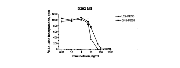
[0019] Figures 6A-C. Figures 6A-C are graphs showing the cytotoxicity of
immunotoxins
L22-PE38 and G49-PE38 on GPNMB+ and on GPNMB- cells. Figure 6A: cytotoxicity
of
the immunotoxins to GPNMB+ cell line D392 MG. Figure 6B: cytotoxicity of the
immunotoxins to GPNMB- cell line HEK293. Figure 6C: cytotoxicity of the
immunotoxins
to GPNMB+ cell line D54MG. All Figures: Squares: G49-PE38 immunotoxin.
Triangles:
L22-PE38 iinmunotoxin. Vertical axes: incorporation of 3H-Leucine, in cpm.
Horizontal
axes: Concentration of immunotoxin, in ng/ml.
[0020] Figures 7A and B. Figure 7A is an aligmnent of the amino acid sequences
of the
heavy chains of antibodies G49, L22, B307, 902V, 201, B308, B305, L04, L12,
and L15
(SEQ ID NOS:l-10), and the sequence of a linker (SEQ ID NO:11) connecting the
heavy
chain to the light chain in the scFvs of these antibodies. Figure 7B is an
alignment of the
amino acid sequences of the light chains of antibodies G49, L22, B307, 902V,
201, B308,
B305, L04, L12, and L15 (SEQ ID NOS:12-21). The framework regions ("FRs") and
complementarity determining regions ("CDRs") for each chain are labeled; the
sequences of
the CDRs are shown in bold face. In Figure 7A, the residues of the scFv G49
heavy chain
CDRl that were mutated to form scFvs B307, 902V, 201, B308, and B305 are
underlined. In
Figure 7B, the residues of the VL CDR3 of G49 that were mutated to form scFvs
L22, B307,
902V, 201, B308, B305, L04, L12, and L15 are underlined.
7
CA 02627890 2008-04-29
WO 2007/053718 PCT/US2006/042735
DETAILED DESCRIPTION
INTRODUCTION
[0021] The human transmembrane glycoprotein nonmetastatic melanoma protein B
("GPNMB ") and a splice variant form in which there is an in-frame insertion
of 12 amino
acids in the extracellular domain of the protein have been found to be highly
expressed in the
cells several forms of brain cancer, as compared to nonnal brain cells. In
particular, both the
protein and its splice variant have been found to be overexpressed in
glioblastoma
multifornies, anaplastic astrocytoinas, anaplastic oligodendrogliomas, and
oligodendroglioma. See, Kuan et al., Proc Amer Assoc Cancer Research 43:277
(2002).
GPNMB is also expressed on some melanoma cells. Accordingly, it would be
useful to be
able to target agents preferentially to cells expressing GPNMB or its splice
variant.
[0022] The present invention provides new antibodies that bind GPNMB and to
its splice
variant with high affinity. It will be appreciated that intact antibodies are
bivalent, while
scFv and dsFv are monovalent, and that creating scFv or dsFv from an intact
antibody
typically results in a consequent loss of affinity compared to the antibody
used as a starting
material. Accordingly, to promote binding of immunoconjugates, such as
immunotoxins, to
the target cells, it is desirable that the antibody from which the scFv or
dsFv is generated has
a high affinity for the target antigen. Thus, the antibodies are useful agents
for targeting
labels, as well as toxins and other therapeutic agents, to GPNMB-expressing
cells.
[0023] Two of the present inventors previously reported that they were able to
generate
monoclonal antibodies against GPNMB. Kuan et al., Proc Amer Acad Cancer Res
44:1116-7
(2003). It turned out, however, that these antibodies did not internalize
well. This made the
antibodies unsuitable for use as the targeting portion of immunotoxins since
they would not
facilitate internalization of the cytotoxin portion of the immunotoxin into
the target cell. As
is appreciated by those of skill in the art, cytotoxins must be internalized
into a cell to kill it.
Unfortunately, the reasons one antibody is internalized and another is not are
not well
understood, and it is not possible to predict which antibodies will
internalize and which will
not. Further, although improving the affinity of the targeting portion of the
immunotoxin
tends to increase the time the immunotoxin binds to the cell and therefore
improves its
opportunity to be internalized, affinity of the targeting portion of the
immunotoxin, by itself,
does not necessarily correlate with the immunotoxins' cell-killing ability.
For example, the
immunotoxin may not be trafficked within the cell in a manner permitting
release of the toxin
8
CA 02627890 2008-04-29
WO 2007/053718 PCT/US2006/042735
portion into the cytosol. The antibodies that were generated by traditional
immunization
formats proved unsuitable for targeting cytotoxins to GPNMB-expressing cells.
[0024] In light of the failure to obtain antibodies that internalized through
monoclonal
antibody approaches, another approach was undertaken. This resulted in the
discovery of the
scFv designated as "G49", and a variant designated as "L22". Further work
resulted in the
discovery of additional variants of G49 or of L22, designated "B307", "902V,"
"201 ",
"B308", "B305," "L04", "L12", and "L15", respectively (the sequences of each
of these
antibodies is discussed in detail below). Surprisingly, and unlike the
antibodies generated by
immunizing animals, these antibodies not only have high affinity for GPNMB,
but also
internalize well. Further, when expressed as a recombinant immunotoxin, G49
had
significant cytotoxic effect on GPNMB-expressing cells, while the others
showed
surprisingly higher cytotoxicity to GPNMB-expressing cells than did a like G49-
based
immunotoxin (except for L15, which had the same cytotoxicity as did G49).
Thus, the anti-
GPNMB antibodies of the invention are surprisingly useful agents for targeting
cytotoxins to
GPNMB-expressing cells.
[0025] It should be noted that, even though the antibodies of the invention
internalize well,
they are still expected to remain on the surface of target cells long enougli
before
internalization so that they are still useful agents for delivery of
detectable labels for detection
of GPNMB-expressing cells in a biological sample or for imaging the location
of GPNMB-
expressing cells in a patient. Thus, while the monoclonal antibodies
previously available
could be used for labeling GPNMB-expressing cells, or for carrying to target
cells
radionuclides or other agents that do not need to enter cells to be effective,
they are not useful
for malcing immunotoxins. In contrast, the antibodies of the invention can be
used for
labeling GPNMB-expressing cells, for delivering to them agents that do not
have to enter the
cell to be effective, and can be used to make immunotoxins. The antibodies of
the invention
therefore have a broader range of uses than the anti-GPNMB antibodies
previously reported
in the art, and have uses for which the antibodies previously available in the
art are
unsuitable.
[0026] Immunotoxins are typically produced by expressing the recombinant
immunotoxins
in E. coli, where they accumulate in inclusion bodies. After the inclusion
bodies are washed
extensively, they are dissolved in guanidine hydrochloride and the protein
renatured and
purified by ion-exchange chromatography and gel filtration. To ease processing
and cost
9
CA 02627890 2008-04-29
WO 2007/053718 PCT/US2006/042735
concerns, it is advantageous if the immunotoxin can be produced with a high
yield. Often,
however, immunotoxins can only be produced with a yield of a few percent.
[0027] In one group of embodiments, therefore, the invention provides the anti-
GPNMB
antibodies designated by the inventors as G49, L22, B307, 902V, 201, B308,
B305, L04,
L12, and L15. The Fv regions of these antibodies are shown in Figures 7A and
B, which set
fortll the sequences of the variable heavy chain of each of these antibodies,
the sequence of
an exemplar peptide (SEQ ID NO: 11) used in the studies reported in the
Examples to link the
antibody heavy and light chains, and the sequences of the variable light
chains of the
antibodies. (For clarity, it is noted that the entire sequence of the variable
heavy or light
chain for each antibody could not be set forth on a single line in Figures 7A
and 7B. The
SEQ ID NO: shown on the first line for the heavy and for the light chain of
each antibody
therefore relate to the sequence of the entire chain, not just the sequence
shown on the first
line. Thus, there is no separate sequence number shown for the second line
since the second
line is a continuation of the sequence already identified by the SEQ ID NO:
for the heavy or
light chain in question.) The four framework regions ("FRs") of each chain of
each antibody
are labeled and numbered, as are the complementarity determining regions
("CDRs") 1, 2 and
3 of each chain. The residues at which the antibodies diverge from those of
G49 are
underlined. As can be seen, in CDR1 of the VH chain, G49, L22, L04, L12, and
L15 have
the same sequence, while B307 has a single substitution (of glycine for the
first serine), 902V
has two, and 201, B308, and B305 all have three. In CDR3 of the VL chain, six
of the nine
variants of G49 have a glutamic acid and a threonine at positions two and
three, respectively,
of the CDR, while two variants mutate all three of the first three positions
of the CDR and
one variant of G49 (L15) contains mutations of just the first two positions of
the CDR.
[0028] As set forth in the Examples, the inventors discovered the G49
antibody, which has
an affinity (KD) for the extracellular domain of GPNMB of 9.1 nM. When made
into an
immunotoxin using a potent cytotoxin, a 38 kD truncated form of Pseudomonas
exotoxin A
lcnown as "PE3 8," the resulting immunotoxin inhibited protein synthesis by
50% at a
concentration of 30 ng/ml in an exemplar GPNMB-expressing cell line (cell line
D392MG)
when the cells were exposed to the immunotoxin for 24 hours. In contrast, at
concentrations
of over 1000 ng/ml, the immunotoxin did not inhibit protein synthesis by 50%
in a control
cell line, HEK293, that does not express GPNMB. (The amount of an agent which
inhibits
protein synthesis by 50% is lcnown as the "IC50" of the agent, and is
considered an important
measure of the cytotoxicity of the agent.) See, Table 5, below.
CA 02627890 2008-04-29
WO 2007/053718 PCT/US2006/042735
[0029] As further shown in the Examples, mutating two residues in the VL CDR3
that are
encoded by codons with nucleotides which fall within a so-called "hot spot
motif' Pu-G-Py-
A/T (wherein "Pu" refers to a purine base and "Py" refers to a pyrimidine
base) resulted in
dramatically increasing the cytotoxicity when the resulting antibody,
designated L22, was
used in place of the G49 antibody in an exemplar immunotoxin. As shown in
Table 5 and
Figure 6, using the same linker peptide and the same toxic moiety to permit
ready
comparison, the cytotoxicity of the L22-PE38 construct was tested against that
of G49-PE38.
Remarlcably, despite only a two amino acid difference between the two
constructs, the L22-
PE38 construct was 5 times as cytotoxic as G49-PE38 on one GPNMB -expressing
cell line
(cell line D392MG), and more than 3 times as cytotoxicity as G49-PE38 on
anotlier
(D54MG). Further, when "hot spot" mutations were made in the VH CDR1, mutating
a
single residue of L22 (resulting in the B307 antibody) was found to increase
cytotoxicity of
the iinmunotoxin another 3 times against the D392MG cell line and 5 times
against the
D54MG cell line, with no apparent increase in cytotoxicity against the control
cell line.
Moreover, mutation of a second residue of the VH CDR1, resulting in the 902V
antibody,
resulted in yet a further doubling of cytotoxicity against the D392MG cell
line of an
immunotoxin made with the resulting antibody, and a further tripling of
cytotoxicity against
the D54MG cell line. As shown by Table 5, the immunotoxin made with the 902V
antibody
was 30 times more cytotoxic to the D392MG cell line than was a like
immunotoxin made
witli G49 as the targeting portion, and was 50 times more cytotoxic to the
D54MG cell line
than was the like immunotoxin made with G49 as the targeting portion.
[00301 The sequences of the VH CDR 1 for the antibodies are SEQ ID NOs:22-28,
respectively. As shown in Figure 7, all the antibodies share the same sequence
for VH
CDR2 (SEQ ID NO:29) and for VH CDR3 (SEQ ID NO:30). As shown in Figure 7, all
the
antibodies also share the same sequences for VL CDRl (SEQ ID NO:3 1) and for
VL CDR2
(SEQ ID NO:32), but show a variation in the first three residues of the VL
CDR3 of G49
(SEQ ID NO:33).
[0031] Persons of skill in the art will recognize that it is the
complementarity determining
regions ("CDRs") that are responsible for an antibody's specificity and
affinity, while the
framework regions contribute more generally to the 3-dimensional shape and
configuration of
the molecule and have less impact on the antibody's specificity and affinity.
Persons of skill
are also aware that, for example, conservative substitutions can typically be
made in the
framework regions (four of which are present in each variable light and heavy
chain), without
11
CA 02627890 2008-04-29
WO 2007/053718 PCT/US2006/042735
significantly affecting antigen binding or specificity. The sequences of each
of the FR
regions of the VH and of the VL chains of the antibodies are shown in Figure
7.
[0032] Persons of skill will also appreciate that the Fv region of the
antibody is the portion
that binds antigen, while the Fc region of the antibody is involved in
opsonization or other
effector fiuictions. Further, persons of skill will appreciate that the Fc
region is relatively
invariant for any given class of immunoglobulin (that is, IgG, IgM, IgA,
etc.). Thus, any
given Fv region could be grafted onto a Fc section to form an intact
immunoglobulin if
desired. Since smaller molecules tend to have better tumor penetration than do
larger
molecules, however, it is usually desirable to use antibody fragments that
retain antigen
recognition rather intact immunoglobulin, as the targeting portion of
immunotoxins intended
for use against solid tumors. Thus, the variable light and the variable heavy
chains that
constitute a Fv region are typically linked, either through a linker, to form
a construct known
as a scFv, or by engineering cysteines into the franiework region to permit
formation of a
disulfide bond between the chains, thereby creating a construct known as a
dsFv.
[0033] It will be appreciated that changes can be made in the antibodies
described herein,
such as changes in the framework regions, without significantly affecting the
ability of the
antibody to bind GPNMB. Thus, an antibody can readily be engineered which has
the CDRs
of the antibodies as shown in Figure 7, but which does not have framework
regions ("FRs")
having the sequence of those of these antibodies as described herein (since
all the antibodies
share the FRs of the G49 antibody, for convenience, the FRs are sometimes
referred to herein
as the FRs of the G49 antibody). To take some simple examples, a practitioner
could make a
conservative substitution of one residue in one FR in one chain of the Fv, or
of one residue in
each FR of one chain, or in each FR in each chain. For example, the
practitioner could
substitute a lysine ("K") for the arginine ("R") which is the last residue
shown for the VL FR4
in Figure 4 to preserve the positive charge the arginine would be expected to
have at
physiological pH. Similarly, an aspartic acid ("D") could be substituted for
the glutamic acid
("E") found at the 12th position in the VH FR1 to provide a substitution
preserving the
negative charge that the glutamic acid residue would be expected to have at
pllysiological pH.
The resulting antibodies could then be readily tested to confirm that the
mutations did not
affect the binding, cytotoxity or yield of immunotoxins made with the mutated
antibody.
Thus, the anti- GPNMB antibodies of the invention encompass antibodies that
bind GPNMB
and that comprise the VH CDR and the VL CDR sequences of the antibodies
described
herein, whether or not the sequence of the FRs is fully that of the G49
antibody.
12
CA 02627890 2008-04-29
WO 2007/053718 PCT/US2006/042735
[0034] The framework regions (non-CDR regions) of antibodies derived from non-
hunian
species can be engineered to replace residues found at particular positions in
the antibodies
the of non-human animals, such as mice, with residues more typically found at
the same
position in human antibodies. Antibodies engineered in these ways are referred
to as
"humanized antibodies" and are preferred, since they have a lower risk of
inducing side
effects and typically can remain in the circulation longer. Methods of
humanizing antibodies
are, however, known in the art and are set forth in, for example, U.S. Patent
Nos. 6,180,377;
6,407,213; 5,693,762; 5,585,089; and 5,530,101. The antibodies described
herein were
developed from a human library and it is expected that the framework regions
will not
provoke an immune response when administered to humans. Persons of skill can,
however,
use the information in the art regarding humanizing residues as a guide to
make modifications
in the frainework regions if desired.
[0035] Further, since the CDRs of the variable regions determine antibody
specificity, the
CDRs can be grafted or engineered into an antibody of choice to confer GPNMB-
binding
specificity upon that antibody. For example, the complementarity determining
regions
(CDRs), i.e., the antigen binding loops, of the antibodies whose sequences are
shown in
Figure 7, or of variants of these antibodies, can be grafted onto a human
antibody framework
of known three dimensional structure, as known in the art (see, e.g.,
W098/45322; WO
87/02671; U.S. Patent No 5,859,205; U.S. Patent No. 5,585,089; U.S. Patent No.
4,816,567;
EP Patent Application 0173494; Jones, et al. Nature 321:522 (1986); Verhoeyen,
et al.,
Science 239:1534 (1988), Riechmann, et al. Nature 332:323 (1988); and Winter &
Milstein,
Nature 349:293 (1991)) to create a GPNMB-binding antibody.
[0036] In some embodiments, the light chain and heavy chain of the variable
region are
joined by a disulfide bond between cysteines engineered into the framework
region to form a
disulfide-stabilized Fv, or "dsFv." Formation of dsFvs is known in the art,
and is taught in,
for example, Pastan, U.S. Patent No. 6,558,672, which is incorporated herein
by reference,
which sets forth a series of positions at which cysteines can be engineered
into the framework
region to facilitate formation of disulfide bonding between the chains. In
light of the '672
patent, as well as the various disulfide stabilized Fvs that are currently in
pre-clinical and
clinical trials, the choice of which particular pair of positions to mutate to
form the dsFv is
considered to be within the slcill of the practitioner. In accordance witli
the '672 patent, in
some embodiments, however, the Fv is engineered with a cysteine replacing the
residue
otherwise present at position 42, 43, 44, 45 or 46 of the light chain, and
engineering a
13
CA 02627890 2008-04-29
WO 2007/053718 PCT/US2006/042735
cysteine to replace the residue otherwise present at position 103, 104, 105,
or 106, of the
heavy chain, as the residues of the antibody would be numbered under the Kabat
system for
numbering antibody residues. On other embodiments, the Fv is engineered to
replace the
residue otherwise present at 43, 44, 45, 46 or 47 of the heavy chain and
mutating a nucleic
acid encoding the second variable region so that cysteine is encoded at
position 98, 99, 100,
or 101 of the light chain (with all positions stated in this paragraph
numbered according to the
Kabat nuinbering system).
[0037] Methods for manufacturing dsFvs are also known in the art. Typically,
the two
chains are expressed from separate plasmids in a prokaryotic host cell, such
as E. coli, and
allowed to bond before the protein is purified from the inclusion bodies.
Making of dsFvs is
exemplified in, for example, Mansfield et al., Blood, 90(5):2020-26 (1997) and
FitzGerald et
al., International Publication Number WO 98/41641.
[0038] In scFv embodiments, the VH and VL chains are expressed as a single
fusion
protein. In some embodiments, the chains are expressed with the VH chain and
the VL chain
expressed sequentially without a spacer or linker. More commonly, the two
chains are
separated by a linker. Conveniently, the linker is a series of glycines
separated by a serine
residue. To facilitate coinparison of the cytotoxicity of the immunotoxins
made with the
antibodies developed in the course of the studies reported herein, all the
immunotoxins were
made with the same linker, GGGGSGGGGSGGSA (SEQ ID NO:11). As is evident from
the
sequence, the linker has two repeats of four glycines followed by a serine (a
motif known
abbreviated as G4S; SEQ ID NO:45). The linker can be varied, for example, by
varying the
number of repeats of the G4S (SEQ ID NO:45) motif, such as by having one,
three, four or
five repeats of the motif. It will be appreciated, however, that scFvs have
been made in the
art for well over a decade and that a multitude of other suitable linlcer
peptides are known in
the art. The choice of the particular linker to be used with the scFvs of the
invention is within
the skill of the practitioner and is not critical to the practice of the
present invention.
[0039] In general, any peptide of about 4 to 20 amino acid residues can be
used so long as
it does not interfere with the proper folding or activity of the scFv, or of
the toxin moiety
when the scFv is made into an immunotoxin. The effect of the linker on the
activity of the
scFv or of the toxin moiety can be readily determined by assaying the binding
of the scFv to
its target and by assaying the cytotoxicity of the toxic moiety on cells
targeted by the scFv. A
decrease in binding affinity of the targeting moiety by more than 25% or a
decrease in
14
CA 02627890 2008-04-29
WO 2007/053718 PCT/US2006/042735
cytotoxicity of the toxin moiety by more than 25%, or both, indicate that the
particular linker
peptide tested is not suitable. Assays for determining the binding
capabilities of numerous
antibodies and ligands are known in the art. For exainple, the binding
affinity of a ligand
employed as the targeting molecule of the immunotoxin may be assayed by
measuring the
ability of the targeting molecule to displace a native ligand from its target
substrate. This
may be accomplished by labeling the native ligand and then incubating cells
bearing the
target receptor with a fixed amount of the labeled ligand and various
concentrations of the
ligand-containing immunotoxin. The amount of bound native ligand can be
determined by
detecting the amount of label bound to the target cell. Unlabeled native
ligand can be run as
a control.
[0040] The improved affinity of the antibodies and antibody fragments provided
by the
present invention can be incorporated into chimeric immunoconjugates to
improve the ability
of the chimeric immunoconjugate to target cells bearing the GPNMB antigen. The
iinmunoconjugates can, for example, bear a detectable label such as a
radioisotope, a
fluorescent moiety, or a reporter enzyme. These labeled immunoconjugates be
used, for
example, in in vitro assays to detect the presence of GPNMB-expressing cells
in a biological
sample or can introduced into a patient to permit imaging the location of
GPNMB-expressing
cells. Once again, the making of immunoconjugates using antibodies and
fragments thereof
is well known in the art and it is assumed that the person of skill is
familiar with the
considerable literature on the subject.
[0041] In another set of in vitro uses, the iminunoconjugate bears a cytotoxin
rather than a
detectable label. Such immunotoxins can be used to purge GPNMB-expressing
cells in a
sample from a patient. The cells can then be cultured or used in further
studies.
[0042] In in vivo uses, iinmunotoxins made with the antibodies or antibody
fragments of
the invention can be used to inhibit the growth and proliferation of cancer
cells bearing the
GPNMB antigen. The high affinity of the antibodies and antibody fragments of
the invention
and the high cytotoxicity of iinmunotoxins made with them means that
relatively small
amounts of the immunotoxins can be administered to achieve a desired
therapeutic effect.
Since side effects are often dose-dependent, the relatively small amount of
iinmunotoxin that
has to be administered to achieve a given tllerapeutic effect should reduce
the occurrence of
side effects in patients being administered the iminunotoxin and a reduction
of the severity of
side effects in patients that do experience them.
CA 02627890 2008-04-29
WO 2007/053718 PCT/US2006/042735
[0043] For ease of comparison, the antibodies of the invention were tested
using the same
cytotoxin, PE38. As discussed in more detail in the section on cytotoxins
below, a number of
variants of Pseudonaonas exotoxin A are known in the art. All share the same
mechanism of
action and all would be expected be equally potent wlien used in in vitro
uses. PE38 and its
variant PE38QQR are somewhat preferred to PE40 for in vivo use against solid
tumors since
they are somewhat smaller and may permit better penetration of the immunotoxin
into the
tumor. In addition to PE, other cytotoxins suitable for use in immunotoxins
are known in the
art and can be used in place of PE38 in creating immunotoxins employing the
anti-GPNMB
antibodies of the invention.
[0044] In some einbodiments, the antibody is a scFv or a dsFv. Many of the
recombinant
immunotoxins produced from constructs of scFv are one-third the size of IgG-
toxin chemical
conjugates and are homogeneous in composition. Elimination of the constant
portion of the
IgG molecule from the scFv results in faster clearance of the immunotoxin
after injection into
animals, including primates, and the smaller size of the conjugates improves
drug penetration
in solid tumors. Together, these properties lessen the side effects associated
with the toxic
moiety by reducing the time in which the immunotoxin (IT) interacts with non-
target tissues
and tissues that express very low levels of antigen.
[0045] These advantages, however, are offset to some degree by the loss of
antigen binding
affinity that occurs when IgGs are converted to scFvs (Reiter et al., Nature
Biotechnol.
14:239-1245 (1996)). Increasing affinity has been shown to improve selective
tumor delivery
of scFvs (Adams et al., Cancer Res. 58:485-490 (1998)), and is likely to
increase their
usefulness in tumor imaging and treatment. Therefore, increasing the affinity
of scFvs and
other targeting moieties (such as dsFvs, Fabs. and F(ab')2 of immunoconjugates
is desirable
to improve the efficiency of these agents in delivering effector molecules,
such as toxins and
other therapeutic agents, to their intended targets. The improved affinity of
the antibodies of
the invention therefore is an important advance in the delivery of labels and
especially toxins
to cells of GPNMB-expressing cancers.
DEFINITIONS
[0046] Units, prefixes, and symbols are denoted in their Systeme International
de Unites
(SI) accepted form. Numeric ranges are inclusive of the numbers defining the
range. Unless
otherwise indicated, nucleic acids are written left to right in 5' to 3'
orientation; amino acid
sequences are written left to right in amino to carboxy orientation. The
headings provided
16
CA 02627890 2008-04-29
WO 2007/053718 PCT/US2006/042735
herein are not limitations of the various aspects or embodiments of the
invention, which can
be had by reference to the specification as a whole. Accordingly, the terms
defined
immediately below are more fully defined by reference to the specification in
its entirety.
[0047] Glycoprotein nonmetastatic melanoma protein B, or "GPNMB", refers to a
highly
glycosylated type I transmembrane protein first discovered a decade ago from a
subtractive
cDNA library of high and low metastatic human melanoma cell lines. Weterman et
al., Int J
Cancer. 60(l):73-81 (1995). The human gpnnzb gene encodes a predicted 560-
amino acid
protein, the deduced arnino acid sequence of which shows that GPNMB is made up
of three
domains: an extracellular domain (464 amino acids) preceded by a signal
peptide, a single
transmembrane region, and a relatively short cytoplasmic domain composed of 53
amino acid
residues. Although the biological function of GPNMB remains to be seen,
transfection of a
minimally.transformed human fetal astrocyte cell line witll gpnmb eDNA
resulted in a
change in the phenotype of the tumor xenografts from minimally invasive to
highly invasive
and metastatic
[0048] Persons of skill will recognize that it is the extracellular domain of
GPNMB which
is the portion exposed on the exterior of the cell and therefore available to
be bound by the
antibodies and compositions of the invention. Unless otherwise required by
context,
therefore,references herein to binding GPNMB refer to binding of the
extracellular domain of
the glycoprotein. For additional specificity, the extracellular domain will
occasionally be
referred to herein as the GPNMBECD. Human GPNMB exists both in its native form
("GPNMBwt") and a splice variant form in which there is a 12-amino acid in-
frame insertion
in the extracellular domain ("GPNMBsv").
[0049] For convenience of reference, as used herein, the term "antibody"
includes whole
(sometimes referred to herein as "intact") antibodies, antibody fragments that
retain antigen
recognition and binding capability, whether produced by the modification of
whole
antibodies or synthesized de novo using recombinant DNA methodologies,
monoclonal
antibodies, polyclonal antibodies, and antibody mimics, unless otherwise
required by context.
The antibody may be an IgM, IgG (e.g. IgG1, IgG2, IgG3 or IgG4), IgD, IgA or
IgE.
[0050] The term "antibody fragments" means molecules that comprise a portion
of an
intact antibody, generally the antigen binding or variable region of the
intact antibody.
Examples of antibody fragments include Fab, Fab', F(ab')2, and Fv fragments;
helix-stabilized
antibodies (see, e.g., Arndt et al., J Mol Biol 312:221-228 (2001); diabodies
(see below);
17
CA 02627890 2008-04-29
WO 2007/053718 PCT/US2006/042735
single-chain antibody molecules ("scFvs," see, e.g., U.S. Patent No.
5,888,773); disulfide
stabilized antibodies ("dsFvs", see, e.g., U.S. Patent No. 5,747,654 and
6,558,672), and
domain antibodies ("dAbs," see, e.g., Holt et al., Trends Biotech 21(11):484-
490 (2003),
Ghahroudi et al., FEBS Lett. 414:521-526 (1997), Lauwereys et al., EMBO J
17:3512-3520
(1998), Reiter et al., J. Mol. Biol. 290:685-698 (1999), Davies and Riechmann,
Biotechnology, 13:475-479 (2001)).
[0051] The term "diabodies" refers to small antibody fragments with two
antigen-binding
sites, which fragments comprise a variable heavy domain ("VH " or "VH")
connected to a
variable liglit domain ("VL" or "VL") in the same polypeptide chain (VH-VL).
By using a
linker that is too short to allow pairing between the two domains on the same
chain, the
domains are forced to pair with the complementary domains of another chain and
create two
antigen-binding sites. Diabodies and their production are described more fully
in, for
example, EP 404,097; WO 93/11161; and Hollinger et al., Proc. Natl. Acad. Sci.
USA, 90:
6444-6448 (1993).
[0052] References to "VH" or a "VH" refer to the variable region of an
immunoglobulin
heavy chain, including of an Fv, scFv , dsFv or Fab. References to "VL" or a
"VL" refer to
the variable region of an immunoglobulin light chain, including of an Fv, scFv
, dsFv or Fab.
The amino acid numbering and CDR delimitation of the G49 antibody was
determined
according to the IMGT database (Lefranc, M.P., IMGT, the international
ImMunoGeneTics
database. Nucleic Acids Res, 31(1): 307-10 (2003)). For nuinbering amino acid
residues of
the antibodies for preparation of disulfide stabilized antibodies, references
to amino acid
positions of the heavy or light chains refer to the numbering of the amino
acids under the
"Kabat" system (Kabat, E., et al., Sequences of Proteins of Immunological
Interest, U.S.
Government Printing Office, NIH Publication No. 91-3242 (1991). Since the
numbering of a
residue under the Kabat system aligns it to other antibodies to permit
determination of the
residues in the framework regions and the CDRs, the number assigned to a
residue under the
system does not necessarily correspond to the number that one might obtain for
a residue in a
given heavy or light chain by simply counting from the amino terminus of that
chain. Thus,
the position of an amino acid residue in a particular VH or VL sequence does
not refer to the
number of amino acids in a particular sequence, but rather refers to the
position as designated
with reference to the Kabat numbering scheme.)
18
CA 02627890 2008-04-29
WO 2007/053718 PCT/US2006/042735
[0053] The phrase "single chain Fv" or "scFv" refers to an antibody in which
the variable
domains of the heavy chain and of the light chain of a traditional two chain
antibody have
been joined to form one chain. Typically, a linker peptide is inserted between
the two chains
to allow for proper folding and creation of an active binding site.
[0054] The term "linker peptide" includes reference to a peptide within an
antibody binding
fragment (e.g., Fv fragment) which serves to indirectly bond the variable
domain of the heavy
chain to the variable domain of the light chain.
[0055] The term "parental antibody" means an antibody of interest which is to
be or has
been mutated or varied to obtain antibodies or fragments thereof which bind to
the same
epitope as the parental antibody, but with higher affinity.
[0056] The term "hotspot" means a portion of a nucleotide sequence of a CDR or
of a
framework region of a variable domain which is a site of particularly high
natural variation.
Although CDRs are themselves considered to be regions of hypervariability, it
has been
learned that mutations are not evenly distributed throughout the CDRs.
Particular sites, or
hotspots, have been identified as locations which undergo concentrated
mutations. The
hotspots are characterized by a number of structural features and sequences.
These "hotspot
motifs" can be used to identify llotspots. Two consensus sequences motifs
which are
especially well characterized are the tetranucleotide sequence RGYW and the
serine sequence
AGY, where R is A or G, Y is C or T, and W is A or T.
[0057] A "targeting moiety" or "targeting portion" is the portion of an
immunoconjugate
intended to target the immunoconjugate to a cell of interest. Typically, the
targeting moiety
is an antibody, or a fragment of an antibody that retains antigen recognition
capability, such
as a scFv, a dsFv, an Fab, or an F(ab ')2.
[0058] Typically, an immunoglobulin has a heavy and light chain. Each heavy
and light
chain contains a constant region and a variable region, (the regions are also
known as
"domains"). Light and heavy chain variable regions contain a"frameworlc"
region
interrupted by three hypervariable regions, also called "complementarity-
determining
regions" or "CDRs". The extent of the framework region and CDRs have been
defined. The
sequences of the framework regions of different light or heavy chains are
relatively
conserved within a species. The framework region of an antibody, that is the
combined
19
CA 02627890 2008-04-29
WO 2007/053718 PCT/US2006/042735
framework regions of the constituent light and heavy chains, serves to
position and align the
CDRs in three dimensional space.
[0059] The CDRs are primarily responsible for binding to an epitope of an
antigen. The
CDRs of each chain are typically referred to as CDRl, CDR2, and CDR3, numbered
sequentially starting from the N-terminus, and are also typically identified
by the chain in
which the particular CDR is located. Thus, a VH CDR3 is located in the
variable domain of
the heavy chain of the antibody in which it is found, whereas a VL CDR1 is the
CDRI from
the variable domain of the light chain of the antibody in which it is found.
[0060] References to "VH" or a "VH" refer to the variable region of an
immunoglobulin
heavy chain, including an Fv, scFv, dsFv or Fab. References to "VL" or a "VL"
refer to the
variable region of an immunoglobulin light chain, including of an Fv, scFv,
dsFv or Fab
[0061] The phrase "single chain Fv" or "scFv" refers to an antibody in which
the variable
domains of the heavy chain and of the light chain of a traditional two chain
antibody have
been joined to form one chain. Typically, a linker peptide is inserted between
the two chains
to allow for proper folding and creation of an active binding site.
[0062] The phrase "disulfide bond" or "cysteine-cysteine disulfide bond"
refers to a
covalent interaction between two cysteines in which the sulfiu atoms of the
cysteines are
oxidized to form a disulfide bond. The average bond energy of a disulfide bond
is about 60
kcal/mol compared to 1-2 kcal/mol for a hydrogen bond.
[0063] The phrase "disulfide stabilized Fv" or "dsFv" refer to the variable
region of an
immunoglobulin in which there is a disulfide bond between the light chain and
the heavy
chain. In the context of this invention, the cysteines which form the
disulfide bond are within
the framework regions of the antibody chains and serve to stabilize the
conformation of the
antibody. Typically, the antibody is engineered to introduce cysteines in the
framework
region at positions where the substitution will not interfere with antigen
binding.
[0064] An antibody immunologically reactive with a particular antigen can be
generated by
recoinbinant methods such as selection of libraries of recombinant antibodies
in phage or
similar vectors, see, e.g., Huse, et al., Science 246:1275-1281 (1989); Ward,
et al., Nature
341:544-546 (1989); and Vaughan, et al., Nature Biotech. 14:309-314 (1996), or
by
immunizing an animal with the a.ntigen or with DNA encoding the antigen.
CA 02627890 2008-04-29
WO 2007/053718 PCT/US2006/042735
[0065] A "toxic moiety" is the portion of a immunotoxin which renders the
immunotoxin
cytotoxic to cells of interest.
[0066] A "therapeutic moiety" is the portion of an iinmunoconjugate intended
to act as a
therapeutic agent.
[0067] The term "therapeutic agent" includes any number of compounds currently
lcnown
or later developed to act as anti-neoplastics, anti-inflammatories, cytokines,
anti-infectives,
enzyme activators or inhibitors, allosteric modifiers, antibiotics or other
agents administered
to induce a desired therapeutic effect in a patient. The therapeutic agent may
also be a toxin
or a radioisotope, where the therapeutic effect intended is, for example, the
killing of a cancer
cell.
[0068] A "detectable label" means, with respect to an immunoconjugate, a
portion of the
immunoconjugate which has a property rendering its presence detectable. For
example, the
immunoconjugate may be labeled with a radioactive isotope which permits cells
in which the
immunoconjugate is present to be detected in immunohistochemical assays.
[0069] The term "effector moiety" means the portion of an immunoconjugate
intended to
have an effect on a cell targeted by the targeting moiety or to identify the
presence of the
immunoconjugate. Thus, the effector moiety can be, for example, a therapeutic
moiety, a
toxin, a radiolabel, or a fluorescent label.
[0070] The term "immunoconjugate" includes reference to a covalent linkage of
an effector
molecule to an antibody. The effector molecule can be a cytotoxin.
[0071] The terms "effective amount" or "amount effective to" or
"therapeutically effective
amount" includes reference to a dosage of a therapeutic agent sufficient to
produce a desired
result, such as inlzibiting cell protein synthesis by at least 50%, or killing
the cell.
[0072] The term "toxin" includes reference to abrin, ricin, Pseudomonas
exotoxin A(PE),
diphtheria toxin (DT), botulinum toxin, or modified toxins thereof. For
example, PE and DT
are highly toxic compounds that typically bring about death through liver
toxicity. PE and
DT, however, can be modified into a form for use as an immunotoxin by removing
the native
targeting component of the toxin (e.g., domain Ia of PE or the B chain of DT)
and replacing it
with a different targeting moiety, such as an antibody.
21
CA 02627890 2008-04-29
WO 2007/053718 PCT/US2006/042735
100731 The term "contacting" includes reference to placement in direct
physical
association.
[0074] An "expression plasmid" comprises a nucleotide sequence encoding a
molecule or
interest, which is operably linked to a promoter.
[0075] As used herein, "polypeptide", "peptide" and "protein" are used
interchangeably and
include reference to a polymer of amino acid residues. The terms apply to
amino acid
polymers in which one or more amino acid residue is an artificial chemical
analogue of a
corresponding naturally occurring amino acid, as well as to naturally
occurring amino acid
polymers. The terms also apply to polymers containing conservative amino acid
substitutions
such that the protein remains functional.
[0076] The term "residue" or "amino acid residue" or "anlino acid" includes
reference to an
amino acid that is incorporated into a protein, polypeptide, or peptide
(collectively "peptide").
The amino acid can be a naturally occurring amino acid and, unless otherwise
limited, can
encompass known analogs of natural amino acids that can function in a similar
manner as
naturally occurring amino acids.
[0077] The amino acids and analogs referred to herein are described by
shorthand
designations as follows in Table A:
Table A: Amino Acid Nomenclature
Name 3-letter 1-letter
Alanine Ala A
Arginine Arg R
Asparagine Asn N
Aspartic Acid Asp D
Cysteine Cys C
Glutamic Acid Glu E
Glutarnine Gln Q
Glycine Gly G
Histidine His H
Homoserine Hse -
Isoleucine Ile I
Leucine Leu L
22
CA 02627890 2008-04-29
WO 2007/053718 PCT/US2006/042735
Lysine Lys K
Methionine Met M
Methionine sulfoxide Met (0)
-
Methionine
methylsulfonium Met (S-Me) -
Norleucine Nle -
Phenylalanine Phe F
Proline Pro P
Serine Ser S
Threonine Thr T
Tryptophan Trp W
Tyrosine Tyr Y
Valine Val V
[00781 A "conservative substitution", when describing a protein refers to a
change in the
amino acid composition of the protein that does not substantially alter the
protein's activity.
Thus, "conservatively modified variations" of a particular amino acid sequence
refers to
amino acid substitutions of those amino acids that are not critical for
protein activity or
substitution of amino acids with otlier amino acids having similar properties
(e.g., acidic,
basic, positively or negatively charged, polar or non-polar, etc.) such that
the substitutions of
even critical amino acids do not substantially alter activity. Conservative
substitution tables
providing functionally similar amino acids are well known in the art. The
following six
groups in Table B each contain amino acids that are conservative substitutions
for one
another:
Table B
1) Alanine (A), Serine (S), Threonine (T);
2) Aspartic acid (D), Glutamic acid (E);
3) Asparagine (N), Glutainine (Q);
4) Arginine (R), Lysine (K);
5) Isoleucine (I), Leucine (L), Methionine (M), Valine (V); and
6) Phenylalanine (F), Tyrosine (Y), Tryptophan (W).
See also, Creighton, Proteins : Structures and Molecular Properties, W.H.
Freeman and Company, New York (2nd Ed., 1992).
23
CA 02627890 2008-04-29
WO 2007/053718 PCT/US2006/042735
[0079] The terms "substantially similar" in the context of a peptide indicates
that a peptide
comprises a sequence with at least 90%, preferably at least 95% sequence
identity to the
reference sequence over a comparison window of 10-20 amino acids. Percentage
of sequence
identity is determined by comparing two optimally aligned sequences over a
comparison
window, wherein the portion of the polynucleotide sequence in the comparison
window may
comprise additions or deletions (i.e., gaps) as compared to the reference
sequence (which
does not comprise additions or deletions) for optimal aligiunent of the two
sequences. The
percentage is calculated by determining the number of positions at which the
identical nucleic
acid base or amino acid residue occurs in both sequences to yield the number
of matched
positions, dividing the number of matched positions by the total number of
positions in the
window of comparison and multiplying the result by 100 to yield the percentage
of sequence
identity.
[0080] The terms "conjugating," "joining," "bonding" or "linking" refer to
making two
polypeptides into one contiguous polypeptide molecule. In the context of the
present
invention, the terins include reference to joining an antibody moiety to an
effector molecule
(EM). The linkage can be either by chemical or recombinant means. "Chemical
means"
refers to a reaction between the antibody moiety and the effector molecule
such that there is a
covalent bond formed between the two molecules to form one molecule, while
"recombinant
means" refers to expression of a nucleic acid resulting in production of a
single, fusion
protein which did not first exist as two separate molecules.
[0081] As used herein, "recombinant" includes reference to a protein produced
using cells
that do not have, in their native state, an endogenous copy of the DNA able to
express the
protein. The cells produce the recombinant.protein because they have been
genetically
altered by the introduction of the appropriate isolated nucleic acid sequence.
The term also
includes reference to a cell, or nucleic acid, or vector, that has been
modified by the
introduction of a heterologous nucleic acid or the alteration of a native
nucleic acid to a forin
not native to that cell, or that the cell is derived from a cell so modified.
Thus, for example,
recombinant cells express genes that are not found within the native (non-
recombinant) form
of the cell, express mutants of genes that are found within the native form,
or express native
genes that are otherwise abnormally expressed, underexpressed or not expressed
at all.
[0082] As used herein, "nucleic acid" or "nucleic acid sequence" includes
reference to a
deoxyribonucleotide or ribonucleotide polymer in either single- or double-
stranded form, and
24
CA 02627890 2008-04-29
WO 2007/053718 PCT/US2006/042735
unless otherwise limited, encompasses known analogues of natural nucleotides
that hybridize
to nucleic acids in a manner similar to naturally occurring nucleotides.
Unless otlierwise
indicated, a particular nucleic acid sequence includes the coinplementary
sequence thereof as
well as conservative variants, i.e., nucleic acids present in wobble positions
of codons and
variants that, when tra.nslated into a protein, result in a conservative
substitution of an amino
acid.
[0083] As used herein, "encoding" with respect to a specified nucleic acid,
includes
reference to nucleic acids which comprise the information for translation into
the specified
protein. The information is specified by the use of codons. Typically, the
amino acid
sequence is encoded by the nucleic acid using the "universal" genetic code.
However,
variants of the universal code, such as is present in some plant, animal, and
fungal
mitochondria, the bacterium M,ycoplasma capricolum (Proc. Nat'l Acad. Sci. USA
82:2306-
2309 (1985), or the ciliate Macronucleus, may be used when the nucleic acid is
expressed in
using the translational machinery of these organisms.
[0084] The phrase "fusing in frame" refers to joining two or more nucleic acid
sequences
which encode polypeptides so that the joined nucleic acid sequence translates
into a single
chain protein which comprises the original polypeptide chains.
[00851 As used herein, "expressed" includes reference to translation of a
nucleic acid into a
protein. Proteins may be expressed and remain intracellular, become a
component of the cell
surface membrane or be secreted into the extracellular matrix or medium.
[0086] By "host cell" is meant a cell which can support the replication or
expression of the
expression vector. Host cells may be prokaryotic cells such as E. coli, or
eukaryotic cells
such as yeast, insect, amphibian, or mammalian cells.
[0087] The phrase "phage display library" refers to a population of
bacteriophage, each of
which contains a foreign cDNA recombinantly fused in frame to a surface
protein. The
phage display the foreign protein encoded by the cDNA on its surface. After
replication in a
bacterial host, typically E. coli, the phage which contain the foreign cDNA of
interest are
selected by the expression of the foreign protein on the phage surface.
[0088] The terms "identical" or percent "identity," in the context of two or
more nucleic
acids or polypeptide sequences, refer to two or more sequences or subsequences
that are the
same or have a specified percentage of amino acid residues or nucleotides that
are the same,
CA 02627890 2008-04-29
WO 2007/053718 PCT/US2006/042735
when compared and aligned for maximum correspondence, as measured using one of
the
following sequence comparison algorithms or by visual inspection.
[0089] The phrase "substantially identical," in the context of two nucleic
acids or
polypeptides, refers to two or more sequences or subsequences that have at
least 60%, more
preferably 65%, even more preferably 70%, still more preferably 75%, even more
preferably
80%, and most preferably 90-95% nucleotide or amino acid residue identity,
when compared
and aligned for maximum correspondence, as measured using one of the following
sequence
comparison algorithms or by visual inspection. Preferably, the substantial
identity exists over
a region of the sequences that is at least about 50 residues in length, more
preferably over a
region of at least about 100 residues, still more preferably over at least
about 150 residues
and most preferably over the full length of the sequence. In a most preferred
embodiment,
the sequences are substantially identical over the entire length of the coding
regions.
[0090] For sequence comparison, typically one sequence acts as a reference
sequence, to
which test sequences are compared. When using a sequence comparison algorithm,
test and
reference sequences are input into a coinputer, subsequence coordinates are
designated, if
necessary, and sequence algorithm program parameters are designated. The
sequence
comparison algorithm then calculates the percent sequence identity for the
test sequence(s)
relative to the reference sequence, based on the designated program
parameters.
[0091] Optimal alignment of sequences for comparison can be conducted, e.g.,
by the local
homology algorithm of Smith & Waterman, Adv. Appl. Math. 2:482 (1981), by tlie
homology
alignment algorithm of Needleman & Wunsch, J. Mol. Biol. 48:443 (1970), by the
search for
siinilarity method of Pearson & Lipman, Proc. Nat'l. Acad. Sci. USA 85:2444
(1988), by
computerized implementations of these algoritluns (GAP, BESTFIT, FASTA, and
TFASTA
in the Wisconsin Genetics Software Package, Genetics Computer Group, 575
Science Dr.,
Madison, WI), or by visual inspection (see generally, Current Protocols in
Molecular
Biology, F.M. Ausubel et al., eds., Current Protocols, ajoint venture between
Greene
Publishing Associates, Inc. and John Wiley & Sons, Inc., (1995 Supplement)
(Ausubel)).
[0092] Examples of algorithms that are suitable for determining percent
sequence identity
and sequence similarity are the BLAST and BLAST 2.0 algorithms, which are
described in
Altschul et al. (1990) J. Mol. Biol. 215: 403-410 and Altschuel et al. (1977)
Nucleic Acids
Res. 25: 3389-3402, respectively. Software for performing BLAST analyses is
publicly
available through the National Center for Biotechnology Information
26
CA 02627890 2008-04-29
WO 2007/053718 PCT/US2006/042735
(http://www.ncbi.nlm.nih.gov/). This algorithm involves first identifying high
scoring
sequence pairs (HSPs) by identifying short words of length W in the query
sequence, which
either match or satisfy some positive-valued threshold score T when aligned
with a word of
the same length in a database sequence. T is referred to as the neigliborhood
word score
threshold (Altschul et al, sups=a). These initial neighborhood word hits act
as seeds for
initiating searches to find longer HSPs containing them. The word hits are
then extended in
both directions along each sequence for as far as the cumulative alignment
score can be
increased. Cumulative scores are calculated using, for nucleotide sequences,
the parameters
M (reward score for a pair of matching residues; always > 0) and N (penalty
score for
mismatching residues; always < 0). For amino acid sequences, a scoring matrix
is used to
calculate the cumulative score. Extension of the word hits in each direction
are halted when:
the cumulative alignment score falls off by the quantity X from its maximum
achieved value;
the cumulative score goes to zero or below, due to the accumulation of one or
more negative-
scoring residue alignments; or the end of either sequence is reached. The
BLAST algorithm
parameters W, T, and X determine the sensitivity and speed of the alignment.
The BLASTN
program (for nucleotide sequences) uses as defaults a wordlength (W) of 11, an
expectation
(E) of 10, M=5, N=-4, and a comparison of both strands. For amino acid
sequences, the
BLASTP program uses as defaults a wordlength (W) of 3, an expectation (E) of
10, and the
BLOSUM62 scoring matrix (see Henikoff & Henikoff, Proc. Natl. Acad. Sci. USA
89:10915
(1989)).
[0093] In addition to calculating percent sequence identity, the BLAST
algorithm also
performs a statistical analysis of the similarity between two sequences (see,
e.g., Karlin &
Altschul, Proc. Nat'l. Acad.. Sci. USA 90:5873-5787 (1993)). One measure of
similarity
provided by the BLAST algorithm is the smallest sum probability (P(N)), which
provides an
indication of the probability by which a match between two nucleotide or amino
acid
sequences would occur by chance. For example, a nucleic acid is considered
similar to a
reference sequence if the smallest sum probability in a comparison of the test
nucleic acid to
the reference nucleic acid is less than about 0.1, more preferably less than
about 0.01, and
most preferably less than about 0.001.
[0094] A further indication that two nucleic acid sequences or polypeptides
are
substantially identical is that the polypeptide encoded by the first nucleic
acid is
immunologically cross reactive witli the polypeptide encoded by the second
nucleic acid, as
described below. Thus, a polypeptide is typically substantially identical to a
second
27
CA 02627890 2008-04-29
WO 2007/053718 PCT/US2006/042735
polypeptide, for example, where the two peptides differ only by conservative
substitutions.
Another indication that two nucleic acid sequences are substantially identical
is that the two
molecules hybridize to each otlier under stringent conditions, as described
below.
[0095] The terin "in vivo" includes reference to inside the body of the
organism from which
the cell was obtained. "Ex vivo" and "in vitro " means outside the body of the
organism from
which the cell was obtained.
[0096] The phrase "malignant cell" or "malignancy" refers to tumors or tumor
cells that are
invasive and/or able to undergo metastasis, i.e., a cancerous cell.
[0097] As used herein, "mammalian cells" includes reference to cells derived
from
mammals including humans, rats, mice, guinea pigs, chimpanzees, or macaques.
The cells
may be cultured in vivo or in vitro.
[0098] The term "selectively reactive" refers, with respect to an antigen, the
preferential
association of an antibody, in whole or part, with a cell or tissue bearing
that antigen and not
to cells or tissues lacking that antigen. It is, of course, recognized that a
certain degree of
non-specific interaction may occur between a molecule and a non-target cell or
tissue.
Nevertheless, selective reactivity, may be distinguished as mediated through
specific
recognition of the antigen. Although selectively reactive antibodies bind
antigen, they may
do so with low affinity. On the other hand, specific binding results in a much
stronger
association between the antibody and cells bearing the antigen than between
the bound
antibody and cells lacking the antigen. Specific binding typically results in
greater than 2-
fold, preferably greater than 5-fold, more preferably greater than 10-fold and
most preferably
greater than 100-fold increase in amount of bound antibody (per unit time) to
a cell or tissue
bearing GPNMB as compared to a cell or tissue lacking GPNMB. Specific binding
to a
protein under such conditions requires an antibody that is selected for its
specificity for a
particular protein. A variety of immunoassay formats are appropriate for
selecting antibodies
specifically immunoreactive with a particular protein. For example, solid-
phase ELISA
immunoassays are routinely used to select monoclonal antibodies specifically
immunoreactive with a protein. See Harlow & Lane, ANTIBODIES, A LABORATORY
MANUAL,
Cold Spring Harbor Publications, New York (1988), for a description of
immunoassay
formats and conditions that can be used to determine specific
immunoreactivity.
[0099] The term "immunologically reactive conditions" includes reference to
conditions
which allow an antibody generated to a particular epitope to bind to that
epitope to a
28
CA 02627890 2008-04-29
WO 2007/053718 PCT/US2006/042735
detectably greater degree than, and/or to the substantial exclusion of,
binding to substantially
all other epitopes. Immunologically reactive conditions are dependent upon the
format of the
antibody binding reaction and typically are those utilized in immunoassay
protocols or those
conditions encountered in vivo. See Harlow & Lane, supra, for a description of
immunoassay formats and conditions. Preferably, the immunologically reactive
conditions
einployed in the methods of the present invention are "physiological
conditions" which
include reference to conditions (e.g., temperature, osmolarity, pH) that are
typical inside a
living mammal or a mammalian cell. While it is recognized that some organs are
subject to
extreme conditions, the intra-organismal and intracellular environment
normally lies around
pH 7 (i.e., from pH 6.0 to pH 8.0, more typically pH 6.5 to 7.5), contains
water as the
predominant solvent, and exists at a temperature above 0 C and below 50 C.
Osmolarity is
within the range that is supportive of cell viability and proliferation.
GLYCOPROTEIN NMB
[0100] The human gpnmb gene encodes a predicted 560-amino acid protein, the
deduced
amino acid sequence of which shows that GPNMB is made up of three domains, a
long
extracellular domain (ECD) preceded by a signal peptide, a single
transmembrane region, and
a relatively short cytoplasmic domain. The human GPNMB amino acid sequence had
homology of 71.1% to DC-HIL (Shikano et al., JBiol Chem, 276:8125-34 (2001)),
69.8% to
Osteoactivin (Safadi et al., J Cell Biochem, 84:12-26 (2001)), 56% to the
precursor of pMel
17 (Kwon et al., Proc Natl Acad Sci USA, 88:9228-32 (1991)), and 51% to QNR-71
(Turque
et al., Embo J, 15:3338-50 (1996)).
[0101] The human GPNMB gene was localized to human chromosome 7q15 (NCBI
Unigene Cluster Hs.82226 GPNMB), a locus involved in the human inherited
disease cystoid
macular dystrophy. Bachner et al suggested that human GPNMB may be a candidate
gene for
the dominant cystoid macular edema since they found high expression of murine
gpnmb
mRNA within the retinal and iris pigment epithelium (Bachner et al., Brain Res
Gene Expr
Patterns, 1:159-65 (2002)).
[0102] The function of GPNMB has not been fully described, and paradoxical
effects have
been noted in transfection studies. Transfection of an in vitro minimally
transformed human
fetal astrocyte line, THRG (Rich et al., Cancer Res, 61:3556-60 (2001); Rich
et al., JBiol
Chem (2003)) with gpnmb cDNA altered the phenotype of both subcutaneous and
intracranial
tumors growing in athymic mice from a minimally invasive to a highly invasive
and
29
CA 02627890 2008-04-29
WO 2007/053718 PCT/US2006/042735
metastatic phenotype. Conversely, transfection of a partial gpnmb eDNA into a
highly
metastatic melanoma cell line resulted in slower subcutaneous tumor growth and
also in
reduction of the potential for spontaneous metastasis in nude mice (Weterman
et al., Int J
Cancer, 60:73-81 (1995)). In studies of lii.gh-grade glioma (HGG) biopsy
samples by some
of the present inventors, gpnmb RNA transcripts were detected in 35/50 GBM
(70%), while
little or no gpnmb mRNA expression was noted in normal brain samples. By ,
immunohistochemical study of a larger HGG group, 75/108 GBM (70%) were
positive for
GPNMB protein expression. Furthermore, quantitative flow cytometric analysis
of fresh
GBM biopsy specimens revealed that cell-surface GPNMB molecular density ranged
from
1.1 to 7.8 x 10⁴ molecules. Its frequent expression in human HGGs and its cell-
surface
localization make GPNMB a good target for antibody-mediated delivery of
cytotoxic agents.
PRODUCTION OF IMMUNOCONJUGATES
[0103] Iminunoconjugates include, but are not limited to, molecules in which
there is a
covalent linkage of a therapeutic agent to an antibody. A therapeutic agent is
an agent with a
particular biological activity directed against a particular target molecule
or a cell bearing a
target molecule. One of skill in the art will appreciate that therapeutic
agents may include
various drugs such as vinblastine, daunomycin and the like, cytotoxins such as
native or
modified Pseudomonas exotoxin or Diphtheria toxin, encapsulating agents,
(e.g., liposomes)
which themselves contain pharmacological compositions, radioactive agents such
as 1251, 32P,
14C, 3H and 35S and other labels, target moieties and ligands.
[0104] The choice of a particular therapeutic agent depends on the particular
target
molecule or cell and the biological effect is desired to evoke. Thus, for
example, in some
embodiments, the therapeutic agent is a cytotoxin which is used to bring about
the death of a
particular target cell. Conversely, where it is merely desired to invoke a non-
lethal biological
response, the therapeutic agent may be conjugated to a non-lethal
pharmacological agent or a
liposome containing a non-lethal pharmacological agent.
[0105] With the therapeutic agents and antibodies herein provided, one of
skill can readily
construct a variety of clones containing functionally equivalent nucleic
acids, such as nucleic
acids which differ in sequence but which encode the same effector molecule
("EM") or
antibody sequence. Thus, the present invention provides nucleic acids encoding
antibodies
and conjugates and fusion proteins thereof.
CA 02627890 2008-04-29
WO 2007/053718 PCT/US2006/042735
A. Recombinant Methods
[0106] The nucleic acid sequences of the present invention can be prepared by
any suitable
method including, for example, cloning of appropriate sequences or by direct
cliemical
synthesis by methods such as the phosphotriester method of Narang, et al.,
Meth. Enzymol.
68:90-99 (1979); the phosphodiester method of Brown, et al., Meth. Enz.ymol.
68:109-151
(1979); the diethylphosphoramidite method of Beaucage, et al., Tetra. Lett.
22:1859-1862
(1981); the solid phase phosphoramidite triester metliod described by Beaucage
& Caruthers,
Tetra. Letts. 22(20):1859-1862 (1981), e.g., using an automated synthesizer as
described in,
for example, Needliain-VanDevanter, et al. Nucl. Acids Res. 12:6159-6168
(1984); and, the
solid support method of U.S. Patent No. 4,458,066. Chemical synthesis produces
a single
stranded oligonucleotide. This may be converted into double stranded DNA by
hybridization
with a complementary sequence, or by polymerization with a DNA polymerase
using the
single strand as a template. One of skill would recognize that while chemical
synthesis of
DNA is limited to sequences of about 100 bases, longer sequences may be
obtained by the
ligation of shorter sequences.
[0107] In a preferred embodiment, the nucleic acid sequences of this invention
are prepared
by cloning techniques. Examples of appropriate cloning and sequencing
techniques, and
instructions sufficient to direct persons of skill through many cloning
exercises are found in
Sambrook, et al., MOLECULAR CLONING: A LABORATORY MANUAL (2ND ED.), Vols. 1-3,
Cold Spring Harbor Laboratory (1989)), Berger and Kimmel (eds.), GUIDE TO
MOLECULAR
CLONING TECHNIQUES, Academic Press, Inc., San Diego CA (1987)), or Ausubel, et
al.
(eds.), CURRENT PROTOCOLS IN MOLECULAR BIOLOGY, Greene Publishing and Wiley-
Interscience, NY (1987). Product information from manufacturers of biological
reagents and
experimental equipment also provide useful information. Such manufacturers
include the
SIGMA chemical company (Saint Louis, MO), R&D systems (Minneapolis, MN),
Pharmacia
LKB Biotechnology (Piscataway, NJ), CLONTECH Laboratories, Inc. (Palo Alto,
CA),
Chem Genes Corp., Aldrich Chemical Company (Milwaukee, WI), Glen Research,
Inc.,
GIBCO BRL Life Technologies, Inc. (Gaithersberg, MD), Fluka Chemica-Biochemika
Analytika (Fluka Chemie AG, Buchs, Switzerland), Invitrogen, San Diego, CA,
and Applied
Biosystems (Foster City, CA), as well as many other coinmercial sources known
to one of
skill.
31
CA 02627890 2008-04-29
WO 2007/053718 PCT/US2006/042735
[0108] Nucleic acids encoding native EM or anti-GPNMB antibodies can be
modified to
form the antibodies or immunoconjugates of the present invention. Modification
by site-
directed mutagenesis is well lcnown in the art. Nucleic acids encoding anti-
GPNMB
antibodies or immunoconjugates can be amplified by in vitro methods.
Amplification
methods include the polymerase chain reaction (PCR), the ligase chain reaction
(LCR), the
transcription-based amplification system (TAS), the self-sustained sequence
replication
system (3SR). A wide variety of cloning methods, host cells, and in vitro
amplification
methodologies are well known to persons of skill.
[0109] In a preferred embodiment, immunoconjugates are prepared by inserting
the cDNA
which encodes an anti-GPNMB scFv antibody into a vector which comprises the
cDNA
encoding the EM. The insertion is made so that the scFv and the EM are read in
frame, that
is in one continuous polypeptide which contains a functional Fv region and a
fiinctional EM
region. In a particularly preferred embodiment, cDNA encoding a diphtheria
toxin fragment
is ligated to a scFv so that the toxin is located at the carboxyl terminus of
the scFv. In more
preferred embodiments, cDNA encoding PE is ligated to a scFv so that the toxin
is located at
the amino terminus of the scFv.
[0110] Once the nucleic acids encoding an EM, anti-GPNMB antibody, or an
immunoconjugate of the present invention are isolated and cloned, one may
express the
desired protein in a recombinantly engineered cell such as bacteria, plant,
yeast, insect and
mammalian cells. It is expected that those of skill in the art are
knowledgeable in the
nuinerous expression systems available for expression of proteins including E.
coli, other
bacterial hosts, yeast, and various higher eucaryotic cells such as the COS,
CHO, HeLa and
myeloma cell lines. No attempt to describe in detail the various methods known
for the
expression of proteins in prokaryotes or eukaryotes will be made. In brief,
the expression of
natural or synthetic nucleic acids encoding the isolated proteins of the
invention will typically
be achieved by operably linking the DNA or cDNA to a promoter (which is either
constitutive or inducible), followed by incorporation into an expression
cassette. The
cassettes can be suitable for replication and integration in either
prokaryotes or eukaryotes.
Typical expression cassettes contain transcription and translation
terminators, initiation
sequences, and promoters useful for regulation of the expression of the DNA
encoding the
protein. To obtain high level expression of a cloned gene, it is desirable to
construct
expression cassettes which contain, at the minimum, a strong promoter to
direct transcription,
a ribosome binding site for translational initiation, and a
transcription/translation terminator.
32
CA 02627890 2008-04-29
WO 2007/053718 PCT/US2006/042735
For E. coli this includes a promoter such as the T7, trp, lac, or lambda
promoters, a ribosome
binding site and preferably a transcription termination signal. For eukaryotic
cells, the
control sequences can include a promoter and preferably an enhancer derived
from
immunoglobulin genes, SV40, cytomegalovirus, and a polyadenylation sequence,
and may
include splice donor and acceptor sequences. The cassettes of the invention
can be
transferred into the chosen host cell by well-known methods such as calcium
chloride
transformation or electroporation for E. coli and calcium phosphate treatment,
electroporation
or lipofection for mammalian cells. Cells transformed by the cassettes can be
selected by
resistance to antibiotics conferred by genes contained in the cassettes, such
as the amp, gpt,
neo and hyg genes.
[0111] One of skill would recognize that modifications can be made to a
nucleic acid
encoding a polypeptide of the present invention (i. e., anti-GPNMB antibody,
or an
immunoconjugate formed using the antibody) without diminishing its biological
activity.
Some modifications may be made to facilitate the cloning, expression, or
incorporation of the
targeting molecule into a fusion protein. Such modifications are well known to
those of skill
in the art and include, for example, termination codons, a methionine added at
the amino
terminus to provide an initiation, site, additional amino acids placed on
either terminus to
create conveniently located restriction sites, or additional amino acids (such
as poly His) to
aid in purification steps.
[0112] In addition to recombinant methods, the antibodies and immunoconjugates
of the
present invention can also be constructed in whole or in part using standard
peptide synthesis.
Solid phase synthesis of the polypeptides of the present invention of less
than about 50 amino
acids in length may be accomplished by attacliing the C-terminal amino acid of
the sequence
to an insoluble support followed by sequential addition of the remaining amino
acids in the
sequence. Techniques for solid phase synthesis are described by Barany &
Merrifield, THE
PEPTIDES: ANALYSIS, SYNTHESIS, BIOLOGY. VOL. 2: SPECIAL METHODS IN PEPTIDE
SYNTHESIS, PART A. pp. 3-284; Menifield, et al. J. Am. Chem. Soc. 85:2149-2156
(1963),
and Stewart, et al., SOLID PHASE PEPTIDE SYNTHESIS, 2ND ED., Pierce Chem. Co.,
Rockford,
111. (1984). Proteins of greater length may be synthesized by condensation of
the amino and
carboxyl termini of shorter fragments. Methods of forming peptide bonds by
activation of a
carboxyl terminal end (e.g., by the use of the coupling reagent N, N'-
dicycylohexylcarbodiimide) are known to those of skill.
33
CA 02627890 2008-04-29
WO 2007/053718 PCT/US2006/042735
B. Purification
[0113] Once expressed, the recombinant immunoconjugates, antibodies, and/or
effector
molecules of the present invention can be purified according to standard
procedures of the
art, including ammonium sulfate precipitation, affinity columns, colunm
chromatography,
and the like (see, generally, R. Scopes et al., PROTEIN PURIFICATION:
PRINCIPLES AND
PRACTICE Springer-Verlag, N.Y. (3rd ed., 1994)). Substantially pure
compositions of at least
about 90 to 95% homogeneity are preferred, and 98 to 99% or more homogeneity
are most
preferred for pharmaceutical uses. Once purified, partially or to homogeneity
as desired, if to
be used therapeutically, the polypeptides should be substantially free of
endotoxin.
[0114] Metliods for expression of single chain antibodies and/or refolding to
an appropriate
active form, including single chain antibodies, from bacteria such as E. coli
have been
described and are well-known and are applicable to the antibodies of this
invention. See,
Buchner, et al., Anal. Biochem. 205:263-270 (1992); Pluckthun, Biotechnology
9:545 (1991);
Huse, et al., Science 246:1275 (1989) and Ward, et al., Nature 341:544 (1989),
all
incorporated by reference herein.
[0115] Often, functional heterologous proteins from E. coli or other bacteria
are isolated
from inclusion bodies and require solubilization using strong denatura.nts,
and subsequent
refolding. During the solubilization step, as is well-known in the art, a
reducing agent must
be present to separate disulfide bonds. An exemplary buffer with a reducing
agent is: 0.1 M
Tris pH 8, 6 M guanidine, 2 mM EDTA, 0.3 M DTE (dithioerythritol). Reoxidation
of the
disulfide bonds can occur in the presence of low molecular weight thiol
reagents in reduced
and oxidized form, as described in Saxena, et al., Biochemistry 9: 5015-5021
(1970),
incorporated by reference herein, and especially as described by Buchner, et
al., supra.
[0116] Renaturation is typically accomplished by dilution (e.g., 100-fold) of
the denatured
and reduced protein into refolding buffer. An exemplary buffer is 0.1 M Tris,
pH 8.0, 0.5 M
L-arginine, 8 mM oxidized glutathione, and 2 mM EDTA.
[0117] As a modification to the two chain antibody purification protocol, the
heavy and
light chain regions are separately solubilized and reduced and then combined
in the refolding
solution. A preferred yield is obtained when these two proteins are mixed in a
molar ratio
such that a 5 fold molar excess of one protein over the otlier is not
exceeded. It is desirable to
add excess oxidized glutathione or other oxidizing low molecular weight
compounds to the
refolding solution after the redox-shuffling is completed.
34
CA 02627890 2008-04-29
WO 2007/053718 PCT/US2006/042735
CYTOTOXINS
[0118] Toxins can be employed with antibodies of the present invention to
yield
immunotoxins. Exemplary toxins include ricin, abrin, diphtheria toxin and
subunits thereof,
as well as botulinum toxins A tlirough F. These toxins are readily available
from commercial
sources (e.g., Sigma Chemical Company, St. Louis, MO). Diphtheria toxin ("DT")
is isolated
from Corynebacterium diphtheriae. Ricin is the lectin RCA60 from Ricinus
communis
(Castor bean). The term also references toxic variants thereof. For example,
see, U.S. Patent
Nos. 5,079,163 and 4,689,401. Ricinus communis agglutinin (RCA) occurs in two
forms
designated RCA60 and RCA120 according to their molecular weights of
approximately 65 and
120 kD, respectively (Nicholson & Blaustein, J. Biochim. Biophys. Acta 266:543
(1972)).
The A chain is responsible for inactivating protein synthesis and killing
cells. The B chain
binds ricin to cell-surface galactose residues and facilitates transport of
the A chain into the
cytosol (Olsnes, et al., Nature 249:627-631 (1974) and U.S. Patent No.
3,060,165).
[0119] Abrin includes toxic lectins from Abrus precatorius. The toxic
principles, abrin a,
b, c, and d, have a molecular weight of from about 63 and 67 kD and are
composed of two
disulfide-linked polypeptide chains A and B. The A chain inhibits protein
synthesis; the B-
chain (abrin-b) binds to D-galactose residues (see, Funatsu, et al., Agr.
Biol. Chem. 52:1095
(1988); and Olsnes, Methods Enzymol. 50:330-335 (1978)).
A. Diphtheria toxin ("DT")
[0120] In some embodiments, the toxin is a mutant form of Diphtheria toxin
("DT"). Most
persons in the developed world have been immunized against Diphtheria, which
results in the
presence of antibodies to DT in the systemic circulation and reduces the
utility of DT as the
toxic moiety of immunotoxins for systemic administration. Due to the blood-
brain barrier,
however, anti-DT antibodies do not tend to interfere with the use of DT-based
immunotoxins
in the brain, and immunotoxin therapy of brain cancers typically involves
localized infusion o
the tumor or of the area around the tumor after the tumor has been resected.
DT-based
immunotoxins of the invention are therefore particularly useful for treating
gliomas or other
brain cancers expressing GPNMB.
[0121] DT is a protein secreted by toxigenic strains of Corynebacterum
diphtheriae. It is
initially synthesized as a 535 amino acid polypeptide which undergoes
proteolysis to form the
toxin, which is composed of two subunits, A and B, joined by a disulfide bond.
The B
subunit, found at the carboxyl end, is responsible for cell surface binding
and translocation;
CA 02627890 2008-04-29
WO 2007/053718 PCT/US2006/042735
the A subunit, which is present on the amino end, is the catalytic domain, and
causes the ADP
ribosylation of Elongation Factor 2 ("EF-2"), thereby inactivating EF-2. Since
EF-2 is
essential for a cell to synthesize proteins, inactivation of the EF-2 in a
cell causes its death.
See generally, Uchida et al., Science 175:901-903 (1972); Uchida et al., J.
Biol. Chem.
248:3838-3844 (1973).
[0122] In a preferred series of embodiments, the mutant form of DT is one in
which is
deficient in the cell binding function but not the cell translocation
function. These include
mutants in which the native receptor-binding domain, which comprises amino
acid residues
384-535, is truncated or wholly removed, and mutants in which one or more
residues critical
for cell binding or translocation are mutated to residues which reduce or
destroy the
functionality of the domain. Various deletion mutants of the native receptor-
binding domain
have been tested in clinical trials, including DT389, a DT in whicll the
carboxyl terminal
sequence beginning at residue 389 is removed (e.g., LeMaistre et al., Blood
91:399-405
(1999)), and a form truncated at residue 388. See, Hall et al., Leukemia
13:629-633 (1999).
The domain can also be truncated commencing at other residues, such as 385,
386, 387, 390,
or 391, or the entire domain, starting at residue 384, can be deleted. Mutants
in which
smaller portions of the domain are deleted can also be used, provided that
they do not retain
non-specific binding activity. The degree to which any particular truncation
or other mutant
retains non-specific binding can be readily measured by standard assays in the
art, such as
that taught by Vallera et al., Science 222:512-515 (1983).
[0123] In a preferred class of embodiments, the mutant DTs contain mutations
at one or
more residues of the native receptor-binding domain which reduce or eliminate
binding of the
molecule to the receptor. These include DT molecules which have mutations in
the B subunit
which result in reduced non-specific binding to cells, such as mutants CRM9,
CRM45,
CRM102, CRM103, and CRM107, as described, for example, by Nicholls and Youle
in
Frankel, ed., GENETICALLY ENGINEERED TOXINs, Marcel Deldcer, Inc., New York,
N.Y.
(1992). In a particularly preferred embodiment, the mutated DT is CRM107.
CRM107
contains an ainino acid substitution of phenylalanine for serine at position
525, resulting in a
more than 1000-fold reduction in cell binding, without affecting the
translocating properties
of the B subunit. The A subunit is unaffected and, when introduced into target
cells, retains
the full toxicity of native DT. Thus, CRM107 is particularly well suited for
use as the toxic
moiety, or component, of immunotoxins. It should be noted, however, that
position 525 of
DT was substituted with each of the natural amino acids and that a number of
amino acids
36
CA 02627890 2008-04-29
WO 2007/053718 PCT/US2006/042735
were found to result in reduced toxicity to cells (toxicity is usually reduced
proportionately to
binding of the toxin and can be used as an alternative measure). Thus, while
the substitution
of phenylalanine for serine resulted in the greatest reduction in toxicity,
many of the other
amino acid substitutions also reduced toxicity and could be used in the
methods herein. Any
particular substitution can of course be tested for non-specific toxicity to
confirm whether it
is suitable for use in the methods of the invention.
[0124] Other positions in the native receptor-binding domain can be mutated in
place of or
addition to position 525 to reduce or eliminate non-specific binding. For
examples, position
508 can be mutated from serine to phenylalanine to reduce binding. While this
mutation
results in the greatest degree of loss of non-specific binding, however, other
amino acid
residue substitutions also reduce binding. Any particular substitution of
another amino acid
for the serine at position 508 can be tested to deterinine the degree to which
it has lost the
ability to bind non-specifically. Standard assays in the art, such as those
taught by Vallera et
al., supra, can be used for these determinations. Moreover, one can form a
double mutant in
which the serine at position 508 and in which the serine at position 525 are
both mutated to
decrease non-specific binding. In a preferred embodiment, the serine at
position 508 and the
serine at position 525 are both mutated to phenylalanine.
B. Pseudomonas exotoxin A and its variants
[0125] In preferred embodiments of the present invention, the toxin is
Pseudonzanas
exotoxin A("PE"). Native PE is an extremely active monomeric protein
(molecular weight
66 kD), secreted by Pseudomonas aeruginosa which inhibits protein synthesis in
eukaryotic
cells. The native PE sequence is set forth in U.S. Patent No. 5,602,095,
incorporated herein
by reference. The method of action is inactivation of the ADP-ribosylation of
elongation
factor 2 (EF-2). The exotoxin contains three structural domains that act in
concert to cause
cytotoxicity. Domain Ia (amino acids 1-252) mediates cell binding. Domain II
(amino acids
253-364) is responsible for translocation into the cytosol and domain III
(amino acids 400-
613) mediates ADP ribosylation of elongation factor 2. The function of domain
lb (amino
acids 365-399) remains undefined, although a large part of it, amino acids 365-
380, can be
deleted without loss of cytotoxicity. See Siegall, et al., J Biol Chem
264:14256-61 (1989).
[0126] The terms "Pseudomonas exotoxin" and "PE" as used herein typically
refer to a PE
that has been modified from the native protein to reduce or to eliminate non-
specific toxicity.
Such modifications may include, but are not limited to, elimination of domain
Ia, various
37
CA 02627890 2008-04-29
WO 2007/053718 PCT/US2006/042735
amino acid deletions in domains Ib, II and III, single amino acid
substitutions and the
addition of one or more sequences at the carboxyl terminus such as KDEL (SEQ
ID NO:42)
and REDL (SEQ ID NO:43). See, e.g., Siegall, et al., supra. Cytotoxic
fragments of PE
include those which are cytotoxic with or without subsequent proteolytic or
other processing
in the target cell (e.g., as a protein or pre-protein). Cytotoxic fragments of
PE include PE40,
PE38 and its variants PE38QQR and PE38KDEL, and PE35, as discussed below. In a
preferred embodiment, the cytotoxic fragment of PE retains at least 50%,
preferably 75%,
more preferably at least 90%, and most preferably 95% of the cytotoxicity of
native PE. In
some preferred embodiments, the cytotoxic fragment is more toxic than native
PE.
[01271 In preferred embodiments, the PE has been modified to reduce or
eliminate non-
specific cell binding, frequently by deleting domain Ia. as taught in U.S.
Patent 4,892,827,
although this can also be achieved, for example, by mutating certain residues
of domain Ia.
U.S. Patent 5,512,658, for instance, discloses that a mutated PE in which
Domain Ia is
present but in which the basic residues of domain Ia at positions 57, 246,
247, and 249 are
replaced with acidic residues (glutamic acid, or "E")) exhibits greatly
diminished non-
specific cytotoxicity. This mutant form of PE is sometimes referred to as
PE4E.
[0128] PE40 is a truncated derivative of PE as previously described in the
art. See, Pai, et
al., Proc. Nat'l Acaa? Sci. USA 88:3358-62 (1991); and Kondo, et al., J. Biol.
Chem.
263:9470-9475 (1988). PE35 is a 35 kD carboxyl-terminal fragment of PE in
which amino
acid residues 1-279 have deleted and the molecule commences with a met at
position 280
followed by amino acids 281-364 and 381-613 of native PE. PE35 and PE40 are
disclosed,
for example, in U.S. Patents 5,602,095 and 4,892,827.
[0129] In some preferred embodiments, the cytotoxic fragment PE38 is employed.
PE38
contains the translocating and ADP ribosylating domains of PE but not the cell-
binding
portion (Hwang, J. et al., Cell, 48:129-136 (1987)). PE38 is a truncated PE
pro-protein
composed of amino acids 253-364 and 381-613 which is activated to its
cytotoxic form upon
processing within a cell (see e.g., U.S. Patent No. 5,608,039, and Pastan et
al., Biochim.
Biophys. Acta 1333:C1-C6 (1997)). The sequence of PE38 can be readily
determined by the
practitioner from the sequence of PE. Persons of skill will be aware that, due
to the
degeneracy of the genetic code, the amino acid sequence of PE38, of its
variants, such as
PE38KDEL (it should be noted that "PE38KDEL" designates a particular PE38
variant in
which the carboxyl terminus ends with the particular residues noted), and of
the other PE
38
CA 02627890 2008-04-29
WO 2007/053718 PCT/US2006/042735
derivatives discussed herein can be encoded by a great variety of nucleic acid
sequences, any
of which can be expressed to result in the desired polypeptide.
[0130] As noted above, some or all of domain lb may be deleted, and the
remaining
portions joined by a linker or directly by a peptide bond. Some of the amino
portion of
domain II may be deleted. And, the C-terminal end may contain the native
sequence of
residues 609-613 (REDLK (SEQ ID NO:44)), or may contain a variation found to
maintain
the ability of the construct to translocate into the cytosol, such as REDL
(SEQ ID NO:43) or
KDEL (SEQ ID NO:42), and repeats of these sequences. See, e.g., U.S. Patents
5,854,044;
5,821,238; and 5,602,095 and WO 99/51643. While in preferred embodiments, the
PE is
PE38, PE4E, or PE40, any form of PE in which non-specific cytotoxicity has
been eliminated
or has been reduced to levels in which significant toxicity to non-targeted
cells does not occur
can be used in the immunotoxins of the present invention so long as it remains
capable of
translocation and EF-2 ribosylation in a targeted cell.
[01311 In preferred embodiments, the PE molecules are further modified to have
a
substitution of an aliphatic amino acid in place of the arginine normally
present at position
490 of the PE molecule. The substitute amino acids can be, for example, G, A,
V, L, or I. G,
A, and I are more preferred substitutes, with A being the most preferred.
Thus, for example,
PE40, PE38, PE38KDEL, PE38QQR, PE4E, PE37, or PE35 can be engineered to have a
G,
A, or I at position 490 to improve the cytotoxicity of the molecule. In
particularly preferred
embodiments, the residue at position 490 is changed to an alanine. The PE may
also be
modified to reduce the immunogenicity of the PE portion of the immunotoxin
when used in
vivo.
i.) Conservatively Modified Variants of PE
[0132] Conservatively modified variants of PE or cytotoxic fragments thereof
have at least
80% sequence similarity, preferably at least 85% sequence similarity, more
preferably at least
90% sequence similarity, and most preferably at least 95% sequence similarity
at the amino
acid level, with the PE of interest, such as PE3 8.
[0133] The term "conservatively modified variants" applies to both amino acid
and nucleic
acid sequences. With respect to particular nucleic acid sequences,
conservatively modified
variants refer to those nucleic acid sequences which encode identical or
essentially identical
amino acid sequences, or if the nucleic acid does not encode an amino acid
sequence, to
essentially identical nucleic acid sequences. Because of the degeneracy of the
genetic code, a
39 1
CA 02627890 2008-04-29
WO 2007/053718 PCT/US2006/042735
large number of functionally identical nucleic acids encode any given
polypeptide. For
instance, the codons GCA, GCC, GCG and GCU all encode the amino acid alanine.
Thus, at
every position where an alanine is specified by a codon, the codon can be
altered to any of
the corresponding codons described without altering the encoded polypeptide.
Such nucleic
acid variations are "silent variations," which are one species of
conservatively modified
variations. Every nucleic acid sequence herein which encodes a polypeptide
also describes
every possible silent variation of the nucleic acid. One of skill will
recognize that each codon
in a nucleic acid (except AUG, which is ordinarily the only codon for
methionine) can be
modified to yield a functionally identical molecule. Accordingly, each silent
variation of a
nucleic acid which encodes a polypeptide is implicit in each described
sequence.
[0134] As to amino acid sequences, one of skill will recognize that individual
substitutions,
deletions or additions to a nucleic acid, peptide, polypeptide, or protein
sequence wlhich
alters, adds or deletes a single amino acid or a small percentage of amino
acids in the encoded
sequence is a "conservatively modified variant" where the alteration results
in the substitution
of an amino acid with a chemically similar amino acid.
ii.) Assaying for Cytotoxicity of PE
[0135] Pseudonzonas exotoxins employed in the invention can be assayed for the
desired
level of cytotoxicity by assays well known to those of skill in the art. Thus,
cytotoxic
fragments of PE and conservatively modified variants of such fragments can be
readily
assayed for cytotoxicity. A large number of candidate PE molecules can be
assayed
simultaneously for cytotoxicity by methods well known in the art. For example,
subgroups of
the candidate molecules can be assayed for cytotoxicity. Positively reacting
subgroups of the
candidate molecules can be continually subdivided and reassayed until the
desired cytotoxic
fraginent(s) is identified. Such methods allow rapid screening of large
numbers of cytotoxic
fragments or conservative variants of PE.
C. Other Therapeutic Moieties
[0136] Antibodies of the present invention can also be used to target any
number of
different diagnostic or therapeutic compounds to cells expressing GPNMB on
their surface.
Thus, an antibody of the present invention, such as an anti-GPNMB scFv, may be
attached
directly or via a linker to a drug that is to be delivered directly to cells
bearing GPNMB.
Therapeutic agents include such compounds as nucleic acids, proteins,
peptides, amino acids
or derivatives, glycoproteins, radioisotopes, lipids, carbohydrates, or
recombinant viruses.
CA 02627890 2008-04-29
WO 2007/053718 PCT/US2006/042735
Nucleic acid therapeutic and diagnostic moieties include antisense nucleic
acids, derivatized
oligonucleotides for covalent cross-linking with single or duplex DNA, and
triplex forming
oligonucleotides.
[0137] Alternatively, the molecule linked to an anti-GPNMB antibody may be an
encapsulation system, such as a liposome or micelle that contains a
therapeutic composition
such as a drug, a nucleic acid (e.g. an antisense nucleic acid), or another
therapeutic moiety
that is preferably shielded from direct exposure to the circulatory system.
Means of
preparing liposomes attached to antibodies are well known to those of skill in
the art. See,
for example, U.S. Patent No. 4,957,735; and Connor, et al., Pharm. Ther.
28:341-365 (1985).
DETECTABLE LABELS
[0138] The higll affinity of the antibodies of the present invention also
makes them suitable
as improved reagents for labeling GPNMB-expressing cells. Antibodies used for
these
purposes may be covalently or non-covalently linked to a detectable label.
Detectable labels
suitable for such use include any composition detectable by spectroscopic,
photochemical,
biochemical, immunochemical, electrical, optical or chemical means. Useful
labels in the
present invention include magnetic beads (e.g. DYNABEADS), fluorescent dyes
(e.g.,
fluorescein isothiocyanate, Texas red, rhodainine, green fluorescent protein,
and the like),
radiolabels (e.g., 3H, iz5h 35S, 14C, or 32P), enzyines (e.g., horse radish
peroxidase, alkaline
phosphatase and others coinmonly used in an ELISA), and colorimetric labels
such as
colloidal gold or colored glass or plastic (e.g. polystyrene, polypropylene,
latex, etc.) beads.
[0139] Means of detecting such labels are well known to those of skill in the
art. Thus, for
example, radiolabels may be detected using photographic film or scintillation
counters,
fluorescent markers may be detected using a photodetector to detect emitted
illumination.
Enzymatic labels are typically detected by providing the enzyme with a
substrate and
detecting the reaction product produced by the action of the enzyme on the
substrate, and
colorimetric labels are detected by simply visualizing the colored label.
CONJUGATION OF TOXINS OR LABELS TO THE ANTIBODY
[0140] In a non-recombinant embodiment of the invention, effector molecules,
e.g.,
therapeutic, diagnostic, or detection moieties, are linked to the anti-GPNMB
antibodies of the
present invention using any number of means known to those of skill in the
art. Both
covalent and noncovalent attachment means may be used witli anti-GPNMB
antibodies of the
present invention.
41
CA 02627890 2008-04-29
WO 2007/053718 PCT/US2006/042735
[0141] The procedure for attaching an effector molecule to an antibody will
vary according
to the chemical structure of the EM. Polypeptides typically contain a variety
of fiuictional
groups; e.g., carboxylic acid (COOH), free amine (-NH2) or sulfliydryl (-SH)
groups, which
are available for reaction with a suitable functional group on an antibody to
result in the
binding of the effector molecule.
[0142] Alternatively, the antibody is derivatized to expose or to attach
additional reactive
functional groups. The derivatization may involve attachment of any of a
number of linker
molecules such as those available from Pierce Chemical Company, Rockford
Illinois.
[0143] A "linker", as used herein, is a molecule that is used to join the
antibody to the
effector molecule. The linker is capable of forming covalent bonds to both the
antibody and
to the effector molecule. Suitable linkers are well known to those of skill in
the art and
include, but are not limited to, straight or branched-chain carbon linkers,
heterocyclic carbon
linkers, or peptide linkers. Where the antibody and the effector molecule are
polypeptides,
the linkers may be joined to the constituent amino acids through their side
groups (e.g.,
through a disulfide linkage to cysteine). However, in a preferred embodiment,
the linkers
will be joined to the alpha carbon amino and carboxyl groups of the terminal
amino acids.
[0144] In some circumstances, it is desirable to free the effector molecule
from the
antibody when the immunoconjugate has reached its target site. Therefore, in
these
circumstances, immunoconjugates will comprise liiikages which are cleavable in
the vicinity
of the target site. Cleavage of the linker to release the effector molecule
from the antibody
may be prompted by enzyinatic activity or conditions to which the
immunoconjugate is
subjected either inside the target cell or in the vicinity of the target site.
When the target site
is a tumor, a linker which is cleavable under conditions present at the tumor
site (e.g. when
exposed to tumor-associated enzymes or acidic pH) may be used.
[0145] In view of the large number of methods that have been reported for
attaching a
variety of radiodiagnostic compounds, radiotllerapeutic compounds, drugs,
toxins, and other
agents to antibodies one skilled in the art will be able to determine a
suitable method for
attaching a given agent to an antibody or other polypeptide.
PHARMACEUTICAL COMPOSITIONS AND ADMINISTRATION
[0146] The antibody and/or immunoconjugate compositions of this invention
(i.e., PE
linked to an anti-GPNMB antibody of the invention) are useful for localized
administration,
42
CA 02627890 2008-04-29
WO 2007/053718 PCT/US2006/042735
such as administration into the brain, or parenteral administration, such as
intravenous
administration or administration into a body cavity.
[0147] The compositions for administration will commonly comprise a solution
of the
antibody and/or immunoconjugate dissolved in a pharmaceutically acceptable
carrier,
preferably an aqueous carrier. A variety of aqueous carriers can be used,
e.g., buffered saline
and the like. These solutions are sterile and generally free of undesirable
matter. These
compositions may be sterilized by conventional, well known sterilization
techniques. The
compositions may contain pharmaceutically acceptable auxiliary substances as
required to
approximate physiological conditions such as pH adjusting and buffering
agents, toxicity
adjusting agents and the like, for example, sodium acetate, sodium chloride,
potassium
cliloride, calcium chloride, sodium lactate and the like. The concentration of
fusion protein
in these formulations can vary widely, and will be selected primarily based on
fluid volumes,
viscosities, body weight and the like in accordance with the particular mode
of administration
selected and the patient's needs.
[0148] Thus, a typical pharmaceutical immunotoxin composition of the present
invention
for intravenous administration would be about 0.1 to 10 mg per patient per
day. Dosages
from 0.1 up to about 100 mg per patient per day may be used. Actual methods
for preparing
administrable compositions will be known or apparent to those skilled in the
art and are
described in more detail in such publications as REMINGTON'S PHARMACEUTICAL
SCIENCE,
19TH ED., Mack Publishing Company, Easton, Pennsylvania (1995).
[0149] For administration of immunoconjugates of the invention, such as an
immunotoxin,
directly into the brain or directly into a brain tunior can be performed by
techniques
conventional in neurosurgery, including stereotactic cannulation and visual
observation
followed by direct injection into sites around the site from which a tumor has
been excised to
kill residual cells. High-flow interstitial microinfusion of immunotoxins to
treat brain
cancers is described in detail, for example, in Laske, D. W. et al., Nat.
Med., 3:1362-1368
(1997). Convection-enhanced delivery, in which a pressure gradient is used to
distribute
immunotoxins in the brain is described in, for example, Kunwar, Acta Neurochir
Suppl.
88:105-11 (2003). Clinical trials administering DT-based and PE-based
immunotoxins to
brain tuinors have been conducted at hospitals around the United States. See,
e.g., Weaver
and Laslce, J Neurooncol. 65(1):3-13 (2003); and Husain and Puri, J
Neurooncol. 65(1):37-48
(2003).
43
CA 02627890 2008-04-29
WO 2007/053718 PCT/US2006/042735
[0150] The compositions of the present invention can be administered for
therapeutic
treatments. In therapeutic applications, compositions are administered to a
patient suffering
from a disease, in an amount sufficient to cure or at least partially arrest
the disease and its
coinplications. An amount adequate to accomplish this is defined as a
"therapeutically
effective dose." Amounts effective for this use will depend upon the severity
of the disease
and the general state of the patient's health. An effective amount of the
compound is that
which provides either subjective relief of a symptom(s) or an objectively
identifiable
improvement as noted by the clinician or other qualified observer.
[0151] Single or multiple administrations of the compositions are administered
depending
on the dosage and frequency as required and tolerated by the patient. In any
event, the
composition should provide a sufficient quantity of the proteins of this
invention to
effectively treat the patient. Preferably, the dosage is administered once but
may be applied
periodically until either a therapeutic result is achieved or until side
effects warrant
discontinuation of therapy. Generally, the dose is sufficient to treat or
ameliorate symptoms
or signs of disease without producing unacceptable toxicity to the patient.
[0152] Controlled release parenteral formulations of the immunoconjugate
compositions of
the present invention can be made as implants, oily injections, or as
particulate systems. For
a broad overview of protein delivery systems see, Banga, A.J., THERAPEUTIC
PEPTIDES AND
PROTEINS: FORMULATION, PROCESSING, AND DELIVERY SYSTEMS, Technomic Publishing
Company, Inc., Lancaster, PA, (1995) incorporated herein by reference.
Particulate systems
include microspheres, microparticles, microcapsules, nanocapsules,
nanospheres, and
nanoparticles. Microcapsules contain the therapeutic protein as a central
core. In
microspheres the therapeutic is dispersed throughout the particle. Particles,
microspheres,
and microcapsules smaller than about 1 m are generally referred to as
nanoparticles,
nanospheres, and nanocapsules, respectively. Capillaries have a diameter of
approximately 5
m so that only nanoparticles are administered intravenously. Microparticles
are typically
around 100 m in diameter and are administered subcutaneously or
intramuscularly. See, e.g.,
Kreuter, J., COLLOIDAL DRUG DELIVERY SYSTEMS, J. Kreuter, ed., Marcel Dekker,
Inc., New
York, NY, pp. 219-342 (1994); and Tice & Tabibi, TREATISE ON CONTROLLED DRUG
DELIVERY, A. Kydonieus, ed., Marcel Dekker, Inc. New York, NY, pp.315-339,
(1992) both
of which are incorporated herein by reference.
44
CA 02627890 2008-04-29
WO 2007/053718 PCT/US2006/042735
[0153] Polymers can be used for ion-controlled release of immunoconjugate
compositions
of the present invention. Various degradable and nondegradable polymeric
matrices for use
in controlled drug delivery are known in the art (Langer, R., Accounts Chenz.
Res. 26:537-542
(1993)). For example, the block copolymer, polaxamer 407 exists as a viscous
yet mobile
liquid at low temperatures but forms a semisolid gel at body temperature. It
has shown to be
an effective vehicle for formulation and sustained delivery of recoinbinant
interleukin-2 and
urease (Johnston, et al., Pharm. Res. 9:425-434 (1992); and Pec, et al., J.
Parent. Sci. Tech.
44(2):58-65 (1990)). Alternatively, hydroxyapatite has been used as a
microcarrier for
controlled release of proteins (Ijntema, et al., Int. J. Pharm. 112:215-224
(1994)). In yet
anotlier aspect, liposomes are used for controlled release as well as drug
targeting of the lipid-
capsulated drug (Betageri, et al., LIPOSOME DRUG DELIVERY SYSTEMS, Technomic
Publishing
Co., Inc., Lancaster, PA (1993)). Numerous additional systems for controlled
delivery of
therapeutic proteins are known. See, e.g., U.S. Pat. No. 5,055,303, 5,188,837,
4,235,871,
4,501,728, 4,837,028 4,957,735 and 5,019,369, 5,055,303; 5,514,670; 5,413,797;
5,268,164;
5,004,697; 4,902,505; 5,506,206, 5,271,961; 5,254,342 and 5,534,496, each of
which is
incorporated herein by reference.
[0154] Among various uses of the immunotoxins of the present invention are
included a
variety of disease conditions caused by specific human cells that may be
eliminated by the
toxic action of the fusion protein. One preferred application for the
immunotoxins of the
invention is to inhibit the growth of malignant cells expressing GPNMB.
Exemplary
malignant cells include those of GPNMB-expressing gliomas and melanomas.
DIAGNOSTIC KITS AND IN VITRO USES
[0155] In another embodiment, this invention provides for kits for the
detection of GPNMB
or an immunoreactive fragment thereof, (i.e., collectively, a"GPNMB protein")
in a
biological sainple. A "biological sample" as used herein is a sample of
biological tissue or
fluid. Such samples include, but are not limited to, tissue from biopsy,
blood, or other
biological fluids containing cells. Biological samples also include sections
of tissues, such as
frozen sections taken for histological purposes. A biological sample is
typically obtained
from a multicellular eulcaryote, preferably a mammal such as rat, mouse, cow,
dog, guinea
pig, or rabbit, and more preferably a primate, such as a macaque, chimpanzee,
or human.
Most preferably, the sample is from a human.
CA 02627890 2008-04-29
WO 2007/053718 PCT/US2006/042735
[0156] Kits will typically comprise an anti-GPNMB antibody of the present
invention. In
some embodiments, the anti-GPNMB antibody will be an anti-GPNMB Fv fragment,
such as
a scFv or dsFv.
[0157] In addition the kits will typically include instructional materials
disclosing means of
use of an antibody of the present invention (e.g. for detection of glioma
cells or melanoma
cells in a biopsy sample). The kits may also include additional components to
facilitate the
particular application for which the kit is designed. Thus, for exainple, the
kit may
additionally contain means of detecting the label (e.g. enzyme substrates for
enzymatic
labels, filter sets to detect fluorescent labels, appropriate secondary labels
such as a sheep
anti-mouse-HRP, or the like). The kits may additionally include buffers and
other reagents
routinely used for the practice of a particular method. Such kits and
appropriate contents are
well known to those of skill in the art.
[0158] In one embodiment of the present invention, the diagnostic kit
comprises an
immunoassay. As described above, although the details of the immunoassays of
the present
invention may vary with the particular format employed, the method of
detecting GPNMB in
a biological sample generally comprises the steps of contacting the biological
sample with an
antibody of the present invention which specifically reacts, under
immunologically reactive
conditions, to GPNMB. The antibody is allowed to bind to GPNMB under
immunologically
reactive conditions, and the presence of the bound antibody is detected
directly or indirectly.
[0159] Due to the increased affinity of the antibodies of the invention, the
antibodies will
be especially useful as diagnostic agents and in in vitro assays to detect the
presence of
GPNMB in biological samples. For example, the antibodies taught herein can be
used as the
targeting moieties of immunoconjugates in immunohistochemical assays to
determine
whether a sample contains cells expressing GPNMB. Detection of GPNMB in
lymphocytes
would indicate eitller that the patient has a cancer characterized by the
presence of GPNMB-
expressing cells, or that a treatment for such a cancer has not yet been
successful at
eradicating the cancer.
[0160] In another set of uses for the invention, immunotoxins targeted by
antibodies of the
invention can be used to purge targeted cells from a population of cells in a
culture. Thus, for
example, cells cultured from a patient having a cancer expressing GPNMB can be
purged of
cancer cells by contacting the culture with immunotoxins which use the
antibodies of the
invention as a targeting moiety.
46
CA 02627890 2008-04-29
WO 2007/053718 PCT/US2006/042735
EXAMPLES
[0161] The following examples are offered to illustrate, but not to limit the
claimed
invention.
Example 1
Isolation of Anti-GPNMB scFv G49
Library and Panning
[0162] To obtain GPNMB-specific scFv, a human synthetic phage display library
of 1.2 x
109 members (Griffin. l library from MRC Center for Protein Engineering,
Cambridge, UK)
was panned on recombinant protein GPNMBEcD defining the extracellular domain
("ECD")
of GPNMB (Weterman et al., Int J Cancer, 60:73-81 (1995)). GPNMBECD protein
was
produced in High FiveTM insect cells (Invitrogen Corp., Carlsbad, CA) and
biotinylated for
use as a target antigen in panning procedure.
[0163] Panning was carried out in a solution according to the method described
previously
(Amersdorfer, P. and Marks, J.D., Methods Mol Biol; 145:219-40 (2000)). After
four rounds
of panning, 12 phage clones were randomly selected to test the reactivity with
GPNMBECD.
9 of 12 were positive by phage ELISA and DNA fingerprinting and sequencing
revealed that
all 9 clones had identical scFv sequence. This clone was designated as G49
(Table 1).
G49 scFv Antibody and BIACore Analysis
[0164] To generate G49 scFv antibody, DNA fragment encoding G49 scFv was
excised
from the corresponding phagemid by Ncol and Notl digestion and ligated into
the Ncol-Notl
sites of the pET22 vector (Novagen, Madison, WI), in which scFv protein is
tagged at the
carboxy terrninus with hexahistidine and myc sequences for purification and
detection.
Plasmids were introduced to E. coli BL21(DE3) Gold cells (Stratagene, La
Jolla, CA). His-
tagged G49 scFv antibody was expressed and purified using metal affinity resin
(BD
TALONTA , BD Biosciences, Palo Alto, CA) according to the manufacturer's
instruction
(Figure 1).
[0165] Binding affinity of purified G49 scFv antibody was measured by surface
plasmon
resonance (BIACore analysis, BlAcore Inc, Piscataway, NJ). G49 scFv antibody
had a KD of
8.4 nM for GPNMBECD protein (Table 2).
47
CA 02627890 2008-04-29
WO 2007/053718 PCT/US2006/042735
G49-PE38 Immunotoxin and Cytotoxicity Assay
[0166] Recombinant immunotoxin G49-PE38 was constructed by fusing G49 scFv to
the
sequences for domains II and III of Pseudomonas exotoxin A (Beers et al., Clin
Cancer Res;
6:2835-43. (2000)) (Figure 2). In the refolding process of immunotoxin, 50 mg
of soluble
G49-PE38 was obtained from 300 mg of solubilized inclusion bodies, giving a
yield of about
17%. By BIACore analysis, purified G49-PE3 8 immunotoxin had a KD of 3.2 nM
for
GPNMBECD protein (Table 2).
[0167] Immunotoxin G49-PE38 was used in cytotoxicity assays on GPNMB-
expressing
glioma cells. D392 MG aa.id D54 MG cells are glioblastoma-derived cell lines.
(Bigner, D. et
al., JNeuropathol Exp Neurol 40(3): 201-29 (1981)). D392 MG glioma cells that
express 2.5
x 105 surface GPNMB molecules per cell defined.by quantitative FACS analysis
were chosen
as a target. G49-PE38 immunotoxin inhibited 50% of protein synthesis at a
concentration of
23 ng/ml on D392 MG cells when the cells were exposed to iminunotoxin for 20
h, while
control anti-Tac(Fv) PE40 immunotoxin did not show cytotoxic activity at up to
1000 ng/ml
(Figure 3). No cytotoxicity was noted on GPNMB-negative cell lines, including
HEK293,
A43 1 and mouse fibroblast NR6 cells, at concentration up to 1000 ng/ml,
indicating that the
cytotoxicity of G49-PE38 is restricted to GPNMB-expressing cells.
Example 2
Affinity Maturation of GPNMB-binding scFv G49
[0168] To obtain mutants of G49 with an increased affinity for GPNMB, random
complementarity determining region (CDR) mutagenesis was carried out.
Light Chain CDR3 Mutagenesis
[0169] CDR3 of the light chain of G49 clone consists of 9 amino acids
containing one
consensus hot spot sequence (Table 3). VLCDR3 was mutated using degenerate
oligonucleotide PCR primers each randomizing tllree consecutive amino acids
(Figure 4 and
Table 4). Three VL libraries, Ll for residue 1-3. L2 for residue 4-6, and L3
for residue 7-9
random mutagenesis, were constructed in pCANTAB5E phagemid system as described
previously (Weterman et al., Int J Cancer, 60:73-81 (1995)). After
transformation of E. coli
TG1, each library contained approximately 1.0 x 10⁶ clones.
48
CA 02627890 2008-04-29
WO 2007/053718 PCT/US2006/042735
[0170] Cell-based panning was performed using GPNMB-expressing glioma cell
line D54
MG as a target (Weterman et al., Int J Cancef , 60:73-81 (1995)). 2 x 10⁷ D54
MG cells,
maintained in zinc option medium supplemented with 10% FBS, were harvested
using 0.02%
EDTA and suspended in 10 ml of Dulbecco's PBS containing 2% BSA. 1 x 10¹⁰ pfu
phages
from each light chain CRD3 library were combined (3 x 10¹⁰ pfu phages in
total) and added
to D54 MG cell suspension. The mixture was rotated at 4 C for 2h. Then, cells
were washed
with 10 ml of 2% BSA/Dulbecco's PBS three times and bound phages were eluted
in ice-cold
50 mM HCl and neutralized. Half of the eluted phages were amplified for use in
the next
round of selection.
[0171] After three rounds of panning, 24 clones were selected randomly and
subjected to
phage rescue to assess their ability to bind to GPNMB. By phage ELISA, 19/24
clones were
positive for GPNMB and 14 clones that had ELISA signal stronger than that of
parental G49
phage were processed for DNA sequencing (Figure 5). All these 14 clones except
for one
clone identical to parental G49 belonged to library Ll and had amino acid
substitution in hot
spot position (Table 4).
Heavy Chain CDR3 Mutagenesis
[0172] G49 heavy chain CDR3 consists of 4 amino acids and contains no hot spot
sequence
(Table 3). A VHCDR3 library was constructed by mutating all these four amino
acids
simultaneously (Figure 4) and panning was carried out as described for VLCDR3
library.
After three rounds of selection, 11/24 clones were positive for GPNMB by phage
ELISA.
However, DNA sequencing revealed that all 11 clones were identical to the
parental G49
scFv.
Cytotoxicity Assay of Selected Mutants
[0173] Three of the 14 mutants (L22, L04 and L12) that had the strongest ELISA
signal
were used to construct immunotoxin and purified immunotoxins were assayed for
their
cytotoxicity on D392 MG and D54 MG cells. Compared with the parental clone G49-
PE38,
one mutant clone, L22-PE38 (Gln-;GIu and Ala->Thr), exhibited improved cell-
killing
activity toward D392 MG and D54 MG by several fold (Table 5, Figure 6). There
was no
cytotoxic activity of L22-PE3 8 on GPNMB-negative HEK293, A43 1, or NR6 cells
(Figu're
6).
49
CA 02627890 2008-04-29
WO 2007/053718 PCT/US2006/042735
[0174] Conclusion: G49 and L22 anti-GPNMB scFv immunotoxins were successfully
produced and showed good cytotoxic activity to various GPNMB-positive cell
lines but not
to GPNMB-negative lines.
Example 3
Development of mutants of L22 with yet higher affinity and cytotoxicity when
made into
immunotoxins
[0175] In addition to the hot spot affinity maturation studies discussed
above, hot spot
affinity maturation studies were conducted in which the VH and VL CDRs 1 and 2
of the L22
antibody sequence were mutated. Thus, the VH CDR1 and 2 and VL CDR1 and 2 were
subjected to hot spot inutatagenesis. Only mutations in two residues of VH
CDR1 resulted in
mutants with higher affinity than the starting, L22, antibody. These clones,
designated B307
and 902V, both were tested as immunotoxins and both resulted in immunotoxins
with
surprisingly better cytotoxicity than that of like immunotoxins made with G49
or the L22
antibody. By DNA sequencing of plasmids rescued from yeast cells, B307 was
found to have
a single substitution of G instead of S at position 31 in the VH CDR1 domain.
(See Table 7).
Co-incubation with 50-fold molar excess of GPNMBECD protein abrogated the
cytotoxicity
of B307-PE38 on D54 MG cells, indicating that the cell-killing activity of
anti-GPNMB toxin
observed is dependent on the specific interaction of the antibody with the
cell surface target
molecule.
[0176] The results of the studies of the affinity of the G49, L22, B307 and
902V antibodies,
and the results of studies comparing the cytotoxicity of immunotoxins using
the antibodies as
the targeting portion of the immunotoxin are set forth in Table 5. The
cytotoxicities shown as
IC50 values reflect the amount of immunotoxin found to inhibit protein
synthesis by 50%
when cells were exposed to immunotoxin for 24 hours. To ensure the comparison
was
meaningful, all of the immunotoxins were made using the same linker between
the targeting
antibody and the cytotoxin, and all the immunotoxins were made with the same
toxin. It is
expected that similar comparative results would obtain with different linkers
and witli
different toxins. As shown in Table 5, when converted to an immunotoxin form,
B307-PE38
exhibited 3- and 5-fold improvement in cytotoxic activity on D392 MG and D54
MG cells,
respectively, compared to a like immunotoxin made with L22, while 902V-PE38
exhibited
double the cytotoxicity of B307-PE38 on D392MG cells and triple the
cytotoxicity of B307-
PE38 on D54MG cells. Similar studies were conducted on immunotoxins made with
CA 02627890 2008-04-29
WO 2007/053718 PCT/US2006/042735
antibodies 201, B308, B305, L04, L12, and L15. The results are shown in Tables
8 and 9,
respectively.
[0177) Accordingly, the anti-GPNMB antibodies of the invention are expected to
form
potent targeting moieties for directing immunoconjugates, including toxin
moieties, to
GPNMB-expressing cells.
Table 1
Panning of Human Synthetic Phage Display Library for GPNMB-specific scFv.
Antigen Incidence of
Number of Phage Concentration Number of Phage GPNMB-binding
Round Panned (pfii) (nM) Eluted (pfu) Clone G49 a)
1 3.7 x 10¹³ 500 5.2 x 10⁶ 0/12(0%)
2 1.0 x10¹² 100 3.1x10³ 0/12(0%)
3 1.0 x 10¹² 20 1.2 x 10⁶ 5/12 (42%)
4 1.0 x10¹² 4 1.1x10⁷ 9/12(750/6)
a) Determined by DNA fingerprinting and sequencing.
Table 2
BIACore Analysis of G49 scFv Antibody and G49-PE38 immunotoxin.
kassoo (1/Ms) k&soc (1/S) KA (1/1V1) KD (M)
049 scFv 9.5 x 10³ 8.0 x 10⁻⁵ 1.2 x 10⁸ 8.4 x 10'9
G49-PE38 9.0 x 10³ 8.3 x 10⁻⁵ 1.1 x 10⁸ 9.1 x 10⁻⁹
51
CA 02627890 2008-04-29
WO 2007/053718 PCT/US2006/042735
Table 3
DNA and Amino Acid Sequence of Light-Chain CDR3 and Heavy-Chain CDR3 of G49
scFv
G49 (VL CDR3) ATG CAA GCT CTA CAA ACT CAC CCT ACG
(DNA)(SEQ ID
NO.: ID NO:46)
G49 (VL CDR3) M Q A L Q T H P T
(Amino acid)
(SEQ ID NO:33)
G49 (VH CDR3) GGG CCT AAT ACG
(DNA) (SEQ ID
NO:47)
G49 (VH CDR3) G P N T
(Amino acid)
(SEQ ID NO:30)
Hot spot with the sequence Pu-G-Py-A/T is underlined.
Table 4
Sequence of Mutant Phage Isolated from Light Chain CDR3 Library
Parental clone G49
(residue) 1 2 3 4 5 6 7 8 9
MQALQTHPT
VL CDR3 libraries
L1 library X X X L Q T H P T(SEQ ID NO:48)
L2 library M QA X X X H P T (SEQ ID NO:49)
L3 library M Q A L Q T X X X (SEQ ID NO:50)
52
CA 02627890 2008-04-29
WO 2007/053718 PCT/US2006/042735
Mutant Incidence in
24 clones
(SEQ ID NO:)
L22 M E T L Q T H P T 1(34)
L04 E P T L Q T H P T 1(35)
L12 A M T L Q T H P T 1(36)
L15 G V A L Q T H P T 1(37)
L21 L P T L Q T H P T 1(51)
L16 G P T L Q T H P T 2(52)
L08 A V A L Q T H P T 1(53)
L24 G L A L Q T H P T 1(54)
L20 G L T L Q T H P T 1(55)
L18 V M T L Q T H P T 1(56)
L07 H M S L Q T H P 1(57)
L10 M Q A L Q T H P T 1 (33; identical to G49)
Lll E R W L Q T H P T 1(58)
Mutated residues are indicated in bold.
Table 5
Binding Affinity and Cytotoxic Activity (IC50) in ng/ml of Anti-GPNMB
Immunotoxins
("IT") on GPNMB+ and GPNMB- (control) cells
Immunotoxin CDRs of Affinity D392 MG D54 MG HEK293
G49 (KD) (IC50 of IT (IC50 of (IC50 of IT in
Mutated in ng/ml) IT in ng/ml)
ng/ml)
G49-PE38 9.1 nM 30 100 > 1000
L22-PE38 VL CDR3 3.7 nM 6 30 > 1000
B307-PE38 VL CDR3 2.9 nM 2 6 > 1000
+ VH
CDRl
902V-PE38 VL CDR3 0.77 nM 1 2 > 1000
+ VH
CDRl
D392 MG and D54 MG are GPNMB+ cell lines, HEK293 cells are GPNMB- cells
used as controls.
53
CA 02627890 2008-04-29
WO 2007/053718 PCT/US2006/042735
Table 6
Yields of Anti-GPNMB Immunotoxins
Clone Scale Inclusion Body Immunotoxin Refold
G49 3 L 300 mg 15.4 mg 5.1%
(50 mg/L) (1.4 mg/L)
L22 3 L 260 mg 14.4 mg 5.5%
(86.6 mg/L) (4.8 mg/L)
B307 3 L 90 mg 3.9 mg 4.3 %
(45 mg/L) (1.95 mg/L)
902V 2L 127 mg 4.7 mg 3.7%
(63.5 mg/L) (2.35 mg/L)
Table 7
Amino Acid Differences in VH CDR1 and VL CDR3 of Anti-GPNMB Immunotoxins
Clone No. VH CDR1 VL CDR3 SEQ ID NOS:*/
for
VH: VL:
G49 S S Y M A Q 1 6
L22 SSY MET 2 7
B307 G S Y M E T 3 8
902V GTY MET 4 9
54
CA 02627890 2008-04-29
WO 2007/053718 PCT/US2006/042735
TABLE 8.
VH CDR1 MUTANTS
Clonee) Positionv) KD (nM)C' IC50 (ng/m1)d)
31 32 33
L22 (parental) S S Y 3.7 6
902V G T Y ND 1
B307 G S Y 1.0 2
201e) A R T 3.0 4
B308 S R T ND 6
B305e) S T T ND 6
a) Five mutant clones that had mean fluorescent intensity in flow cytometry
higher than the
cells expressing parental clone L22.
b) Amino acid numbering determined by IMAT and refers to the first three
positions of the VH
CDRl, as shown in Figure 7.
c) Determined by BIACore on the corresponding immunotoxins.
d) Determined by cytotoxicity assay using antibody-PE38 immunotoxin on D392 MG
cells.
e) Clones were selected by yeast surface display and flow cytometry.
ND; Not determined.
TABLE 9
BIOPANNING OF VL CDR3 MUTANT LIBRARY BY PHAGE DISPLAY;
SEQUENCE, BINDING AFFINITY, AND CYTOTOXIC ACTIVITY OF MUTANT
SCFVS AND IMMUNOTOXINS.
Positionb) Clonea~ KD (nM) ) IC50 (ng/ml)d)
105 106 107
G49 (parental) M Q A 8.4 30
L22 M E T 3.7 6
L04 E P T 15
L12 A M T 20
L15 G V A 30
L21 L P T ND ND
L16 G P T ND ND
a~ Six mutant clones that had phage ELISA signals higher than 3-fold increase
over the
parental G49 phage.
b) Amino acid numbering determined by IMAT.
') Determined by BIACore using corresponding immunotoxin.
d) Determined by cytotoxicity assay of antibody-PE3 8 on D392 MG cells.
CA 02627890 2008-04-29
WO 2007/053718 PCT/US2006/042735
ND; Not determined due to low yield of immunotoxin.
[0178] It is understood that the examples and embodiments described herein are
for
illustrative purposes only and that various modifications or changes in light
thereof will be
suggested to persons skilled in the art and are to be included within the
spirit and purview of
this application and scope of the appended claims. All publications, patents,
and patent
applications cited herein are hereby incorporated by reference in their
entirety for all
purposes.
56
DEMANDES OU BREVETS VOLUMINEUX
LA PRESENTE PARTIE DE CETTE DEMANDE OU CE BREVETS
COMPREND PLUS D'UN TOME.
CECI EST LE TOME 1 DE 2
NOTE: Pour les tomes additionels, veillez contacter le Bureau Canadien des
Brevets.
JUMBO APPLICATIONS / PATENTS
THIS SECTION OF THE APPLICATION / PATENT CONTAINS MORE
THAN ONE VOLUME.
THIS IS VOLUME 1 OF 2
NOTE: For additional volumes please contact the Canadian Patent Office.