Note: Descriptions are shown in the official language in which they were submitted.
CA 02629283 2008-05-08
WO 2007/059084 PCT/US2006/044094
Methods for preparing cord matrix stem cells (CMSC) for long term storage and
for preparing a segment of umbilical cord for cryopreservation
Field of the invention
Methods and kits for preparing cord matrix stem cells for cryopreservation are
provided.
Background
Stem cells are considered potentially useful for treatment of a large variety
of
human and animal conditions, for example, replacement and repair of tissues
such as
pancreatic islets, severed nerve cells, skin grafts for'burns or abrasions,
and
hematopoietic cells following chemotherapy and radiation. Cells obtained from
various sources, for example, embryonic stem cells, placenta stem cells,
amniotic
stem cells, cord blood stem cells, cord matrix stem cells and other forms of
adult stem
cells generally have ability to differentiate into a variety of tissue types
and
potentially entire organs.
Although embryonic stem cells hold promise for tissue and organ generation,
stem cells with early mesenchymal character, which are obtained at the time of
birth
from extra-embryonic tissue, may have similar capabilities if manipulated
appropriately. These "peri-natal" tissues such as the umbilical cord and
placenta
structures, which are generally discarded after delivery, contain early
mesenchymal
stem cells that are believed to have a greater potential for plasticity than
post-natal
inesenchymal cells. Early mesenchymal cells express early transcriptional
genes, and
as einerging technologies such as nuclear reprogramming could direct their
development into tissues of embryonic origin, these cells, generally discarded
after
birth, could become a valuable source for future tissue generation.
Umbilical cord blood (UCB) is a rich source of biological materials including
cells such as hematopoietic stem cells, and is readily available from placenta
following childbirth. Public cord blood banks have been established in the
United
States and abroad to collect, process and store UCB for use in
transplantation. To
date, more than 3000 UCB transplants have been performed in children and
adults
around the world (Kurtzberg J et al., N Engl J Med 335: 157, 1996; Gluckman E
et
al., Exp Hemato132: 397, 2004; Gluckman E et al., Rev Clin Exp Hematol 5: 87,
CA 02629283 2008-05-08
WO 2007/059084 PCT/US2006/044094
2001; Laughlin MJ et al., N Engl J Med 344: 1815, 2001; and Barker JN et al.,
Blood
105: 1343, 2005), used to treat patients with leukemia and lymphoma. Cord
blood is a
stem cell source for those patients who do not have a sibling donor, or cannot
wait for
a long search to find a matched marrow donor. UCB cells induce less incidence
of
graft versus host disease than blood or marrow stem cells and hence allow
transplantation across HLA barriers comnionly found among human populations.
Marrow stromal cells compose a heterogenous population, and include:
reticular endothelial cells, fibroblasts, adipocytes, and osteogeneic
precursor cells,
which provide growth factors, cell-to-cell interactions, and matrix proteins,
which are
derived from common precursor cells that reside in the bone marrow
microenvironment and are referred to as mesenchymal stem cells (MSC; Pittenger
MF
et al., Science 284: 143, 1999; and Muraglia A et al., J Cell Sci 113: 1161,
2000).
Similar cells have been found in the lung (in't Anker PS et a1., Exp Hematol
31: 881,
2003), in UCB (Noort WA et al., Exp Hematol 30: 870, 2002) and in the placenta
(Li,
C et al., Exp Hematol 32: 657, 2004). MSC can be distinguished from
hematopoietic
stem cells based on a lack of CD34 expression, and are negative also for CD45,
and
are positive for CD73, CD 105 and MHC class I antigens. MSC exhibit
multilineage
differentiation capacity and are able to generate progenitors with more
restricted
development potential, including fibroblasts, osteoblasts, and chondrocyte
progenitors
(Pittenger MF, et al., Science 284: 143, 1999; and Muraglia A et al., J Cell
Sci 113:
1161, 2000), and are able to generate a variety of differentiated cell types,
for
example, those found in embryonic genn layers, such as bone, cartilage, fat,
tendon,
muscle, marrow stroma and even cardiomyocytes.
Early pluri-potential MSC from peri-natal tissues such as the umbilical cord
(Wang H-S, Stem Cells 22:1330-1337, 2004), placenta (Zhang Y et al, Exp
Hematol
32:657-664, 2004) amnion (Miki T, Stem Cells 23:1549-1559, 2005) or even cord
blood (Koegler G et al., Exp Hematol 33: 573-583, 2005) may contain stem cells
that
could be manipulated either by external factors or at the gene level to
develop into
different cell types that can be used for tissue generation similar to or
instead of
embryonic stem cells.
Cord matrix stem cells (CMSC) are mesenchymal-like cells that are located in
the circumference of the umbilical cord. CMSC express characteristic surface
markers
2
CA 02629283 2008-05-08
WO 2007/059084 PCT/US2006/044094
(CD44, CD73, CD 105) and integrin markers (CD29, CD51), and lack certain
hematopoietic lineage markers (CD34 and CD45).
Culture or cryopreservation of cells in the preserice of serum or plasma that
is
xenogeneic (i.e. fetal calf or fetal bovine serum or plasma), or even
allogeneic,
changes the pattern of expression of genes, in addition to inducing an immune
response. Addition of fetal calf or bovine serum or plasma to CMSC was found
to
induce an unstable transcriptional profile (Shahdadfar A et al., Stem Cells
23: 1357,
2005) and lead to over-expression of collagen, changing the adherence
characteristics
of the cells. Thus, cells contacted with a xenogeneic or allogeneic serum or
plasma
display significantly different cell expression profiles from cells prior to
this process,
and are substantially altered physiologically, functionally, and even
genetically, as a
result of contact with allogeneic or xenogeneic materials. See U. S. patent
application
publication numbers 2003/0161818; 2005/0148074; and 2005/0054098.
There is a need for a method of isolating and cryopreserving CMSC and cells
from UCB under current good manufacturing practices (cGMP) and current good
tissue practices (cGTP), and under conditions that do not affect the
biological
characteristics of the cells for use for therapeutic purposes.
Summary
The invention in one embodiment provides a method for preparing cord matrix
stem cells (CMSC) for cryopreserving, the method including steps of contacting
the
CMSC with a cryoprotectant and cord blood serum or plasma, wherein the serum
or
plasma is obtained from a source autologous in origin to the CMSC. The
cryoprotectant is chosen from, for example, dimethyl sulfoxide, glycerol,
ethylene
glycol, or propylene glycol.
In a related embodiment, CMSC are isolated from a plurality of locations
along an entire circumference of a transverse section of an umbilical cord. In
a related
embodiment, the source is human.
In another related embodiment, after obtaining the CMSC from the source, the
CMSC are cryopreserved without culturing the cells to expand the cell number.
In an alternative embodiment, prior to cryopreserving, the CMSC cell number
is expanding by culturing. Expanding the CMSC includes culturing the cells,
for
example, for at least one day, for.example, or for at least two days.
3
CA 02629283 2008-05-08
WO 2007/059084 PCT/US2006/044094
In another related embodiment, obtaining the CMSC further includes, prior to
cryopreserving, dissecting the cord to obtained resulting fragments, and
isolating the
CMSC from the fragments. Alternatively, the fragments are cryopreserved prior
to
isolating the CMSC.
In general, cord, blood and/or plasma are contacted using sterile technique,
sterile apparatus, and sterile buffers, wherein the buffers are adjusted to
physiological
pH and osmolarity.
Another embodiment of the invention provided herein is a method of
cryopreserving, separately or together, a plurality of types of stem cells
from a
subject, the method including steps of apportioning the types of stem cells
into a
separate chamber of a container comprising a plurality of chambers, wherein
each of
the chambers is separately accessible. The container is in one embodiment a
plastic
bag, and the separated chambers are separable compartments of the bag. The
types of
stem cells are obtained from sources including cord, matrix, placenta, cord
matrix
stem cells (CMSC) and blood cells.
Another embodiment of the invention provided herein is a method of
preparing an umbilical cord obtained from an animal subject for
cryopreservation, the
metlzod including steps of: preparing a plurality of segments of the cord;
dissecting
each of the plurality of segments, wherein a plurality of resulting cord
fragment
preparations are obtained from each of the segments; and cryopreserving
separately
each of the plurality of fragment preparations, wherein the umbilical cord is
cryopreserved. In one embodiment, the segments are less than about 2 cm in
length.
In an alternative embodiment, the segments are less than about 1 cm in length.
In another embodiment, cord matrix stem cells (CMSC) are isolated from the
fragments after cryopreserving. In general, the cord is contacted with sterile
plasticware or glassware, and sterile buffer of physiological pH and
osmolarity prior
to dissecting.
In another embodiment, the segments are taken from all or a portion of a
circumferential transverse section of the cord.
Another embodinient of the invention provided herein is a kit including a
plurality of chambers such that each chamber contains at least one
cryopreserved
material selected from the group of cord matrix stem cells (CMSC) and cord
blood
cells, and the CMSC and cord blood cells are obtained from an autologous
source, and
4
CA 02629283 2008-05-08
WO 2007/059084 PCT/US2006/044094
the chambers comprise separate compartments attached within a container, each
chamber separably accessible so that within each chamber are provided
independently
with respect to the remainder of the chambers. In a related embodiment, each
chamber
contains a unit dose of CMSC.
Another embodiment of the invention provided herein is a kit that has a
plurality of chambers each including cryopreserved cord matrix stem cells
(CMSC)
and cord blood cells, such that the chambers have separate compartments that
are
attached and are within a plastic bag, and the CMSC and cord blood cells
within each
chamber are autologous, such that each chamber is separately openable and CMSC
within each chamber are used independently with respect to the remainder of
the
chambers. In a related embodiment, each chamber contains a unit dose of CMSC.
In
another related enibodiment, the CMSC and/or cord blood cells in the plurality
of
chambers are from an autologous source.
Another embodiment of the invention provided herein is a method of
increasing the number of hematopoietic cells, the method including
transfecting at
least one gene into feeder cells thus improving ability of the feeder cells to
serve as a
feeder layer; and culturing the hematopoietic cells with the feeder layer, so
that the
number of hematopoietic cells is increased. In a related embodiment of the
method,
culturing further includes using a blood product that is autologous to the
hematopoietic cells or the feeder cells.
In yet another related embodiment of the method, the gene encodes at least
one protein selected from the group consisting of granulocyte-colony
stimulating
factor (G-CSF), granulocyte macrophage cell stimulating factor (GM-CSF), stem
cell
factor (SCF), thrombopoietin (TPO), erythropoietin (EPO), epidermal growth
factor
(EGF), keritinocyte growth factor (KGF), and other proteins that support the
expansion and proliferation of cells.
In still another related embodiment of the method, the feeder cells are
Wharton's Jelly cells. In another related embodiment, the hematopoietic cells
are
CD34+ hematopoietic progenitor cells. In another related embodiment, the
method
further includes culturing the CD34+ hematopoietic progenitor cell and
developing the
cells into least one cell type selected from the group consisting of natural
killer cells,
T cells, and dendritic cells. In still another related embodiment of the
method, the
hematopoietic cells and the feeder cells are autologous.
5
CA 02629283 2008-05-08
WO 2007/059084 PCT/US2006/044094
Another embodiment of the invention provided herein is a method of
preparing feeder cells, the method including, genetically manipulating feeder
cells,
such that the genetic manipulating results in improving an ability of the
feeder cells to
serve as a feeder layer. In a related embodiment of the method, the feeder
cells are
genetically manipulated by transfecting genes into the feeder cells encoding
at least
one of granulocyte-colony stimulating factor (G-CSF), granulocyte macrophage
cell
stimulating factor (GM-CSF), stem cell factor (SCF), thrombopoietin (TPO),
erythropoietin (EPO), EGF, KGF, and other proteins that support the expansion
and
proliferation of cells.
In another related embodiment of the method, prior to manipulating, the
method includes isolating the feeder cells from human umbilical cord. In yet
another
related embodiment of the method, isolating the feeder cells involves
obtaining
Wharton's Jelly cells.
Brief descri-ption of the drawingus
Figure 1 is a photomicrograph of a cross-section of an umbilical cord.
Figure 2 is a drawing of a multi-chanlbered container.
Figure 3 is a photomicrograph of CMSC.
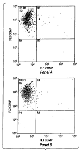
Figure 4 is a set of graphs showing flow cytometric profiles of CMSC.
Detailed description
Because of potential uses of CMSC for therapeutic purposes in humans or
other mammals, there is a need in the art for a method of isolating and
cryopreserving
these cells in compliance with cGMP and cGTP standards and for further methods
that do not change the relevant biological characteristics of these cells.
Prior art
methods pertaining to cells obtained from umbilical cord do not address the
issues of
storing CMSC under conditions that conform to FDA standards and that maintain
their biologic characteristics.
Umbilical cord blood (UCB) stem cells provide a readily available source for
hematopoietic stem cells. UCB has a number of proven advantages as a source of
hematopoietic stem cells for transplantation. Oiie advantage is that UCB is an
abundantly available source of stem cells that is currently discarded and can
be
harvested at no risk to the mother or infant. In contrast, in bone marrow and
6
CA 02629283 2008-05-08
WO 2007/059084 PCT/US2006/044094
peripheral blood donations there is a risk imposed on the donor associated
with the
procedure, in addition to the inconvenience.
'Another advantage of UCB is that major infectious agents, such as
cytomegalovirus (CMV), are much less common in the newborn than adults, and
are
less likely to contaminate UCB. UCB units, typed, cryopreserved and banked,
also are
available on demand, eliminating delays and uncertainties that complicate
marrow
collection from unrelated donors. At present, UCB can be delivered for
infusion
within days of initiation of a search. This compares with a median of 3- 4
months
from search to delivery of stem cells through registries of volunteer adult
donors.
Frozen UCB also can be easily shipped, stored at the treating institution, and
thawed
for use when needed, compared to freshly donated bone marrow which has a
limited
shelf-life of one day or less, necessitating coordination between harvesting
surgeons,
transportation, and transplantation teams.
A further advantage of UCB as a source of stem cells is that the intensity of
graft-versus-host reactivity of fetal lymphocytes appears to be less than that
of adult
cells and consequently fetal lymphocytes are more tolerant of HLA
incompatibility.
Published studies have shown that transplantation of UCB matched at 4 to 5 out
of 6
antigens results in a siinilar incidence of GvHD to transplantation of
unrelated bone
marrow fully matched at 6 out of 6 antigens (Gluckman E et al., Exp Hematol
32:
397, 2004; Gluckman E et al., Rev Clin Exp Hematol 5: 87, 2001; and Laughlin
MJ et
al., N Engl J Med 344: 1815, 2001). However, extent of engraftment of cells
over a
prolonged period of time continues to be a problem accounting for morbidity
and
mortality. At present, shortening of the engraftment period is achieved by
providing
sufficient numbers of UCB cells, which restricts the recipient pool to
clzildren and
small adults.
Although research is ongoing to obtain methods that expand ex vivo the
number of available UCB stem cells, these approaches have resulted in the
expansion
primarily of committed progenitor cells, with no significant beneficial impact
on the
time of bone marrow recovery. Efforts to accelerate the pace of engraftment
via ex
vivo expansion of UCB units have not improved clinical outcomes (Gluckman E et
al., Rev Clin Exp Hematol 5: 87, 2001; and Laughlin MJ et al., N Engl J Med
344:
1815, 2001). Evidence in both animal models and human studies suggests that
methods utilizing cytokines such as granulocyte-colony stimulating factor (G-
CSF),
7
CA 02629283 2008-05-08
WO 2007/059084 PCT/US2006/044094
stem cell factor (SCF), and tlirombopoietin (TPO) in liquid cultures expand
predominantly short-term committed hematopoietic progenitors, at the expense
of
long-term progenitors, which are the cells that will lead to sustained
hematopoiesis
(Williams DA, Blood 81: 3169, 1993; McNiece IK, Exp Hematol 30: 612, 2002;
Von Drygalski A et al., Stem Cells Dev 13: 101, 2004; and Tisdale JF et al.,
Blood
92: 1131, 1998). However, another disadvantage hampering the exploration of ex
vivo
stem cell expansion approach is the availability of clinical growth factors.
In addition,
the majority of cord blood banks preserve only a single unit of frozen
material from a
donor source. Clinical trials have typically expanded only a fraction (10-60%)
of the
frozen cells, with the remainder infused unmanipulated.
Ex vivo expansion of cord blood stem cells is accomplished by using bone
marrow derived MCS as a feeder layer (Robinson SN et al., Bone Marrow
Transplant
37: 359, 2006). MSC were generated from adult bone marrow, and when serving as
a
monolayer platform for UCB cells together with cytokines (usually a
combination of
an interleukin such as IL-3, IL-6, and with G-CSF, SCF, FLT-3L, EPO), resulted
in
faster engraftment. Flow immunocytometric analysis shows that mice that
received
UBC cells expanded by culture on a layer of MSC had about three times as many
human cells (CD45 positive) in the marrow after the transplant, than mice that
received an infusion of uncultured cells (Kadereit S etal., Stein Cells 20:
573, 2002).
Stroma contact of hematopoietic stem cells was found to be superior to culture
in
cytokine supplemented (McNiece I et al., Cytotherapy, 6: 311, 2004; Yildirim S
et al.,
Bone Marrow Transplant 36: 71, 2005; Breems DA et al., Blood 91: 111, 1998;
Zhang Y et al., Exp Hematol 32: 657, 2004; and Kanai M et al., Bone Marrow
Transplant 26: 83 7, 2000).
To solve the problem of long engraftment, anotlier approach taken in animal
models is co-injection of MSC with UBC. Thus mice were administered non
culture-
expanded fetal lung-derived CD34 negative MSC (in't Anker PS et al., Exp
Hematol
31: 881, 2003). Results showed that transplantation of a mixture of human UCB
CD34+ cells (at either of four concentrations, 0.03, 0.1, 0.3, and 1 X 10⁶) in
the
presence of MSC (106) resulted in significantly faster engraftment in bone
marrow of
NOD/SCID mice, tha.n that observed after transplantation with control UCB
CD34+
cells alone (n = 22 versus 29 days, p < 0.05). The most pronounced effect on
bone
marrow engraftment was observed after transplantation of relatively low doses
of
8
CA 02629283 2008-05-08
WO 2007/059084 PCT/US2006/044094
CD34+ UCB cells (0.03-0.1 X 10⁶). Co-transplantation of MSC resulted in a
three-
fold to four-fold increase in the percentage of human CD45+ cells in the bone
marrow
(14% versus 4.7% at 0.03 x 10⁶ cells, and 40% versus 10% at 0.1 X 10⁶
CD34+cells, p
< 0.001). However, the majority of the infused lung-derived MSC were observed
in
the lung and not in the bone marrow. Improved engraftment was observed when a
lower number of CD34+ cells (<lx106) were infused into irradiated NOD/SCID
mice
infused with a mixture of human mobilized peripheral blood hematopoietic stem
cells
and culture-expanded MSC harvested from adult bone marrow (Angelopoulou M et
al., Exp Hematol 31: 413, 2003). See also in't Anker PS et al., Exp Hematol
31: 881,
2003. Further, co-transplantation of human stromal cells into pre-immune sheep
supported faster recovery after marrow transplant (Maitra B et al., Bone
Marrow
Transplant 33: 597, 2004). Clinical feasibility has been shown by co-
transplanting
culture expanded HLA identical mesenchymal stem cells with marrow stem eells
in
patients with hematopoietic malignancies (Lazarus HM et al., Biol Blood Marrow
Transplant 11: 389, 2005).
An additional inethod of potentially enhancing engraftment of a suboptimal
dose of UCB cells is direct intraosseous infusion, or intra-bone marrow
transplant.
Bone marrow transplant directly into bone was shown long ago, however this
procedure was abandoned for its morbidity, especially after discovery that
intravenous
infusion yielded comparable or superior results (Kadereit S et al., Stem Cells
20: 573,
2002). Stem cells are known to transit intravenously through various organs
before
reaching their final destination in the bone marrow, however up to 90% of
infused
hematopoietic stem cells will lodge in the lungs. Investigators have therefore
re-
considered injecting stem cells directly into the marrow space. Stem cells are
directly
inserted into the bone marrow microenvironment, which is known to contain
molecular cues to direct hematopoiesis. Studies in mice have shown that this
approach
results in faster engraftment and long-term engraftment of the injected stem
cells
(Levac K et al., Exp Hematol. 33: 1417, 2005; and Wang J et al., Blood 101:
2924,
2003). Initial clinical trials have injected bone marrow cells into the
pelvis.
Surprisingly, volumes as large as one liter were tolerated without significant
side
effects (Hagglund H et al., Bone Marrow Transplant 21: 331, 1998). Although no
benefit was seen with respect to shortening of the engraftrnent time, these
studies
were not designed to analyze such a benefit, as patients received a full
marrow
9
CA 02629283 2008-05-08
WO 2007/059084 PCT/US2006/044094
transplant in a conventional way at the same time, representing sufficient
numbers of
stem cells to guarantee timely engraftment.
It is here envisioned that UCB cells may be utilized as a stem cell source in
this setting, and it is here further envisioned that co-infusion with MSC into
the
marrow along with umbilical cord blood could lead to enhanced engraftment.
Providing injection of UCB cells directly into the marrow to accelerate
engraftment
would allow a transplant to be perfomied with suboptimal umbilical cord blood
stem
cell numbers.
Examples herein use co-transplantation of umbilical cord matrix (UCM) cells,
a type of mesenchymal cell that is obtained from the Wharton's Jelly of the
umbilical
cord, to support faster engraftment of UCB cells arnd thereby facilitate
transplantation
into recipients that are larger adults. These cells optimize UCB cell homing
and blood
cell production, under conditions where only limited numbers of UCB cells have
been
transplanted.
Expression of genes found in early development and required for self renewal
and pluripotency, such as Oct-4 and nanoc, was observed in MSC obtained from
peri-
natal tissues, materials that are usually discarded after birth, such as the
umbilical
cord, placenta, ainnion, and chorion (Wang H-S, Stem Cells 22:1330-1337, 2004;
Zhang Y et al, Exp Hematol 32:657-664, 2004; Miki T, Stem Cells 23:1549-1559,
2005; and Koegler G et al., Stem Cells 33: 573-583, 2005). Further, MSC
express
genes associated with each of the three principal germinal layers: ectoderm,
mesoderm and endoderm, and are presumably in a state of transition to the
-mesenclzyme found at a later development state in bone marrow. Without being
limited by any particular mechanism or theory, those genes could be
manipulated and
activated in a nlethod causing the cells to differentiate along each of a
plurality of cell
lineages.
For example, it is here envisioned that early MSC can be programmed to
develop into insulin secreting cells. Alternatively, peri-natal MSC have the
potential
for use to improve engraftment after bone marrow and stem cell transplant.
Delayed
engraftment can be a significant problem, especially after cord blood
transplant. Co-
transplantation of cord blood cells together with peri-natal MSC speeds
engraftment
and facilitates transplantation, particularly in transfusions where only
limited numbers
of hematopoietic stem cells are available. The MSC that make up the bone
marrow
CA 02629283 2008-05-08
WO 2007/059084 PCT/US2006/044094
stroma can provide an essential structural network for hematopoietic stem
cells in
addition to producing cytokines that support their maturation and
differentiation.
MSC can also be used for down-regulating the immune response using bone marrow
derived MSC in autoimmune diseases and graft-versus-host disease after bone
marrow transplantation.
Different types of mesenchymal-like cells have been from isolated from
umbilical cords, for example, by a method in which vessels of the umbilical
cord are
first removed and discarded to harvest the remaining tissue, known as
Wharton's Jelly
(Mitchell et al., Stem Cells 21: 50-60, 2003). Peri-natal MSC, and in
particular MSC
from the umbilical cord, can easily be obtained after delivery. A small amount
of cord
tissue provides sufficient cells for expansion, and can be frozen and stored
along with
cord blood of a newborn. Cells can be thawed and processed when needed at a
later
point. Such cells provided by the methods herein are advantageous because they
are
autologous and therefore carry no risk of rejection.
A cross-section of an umbilical cord 10 is shown in Figure 1. A majority of
the
tissue in the cord consists of the Wharton's Jelly 11, which surrounds the
umbilical
veins 12 and artery 13. Wharton's Jelly includes connective tissue of the
umbilical
cord, a mixture of a gelatinous intercellular substance, collagen fibers,
hyaluronic
acid, and cells such as myofibroblasts and fibroblasts. The Wharton's Jelly
mixture
acts as a physical buffer, preventing kinking of the umbilical cord and
thereby
preventing disruption of maternal-fetal circulation (Sackier et al., U.S.
patent number
5,612,028). It is here proposed that Wharton's Jelly cells are very early stem
cells.
The first "blood islands" or developmental site of hematopoiesis is the
extraembryonic yolk sac followed by the aortic-gonad-mesonephros (AGM). The
region is thought to produce populations of mesenchymal cells, vascular
progenitors
and perhaps hemangioblasts. From the AGM region there is a migration of
precursors
to the fetal liver through the allantois. During or shortly after this
migration a portion
of these multipotential progenitors are trapped in the Wharton's Jelly of the
developing placenta and umbilical cord.
A limiting factor in commercial development of cord blood transplant is the
low number of hematopoietic stem cells, which can lead to delayed engraftment
and
decreased survival. The cord stem cell number frequently is insufficient to
transplant
adult patients. Examples herein show methods to isolate a type of 'support'
cells
11
CA 02629283 2008-05-08
WO 2007/059084 PCT/US2006/044094
from the Wharton's Jelly of the umbilical cord. Examples show that Wharton's
Jelly
cells can increase the number and function of blood-fomling stem cells. Mice
are
simultaneously transplanted with cord blood cells with Jelly cells, to show
faster
engraftment and allow to transplant adults for whom there are not enough stem
cells
in the cord blood.
Cord blood transplants are done worldwide, mostly in children and small
adults, as the number of stem cells in the banked units is frequently too low
to support
timely engraftment in larger adults. Stem cells currently are not present in
the cord
blood in sufficient amounts to support hematopoiesis in larger individuals,
limiting
more widespread use of cord blood cells for transplantation in adults. Even
with
optimization of the collection process, the majority of collections are not
sufficient for
larger adults. Attempts to expand cord blood cells ex vivo in a cytokine
cocktail have
met with only limited success. The volume of cryopreserved cord blood units
and the
large body size of most adult patients limit the dose of cells (number of
cells per
kilogram of body weight) that can be infused to establish donor hematopoiesis.
Limited cell doses lead to prolonged engraftment times, increased risk of
engraftment
failure and consequent increased risks to patients.
Transplant centers therefore have certain guidelines in place that define a
minimum number of cells. Single units for infusion generally have a
cryopreserved
cell dose greater than 2.0 x 10⁷ mononuclear cells (MNC) per kilogram of
recipient
body weight. Thus there is a probability of only 4 % of finding a
transplantable cord
blood unit in the current registries, of sufficient size for for a 70 kg
adult, compared to
94% probability for a 10 kg child. (Thomas ED, IntJHematol 81: 89, 2005).
Even if a cell dose of more than 2.0 x 10⁷ MNCIkg is transplanted, the median
time to recover more than 500/mm3 neutrophils is 25 days and 59 days to
achieve a
platelet count of 20,000/mm3 (Kurtzberg J et al., N Engl J Med 335: 157
(1996);
Gluckman E et al., Exp Hematol 32: 397, 2004; Gluckman. E et al., Rev Clin Exp
Hematol 5: 87, 2001; Laughlin MJ et al., N Engl J Med 344: 1815, 2001; and
Barker JN, et al., Blood 105: 1343, 2005). This is a median value, suggesting
that
50% of patients will take even longer for their marrow to recover. This
prolonged
marrow recovery increases the risk of infections as well as costs related to
blood and
platelet support, extended hospitalization and frequent hospital readmissions.
The
transplantation of two unrelated cord blood units has shown some shortening of
12
CA 02629283 2008-05-08
WO 2007/059084 PCT/US2006/044094
engraftment but this effect is not dramatic and carries a higher costs. Rates
of acute
GvHD are similar to those reported for matched unrelated transplant allogeneic
transplant (Kurtzberg J et al., N Engl J Med 335: 157, 1996; Gluckman E et
al., Exp
Hematol 32: 397, 2004; Gluckman E et al., Rev Clin Exp Hematol 5: 87, 2001;
Laughlin MJ et al., N Engl J Med 344: 1815, 2001; and Barker JN, et al., Blood
105:
1343, 2005). Most recipients of cord blood units are mismatched at one or two
of the
six HLA loci (i.e., each of loci HLA-A, HLA-B, and HLA-DR on each of two
paired
chromosomes). Such HLA antigen incompatibility in matched unrelated
transplants
is associated with poor outcomes, due to graft failure and GvHD. Tolerance of
HLA-
incompatibility by a cord blood graft makes cord blood valuable as a stem cell
source.
Even witliin the relatively small pool of banked cord units, matching a
minimum of
four or five antigens instead of all six greatly increases the likelihood that
a match
will be found.
Wharton's Jelly or umbilical cord matrix represents a rich source of primitive
multipotent MSC like progenitor cells which are currently not widely
appreciated as a
source of MSC. MSC cells were characterized by several investigators (Eyden, J
Submicrosc Cytology 26: 347, 1994; Wang HS et al., Stem Cells 22: 1330, 2004;
Weiss ML, et al., Stem Cells 24: 781-792, 2006; Weiss ML et al., Exp Neurol
182:
288, 2003; Fu YS et al., J Biomed Sci. 11: 652, 2004; Fu YS et al., Stem Cells
24:
115, 2005; Sarugaser R et al., Stem Cells 23: 220, 2005; and Carlin R et al.,
Reprod
Biol Endocrinol 4: 8, 2006). MSC from adult bone marrow are rare (less than
0.001%
of cells) and inust be harvested from adult volunteers. The cells do not
appear to be
immortal, leading researchers to search for more viable sources of MSC that
potentially could support cord blood cell engraftment. A MSC-like cell has
been
isolated from the UCB and termed an unrestricted somatic stem cell (USSC)
(Kogler
G. et al., Exp Hematol 33: 573, 2005). However the recovery of those cells is
relatively low and only one third of fresh cord blood specimens will yield
USSC upon
culture.
Cells from cord have a much longer life span than bone marrow derived MSC,
and express the transcription factors Oct-4 and nanog that are important for
maintaining an undifferentiated state and pluripotent capacity. The Wharton's
Jelly
contains a large amount of early MSC that are obtained from cord which is
otherwise
discarded after delivery (Koegler G et al., Exp Hematol 33: 573-583, 2005).
These
13
CA 02629283 2008-05-08
WO 2007/059084 , PCT/US2006/044094
cells display pluri-potent capacity, with potential applications such as use
in spinal
cord injuries, to accelerate wound healing or to treat Parkinson's disease
(Weiss ML
et al., Stem Cells 24: 781-792, 2006). Beyond using these early pluripotential
MSC
merely for regenerative medicine, they may be used also as carriers of
targeted
molecules, cytokines and drugs. Examples are molecules that increase
angiogenesis
or prevent scarring and fibrosis. Peri-natal MSC can easily be transduced and
can be
used as vehicles for either short-term or long-term expression of genes of
interest.
Since MSC are known to target sites of inflammation, and cancerous cells
generally initiate a state of inflammation around them, MSC migrate to tumor
sites.
MSC obtained from bone marrow and transfected with an interferon gene have
been
shown in a murine model to travel to malignant sites and release a cytokine
locally,
resulting in an anti-tumor effect (Deans RJ et al., Exp Hematol 28: 875-884,
2000).
MSC can be used as biological pumps to inhibit degenerative and support
restorative
events. Genetic manipulation of these cells extends the life span. Even
without
manipulation, these cells are capable of at least twice as many doublings as
MSC
obtained from bone marrow that is more mature. In addition, peri-natal MSC
having
low immunogenicity are useful as allogeneic donor cells, to establish cell
lines for
further manipulation.
Peri-natal MSC are here envisioned to have a further role in generating more
complex tissues, for which certain scaffolds such as bone and vessels are
supplied.
Alternatively the cells may serve as vehicles for delivery of site directed
morphogenic
proteins. Peri-natal cells per se take on features of embryonic stem cells by
'nuclear
reprograsnming' at the genetic level (Deinbinski JL et al., Cytotherapy 888,
2006).
Certain progenitor cells remain responsive to embryonic transcription factors
(Hochedlinger K et al., Nature 441:1061-1067, 2006). Somatic cells regress
when the
transcription factor Oct-4 is turned off. As Oct-4 is expressed in peri-natal
MSC from
extra-embryonic tissue, reprograinining MSC is envisioned herein to involve
turn-off
of Oct-4. MSC are used to provide the framework (stroma) so that tissue
specific stem
cells of multi-potential capacity differentiate into a fully functional
tissue.
Mesenchymal stem cell-like cells surrounding the vasculature of the cord have
been isolated from the umbilical cord (Romanov et al., Stem Cells 21: 105-110,
2003). Collagenase digestion from within the umbilical vein has been used to
obtain a
mixed population of vascular endothelial and sub-endothelial cells.
14
CA 02629283 2008-05-08
WO 2007/059084 PCT/US2006/044094
A procedure to collect Wharton's Jelly from the umbilical cord under sterile
conditions is shown in U.S. patent application publication number
2003/0161818. In
this procedure, the cord is cut transversely with a scalpel, and each section
is
transferred to a sterile container containing phosphate buffered saline (PBS)
with
CaC12 (0.1 g/1) and MgC126H2O (0.1 g/1) to remove surface blood from the
section
with gentle agitation. The section is then removed to a sterile-surface where
the outer
layer of the section is incised along the longitudinal axis of the cord, and
blood
vessels of the umbilical cord (two veins and an artery) are removed by
dissection, for
example, with sterile forceps and dissecting scissors. Wharton's Jelly is
collected into
a sterile container, or cut into smaller sections, of size such as 2-3 mm3 for
culturing
the included cells.
Umbilical cord matrix (UCM) cells express CD44, CD29, CD51 and not
heinatopoietic lineage markers (CD34, CD45, CD3, CD5, CD14, CD19). Further,
UCM express MSC markers (SH2 also known as CD105, SH3 also known as CD73).
These cells are here envisioned to be used to differentiate into
cardiomyocytes,
cartilage cells, adipocytes, cells of osteogenic lineage as well as nerve
cells (Weiss
ML et al., Exp Neurol 182: 288, 2003; Fu YS, et al., J Biomed Sci. 11: 652,
2004; Fu
YS et al., Stem Cells 24: 115, 2005; and Sarugaser R et al., Stem Cells 23:
220, 2005).
UCM cells of the Wharton's Jelly, like MSC, express intermediate levels of
human leukocyte antigen (HLA) major histocompatibility complex (MHC) class I
molecules and very low levels of (HLA) class II and Fas ligand; UCM cells do
not
express the co-stimulatory molecules B7-1, B7-2, CD40 or CD40L and are
therefore
not immunogeneic, as these co-stimulatory molecules are required for a full T-
cell
response. (Le Blaizc K et al., Scand. J. Immunol. 57: 11, 2003; and Glennie S
et al.,
Blood 105:2821, 2005).
Umbilical cord blood (UCB) is a viable source of hematopoietic stem cells for
transplantation of children and adults undergoing treatment for hematological
malignancies. However only 4% of adults 70kg and over have a UCB unit
available
which contains the widely accepted minimum cell dose of 1.5x10⁷ mononuclear
cells
per kilogram. Co-transplantation of hematopoietic stem cells with mesenchymal
stem
cells may enhance engraftment and therefore decrease transplant-related
morbidity
and mortality from delayed leukocyte recovery associated with a low pre-
transplant
cell dose.
CA 02629283 2008-05-08
WO 2007/059084 PCT/US2006/044094
Umbilical cord matrix (UCM) cells, found in the Wharton's Jelly, were easily
and reliably extracted from minced pieces of cord by culture in RPMI + 20%
fetal
bovine serum at 37 C and 5% humidified CO2. It was observed that UCM cells
best
expanded in medium containing 20% FBS. This procedure can also be used to
expand UCM cells in human serum, autologous serum, and the serum-free
commercially available medium X-VIVO 10. Small (1-3mm) minced pieces of
umbilical cord can be cyropreserved at the time of delivery in 10% DMSO
solution.
UCM cells exhibit a fibroblast morphology and express markers common to
mesenchymal stem cells: CD73 (SH3), CD105 (SH2), CD 29, CD44, CD49b, CD117,
CD166, STRO-1 and HLA-DR. UCM are negative for CD14, CD 19, CD34, and
CD45. Morphology and cell surface marker expression is stable after greater
than
fifteen passages.
The present invention in certain enlbodiments provides methods and
compositions for preparing CMSC in coinpliance with cGMP and cGTP conditions
and practices, and materials that comply with the standards as regulated by
the FDA,
for use of these cells in humans for therapeutic purposes. The methods
provided
herein use cord blood serum or plasma of autologous origin, or use autologous
serum
or plasma, to add to the cells for culture or long term storage of CMSC. Prior
art
procedures have used serum or plasma from a non-human animal, or have used non-
autologous serum or plasma (such as isologous, or allogeneic). However, use of
animal serum or plasma is not ideal, for example, because of the possible
presence of
infectious particles.
The term "autologous" as used herein refers to rriaterials that are taken from
the same subject, for example, two or more biological samples taken from the
same
human.
The term "allogeneic" as used herein means materials taken from two different
subjects of the same species, for example, two different human subjects, and
generally
assumes that the two subjects are genetically independent, i.e., are not
identical twins
or organismal clones.
The term, "xenogeneic" as used herein means materials taken from subjects of
different species, for example, transfusion or implantation of material of
porcine,
bovine or canine origin into a species different than the source of the
implant.
16
CA 02629283 2008-05-08
WO 2007/059084 PCT/US2006/044094
Allogeneic stem cell transplantation from a matched donor following
myeloablative and non-myeloablative conditioning therapy has proven curative
when
used as part of a treatment for a number of inherited and acquired
hematological
disorders (Thomas ED, Int J Hematol 81: 89, 2005; and Resnick et al., Transpl
Immunol 3: 207, 2005). The success of allogeneic transplantation is largely
determined by compatibility between donor and recipient, which predicts the
risk of
severe and potentially fatal graft-versus-host disease.
About 75,000 cord blood units are stored in public banks. "The Stem Cell
Therapeutic and Research Act" will allocate $79 million dollars for
acquisition of
150,000 cord blood units that are believed to be necessary to broaden the
donor pool
to include recipients of all racial backgrounds and establish the "National
Cell
Transplantation Program" (Cord Blood-Establishing a National Hematopoietic
Stem
Cell Bank Program, The National Academies Press, Washington, DC, 2005).
Unfortunately, less than one third of patients needing an allogeneic
transplant have a
compatible donor available in their family. Registries have been established
to match
patients with coznpatible volunteer i.e. unrelated bone marrow/stem cell
donors, but
many patients, especially patierits of non-Caucasian background, still lack
stem cell
donors. African-American and Asian donors are still underrepresented in
existing
bone marrow registries. Because of a lack of matched unrelated donors for
minorities; the lead time necessary to acquire and process the hematopoietic
stem
cells from a volunteer, and the many advantages of UCB transplantation as
listed
above, continued advances related to UCB transplantation is needed to extend
curative therapy to patients with hematologic malignaicies and other
hematologic
disorders. Additionally, there can be a three to four month delay while the
donor is
contacted, tested, and arrangements for stem cell collections are made. Many
patients
cannot wait that long if their disease is progressing.
Further, general prior use of fetal bovine serum or plasma carries the risk of
transmitting prion diseases and zoonoses, and xenogeneic proteins from an
animal or
allogeneic human serum or plasma may initiate immune responses in a subject.
In
addition to being a source of prions and other infectious particles, FCS is
known to
change the gene expression and functional characteristics of MSC (Shalldadfar
A et
al., Stem Cells 23: 1357, 2005). Alternative prior art procedures have used
allogeneic
human serum or plasma, however this material has been shown to be detrimental
to
17
CA 02629283 2008-05-08
WO 2007/059084 PCT/US2006/044094
the growth and function of CMSC. The present invention fitrther provides
methods
and composition for the preparation of CMSC and cord blood cells and for
subsequent
long-term storage of these sources of stem cells obtained from the same donor
in the
same storage devise.
According to various embodiments of the methods provided herein, the
sources of umbilical cord blood cells and CMSC are autologous, i.e. are
obtained
from the same donor. In certain embodiments, both the cells and CMSC are
cryopreserved in the same container, for example in separate clianlbers of a
multi-
chamber container such as a freezer bag, using serum or plasma from the
autologous
cord donor for cryopreservation, generally admixed with a cryoprotectant. The
container includes a mechanism such as a hermetically sealed plastic segment
between each chamber of a bag. The plastic bridge between the chambers is
large
enough to allow opening, or even physical detachment, of a single storage
chamber at
aiiy later time with continued cryopreservation of remaining chambers. Each
chamber
of the multi-chamber container also has a separate entry port.
In the methods provided herein, CMSC are extracted from the entire
circumference of the utnbilical cord of a mammal. The cord can first be
divided into
segments for storage and ease of manipulation. CMSC are prepared from each of
a
plurality of the short segments of the cord, by dissecting or mincing, i.e.
dissecting
each section of the umbilical tissue into small fragments, the umbilical
tissue prior to
cryopreservation.
Procedures for obtaining CMSC from the cord in the past have generally
included mechanical extraction or enzymatic separation, following which cells
are
expanded in culture, for example, for several days, and are subsequently
frozen for
future use. However, ex vivo culture procedures used prior to cryopreservation
carry
a risk of contamination, and pose logistic problems, for example, a
requirement that
the cord blood and the umbilical cord arrive the same day for banking.
Therefore a
process or metllod that allows cryopreservation of fresh cord tissue would
represent a
significant improvement.
Prior attempts to freeze small segments of the cord have involved injecting
cryoprotectant into the interior of the cord via a needle inserted into the
cavity.
However, recovery of the CMSC after thawing was observed to be minimal, and
the
yield and quality of the cells were highly variable. The cord segments
obtained by
18
CA 02629283 2008-05-08
WO 2007/059084 PCT/US2006/044094
this method also were not found to be suitable for storage in a bag or other
standard
container for long-term storage.
Prior art references relating to the collection and storage of cord cells have
not
addressed the issue of use of xenobiotic materials, and use of animal serum or
plasma
remains routine. Methods herein are advantageous in using chemicals and
solutions
that are well characterized and are prepared by methods approved by the FDA
for use
in humans.
Examples
Example 1: Methods for co-transplantation of human umbilical cord matrix (UCM)
cells with umbilical cord blood (UCB) cells to obtain improved weed of
engraftment.
UCB are collected via cannulation of umbilical cord vessels at delivery.
Mononuclear cells (MNCs) are isolated using Ficoll-Paque (Arnersham
Biosciences).
Flow cytometry is performed on the MNCs to determine the number of CD34+
cells.
The MNCs are stored in 10% dimethylsulfoxide (DMSO) in liquid nitrogen until
ready for use. The UCB mononuclear cells are thawed, rinsed in fetal bovine
serum
(FBS) except as indicated below, and then resuspended in Dulbecco's Phosphate-
Buffered Saline (PBS) prior to injection. Addition of FBS to buffers herein is
according to conventional preparation of media, and is omitted in exaniples
herein
describing use of autologous cells, blood, and blood products.
Fresh umbilical cords are rinsed in saline and cut into pieces approximately
one centimeter in length. The umbilical arteries and the umbilical vein are
removed
and the remaining tissue is placed in six well plates in RMPI plus 20% FBS and
antibiotics (penicillin 100 g/mL, streptomycin 10 g/mL, amphotericin B 250
g/mL) and incubated at 37 C in 5% CO2. UCM cells migrate from the cord and
adhere to the plastic wells for about one week. The supernatant and the cord
are
discarded and cells are detached from the plate using 0.25% trypsin-EDTA
(Invitrogen). UCM cells are expanded in plastic flasks using the
aforementioned
culture conditions. Flow cytonietry is performed using CD73 (SH3), CD105
(SH2),
CD 29, CD44, CD49b, CD14, CD34, CD45 as an assay for homogeneity.
Cells prepared as described above are injected via either of two different
routes: intravenous (IV) or intra-bone marrow (IBM). Recipients are eight to
ten
week-old mice sublethally irradiated with 3.5 Gy from a 137Cs source (2.115
Gy/min).
Intravenous injection is via the lateral tail vein of mice. IBM injection is
perforrned
19
CA 02629283 2008-05-08
WO 2007/059084 PCT/US2006/044094
as described by Levac et al. (Levac, K et al., Exp Hematol. 33: 1417, 2005) as
follows. Mice are anesthetized with an intraperitoneal injection of 0.015 mL/g
body
weight of a 2.5% solution of tribromoethanol. The right hind leg is shaved and
disinfected. The knee is flexed to 90 degrees and a hole is drilled into the
femur with
a short 27-gauge needle attached to a 3-mL syringe filled with PBS. The first
needle
is removed and a 28-gauge needle with a 0.3niL insulin syringe containing the
cells is
inserted into the femur. The cell dose injected for a total volume of 30-50
L. The
skin is closed with 6-0 vicryl suture (Ethicon).
The organ distribution of injected UCM cells after each mode of injection is
determined by immunohistology of different target organs, including bone
marrow,
spleen, liver and lung. For identifying human UCM, huinan UCM cells that have
been
retrovirally transfected or marked with the green fluorescent protein (GFP)
gene are
used. The presence of GFP protein on GFP-tranduced cells in mouse tissue
sections is
assessed by assaying with a rabbit anti-GFP antibody. The human origin of
these cells
in mouse tissues is assessed by an antibody directed against human (32-
microglobulin.
Since GFP expression in tissue may be unstable, a second method to
determine organ distribution of injected UCM cells is also used. Thus organ
distribution is also determined by injecting male lluman UCM cells into female
mice
and assessing for presence of the human Y-chromosome by PCR.
The time points assessed after injection are each of 2 days, 7 days and 4
weeks. Since the IV infusion of UCM cells results in the majority of cells
being
sequestered in the lung and/or spleen before reaching the bone marrow, the UCM
cells are also injected directly into bone as described above.
Initially, three different concentrations of UCB cells are injected to
establish
the length of time required for engraftment for varying cell doses, at each of
IV and
IBM routes of administration. A lower cell dose may be required for
engraftment
following IBM route of delivery. Twelve to twenty-four hours after
irradiation, either
5 x 10⁵, 106, or 5x10⁶ UCB cells resuspended in PBS are injected into the tail
vein of
mice.
Once the engraftment kinetics at each UCB concentration has been
established, the dose that gives delayed engraftment is combined and co-
injected with
106 UCM. Control groups include mice receiving a dose of UCB cells that has
been
shown to establish delayed engraftment, a group that has been shown to provide
CA 02629283 2008-05-08
WO 2007/059084 PCT/US2006/044094
timely engraftment, and a group that received PBS with no UCB cells.
Engraftment is
documented at 2, 3 and 4 weeks after cell infusion. Peripheral blood (50 L)
is
collected from the submandibular plexus and a CBC is performed using the
Hemavet
850 (CDC Technologies Inc. Oxford, CT). Furtller, the percentage of human
CD45+
cells in murine blood is counted by flow cytometry. Differences in the human
CD45
cell engraftment are determined by calculating the areas under the curves
(AUCs) at
each different time point. Mice are sacrificed after 6 weeks and bone marrow
is
collected by flushing both femurs and pelvis with RPMI medium. Single-cell
suspensions from spleen, lung, and liver are prepared. The cell suspensions
are
stained with mouse anti-human monoclonal antibodies for flow cytometric
analysis.
PE or FITC-conjugated antibodies include monoclonal antibodies against CD45,
CD34, CD 19, CD33, and CD38 (Becton-Dickinson).
Initial experiments are with autologous materials and recipients, i.e. UCM
cells and UCB cells are from the same donor. As a control, allogeneic co-
transplantation, i.e. UCB from one donor and UCM from another, is also
performed.
Example 2: Transplantation of varying concentrations of human UCB cells to
determine engraftment delay
Initially human UCB cells at various doses are transplanted as the sole source
of cells to determine cell doses that allow full engraftment and to determine
a
suboptimal cell concentration at which engraftment is delayed or will no
longer occur.
Two routes of injections are tested: intravenously and intra-bone marrow.
Mice are transplanted with UCB in order to establish engraftment kinetics.
Mice in each of three experimental groups of mice are injected with either 5
x105, 106,
or 5 x10⁶ cells. As controls, a single mouse is irradiated and receives a
saline
injection, and another single mouse is not irradiated and receives a saline
injection.
The experiment is done both IV and IBM, for example, a total of 28 mice in an
experiment using four mice per experimental group. If all IBM mice have rapid
engraftment at the lower dose, doses of 105 cells or even 5 x10⁴ cells are
used with
similar controls.
Statistical analyses are performed using table curve software (SPSS). The
kinetics of human CD45 cell engraftment are evaluated by calculating areas
under the
curves (AUCs) at three time points (2 weeks, 4 weeks, 6 weeks). Differences in
21
CA 02629283 2008-05-08
WO 2007/059084 PCT/US2006/044094
CD45+ cell AUCs between mouse groups are assessed using the Mann-Whitney rank
sum test. Other statistics tests are performedusing SPSS. When indicated,
values are
reported as mean standard deviation (SD). Statistical significance is set
for P < .05.
Following observing a positive effect on engraftment with autologous UCM, the
experiments are repeated using allogeneic UCM cells.
Example 3: Analyzing effect of number of autologous UCM cells on engraftment
rate
in co-transplantation with UCB
Three groups of eight mice are injected with 5 x10⁵, 106, or 5 x10⁶ UCB cells.
For each cell dose, four mice are injected with 106 UCM cells and four mice
are not
injected with UCM cells. Three controls are further performed at each UCB
dose: an
irradiated mouse that does not receive UCB and is injected with saline, a
mouse that is
not irradiated and receives saline, and a mouse that is irradiated and
receives the
UCM. The experim.ent is performed using each route of administration, both IV
and
IBM, and is also performed in an autologous fashion (UCM and UCB from same
donor) and an allogeneic fashion (UCM and UCB from different donors), for a
total
of 132 inice.
Statistical analyses are performed using table curve software (SPSS). The
kinetics of human CD45 cell engraftment are evaluated by calculating areas
under the
curves (AUCs) at different time points (2 weeks, 4 weeks, 6 weeks).
Differences in
CD45+ cell AUCs between mouse groups are assessed using the Mann-Whitney rank
sum test. Other statistics tests are performed using SPSS. When indicated,
values are
reported as mean + standard deviation (SD). Statistical significance is set
for P < .05.
If any positive effect on engraftment is seen with autologous UCM, the same
experiments are conducted with allogeneic UCM cells.
Exam-ple 4: Affect of co-transplantation of UCB CD34} cells and autologous UCM
cells on engraftment in vivo
UCM cells were grown in culture and were shown to produce more GM-CSF
and G-CSF than similar numbers of adult bone marrow mesenchymal stem cells.
The
data showed that the UCM derived cells produced 178 pg/mL of GM-CSF compared
to adult bone marrow mesenchymal stem cells, that produced 77 pg/mL; and the
22
CA 02629283 2008-05-08
WO 2007/059084 PCT/US2006/044094
UCM derived cells produced 82.6 pg/mL G-CSF, compared to adult bone marrow
cells that produced 7.9 pg/mL respectively.
Recipient mice of strain NOD/SCID were treated with anti-NK 1.1 antibodies,
and were irradiated with 350 cGy. These were then injected with suboptimal
(1x10⁴)
numbers of cord blood CD34+ cells with and without 1x10⁶ autologous UCM cells,
extracted from the same umbilical cord as the cord blood CD34+ cells. Bone
marrow
was harvested at six weeks post transplant from both femurs and tibias and
peripheral
blood was obtained via cardiac puncture. The percentage of human CD45+ cells
in the
bone marrow and the peripheral blood was assessed by flow cytometry.
The data showed that control NOD/SCID mice transplanted with 1x10⁴ cord blood
CD34+ cells alone had 3.0% human CD45+ cell engraftment in the bone marrow and
3.6% human CD45+ cells in the peripheral blood, while NOD/SCID mice
transplanted
with 1x10⁴ CD34+ cells and 1x10⁶ UCM cells had an average of 27.3% husnan
CD45+
cell engraftment in the bone marrow and 3.9% human CD45+ cells in the
peripheral
blood. These results indicate that improved engraftment in vivo was observed
with
co-transplantation of suboptimal numbers of umbilical cord blood CD34+ cells
and
autologous umbilical cord matrix cells, compared to control transplantation of
suboptimal numbers of umbilical cord CD34+ cells alone.
Example 5: Methods for developing conditions for culture and expansion of UCM
cells for clinical use.
Human serum at different concentrations (5%, 10%, 20%) is tested with
respect to its ability to support expansion of human UCM cells (autologous or
isologous) in culture, and results are compared to FBS, at each of the
concentrations,
and as a control in absence of serum. Using current conventional methods, UCM
cells
are grown in RPMI 1640 as mediw.n supplement. This example uses X-Vivo 10
(Cambrex Corporation) as a base medium instead of RPMI 1640, as X-Vivo 10 has
been employed for studies with human cells and a drug master file for X-Vivo
10 is
deposited with the FDA. Cell growth is evaluated and growth behaviors (cell
count
and doubling time) are evaluated daily, and the effect of different media on
flowcytometric profile is analyzed.
The ability of the UCM cells to differentiate into bone tissue (osteogenesis
assay) is used as a marker of intact and fiulctional UCM cells. In this assay
UCM cells
23
CA 02629283 2008-05-08
WO 2007/059084 PCT/US2006/044094
for this test are contacted with medium containing dexamethasone (0.1mM), L-
ascorbic acid 2-phosphate 0.05 mM and beta-glycerol phosphate (3mM). After 21
days the cells are fixed in 3.7 % formaldehyde and then stained in 6% silver
nitrate
and exposed to UV light (20 min), and stained cells are counted.
Further, additives to the medium such as amino acids or epidermal growth
factor (EGF), platelet derived growth factor (PDGF), leukemia inhibitory
factor (LIF)
or other growth factors are tested for obtaining optimum cell proliferation
without the
presence of FBS. Culture conditions that provide an aseptic closed system are
used to
reduce airborne contamination. Only cGMP and cGTP grade or appropriately
qualified reagent such as trypsin and plastic ware are used. In order to make
the
process cGMP and cGTP compliant, a closed system with bags or a hollow fiber
type
bioreactor type system are used.
Example 6: Co-injection of UCB and UCM cells to establish eng-raftment in
larger
recipients
The examples herein are designed to show that co-injection of suboptimal
numbers of UCB cells together with UCM cells can successfully establish
engraftment, in larger recipients. For this purpose, recipients are transfused
with only
suboptimal amounts UCB, generally those units having MNC numbers of less than
1.5 x 10⁷ /kg. Accelerated engraftment with optimal UCB nunlbers and the
maintenance of.fun.ctional characteristics of UCM cells after switching
culture
condition to cGMP and cGTP compliant conditions are also examined. Cells are
manufactured by a facility in compliance with cGMP and cGTP, and further
restricted
to human MSC culture and expansion and designated by the NHLBI to provide such
a
cell therapy service to other clinical centers.
A phase I clinical trial is performed in which patients receive a standard
cord
blood transplant together with each of increasing numbers of UCM cells. Due to
the
current banking situation where no UCM are stored, the initial clinical trial
uses
allogeneic UCM cells. It is shown herein that sufficient number of cells are
obtained
from a small piece of cord and prepared and stored under cGMP conditions
requiring
minimal manipulation. Therefore it is contemplated herein that further
clinical trials
use autologous UCM cells.
24
CA 02629283 2008-05-08
WO 2007/059084 PCT/US2006/044094
In a clinical trial setting, three to four different dose levels of UCM cells
are
given along with UCB cells to cohorts of three patients in each group. The
objective
of the initial trial is to determine safety of the UCM infusions. The initial
patients are
those who receive a standard cord blood transplant with a sufficient number of
UCB
cells. Once this phase I trial is concluded, a phase II trial analyzes
efficacy by
transplanting a group of patients characterized in that only a suboptimal
number of
UCB cells are stored or are available (< 1 x10⁷ MNC/kg), and these patients
are
transplanted with the UCB together with a fixed dose of UCM cells.
Exanple 7: Methods for preparation of CMSC in autologous cordplasma or serum
for long-term cryoaenic storage
Autologous cord blood serum or plasma is shown herein by the methods
provided as useful for long-term storage of CMSC. The CMSC are obtained by
different methods from the cord after delivery, for example, dissecting
(mincing or
cutting the cord) into fragments (small pieces) followed by addition of a
cryoprotectant solution.
Alternatively to stored CMSC, a fresh supply of CMSC is obtained by
mechanical or enzymatic extraction from the store or fresh cord and cultured
for one
or more days in serum free medium that is FDA approved to expand their numbers
before being cryopreserved.
An exemplary cryoprotectant for use in the methods herein prepared as
follows. Autologous cord blood plasma or serum (80-95%) that has been
centrifuged
is filtered through a 0.2 m membrane and is mixed with diniethylsulfoxide
(DMSO;
5-15%); and hydroxyethyl starch (HES; 3-8%). Because human serum or plasma
obtained from an allogeneic donor causes significant changes in the pattern of
gene
expression in human matrix cells, affecting biological properties, the methods
herein
address that issue by using only autologous serum, individual plasma, or cord
blood
from the same source, i.e., from the same individual donor, for the pur.pose
of
preparing the cells for long term storage.
The cord fragments or the isolated or cultured CMSC are frozen under
controlled rate conditions, i.e., the external temperature is reduced
systematically with
specifically timed intervals of incubation at each lower temperature until the
target
freezing temperature is obtained.
CA 02629283 2008-05-08
WO 2007/059084 PCT/US2006/044094
Exam-ole 8: Storage of cord blood stem cells and CMSC in separate compartments
of
a multi-compartment container
Autologous CMSC and cord blood cells, i.e. obtained from the same
individual are stored in the same container at the time of banking to maximize
convenience, and to avoid unwanted mixing and contamination.
The method of using a plastic bag for cryogenic storage having at least two
chambers is suitable for cryopreserved minced cord fragments. Each chainber is
accessible via a separate port and equipped with an identifier. The plurality
of
chambers allow storage of cord fragments, and/or stem cells from the
autologous,
same source or heterologous or allogeneic, from different sources of the same
origin,
in this case umbilical cord blood mononuclear cells aiid CMSC.
The storage container is a standard cryopreservation bag that however is
separated into a number of discrete chambers. Figure 2 shows a cryogenic bag
20
(Pall Corporation, East Hills, NY) that can be used for long-term storage of
cord
blood cells and CMSC from the same donor. The bag 20 contains segments 21 that
have patient-specific data engraved in the lining. The bag has a smaller
compartment
22 and a larger compartment 23 for storing cord blood cells and CMSC. A solid
plastic lining separates each cliamber, each of which is individually
removable and
individually openable. A user thereby removes sainples as needed by breaking
away
or cutting off one chalnber, thereby processing only the amount of stem cells
that are
frozen in that particular chamber.
Alternatively, as each chamber is equipped with a corresponding separate
entry port, the user accesses that discrete chamber. Each chamber fiu-ther
includes a
patient/donor identifier and other relevant data attached to it and the
identifier
optionally includes additional information.
The methods herein can be used selectively to provide or remove one or a
small number from among inultiple iterations of chambers of frozen stem cells
from
the same donor. These are accessed for further cell manipulations including
direct
therapeutic administration, or alternatively, cell expansion, use as a feeder
layer, or
further culture to differentiate the cells into suitable transplants for
various tissues,
such culture including culture in the presence of well known differentiation
factors
such as epidermal growth factor (EGF), insulin-like growth factor (IGF) and
keratinocyte growth factor (KGF).
26
CA 02629283 2008-05-08
WO 2007/059084 PCT/US2006/044094
An exemplary use of stem cells from the umbilical cord is support of a stem
cell transplant. This use has in the past been limited, however, because the
number of
cord blood stem cells obtained and/or stored is frequently too low for larger
recipients. Using the multi-compartment storage system in this situation, the
CMSC
from an additional chamber are thawed to use as a stromal (feeder) layer, to
support
cell number expansion by culture of cord blood stem cells.
The methods herein provide preparation of CMSC for cryopreservation that
are performed under conditions that conform to FDA standards for current good
manufacturing practices. This method involves using only reagents, plasticware
and
procedures that are approved for use with human cells. The methods provided
lierein
do not use animal serum or plasma components or allogeneic serum or plasma
from a
corresponding mammalian individual. Such non-autologous components are known
to
be detrimental to the number and biological functions characteristic of CMSC.
Example 9: Method of preparing a sezment of the umbilical cord for lon -tg
ei7n
cryogenic storage
A method was developed to prepare a small section of the cord (about 1 cm)
with minimal maiiipulation compliant with cGMP conditions. An embodiment of
the
method herein includes, preparation of small cord segments, followed by
dissecting,
i.e. mincing, the small segment of the cord. This involves mincing of the cord
with a
scissor and freezing the small pieces in 10% DMSO and autologous (cord) plasma
(to
avoid exposure to allogeneic and/or animal serum or plasma). The entire
circumference of the umbilical cord is utilized herein to obtain CMSC. After
the cord
is received from the donor, a plurality of small lengths is produced and each
is
subjected to a dissection or mincing process, resulting in a plurality of sets
of
fragments of sufficiently small size to result in even and consistent exposure
of each
of the CMSC fragments to cryoprotectant solution described herein or an
equivalent.
Reducing the fragment size was found to result in excellent recovery and yield
of
CMSC cells after thawing. This technique therefore fulfils the requirement for
a
clinical trial where we would need UCM cells from cords prepared under cGMP
conditions.
The homogenization or mincing process was performed, for example, using an
instrument in a plastic cartridge designed for single use. Alternatively the
fragments
27
CA 02629283 2008-05-08
WO 2007/059084 PCT/US2006/044094
were dissected manually, using, by way of example but not restricted to, a
scissor,
scalpel, disposable lancet, or other similar instrument. The fragments were
then
transferred to a container suitable for cryopreservation and long-term
storage. The
fragments containing CMSC were cryopreserved under controlled rate condition.
After thawing, cells were expanded, by cell culture of the fragments, and the
cells so
obtained were found to express appropriate surface markers and display
functional
characteristics. '
Example 10: Culture methods and biological characteristics
Isolation of UCM cells from the Wharton Jelly was performed using the entire
umbilical cord obtained from full-term deliveries. The cells were extracted
and placed
into 2 inch microtiter plates and kept at 37 C (5% vol/vol CO2) in media
containing
RPMI 1640/20% FCS. After two passages cells were transferred into flasks.
Cultures
without addition of cytokines were kept at 37 C (5% vol/vol C02), and three
fifths of
the medium is renewed every 3 to 4 days. When grown to confluence, cells were
detached with trypsin/EDTA and re-plated after washing, or cryopreserved in
10%
dimethylsulfoxide, 25% FCS, and 65% RPMI medium.
Figure 3 shows a growth pattern of UCM cells irioculated into culture dishes
containing RPMI growth medium/20% FCS. Culture-expanded UCM cells adhered to
plastic and were found to have fibroblast like features, as shown in Figure 3.
The
large dark spots represent areas of intense cell production.
As shown by flow cytometry, cells were negative for the surface antigens
CD34, CD45, CD14, CD40, CD80 and CD86. Thus these cells were found to be early
stage stem cells, expressing the MSC markers CD73 and CD 105. Figure 4 shows a
flow cytometric profile of UCM cells. In Figure 4 Panel A, the abscissa
represents the
CD34 cells and the ordinate represents CD73. hi Figure 4 Panel B, the abscissa
in
represents the CD34 cells and the ordinate represents CD105 cells. The cells
stained
positive for CD 105 (SH2), CD73 (SH3) and CD44. Further data indicated that
the
cells differentiated under appropriate conditions into adipocytes and
osteoblasts.
Similar examples of isolation and characterization of UCM from Wharton's
Jelly are shown using human serum in place of PCS.
28
CA 02629283 2008-05-08
WO 2007/059084 PCT/US2006/044094
Example 11: Culture usingirradiated feeder cells
This example was performed using UCB cells that were expanded, by culture,
on irradiated UCM feeder layers. CD34 cells were obtained from UCB using the
Miltenyi immunomagnetic separation device (Miltenyi Biotec GmbH, Bergisch
Gladbach, Germany). The CD34 enriched cells were plated on the irradiated UCM
(autologous setting). Hematopoietic colonies were quantified after two weeks
in
medium containing methylcellulose. The data summarized below in Table 1 show
that
significantly more colonies were generated when an UCM feeder layer was
present.
CFU is mean number of hematopoietic colonies; CFU-E is number of erythrocyte
CFUs; CFU-GEMM is number of granulocyte, erythrocyte, monocyte and
megakaryocyte CFUs; and CFU-GM is granulocyte-macrophage CFUs. Analyses
were performed on Day 14. The mean colony count of three examples is
presented.
Table 1
UCB no UCM UCB Plus UCM Fold Increase
CFU-E 55 137 2.49
CFU-GEMM 21 39 1.85
CFU-GM 120 242 2.01
Example 12: Method of using Wharton's Jelly cells as a feeder layer to
increase
number of expanded hematopoietic cells
Umbilical cord matrix (UCM) was obtained as shown in Example 1 and other
examples above. Natural killer (NK) cells were isolated from peripheral or
cord
blood using the Miltenyi immunomagnetic separation device (Miltenyi Biotec
GmbH,
Bergisch Gladbach, Germany).
Cells from Wharton's Jelly were obtained for use as a feeder layer by a
procedure to collect Wharton's Jelly from the umbilical cord under sterile
conditions
as shown in U.S. patent application publication number 2003/0161818. The cord
was
cut transversely with a scalpel, a.nd each section was transferred to a
sterile container
containing phosphate buffered saline (PBS) with CaC12 (0.1 g/1) and MgC126H2O
(0.1
g/1) to remove surface blood from the section with gentle agitation. The
section was
then removed to a sterile surface, the outer layer of the section was incised
along the
longitudinal axis of the cord, and blood vessels of the umbilical cord (two
veins and
an artery) were removed by dissection. Wharton's Jelly was collected into a
sterile
29
CA 02629283 2008-05-08
WO 2007/059084 PCT/US2006/044094
container, or cut into smaller sections, of size such as 2-3 mm3 for culturing
the
included cells.
Genes for introduction into feeder layer cells were transfected into Wharton's
Jelly cells by electroporation, a method well-known in the art, for example,
Toneguzzo et al. 1986, PNAS 83:3496-3499. Viability of cells was assessed by
flow
cytometry using standard methods.
The NK cells were plated on the Wharton's Jelly cells in medium containing
methylcellulose. Hematopoietic colonies were quantified after two weeks of
culture.
The number of expanded NK cells was found to have been increased when
genetically
manipulated Wharton's Jelly cells were used as the feeder layer, compared to
NK
cells cultured with control feeder cells not transfected, or feeder cells
transfected with
the vehicle vector only.
Example 13: Function of Natural Killer cells ex-panded with feeder cells for
use after
cord blood transplant
In treatment of certain diseases such as leukemia, bone marrow transplants are
standard therapeutic procedures. Further, Natural Killer (NK) cell-mediated
cytotoxicity can control the leukemia relapse, and protect the recipient
patient from
graft-versus-host disease (GVHD) that is observed to occur after allogeneic
stem cell
transplant.
Cord blood (CB) is rich in NK cells that have properties of proliferation and
cytotoxicity similar to those of adult blood NK cells. Hence these cells are
attractive
for developing strategies to eliminate residual disease after cord blood
transplant.
In this example, CB mononuclear cells were CD3 depleted and cells remaining
were cryopreserved as described herein by immunomagnetic microbead selection
(Miltenyi Biotec, Auburn, CA). Cells were thawed, and were plated for NK
expansion with a feeder layer of irradiated umbilical cord mesenchymal (UCM)
cells,
the UCM having been obtained either from the same (autologous) or from an
uiirelated (allogeneic) cord donor, and having been cultured in presence or
absence of
each of IL-2 (1000 IU/ml), IL-15 (10 ng/ml), IL-3 (lOng/ml) and Flt3
(lOng/ml).
Control NK cells were plated in the absence of feeder cells.
It was observed at a median of 19 days of culture (range 14-21 days), that
there was a significantly greater extent of expansion (range 3.5-72 fold) of
CA 02629283 2008-05-08
WO 2007/059084 PCT/US2006/044094
CD56+/CD3' cells in cultures with the UCM feeder layer, and in the presence of
cytokines, compared to controls (mean 21.2 :L 20.8 fold increase, compared to
1.6 ~
0.9 fold increase with feeder layer only, and 1.8 0.89 fold increase with
cytokines
only, p=0.039 and p=0.041 respectively). There was no significant difference
observed in NK expansion between autologous and allogeneic UCM feeder layers
(29.6 ::L 26.8 compared to 12.8 8.9 fold, p=0.243).
Expanded CB-NK cells were then tested for biological funetion viz.,
cytotoxicity using K562 cells. K562 cells are an established cell line that
was derived
from a patient having chronic myeloid leukemia, and a colorimetric assay with
fluorescent dye PKH67-GL (Sigma, St. Louis, MO) was used to assess cytotoxic
NK
capability. CB-NK cells expanded by culture either with autologous or
allogeneic
UCM feeder layers were found to display enhanced cytotoxicity compared to
controls
plated with cytokines only (91.78 0.7% compared to 82.5~:1.8%, p=0.003 and
89 2.3% compared to 83.7--L-0.18%, p=0.056, respectively).
In order to test wlzether expanded transfected CB-NK cells are useful for
potentially targeting malignant cells, expanded CB-NK cells were
electroporated,
with mRNA transctibed from plasmid green fluorescent proetin (GFP) DNA by in
vitro transcription. Flow cytometry was used to detect viability, which was
94%, 92%
and 93% for non-transfected, GFP-DNA and GFP-mRNA samples respectively. GFP-
mRNA expression at 24 hours was observed to be significantly higher (range
36.6-
50.8%, mean 42.8d:5.2%) compared to GFP-cDNA controls (mean 4.2- 0.35%,
p<0.001). Mean GFP-inRNA expression was 35%, 31% and 16.5% at 48, 72 and 144
hours respectively.
In summary, CB-NK cells were substantially expanded by culture with a
feeder layer of UCM cells, and cytotoxicity was preserved. Further, the
expanded
cells were also capable of being genetically modified by transfection with
mRNA of a
gene of interest.
It will furtllermore be apparent that other and further forms of the
invention,
and einbodiments other than the specific and exemplary einbodiments described
above, may be devised without departing from the spirit and scope of the
appended
claims and their equivalents, and therefore it is intended that the scope of
this
invention encompasses these equivalents and that the description and claims
are
31
CA 02629283 2008-05-08
WO 2007/059084 PCT/US2006/044094
intended to be exemplary and should not be construed as further limiting. The
contents of all references cited herein are incorporated by reference.
32