Note: Descriptions are shown in the official language in which they were submitted.
CA 02629451 2008-05-12
WO 2007/059065 PCT/US2006/044059
DIAGNOSTIC METHOD FOR PROTEINACEOUS BINDING PAIRS,
CARDIOVASCULAR CONDITIONS AND PREECLAMPSIA
Techiiical Field of the Invention
The Field of the invention relates to the detection of placental growth factor
(PIGF), soluble fms-liice tyrosine kinase (sFlt-1), and related molecules in
biological
samples that are preferably obtained from patients.
Background of the Invention
Immunodiagnostics enables the detection, diagnosis, prognosis of diseases,
dysfunctions, and other conditions afflicting or affecting animals, including
humans. It
has become highly desirable to perform immunodiagnostics testing with the aid
of
automated testing equipment that minimizes the investigator's time handling
samples and
data. The rapid commercial growth of immunodiagnostics since 1980 has been
made
possible in part by technology permitting the rapid and efficient isolation of
antibodies
and/or antibody fragments that bind with sufficient specificity to markers
found in
biological samples, so that the marker can be recognized. Even more desirable
for some
inununodiagnostics testing has been the use of monoclonal antibodies, which in
many
instances allows the slcilled artisan to carefully tailor the performance,
specificity, and
sensitivity of an assay to particular needs. Antibodies also tend to be
predictable
inolecules that are somewhat amenable to improvement by genetic re-
engineering.
Hence, they have become essential elements of modem immunodiagnostics agents.
Other reagents are available for the detection of markers in biological
samples, but
the need to carefully characterize these agents and develop unique techniques
for their use
in immunoassays has somewhat discouraged their use in modem immunodiagnostics.
This is particularly true when the non-antibody reagent is a polypeptide or
protein.
VEGF and P1GF belong to a family of regulatory peptides that can control blood
vessel formation and vascular permeability. These proteins are believed to
interact with
Flt-I and KDR/FLK1 to acliieve this function (Mattei et al., Genomics, 32, 168-
169,
(1996)). There are currently 3 putative isoforms of P1GF identified: PIGF1,
P1GF2, and
P1GF3. PIGF2 can bind with heparin. P1GF2 is believed to be capable of binding
neuropilin-1 in human umbilical vein endothelial cells in a heparin-dependent
fashion.
1
CA 02629451 2008-05-12
WO 2007/059065 PCT/US2006/044059
Neuropilin-1 is also believed to be able to bind with P1GF1 with lower
affinity (Migdal et
al., J Biol Chem, 273, 22272-22278 (1998)).
P1GF is believed to be capable of stimulating angiogenesis and collateral
growth
in ischemic heart and limb with good efficiency (Luttun et al., Nature Med 8,
831-840
(2002)). Activation of Flt-1 by P1GF can cause angiogenesis. Both VEGF and
P1GF bind
to Flt-1, however, P1GF binding with Flt-1 is believed to cause different
biological effects
than VEGF binding to Flt-1.
In pregnant women suffering from preeclampsia, increased soluble Flt-1 (sFlt-
1)
may cause decreased circulating levels of free VEGF and especially P1GF,
resulting in
endothelial cell dysfunction that could be relieved by exogenous VEGF and P1GF
(Maynard et al., J Clin Invest, 111, 649-658 (2003)). Serum levels of P1GF
were
significantly lower in women who later had preeclampsia, than in women who did
not
later develop preeclampsia, in a study reported by Levine et al. (New Eng J
Med, 350,
672-683 (2004)). The study suggested that the difference might be perceptible
by about
13 to about 16 weeks of gestation, and the greatest difference in P1GF levels
was apparent
closer to the onset of preeclampsia. Levine et al. also suggested that an
increase in the
total sFlt-1 level in the blood was also more pronounced in the preeclamptic
women.
Levine et al. therefore suggested that increased levels of total sFlt-1 and
lower levels of
PIGF could predict the subsequent development of preeclampsia.
sFlt-1 is believed to be an alternately spliced form of Flt-1 resulting in a
soluble
variant of the Flt-1 protein and can bind both vascular endothelial growth
factor (VEGF)
with high affinity (Kendall et al., Biophys Res Commun, 226, 324-328 (1996))
and P1GF.
Domain deletion studies of the sFlt-1 have shown that (s)Flt-1 domains 2 and 3
permit
binding to VEGF with almost the same affinity as sFlt-1 and that domain 2
alone binds
only 60-fold less tightly than the full-length sFlt-1.
Summary of the Invention
The invention involves the use of a proteinaceous binding partner, other than
a
portion of an antibody, used to detect the quantity or concentration of a
second binding
partner, other than a portion of an antibody, in a biological sample. Only one
antibody or
portion thereof is preferably used in the inventive method. Preferred binding
partners of
the invention include, but are not limited to, placental growth factor (P1GF)
and soluble
2
CA 02629451 2008-05-12
WO 2007/059065 PCT/US2006/044059
fms-like tyrosine kinase (sFlt-1), which is a portion of Flt-1 generated by
alternative
splicing of the Flt-1 gene product and is capable of binding with PIGF.
In certain preferred embodiments, the invention also provides a method of
determining the quantity of sFlt-1 that is not bound to P1GF ("free sFlt-1")
and a method
of detennining the quantity of PIGF that is not bound to sFlt-1 ("free P1GF").
Moreover, the invention provides a inethod of deterinining the ratio of free
sFlt-1
to free PIGF.
In another preferred embodiment, the ratio of free sFlt-1 to free P1GF is used
to
diagnose, predict, monitor, or monitor tlierapy of preeclampsia.
Other proteinaceous binding pairs amenable for detection or quantitation in
accordance with the invention include but are not limited to atrial
natriuretic peptide
(ANP), brain natriuretic peptide (aka, b-type natriuretic peptide) (BNP) with
natriuretic
peptide receptor a/guanylate cyclase a(NPRl) (also known as atrial natriuretic
peptide
receptor, type a(ANPR.A or NPRA), as atrionatriuretic peptide receptor, Type A
and as
GUC2A, which is believed to map to gene locus 1q21-q22; and insulin-like
growth factor
receptor (IGF-1) and its receptor (IGFR1).
Brief Description of the Drawinlzs
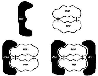
Figure 1 illustrates the ability of sFlt-1 to interact with P1GF. Altliough
the
binding of two molecules of sFlt-1 with a homodimer of P1GF has been suggested
by
observations of experimental systems, the inventors recognize that this state
may not exist
in vivo or may exist at insignificant levels, but is presented in Fig. 1
because the inventive
method is useful for the detection of such complexes, if they exist.
Figure 2 depicts a histograzn of P1GF levels observed by an immunoassay
employing a monoclonal antibody used to capture free P1GF and a polyclonal
antibody
used to detect P1GF in a small number of human sainples collected and
investigated under
ethically appropriate conditions. Figure 2 demonstrates that low levels of
P1GF are
associated with preeclamptic pregnancies (PE), and also that the ability to
separate non-
preeclamptic (Normal) from preeclamptic pregnancies using two antibodies to
PLGF is in
need of improvement.
Figure 3 depicts a histogram of data collected using a monoclonal antibody as
a
first sbp for sFlt-1 and a polyclonal antibody as a second sbp for sFlt-1.
These data show
3
CA 02629451 2008-05-12
WO 2007/059065 PCT/US2006/044059
that there is a significant overlap in the range of total sFlt-1 values for
non-preeclamptic
pregnant women (Normal) with the range of total sFlt-1 values for
preeclainptic women
(PE). According to these data, there would be a need to inlprove the ability
to
discriminate between normal and preeclamptic pregnancies based on inspection
of sFlt-1
levels observed by an inununoassay using two antibodies to sFlt-1 in
diagnostic sainples
obtained from pregnant women.
Figure 4 depicts data obtained in a manner similar to that of the data
depicted in
Figure 3, except that two monoclonal antibodies to sFlt-1 were used instead of
a
combination of a polyclonal and a monoclonal antibody. The data presented in
Figures 3
and 4 indicate that an immunoassay for sFlt-1 comprising a polyclonal and
monoclonal
antibody for sFlt-1 outperforms a similar immunoassay comprising two
monoclonal
antibodies. Even though based on general principles one would expect that this
assay
would provide better quantitation than the assay of Figure 3, it surprisingly
provide less
ability to discriminate non-preeclamptic specimens from preeclamptic
specimens.
Figure 5 depicts a histogram of data collected using one preferred einbodiment
of
the present invention. These data were collected with an immunoassay
comprising a
microparticle-labeled monoclonal antibody to sFlt-1 so that total sFlt-1 in
the sample
would be bound to the microparticle. The bound sFlt-1 was detected by sFlt-l-
binding
portion of P1GF labeled with acridinium. This assay determines the amount of
free sFlt-1
in the specimen. These data show that there is a significant reduction in the
overlap of
fiee sFlt-1 values for non-preeclamptic pregnant women (Normal) with the range
of
values of free sFlt-1 for preeclamptic women (PE). According to these data,
use of a
portion of P1GF as a sbp for free sFlt-1 significantly iinproves the ability
to discriminate
between normal and preeclamptic pregnancies based on inspection of sFlt-1
levels in
diagnostic sainples obtained from pregnant women.
Figure 6 collects the data described above and presents it in a single graphic
representation.
Figure 7 normalizes the data presented in Figure 6 to a single non-
preeclamptic
sample.
Figure 8 compares data collected from the "most normal" preeclainptic woman in
the study ("mP-20") to data collected from the sainples of non-preeclamptic
women.
Measureinents of sFlt-1 are of total sFlt-1 for the monoclonal+polyclonal
antibody fonnat
4
CA 02629451 2008-05-12
WO 2007/059065 PCT/US2006/044059
of this assay, and for the monoclonal+inonoclonal format of this assay,
whereas for the
"Free Recpt" data, an sFlt-l-binding portion of P1GF was used as one sbp in a
sandwich
immunoassay, and therefore, bound only to free sFlt-1. These data show that,
in
accordance with aspects of tla.e present invention, the ratio of free sFlt-1
to free P1GF
(ranging from about 0 to about 1.0) is a better predictor of lower risk (i.e.,
normal
pregnancies) than the ratio of P1GF to total sFlt-1 in the sample.
Figure 9 depicts in tabular form the increased ability of the present
invention to
discriminate non-preeclamptic from preeclamptic samples. These data show that
for non-
preeclainptic specimens determined with a two-antibody based immunoassay the
ratio of
free sFlt-1 to free PIGF is observed to be higher than when using embodiments
of the
invention. Accordingly, these data show that the invention provides superior
discrimination of non-preeclamptic specimens from preeclamptic samples.
Detailed Description of the Invention
In describing the invention, the following terms and abbreviations will be
used.
Abbreviations used to Describe the Invention
The abbreviation "sbp" refers to a "specific binding partner". All biological
materials have some affinity for each other, however, specific binding
partners are those
that bind together in a specific way to perforin a biological function. For
example, sFlt-1
binds to P1GF with a biologically significant affinity, and by so doing, is
able to modulate
the biological activity of P1GF. Accordingly, sFlt-1 and PIGF are specific
binding
partners. Similarly, the membrane bound counterpart to sFlt-1, i.e., Flt-1, is
also a sbp of
PIGF. Moreover, VEGF is also a sbp with Flt-1. Another type of sbp that is
useful in the
context of the invention is an antibody and the molecule comprising the
epitope to which
it binds. While this type of sbp is essential to the function of some of the
embodiments of
the present invention, most embodiments of the invention are directed to
determining the
presence or quantity of one specific binding partner by detecting under assay
conditions
the binding to the otller sbp, wherein neither the first or second sbp is an
antibody or
portion of an antibody.
The term "proteinaceous" is used herein to describe polypeptides, protein
fragments, and proteins which are large enough to forin at least one helix,
sheet, or other
significant (polypeptidyl) secondary structure. The term proteinaceous
includes both
5
CA 02629451 2008-05-12
WO 2007/059065 PCT/US2006/044059
unmodified and modified polypeptides, such as glycosylated, proteolytically
cleaved,
prenylated, and dimerized polypeptides and proteins. Each proteinaceous
binding
partner preferably comprises at least one element of secondary structure, such
as a helical
or sheet structure (e.g., by way of example, an a-helix or (3-sheet).
Accordingly, each
proteinaceous binding partner preferably comprises at least 8 ainino acid
residues, more
preferably at least 50 amino acid residues, and yet more preferably at least
100 ainino
acid residues. A proteinaceous binding partner can also comprise multiple
polypeptide
strands folded togetller so as to form a coinplex protein.
The tei7ns "biological specimen" or "biological sample" are used
interchangeably
(unless expressly indicated to the contrary), and refer to any material
originating from a
human or other animal that contains proteinaceous molecules. The inventive
metllod can
usefully be performed on any suitable sample, including without limitation
sewage,
clothing, carpeting, respiratory condensates, and tissue biopsies. Preferred
"biological
sanlples" of the present invention include blood, blood plasma, blood serum,
urine, feces,
lymph, and saliva. When substantially solid biological samples are used, it
will most
commonly be preferable to extract or solubilize a portion of the sample prior
to
perfoirning or continuing the inventive method.
The term "P1GF" refers to placental growth factor, and is sometimes referred
to as
PGF. The gene encoding P1GF is currently believed to map to gene locus 14q24-
q31.
P1GF encoinpasses all three isoforms culTently lcnown in the art and any
others that are
currently not well characterized to the extent that they bind with the P1GF
sbp used in any
particular embodiment of the invention.
The term "label" or "detectable label" means any suitable molecule allowing
the
direct or indirect quantitative or relative measurement of the molecule to
which it is
attached. Suitable labels useful in the context of the invention include
solids, enzymes,
enzyme substrates, enzyme inhibitors, coenzymes, enzyme precursors,
apoenzymes,
fluorescent substances, pigments, chemiluminescent compounds, luminescent
substances,
coloring substances, magnetic substances, metal particles such as gold
colloids,
radioactive substa.nces, and the lilce. Useful enzymatic markers include
without limitation
dehydrogenases, oxidoreductases such as reductases and oxidases; transferases
that
catalyze the transfer of functional groups, such as amino, carboxyl, methyl,
acyl, and
phosphate groups; hydrolases that hydrolyze bonds such as ester, glycoside,
ether, and
6
CA 02629451 2008-05-12
WO 2007/059065 PCT/US2006/044059
peptide bonds; lyases; isomerases; ligases; and the like. Multiple enzyines
can also be
used in a conjugated form for detection.
Useful solid labels include but are not limited to microtiter plates,
particles,
microparticles and microscope slides.
When the detectable marker is an enzyme, detection of the labeled molecule
also
can be facilitated by enzyinatic cycling. For example, when the detectable
label is
alkaline phosphatase, measurements can be made by observing the fluorescence
or
luminescence generated from a suitable substrate, such as an tunbelliferone
derivative.
Useful umbelliferone derivatives include without limitation 4-methyl-
umbellipheryl
phosphate.
Other useful labels include phosphorylated phenol derivatives such as
nitrophenyl
phosphate, luciferin derivatives, dioxetane derivatives.
Preferred fluorescent and chemiluminescent labels useful in the context of the
invention include fluorescein isotlliocyanate; rhodamine derivatives such as
rhodamine B
isothiocyanate and tetramethyl rhodainine isotliiocyanate; dancyl chloride (5-
(dimethylamino)-1-naphtalenesulfonyl chloride), dancyl fluoride, fluorescamine
(4-
phenylspiro[furan-2(3H), 1'-(3'H)-isobenzofuran]-3,3'-dione);
phycobiliproteins such as
phycocyanine and physoerythrin; acridinium salts; luminol compounds such as
lumiferin,
luciferase and aequorin; imidazoles; oxalic acid esters; chelate coinpounds of
rare earth
elements such as europium (Eu), terbium (Tb) and samarium (Sm); and coumarin
derivatives such as 7-amino-4-methylcoumarin.
Accordingly, it will be appreciated that a wide variety of detectable markers
useful in the context of the present invention are available. It will also be
appreciated that
any suitable detection means can be used to quantify the amount of a molecule
attached to
a detectable label, such as but not limited to the use of electrodes,
spectrophotometric
measurement of color, light, or absorbance, and visual inspection.
Specific Embodiments of the Invention
The present invention provides a method of determining the concentration or
amount of a proteinaceous specific binding-pair present in a biological
specimen. The
binding-pair comprises two proteinaceous moieties (i.e., a first proteinaceous
moiety and
a second proteinaceous moiety) that preferably bind directly to each other and
neither
7
CA 02629451 2008-05-12
WO 2007/059065 PCT/US2006/044059
proteinaceous binding partner is an antibody (or fragment of an antibody). Any
suitable
proteinaceous binding partners, including polypeptides and proteins, can be
used in the
inventive method. Antibodies and portions of antibodies can be used in the
inventive
method, but preferably less than two antibodies or portions thereof are used.
Alternatively, when more than two antibodies are used, one of the two
proteinaceous specific binding partners of the method is labeled with the
detectable
inarlcer that is observed in the method. For example, when absorbance at a
particular
wavelength is used, the chroinophore is linked to the either the first or
second sbp otlier
than through use of an antibody or portion thereof. Similarly, if a
scintillation or Geiger
counter is used to detect a radioactive label (e.g., 125I) , the sbp is
indirectly, or more
preferably directly, attached directly to the second sbp. When the first or
second sbp is
labeled indirectly tluough a means other than a first or second antibody, or
portion of an
antibody, then the directly labeled moiety is preferably either one having
biospecific
affinity for the first or second sbp (i.e., a third sbp) or is one having
stringent affinity for a
coinponent of the first or second sbp. One exainple of an indirect label
system having
stringent affinity includes biotin and avidin. In this non-limiting example,
the second sbp
can be labeled with biotin and an avidin=like moiety (e.g., avidin,
streptavidin, extravidin)
can be directly labeled (with, e.g., an acridiniuin ester or otlier
chemiluminescent
acridinium derivative).
In a first embodiment of the inventive method, the first proteinaceous
specific
binding partner is labeled with a detectable label, which therefore forms a
labeled moiety.
The labeled moiety is contacted witli the biological specimen and the degree
of binding
between the labeled moiety and the component of the biological specimen that
is the
second binding partner is detemlined. The degree of binding indicates either
the
concentration or amount of the first binding partner or the second binding
partner present
in the specimen. The determination of the degree of binding can be a relative
value, but
is preferably quantitative. This degree of binding can be determined by any
suitable
means, such as by causing the bound pair to agglutinate, bind to a solid
support, or
migrate at a differential rate through a liquid medium. Preferably, the degree
of binding
is determined by causing the bound pair to adhere to a solid support,
separating the
unbound labeled moiety from the bound label moiety and determining either the
bound or
unbound (or botli) fraction of the labeled moiety.
8
CA 02629451 2008-05-12
WO 2007/059065 PCT/US2006/044059
The binding pair used in the invention is preferably not a pair of proteins
that
interact in the mammalian inunune system such as the T cell receptor and major
histocompatibility complex, CD40 and CD40L, or an antibody and its target.
The method optionally can be perfoilned with a cartridge, test strip, or in a
unitary
package adapted to be used by a semi-automated or fully-automated
immunoanalyzer.
Automated diagnostic assays in the context of the invention are preferably
performed in a
system that delivers samples and reagents to a reaction vessel, perfoi7ns
incubations, and
optionally washes unbound labeled moiety from the bound labeled moiety,
without user
intervention, once the sample and reagents are inserted into the system. Such
a system
optionally can be distinguished from manual or less-automated systems by the
ability of
the system to perform at least eight assays, preferably at least 16 assays,
more preferably
at least 64 assays, and most preferably at least 128 assays in a 48-hour
period without
user intervention after inserting the sample and the reagents into the system.
The system
is preferably also able to calculate the concentration or quantity of the
target protein of
the binding pair automatically, i.e., without the need for huinan calculation
or input once
the samples are loaded into the system.
The method also can be performed in a cartridge foi7nat or in a test strip
assay. In
such an assay, the assay reagents are preferably provided as a unit-dose
loadable into
disposable instrument and the unit-dose contains all the reagents necessary to
assay to
perforin the method. Such a unit dose instrunient for example can comprise a
plastic
liousing comprising a disposable membrane-like structure of nylon,
nitrocellulose, or
other suitable material. The sample can be preprocessed or loaded directly
onto a loading
zone. The sample can then optionally flow across the membraiie-like structure
tlirough a
plurality of zones contained on the membrane. The membrane-like structure
optionally
further contains a detergent or lateral flow-aid and also optionally contains
an absorbant
to collect excess fluid and/or encourage the lateral flow across the
meinbrane.
Additionally, the inventive inethod can be performed with inulti-pack systems
in which
each pack comprises sufficient reagents to perform 2, 4, 8, 10, or 12 assays,
or preferably
one assay.
The method can also be perforined in a microfluidic device designed to analyze
samples in the microliter range (e.g., less than 50 L, preferably less than
12 L, and
optionally less than 4 L of fluid). Such inicrofluidic devices can optionally
contain flow
9
CA 02629451 2008-05-12
WO 2007/059065 PCT/US2006/044059
aids, propulsion devices (including but not limited to expansion gels, waxes,
and gases),
nanovalving and the lilce to assist the transportation of the biological fluid
or assay
reagents or both through the microfluidic device.
The inethod optionally can be configured as a sandwich assay. Sandwich assays
comprise binding the labeled moiety to the other specific binding partner of
the binding
pair, and another specific labeling reagent. Multiple sandwich assays are
within the scope
of the invention. Mainly for the salce of illustration and ease of
coinprehension, but not
by limitation, the following sandwich assays are illustrated by the use of
P1GF as the first
binding partner and sFlt-1 as the second binding partner. However, any
suitable pair of
proteinaceous specific binding partners can be substituted for the P1GF and/or
sFlt-1 in
the following illustrations.
In a first sandwich assay within the scope of the invention an antibody to
P1GF (a-
P1GF) is bound to a microtiter plate which antibody is previously or
subsequently bound
to P1GF or an sFlt-l-binding fragment thereof. In the context of the
invention, the P1GF
is thus labeled witli a solid substrate and is a labeled moiety. The
microtiter plate can be
optionally washed to ensure that substantially no free P1GF is on the
microtiter plate. A
sample is contacted to the plate, wliich sample is lcnown to contain, or
suspected of
containing sFlt-1. Thus, the use of P1GF:sFIt-1 binding is used in the assay.
After
washing, the quantity of sFlt-1 in the sample can be detected by contacting
the plate with
a labeled antibody. The antibody can be labeled with any suitable detectable
label.
In a second sandwich assay within the scope of the invention the microtiter
plate
of the first sandwich assay is replaced a microparticle. Preferred
microparticles include
but are not limited magnetic microparticles, particularly those averaging
between 0.2 and
7.0 microns in size, haptenated microparticles, microparticles impregnated by
one or
preferably at least two fluorescent dyes (particularly those that can be
identified after
individual isolation in a flow cell and excitation by a laser), ferrofluids
(i.e., magnetic
particles less than about 0.1 m in size), and other microparticles rernovable
by
collectable or removable by filtration.
In a third and fourth saiidwich assay within the scope of the invention, the
P1GF is
conjugated directly to the microtiter plate or to the inicroparticle,
respectively.
In a fifth and sixth sandwich assay, the P1GF is biotinylated or labeled with
a
suitable hapten, such as for example, adamantine, fluorescein isothiocyanate,
or
CA 02629451 2008-05-12
WO 2007/059065 PCT/US2006/044059
carbazole. This allows the formation of aggregates when contacted with a multi-
valent
antibody or (strept)avidin containing moiety, or alternatively allows easy
attachment of
the P1GF to a solid substrate sucll as a microtiter plate, microscope slide,
or microparticle.
In embodiments employing aggregation, any suitable separation or detection
means can
be used, such as precipitation or filtration of the aggregates or liquid
cliroinatography of
the aggregates.
Similarly, the inventive method coinprises competitive inliibition assays. A
competitive inhibition assay can be configured with a single specific binding
partner or
also as a sandwich assay. Useful coinpetitive inhibition assays include those
in which a
labeled second specific binding partner (or fragment thereof) ("labeled 2d
sbp") is
synthesized or isolated from a source other than the biological sample to be
assayed, and
labeled with a direct or indirect label. The amount of the 2"d sbp in the
tested biological
sample is then determined by measuring the extent to which the labeled 2"d sbp
is
prevented from binding to the first sbp. By way of illustration, and not
liinitation, and
using the illustrative convention used above, sFlt-1 or a P1GF-binding
fragment thereof
can be labeled with any suitable detectable label, including without
limitation those
discussed above. When immobilized P1GF is contacted with a biological sample,
the
sFlt-1 in the biological sainple will compete with the labeled sFlt-1 for
binding to the
P1GF. The reduction in label binding to the immobilized P1GF then indicates
the anlount
of sFlt-1 in the biological sample which is known to contain, or is suspected
of
containing, s-Flt-1.
The skilled artisan will appreciate, therefore, that the invention provides
many
embodiments in which the bindiiig interaction of a first polypeptide or
protein with a
second polypeptide or protein is used to measure the amount or concentration
of the
second polypeptide or protein. The specific binding partners used to
illustrate
embodiments of the invention above, i.e., P1GF and sFlt-1, are of course among
the
preferred specific binding partners that are suitable for use with the
invention. Additional
preferred einbodiments include other angiogenic growth factors and their
receptors, such
as without limitation, VEGF-A, VEGF-B, VEGF-C, VEGF-D, VPF and the like.
Similarly, the biologically significant variants of the growth factors, such
as without
liinitation, VEGF-A206, VEGF-A189, VEGF-Al65, VEGF-A145, and VEGF-A121 are
preferred specific binding partners of the invention. Similarly, receptors for
these
11
CA 02629451 2008-05-12
WO 2007/059065 PCT/US2006/044059
nlolecules, such as KDR, or Flk-1 (fms tyrosine-like kinase-1) (also VEGFR or
VEGFR2)
are preferred first or second binging partners of the present invention.
Other preferred specific binding partners of the invention include natriuretic
factors, a natriuretic factor receptor, an insulin-like growth factor (IGF),
or an IGF-like
receptor. Examples of natriuretic factors include atrial natriuretic peptide
(ANP), B-type
or brain natriuretic peptide (BNP), and c-type natriuretic peptide (CNP). Any
suitable
member of the IGF, IGFR, and IGFBP family can be used as the first or second
specific
binding member or both.
It will be readily appreciated that the first specific binding partner or the
second
specific binding partner can be a cllimera or fusion comprising amino acid
residues from
other polypeptides. Similarly, the specific binding partners can be full-
length or
truncated. Particularly preferred truncations useful in the context of the
invention are
those that cleave a transmembrane region from a soluble extracellular domain
of the
protein, although the method can also be performed using membrane-bound or
bindable
binding partners. When a specific binding partner is membrane bound the
membrane
optionally can be part of a cellular structure, synthetic, or removed from a
cellular context
(e.g., in a vesicle, liposome, or einulsion). This extracellular domain itself
can be "full
length", truncated at the N-terininus or C-terminus or both, and can be fused
to exogenous
polypeptides.
The invention also provides a method of deterinining the concentration of sFlt-
1
that does not have a specific binding partner bound to the P1GF binding site
of sFlt-1.
The method includes contacting a sample that is known or suspected of
containing sFlt-1
that does not have a specific binding partner bound to the P1GF binding site
of sFlt-1 with
a first specific binding partner (sbp) of sFlt-1 capable of forining a
sbp:sFlt-1 complex
and with a second sbp, wherein the second sbp is specifically labeled with a
detectable
label or a solid structure. The first or second sbp is P1GF or an sFlt-1-
binding fragment of
P1GF. The first sbp, the second sbp, and the sFlt-1, if present, then form a
teniary
complex which is detected as an indication of the ainount of sFlt-1 in the
sample.
In one preferred embodiment the total sFlt-1 in the biological sample is
captured
with an Flt-1 binding antibody and the portion of sFlt-1 in the sainple that
is not bomid to
P1GF or a similar binding partner that binds to the P1GF-binding site of sFlt-
1 is detected
with a labeled fragment of the epidermal growth factor (EGF) superfamily.
Suitable
12
CA 02629451 2008-05-12
WO 2007/059065 PCT/US2006/044059
members of the EGF superfainily include, but are not limited to, any suitable
portion of
P1GF or VEGF. In yet more preferred einbodiments that EGF superfamily inember
is
labeled with a luininophore or an enzyine capable of producing a detectable
product, such
as without limitation, horse radish peroxidase, fluorescein, or acridinium.
Another prefeiTed embodiment comprises affixing P1GF on a solid surface, such
as without limitation a microparticle. This reagent will capture only free
sFlt-1. Without
desiring to be bound by any particular theory it is believed that the sFlt-1
in the tested
biological sainple does not bind to the solid-bound P1GF because the binding
site between
P1GF and sFlt-1 is already bound in non-free sFlt-1. The coinplex can then be
detected in
any suitable manner. Suitable direct and indirect detection reagents include
an antibody,
antibody-fragment, or aptamer to the sFlt-1.
Any of the reagents in the foregoing embodiments can be readily biotinylated
through prior art methods. Accordingly, the P1GF can be biotinylated which
will
facilitate its fixture to a solid phase or another detectable molecule, and
the detection
reagent can be biotinylated so that it is detectable with a specific binding
partner for
biotin. Preferred specific binding partners for biotin in the context of the
invention
include antibodies and aptainers to biotin, avidin and strepavidin.
The structure of sFlt-1 is well-lcnown in the art (See, Wiesinaim et al.,
Cell, 91,
695-704 (1997); Davis-Smyth et al., EMBO J, 15, 4919-4927 (1996); Barleon et
al., J
Biol Chein, 272, 10382-10388 (1997); Cunningham et al., Biochem Biophys Res
Conunun, 231, 596-599 (1997); Fuli et al. (cited within Wiesmann et al.)). A
preferred
sFlt-1 specific binding par-tYler is any suitable sFlt-l-binding fragment to
P1GF. Preferred
sFlt-l-binding fragments of polypeptides comprising at least about 90% of the
second
and third domains of sFlt-1. Truncated polypeptides of P1GF are also
preferred, such as
the 21St amino acid of P1GF through domain 3 of P1GF. Either or both of the
P1GF and
sFlt-l-binding fiagnlent of P1GF can be labeled as disclosed elsewhere
herein..
To facilitate detection of the interaction of the P1GF capture or detection
reagent
and the sFlt-1 that was free in the tested biological sainple, an additional
reagent can be
added which is labeled by binding to a solid surface or -a detectable label.
Labeled
antibodies are among the preferred additional reagents.
The invention also provides an immunoassay based on the competitive
inliibition
of a labeled sFlt-1 moiety by the quantity of sFlt-1 in the tested biological
sainple that
13
CA 02629451 2008-05-12
WO 2007/059065 PCT/US2006/044059
does not have P1GF bound to the sFlt-1 ("free sFlt-1"). The method comprises
contacting
a sample that contains free sFlt-1 with a first sbp, in which the first sbp
contains an sFlt-1-
bindin.g fragment of P1GF and a second sbp, in which the second sbp contains a
fragment
of sFlt-1 that is capable of binding to the sFlt-1 binding fragment of P1GF
where at least
the first sbp or the second sbp is labeled. The concentration of sFlt-1
present in the
sainple is then determined by measuring the decrease in binding between the
first sbp and
the second sbp caused by the sanlple.
The invention also provides a method of deteimining the ratio of free sFlt-1
to
total sFlt-1 in a sample. The method comprises (i) determining the amount of
sFlt-1
according to any of the foregoing embodiments, (ii) detennining the total
ainount of sFlt-
1 in the sanzple, and comparing the result of part (i) to part (ii). Any
suitable method can
be used to determine the total amount of sFlt-1 in the sainple. Suitable
methods for
carrying out this step include, but are not liniited to, sandwich immunoassays
and
competitive inhibition assays. If at leastone antibody used in an immunoassay
to
deterinine the total sFlt-1 present in the assay binds to the binding site of
P1GF or another
factor present or possibly present in the biological sample (e.g., an anti-
idiotypic antibody
specific for the active site of a first or second sbp), then a portion of the
sample optionally
can be denatured to disrupt the binding of the sFlt-1 to other proteins in the
biological
sample. In this instance, any suitable technique can be used to denature the
sFlt- 1 such
that proteins that would block the antibody binding to the active site of sFlt-
1 are
released. Suitable techniques include adding acid, base, salt, detergents or
surfactants,
organic bases or a combination of the foregoing and are within the skill of
those in the art.
To facilitate the binding of an antibody or another sbp used as a diagnostic
reagent, the
denaturant used to disrupt the binding of sFlt-1 to the sbp in the sample is
preferably
readily neutralized or removed from the sample. Preferably, however, the one
or more
antibodies used in an immunoassay to determine total sFlt-1 does not bind to
the P1GF
binding site of sFlt-1. The skilled artisan will appreciate that still other
methods of
measuring total sFlt-1 in the sample are readily available and within the
scope of the
present invention. Accordingly, the invention enables both the direct and
indirect
determination of each of (i) free sFlt-1, (ii) bound sFlt-1, and (iii) total
sFlt-1. Any of
these sFlt-1 values optionally can be fiuther conlpared to the concentration
of an EGF
superfamily nleinber, uicluding without limitation VEGF, and preferably P1GF.
14
CA 02629451 2008-05-12
WO 2007/059065 PCT/US2006/044059
In a particularly preferred einbodiinent, an anti-sFlt immunoreagent is
attached to
a magnetic microparticle, and the biological specimen is contacted to the anti-
sFlt-1
bound microparticle such that the sFlt-1 in the sample is bound to the
magnetic
microparticle. The complex can then be optionally washed in a suitable
solution or buffer
one or more times to remove unbound molecules that could interfere with the
assay.
Then labeled P1GF is contacted to inicroparticle containing coinplex and
unbound labeled
P1GF is removed or washed away from the magnetic microparticle. The amount of
labeled P1GF bound to the magnetic microparticle then serves as an indication
of the
quantity of free sFlt-1 in the biological specimen because sFlt-1 bound by a
sbp (which
spb binds to the P1GF binding-site of sFlt-1) cannot efficiently bind the
labeled P1GF.
In another embodiment of the claimed invention, the total sFlt-1 and the
portion of
the sFlt-1 bound to P1GF is measured. Any suitable metliod can be used to
detennine the
quantity of total and P1GF-bound sFlt-1 ("bound sFlt-1") present in the
sample. One
suitable method to deterinine the quantity of bound sFlt-1 in the sample is to
detect the
formation of a coinplex having at least three components including an anti-
P1GF
antibody, the bound sFlt-1 (which itself comprises at least sFlt-1 and P1GF),
and an anti-
sFlt-1 antibody. That is, to employ a two-antibody sandwich assay in which one
antibody
is specific for P1GF and at least one antibody is specific for sFlt-1. In
accordance with
other preferred embodiments of the invention, the antibodies are each
preferably labeled
with detectable labels. In an even more preferred enlbodiinent of this
embodiment, one
antibody is labeled by attachment to a solid substrate and at least one
antibody is labeled
by conjugation to another label referred to herein.
In another embodiment, the detection of free sFlt-1 is performed with an
antibody
that binds to an epitope that is not accessible (i.e., hidden) when P1GF is
bound to the
sFlt-1. In this way respect, the assay of the invention is any traditional
sandwich,
coinpetitive inhibition, or other conventional immunoassay (for sFlt-1),
except that it only
measures free sFlt-1. This allows coinparison of the quantity of free sFlt-1
to the quantity
of total P1GF, or more preferably, to the quantity of free P1GF. In further
aspects of this
embodiment, an antibody to the sFlt-1 binding site of P1GF can be substituted
for the
portion of the sFlt-1 used in other embodiments of this invention.
The measurements of P1GF, and sFlt-1, including without limitation the
ineasurements bound and free states of these molecules can be used for any
suitable
CA 02629451 2008-05-12
WO 2007/059065 PCT/US2006/044059
purpose. For example, the measurement of these marlcers can be used to predict
the
course of angina following a major cardiovascular event such as a non-lethal
myocardial
infarction. Similarly, the ability to measure these markers can be used to
better
understand the mode of action of heart inedicines. Moreover, the accurate
measurement
of these markers permits more detailed investigations into the inechanisms of
restenosis
and neovascularization. The measurement of P1GF and sFlt-1 could find the
greatest
significance in demonstrating a lower risk of preeclampsia in pregnant women.
Preeclampsia affects about 5% of all pregnant women, and in some ethnic groups
affects as many as about 10% of all pregnant women. The effects of
preeclainpsia can be
severe and sometimes include death. Accordingly, there is a need to better
separate
normal pregnancies from pregnancies at high risk for preeclainpsia.
The present inventors have discovered that the ratio of free sFlt-1 to free
P1GF is a
better predictor of risk of preeclainpsia than is the ratio of total sFlt-1 to
free P1GF.
Because the quantity of free sFlt-1 is matllematically related to the quantity
of total sFlt-1
and bound sFlt-l, these values can be used as a surrogate for the quantity of
free sFlt-1,
and can be compared to the quantity of P1GF in a biological sample within the
scope of
the present invention.
Many proteins of interest for medical diagnostics are present in low
concentrations, e.g., at from less than 1 pg/inL to 0.1 mg/mL. Some of these
proteins will
bind to a protein receptor witli affinities similar to that observed for
antibody-antigen
interactions. In an analogous fashion to enzymatic activity, it is possible to
measure the
amount of a free protein or the amount that is bound its native receptor.
Preeclampsia is a disease of late pregnancy that is currently diagnosed based
on
clinical symptoms of high blood pressure and protein in the urine. Recent
literature has
proposed that the precipitating event of the disease is a decrease in
circulating levels of
the angiogenic proteins Vascular Endothelial Growth Factor (VEGF) and
Placental
Growth Factor (P1GF). The resulting lack of vascularization in the placenta is
then
suggested to be responsible for the increase in blood pressure and
proteinuria, clinically
known as preecla.inpsia. The decrease in these two proteins is apparently due
to the
increased concentration of the soluble form of the receptor soluble fins-like
tyrosine
kinase 1(sFlt-1). The present invention covers an approach to measuring sFlt-
1, which is
free or bound to P1GF and its use as an assay component for measuring free and
bound
16
CA 02629451 2008-05-12
WO 2007/059065 PCT/US2006/044059
forms of P1GF. For detection of preeclampsia, the most relevant inforination
is the levels
of PIGF in relationship to that of the sFlt-l which is not bound to P1GF. High
concentrations of free sFlt-1 indicate that the P1GF concentrations are likely
to be low due
to the presence of a large excess of free receptor.
In the preferred embodiment, an antibody is used to bind all the circulating
sFlt-1
(either bound or unbound to P1GF). A conjugate of a signal generating moiety
and P1GF
is then allowed to interact with the sFlt-1 bound to the solid phase. In this
exainple, only
the sFlt-1 free of P1GF would bind the conjugated P1GF. The unbound P1GF is
then
washed away and the necessary steps are talfen to reveal the concentration of
P1GF-
conjugate.
The above format could also be constructed using VEGF, in the same manner as
the P1GF as conjugate. Furthermore it would be possible to also use the
heterodimer of
VEGF and P1GF.
Anotlier form of the assay would be to use P1GF bound to a solid phase and
then
capture any free P1GF which can then be detected with an conjugated antibody
that binds
sFlt-l. The same format can use VEGF on the solid phase.
Anotller form of the assay would measure free P1GF or VEGF by attaching the
sFlt-1 to a solid phase and then capturing any free growth factor that is not
bound to a
soluble receptor. Detection again can be performed with a conjugated antibody
that binds
to the growth factor. This form of the assay would be specific for the
biologically active
form of the growth factors. In this forinat any degraded growth factor would
not be
detected improving the specificity of the assay to the biological event that
causes
preeclainpsia.
When the relevant biological question is the aniount of P1GF bound to
receptor, it
is also possible to use a solid phase that would capture sFlt-1 as in the
first example and
then use a conjugated antibody against P1GF or VEGF to measure the amount of
bound
growth factor. The utility of the approach would depend upon the successful
correlation
of disease state witli the species measured.
Another fomz of the assay is to use immobilized receptor in a competitive
format
wliere the free ligand in the sample competes with labeled ligand. For example
sFlt-1 on
a solid phase be used to capture either the P1GF in the sample in a
competitive forinat
17
CA 02629451 2008-05-12
WO 2007/059065 PCT/US2006/044059
wit11 labeled P1GF. This form of the assay would eliminate the requirement of
an
antibody in the assay.
The converse of the above exainple would be to inunobilize with P1GF or VEGF
and add sample and conjugated sFlt-1. In this format, free sFlt-1 would
compete for sites
on the solid phase.
The sources of the protein used in the assay could be derived from patient
samples
however the use of recombinant proteins expressed in either cell culture or in
bacteria
would be more practical approach.
The approach described here could be used to interrogate sainples with regards
to
either receptor or associated ligand activity so long as the affinity between
the ligand and
the receptor is sufficiently high to perinit the use of wash steps without
such loss of the
bound material to such an extent that it could not be detected in the signal
generation of
the assay protocol.
Assays that depend upon the inherent biological binding activity of the
targeted
proteins may provide superior infoi7nation to assess the clinical situation of
a patient.
When a disease or medical condition involves a protein receptor, assays that
measure the
relative amount of that biological activity can be expected to lead to a more
accurate
clinical picture as compared to only lcnowing the mass of the protein.
In accordance with the foregoing metllods, the present invention also provides
an
iminunoassay comprising two proteinaceous specific binding partners, wherein
at least
one sbp is detectably labeled.
Additionally, in accordance with the foregoing the invention provides a
composition of matter for determining the ratio of free sFlt-1 to free P1GF,
as well as
compositions of matter for determining the total (i) sFlt-1 and bound sFlt-1
or (ii) the total
P1GF and bound P1GF, or both (i) and (ii).
Examples
The invention is illustrated witli data obtained from various immunoassays for
total P1GF, free P1GF, total sFlt=1, and free sFlt-1. A selection of these
data deemed to be
most illustrative of the invention and inventive concepts are presented in the
attached
drawings and discussed briefly in the Brief Description of the Drawings. As is
clear from
18
CA 02629451 2008-05-12
WO 2007/059065 PCT/US2006/044059
the entirety of this description of the invention, the exainples are meant to
illustrate the
claimed invention rather than to limit its scope.
Further Exainples
The following additional examples provide more detail regarding two preferred
embodiments of the present invention.
EXAMPLE 1
This exainple illustrates the inventive method in an assay used to detect free
sFlt-1
in a biological sample using a portion of P1GF as a sbp for sFlt-1.
A monoclonal antibody against sFlt-1 was coated on magnetic carboxyl-latex
microparticles (4.7 microns) at a protein concentration of 0.lmg/mL of
microparticles at a
concentration of 1% by weight in 50mM sodium MES (2-morpholinoetlianesulfonic
acid)
at pH 6Ø After 10 minutes, EDAC, (ethyl-3-(-3-
diinethylaminopropyl)carbodiunide)
was added and allowed to react for one hour before washing the particles with
phosphate
buffered saline. The particles were then diluted to 0.1 % in a buffer for use
in an
automated immunochemical analyzer.
Acridinylated P1GF was prepared by dissolving P1GF in phosphate buffered
saline
and incubation with an acridinium-ester at a mass ratio of P1GF to acridiniuin
of 150,00/1.
The conjugate was then purified by HPLC chromatography and diluted to a
concentration
of approximately 75 ng/mL.
The following series of steps are then perfonned. A 0.05 mL aliquot of sample
is
added to a reaction vessel to which 0.05 mL of the 0.1 % labeled
microparticles is added.
The reaction mixture is incubated for 18 minutes at 37 degrees centigrade. A
magnet
holds the particles while the reaction solution is removed. After the
particles are washed,
a 0.05 mL aliquot of conjugate solution is added. After incubation for 4
minutes, the
particles are once again held to a magnet and the pellet of microparticles the
conjugate
solution is removed followed by washing of the particles once again. The
remaining
acridinium label is caused to emit light after the addition of sodium
hydroxide and
hydrogen peroxide. The photons released are measured and is linearly related
to the
calibrators run in the identical way.
Data were collected on test samples and compared to the results obtained with
conventional immunoassays. The data suggested that the inventive method better
19
CA 02629451 2008-05-12
WO 2007/059065 PCT/US2006/044059
discriminated non-preeclamptic samples from preeclamptic samples, particularly
when
observing the ratio of free sFlt-1 to free PLGF concentrations.
EXAMPLE 2
This example illustrates the inventive method in an assay used to detect free
P1GF
in a biological sample using a portion of sFlt-1 as a sbp for P1GF.
Para.inagnetic latex microparticles (4.7 microns), derivatized witli carboxyl
functional groups, was coated with aia.ti-sFlt-1 antibody (containing domains
1-3 of the
fins-like tyrosine kinase 1) at a protein concentration demonstrated to be
sufficient to
maximize the ainount of protein absorbed to the surface area of the
microparticles (2%
solids by weight) in 50mM MES, pH 5.5. In another einbodiment, the sFlt-1
could be
bound directly to a solid substrate. After 10 minutes, the non-absorbed sFlt-1
was
removed by washing the particles tnultiple times with MES buffer. Following
washing
the particles, EDAC was added and allowed to react forming a covalent coupling
of the
sFlt-1 molecules to the particles. The particles were then waslied with
phosphate buffered
saline to stop the reaction and remove unreacted EDAC. The particles are then
diluted to
0.1 Jo in buffer for use in an automated inununochemical analyzer.
Acridinium-labeled anti-P1GF antibody was prepared by incubating an polyclonal
antibody (alternatively a monoclonal antibody could be used) with an
acridinium-ester at
a molar ratio of acridinium to antibody ranging from 1 to 100. Unconjugated
acridinium
was then separated from the acridinium-labeled antibody conjugate by size
chromatography. The purified conjugate was then diluted in buffer to a
concentration
yielding the maximuin signal to noise ratio in the assay.
The following series of steps were then performed. A 0.1 mL aliquot of sample
was added to a reaction vessel to which 0.05 mL of the 0.1% labeled
microparticles was
added. The reaction mixture was incubated for 18 minutes at 37 degrees
centigrade.
Utilizing the parainagnetic property of the particles, a magnet attracts and
holds the
particles against the side of the reaction vessel while the reaction solution
is removed.
After the particles are washed, buffer is dispensed; a 0.05 mL aliquot of
conjugate
solution is added; the magnet removed and the mixture vortexed. After a 4-
minute
incubation, excess conjugate was removed by particle attraction to a magnet,
washing and
resuspension. The particles, now containing the sFlt-1/P1GF/anti-P1GF antibody
CA 02629451 2008-05-12
WO 2007/059065 PCT/US2006/044059
(acridiniuin-labeled) sandwich, was then exposed to reactants causing the
acridinium to
emit light. The cheiniluminescence, measured by the instrument, is directly
proportional
to the amount of P1GF (fiee) in the sample.
Data were collected on test sainples and compared to the results obtained
witli
conventional irrununoassays. The data suggested that the inventive method
better
distinguished the preeclamptic state from the non-preeclamptic state by
measuring the
biological activity of the prophylactic or causative biological agent rather
than using
antibodies against the agent that would not necessarily distinguish between
active and
inactive forins of the protein.
All patents, patent applications, and publications mentioned herein are hereby
incorporated by reference.
The present invention is amenable to many variations and includes
modifications
that can be derived from the description herein by a person skilled in the
art. All such
variations and modifications are considered to be witllin the scope and spirit
of the
present invention as defined by the following claims.
21