Note: Descriptions are shown in the official language in which they were submitted.
CA 02629715 2013-09-25
ANTI-ALPHA2 INTEGRIN ANTIBODIES AND THEIR USES
CROSS REFERENCE TO RELATED APPLICATIONS
[01]
TECHNICAL FIELD
[02] The present invention generally relates to antibodies directed to a2p1
integrin and
their uses, including humanized anti-alpha 2 (a2) integrin antibodies and
methods of
treatment with anti-a2 integrin antibodies.
BACKGROUND OF THE INVENTION
[03] The
integrin a2í31 (Very late antigen 2; VLA-2) is expressed on a variety of cell
types including platelets, vascular endothelial cells, epithelial cells,
activated
monocytes/macrophages, fibroblasts, leukocytes, lymphocytes, activated
neutrophils and
mast cells. (Hemler, Annu Rev lmmunol 8:365:365-400 (1999); Wu and Santoro,
Dev.
Dyn. 206:169-171 (1994); Edelson et. aL, Blood. 103(6):2214-20 (2004);
Dickeson et al,
Cell Adhesion and Communication. 5: 273-281 (1998)). The most typical ligands
for
a2í31 include collagen and laminin, both of which are found in extracellular
matrix.
Typically the I-domain of the a2 integrin binds to collagen in a divalent-
cation dependent
manner whereas the same domain binds to laminin through both divalent-cation
dependent and independent mechanisms. (Dickeson et al, Cell Adhesion and
Communication. 5: 273-281 (1998)) The specificity of the a2í31 integrin varies
with cell
type and serves as a collagen and/or laminin receptor for particular cell
types, for
example a2í31 integrin is known as a collagen receptor for platelets and a
laminin
receptor for endothelial cells. (Dickeson et al, J Biol. Chem. 272: 7661-7668
(1997))
Echovirus-1, decorin, E-cadherin, matrix metalloproteinase I (MMP-I),
endorepellin and
multiple collectins and the C1q complement protein are also ligands for a2í31
integrin.
(Edelson et al., Blood 107(1): 143-50 (2006)) The a2í31 integrin has been
implicated in
several biological and pathological processes including collagen-induced
platelet
aggregation, cell migration on collagen, cell-dependent reorganization of
collagen fibers
as well as collagen-dependent cellular responses that result in increases in
cytokine
expression and proliferation, (Gendron, J. Biol. Chem. 278:48633-48643 (2003);
Andreasen et al., J. lmmunol. 171:2804-2811 (2003); Rao et al., J. lmmunol.
165(9):4935-40 (2000)), aspects of T-cell, mast cell, and neutrophil function
(Chan et. al.,
CA 02629715 2008-05-14
WO 2007/056858 PCT/CA2006/001876
2
J. Immunol. 147:398-404 (1991); Dustin and de Fougerolles, Curr Opin Immunol
:286-
290 (2001), Edelson et. al., Blood. 103(6):2214-20 (2004), Werr et al., Blood
95:1804-
1809 (2000), aspects of delayed type hyersensitivity contact hypersensitivity
and
collagen-induced arthritis (de Fougerolles et. al., J. Clin. Invest. 105:721-
720 (2000);
Kriegelstein et al., J. Clin. Invest. 110(12):1773-82 (2002)), mammary gland
,ductal
morphogenesis (Keely et. al., J. Cell Sci. 108:595-607 (1995); Zutter et al.,
Am. J. Pathol.
155(3):927-940 (1995)), epidermal wound healing (Pilcher et. a/., J. Biol.
(:;hem.
272:181457-54 (1997)), and processes associated with VEGF-induced angiogc
nesis
(Senger et al., Am. J. Pathol. 160(1):195-204 (2002)).
[04] lntegrins are heterodimers comprised of one a and one 13 subunit, and
comprise a
large family of cell surface proteins that mediate cell adhesion to
extracellular matrix
(ECM) as well as plasma proteins and are central to some types of cell-cell
interactions.
lntegrins interact with ECM components through their extracellular domains.
(Pozzi &
Zent, Exp Nephrol. 94:77-84 (2003)) Upon binding to ligands, integrins
tran:iduce
intracellular signals to the cytoskeleton that modify cellular activity in
response to 1:hese
cellular adhesion events, referred to as outside-in signaling (see, e.g.,
Hemler, Ann I Rev
Immunol 8:365:365-400 (1999); Hynes, Cell. 110(6):673-87 (2002)). Such
signaling can
also activate other integrin subtypes expressed on the same cell, referred to
as inside-out
signaling. Inside-out signaling further occurs via regulatory signals that
originate within
cell cytoplasm such as a disruption of the clasp between an a and p subunit,
which are
then transmitted to the external ligand-binding domain of the receptor.
lntegrins can play
important roles in the cell adhesion events that control development, organ
morphogenesis, physiology and pathology as well as normal tissue homeostasi.,
and
immune and thrombotic responses, and in addition, they serve as environmental
sc. nsors
for the cell. These proteins are characterized as being in a closed
conformation under
normal conditions that, upon activation undergo rapid conformational chango
that
exposes the ligand binding site. X-ray crystal structure is a recent tool that
has been
used in the study of integrin structure and mechanisms of activation. The
understii nding
of integrin structural features facilitates the better understanding of
binding sites,
differentiated states and their active and inactive formations. In general,
the bindir g site
for ligand/counter-receptor for all integrins lies within the a domain and is
comprised of a
metal ion dependent binding site, referred to as the MIDAS domain (Dembo el
al, J
Biol.Chem. 274, 32108-32111 (1988); Feuston et al., J. Med. Chem. 46:5316-
.5325
(2003); Gadek et al., Science 295(5557):1086-9 (2002)); Gurrath et al., Eur.
J. Bio:;hem.
210:911-921 (1992)). In the a subunits of the collagen-binding integrins,
which include
al, a2, al and all integrins, the MIDAS site is located within an extra
inserted d :main
CA 02629715 2008-05-14
WO 2007/056858 PCT/CA2006/001876
3
at the N-terminus known as the I, A or I/A domain, a feature they share with
the a
subunits of the leukocyte 132 family of integrins (Randi and Hogg, J Biol Chem
269:
12395-8 (1994), Larson et al J Cell Biol. 108(2):703-12 (1989), Lee et al., J
Biol Chem.
269: 12395-8 (1995); Emsley et al, J. Biol. Chem. 272:28512-28517 (1997) an
Cell
100:47-56 (2000)). The I domains are structurally homologous to the A1 domain
von
Willebrandt factor, with a Rossman-fold topology of six 13-sheet strands
surrounded by
seven a-helices (Colombatti and BonaIdo, Blood 77(11):2305-15 (1991); Larson
el al, J
Cell Biol. 108(2):703-712 (1989); Emsley et al, J. Biol. Chem. 272:28512-28517
('I997);
Nolte et al; FEBS Letters, 452(3):379-385 (1999)). The collagen-binding
integrins have
an additional a-helix known as the aC helix (Emsley et al, J. Biol. Chem.
272:213512-
28517 (1997) and Cell 100:47-56 (2000); Nolte et al; FEBS Letters, 452(3):379-
385
(1999)).
[05] Integrin/ligand interactions can facilitate leukocyte extravasation
into inf ,amed
tissues (Jackson et al., J. Med. Chem. 40:3359-3368 (1997); Gadek et al.,
Science
295(5557):1086-9 (2002), Sircar et al., Bioorg. Med. Chem. 10:2051-2066
(20021), and
play a role in downstream events following the initial extravasation of
leukocytes frcrn the
circulation into tissues in response to inflammatory stimuli, including
migration,
recruitment and activation of pro-inflammatory cells at the site of
inflammation (Eblqi J.A.,
Curr. Phar. Des. 11(7):867-880 (2005)). Some antibodies that block a2131
integrir were
reported to show impact on delayed hypersensitivity responses and efficacy in
a nurine
model of rheumatoid arthritis and a model of inflammatory bowel disease
(Kriegels.:ein et
al., J. Clin. Invest. 11O(12):1773-82(2002); de Fougerolles et. al., J. Clin.
Invest. 10!!;:721-
720 (2000) and were reported to attenuate endothelial cell proliferation and
migra :ion in
vitro (Senger et al., Am. J. Pathol. 160(1):195-204 (2002), suggesting that
the blocking of
a2131 integrin might prevent/inhibit abnormal or higher than normal angiogenes
is, as
observed in various cancers.
[06] Platelets normally circulate in the blood in an inactive resting
state, however, they
are primed to respond rapidly at sites of injury to a wide variety of
agonists. Upon
stimulation, they undergo shape changes and become highly reactive with plasma
proteins, such as fibrinogen and von Willebrand factor (vWf), other platelets,
and the
endothelial lining of the vessel wall. These interactions all cooperate to
facilitate the rapid
formation of a hemostatic fibrin platelet plug (Cramer, 2002 in Hemostasi!;
and
Thrombosis, 4th edition). Upon binding ligand, platelet receptors transduce
outhide-in
signal pathways which in turn, trigger inside-out signaling that results in
activa=lion of
secondary receptors such as the platelet fibrinogen receptor, allbI33
integrin, leac ing to
platelet aggregation. Antibodies or peptide ligand mimetics that bind to or
intera(:t with
CA 02629715 2008-05-14
WO 2007/056858 PCT/CA2006/001876
4
platelet receptors are anticipated to induce a similar signaling cascade
leading to platelet
activation. Even minor activation of platelets can result in platelet
thrombotic respcnses,
thrombocytopenia and bleeding complications.
[07] a2131 integrin is the only collagen-binding integrin expressed on
platelets arid has
been implicated to play some role in platelet adhesion to collagen and
hemc3tasis
(Gruner et al., Blood 102:4021-4027 (2003); Nieswandt and Watson, Blood
102(21:449-
461 (2003); Santoro et al., Thromb. Haemost. 74:813-821 (1995); Siljander et
al., Blood
15:1333-1341 (2004); Vanhoorelbeke et al., Curr. Drug Targets Cardiovasc.
Haeinatol.
Disord. 3(2):125-40 (2003)). In addition, platelet a2131 may play a role in
the regulalion of
the size of the platelet aggregate (Siljander et al., Blood 103(4):1333-1341
(2004)).
[08] a2r31 integrin has also been shown as a laminin-binding integrin
expressIA on
endothelial cells (Languino et al., J Cell Bio. 109:2455-2462 (1989)).
Endothelia cells
are thought to attach to laminin through an integrin-mediated mechanism,
however It has
been suggested that the a2 I domain may function as a ligand-specific sequence
involved
in mediating endothelial cell interactions (Bahou et al., Blood. 84(11):3734-
3741(19cA)).
[09] It is anticipated that a therapeutic antibody that binds a2131
integrin, including the
a2131 integrin on platelets, could result in bleeding complications. For ex
Ei mple,
antibodies targeting other platelet receptors such as GPlb (Vanhoorelbeke et
al., Curr.
Drug Targets Cardiovasc. Haematol. Disord. 3(2):125-40 (2003) or GP Ilb/Illa
(Schell et
al., Ann. Hematol. 81:76-79 (2002), Nieswandt and Watson, Blood 102(2):4119-
461
(2003), Merlini et al., Circulation 109:2203-2206 (2004)) have been associated
with
thrombocytopenia, although the mechanisms behind this are not well understood.
It has
been hypothesized that binding of an antibody to a platelet receptor can alter
its three
dimensional structure, and expose normally unexposed epitopes which then leads
to
platelet elimination (Merlini et al., Circulation 109:2203-2206 (2004).
Indeed, the bleeding
complications associated with oral doses of GP Ila/Illb antagonists have been
des :;ribed
as the "dark side" of this class of compounds (Bhatt and Topol, Nat. Rev. Drug
Ciscov.
2(1):15-28 (2003)). If
a2131 integrin plays an important role in the movem 'int of
leukocytes through inflammatory tissue, it would be desirable to develop
therapeutic
agents that could target a2131 for diseases a2131 integrin-associated
disorders and/or
cellular processes associated with the disorders, including cancer,
inflammatory dis.aases
and autoimmune diseases, if such agents would not activate platelets. Thus,
there, is a
need in the art for the development of compounds capable of targeting a2131 in
:egrin,
such as the a2131 integrin on leukocytes, which would not be associated with
adverse
bleeding complications.
CA 02629715 2008-05-14
WO 2007/056858 PCT/CA2006/001876
[101 The anti-human a2131 integrin blocking antibody BHA2.1 was first descrind
by
Hangan et al., (Cancer Res. 56:3142-3149 (1996)). Other anti- a2(31 integrin
antibodies
are known and have been used in vitro, such as the commercially available
antibodies
AK7 (Mazurov et al., Thromb. Haemost. 66(4):494-9 (1991), P1E6 (Wayner et al.,
l. Cell
Biol. 107(5):1881-91 (1988)), 10G11 (Giltay et al., Blood 73(5):1235-41 (1989)
ard A2-
11E10 (Bergelson et al., Cell Adhes. Commun. 2(5):455-64 (1994). Hangan !?t
al.,
(Cancer Res. 56:3142-3149 (1996)) used the BHA2.1 antibody in vivo to study
the clffects
of blocking a2131 integrin function on the extravasation of human tumor cells
in thc liver,
and the ability of these tumor cells to develop metastatic foci under antibody
teal ment.
The Hal/29 antibody (Mendrick and Kelly, Lab Invest. 69(6):690-702 (1993)),
spec fic for
rat and murine a2131 integrin, has been used in vivo to study the upregulation
of a2131
integrin on T cells following LCMV viral activation (Andreasen et al., J.
Immunol.
171:2804-2811 (2003)), to study SRBC-induced delayed type hypersensitivity and
IFITC-
induced contact type-hypersensitivity responses and collagen-induced arthriti
i; (de
Fougerolles et. al., J. Clin. Invest. 105:721- 720 (2000)), to study the role
of a2131 irtegrin
in VEGF regulated angiogenesis (Senger et al., Am. J. Pathol. 160(1):195-204
(2002);
Senger et al., PNAS 94(25): 13612-7 (1997)), and to study the role of a2131
integrin in
PMN locomotion in response to platelet activating factor (PAF) (Werr et al.,
Blood
95:1804-1809 (2000)).
[11] The use of murine monoclonal antibodies, such as those described aboye,
as
human therapeutic agents in non-immunocompromized patients has been limited
:)y the
robust immune responses directed against administered murine antibodies,
particularly in
repeated administration. This response cannot only curtail the effective half-
life of the
murine antibody in circulation but also can lead to profound injection site
and/or
anaphylactic responses (Shawler et al., J. Immunol. 135(2):1530 (1985)). In
addition, the
rodent effector functions associated with the constant regions (Fc) are much
less et ;active
than their human counterparts when administered to humans, resulting in a
1::ss of
potentially desirable complement activation and antibody-dependent, cell-
mediated
cytotoxicity (ADCC) activity.
[12] Thus, there is a need for the development of antibodies directed against
a2131
integrin, including for treatment of a2131 integrin-associated disorders,
mechanism and
cellular processes including inflammatory diseases and autoimmune distioses.
Moreover, it would be desirable to develop anti-a2131 integrin antibodies that
would not be
associated with the development of an anti-murine antibody response in a
patient.
BRIEF DESCRIPTION OF THE DRAWINGS
CA 02629715 2008-05-14
WO 2007/056858 PCT/CA2006/001876
6
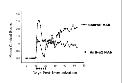
[13] Figure 1: Graphical results of studies of effects of anti-a2 integrin
antibody on
paralytic disease in mouse EAE model when administered at first sign of
disease (See
Example 7).
[14] Figure 2: Graphical results of studies of effects of anti-a2 integrin
on paralytic
disease when administered during induction phase (See Example 7).
SUMMARY OF THE INVENTION
[15] The present invention provides anti-alpha 2 (a2) integrin antibodies and
methods
for their use, notably humanized anti-alpha 2 (a2) integrin antibodies and
methods for
their use.
[16] In certain embodiments, the anti-a2 integrin antibody includes one or
more h iman
constant regions (e.g., CL and/or CH) and a light chain variable region
comprising the
amino acid sequence of SEQ ID NO:19 and/or a heavy chain variable region
comprising
the amino acid sequence of SEQ ID NO:21 or amino acid sequence variants
th,Dreof.
Various forms of the antibody are contemplated herein. For example, the anti-
a2 in tegrin
antibody may be a full length antibody (e.g., comprising human immunoglobulin
constant
regions) or an antibody fragment (e.g. Fab or F(ab1)2 or Fab' or Fv or scFv
fragments).
Furthermore, the antibody may be labeled with a detectable label, immobilized
on EI solid
phase and/or conjugated with a heterologous compound (such as a cytotoxic
agent)
[17] Diagnostic and therapeutic uses for anti-a2 integrin antibodies are
contemplated
as well as prophylactic or preventative uses. For diagnostic uses, a method
for
determining the presence of a261 integrin protein is provided comprising expo
E ing a
sample suspected of containing the a261 integrin protein to an anti-a2
integrin an. 'body
and determining binding of the antibody to the sample. For this use, a kit is
provided
comprising an anti-a2 integrin antibody and instructions for using the
antibody to detect
the a261 integrin protein. Therapeutic uses included but are not limted to the
treatment
of a261 integrin-associated disorders, mechanisms, and cellular processes inc
tiding
inflammatory diseases and autoimmune diseases, particulary multiple sclerosis.
[18] Gene therapy applications for anti-a2 integrin antibodies are contemr:
lated.
Various vectors (e.g., retroviral vectors, chromsomes) encoding the anti-a261
hea),y and
light chain gene sequences, may be transferred to cells (e.g., fibroblasts,
stem Gels) to
generate populations of cells secreting anti-a261 MAb. These cells may possess
specific
"homing" properties to different cell types, tissues, and/or organs. These an1
body-
producing cells in turn may be introduced into a patient for localized
delivery of thti, anti-
a261 MAb. As an example, mesenchymal stem cells modified with an anti- a261
MAb
CA 02629715 2008-05-14
WO 2007/056858 PCT/CA2006/001876
7
vector could be injected into the brain of a patient suffering from multiple
sclerosis The
stem cells differentiate into neural cells and secrete the anti-a2131 MAb to
treat the
inflammation associated with the multiple sclerosis. In addition, anti-a2f31
may be
conjugated to viruses encoding therapeutic genes (e.g., ricin). The modified
viruses
would bind specifically to cells expressing a2131 on the cell surface,
enabling incriased
transgene transfer efficiency. Further, immunoconjugates composed of anti-
a2[31
antibody-liposome complexes encapsulating nucleic acids encoding therapeutic
qenes
may be introduced intravenously into a patient. The anti-a2f31-immunoconjugate
'would
bind to cells expressing a2131 integrin and facilitate efficient uptake of the
therapeutic
genes.
[19] Further provided is an isolated nucleic acid encoding an anti-a2
integrin antibody;
a vector comprising that nucleic acid, optionally operably linked to control
sequences
recognized by a host cell transformed with the vector; a host cell comprising
that valor; a
process for producing the anti-a2 integrin antibody comprising culturing the
host cell so
that the nucleic acid is expressed and, optionally, recovering the antibody
from tho host
cell culture (e.g., from the host cell culture medium).
Also provided is a composition comprising a humanized anti-a2 integrin
antibody iind a
pharmaceutically acceptable carrier or diluent. Compositions for therapeutic
uses may be
sterile and may be lyophilized. Further provided is a method for treating an
a2[31 iMegrin-
associated disorder, comprising administering to a subject a pharmaceutically
effective
amount of an anti-a2 integrin antibody such as a humanized anti-a2 integrin
antibpdy to
the mammal. For such therapeutic uses, other agents (e.g., another a2f31
antagonist) may be co-administered to the mammal either before, after, or
simultanoously
with, the anti-a2 integrin antibody.
[20] Also provided is a humanized anti-a2 integrin antibody comprising a heavy
chain
variable region comprising the amino acid sequence of (a) H :;;DR2
(VIWARGFTNYNSALMS, SEQ ID NO:2), (b) HCDR1 (GFSLTNYGIH, SEQ ID l'µ10:1),
HCDR2 (VIWARGFTNYNSALMS, SEQ ID NO:2) and HCDR3 (ANDGVYYAMDY, S EQ ID
NO:3), or (c) SEQ ID NO:40.
[21] In an embodiment, the above-mentioned heavy chain variable region corn
prises
the amino acid sequence of SEQ ID NO:185.
[22] In a further embodiment, the above-mentioned heavy chain variable
region
comprises the amino acid sequence of SEQ ID NO:185 in which (a) position 71 is
Lys, (b)
position 73 is Asn, (c) position 78 is Val, or (d) any combination of (a)-(c).
CA 02629715 2008-05-14
WO 2007/056858 PCT/CA2006/001876
8
[23] In a further embodiment, the above-mentioned heavy chain variable region
comprises an amino acid sequence selected from SEQ ID NOs:70-79 and SEQ ID
NOs:109-111.
[24] In an embodiment, the above-mentioned anti-a2 integrin antibody
further
comprises a FW4 region comprising the amino acid sequence WGQGTLVTVSS (SlQ ID
NO:13).
[25] In an embodiment, the above-mentioned anti-a2 integrin antibody comprisos
the
amino acid sequence of HCDR1 (SEQ ID NO:1), HCDR2 (SEQ ID NO:2) and HCDR3
(SEQ ID NO:3).
[26] In an embodiment, the above-mentioned anti-a2 integrin antibody t
Jrther
comprises a light chain.
[27] The invention further provides a humanized anti-a2 integrin antibody
compri!iing a
light chain variable region comprising the amino acid sequence of (a) an LCDR1
selected
from SANSSVNYIH (SEQ ID NO:4) or SAQSSWNYIH (SEQ ID NO:112), (b) LCDR2
(DTSKLAS; SEQ ID NO:5) and (c) LCDR3 (QQWTTNPLT, SEQ ID NO:6).
[28] In an embodiment, the above-mentioned light chain variable region
comprisfts the
amino acid sequence of SEQ ID NO:186.
[29] In an embodiment, the above-mentioned light chain variable region
comprisis the
amino acid sequence of SEQ ID NO:186 in which (a) position 2 is Phe, (b)
position 45 is
Lys, (c) position 48 is Tyr, or (d) any combination of (a)-(c).
[30] In an embodiment, the above-mentioned light chain variable region
comprises an
amino acid sequence selected from SEQ ID NO:41, SEQ ID NOs:80-92 and SEQ ID
NO:108.
[31] In an embodiment, the above-mentioned humanized anti-a2 integrin an
tibody
further comprises a FW4 region comprising the amino acids sequence FGQGTKVEIK
of
SEQ ID NO:38.
[32] In an embodiment, the above-mentioned humanized anti-a2 integrin
antibody
comprises the amino acid sequence of LCDR1 (SEQ ID NO:4), LCDR2 (SEQ ID NO:5)
and LCDR3 (SEQ ID NO:6)
[33] In an embodiment, the above-mentioned humanized anti-a2 integrin
antibody
further comprises a heavy chain.
[34] The invention further provides a humanized anti-a2 integrin antibody
comprising:
(i) a heavy chain variable region comprising the amino acid sequence of (a)
H:.:DR2
(VIWARGFTNYNSALMS, SEQ ID NO:2), (b) HCDR1 (GFSLTNYGIH, SEQ ID l'40:1),
HCDR2 (VIWARGFTNYNSALMS, SEQ ID NO:2) and HCDR3 (ANDGVYYAMDY, S EQ ID
NO:3), or (c) SEQ ID NO:40; and
CA 02629715 2008-05-14
WO 2007/056858 PCT/CA2006/001876
9
(ii) a light chain variable region comprising the amino acid sequence of (a)
an LCDR1
selected from SANSSVNYIH (SEQ ID NO:4) or SAQSSWNYIH (SEQ ID NO:112), (b)
LCDR2 (DTSKLAS; SEQ ID NO:5) and (c) LCDR3 (QQWTTNPLT, SEQ ID NO:6).
[35] Also provided is the above-mentioned humanized anti-a2 integrin antibody,
wherein (a) the heavy chain variable region comprises the amino acid sequence
ol SEQ
ID NO:185, (b) the light chain variable region comprises the amino acid
sequence or SEQ
ID NO:186, or (c) both (a) and (b).
[36] Also provided is the above-mentioned humanized anti-a2 integrin antrbody,
wherein (i) the heavy chain variable region comprises the amino acid sequence
ol SEQ
ID NO:185 in which (a) position 71 is Lys, (b) position 73 is Asn, (c)
position 78 is Val, or
(d) any combination of (a)-(c); (ii) the light chain variable region comprises
the amino acid
sequence of SEQ ID NO:186 in which (a) position 2 is Phe, (b) position 45 is
Lys, (c)
position 48 is Tyr, or (d) any combination of (a)-(c); or (iii) both (i) and
(ii).
[37] Also provided is the above-mentioned humanized anti-a2 integrin ani body,
wherein (a) the heavy chain variable region comprises an amino acid sequence
se acted
from SEQ ID NOs:70-79 and SEQ ID NOs:109-111; (b) the light chain variable
region
comprises an amino acid sequence selected from SEQ ID NO:41, SEQ ID NOs:130-92
and SEQ ID NO:108; or (c) both (a) and (b).
[38] In an embodiment, the above-mentioned anti-a2 integrin antibody
recognizei, the I
domain of human a2 integrin.
[39] In an embodiment, the above-mentioned anti-a2 integrin antibody binds
a2131
integrin.
[40] In an embodiment, the above-mentioned anti-a2 integrin antibody binds
an
epitope of a2 integrin, the epitope comprising:
(a) a Lys residue corresponding to position 192 of the a2 integrin amino acid
seq Jence
set forth in SEQ ID NO:8 or position 40 of the a2 integrin I domain amino acid
seq uence
set forth in SEQ ID NO:11;
(b) an Asn residue corresponding to position 225 of the a2 integrin amino acid
seq Lience
set forth in SEQ ID NO:8 or position 73 of the a2 integrin I domain amino acid
seq uence
set forth in SEQ ID NO:11;
(c) a Gln residue corresponding to position 241 of the a2 integrin amino acid
sequence
set forth in SEQ ID NO:8 or position 89 of the a2 integrin I domain amino acid
sequence
set forth in SEQ ID NO:11;
CA 02629715 2008-05-14
WO 2007/056858 PCT/CA2006/001876
(d) a Tyr residue corresponding to position 245 of the a2 integrin amino acid
sequence
set forth in SEQ ID NO:8 or position 93 of the a2 integrin I domain amino acid
sequence
set forth in SEQ ID NO:11;
(e) an Arg residue corresponding to position 317 of the a2 integrin amino acid
sequence
set forth in SEQ ID NO:8 or position 165 of the a2 integrin I domain amino
acid sequence
set forth in SEQ ID NO:11;
(f) an Asn residue corresponding to position 318 of the a2 integrin amino acid
sequence
set forth in SEQ ID NO:8 or position 166 of the a2 integrin I domain amino
acid sequence
set forth in SEQ ID NO:11; or
(g) any combination of (a) to (f).
[41] Also provided is an anti-a2 integrin antibody, wherein the antibody
binds an
epitope of a2 integrin, the epitope comprising:
(a) a Lys residue corresponding to position 192 of the a2 integrin amino acid
sequence
set forth in SEQ ID NO:8 or position 40 of the a2 integrin I domain amino acid
seq Jence
set forth in SEQ ID NO:11;
(b) an Asn residue corresponding to position 225 of the a2 integrin amino acid
seq Jence
set forth in SEQ ID NO:8 or position 73 of the a2 integrin I domain amino acid
seq .ience
set forth in SEQ ID NO:11;
(c) a Gln residue corresponding to position 241 of the a2 integrin amino acid
seq Jence
set forth in SEQ ID NO:8 or position 89 of the a2 integrin I domain amino acid
seq Jence
set forth in SEQ ID NO:11;
(d) a Tyr residue corresponding to position 245 of the a2 integrin amino acid
seq Jence
set forth in SEQ ID NO:8 or position 93 of the a2 integrin I domain amino acid
seq Jence
set forth in SEQ ID NO:11;
(e) an Arg residue corresponding to position 317 of the a2 integrin amino acid
seq uence
set forth in SEQ ID NO:8 or position 165 of the a2 integrin I domain amino
acid seq uence
set forth in SEQ ID NO:11;
(f) an Asn residue corresponding to position 318 of the a2 integrin amino acid
seq uence
set forth in SEQ ID NO:8 or position 166 of the a2 integrin I domain amino
acid sec uence
set forth in SEQ ID NO:11; or
(g) any combination of (a) to (f).
CA 02629715 2008-05-14
WO 2007/056858 PCT/CA2006/001876
11
[42] In an embodiment, the above-mentioned humanized anti-a2 integrin antibocy
is a
full length antibody.
[43] In an embodiment, the above-mentioned humanized anti-a2 integrin antibody
is
an antibody fragment.
[44] In an embodiment, the above-mentioned humanized anti-a2 integrin
antibody is
bound to a detectable label.
[45] In an embodiment, the above-mentioned humanized anti-a2 integrin
antibody is
immobilized on solid phase.
[46] In an embodiment, the above-mentioned humanized anti-a2 integrin arr
ibody
inhibits binding of a2 or a2[31 integrin to an a2131 integrin ligand.
[47] In an embodiment, the above-mentioned a2131 integrin ligand is
selectec from
collagen, laminin, Echovirus-1, decorin, E-cadherin, matrix metalloproteinase
I (NA ,AP-I),
endorepellin, collectin and C1q complement protein.
[48] The invention further provides a method for determining whether a sample
contains a2 integrin, a2r31 integrin, or both, comprising contacting the
sample with the
above-mentioned humanized anti-a2 integrin antibody and determining whether
the
antibody binds to the sample, said binding being an indication that the sample
contains
a2 integrin, a2131 integrin, or both.
[49] The invention further provides a kit comprising the above-mentioned
humanized
anti-a2 integrin, optionally further comprising instructions for its use to
detect a2 or a2l31
integrin protein.
[50] The invention further provides an isolated nucleic acid encoding a
humanized anti-
a2131 integrin antibody mentioned above.
[51] The invention further provides a vector comprising the above-mentioned r
acid.
[52] The invention further provides a host cell comprising the above-mentioned
r ucleic
acid or vector.
[53] The invention further provides a process of producing a humanized nti-a2
integrin antibody comprising culturing the above-mentioned host cell under
conditions
permitting expression of the antibody. In an embodiment, the methiod further
comprises
recovering the humanized anti-a2 integrin antibody from the host cell. In a I
urther
embodiment, the method further comprises recovering the humanized anti-a2
integrin
antibody from the host cell culture medium.
[54] The invention further provides a screening method comprising: detecting
binding
of a2 or a2r31 integrin to an antibody comprising the VL region of SEQ ID
NO:19 aid the
VH region of SEQ ID NO:21 in the presence versus the absence of a test antibod
v; and
CA 02629715 2008-05-14
WO 2007/056858 PCT/CA2006/001876
12
selecting the test antibody if its presence correlates with decreased binding
of the a2 or
a2I31 integrin to the antibody comprising the VL region of SEQ ID NO:19 and
tha VH
region of SEQ ID NO:21. In an embodiment, the a2 or a2131 integrin is
immobilize(; on a
solid support.
[55] The invention further provides a screening method comprising:
detecting binding
of a2r31 integrin to collagen in the presence of a test antibody, wherein test
antibody
refers to an antibody that binds to an a2 I domain; detecting binding of the
test antibody
to the a2 I domain in the presence of Mg ++ ions; detecting binding of the
test antibody to
the a2 I domain in the presence of Ca ++ ions; detecting binding of the test
antibody lo the
a2 I domain in the presence of cation-free media; and selecting the test antib
xly if
inhibits the binding of a2131 integrin to collagen and binds to the a2 I
domain n the
presence of Mg ++ ions and Ca ions and cation-free media.
[56] The invention further provides a composition comprising the above-
mentioned
humanized anti-a2 integrin antibody and a pharmaceutically acceptable carrier.
[57] The invention further provides a method of treating an a2I31 integrin-
asso ::iated
disorder in a subject, the method comprising administering to the subject a
therapeutically
effective amount of the above-mentioned anti-a2 integrin antibody or
composition.
[58] The invention further provides a method for inhibiting leukocyte bind'
ng to
collagen comprising administering to a subject an amount of the above-
mentioned anti-
a2f31 integrin antibody effective to inhibit the binding of the leukocytes to
collagen.
[59] The invention further provides a use of the above mentioned humanized
anti-a2
integrin antibody as a medicament.
[60] The invention further provides a use of the above mentioned humanized
anti-a2
integrin antibody or composition for the treatment of an a2131 integrin-
associated dis:wder.
[61] The invention further provides a use of the above mentioned humanized
anti-a2
integrin antibody or composition for the preparation of a medicament for the
treatment of
an a2131 integrin-associated disorder.
[62] The invention further provides a composition for the treatment of an
a2131 inlegrin-
associated disorder, the composition comprising the above-mentioned humanized
anti-a2
integrin antibody and a pharmaceutically acceptable carrier or diluent.
[63] The invention further provides a package comprising the above-men=:ioned
humanized anti-a2 integrin antibody or composition together with instructions
for the
treatment of an a2131 integrin-associated disorder.
CA 02629715 2014-08-25
12a
[63.1] Also provided is a use of the above-mentioned humanized anti-a2
integrin
antibody for the manufacture of a medicament for the treatment of
inflammation, an
inflammatory disease, an autoimmune disease, or for decreasing angiogenesis.
[63.2] Also provided is a use of the above-mentioned humanized anti-a2
integrin
antibody or the above-mentioned composition, for the treatment of
inflammation, an
inflammatory disease, an autoimmune disease, or for decreasing angiogenesis.
[63.3] Also provided is a composition for the treatment of inflammation, an
inflammatory
disease, an autoimmune disease, or for decreasing angiogenesis, the
composition
comprising the above-mentioned humanized anti-a2 integrin antibody and a
pharmaceutically acceptable carrier or diluent.
[63.4] In embodiments, the inflammatory or autoimmune disease is inflammatory
bowel
disease, Crohn's disease, ulcerative colitis, optical neuritis, spinal cord
trauma,
rheumatoid arthritis, or multiple sclerosis.
CA 02629715 2008-05-14
WO 2007/056858 PCT/CA2006/001876
13
[64] In embodiments, the a261 integrin-associated disorder is selected from
inflammatory disease, autoimmune disease and a disease characterized by
abnormal or
increased angiogenesis.
[65] In embodiments, the a2131 integrin-associated disorder is selected from
inflammatory bowel disease, Crohn's disease, ulcerative colitis, reactions to
tram plant,
optical neuritis, spinal cord trauma, rheumatoid arthritis, systemic lupus
erythemvitosus
(SLE), diabetes mellitus, multiple sclerosis, Reynaud's syndrome,
expeririental
autoimmune encephalomyelitis, Sjorgen's syndrome, scleroderma, juvenile onset
diabetes, diabetic retinopathy, age related macular degeneration,
cardiovascular diE.ease,
psoriasis, cancer as well as infections that induce an inflammatory response.
[66] In embodiments, the a261 integrin-associated disorder is selected from
multiple
sclerosis (e.g., characterized by relapse, acute treatment, delayed
treatment), rheuriatoid
arthritis, optical neuritis and spinal cord trauma.
[67] In embodiments, the above-mentioned method is not associated with (a)
platelet
activation, (b) platelet aggregation, (c) a decrease in circulating platelet
count, (d)
bleeding complications, or (e) any combination of (a) to (d).
[68] In an embodiment, the above-mentioned anti-a2 integrin antibody
comprises a
heavy chain comprising SEQ ID NO:174 or SEQ ID NO:176 and a light chain
comprising
SEQ ID NO:178.
[69] In an embodiment, the above-mentioned anti-a2 integrin antibody
complAively
inhibits the binding of an antibody comprising the UL region of SEQ ID NO:19
and tie VH
region of SEQ ID NO:21 to human a2131 integrin or the I domain thereof.
[70] In an embodiment, the above-mentioned method is associated with an
alleviation
of a flare or neuroligical sequelae associated with multiple sclerosis.
[71] In an embodiment, the above-mentioned anti-a2 integrin antibody
inhibils the
binding of a261 integrin to collagen and is not a ligand mimetic.
[72] Also provided is a method of targeting a moiety, such as a molecule, p
'otein,
nucleic acid, vector, composition, complex, etc., to a site characterized by
the presence of
an a2f31 integrin ligand, the method comprising attaching or binding the
moiety to the
above-mentioned humanized anti-a2 integrin antibody.
[73] Also provided is an a2 integrin epitope that binds an anti-a2 integrin
anlibody,
wherein the epitope does not comprise the ligand-binding site of a2 integrin.
In
embodiments, binding to the epitope is not associated with (a) platelet
activation, (b)
platelet aggregation, (c) a decrease in circulating platelet count, (d)
blueding
complications, (e) a2 integrin activation, or (f) any combination of (a) to
(e).
CA 02629715 2008-05-14
WO 2007/056858 PCT/CA2006/001876
14
[74] Preferred antibodies bind to the I-domain of human a2131 integrin. In
particulc 1r, the
preferred antibodies are able to block a2-dependent adhesion of cells to the
extracollular
matrix (ECM), particularly to at least one or both of collagen and laminin.
Humanized
antibodies are provided, including antibodies based on an antibody referred to
heroin as
TMC-2206. Anti-a2 integrin antibodies are provided that are highly specific
for b man
a2131 integrin, and whose administration is not associated with undesired
effects such as
bleeding complications or complications due to cellular activation. The
binding specificity
(e.g., epitope specificity) of these antibodies is associated with their
unexpected non-
hemorrhagic profile.
[75] The humanized anti-a2p1 integrin antibody may have a heavy chain variable
region comprising the amino acid sequence of HCDR1 (GFSLTNYGIH; SEQ ID 110:1)
and/or HCDR2 (VIWARGFTNYNSALMS; SEQ ID NO:2) and/or H CDR3
(ANDGVYYAMDY; SEQ ID NO:3). The humanized anti-a2p1 integrin antibody may have
a light chain variable region comprising the amino acid sequence of LCDR1
(SANSSVNYIH; SEQ ID NO:4 or SAQSSWNYIH; SEQ ID NO:112) and/or LCDR2
(DTSKLAS; SEQ ID NO:5) and/or LCDR3 (QQWTTNPLT; SEQ ID NO:6). In certain
embodiments, the humanized anti-a2p1 integrin antibodies have a heavy ohain
comprising HCDR1 (GFSLTNYGIH; SEQ ID NO:1) and/or HCDR2
(VIWARGFTNYNSALMS; SEQ ID NO:2) and/or HCDR3 (ANDGVYYAMDY; SE .Q ID
NO:3) and a light chain variable region comprising the amino acid sequence of
LCDR1
(SANSSVNYIH; SEQ ID NO:4) and/or LCDR2 (DTSKLAS; SEQ ID NO:5) and/or LCDR3
(QQWTTNPLT; SEQ ID NO:6). In other embodiments, the antibody comprises an
amino
acid sequence variant of one or more of such CDRs, which variant comprises
crie or
more amino acid insertion(s) within or adjacent to a CDR residue and/or
deletion(s) within
or adjacent to a CDR residue and/or substitution(s) of CDR residue(s) (with
substitulion(s)
being the preferred type of amino acid alteration for generating such
variants).
DETAILED DESCRIPTION OF THE INVENTION
[76] The present invention provides antibodies specifically reactive with
human alpha 2
(a2) integrin, including humanized antibodies, and methods for their use. The
humanized
antibodies may have human framework regions (FWs) and complementarity
determining
regions (CDRs) from a non-human antibody, typically a mouse, specifically
reactiv with
human a2 integrin. Nucleotide sequences encoding, and amino acid sequ ences
comprising heavy and light chain antibodies are provided. In preferred
embodiment i;, one
or more of the CDR regions are derived from or based on the murine antibody
secreted
by the hybridoma clone, BHA2.1 [referred to herein as the TMC-2206 antibody].
Further
CA 02629715 2008-05-14
WO 2007/056858 PCT/CA2006/001876
provided are antibodies having similar binding properties and antibodies (or
other
antagonists) having similar functionality as the antibodies disclosed herein.
Preferred
anti-a2 integrin antibodies include those that (a) bind to the I domain of a2
integr n, (b)
inhibit the function of a2 integrin (e.g., collagen or laminin binding), (c)
bind to a2 integrin
on resting platelets without inducing platelet activation and (d) recognize
the b riding
epitope of TMC-2206 (e.g., compete with TMC-2206 for the binding to a2
integrin). Such
antibodies may bind preferentially to the inactive or closed conformation of
the target a2
integrin molecule without competing for the ligand binding site. Unexpected
advar =tages
of anti-a2 integrin antibodies as described herein that bind preferentially to
the Glosed
conformation of the a2131 integrin and/or bind to a2I31 integrin without
competing far the
ligand binding site (e.g., are not a ligand mimetic) include preventing
potential p atelet
activation, platelet aggregation, decreases in circulating platelet count
and/or bleeding
complications in a treated subject.
[77] "Bleeding complications" as used herein refers to any adverse effect on
blood
levels and physiology, including platelet thrombotic responses, thrombocyto
increased time to clot, increased bleeding time and blood loss that limit
therapeutic ise of
the anti-a2 integrin antibody.
[78] a2131 integrin is a molecule comprised of an a2 integrin subunit (see,
e.g., S EQ ID
NO:7, for DNA sequence and SEQ ID NO:8 for protein sequence of human a2) from
the
family of alpha integrins, and a 131 integrin subunit (see, e.g., SEQ ID NO:9
for DNA
sequence and SEQ ID NO:10 protein sequence of human f31) from the family ol
beta
integrins, and may be from any subject including a mammal, but preferably is
1.om a
human. The a2131 integrin may be purified from any natural source, or may be
pro luced
synthetically (e.g., by use of recombinant DNA technology). The nucleic acid
ooding
sequences for a2 integrin and for 131 integrin are described in Takada and
Hemler .11. Cell
Biol. 109(1):397-407 (1989; GenBank submission X17033; subsequently updated tc
entry
NM 002203) and Argraves, W.S, J. Cell. Biol. Sep 105(3):1183-90 (1987; Ge
'tank
submission X07979.1 and related sequences representing alternatively spliced
var ants),
respectively.
[79] The 'I' domain of the a2131 integrin molecule refers to a region of
this a2I31 ir tegrin
molecule within the a2 subunit, and is described, for example, in Kamata et
al., .1 Biol.
Chem. 269:9659-9663(1994); Emsley et al., J. Biol. Chem. 272:28512 (1997) ard
Cell
101:47 (2000). The amino acid sequence of a human I domain of a2 integrin is
shown as
SEQ ID NO:11 (see also, e.g., SEQ ID NO: 107). The I domain of a2 integrin
contains a
MIDAS type of ligand binding site (Metal Ion Dependent Adhesion Site) which
has a
requirement and a specificity for a given divalent cation to support ligand
binding. The
CA 02629715 2008-05-14
WO 2007/056858 PCT/CA2006/001876
16
amino acid sequences for an I domain of a2 integrin in rat shown as SEQ ID
NO:9:i; (see
also, e.g., SEQ ID NO:113) and in mouse shown as SEQ ID NO:94 (see also, e.g.,
SEQ
ID NO:114) are shown in Table 28. Cynomolgus monkey and rhesus monkey I dmain
sequences were cloned from the leukocyte fraction derived from whole blood
arid are
provided in SEQ ID NO:103 (DNA), SEQ ID NO:171 (amino acid) for cynomolgu i;
and
SEQ ID NO:104 (DNA), SEQ ID NO:172 (amino acid) for rhesus, respectively.
[80] A TMC-2206 (BHA2.1) epitope refers to a region of the I domain of human
a2
integrin to which the TMC-2206 antibody binds. This
epitope spans a iregion
encompassing amino acid residues, K40, N73, Q89, Y93, R165, and N166 and
optionally,
other amino acid residues of the a2 integrin l domain.
[81] An a2 integrin-associated disorder refers to a disorder, disease, or
condition that
involves a2 integrin-dependent processes/function (e.g., binding, activity)
that miildiate
aberrant cellular reactions within target tissue. Examples of a2 integrin-
dependent
processes involved in disease include collagen-dependent cellular responses
st.gt as
those involved in increases in cytokine expression and proliferation, aspects
of 1=-cell-,
mast cell- and neutrophil-function, inflammatory disorders, mammary gland
luctal
morphogenesis, epidermal wound healing, and angiogenesis. Examples of a2
inlegrin-
associated disorders include, but are not limited to, inflammatory diseases or
dis.:)rders
including but not limited to inflammatory bowel disease (such as Crohn's
diseas3 and
ulcerative colitis), reactions to transplant (including transplant rejection),
optic nouritis,
spinal cord trauma, rheumatoid arthritis, multiple sclerosis (including
treatmont of
neurological sequelae associated therewith as well as multiple sclerosis
characterbiad by
relapse), autoimmune diseases or disorders (including systemic lupus
erythemotosus
(SLE), diabetes mellitus, Reynaud's syndrome,
experimental autoirr mune
encephalomyelitis, Sjorgen's syndrome, scleroderma), juvenile onset diabetes,
and
disorders associated with abnormal or higher than normal angiogenesis (such as
diabetic
retinopathy, age related macula degeneration, cardiovascular disease,
psoriasis,
rheumatoid arthritis and cancer) as well as infections that induce an
inflamnatory
response.
[82] Treatment of an a2131 integrin-associated disorder refers to both
therapeutic use
and prophylactic or preventative use of the anti-a2 integrin antibodies
described l'arein.
Those in need of treatment include those already diagnosed with the disorder
as viell as
those in which the onset of the disorder is to be prevented or delayed.
[83] A mammal, including for purposes of treatment, refers to any animal
classified as
a mammal, including humans, domestic and farm animals, and zoo, sports or pet
animals
such as dogs, horses, cats, cows etc. Preferably, the mammal is human.
CA 02629715 2008-05-14
WO 2007/056858 PCT/CA2006/001876
17
[84] Intermittent or periodic dosing is a dosing that is continuous for a
certain period of
time and is at regular intervals that are preferably separated more than by
one day.
[85] The term antibody or immunoglobulin is used in the broadest sense, and
covers
monoclonal antibodies (including full length monoclonal antibodies),
poly:lonal
antibodies, multispecific antibodies, and antibody fragments so long as they
exhibit the
desired biological activity. Antibody fragments comprise a portion of a full
length ant body,
generally an antigen binding or variable region thereof. Examples of antibody
fragments
include Fab, Fab', F(alb1)2, and Fv fragments, diabodies, linear antibodies,
single-chain
antibody molecules, single domain antibodies (e.g., from camelids), shark NAR
single
domain antibodies, and multispecific antibodies formed from antibody
fragments.
Antibody fragments can also refer to binding moieties comprising CDRs or a
ligen
binding domains including, but not limited to, VH regions (VH, VH-VH),
antii:alins,
PepBodiesTm, antibody-T-cell epitope fusions (Troybodies) or Peptibodies.
[86] A monoclonal antibody refers to an antibody obtained from a population of
substantially homogeneous antibodies, e.g., the individual antibodies
comprisirg the
population are identical except for possible naturally occurring mutations
that may be
present in minor amounts. Monoclonal antibodies are highly specific, being
directed
against a single antigenic site. Furthermore, in contrast to conventional
(e.g., polyolonal)
antibody preparations which typically include different antibodies directed
against different
determinants (e.g., epitopes) on an antigen, each monoclonal antibody is
directed against
at least a single determinant on the antigen. The modifier "monoclonal"
indicatifs the
character of the antibody as being obtained from a substantially homogeneous
population
of antibodies, and is not to be construed as requiring production of the
antibody by any
particular method. For example, monoclonal antibodies may be made by the
hybrdoma
method first described by Kohler et al., Nature 256:495 (1975), or may be made
by
recombinant DNA methods (see, e.g., U.S. Patent No. 4,816,567).
Monoclonal
antibodies may also be isolated from phage antibody libraries, for example,
using the
techniques described in Clackson et al., Nature 352:624-628 (1991) and Marks
ei al., J.
Mol. Biol. 222:581-597 (1991). Monoclonal antibodies can also be isolated
using the
techniques described in U.S. Patent Nos. 6,025,155 and 6,077,677 as well as
U.S. Patent
Application Publication Nos. 2002/0160970 and 2003/0083293 (see also, e.g.,
Lindenbaum, et al., Nucleic Acids Research 32 (21):0177 (2004)).
[87] Monoclonal antibodies can include chimeric antibodies in which a
portion of the
heavy and/or light chain is identical with or homologous to corresponding
sequences in
antibodies derived from a particular species or belonging to a particular
antibody cl fiss or
subclass, while the remainder of the chain(s) is identical with or homologbus
to
corresponding sequences in antibodies derived from another species or
belonging to
CA 02629715 2013-09-25
18
another antibody class or subclass, as well as fragments of such antibodies,
so long as
they exhibit the desired biological activity (see, e.g., U.S. Patent No.
4,816,567; and
Morrison et al., Proc. Natl. Acad Sci. USA 81: 6851-6855 (1984) for mouse-
human
chimeric antibodies).
[88] A hypervariable region refers to the amino acid residues of an antibody
which are
responsible for antigen-binding. The hypervariable region comprises amino acid
residues
from a complementarity determining region or CDR (e.g., residues 24-34 (L1),
50-56 (L2)
and 89-97 (L3) in the light chain variable domain and 31-35 (H1), 50-65 (H2)
and 95-102
(H3) in the heavy chain variable domain; Kabat et al., Sequences of Proteins
of
Immunological Interest, 5th Ed. Public Health Service, National Institutes of
Health,
Bethesda, Md. (1991)) and/or those residues from a hypervariable loop (e.g.,
residues
26-32 (L1), 50-52 (L2) and 91-96 (L3) in the light chain variable domain and
26-32 (H1),
53-55 (H2) and 96-101 (H3) in the heavy chain variable domain; Chothia and
Lesk J. Mol.
Biol. 196: 901-917 (1987)). Framework or FR residues are those variable domain
residues other than the hypervariable region residues. For antibodies
described herein,
the CDR and framework regions are identified based on the Kabat numbering
system
except that the CDR1 of the heavy chain is defined by Oxford Molecular's AbM
definition
as spanning residues 26 to 35. The Oxford Molecular's AbM antibody modeling
software
(Martin et al., Proc. Natl Acad. Sci. USA, 86, 9268-9272 (1989); Martin et
al., Methods
Enzymol., 203, 121-153 (1991); Pedersen et al., lmmunomethods, 1, 126 (1992);
and
Rees et al., In Sternberg M.J.E. (ed.), Protein Structure Prediction. Oxford
University
Press, Oxford, 141-172. (1996)) combines the Kabat CDR and the Chothia
hypervariable
region numbering systems to define CDRs.
[89] Humanized forms of non-human (e.g., murine) antibodies may be chimeric
antibodies which contain minimal sequence derived from non-human
immunoglobulin.
For the most part, humanized antibodies are human immunoglobulins (recipient
or
acceptor antibody) in which hypervariable region residues of the recipient are
replaced by
hypervariable region residues from a non-human species (donor antibody) such
as
mouse, rat, rabbit or nonhuman primate having the desired specificity,
affinity, and
capacity. In addition, individual or groups of Fv framework region (FR)
residues of the
human immunoglobulin may be replaced by corresponding non-human residues.
Furthermore, humanized antibodies may comprise residues which are not found in
the
recipient antibody or in the donor antibody. These modifications are made to
further
refine antibody performance. In
general, the humanized antibody will comprise
substantially all of at least one, and typically two, variable regions or
domains, in which all
or substantially all of the hypervariable loops correspond to those of a non-
human
immunoglobulin and all or substantially all of the FR regions are those of a
human
CA 02629715 2008-05-14
WO 2007/056858 PCT/CA2006/001876
19
immunoglobulin sequence. The humanized antibody optionally also will comprise
al least
a portion of an immunoglobulin constant region (e.g., Fc), typically that of a
human
immunoglobulin (see, e.g., Queen et aL, Proc. Natl. Acad. Sci. USA 86:10029
(1989), and
Foote and Winter, J. Mol. Biol. 224: 487 (1992)).
[90] Single-chain Fv or scFv antibody fragments may comprise the VH and VL
rions
or domains of antibody, wherein these domains are present in a single
polypeptide i::hain.
Generally, the Fv polypeptide further comprises a polypeptide linker between
the VII and
VL domains which enables the scFv to form the desired structure for antigen
binding (for a
review, see, e.g., Pluckthun in The Pharmacology of Monoclonal Antibodies, vol
113,
Rosenburg and Moore eds. Springer-Verlag, New York, pp. 269-315 (1994)).
[91] Diabody refers to small antibody fragments with two antigen-binding
sites, which
fragments comprise a heavy chain variable domain (VH) connected to a light
chain
variable domain (VL) in the same polypeptide chain (VH - VL). By using a
linker that is too
short to allow pairing between the two domains on the same chain, the domains
are
forced to pair with the complementary domains of another chain and create two
ar
binding sites. Diabodies are described more fully in, for example, EP 404,097;
WO
93/11161; and Hollinger et al., Proc. Natl. Acad. Sci. USA 90: 6444-6448
(1993).
[92] Linear antibody refers to antibodies such as those described in Zapata
9t al.,
Protein Eng. 8(10): 1057-1062 (1995). Briefly, these antibodies comprise a
pair of
tandem Fd segments (VH -CH1- VH -CH1) which form a pair of antigen binding re
;lions.
Linear antibodies can be bispecific or monospecific.
[93] An isolated antibody refers to one which has been identified and
separated 9nd/or
recovered from a component of its natural environment. Contaminant components
of its
natural environment are materials which would interfere with diagnostic or
therapeutic
uses for the antibody, and may include enzymes, hormones, and other
proteinaceDus or
nonproteinaceous solutes. In preferred embodiments, the antibody will be
purified (1) to
greater than 95% by weight of antibody as determined by the Lowry method, an
most
preferably more than 99% by weight, (2) to a degree sufficient to obtain at
least 15
residues of N-terminal or internal amino acid sequence by use of a spinning
cup
sequenator, or (3) to homogeneity by SDS-PAGE under reducing or nonreclucing
conditions using Coomassie blue or, preferably, silver stain. Isolated
antibody imludes
the antibody in situ within recombinant cells since at least one component of
the
antibody's natural environment will not be present. Ordinarily, however,
isolated antibody
will be prepared by at least one purification step.
[94] An epitope tagged antibody refers to one wherein the antibody of the
invention is
fused to an epitope tag. The epitope tag polypeptide has enough residues to
prov 'le an
epitope against which an antibody thereagainst can be made, yet is short
enough such
CA 02629715 2013-09-25
that it does not interfere with activity of the anti-a2131 integrin antibody.
The epitope tag
preferably is sufficiently unique so that the antibody thereagainst does not
substantially
cross-react with other epitopes. Suitable tag polypeptides generally have at
least 6
amino acid residues and usually between about 8-50 amino acid residues
(preferably
between about 9-30 residues). Examples include the flu HA tag polypeptide and
its
antibody 12CA5 (Field et aL, Mol. Cell. Biol. 8: 2159-2165 (1988)); the c-myc
tag and the
8F9, 3C7, 6E10, G4, B7 and 9E10 antibodies thereto (Evan et al., Mol. Cell.
Biol.
5(12):3610-3616 (1985)); and the Herpes Simplex virus glycoprotein D (gD) tag
and its
antibody (Paborsky et al., Protein Engineering 3(6): 547-553 (1990)). In
certain
embodiments, the epitope tag is a salvage receptor binding epitope which is an
epitope of
the Fc region of an IgG molecule (e.g., lgGi, IgG2, IgG3, or IgG4) that is
responsible for
increasing the in vivo serum half-life of the IgG molecule.
[95] A cytotoxic agent refers to a substance that inhibits or prevents the
function of
cells and/or causes destruction of cells. The can include radioactive isotopes
(e.g., 1311,
125.,
I 90Y and 186Re), chemotherapeutic agents, and toxins such as enzymatically
active
toxins of bacterial, fungal, plant or animal origin, or fragments thereof. A
non-cytotoxic
agent refers to a substance that does not inhibit or prevent function of cells
and/or does
not cause destruction of cells. A non-cytotoxic agent may include an agent
that can be
activated to become cytotoxic. A non-cytotoxic agent may include a bead,
liposome,
matrix or particle (see, e.g., U.S. Patent Publications 2003/0028071 and
2003/0032995).
Such agents may be conjugated, coupled, linked or associated with an anti-
a2í31 integrin
antibody as described herein.
[96] A chemotherapeutic agent refers to a chemical compound useful in the
treatment
of cancer. Examples of chemotherapeutic agents include but are not limited to
Adriamycin, Doxorubicin, 5-Fluorouracil,
Cytosine arabinoside ("Ara-C"),
Cyclophosphamide, Thiotepa, Taxotere (docetaxel), Busulfan, Cytoxin, Taxol,
Methotrexate, Cisplatin, Melphalan, Vinblastine, Bleomycin, Etoposide,
lfosfamide,
Mitomycin C, Mitoxantrone, Vincreistine, Vinorelbine, Carboplatin, Teniposide,
Daunomycin, Carminomycin, Aminopterin, Dactinomycin, Mitomycins, Esperamicins
(see
U.S. Pat. No. 4,675,187), Melphalan and other related nitrogen mustards.
[97] A prodrug refers to a precursor or derivative form of a pharmaceutically
active
substance that is less cytotoxic to tumor cells compared to the parent drug
and is capable
of being enzymatically activated or converted into the more active parent form
(see, e.g.,
Wilman, "Prodrugs in Cancer Chemotherapy" Biochemical Society Transactions,
14, pp.
375-382, 615th Meeting Belfast (1986) and Stella et al., "Prodrugs: A Chemical
Approach
to Targeted Drug Delivery," Directed Drug Delivery, Borchardt et al., (ed.),
pp. 247-267,
CA 02629715 2008-05-14
WO 2007/056858 PCT/CA2006/001876
21
Humana Press (1985). Prodrugs include, but are mot limited to, phosphate-
contiiiining
prodrugs, thiophosphate-containing prodrugs, sulfate-containing prodrugs,
peptide-
containing prodrugs, D-amino acid-modified prodrugs, glycosylated prodrugs, 8-
lectam-
containing prodrugs, optionally substituted phenoxyacetamide-containing prodru
=Js or
optionally substituted phenylacetamide-containing prodrugs, 5-fluorocytosine
and ot ler 5-
fluorouridine prodrugs which can be converted into the more active cytotoxic
free drug.
Examples of cytotoxic drugs that can be derivatized into a prodrug form can be
those
chemotherapeutic agents described above.
[98] A label refers to a detectable compound or composition which is
conjugaled or
coupled directly or indirectly to the antibody. The label may itself be
detectable by itself
(e.g., radioisotope labels or fluorescent labels) or, in the case of an
enzymatic label may
catalyze chemical alteration of a substrate compound or composition which is
detectable.
[99] Solid phase refers to a non-aqueous matrix to which the antibody of the
present
invention can adhere. Examples of solid phases encompassed herein include
those
formed partially or entirely of glass (e.g., controlled pore glass),
polysaccharides (e.g.,
agarose), polyacrylamides, polystyrene, polyvinyl alcohol and silicones. In
certain
embodiments, depending on the context, the solid phase can comprise the well
of an
assay plate; in others it is a purification column (e.g., an affinity
chromatography column).
This term also includes a discontinuous solid phase of discrete particles,
such as those
described in U.S. Patent No. 4,275,149.
[100] A liposome refers to a small vesicle composed of various types of
lipids,
phospholipids and/or surfactant which is useful for delivery of a drug (such s
the
antibodies of the invention and, optionally, a chemotherapeutic agent) to a
mammal. The
components of the liposome are commonly arranged in a bilayer formation,
similar to the
lipid arrangement of biological membranes.
[101] An isolated nucleic acid molecule refers to a nucleic acid molecule I
'let is
identified and separated from at least one contaminant nucleic acid molecule
with which it
is ordinarily associated in the natural source of the antibody nucleic acid.
An is Dieted
nucleic acid molecule is other than in the form or setting in which it is
found in rature.
Isolated nucleic acid molecules therefore are distinguished from the nucleic
acid mcdecule
as it exists in natural cells. However, an isolated nucleic acid molecule
includes a rlucleic
acid molecule contained in cells that ordinarily express the antibody where,
for ex;iimple,
the nucleic acid molecule is in a chromosomal location different from that of
natural ,:;ells.
[102] A viral vector refers to a vehicle for the transfer of a nucleic acid
(e.g. DNA or
RNA) to cells through viral infection or transduction. Examples of viral
vectors 'include
retroviruses, adenoviruses, pox viruses, and baculovirus.
CA 02629715 2008-05-14
WO 2007/056858 PCT/CA2006/001876
22
[103] A non-viral vector refers to a nucleic acid vehicle such as a CAN,
plasnilid or
chromosome that is delivered to cells by non-viral methods such as
electroporation,
injections, and cationic reagent mediated transfection.
[104] Expression control sequences refer to those DNA sequences necessary for
the
expression of an operably linked coding sequence in a particular host
organism. The
control sequences that are suitable for prokaryotes, for example, include a
promoter,
optionally an operator sequence, and a ribosome binding site. Eukaryotic cells
are I, nown
to utilize promoters, polyadenylation signals, and enhancers.
[105] A nucleic acid is operably linked when it is placed into a functional
relationship
with another nucleic acid sequence. For example, DNA for a presequence or
secretory
leader is operably linked to DNA for a polypeptide if it is expressed as a
preproteiln that
participates in the secretion of the polypeptide; a promoter or enhancer is
operably inked
to a coding sequence if it affects the transcription of the sequence; or a
ribosome b nding
site is operably linked to a coding sequence if it is positioned so as to
facilitate trans ation.
Generally, operably linked DNA sequences are contiguous, and, in the case of a
secretory leader, contiguous and in reading phase. However, enhancers do not
hiwe to
be contiguous. Linking is accomplished by ligation at convenient restriction
sites. I such
sites do not exist, the synthetic oligonucleotide adaptors or linkers are
uF..ed in
accordance with conventional practice.
[106] A further aspect of the present invention is the treatment of a2131
inlegrin-
associated disorders by administering to a subject a nucleic acid molecule
encoding an
anti-a2 integrin antibody of the invention. Suitable methods of administration
include
gene therapy methods (see below).
[107] A nucleic acid of the invention may be delivered to cells in vivo using
methods
such as direct injection of DNA, receptor-mediated DNA uptake, viral-me liated
transfection or non-viral transfection and lipid based transfection, all of
which may involve
the use of gene therapy vectors. Direct injection has been used to introduce
naked DNA
into cells in vivo (see e.g., Acsadi et al. (1991) Nature 332:815-818; Wolff
et al. 1990)
Science 247:1465-1468). A delivery apparatus (e.g., a "gene gun") for
injecting DNA into
cells in vivo may be used. Such an apparatus may be commercially available
(e.g., from
BioRad). Naked DNA may also be introduced into cells by complexing the DNh to
a
cation, such as polylysine, which is coupled to a ligand for a cell-surface
receptor (ti ee for
example Wu, G. and Wu, C. H. (1988) J. Biol. Chem. 263:14621; Wilson el al.
(192) J.
Biol. Chem. 267:963-967; and U.S. Pat. No. 5,166,320). Binding of the DNA-
ligand
complex to the receptor may facilitate uptake of the DNA by receptor-me liated
endocytosis. A DNA-ligand complex linked to adenovirus capsids which disrupt
endosomes, thereby releasing material into the cytoplasm, may be used to avoid
CA 02629715 2008-05-14
WO 2007/056858 PCT/CA2006/001876
23
degradation of the complex by intracellular lysosomes (see for example Curiel
el al.
(1991) Proc. Natl. Acad. Sci. USA 88:8850; Cristiano et al. (1993) Proc. Natl.
Acad. Sci.
USA 90:2122-2126).
[108] Defective retroviruses are well characterized for use as gene therapy
vecto 's (for
a review see Miller, A. D. (1990) Blood 76:271). Protocols for producing
recombinant
retroviruses and for infecting cells in vitro or in vivo with such viruses can
be found in
Current Protocols in Molecular Biology, Ausubel, F. M. et al. (eds.) Greene
Publishing
Associates, (1989), Sections 9.10-9.14 and other standard laboratory manuals.
Exa 'nples
of suitable retroviruses include pLJ, pZIP, pWE and pEM which are well known
to those
skilled in the art. Examples of suitable packaging virus lines include
.psi.Crip, .p.ACre,
.psi.2 and .psi.Am. Retroviruses have been used to introduce a variety of
genes into
many different cell types, including epithelial cells, endothelial cells,
lymphocytes,
myoblasts, hepatocytes, bone marrow cells, in vitro and/or in vivo (see for ex
iimple
Eglitis, et al. (1985) Science 230:1395-1398; Danos and Mulligan (1988) Proc.
Natl.
Acad. Sci. USA 85:6460-6464; Wilson et al. (1988) Proc. Natl. Acad. Sci. USA
85:3014-
3018; Armentano et al. (1990) Proc. Natl. Acad. Sci. USA 87:6141-6145; Huber
et al.
(1991) Proc. Natl. Acad. Sci. USA 88:8039-8043; Ferry et al. (1991) Proc.
Natl. Acad. Sci.
USA 88:8377-8381; Chowdhury et al. (1991) Science 254:1802-1805; van
Beusect.em et
al. (1992) Proc. Natl. Acad. Sci. USA 89:7640-7644; Kay et al. (1992) Human
Gene
Therapy 3:641-647; Dai et al. (1992) Proc. Natl. Acad. Sci. USA 89:10892-
10895; Hwu et
al. (1993) J. Immunol. 150:4104-4115; U.S. Pat. No. 4,868,116; U.S. Pat. No.
4,980,286;
PCT Application WO 89/07136; PCT Application WO 89/02468; PCT Applicatiob WO
89/05345; and PCT Application WO 92/07573).
[109] For use as a gene therapy vector, the genome of an adenovirus may be
manipulated so that it encodes and expresses a nucleic acid compound of the
invontion,
but is inactivated in terms of its ability to replicate in a normal lytic
viral life cycle. See for
example Berkner et al. (1988) BioTechniques 6:616; Rosenfeld et al. (1991)
Si:ience
252:431-434; and Rosenfeld et al. (1992) Cell 68:143-155. Suitable adenoviral
v ctors
derived from the adenovirus strain Ad type 5 dI324 or other strains of
adenovirus (e.g.,
Ad2, Ad3, Ad7 etc.) are well known to those skilled in the art. Recombinant
adenoviruses
are advantageous in that they do not require dividing cells to be effective
gene &livery
vehicles and can be used to infect a wide variety of cell types, including
airway epithelium
(Rosenfeld et al. (1992) cited supra), endothelial cells (Lemarchand et al.
(1992) Proc.
Natl. Acad. Sci. USA 89:6482-6486), hepatocytes (Herz and Gerard (1993) Proc
Natl.
Acad. Sci. USA 90:2812-2816) and muscle cells (Quantin el al. (1992) Proc.
Natl. Acad.
Sci. USA 89:2581-2584).
CA 02629715 2008-05-14
WO 2007/056858 PCT/CA2006/001876
24
[110] Adeno-associated virus (AAV) may be used as a gene therapy vector for
&livery
of DNA for gene therapy purposes. AAV is a naturally occurring defective
virt.u; that
requires another virus, such as an adenovirus or a herpes virus, as a helper
vims for
efficient replication and a productive life cycle (Muzyczka et al. Curr.
Topics in Micro. and
Immunol. (1992) 158:97-129). AAV may be used to integrate DNA into non-
dividinc cells
(see for example Flotte et al. (1992) Am. J. Respir. Cell. Mol. Biol. 7:349-
356; Samu ski et
al. (1989) J. Virol. 63:3822-3828; and McLaughlin et al. (1989) J. Virol.
62:1963-197÷. An
AAV vector such as that described in Tratschin et al. (1985) Mol. Cell. Biol.
5:3251-3260
may be used to introduce DNA into cells (see for example Hermonat et al.
(1984) Proc.
Natl. Acad. Sci. USA 81:6466-6470; Tratschin et al. (1985) Mol. Cell. Biol.
4:2072-.2081;
Wondisford et al. (1988) Mol. Endocrinol. 2:32-39; Tratschin et al. (1984) J.
Virol. 5 ' :611-
619; and Flotte et al. (1993) J. Biol. Chem. 268:3781-3790). Lentiviral gene
therapy
vectors may also be adapted for use in the invention.
[111] General methods for gene therapy are known in the art. See for example
U.S.
Pat. No. 5,399,346 by Anderson et al. A biocompatible capsule for delivering
genetic
material is described in PCT Publication WO 95/05452 by Baetge et al. Methods
of gene
transfer into hematopoietic cells have also previously been reported (see
Clapp, D. N., et
al., Blood 78: 1132-1139 (1991); Anderson, Science 288:627-9 (2000); and
Cava2zana-
Calvo et al., Science 288:669-72 (2000)).
[112] Cell, cell line, and cell culture are often used interchangeably and all
such
designations include progeny. Transformants and transformed cells (e.g.,
obtain ;:d by
transfection, transformation or transduction of nucleic acids, vectors, virus,
etc.) include
the primary subject cell and cultures derived therefrom without regard for the
num Der of
transfers. It is also understood that all progeny may not be precisely
identical in DNA
content, due to deliberate or inadvertent mutations. Mutant progeny that have
the same
function or biological activity as screened for in the originally transformed
cell are
included. Where distinct designations are intended, it will be clear from the
context.
[113] Humanized antibodies as described herein include antibodies that have vc
riable
region frameworks derived from a human acceptor antibody molecule, hypervaria
Die or
CDR sequences from a donor murine antibody, and constant regions, if present,
derived
from human sequences.
[114] Antibodies of the present invention have been constructed comprising
CDR; from
both the heavy chain variable and light chain variable regions of the murine
monoclonal
antibody clone BHA2.1 (Hangan et al., Cancer Res. 56:3142-3149 (1996)).
Preferred
starting materials for constructing antibodies are anti-a2 integrin antibodies
such as those
secreted by the BHA2.1 hybridoma (e.g., TMC-2206) that are function-blocking
antibodies
CA 02629715 2008-05-14
WO 2007/056858 PCT/CA2006/001876
directed against human a2 integrin and are dependent for binding and activity
c:11 the
presence of an intact I-domain within the targeted a2 integrin. Preferred are
antibodies
with the epitope specificity of TMC-2206 (or BHA2.1), including antibodies
which bind to
the inactive conformation of the a2 integrin molecule, and/or do not act as
gand
mimetics. Preferred are antibodies with the epitope specificity of TMC-2206
(or BI- A2.1)
that, although they interact with a2131 integrin present on both leukocytes
and pla:elets,
do not cause platelet activation, impair aggregation of activated platelets on
collagen,
have minimal or no effect on bleeding and/or are not associated with ble eding
complications at administered concentrations, including therapeutic doses in
vivo.
[115] Antibodies may be constructed wherein the human acceptor molecule for
tha light
chain variable region is selected based on homology considerations between pc
tential
acceptor molecule variable regions and with the light chain variable region of
the rnurine
antibody. Germline candidate human acceptor molecules are preferred to raduce
potential antigenicity. Germline databases are made up of antibody sequences
thel read
through the end of the heavy chain FW3 region and partially into the CDR3
seqt. ence.
For selection of a FW4 region, it is preferred to search databases of mature
an tibody
sequences which have been derived from the selected germline molecule, and
also
preferred to select a reasonably homologous FW4 region for use in the recom
Anent
antibody molecule. Human acceptor molecules are preferably selected from the
same
light chain class as the murine donor molecule, and of the same canonical
structural class
of the variable region of the murine donor molecule. Secondary considerations
for
selection of the human acceptor molecule for the light chain variable region
include
homology in CDR length between the murine donor molecule and the human
ac,::eptor
molecule. Human acceptor antibody molecules are preferably selected by
homology
searches to the V-BASE database, and other databases such as the Kabat and the
public
NCB! databases may be used as well. For humanized anti-a2 integrin antibodies
w .th the
same or similar epitope specificity and/or functional properties as TMC-2206,
a preferred
light chain human acceptor molecule is SEQ ID NO:37 with the germline artibody
sequence A14 for the FW 1-3 region and the sequence FGQGTKVEIK for FW4 (S iEQ
ID
NO:38) which represents a common FW-4 of mature kappa 1 light chains (e.g.,
light chain
sequence AAB24132 (NCBI entry gi/259596/gb/AAB24132).
[116] Antibodies may be constructed wherein the human acceptor molecule for
the
heavy chain variable region is selected based on homology considerations
between
potential acceptor molecule variable regions and the heavy chain variable
region of the
murine antibody. Germline candidate human acceptor molecules are preferred to
nduce
potential antigenicity. Germline databases are made up of antibody sequences
that read
CA 02629715 2013-09-25
,
26
through the end of the heavy chain FVV3 region and partially into the CDR3
sequence.
For selection of a FVV4 region, it is preferred to search databases of mature
antibody
sequences which have been derived from the selected germline molecule, and
also
preferred to select a reasonably homologous FW4 region for use in the
recombinant
antibody molecule. Human acceptor molecules are preferably selected from the
same
heavy chain class as the murine donor molecule, and of the same canonical
structural
class of the variable region of the murine donor molecule. Secondary
considerations for
selection of the human acceptor molecule for the heavy chain variable region
include
homology in CDR length between the murine donor molecule and the human
acceptor
molecule. Human acceptor antibody molecules are preferably selected by
homology
search to the V-BASE database, although other databases such as the Kabat and
the
public NCB! databases may be used as well. For anti-a2 integrin antibodies
with the
same or similar epitope specificity and/or functional properties as TMC-2206,
a preferred
heavy chain acceptor molecule is SEQ ID NO:39 with the germline antibody
sequence 4-
59 for the FW 1-3 region (SEQ ID NO:12) and antibody, CAA48104.1 (NCBI entry,
gi/33583/emb/CAA48104.1) a mature antibody derived from the 4-59 germline
sequence
for the FW 4 region (SEQ ID NO:13).
[117] Methods for humanizing a nonhuman a2 integrin antibody are described
herein,
including in the Examples below. In order to humanize an anti-a2 integrin
antibody, the
nonhuman antibody starting material is obtained, including by preparation from
immunization or by purchase of commercially available antibodies.
Exemplary
techniques for generating antibodies are described herein.
[118] The a2f31 integrin antigen to be used for production of antibodies may
be, for
example, a soluble form of a2131 integrin or other fragment of a2131 integrin
(e.g., an a2131
integrin fragment comprising a human a2 integrin I-domain (SEQ ID NO:11); see
also,
e.g., SEQ ID NO: 107). Other forms of a2 integrin useful for generating
antibodies will be
apparent to those skilled in the art based on the sequence of a2 integrin
(e.g., a human
a2 integrin as in SEQ ID NO:8).
(119] Polyclonal antibodies are preferably raised in animals by multiple
subcutaneous
(sc), intravenous (iv) or intraperitoneal (ip) injections of the relevant
antigen with or
without an adjuvant. It may be useful to conjugate the relevant antigen to a
protein that is
immunogenic in the species to be immunized, e.g., keyhole limpet hemocyanin,
serum
albumin, bovine thyroglobulin, or soybean trypsin inhibitor using a
bifunctional or
derivatizing agent, for example, maleimidobenzoyl sulfosuccinimide ester
(conjugation
through cysteine residues), N-hydroxysuccinimide (through lysine residues),
CA 02629715 2008-05-14
WO 2007/056858 PCT/CA2006/001876
27
glutaraldehyde, succinic anhydride, SOCl2, or R1N=C=NR, where R and R1 are
dillerent
alkyl groups.
[120] Animals may be immunized against the antigen, immunogenic conjugatos, or
derivatives by combining the antigen or conjugate (e.g., 100 pg for rabbits or
5 i.ig for
mice) with 3 volumes of Freund's complete adjuvant and injecting the solution
intradermally at multiple sites. One month later the animals are boosted with
the aritigen
or conjugate (e.g., with 1/5 to 1/10 of the original amount used to immunize)
in Fround's
complete adjuvant by subcutaneous injection at multiple sites. Seven to 14
days laler the
animals are bled and the serum is assayed for antibody titer. Animals are
boosted until
the titer plateaus. Preferably, for conjugate immunizations, the animal is
boosted wil:h the
conjugate of the same antigen, but conjugated to a different protein and/or
thro igh a
different cross-linking reagent. Conjugates also can be made in recombinant
cell culture
as protein fusions. Also, aggregating agents such as alum are suitably used to
en lance
the immune response.
[121] Monoclonal antibodies may be made using the hybridoma method first des
::ribed
by Kohler et al., Nature, 256: 495 (1975), or may be made by recombinant DNA
methods
(e.g., U.S. Patent No. 6,204,023). Monoclonal antibodies may also be made
using the
techniques described in U.S. Patent Nos. 6,025,155 and 6,077,677 as well as
U.S. Patent
Application Publication Nos. 2002/0160970 and 2003/0083293 (see also, e.g.,
Lindenbaum, et al., Nucleic Acids Research 32 (21):0177 (2004)).
[122] In the hybridoma method, a mouse or other appropriate host animal, such
as a
rat, hamster or monkey, is immunized (e.g., as hereinabove described) to
elicit
lymphocytes that produce or are capable of producing antibodies that will
specificall If bind
to the antigen used for immunization. Alternatively, lymphocytes may be
immunh:ed in
vitro. Lymphocytes then are fused with myeloma cells using a suitable fusing
agent, such
as polyethylene glycol, to form a hybridoma cell (see, e.g., Goding,
Monoclonal
Antibodies: Principles and Practice, pp.59-103 (Academic Press, 1986)).
[123] The hybridoma cells thus prepared are seeded and grown in a suitable c
ulture
medium that preferably contains one or more substances that inhibit the
grov.th or
survival of the unfused, parental myeloma cells. For example, if the parental
my )loma
cells lack the enzyme hypoxanthine guanine phosphoribosyl transferase (HGP :ZT
or
HPRT), the culture medium for the hybridomas typically will include
hypoxanthine,
aminopterin, and thymidine (HAT medium), which substances prevent the growth
of
HGPRT-deficient cells.
[124] Preferred myeloma cells are those that fuse efficiently, support stable
high-level
production of antibody by the selected antibody-producing cells, and are
sensitivo to a
medium such as HAT medium. Among these, preferred myeloma cell lines are
riurine
CA 02629715 2008-05-14
WO 2007/056858 PCT/CA2006/001876
28
myeloma lines, such as those derived from MOP-21 and M.C.-11 mouse tumors
avaable
from the Salk Institute Cell Distribution Center, San Diego, Calif. USA, and
SP-2 or X63-
Ag8-653 cells available from the American Type Culture Collection, Rockville,
Md. USA.
Human myeloma and mouse-human heteromyeloma cell lines also have been
described
for the production of human monoclonal antibodies (e.g., Kozbor, J. Immunol.,
133 3001
(1984); Brodeur et al., Monoclonal Antibody Production Techniques and
Applications, pp.
51-63 (Marcel Dekker, Inc., New York, 1987)).
[125] Culture medium in which hybridoma cells are growing is assayed for
produclion of
monoclonal antibodies directed against the antigen. Preferably, the binding
specifbity of
monoclonal antibodies produced by hybridoma cells is determined by
immunoprecipitation or by an in vitro binding assay, such as
radioinnnnunoassay (FHA) or
enzyme-linked immunoabsorbent assay (ELISA).
[126] The binding affinity of the monoclonal antibody can be determined, for
exunple,
by the Scatchard analysis of Munson et al., Anal. Biochem., 107: 220 (1980).
[127] After hybridoma cells are identified that produce antibodies of the
dosired
specificity, affinity, and/or activity, the clones may be subcloned by
limiting dilution
procedures and grown by standard methods (Goding, Monoclonal Antibodies:
Principles
and Practice, pp.59-103 (Academic Press, 1986)). Suitable culture media fcr
this
purpose include, for example, D-MEM or RPMI-1640 medium. In addition, the hybr
doma
cells may be grown in vivo as ascites tumors in an animal.
[128] The monoclonal antibodies secreted by the subclones are suitably
separated from
the culture medium, ascites fluid, or serum by conventional immunoglobulin
purifi ;:ation
procedures including, for example, protein A chromatography, hydrophobic
interaction
chromatography, hydroxylapatite chromatography, gel electrophoresis, dialysis,
)rIcl/or
affinity chromatography.
[129] DNA encoding the monoclonal antibodies is readily isolated and sequenced
using
conventional procedures (e.g., by using oligonucleotide probes that are
capable of
binding specifically to genes encoding the heavy and light chains of the
monoclonal
antibodies). The hybridoma cells serve as a preferred source of such DNA. Once
isolated, the DNA may be placed into expression vectors, which are then
transfectc d into
host cells such as E. coli cells, simian COS cells, Chinese hamster ovary
(CHO) cells, or
myeloma cells, including those that do not otherwise produce immunoglobulin
proti:in, to
obtain the synthesis of monoclonal antibodies in the recombinant host cells.
Recom ainant
production of antibodies is described in further detail below.
[130] The Examples herein describe methods for humanization of an exemplary
zanti-a2
integrin antibody. In certain embodiments, it may be desirable to generate
aminci acid
CA 02629715 2008-05-14
WO 2007/056858 PCT/CA2006/001876
29
sequence variants of the humanized antibody, particularly where these improve
the
binding affinity or other biological properties of the humanized antibody.
[131] Amino acid sequence variants of humanized anti-a2131 integrin antibody
are
prepared by introducing appropriate nucleotide changes into a humanized anti
.a2131
integrin antibody DNA, or by peptide synthesis. Such variants include, for
example,
deletions from, and/or insertions into and/or substitutions of, residues
within the amino
acid sequences shown for the anti-a2 integrin antibody TMC-2206 (e.g., derived
fr)m or
based on variable region sequences as shown in SEQ ID NOS: 19 and 21). Any
combination of amino acid deletion, insertion, and substitution is made to
arrive it the
final construct, provided that the final construct possesses the desired
characteristics.
The amino acid changes also may alter post-translational processes of the hum
i nized
anti-a2 integrin antibody, such as changing the number or position of
glycosylation sites.
[132] There are a number of methods used to make antibodies human or hum i n-
like
(e.g., "humanization"). Approaches to humanize antibodies have varied over the
!ears.
One approach was to generate murine variable regions fused to human constant
regions,
so-called murine-human Fc chimeras (see, e.g., Morrison et al, Proc. Natl.
Aca(: Sci.
USA 81:6851-6855 (1984); U.S. Patent No, 5,807,715). Another approach
exploitod the
fact that CDRs could be readily identified based on their hypervariable nature
(Ka pat et
al, J. Biol. Chem. 252:6609-6616 (1977)), Kabat, Adv. Protein Chem. 32:1-75
(1978)) and
canonical structure (Chothia and Lesk, J. Mol. Biol. 196(4):901-17 (1987);
Lazakani et al.,
J. Mol. Biol. 272:929 (1997) and humanized by grafting just the non-human CDR
rE gions
(referred to as donor CDRs) onto a human framework (referred to as ac:;eptor
frameworks) as shown, for example by Jones et al., Nature 321(6069):522-5
(1986), (see,
e.g., U.S. Patent No. 5,225,539; U.S. Patent No. 6,548,640). The
six CDR loops are
presented in a cluster, and based on crystallographic analysis, critical
framework re :i 'dues
within the so-called "Vernier" zone flanking the CDRs or in the heavy-light
chain intuface
can be readily identified (see, e.g., Chothia and Lesk, J. Mol. Biol.
196(4):901-17 ( ' 987);
Chothia et al., J. Mol. Biol. 186(3):651-63 (1985); Chothia et aL, Nature
342(6252):877-83
(1989)). These residues can be back-mutated to the murine residue to restore
the cDrrect
relative orientation of the six CDRs (see, e.g., Verhoyen et al., Science
239(4847):1534-6
(1988); Reichman et al., Nature 332(6162):323-7 (1988); Tempest et al.,
Biotechr Dlogy
(NY) 9(3):266-71 (1991)). Since variable regions can be classified in families
tha bear
relatively high homology between mouse and human (reviewed in e.g., Pascuh and
Capra Adv. Immunol. 49:1-74 (1991)), these early studies also indicated that
the potential
for loss in affinity could be minimized in the grafted antibody by selecting
the human
germline sequence with the highest homology to the murine antibody of interest
for use
CA 02629715 2008-05-14
WO 2007/056858 PCT/CA2006/001876
as the human acceptor molecule (see, e.g., U.S. Patent No. 5,225,539; Verhoyen
at al.,
Science 239(4847):1534-6 (1988)).
[133] Family homologies and structural relationships between frameworks that
impact
correct presentation of a given type of CDR canonical structure have been
reported (see,
e.g., Al-Lazakani et al., J. Mol. Biol. 273(4):927-48 (1997) and references
thorein).
Preferably, a best fit human or germline sequence is chosen. Available
databaaes of
antibody germline sequences may be used to determine the family subtype of a
given
murine heavy and light chain and to identify best fit sequences useful as
human acc:eptor
frameworks within that human subfamily. Both the linear amino acid homology
:)f the
donor and acceptor frameworks as well as the CDR canonical structure are
prefilrably
taken into account.
[134] Exemplary heavy chain residues which may be substituted in a humanized
anti-a2
integrin antibody include any one or more of the following framework residue
numbers:
H37, H48, H67, H71, H73, H78 and H91 (Kabat numbering system). Preferably a
least
four of these framework residues are substituted. A particularly preferable !i
et of
substitutions for the heavy chain in humanized anti-a2 integrin antibodies as
exemplified
herein is H37, H71, H73 and H78. Similarly, residues in the light chain can
also be
substituted. Exemplary light chain residues for substitution include any one
or m pre of
the following residue numbers: L1, L2, L4, L6, L46, L47, L49 and L71.
Preferably a: least
three of these framework residues are substituted. A particularly preferable
:i,et of
substitutions for the light chain in humanized anti-a2 integrin antibodies as
exemplified
herein is L2, L46 and L49.
[135] A useful method for identification of certain residues or regions of a
humanized
anti-a2 integrin antibody that are preferred locations for mutagenesis is
called "alanine
scanning mutagenesis" (see, e.g., Cunningham and Wells Science, 244: 1081085
(1989)). Here, a residue or group of target residues are identified (e.g.,
charged re!;idues
such as arg, asp, his, lys, and glu) and replaced by a neutral or negatively
charged 3mino
acid (preferably alanine or polyalanine) to affect the interaction of the
amino acid with
a2131 integrin antigen. Those amino acid locations demonstrating functional
sensitivity to
the substitutions then are refined by introducing further or other variants
at, or far, the
sites of substitution. Thus, while the site for introducing an amino acid
sequence va lation
is predetermined, the nature of the mutation per se need not be predetermined.
For
example, to analyze the performance of a mutation at a given site, ala scann
ng or
random mutagenesis is conducted at the target codon or region and the
expressed
humanized anti-a2 integrin antibody variants are screened for the desired
activity.
CA 02629715 2008-05-14
WO 2007/056858 PCT/CA2006/001876
31
[136] Amino acid sequence insertions include amino- and/or carboxyl-terminal
ft.'sions
ranging in length from one residue to polypeptides containing a hundred or
more
residues, as well as intrasequence insertions of single or multiple amino acid
residues.
Examples of terminal insertions include a humanized anti-a2 integrin antibody
with an N-
terminal methionyl residue or the antibody fused to an epitope tag. Other
insertional
variants of a humanized anti-a2 integrin antibody molecule include the fusion
to the N- or
C-terminus of a humanized anti-a2 integrin antibody of an enzyme or a
polypeptide .ahich
increases the serum half-life of the antibody (see below).
[137] Another type of variant is an amino acid substitution variant. These
variants have
at least one amino acid residue in a humanized anti-a2 integrin antibody
molecule
removed and a different residue inserted in its place. The sites of greatest
inter c st for
substitutional mutagenesis include the hypervariable loops, but framework
alterations are
also contemplated. Hypervariable region residues or framework residues
involved in
antigen binding are generally substituted in a relatively conservative manner.
Such
conservative substitutions are shown below under the heading of "preferred
substitutions". If such substitutions result in a change in biological
activity, then more
substantial changes, denominated "exemplary substitutions" or as further
des.::ribed
below in reference to amino acid classes, are introduced and the products
screened.
Original Exemplary Preferred
Residue Substitutions Substitutions
Ala (A) val; leu; ile Val
Arg (R) lys; gin; asn Lys
Asn (N) gin; his; lys; arg Gin
Asp (D) giu Giu
Cys (C) = ser Ser
Gln (Q) asn Asn
Glu (E) asp Asp
Gly (G) = pro; ala Ala
His (H) asn; gin; lys; arg Arg
leu; val; met; ala;
Ile (I) Leu
phe; norieucine
norieucine; ile; val;
Leu (L) Ile
met; ala; phe
Lys (K) arg; gin; asn Arg
Met (M) = leu; phe; ile leu
Phe (F) leu; val; ile; ala; tyr Leu
Pro (P) ala Ala
Ser (S) thr Thr
Thr (T) ser Ser
Trp (W) tyr; phe Tyr
Tyr (Y) trp; phe; thr; ser Phe
ile; leu; met; phe;
Val (V) Leu
ala; norleucine
[138] Substantial modifications in the biological properties of the antibody
are
accomplished by selecting substitutions that differ significantly in their
effea on
CA 02629715 2008-05-14
WO 2007/056858 PCT/CA2006/001876
32
maintaining (a) the structure of the polypeptide backbone in the area of the
substilution,
for example, as a sheet or helical conformation, (b) the charge or
hydrophobicity clf the
molecule at the target site, or (c) the bulk of the side chain. Naturally
occurring residues
are divided into groups based on common side-chain properties: (1)
hydrophobic:
norleucine, met, ala, val, leu, ile; (2) neutral hydrophilic: cys, ser, thr;
(3) acidic: asp, glu;
(4) basic: asn, gln, his, lys, arg; (5) residues that influence chain
orientation: gly, pro; and
(6) aromatic: trp, tyr, phe.
[139] Non-conservative substitutions will entail exchanging a member of one of
I.hese
classes for another class. Any cysteine residue not involved in maintaining
the proper
confirmation of a humanized anti-a2 integrin antibody also may be substituted,
ger erally
with serine, to improve the oxidative stability of the molecule and prevent
aberrant
crosslinking. Conversely, cysteine bond(s) may be added to the antibody to
imprclve its
stability (particularly where the antibody is an antibody fragment such as an
Fv fragment).
[140] Another type of amino acid variant of the antibody alters the original
glycosMation
pattern of the antibody. By altering is meant deleting one or more
carbohydrate moieties
found in the antibody and/or adding one or more glycosylation sites that are
not pretent in
the antibody.
[141] Glycosylation of antibodies is typically either N-linked or 0-linked. N-
linked refers
to the attachment of the carbohydrate moiety to the side chain of an
asparagine rei.idue.
The tripeptide sequences asparagine-X-serine and asparagine-X-threonine, where
X is
any amino acid except proline, are the recognition sequences for enzymatic
attacliment
of the carbohydrate moiety to the asparagine side chain. Thus, the presence of
either of
these tripeptide sequences in a polypeptide creates a potential glycosylation
sile. ()-
linked glycosylation refers to the attachment of one of the sugars N-
aceylgalactosc mine,
galactose, or xylose to a hydroxyamino acid, most commonly serine or
threonine,
although 5-hydroxyproline or 5-hydroxylysine may also be used.
[142] Addition or deletion of glycosylation sites to the antibody is
conveniently
accomplished by altering the amino acid sequence such that it contains or
lacks one or
more of the above-described tripeptide sequences (for N-linked glycosylation
sites) The
alteration may also be made by the addition of, substitution by, or deletion
of, one or more
serine or threonine residues to the sequence of the original antibody (for 0-
linked
glycosylation sites). Nucleic acid molecules encoding amino acid sequence
variants of
humanized anti-a2 integrin antibody are prepared by a variety of methods known
in the
art. These methods include, but are not limited to, isolation from a natural
source On the
case of naturally occurring amino acid sequence variants) or preparaticin by
oligonucleotide-mediated (or site-directed) mutagenesis, PCR mutagenesis, or
ca i;sette
CA 02629715 2008-05-14
WO 2007/056858 PCT/CA2006/001876
33
mutagenesis of an earlier prepared variant or a non-variant version of
humanized a iti-a2
integrin antibody.
[143] Ordinarily, amino acid sequence variants of a humanized anti-a2 integrin
an body
will have an amino acid sequence having at least 75% amino acid sequence
identil with
the original humanized antibody amino acid sequences of either the heavy or
tho light
chain (e.g., variable region sequences as in SEQ ID NO:21 or SEQ ID N0:19,
respectively), more preferably at least 80%, more preferably at least 85%,
more
preferably at least 90%, and most preferably at least 95%, including for
example, 80%,
81%, 82%, 83%, 84%, 85%, 86%, 87%, 88%, 89%, 90%, 91%, 92%, 93%, 94%, 95%,
96%, 97%, 98%, 99%, and 100%. Identity or homology with respect to this
sequence is
defined herein as the percentage of amino acid residues in the candidate
sequenc e that
are identical with the humanized anti-a2 integrin residues, after aligning the
sequonces
and introducing gaps, if necessary, to achieve the maximum percent sequence
idontity,
and not considering any conservative substitutions (as described above) as
part of the
sequence identity. None of N-terminal, C-terminal, or internal extensions,
deletions, or
insertions into the antibody sequence shall be construed as affecting sequence
identity or
homology. Thus sequence identity can be determined by standard methods tht are
commonly used to compare the similarity in position of the amino acids cl two
polypeptides. Using a computer program such as BLAST or FASTA, two
polypeptides
are aligned for optimal matching of their respective amino acids (either along
the full
length of one or both sequences, or along a pre-determined portion of one or
both
sequences). The programs provide a default opening penalty and a default gap
pc nalty,
and a scoring matrix such as PAM250 (a standard scoring matrix; see Dayhoff et
al., in
Atlas of Protein Sequence and Structure, vol 5, supp. 3 (1978)) can be us ed
in
conjunction with the computer program. For example, the percent identity can
the be
calculated as: the total number of identical matches multiplied by 100 and
then divided by
the sum of the length of the longer sequence within the matched span and the
num :)er of
gaps introduced into the longer sequences in order to align the two sequences.
[144] Antibodies having the characteristics identified herein as being
desirable in a
humanized anti-a2 integrin antibody are screened for by methods as described
herein.
For example, methods for screening candidate anti-a2 integrin antibodies for
pre.erred
characteristics and functionalities are provided that include screening for
antibodies ,vhich
bind to the epitope on a2131 integrin bound by an antibody of interest (e.g.,
those vhich
compete with, inhibit or block binding of the TMC-2206 antibody to a2131
intogrin).
Exemplary methods and materials are described in Example 13. Cross-blocking a
i;says
can be performed and are described, for example, in Antibodies, A Laboratory
Manual,
CA 02629715 2008-05-14
WO 2007/056858 PCT/CA2006/001876
34
Cold Spring Harbor Laboratory, Ed Harlow and David Lane (1988). In addition,
or
alternatively, epitope mapping, for example, as described in Champe et al., J.
Biol. Chem.
270:1388-1394 (1995), can be performed to determine whether the antibody binds
an
epitope of interest (see, e.g., Example 12 for epitope mapping studies of TMC-
2206).
[145] Immobilized a2p1 integrin can similarly be used to determine relative
binding
potencies by measuring K, values in competition assays (see, e.g., Example 2).
For
example, fluorescently labeled Eu-TMC-2206 is used in the presence of
vi:trying
concentrations of unlabeled candidate antibody, for example, using an assay
system
similar to that described above. Following a specified incubation time, the
amount of
bound Eu-TMC-2206 is determined. The inhibition curves are fitted with the
"one site
competition" model using Prism software (GraphPad, Inc. CA) to obtain IC50
values Eind to
calculate the K1 using the equation of Cheng and Prusoff (Biochem, Pharnacol.
22(23):3099-108(1973)).
[146] It is desirable to prepare, identify and/or select humanized anti-a2
integrin
antibodies which have beneficial binding properties, for example, under
conditions as
described in Example 2, wherein candidate antibodies are tested for their
ability to block
a231-integrin mediated cell adhesion in comparison to TMC-2206 and the mouse-
human
chimeric antibody derived from TMC-2206 as described in Example 2. For en
mple,
CHO cells expressing human a2 integrin and endogenous hamster [31 (Symington
et al.,
J. Cell Biol. 120(2):523-35 (1993)) are prepared and labeled with CFSE
(Molecule
Probes, OR). Labeled cells are prepared and the cell concentration is
adjusted; ce is are
kept in the dark until used. A collagen-coated plate (rat-tail collagen Type
I; BD
Biosciences) is prepared and each serially diluted antibody solution is added
lo the
collagen plate. Labeled cells are then added to the well and the plate is
incubated. After
washing, cells are lysed and the fluorescence intensity (excitation, 485 nm;
emission, 535
nm) is read. The inhibitory activity of each antibody is calculated.
[147] Additionally, binding constants of the candidate antibodies for the
immobilized
a2131 integrin ligand can be calculated as described in Example 2. Wells in a
93 well
microtiter plate are coated with platelet a2p1-integrin (custom-coated with
human p atelet
a2131 by GTI Inc., WI) and then blocked. For example, to determine the
affinity of TMC-
2206 for its a2 integrin antigen, fluorescently labeled TMC-2206 or isotype
contrn1 IgG
antibody are used (see Examples below). The fluorescently labeled antibody,
inc uding
Eu-TMC-2206 or Eu-isotype control IgG, is applied to the blocked a2131-
integrin microtiter
plates. After incubating the sealed plates to allow the antibody-antigen
interaction to
reach equilibrium, samples are transferred from each well into a fresh well
containing an
enhancement solution for the measurement of free (unbound) label. The
enhancment
CA 02629715 2008-05-14
WO 2007/056858 PCT/CA2006/001876
solution is also added to the emptied wells for the measurement of bound
label. The Kd
values of the anti-a2 integrin antibody is calculated by Scatchard analysis.
The Illative
affinity of TMC-2206 derivatives (including humanized antibodies derived from
or hased
on TMC-2206) can be determined by determining the Ki value in a competition Ei
ssay.
For example, for the competition assay, Eu-labelled TMC-2206 is added to a2131-
cpated
wells in the presence of unlabelled anti-a2 integrin antibodies, including TMC-
2206 or
chimeric (including humanized) antibodies derived from or based on TMC-22C15,
or
isotype control IgG antibody at various concentrations. After a period of
incubation to
reach equilibrium, the wells are washed and the bound labeled antibody level s
are
measured as retained Eu label in each well. The Ki value can be derived from
the EC50
values using the Kd value obtained for the Eu-TMC-2206 antibody by the direct
binding
studies as described above.
[148] In certain embodiments, the humanized anti-a2 integrin antibody is an
anlibody
fragment. Various techniques have been developed for the production of
anlibody
fragments. Traditionally, these fragments were derived via proteolytic
digestion of intact
antibodies (see, e.g., Morimoto et aL, Journal of Biochemical and Biophysical
MEilhods
24: 107-117 (1992) and Brennan et al., Science 229: 81 (1985)). However, these
fragments can be produced directly by recombinant host cells, such as bacteria
(see,
e.g., Better et al., Science 240(4855):1041-1043 (1988); U.S. Patent No.
6,204,020. For
example, Fab'-SH fragments can be directly recovered from E. coli and
chemically
coupled to form F(abi)2 fragments (Carter et al., Bio/Technology 10: 163-167
(1)92)).
According to another approach, F(ab1)2 fragments can be isolated directly from
recombinant host cell culture. Other techniques for the production of antibody
fragments
will be apparent to the skilled practitioner.
[149] In some embodiments, it may be desirable to generate multispecific
(e.g.,
bispecific) humanized anti-a2 integrin antibodies having binding specificities
for al: least
two different epitopes. Exemplary bispecific antibodies (e.g., with two
different binding
arms) may bind to two different epitopes of the a2131 integrin protein.
Alternately, an anti-
a2 integrin arm may be combined with an arm which binds to a triggering
moleculc on a
leukocyte such as a T-cell receptor molecule (e.g., CD2 or CD3), or Fc
receptors fcir IgG
(FcyR), such as FcyR1 (CD64), FcyRII (CD32) and FcyRIII (CD16) so as to focus
ciAlular
defense mechanisms on a cell which has a2131 integrin bound to its surface.
Bispecific
antibodies can be used to localized cytotoxic agents to cells with a2131
integrin bound to
their surface. These antibodies possess a a2r31 integrin binding arm and an
arm vhich
binds the cytotoxic agent (e.g., gelonin, saporin, anti-interferon alpha,
vinca alkaloid ricin
CA 02629715 2008-05-14
WO 2007/056858 PCT/CA2006/001876
36
A chain, or radioisotope hapten). Bispecific antibodies can be prepared as
full length
antibodies or antibody fragments (e.g., F(ab')2 bispecific antibodies).
[150] According to another approach for making bispecific antibodies, the
intilrface
between a pair of antibody molecules can be engineered to maximize the
percentage of
heterodimers which are recovered from recombinant cell culture. The preferred
inthrface
comprises at least a part of the CH3 domain of an antibody constant domain. In
this
method, one or more small amino acid side chains are replaced with larger side
e,hains
(e.g., tyrosine or tryptophan). Compensatory cavities of identical or smaller
size :o the
large side chain(s) are created on the interface of the second antibody by
replacing large
amino acid side chains with smaller ones (e.g., alanine or threonine). This
provi les a
mechanism for increasing the yield of the heterodimers over other unwanted end-
products such as homodimers (see, e.g., W096/27011).
[151] Bispecific antibodies include cross-linked or heteroconjugate
antibodie;i For
example, one of the antibodies in the heteroconjugate can be coupled to
avidin, the other
to biotin. Heteroconjugate antibodies may be made using any convenient cross-
linking
methods. Suitable cross-linking agents are well known in the art, and are
disclosed, for
example, in U.S. Patent No. 4,676,980 along with a number of cross-linking
techniques.
[152] Techniques for generating bispecific antibodies from antibody fragments
have
also been described in the literature. Bispecific antibodies can be prepared
using
chemical linkage. For example, Brennan et al., (Science 229:81 (1985))
describe a
procedure wherein intact antibodies are proteolytically cleaved to generate
1::(a131)2
fragments. These fragments are reduced in the presence of the dithiol
complexing agent
sodium arsenite to stabilize vincal dithiols and prevent intermolecular
disulfide form ,ation.
The Fab' fragments generated are then converted to thionitrobenzoate (TNB)
derivatives.
One of the Fab'-TNB derivatives is then reconverted to the Fab'-thiol by
reduction with
mercaptoethylamine and is mixed with an equimolar amount of the other Fab'-TNB
derivative to form the bispecific antibody. The bispecific antibodies produced
can lac used
as agents for the selective immobilization of enzymes.
[153] Fab'-SH fragments, recovered from E. coli, can be chemically coupled tc
form
bispecific antibodies. For example, Shalaby et al., (J. Exp. Med. 175:217-225
( 992))
describe the production of a fully humanized bispecific antibody F(abl)2
molecule. Where
each Fab' fragment was separately secreted from E. coli and subjected to
ditexted
chemical coupling in vitro to form the bispecific antibody. The bispecific
antibod!o thus
formed was able to bind to cells overexpressing the HER2 receptor and normal
human T
cells, as well as trigger the lytic activity of human cytotoxic lymphocytes
against ti Liman
breast tumor targets.
CA 02629715 2008-05-14
WO 2007/056858 PCT/CA2006/001876
37
[154] Various techniques for making and isolating bispecific antibody
fragments d rectly
from recombinant cell culture have also been described. For example,
bispecific
antibodies have been produced using leucine zippers (see, e.g., Kostgelny et
L, J.
lmmunol. 148(5):1547-1553 (1992)). The leucine zipper peptides from the Fos an
1 Jun
proteins were linked to Fab' portions of two different antibodies by gene
fusion. The
antibody homodimers were reduced at the hinge region to form monomers and thim
re-
oxidized to form antibody heterodimers. This method can also be utilized fc r
the
production of antibody heterodimers. The diabody technology (see, e.g.,
Hollinger ot al.,
Proc. Natl. Acad. Sci. USA 90:6444-6448 (1993) has provided an alternative
mechanism
for making bispecific antibody fragments. The fragments comprise a heavy chain
variable
region (VH) connected to a light-chain variable region (VL) by a linker which
is too short
to allow pairing between the two domains on the same chain. Accordingly, the
VI-I and
VL domains of one fragment are forced to pair with the complementary VL arid
VH
domains of another fragment, thereby forming two antigen-binding sites.
Another
strategy for making bispecific antibody fragments by the use of single-chain
Fv (1=v or
scFv) dimers also has been reported (see, e.g., Gruber et al., J. lmmunol.
152:5368
(1994)). Alternatively, the bispecific antibody, may be a linear antibody, for
example,
produced as described in Zapata et al., Protein Eng. 8(10):1057-1062 (1995).
[155] Antibodies with more than two valencies are contemplated. For example,
trispecific antibodies can be prepared (see, e.g., Tutt et al., J. lmmunol.
147:60 (199 I )).
[156] Other modifications of the humanized anti-a2 integrin antibodies, are
contemplated. For example, it may be desirable to modify the antibody with
resp ct to
effector function, so as to enhance or decrease the effectiveness of the
antibody, for
example, in treating cancer. Cysteine residue(s) may be introduced in the Fc
Ngion,
thereby allowing interchain disulfide bond formation in the region. The
homodimeric
antibody thus generated may have improved internalization capability and/or
incrl;ased
complement mediated cell killing (CMC) and/or antibody-dependent cellular
cytotoxicity
(ADCC) (see e.g., Caron et al., J. Exp. Med. 176:1191-1195 (1992) and Shopes,
B.J.
lmmunol. 148:2918-2922 (1992)). Homodimeric antibodies with enhanced anti-
:umor
activity may also be prepared using heterobifunctional cross-linkers (see,
e.g., I hose
described in Wolff et al., Cancer Research 53:2560-2565 (1993)).
Alternatively, an
antibody can be engineered which has dual Fc regions and may thereby have
enNinced
CMC and/or ADCC capabilities (see, e.g., Stevenson et al., Anti-Cancer Drug
Dasign
3:219-230 (1989)).
[157] lmmunoconjugates comprising a humanized anti-a2 integrin antibody
conjigated
to a moiety, e.g., a molecule, composition, complex, or agent, for example a
cytotoxic
CA 02629715 2008-05-14
WO 2007/056858 PCT/CA2006/001876
38
agent such as a chemotherapeutic agent, toxin (e.g., an enzymatically active
tc Kin of
bacterial, fungal, plant or animal origin, or fragments thereof), or a
radioactive isotope
(e.g., a radioconjugate), for the targeting of the agent to an anti-a2
integrin-exprossing
cell, tissue or organ. Such an immunoconjugate may be used in a method of
targeting
the moiety or agent to a particular site of action characterized by the
presence of 112 or
a261 integrin.
[158] Chemotherapeutic agents useful in the generation of such
immunoconjuigates
have been described above. Enzymatically active toxins and fragments thereof
which
can be used include diphtheria A chain, nonbinding active fragments of
diphtheria toxin,
exotoxin A chain (from Pseudomonas aeruginosa), ricin A chain, abrin A chain,
mocleccin
A chain, alpha-sarcin, Aleurites fordii proteins, dianthin proteins, Phytolaca
americana
proteins (PAPI, PAPII, and PAP-S), momordica charantia inhibitor, curcin,
crotin,
sapaonaria officinalis inhibitor, gelonin, mitogellin, restrictocin,
phenomycin, enomycin or
the tricothecenes. A variety of radionuclides are available for the production
of
radioconjugated anti-alpha 2 integrin antibodies. Examples include 212 Bi,
131.n,
1 or
186Re.
[159] Conjugates of the antibody and cytotoxic agent are made using a varioty
of
bifunctional protein coupling agents such as N-succinimidy1-3-(2-
pyridyldithiol) propionate
(SPDP), iminothiolane (IT), bifunctional derivatives of imidoesters (such as
dinethyl
adipimidate HCL), active esters (such as disuccinimidyl suberate), aldehydes
(such as
gluteraldehyde), bis-azido compounds (such as bis (p-azidobenzoyl)
hexanediainine),
bis-diazonium derivatives (such as
bis-(p-diazoniumbenzoyl)-ethylenedia thine),
diisocyanates (such as tolyene 2,6-diisocyanate), or bis-active fluorine
compounds (such
as 1,5-difluoro-2,4-dinitrobenzene). For example, a ricin immunotoxin can be
prepared
as described in Vitetta et al., Science 238:1098 (1987).
Carbon-14-labelod 1-
isothiocyanatobenzy1-3-methyldiethylene triaminepentaacetic acid (MX-DTPA) s
an
exemplary chelating agent for conjugation of radionuclide to the antibody
(see, e.g.,
W094/11026).
[160] In another embodiment, the antibody may be conjugated to a receptor
(such as
streptavidin) for utilization in pretargeting a2 integrin-expressing cell,
tissue or organ
wherein the antibody-receptor conjugate is administered to the patient,
followod by
removal of unbound conjugate from the circulation using a clearing agent and
then
administration of a ligand (e.g., avidin) which is conjugated to an agent, for
example a
cytotoxic agent (e.g., a radio-nuclide).
[161] The anti-a2 integrin antibodies disclosed herein may also be formulatod
as
immunoliposomes. Liposomes containing the antibody are prepared by methods
known in
CA 02629715 2008-05-14
WO 2007/056858 PCT/CA2006/001876
39
the art, such as described in Epstein et al., Proc. Natl. Acad. Sci. USA 82:
3688 (l985);
Hwang et al., Proc. Natl. Acad. Sci. USA 77: 4030 (1980); and U.S. Patent Nos.
4,485,045 and 4,544,545. Liposomes with enhanced circulation time are
disclosed i n U.S.
Patent No. 5,013,556.
[162] Particularly useful liposomes can be generated by the reverse phase
evaporation
method with a lipid composition comprising phosphatidylcholine, cholesterol
and PEG-
derivatized phosphatidylethanolamine (PEG-PE). Liposomes are extruded through
filters
of defined pore size to yield liposomes with the desired diameter. Fab'
fragments of an
anti-a2 integrin antibody can be conjugated to the liposomes as described in
Martin et aL,
J. Biol. Chem. 257: 286-288 (1982) via a disulfide interchange reaction. A
chemotherapeutic agent (e.g., doxorubicin) is optionally contained within the
liposome
(see, e.g., Gabizon et al., J. National Cancer Inst. 81(19): 1484 (1989)).
[163] Humanized anti-a2 integrin antibodies may also be used in Antibody
Directed
Enzyme Prodrug Therapy (ADEPT) by conjugating the antibody to a prodrug-
actwating
enzyme which converts a prodrug (e.g., a peptidyl chemotherapeutic agent, see
e.g.,
W081/01145) to an active drug. (see, e.g., W088/07378 and U.S. Patent No.
4,972 78).
The enzyme component of the immunoconjugate useful for ADEPT includes any
enzyme
capable of acting on a prodrug in such a way so as to covert it into its more
active form.
Enzymes that are useful include, but are not limited to, alkaline phosphatase
uselul for
converting phosphate-containing prodrugs into free drugs; arylsulfatase usef
.11 for
converting sulfate-containing prodrugs into free drugs; cytosine deaminase
useld for
converting non-toxic 5-fluorocytosine into the anti-cancer drug, 5-
fluorouracil; protases,
such as serratia protease, thermolysin, subtilisin, carboxypeptidases and
cathopsins
(such as cathepsins B and L), that are useful for converting peptide-
containing prciirugs
into free drugs; D-alanylcarboxypeptidases, useful for converting prodrugs
that contain D-
amino acid substituents; carbohydrate-cleaving enzymes such as 13-
galactosidasa and
neuraminidase useful for converting glycosylated prodrugs into free drugs; 13-
lactEirnase
useful for converting drugs derivatized with 13-lactams into free drugs; and
penicillin
amidases, such as penicillin V amidase or penicillin G amidase, useful for
converting
drugs derivatized at their amine nitrogens with phenoxyacetyl or phenylacetyl
groups,
respectively, into free drugs. Alternatively, antibodies with enzymatic
activity, also 1; nown
as abzymes, can be used to convert the prodrugs of the invention into free
active drugs
(see, e.g., Massey, Nature 328: 457-458 (1987)). Antibody-abzyme conjugates am
be
prepared as described herein, including for delivery of the abzyme to a a2
inIgrin-
expressing cell, tissue or organ.
CA 02629715 2008-05-14
WO 2007/056858 PCT/CA2006/001876
[164] Enzymes may be covalently bound to the anti-a2 integrin antibodies by
techniques well known in the art, including the use of the heterobifunctional
crossl nking
reagents discussed above. Alternatively, fusion proteins comprising at least
the antigen
binding region of an anti-a2 integrin antibody linked to at least a
functionally active portion
of an enzyme can be constructed using recombinant DNA techniques well known in
the
art (see, e.g., Neuberger et al., Nature 312: 604-608 (1984)).
[165] In certain embodiments of the invention, it may be desirable to use an
an :ibody
fragment, rather than an intact antibody, for example, to increase tissue or
lumor
penetration. It may also be desirable to modify the antibody fragment in order
to increase
its serum half-life. This may be achieved by incorporation of a salvage
receptor b nding
epitope into the antibody fragment, for example, by mutation of the
appropriate rec ion in
the antibody fragment or by incorporating the epitope into a peptide tag that
is then =fused
to the antibody fragment at either end or in the middle, for example, by DNA
or plvtide
synthesis (see, e.g., W096/32478).
[166] Covalent modifications of the humanized anti-a2 integrin antibodies may
be made,
for example, by chemical synthesis or by enzymatic or chemical cleavage of the
an1 body.
Other types of covalent modifications of the antibody are introduced into the
molec by
reacting targeted amino acid residues of the antibody with an organic
derivatizing ,3gent
that is capable of reacting with selected side chains or the N- or C-terminal
res dues.
Cysteinyl residues, for example, most commonly are reacted with a-
haloacetatei. (and
corresponding amines), such as chloroacetic acid or chloroacetamide, to give
carboxymethyl or carboxyamidomethyl derivatives.
Cysteinyl residues alsa are
derivatized by reaction with bromotrifluoroacetone, a-bromo-f3-(5-
imidozoypprcloionic
acid, chloroacetyl phosphate, N-alkylmaleimides, 3-nitro-2-pyridyl disulfide,
methyl 2-
pyridyl disulfide, p-chloromercuribenzoate, 2-chloromercuri-4-nitrophenol, or
chlDro-7-
nitrobenzo-2-oxa-1,3-diazole. Histidyl residues, for example, are derivatized
by re 3ction
with diethylpyrocarbonate at pH 5.5-7.0 because this agent is relatively
specific fpr the
histidyl side chain. Para-bromophenacyl bromide also is useful; the reaction
is prei irably
performed in 0.1 M sodium cacodylate at pH 6Ø Lysinyl and amino-terminal
residues,
for example, are reacted with succinic or other carboxylic acid anhydrides.
Derivat zation
with these agents has the effect of reversing the charge of the lysinyl
residues. Other
suitable reagents for derivatizing a-amino-containing residues include
imidoesters such
as methyl picolinimidate, pyridoxal phosphate, pyridoxal, chloroborohydride,
trinitrobenzenesulfonic acid, 0-methylisourea, 2,4-pentanedione, and transam
nase-
catalyzed reaction with glyoxylate. Arginyl residues, for example, are
modifitx1 by
reaction with one or several conventional reagents, among them phenylglyoxa ,
2,3-
CA 02629715 2008-05-14
WO 2007/056858 PCT/CA2006/001876
41
butanedione, 1,2-cyclohexanedione, and ninhydrin. Derivatization of arginine
re E idues
requires that the reaction be performed in alkaline conditions because of the
high u Ka of
the guanidine functional group. Furthermore, these reagents may react with the
groups of
lysine as well as the arginine epsilon-amino group. Tyrosyl residues, for
example, are
specifically modified with particular interest in introducing spectral labels
into I 1'rosyl
residues by reaction with aromatic diazonium compounds or tetranitromethane.
Most
commonly, N-acetylimidizole and tetranitromethane are used to form 0-acetyl
tyrosyl
species and 3-nitro derivatives, respectively. Tyrosyl residues are iodinated
using 1251 or
1311 to prepare labeled proteins for use in radioimmunoassay. Carboxyl side
groups, for
example, aspartyl or glutamyl, are selectively modified by reaction with
carbodiimidos (R-
N=C=N-R'), where R and R' are different alkyl groups, such as 1-cyclohexy1-3-
(2-
morpholiny1-4-ethyl) carbodiimide or 1-ethy1-3-(4-azonia-4,4-dimethylpentyl)
carbodiimide.
Furthermore, aspartyl and glutamyl residues are converted to asparaginyl and
glut minyl
residues by reaction with ammonium ions. Glutaminyl and asparaginyl residues
are
frequently deamidated to the corresponding glutamyl and aspartyl residues,
respec tively.
These residues are deamidated under neutral or basic conditions. The
deamidatec form
of these residues falls within the scope of this invention. Other
modifications iriclude
hydroxylation of proline and lysine, phosphorylation of hydroxyl groups of
seryl or threonyl
residues, methylation of the a-amino groups of lysine, arginine, and histidine
side chains
(T. E. Creighton, Proteins: Structure and Molecular Properties, W.H. Freeman &
Co , San
Francisco, pp. 79-86 (1983)), acetylation of the N-terminal amine, and
amidation cif any
C-terminal carboxyl group.
[167] Another type of covalent modification involves chemically or
enzymatically
coupling glycosides to the antibody. These procedures are advantageous in that
thay do
not require production of the antibody in a host cell that has glycosylation
capabiliti i)s for
N- or 0-linked glycosylation. Depending on the coupling mode used, the
sugar(s) may be
attached to (a) arginine and histidine, (b) free carboxyl groups, (c) free
sulfhydryl groups
such as those of cysteine, (d) free hydroxyl groups such as those of serine,
threoni le, or
hydroxyproline, (e) aromatic residues such as those of phenylalanine, tyrosir
la, or
tryptophan, or (f) the amide group of glutamine (see, e.g., W087/05330; Aplin
and
Wriston, CRC Crit. Rev. Biochem., pp. 259-306 (1981)).
[168] Removal of any carbohydrate moieties present on the antibody may be
accomplished, for example, chemically or enzymatically. Chemical
deglycos)dation
requires exposure of the antibody to the compound trifluoromethanesulfonic
acid, or an
equivalent compound. This treatment results in the cleavage of most or all
sugars except
the linking sugar (N-acetylglucosamine or N-acetylgalactosamine), while
leaving the
antibody intact (see, e.g., Hakimuddin, et al., Arch. Biochem. Biophys. 259:
52 ('1987);
CA 02629715 2008-05-14
WO 2007/056858 PCT/CA2006/001876
42
Edge et al., Anal. Biochem., 118: 131 (1981)). Enzymatic cleavage of
carbohydrate
moieties on antibodies can be achieved by the use of a variety of endo- and
exo-
glycosidases, (see, e.g., Thotakura et al., Meth. Enzymol. 138: 350 (1987)).
[169] Another type of covalent modification of the antibody comprises linking
the
antibody to one of a variety of nonproteinaceous polymers, such as
polyethylene glycol,
polypropylene glycol, or polyoxyalkylenes (see, e.g., U.S. Patent Nos.
4,640,835;
4,496,689; 4,301,144; 4,670,417; 4,791,192 or 4,179,337).
[170] Isolated nucleic acid(s) encoding a humanized anti-a2 integrin antibody,
as well
as vectors and host cells comprising the nucleic acid, and recombinant
techniques .lor the
production of the antibody are described herein. For recombinant production of
the
antibody, the nucleic acid(s) encoding the antibody are isolated and inserted
Ito a
replicable vector for further cloning (amplification of the DNA) or for
expression. DNA
encoding the antibody is readily isolated and sequenced using conventional
procedures
(e.g., by using oligonucleotide probes that are capable of binding
specifically to genes
encoding the heavy and light chains of the antibody). Many vectors are
available . The
vector components generally include, but are not limited to, one or more of
the following:
a signal sequence, an origin of replication, one or more marker genes, an
enhancer
element, a promoter, and a transcription termination sequence.
[171] An anti-a2 integrin antibody may be produced recombinantly, including as
a .usion
polypeptide with a heterologous polypeptide, which is preferably a signal
sequer ce or
other polypeptide having a specific cleavage site at the N-terminus of the
mature protein
or polypeptide. The heterologous signal sequence selected preferably is one
that is
recognized and processed (e.g., cleaved by a signal peptidase) by the host
cell For
prokaryotic host cells that do not recognize and process a eukaryotic signal
sequence
(e.g., an immunoglobulin signal sequence), the signal sequence is substituted
by a
prokaryotic signal sequence including, for example, pectate lysase (such as
)elB),
alkaline phosphatase, penicillinase, Ipp, or heat-stable enterotoxin II
leaders. For yeast
secretion, a yeast signal sequence may be utilized, including, for example,
the yeast
invertase leader, a factor leader (including Saccharomyces and Kluyveromyces a-
factor
leaders), or acid phosphatase leader, the C. albicans glucoamylase leader, or
the :iignal
described in W090/13646. In mammalian cell expression, mammalian signal
sequlmces
as well as viral secretory leaders, for example, the herpes simplex gD signa ,
are
available and may be utilized. The DNA for such a precursor region (e.g., the
!iignal
sequence) is ligated in reading frame to DNA encoding an anti-a2 integrin
antibody.
[172] Both expression and cloning vectors contain a nucleic acid sequence that
enables
the vector to replicate in one or more selected host cells. Generally, in
cloning ve3tors,
CA 02629715 2008-05-14
WO 2007/056858 PCT/CA2006/001876
43
this sequence is one that enables the vector to replicate independently of the
host
chromosomal DNA, and includes origins of replication or autonomously
replicating
sequences. Such sequences are well known for a variety of bacteria, yeast, and
viruses.
For example, the origin of replication from the plasmid pBR322 is suitable for
most ,)ram-
negative bacteria, the 2 p plasmid origin is suitable for yeast, and various
viral crigins
(SV40, polyoma, adenovirus, VSV or BPV) are useful for cloning vectors in
mammalian
cells. Generally, the origin of replication component is not needed for
mammalian
expression vectors (e.g., the 5V40 origin may typically be used only because
it contains
the early promoter).
[173] Expression and cloning vectors may contain a selection gene, also termed
a
selectable marker. Typical selection genes encode proteins that (a) confer
resistance to
antibiotics or other toxins, e.g., ampicillin, neomycin, methotrexate, or
tetracyclina, (b)
complement auxotrophic deficiencies, or (c) supply critical nutrients not
available from
complex media, (e.g., the gene encoding D-alanine racemase for Bacilli).
[174] One example of a selection scheme utilizes a drug to arrest growth of a
hot cell.
Those cells that are successfully transformed with a heterologous gene produce
a protein
conferring drug resistance and thus survive the selection regimen. Examples of
such
dominant selection use the drugs methotrexate, neomycin, histidinol,
puronycin,
mycophenolic acid and hygromycin.
[175] Another example of suitable selectable markers for mammalian cells are
I:hose
that enable the identification of cells competent to take up the anti-a2
integrin art body
nucleic acid, such as DHFR, thymidine kinase, metallothionein-I and -II,
prefuably
primate metallothionein genes, adenosine deaminase, ornithine decarboxylase,
etc.
[176] For example, cells transformed with the DHFR selection gene are first
identified
by culturing all of the transformants in a culture medium that contains
methotrexate :Mtx),
a competitive antagonist of DHFR. An appropriate host cell when wild-type DH
FR is
employed is the Chinese hamster ovary (CHO) cell line deficient in DHFR
activity.
[177] Alternatively, host cells (particularly wild-type hosts that contain
endog mous
DHFR) transformed or co-transformed with DNA sequences encoding anti-a2
irilegrin
antibody, wild-type DHFR protein, and another selectable marker such as
aminoglycoside
3'-phosphotransferase (APH) can be selected by cell growth in medium contair
'rig a
selection agent for the selectable marker, including an aminoglycosidic
antibiotic, such as
kanamycin, neomycin, or G418 (see e.g., U.S. Patent No. 4,965,199).
[178] One suitable selection gene for use in yeast is the trp1 gene present in
the 'yeast
plasmid YRp7 (Stinchcomb et al., Nature, 282: 39 (1979)). The trp1 gene
provides a
selection marker for a mutant strain of yeast lacking the ability to grow in
tryptophE n, for
CA 02629715 2008-05-14
WO 2007/056858 PCT/CA2006/001876
44
example, ATCC No. 44076 or PEP4-1 (see, e.g., Jones, Genetics, 85: 12
(1977.:1) The
presence of the trp1 lesion in the yeast host cell genome then provides an
eflective
environment for detecting transformation by growth in the absence of
tryptcrphan.
Similarly, Leu2-deficient yeast strains (ATCC 20,622 or 38,626) are
complemenled by
known plasmids bearing the Leu2 gene.
[179] In addition, vectors derived from the 1.6 p circular plasmid pKD1 can be
used for
transformation of Kluyveromyces yeasts. Alternatively, an expression system
for I arge-
scale production of recombinant calf chymosin was reported for K. lactis by Va
i den
Berg, BiofTechnology, 8:135 (1990). Stable multi-copy expression vectors for
secre=:ion of
mature recombinant human serum albumin by industrial strains of Kluyveromyces
have
also been disclosed (see, e.g., Fleer et al., BiofTechnology, 9: 968-975
(1991)).
[180] Expression and cloning vectors usually contain a promoter that is
recognized by
the host organism and is operably linked to the anti-a2 integrin antibody
nucleic: acid.
Promoters suitable for use with prokaryotic hosts include the arabinose
promoter (e.g.,
araB), phoA promoter, P-lactamase and lactose promoter systems, alkaline
phosphatase,
a tryptophan (trp) promoter system, and hybrid promoters such as the tac
promoter.
However, other known bacterial promoters are suitable. Promoters for use in ba
systems also will contain a Shine-Dalgamo (S.D.) sequence operably linked to
ths DNA
encoding the anti-a2 integrin antibody.
[181] Promoter sequences are known for eukaryotes. Most eukaryotic genes have
an
AT-rich region located approximately 25 to 30 bases upstream from the site
I''here
transcription is initiated. Another sequence found 70 to 80 bases upstream
from tho start
of transcription of many genes is a CNCAAT (SEQ ID NO:115) region where N rri
ay be
any nucleotide. At the 3' end of most eukaryotic genes is an AATAAA (SEQ ID
NO:116)
sequence that may be the signal for addition of the poly A tail to the 3' end
of the coding
sequence. Such sequences are suitably inserted into eukaryotic expression
vectors
[182] Examples of suitable promoter sequences for use with yeast hosts include
b it are
not limited to the promoters for 3-phosphoglycerate kinase or other glycolytic
enzymes,
such as enolase, glyceraldehyde-3-phosphate dehydrogenase, hexokinase,
pyruvate
decarboxylase, phosphofructokinase, glucose-6-phosphate
isomerase, 3-
phosphoglycerate mutase, pyruvate kinase, triosephosphate isomerase,
phosphoglucose
isomerase, and glucokinase. Other yeast promoters, which are inducible
promoters
having the additional advantage of transcription controlled by growth
conditions, are the
promoter regions for alcohol dehydrogenase 2, isocytochrome C, acid
phosphotase,
degradative enzymes associated with nitrogen metabolism, metallothionein,
glyceraldehyde-3-phosphate dehydrogenase, and enzymes responsible for maltosi?
and
CA 02629715 2008-05-14
WO 2007/056858 PCT/CA2006/001876
galactose utilization. Suitable vectors and promoters for use in yeast
expression are
further described in EP 73,657. Yeast enhancers also are advantageously used
with
yeast promoters.
[183] Anti-a2 integrin antibody transcription from vectors in mammalian host
ciDlls is
controlled, for example, by promoters obtained from the genomes of viruses su
::h as
polyoma virus, fowlpox virus, adenovirus (such as Adenovirus 2), bovine
papilloma virus,
avian sarcoma virus, cytomegalovirus, a retrovirus, hepatitis-B virus or
Simian Virds 40
(SV40), from heterologous mammalian promoters, for example, the actin promoter
or an
immunoglobulin promoter, from heat-shock promoters, provided such promoter;
are
compatible with the host cell systems. The early and late promoters of the
SV40 viris are
conveniently obtained as an SV40 restriction fragment that also contains the
SV41:1 viral
origin of replication. The immediate early promoter of the human cytomegalovi
.us is
conveniently obtained as a HindlIl E restriction fragment. A system for
expressing DNA
in mammalian hosts using the bovine papilloma virus as a vector is disclosed
ir. U.S.
Patent No. 4,419,446, and a modification of this system is described in U.S.
Patent No.
4,601,978 (see, also Reyes et al., Nature 297: 598-601 (1982) on expression of
human f3-
interferon cDNA in mouse cells under the control of a thymidine kinase
promoter from
herpes simplex virus). Alternatively, the rous sarcoma virus long terminal
repeat c an be
used as the promoter.
[184] Transcription of DNA encoding an anti-a2 integrin antibody by higher
eukaryotes
is often increased by inserting an enhancer sequence into the vector. Many
enhancer
sequences are now known from mammalian genes (globin, elastase, albumin, a-
fetoprotein, and insulin). Often, however, an enhancer from a eukaryotic cell
vi =us is
used. Examples include the SV40 enhancer on the late side of the replication
orig In (bp
100-270), the cytomegalovirus early promoter enhancer, the polyoma enhancer cn
the
late side of the replication origin, and adenovirus enhancers (see, also,
e.g.,
Nature 297: 17-18 (1982) on enhancing elements for activation of eukaryotic
promoters).
The enhancer may be spliced into the vector at a position 5' or 3' to the anti-
a2 ihlegrin
antibody-encoding sequence, but is preferably located at a site 5' from the
promoter.
Other gene regulation systems well known in the art (e.g. inducible systems,
su ::h as
tetracycline inducible systems and GeneSwitchTM) can be used to control the
transci lotion
of DNA encoding an anti-a2 integrin.
[185] Expression vectors used in eukaryotic host cells (yeast, fungi, insect,
plant,
animal, human, or nucleated cells from other multicellular organisms) will
also omtain
sequences necessary for the termination of transcription and for stabilizing
the m RNA.
Such sequences are commonly available from the 5' and, occasionally 3',
untrandated
CA 02629715 2008-05-14
WO 2007/056858 PCT/CA2006/001876
46
regions of eukaryotic or viral DNAs or cDNAs. These regions contain nuclootide
segments transcribed as polyadenylated fragments in the untranslated portion
the
mRNA encoding an anti-a2 integrin antibody. One useful transcription
termination
component is the bovine growth hormone polyadenylation region (see, e.g.,
W094/11026
and the expression vector disclosed therein).
[186] Suitable host cells for cloning or expressing the DNA in the vectors
herein are the
prokaryote, yeast, or higher eukaryote cells as described above. Suitable
prokaryoles for
this purpose include eubacteria, including gram-negative or gram-positive
organisms, for
example, Enterobacteriaceae such as Escherichia, e.g., E. coli, Enterobacter,
Klebsiella, Proteus, Salmonella, e.g., Salmonella typhimurium, Serratia, e.g.,
Si!?rratia
marcescans, and Shigella, as well as Bacilli such as B. subtilis and B.
lichenitormis,
Pseudomonas such as P. aeruginosa, and Streptomyces. Suitable E. coli cloning
hosts
include E. coli 294 (ATCC 31,446), E. coli B, E. coli X1776 (ATCC 31,537), and
E coil
W3110 (ATCC 27,325).
[187] In addition to prokaryotes, eukaryotic microbes such as filamentous
fungi or 'yeast
are suitable cloning or expression hosts for anti-alpha 2 integrin antibody-
encoding
vectors. Saccharomyces cerevisiae, or common baker's yeast, is the most
commonly
used among lower eukaryotic host microorganisms. However, a number of other
gclnera,
species, and strains are commonly available and useful, such as
Schizosaccharoilyces
pombe; Kluyveromyces hosts including K. lactis, K. fragilis (ATCC 12,424), K.
bulgaricus
(ATCC 16,045), K. wickeramii (ATCC 24,178), K. waltii (ATCC 56,500), K.
drosophIlarum
(ATCC 36,906), K. thermotolerans, or K. marxianus; yarrowia (EP 402,226);
Pichia
pastoris (EP 183,070); Candida; Trichoderma reesia (EP 244,234); Neurospora
cassa;
Schwanniomyces such as Schwanniomyces occidentalis; and filamentous fungi
including
Neurospora, Penicillium, Tolypocladium, or Aspergillus hosts such as A.
nidulans or A.
niger.
[188] Suitable host cells for the expression of glycosylated anti-a2 integrin
antiboc y are
derived from multicellular organisms. Examples of invertebrate cells include
plan' and
insect cells. Numerous baculoviral strains and variants and corresponding perm
ssive
insect host cells from hosts such as Spodoptera frugiperda (caterpillar),
Aedes alvypti
(mosquito), Aedes albopictus (mosquito), Drosophila melanogaster (fruiffly),
and Bombyx
mori have been identified. A variety of viral strains for transfection are
publicly available,
for example, the L-1 variant of Autographa califomica NPV and the Bm-5 stmin
of
Bombyx mori NPV, and such viruses may be used, particularly for transfection
of
Spodoptera frugiperda cells.
CA 02629715 2008-05-14
WO 2007/056858 PCT/CA2006/001876
47
[189] Plant cell cultures of cotton, corn, potato, soybean, petunia, tomato,
and tc Dacco
can also be utilized as hosts.
[190] However, interest has been greatest in vertebrate cells, and propagation
of
vertebrate cells, including a variety of mammalian cells, has become routine
prooHdure.
Examples of useful mammalian host cells include: a monkey kidney CV1 line
transfArned
by SV40 (e.g., COS-7, ATCC CRL 1651); a human embryonic kidney line 293 c r
293
cells subcloned for growth in suspension culture (see e.g., Graham et al., J.
Gen Virol.
36: 59 (1977)); baby hamster kidney cells (e.g., BHK, ATCC CCL 10); Chinese
hamster
ovary (CHO) cells, including CHO cells lacking DHFR (see, e.g., DHFR Urlaub !g
al.,
Proc. Natl. Acad. Sci. USA 77: 4216 (1980)); mouse sertoli cells ((e.g., TM4,
Mather, Biol.
Reprod. 23: 243-251 (1980)); monkey kidney cells (e.g., CV1 ATCC CCL 70);
P.frican
green monkey kidney cells (e.g., VERO-76, ATCC CRL-1587); human cervical
carcinoma
cells (e.g., HELA, ATCC CCL 2); canine kidney cells (e.g., MDCK, ATCC CCI.
34);
buffalo rat liver cells (e.g., BRL 3A, ATCC CRL 1442); human lung cells (e.g.,
1,V138,
ATCC CCL 75); human liver cells (e.g., Hep G2, HB 8065); mouse mammary tumor
(e.g.,
MMT 060562, ATCC CCL51); TRI cells (see, e.g., Mather et al., Annals N.Y Acad.
Sci.
383: 44-68 (1982)); MRC 5 cells; FS4 cells; or a human hepatoma line (e.g.,
Hep G:).
[191] Host cells are transformed with an above-described expression or cloning
victors
for anti-a2 integrin antibody production and cultured in conventional nutrient
media
modified as appropriate for inducing promoters, selecting transformants and/or
amplifying
the genes encoding the desired sequences.
[192] The host cells used to produce an anti-a2 integrin antibody may be
culture :i in a
variety of media. Commercially available media such as Ham's F10 (Sigma), M
nimal
Essential Medium ((MEM), (Sigma), RPMI-1640 (Sigma), and Dulbecco's Mc dified
Eagle's Medium ((DMEM), Sigma) are suitable for culturing the host cells. In
addition, any
of the media described in Ham et al., Meth. Enz. 58: 44 (1979), Barnes et al.,
Anal.
Biochem. 102: 255 (1980), U.S. Patent Nos. 4,767,704; 4,657,866; 4,927,762;
4,56I%655;
or 5,122,469; W090103430; WO 87/00195; or U.S. Patent Re. No. 30,985 may be
used
as culture media for the host cells. Any of these media may be supplementod as
necessary with hormones and/or other growth factors (such as insulin, transfer
in, or
epidermal growth factor), salts (such as sodium chloride, calcium, magnesium,
and
phosphate), buffers (such as HEPES), nucleotides (such as adenosine and
thymidine),
antibiotics (such as GENTAMYCINTm drug), trace elements (defined as inoganic
compounds usually present at final concentrations in the micromolar range),
and glucose
or an equivalent energy source. Any other necessary supplements may also be
included
at appropriate concentrations that would be known to those skilled in the art.
Culture
CA 02629715 2008-05-14
WO 2007/056858 PCT/CA2006/001876
48
conditions, such as temperature, pH, and the like, are selected by those
skilled in the art,
including those culture conditions previously used with the host cell select
cd for
expression.
[193] Anti-a2 integrin antibodies can be purified from cells, including
microbial or
mammalian cells using, for example, protein A chromatography, ion exchlange
chromatography, hydrophobic interaction chromatography, gel electrophoresis,
dialysis,
and/or affinity chromatography. The suitability of protein A as an affinity
ligand depends
on the species and isotype of any immunoglobulin Fc domain that is present lc
the
antibody. Protein A can be used to purify antibodies that are based on human
y1, y2, or
y4 heavy chains (see, e.g., Lindmark et al., J. lmmunol. Meth. 62:1-13
(1983)). Pro G
is useful for mouse isotypes and for human y3 (see, e.g., Guss et al, EMBO J.
5.1516-
1517 (1986)). The matrix to which the affinity ligand is attached is most
often ag)rose,
but other matrices are available. Mechanically stable matrices such as
controllec pore
glass or poly(styrenedivinyl)benzene allow for faster flow rates and shorter
procossing
times than can be achieved with agarose. Where the antibody comprises a CH3
dcmain,
the Bakerbond ABXTM (J.T. Baker, Phillipsburg, N.J.) is useful for
purification. Protein
purification can include one or more of the following techniques such as
fractionati cn on
an ion-exchange column, ethanol precipitation, Reverse Phase HPLC,
chromatography
on silica, chromatography on heparin SEPHAROSETM, chromatography on an an on
or
cation exchange resin (e.g., a polyaspartic acid column), chromatofocusing,
SDS-F: AGE,
ammonium sulfate precipitation and/or hydrophobic interaction chromatography.
For
example, it may be useful following any purification step(s), to subject a
mixture
comprising the antibody of interest and contaminants to low pH hydrophobic
interaction
chromatography using an elution buffer at a pH between about 2.5-4.5,
prefarably
performed at low salt concentrations (e.g., from about 0-0.25M salt).
[194] Formulations of an anti-a2 integrin antibody, including those for
therapeutic
administration, are prepared for storage by mixing the antibody having the
desired d Dgree
of purity
with optional physiologically acceptable carriers, diluents, excipier Is or
stabilizers (Remington's Pharmaceutical Sciences 16th edition, Osol, A. Ed.
(1981:))), in
the form of lyophilized formulations or aqueous solutions. Acceptable
carriers, dilJents,
excipients, or stabilizers are nontoxic to recipients at the dosages and
concentrations
employed, and include buffers such as phosphate, citrate, and other organic
acids;
antioxidants including ascorbic acid and methionine; preservatives (such as
octadecyldimethylbenzyl ammonium chloride; hexamethonium chloride;
benzalkanium
chloride, benzethonium chloride; phenol, butyl or benzyl alcohol; alkyl
parabens such as
methyl or propyl paraben; catechol; resorcinol; cyclohexanol; 3-pentanol; and
m-cresol);
CA 02629715 2008-05-14
WO 2007/056858 PCT/CA2006/001876
49
low molecular weight (less than about 10 residues) polypeptides; proteins,
such as ;erum
albumin, gelatin, or immunoglobulins; hydrophilic polymers such as
polyvinylpyrrolidone;
amino acids such as glycine, glutamine, asparagine, histidine, arginine, or
1,fsine;
monosaccharides, disaccharides, or other carbohydrates including glucose,
manna se, or
dextrins; chelating agents such as EDTA; sugars such as sucrose, mannitol,
trehalose or
sorbitol; salt-forming counter-ions such as sodium; metal complexes (e.g., Zn-
protein
complexes); and/or non-ionic surfactants such as TVVEENTm, PLURONICS 1'1 or
polyethylene glycol (PEG).
[195] The antibody formulation may also contain more than one active compound
for
the particular indication being treated, preferably those with complementary
activities that
do not adversely affect each other. It may be desirable to use anti-a2
integrin antibDcly in
addition to one or more agents currently used to prevent or treat the disorder
in question.
In addition, it may be desirable to further provide an immunosuppressive
agent. Such
molecules are suitably present in combination in amounts that are effective
for the
purpose intended.
[196] The active ingredients may also be entrapped in microcapsule prepare :I,
for
example, by coacervation techniques or by interfacial polymerization, for
example,
hydroxymethylcellulose or gelatin-microcapsule and poly-(methylmethacylate)
microcapsule, respectively, in colloidal drug delivery systems (for example,
lipos:)mes,
albumin microspheres, microemulsions, nano-particles or nanocapsules) ::r in
macroemulsions. Such techniques are disclosed, for example, in Remington's
Pharmaceutical Sciences 16th edition, Osol, A. Ed. (1980).
[197] Formulations to be used for in vivo administration are preferably
sterile. This is
readily accomplished, for example, by filtration through sterile filtration
membranes.
[198] Sustained-release preparations may be prepared. Suitable examples of
sustained-release preparations include semipermeable matrices of solid
hydrophobic
polymers containing the antibody, which matrices are in the form of shaped
articles e.g.,
films, or microcapsule. Examples of sustained-release matrices include
polyesters,
hydrogels (for example, poly(2-hydroxyethyl-methacrylate), or
poly(vinylalcohol)),
polylactides (U.S. Patent No. 3,773,919), copolymers of L-glutamic acid and y
ethyl-L-
glutamate, non-degradable ethylene-vinyl acetate, degradable lactic acid-
glycolic acid
copolymers such as the Lupron DePOtTM (injectable microspheres composed of
lactic
acid-glycolic acid copolymer and leuprolide acetate), and poly-D-(+3-
hydroxybutyric acid.
While polymers such as ethylene-vinyl acetate and lactic acid-glycolic acid
enable release
of molecules for over 100 days, certain hydrogels release proteins for shorter
time
periods. When encapsulated antibodies remain in the body for a long time,
the),, may
denature or aggregate as a result of exposure to moisture at 37 C, resulting
in a loss of
CA 02629715 2013-09-25
biological activity and possible changes in immunogenicity. Rational
strategies can be
devised for stabilization depending on the mechanism involved. For example, if
the
aggregation mechanism is discovered to be intermolecular S-S bond formation
through
thio-disulfide interchange, stabilization may be achieved by modifying
sulfhydryl residues,
lyophilizing from acidic solutions, controlling moisture content, using
appropriate
additives, and developing specific polymer matrix compositions.
[199] The anti-a2 antibodies may be used as affinity purification agents. In
this process,
the antibodies are immobilized on a solid phase such a SephadexTM resin or
filter paper,
using methods well known in the art. The immobilized antibody is contacted
with a
sample containing the 0.2131 integrin protein (or fragment thereof) to be
purified, and
thereafter the support is washed with a suitable solvent that will remove
substantially all
the material in the sample except the a2I31 integrin protein, which is bound
to the
immobilized antibody. Finally, the support is washed with another suitable
solvent, such
as glycine buffer at pH 5.0, that will release the a2I31 integrin protein from
the antibody.
[200] Anti-a2 integrin antibodies may also be useful in diagnostic assays for
a2I31
integrin protein, e.g., detecting its expression in specific cells, tissues,
or serum. For
diagnostic applications, the antibody typically will be labeled with a
detectable moiety.
Numerous labels are available which can be generally grouped into the
following
categories of radioisotopes, fluorescent labels and enzyme-substrate labels.
Radioisotopes, such as 35 S, 14C, 125.,
1 3H, and 1311, are useful labels. The antibody can be
labeled with the radioisotope, for example, using the techniques described in
Current
Protocols in Immunology, Volumes 1 and 2, Coligen et al., Ed. Wiley-
Interscience, New
York, N.Y., Pubs. (1991) and radioactivity can be measured, for example, using
scintillation counting. Fluorescent labels such as rare earth chelates
(europium chelates)
or fluorescein and its derivatives, rhodamine and its derivatives, dansyl,
Lissamine,
phycoerythrin and Texas Red are also useful. The fluorescent labels can be
conjugated
to the antibody, for example, using the techniques disclosed in Current
Protocols in
Immunology, supra. Fluorescence can be quantified, for example, using a
fluorimeter.
Various enzyme-substrate labels are also useful (see, e.g., U.S. Patent No.
4,275,149 for
a review). The enzyme generally catalyzes a chemical alteration of the
chromogenic
substrate which can be measured using various techniques. For example, the
enzyme
may catalyze a color change in a substrate, which can be measured
spectrophotometrically. Alternatively, the enzyme may alter the fluorescence
or
chemiluminescence of the substrate. Techniques for quantifying a change in
fluorescence
are described above. The chemiluminescent substrate becomes electronically
excited by
a chemical reaction and may then emit light which can be measured (e.g., using
a
CA 02629715 2008-05-14
WO 2007/056858 PCT/CA2006/001876
51
chemiluminometer) or donates energy to a fluorescent acceptor. Examples of
enzvnatic
labels include luciferases (e.g., firefly luciferase and bacterial luciferase;
U.S. Patent No.
4,737,456), luciferin, 2,3-dihydrophthalazinediones, malate dehydrogenase,
urease,
peroxidase such as horseradish peroxidase (HRPO), alkaline phosphatase, p-
galactosidase, glucoamylase, lysozyme, saccharide oxidases (e.g., glucose
oxidase,
galactose oxidase, and glucose-6-phosphate dehydrogenase), heterocydic
oxic;lases
(such as uricase and xanthine oxidase), lactoperoxidase, microperoxidase, and
the like.
Techniques for conjugating enzymes to antibodies are described, for example,
in
O'Sullivan et al., Methods for the Preparation of Enzyme-Antibody Conjugates
for Lise in
Enzyme Immunoassay, in Methods in Enzym. (ed J. Langone & H. Van Vur akis),
Academic press, N.Y., 73: 147-166 (1981). Examples of enzyme-substrate
combinetions
include, for example: (i) Horseradish peroxidase (HRPO) with hydrogen
peroxidaso as a
substrate, wherein the hydrogen peroxidase oxidizes a dye precursor (e.g.,
orthophenylene diamine (OPD) or 3,3', 5,5'-tetramethyl benzidine hydrochloride
(TIV1B));
(ii) alkaline phosphatase (AP) with para-Nitrophenyl phosphate as chromcgenic
substrate; and (iii) P-D-galactosidase (I3-D-Gal) with a chromogenic substrate
(e g., p-
nitrophenyl-P-D-galactosidase) or fluorogenic substrate 4-methylumbellifery -
43-D-
galactosidase. Numerous other enzyme-substrate combinations are available to
lhose
skilled in the art (see, e.g., U.S. Patent Nos. 4,275,149 and 4,318,980 for a
goneral
review).
[201] Sometimes, a label is indirectly conjugated with the antibody. The
skilled artisan
will be aware of various techniques for achieving this. For example, the
antibody can be
conjugated with biotin and any of the three broad categories of labels
mentioned dbove
can be conjugated with avidin, or vice versa. Biotin binds selectively to
avidin and thus,
the label can be conjugated with the antibody in this indirect manner.
Alternativc.ly, to
achieve indirect conjugation of the label with the antibody, the antibody can
be
conjugated with a small hapten (e.g., digoxin) and one of the different types
of labels
mentioned above can be conjugated with an anti-hapten antibody (e.g., anti-
digoxin
antibody). Thus, indirect conjugation of the label with the antibody can be
achieved.
[202] An anti-a2 integrin antibody need not be labeled, and the presence
thereof can be
detected using a labeled antibody which binds to the anti-a2 integrin
antibody. A .iti-a2
integrin antibodies may be employed in any known assay method, such as
comptiCive
binding assays, direct and indirect sandwich assays, and immunoprecipitation a
i;says
(see, e.g., Zola, Monoclonal Antibodies: A Manual of Techniques, pp.147-158
;CRC
Press, Inc. 1987)). Competitive binding assays rely on the ability of a
labeled standard to
compete with the test sample analyte for binding with a limited amount of
antibody. For
CA 02629715 2008-05-14
WO 2007/056858 PCT/CA2006/001876
52
example, the amount of a2131 integrin protein in the test sample is inversely
proputional
to the amount of standard that becomes bound to the antibodies. To facilitate
determining
the amount of standard that becomes bound, the antibodies generally are insolu
before or after the competition, so that the standard and analyte that are
bound to the
antibodies may conveniently be separated from the standard and analyte which
ramain
unbound. Sandwich assays involve the use of two antibodies, each capable of
binding to
a different immunogenic portion, or epitope, of the protein to be detected. In
a sar dwich
assay, the test sample analyte is bound by a first antibody which is
immobilized on solid
support, and thereafter a second antibody binds to the analyte, thus forming
an insoluble
three-part complex (see, e.g., U.S. Patent No. 4,376,110). The second
antibod!( may
itself be labeled with a detectable moiety (e.g., direct sandwich assays) or m
ay be
measured using an anti-immunoglobulin antibody that is labeled with a
detectable moiety
(e.g., indirect sandwich assay). For example, one type of sandwich assay is an
IELISA
assay, in which case the detectable moiety is an enzyme.
[203] For immunohistochemistry, a tissue sample, including a tumor sample, may
be
fresh or frozen or may be embedded in paraffin and fixed with a preservative
such as
formalin.
[204] Anti-a2 integrin antibodies may also be used for in vivo diagnostic
&mays.
Generally, the antibody is labeled with a radionuclide (1111n,
99TC, 14C, 1311, 1251, 3H, :12P or
35S) so that the tissue, for example, a tumor, can be localized using
immunoscintiogiraphy.
[205] As a matter of convenience, an anti-a2 integrin antibody can be provided
in a kit,
such as a packaged combination of reagents in predetermined amounts with
instruclions,
including for performing a diagnostic assay. Where the antibody is labeled
with an
enzyme, the kit will include substrates and cofactors required by the enzyme
(e.g., a
substrate precursor which provides the detectable chromophore or fluorophore).
Other
additives may be included in the kit such as stabilizers, buffers (e.g., a
block bulfer or
lysis buffer) and the like. The relative amounts of the various reagents
provided in i he kit
may be varied widely, for example, to provide for concentrations in solution
of the
reagents which substantially optimize the sensitivity of the assay. The
reagents may be
provided as dry powders, usually lyophilized, including excipients, for
example, which on
dissolution will provide a reagent solution having the appropriate
concentration.
[206] An anti-a2 integrin antibody may be used to treat various a2í31 integrin
associated
disorders as described herein. The anti-a2 integrin antibody is administered b
0, any
suitable means, including parenteral, subcutaneous, intraperitoneal,
intrapulmonary, or
intranasal. If desired for local immunosuppressive treatment, intralesional
administration
of the antibody (including perfusing or otherwise contacting the graft with
the anlibody
CA 02629715 2008-05-14
WO 2007/056858 PCT/CA2006/001876
53
before transplantation) is done. Parenteral administration includes intramm
cular,
intravenous, intraarterial, intraperitoneal, or subcutaneous administration.
In addition, the
anti-a2 integrin antibody is suitably administered by pulse infusion, for
example, with
declining doses of the antibody. Preferably the dosing is given by injections,
most
preferably intravenous or subcutaneous injections. This may depend in part on
whether
the administration is brief or chronic.
[207] For the prevention or treatment of disease, the appropriate dosage of
antibody will
depend on the type of disease to be treated, as defined above, the severity
and course of
the disease, whether the anti-a2 integrin antibody is administered for prevent
ve or
therapeutic purposes, previous therapy, the patient's clinical history and
response :o the
antibody, and the discretion of the attending physician. The antibody is
sutably
administered to the patient at one time or over a series of treatments.
[208] Depending on the type and severity of the disease [from about 1 pig/kg
to about 15
mg/kg or from about 0.05 pg/kg to about 20 mg/kg] of antibody is an initial
candidate
dosage for administration to the subject, whether, for example, by one or more
separate
administrations, or by continuous infusion. A typical daily dosage might range
[from
about 1 ptg/kg to about 100 mg/kg] or more, depending on the factors mentioned
above.
For repeated administrations over several days or longer, depending on the
conditic ri, the
treatment is sustained until a desired suppression of disease symptoms occurs.
However,
other dosage regimens may be useful. The progress of this therapy is readily
mon Itored
by those skilled in the art.
[209] An anti-a2 integrin antibody composition will be formulated, dosed, and
administered in a fashion consistent with good medical practice. Factors for
consideration
in this context include the particular disorder being treated, the particular
mammal being
treated, the clinical condition of the individual patient, the cause of the
disorder, the ;;ite of
delivery of the agent, the method of administration, the scheduling of
administration,
results from pharmacological and toxicity studies and other factors known to
medical
practitioners. A therapeutically effective amount of the antibody to be
administeied is
determined by consideration of such, and is the minimum amount necessary to
provent,
ameliorate, or treat an a2131 integrin-associated disorder. Such amount is
preforably
below the amount that is toxic to the host or renders the host significantly
more
susceptible to infections.
[210] The anti-a2 integrin antibody need not be, but may be optionally
formulated, co-
administered or used as an adjunct therapy with one or more agents currently
used to
prevent or treat the disorder in question. For example, in rheumatoid
arthritiE , the
antibody may be given in conjunction with a glucocorticosteroid, Remicaid o =
any
CA 02629715 2013-09-25
54
approved treatment for rheumatoid arthritis. For multiple sclerosis, the
antibody may be
given in conjunction with an interferon13, Avonex, Copaxon, or other approved
therapies
for treatment of the signs and symptoms of multiple sclerosis. For
transplants, the
antibody may be administered concurrently with or separate from an
immunosuppressive
agent as defined above, such as cyclosporin A, to modulate the
immunosuppressant
effect. Alternatively, or in addition, cc2[31 integrin antagonists may be
administered to the
mammal suffering from an a2i31 integrin-associated disorder. The effective
amount of
such other agents depends on the amount of anti-a2 integrin antibody present
in the
formulation, the type of disorder or treatment, and other factors discussed
above. These
are generally used in the same dosages and with administration routes as used
hereinbefore or about from 1 to 99% of the heretofore employed dosages.
(211] An article of manufacture containing materials, including an anti-a2
integrin
antibody, useful for the treatment of the disorders described above is
provided. The
article of manufacture comprises a container and a label. Suitable containers
include, for
example, bottles, vials, syringes, and test tubes. The containers may be
formed from a
variety of materials such as glass or plastic. The container holds a
composition which is
effective for treating the condition and may have a sterile access port (for
example the
container may be an intravenous solution bag or a vial having a stopper
pierceable by a
hypodermic injection needle). The active agent in the composition is an anti-
alpha 2
integrin antibody. The label on, or associated with, the container indicates
that the
composition is used for treating the condition of choice. The article of
manufacture may
further comprise a second container comprising a pharmaceutically-acceptable
buffer,
such as phosphate-buffered saline, Ringer's solution or dextrose solution. It
may further
include other materials desirable from a commercial and user standpoint,
including other
buffers, diluents, filters, needles, syringes, and package inserts with
instructions for use.
[212] The principles described above have been applied, for example, to the
anti-a2
integrin antibody secreted by the BHA2.1 hybridoma (Hangan et al., Cancer
Res., 56(13):
3142-9 (1996)). This antibody binds to human and rat oc2131 integrin, but does
not bind
the murine counterpart. The antibody so produced by the BHA2.1 hybridoma is
referred
to herein as TMC-2206 and is commercially available from Chemicon (now part of
Millipore, catalog number MAB1998). Chimeric, including humanized, variants of
TMC-
2206 were produced and subjected to in vitro analysis. Studies were also
carried out in
vivo, using either the TMC-2206 antibody or a similar antibody, including one
capable of
recognizing murine a2f31 integrin. The following examples are offered by way
of
illustration and not by way of limitation.
CA 02629715 2008-05-14
WO 2007/056858 PCT/CA2006/001876
EXAMPLE 1
[213] Antibodies with specificity for a2f31 integrin were designed and
prepared. The
previously unknown sequences of variable regions of a murine antibody
designated IMC-
2206 secreted by the hybridoma BHA2.1 were determined as described herein. The
VH
and VL cDNAs were cloned from mRNA from BHA2.1 hybridoma cells by RT-PCRIJsing
one set of primers corresponding to amino acids at the N-terminus of the
murine variable
region of the heavy (VH) or light (VL) chain, and a second set of primers
corresponding to
the respective heavy y1 and K light chain constant regions. The sequence was
determined from cDNA that had been synthesized from mRNA isolated according to
standard methods is described herein.
[214] Cytoplasmic mRNA was isolated from approximately 1 million (1 x 10⁶) BI-
IA2.1
hybridoma cells expressing TMC-2206 using standard molecular techniques for
I:hose
skilled in the art. To isolate poly A mRNA, cells were lysed in 5M guaniclnium
thiocyanate, mixed with oligo (dT) cellulose (Ambion, TX) and incubated at
room
temperature with gentle agitation for 60 minutes. The oligo(dT) cellulose-
bound p :)Iy(A)
RNA was pelleted, washed, then applied to a wash spin column (Ambion, TX). The
column was centrifuged at 3000 xg for 1 minute, then the RNA eluted with 200
p.L of 10
mM Tris, 1mM EDTA (TE) buffer, pH 8.0 and precipitated with 0.1 volume ol 5 M
ammonium acetate (NH4Ac) and 2.5 volumes of 100% ethanol at ¨20 C. The RN411,
was
pelleted by centrifugation, dried and dissolved in DEPC-treated water.
[215] cDNAs were synthesized from the isolated BHA2.1 mRNA via reverse
transcription initiated with primers based on the either the N-terminus of the
riturine
variable region of the heavy (VH) or light (VL) chain, and a second set of
primers
corresponding to the murine yl heavy or lc light chain constant regions. The
sequence of
the BHA2.1 antibody was unknown, thus, commercial degenerate antibody primers
for
the N-terminus of murine light and heavy chain variable regions were used
(Light primer
mix, #27-1583-01 and Heavy Primer mix, #27-1586-01, from Amersham Bioscienais)
as
shown in Table 1. These primers are reported to encompass the heterogeneous
amino
acid composition at the N-terminus of the murine light and heavy chains,
respectively.
RT-PCR reactions (Qiagen RT kit) were set up as follows: 0.5 g of mRNA, 10
1_ of 5x
RT buffer, 2 L of 10 mM of dNTP mix, 5 1AL of each 10 mM primer solution and
2 f.t1.. of
enzyme mix in 50 vit of total volume. The reaction was initiated with reverse
transcription
at 50 C for 30 minutes followed by a PCR activation step at 95 C for 15 minute
t; and
ended with a PCR program suitable for the degenerate primer mixes used to
amplify the
variable regions of both heavy and light chains: 94 C for 30 seconds, 56 C
.ior 30
seconds and 72 C for 1 minute for 28 cycles with a final extension run for 10
minutes at
CA 02629715 2008-05-14
WO 2007/056858
PCT/CA2006/001876
56
72 C. Subsequent PCR reactions used the primer pairs listed in Table 2, which
were
synthesized by Retrogen (San Diego, CA). All primers are listed as 5' to 3'
TABLE 1
Primer name Nucleotide sequences (5 - 3')
VHL- for CCATGGCTGTCTTGGGGCTGCTCTTCT
(SEQ ID NO:14)
HC-rev GGGGCCAGTGGATAGAC
(SEQ ID NO:15)
VLL-for CCATGGATTTTCAAGTGCAGATTTTCAG
(SEQ ID NO:16)
LCx-rev GTTGGTGCAGCATCAGC
(SEQ ID NO:17)
TABLE 2
Primer name Nucleotide sequences (5' - 3')
Igx-S TCGAGCCACCATGGAGACAGACACACTCCTGCTATGGGTACTGCTGCTCTGGG
(SEQ ID NO:27) TTCCAGGTTCCACTGGAGACGCG
Igx-AS AATTCGCGTCTCCAGTGGAACCTGGAACCCAGAGCAGCAGTACCCATAGCAGG
(SEQ ID NO:28) AGTGTGTCTGTCTCCATGGTGGC
TMC-2206-r5'
CCCGAATTCACAGGTGCAGTTGAAGGAGTCA
(SEQ ID NO:22)
TMC-2206-r3' CGGGATCCTTAGGATCATTTACCAGGAGAGTGGGA
(SEQ ID NO:23)
TMC-2206-k5' CCCGAATTCACAATTTGTTCTCACCCAGTCT
(SEQ ID NO:24)
TMC-2206-k3'
CGGGATCCTTATCTCTAACACTCATTCCTGTTGAA
(SEQ ID NO:25)
TMC-2206VH-
hIgG1/4Fc-SalI CTTGGTCGACGCTGAGGAGACGGTGACTGAGGT
(SEQ ID NO:29)
TMC2206VL-hKc-SalI
TCGTTTGATGTCGACCTTGGTCCCAGCACCGAACGTGAG
(SEQ ID NO:32)
hIgG1/4Fc-SalI-F
TCAGCGTCGACCAAGGGCCCATCSGTCTTC
(SEQ ID NO:30)
hIgG1/4Fc-NotI-R
AAGGGAAGCGGCCGCTTATCATTTACCCYGAGACAGGGAGAGGCTCTT
SEQ ID NO:31)
hKc-SalI-F
ACCAAGGTCGACATCAAACGAACTGTGGCTGCACC
(SEQ ID NO:33)
Kappa-F CGAACTGTGGCTGCACCATCTGTCTT
(SEQ ID NO:95)
Kappa-BamHI-R AATTCGGATCCTTACTAACACTCTCCCCTGTTGAAGCTCTT
(SEQ ID NO:96)
hKc-NotI-R
AAGGGAAGCGGCCGCTTATCARCACTCTCCCCTGTTGAAGCTCTT
SEQ ID NO:34)
TMC-2206VLwt-hKc-F
AGGGTGGAGCTGAAACGAACTGTGGCTGC
(SEQ ID NO:35)
TMC-2206VLwt-hKc-R
TCGTTTCAGCTCCACCCTGGTCCC
(SEQ ID NO:36)
[216] PCR products of approximately 350 bp in length were obtained for both VI-
I and
VL. These PCR products were recovered from a 1% agarose gel, cloned intc) the
pCR2.1- TOPO cloning vector (Invitrogen, CA) and sequenced.
[217] The sequencing was performed on a CEQ DNA sequencer using M13 forward
and reverse primers (Invitrogen, CA). Plasmid DNA was made from 1.5 mL
bal:terial
cultures using Qiagen kits according to manufacturer's directions.
Approximately 300 ng
of DNA were used for each PCR sequencing reaction, typically in a volume of 10
IA. The
DNA was denatured at 96 C for 2 minutes and then mixed with sequencing primer
at a
final concentration of 0.3 M. Four pl. of DTCS Quick Start Master Mix
(Beckman
CA 02629715 2013-09-25
57
Coulter, Fullerton, CA) were added to the mix and sequencing proceeded for 30
cycles:
96 C for 20 seconds, 50 C for 20 seconds and 60 C for 2 minutes. The
sequencing
reactions were precipitated with ethanol in the presence of sodium acetate
(NaAc), EDTA
and glycogen. The
pellet was washed twice with 70% ethanol, air-dried, and
resuspended in 20 IAL of the Sample Loading Solution (provided in the kit).
Eight
individual VH and VL clones were sequenced by standard techniques, and the
deduced
amino acid sequences of VH (SEQ ID NO:21) and VL (SEQ ID NO:19) are shown in
Tables 3 and 4, respectively. The sequences obtained from all eight clones
were
identical except for the first one or two amino acids. In the VL clones, Glu-
Asn or Gin-
Phe occurred in equal frequency. In the VH clones, Gin or Glu occurred in
equal
frequency. The sequences were checked against the NCB' protein BLAST database
(http://www.ncbi.nih.qov/BLAST/, Ye et al., Nucleic acids Res., Jul 1: 34 (Web
Server
Issue): W6-9). All sequences along with the query (e.g. VH or VL of TMC-2206)
were
aligned by CLUSTALW (Multiple Sequence Alignment) (Aiyar, Methods Mol Biol.,
132:221-41 (2000)). The cloned inserts showed the best match with murine heavy
(IgG1)
and light (K) chains, which was the expected isotype. The sequences of the
cloned VH
and VL regions suggested likely leader and flanking constant region sequences,
which
were used to design more exact primers to clone the entire heavy and light
variable
region of TMC-2206 from the hybridoma mRNA. All primers were synthesized by
Retrogen (San Diego, CA). The primer pair, VHL-
for
CCATGGCTGTCTTGGGGCTGCTCTTCT (SEQ ID NO:14) and HC-rev
GGGGCCAGTGGATAGAC (SEQ ID NO:15; from mouse Fcy CH1), was used to re-clone
the heavy chain variable region and the primer pair, VLL-for
CCATGGATTTTCAAGTGCAGATTTTCAG (SEQ ID NO:16) and LCK-rev
GTTGGTGCAGCATCAGC (SEQ ID NO:17), was used to re-clone the light chain
variable
region from the hybridoma mRNA using the same PCR conditions outlined above.
Sequencing of the products confirmed the identity of the first two residues in
the TMC-
2206 VL to be L1-Q and L2-F and the identity of the first two residues in the
heavy chain
to be H1-Q and H2-V. The remaining nucleotide sequences were identical to
those
cloned using the degenerate primer mixes.
TABLE 3
Name FW1 HCDR1 FW2 HCDR2
1 2 5 6
Kabat No:
1234567890123456789012345 6789012345 67890123456789 0123456789012345
TMC-2206 VH
QVQLKESGPGLVAPSQSLSITCTVS GFSLTNYGIH WVRQPPGKGLEWLG VIWARGFTNYNSALMS
(SEQ ID NO:21)
Name FW3 HCDR3 FW4
8 11--
CA 02629715 2013-09-25
58
Name FW3 HCDR3 FW4
Kabat No: 67890123456789012ABC345678901234 567890ABC12
34567890123
TMC-2206 VH RLIITKDNSQSQVFLKMNSLQPDDSATYFCAR ANDGVYYAMDY
WGQGTSVTVSS
(SEQ ID NO:21)
TABLE 4
Name FW1 LCDR1 FW2 LCDR2
1 3---- 4 5
Kabat No: 12345678901234567890123 45678901234
567890123456789 0123456
TMC-2206 VL QFVLTQSPAFLSASPGEKVTMTC SANS-
SVNYIR WYQQKSGTSPKKWIY DTSKLAS
(SEQ ID NO:19)
Name FW3 LCDR3 FW4
7 8 -9 -10
Kabat No: 78901234567890123456789012345678 901234567 8901234567
TMC-2206 VL GVPVRFSGSGSGTSYSLTISSMETEDAATYYC QQWTTNPLT FGAGTRVELK
(SEQ ID NO:19)
[218] The cloned VL region was 106 amino acids and the VH was 119 amino acids
in
length. As shown in Tables 3 and 4, there are three CDRs (CDR1-3) and four
frameworks (FW1-4) in both the cloned heavy (VH) and light (VL) variable
regions.
Frameworks and CDRs were identified based on the Kabat numbering system (Kabat
et
al., 1983) except that the CDR1 of the heavy chain was defined by the Oxford
Molecular's
AbM definition as spanning residues 26 to 35. The Oxford Molecular's AbM
antibody
modeling software, Martin et al., Proc. Natl Acad. ScL USA, 86, 9268-9272
(1989); Martin
et al., Methods Enzymol., 203, 121-153 (1991); Pedersen et al., lmmunomethods,
1, 126
(1992); and Rees et al., In Sternberg M.J.E. (ed.), Protein Structure
Prediction. Oxford
University Press, Oxford, 141-172. (1996)) combines the Kabat CDR and the
Chothia
hypervariable region numbering systems to define CDRs. For numbering
consistency,
insertions in both framework regions and CDRs relative to the standards are
named as
the residue position followed by an alphabetic sequence (for example, residues
82A, 82B,
82C are inserted between residues 82 and 83 in the heavy chain as shown in
Table 3).
Both the VH and VL sequences have relatively short CDR3s. There is a potential
glycosylation site (Asp-Ser-Ser, NSS) within the CDR1 of the cloned light
chain. This is
consistent with the observation that the TMC-2206 light chain has a molecular
weight of
29 kD by SDS-PAGE that can be shifted by endoglycosidase treatment to 25 kD
(typical
molecular weight of antibody light chains).
[219] To confirm that the cloned sequences represented the bioactive VH and VL
of the
TMC-2206 antibody, the antibody purified from the hybridoma medium was
subjected to
Edman degradation N-terminal peptide sequencing. The deduced amino acid
sequence
of both the VH and VL clones indicated the likely presence of an N-terminal
glutamine on
CA 02629715 2008-05-14
WO 2007/056858 PCT/CA2006/001876
59
each, which raised the possibility of N-terminal blockage arising from
cyclization of Hie N-
terminal glutamine residue to yield pyroglutamate (pG1u). Therefore, to remove
any
potentially cyclized terminal glutamine, the protein was subjected to
pyrogluts mate
aminopeptidase digestion using a heat tolerant enzyme from the thermophilic
Pyroc:)ccus
furiosus before subjecting the heavy and light chains to N-terminal peptide
segue Icing.
Purified pyroglutamate aminopeptidase (0.01 U) from Pyrococcus furiosus
(Sigma, St.
Louis, MO) was reconstituted in 50 ;IL Digestion Buffer (50 mM sodium
phosphale, pH
7.0, 1 mM EDTA and 10 mM dithiothreitol (DTT)). A preparation of TMC-2206 was
digested using a 1:100 molar ratio of pyroglutamate aminopeptidase : protein
at 95 C for
1 hour. The digested proteins were resolved using a standard 10% SDS-PAGE gel
(Tris-
glycine, BioRad Laboratories, Hercules, CA) with sodium mercaptoacetate (0.1 g
150
mL of Running Buffer) in the upper reservoir. The gel was then blotted onto
lmmobilon P
PVDF membrane (Millipore, Billerica, MA) in Transfer Buffer (10 mM CAPS, pH
10,5, 0.5
g/L DTT and 15% methanol) at 250 mAmp for 1 hour. The blot was stained using a
fresh
solution of 0.1% Ponceau S in 1% acetic acid for 1 minute followed by
destaining n 1%
acetic acid. The blot was subjected to peptide sequencing where it was found
thal 20 of
the first 21 N-terminal amino acids of the light chain were successfully
sequenced and
showed exact identity with the deduced peptide sequence obtained by cloning.
This
confirmed the identity of the first amino acid in the cloned VL to be a Glu.
The
pyroglutamate aminopeptidase digested VH failed to yield any peptide sequence
da[a.
EXAMPLE 2
[220] Chimeric antibodies with specificity for 2131 integrin were designed
and prepared,
including mouse-human chimeric antibodies. VH and VL regions of the cloned
TMC=.2206
as described in Example 1 were used to design and prepare chimeric heavy and
light
chains, respectively, using standard molecular cloning techniques (see, e.g.
Molocular
Biology Manual by Sambrook and Russell, 2001).
[221] Heavy and light chains were cloned with the introduction of restriction
sits as
follows. The primers, TMC-2206-r5' CCCGAATTCACAGGTGCAGTTGAAGGAGTCA
SEQ ID NO:22) and TMC-2206-r3' CGGGATCCTTAGGATCATTTACCAGGi,GAG
TGGGA (SEQ ID NO:23), were used to clone out the TMC-2206 heavy chain by
RT=PCR
from BHA2.1 hybridoma mRNA and the primers TMC-2206-k5'
CCCGAATTCACAATTTGTTCTCACCCAGTCT (SEQ ID NO:24) and TMC-2206-k3'
CGGGATCCTTATCTCTAACACTCATTCCTGTTGAA (SEQ ID NO:25) were used to
clone out the TMC-2206 light chain. These primers introduced EcoRI and BamHI
si les at
the 5' and 3' ends, respectively, to allow cloning of the cloned heavy and
light chains into
the pIRES2-GFP and pIRES2-Ds Red mammalian expression vectors (Clontech,
catalog
CA 02629715 2008-05-14
WO 2007/056858 PCT/CA2006/001876
nos. 632306 and 632420), respectively. Both vectors were engineered to carry
an Igx
leader sequence METDTLLLWVLLLWVPGGSTGD (SEQ ID NO:26).
[222] To isolate the mRNA, approximately 1 million hybridoma cells expressing
TMC-
2206 were pelleted at low speed (10 minutes at 800 rpm), washed with PBS, and
lysed
with 1 mL of Trizol (lnvitrogen, CA). After vigorously vortexing, the cell
suspensio .1 was
extracted with 0.2 mL of chloroform and after centrifugation (14,000 rpm for 5
minutes at
4 C), the supernatant was transferred to a new tube where the RNA was
precipitated by
mixing with 0.5 mL isopropanol followed by centrifugation (14,000 rpm for 10
minutes at
4 C). The RNA pellet was washed with 1 mL 75% ethanol and dissolved in 50 pL
[EPC-
treated H20.
[223] The RT-PCR reaction (Qiagen RT kit) was performed as described above
using
0.5 lig of RNA, 10 1.1L of 5x RT buffer, 2 pt of 10 mM of dNTP mix, 5 1_ of
each 10 mM
primer solution and 2 vtL of enzyme mix in a total volume of 50 pl. PCR
products were
digested with EcoRI and BamHI restriction enzymes and the purified fragments
fro 'n 1 /o
agarose gel were then ligated into the EcoRI/BamH1 sites of the pIRES2-GFP (
leavy
chain) and pIRES2-Ds Red (light chain) vectors. Subsequent sequencing of the
variable
regions confirmed that no mutations had been introduced by the RT-PCR.
[224] pCI-neo (Promega, catalog no. E1841) was chosen as the expression vector
for
cloning chimeric, including humanized, antibody molecules based on or derived
from
TMC-2206 as described below. To reduce the possibility of introducing
mutations into the
constant regions through PCR, cloning cassettes were prepared for both the VH
ar d VL.
First, the DNA encoding an Igx leader (SEQ ID NO:26) was cloned into the Xhc,1
and
EcoRI cloning sites of pCI-neo using the oligonucleotides Igx-S (SEQ ID NO:27)
an :1 Igx-
AS (SEQ ID NO:28) listed in Table 2, which were annealed to each other and
then !gated
directly into Xhol-EcoRI digested pCI-neo using T4 ligase. This provided the
parental
vector for all subsequent cloning steps. From this, two expression cassettes
were made:
one for cloning in the VH regions adjacent to a human IgG1 Fc (hFc) and the
second for
cloning in the VL regions upstream from the constant region of the human kappa
chain
(hKc).
[225] There are no EcoRI, Xbal, Hindi! or Sall sites found on sequences of h
Liman
IgG1 Fc (hFc) or the constant region of the kappa chain (hxc), therefore any
one of these
restriction sites could be introduced at the 5' end of the constant regions to
facilitate
cloning. Sall was chosen as the cloning site since this would minimize the
number of
changes in amino acids at the variable-constant junction. For the heavy chain
chimera,
introduction of a Sall site at the mouse VH-human Fc junction was accomplished
without
causing any change in amino acid sequence. First, an EcoRI-Sall VH fragmen=:
was
CA 02629715 2008-05-14
WO 2007/056858 PCT/CA2006/001876
61
made by PCR using the primer pairs TMC-2206-r5' (SEQ ID NO:22) and TMC22 )6VH-
hIgG1/4Fc-Sall (SEQ ID NO:29) shown in Table 2 to introduce a Sall restriction
site at the
3' end of the murine VH sequence using the cloned heavy chain in the pIRES-GFP
vector
as a template. The human IgG1 Fc was obtained from amplification of IMAGE
clone
20688 (Invitrogen, Catalog No. 4764519) DNA using the primers shown in Table
2,
hIgG1/4Fc-Sall-F (SEQ ID NO:30) and hIgG1/4Fc-Notl-R (SEQ ID NO:31). The two
PCR
products were digested with EcoRI/Sa/1 and Sall/Not!, respectively, purified,
and I gated
with EcoRI/Notl digested pC1-neo-Igx vector. The resulting vector was named
pCI-neo-
Igx-TMCVH-hFc.
[226] For the light chain chimera, it was not possible to design a Sall site
\without
changing two amino acids at the VL-KC junction, E105D and L1061. ThiE was
accomplished by generating a PCR product using the primers shown in Table 2,
TMC-
2206-k5' (SEQ ID NO:24) and TMC2206VL-hKc-Sa/I (SEQ ID NO:32) to ampli 'y the
2206VL region from the plasmid, pIRES-DsRed2-TMC-2206LC from above. The PCR
product was digested with EcoRI/Sa/1 separated on a 1% agarose gel, purified
with a Gel
Extraction Kit (Qiagen) and ligated with the human Igic light chain constant
region
amplified from the IMAGE clone #4704496 (ATCC) using the primers hKc-Sa/I-F
(SEQ ID
NO:33) and hKc-Notl-R (SEQ ID NO:34) and the vector described above, pCL-neo-
The resulting plasmid was named pCI-neo-Igic-TMC2206VL-hKc.
[227] To evaluate whether the two amino acid change at the VL-KC junction
,vould
impact antibody activity, a second light chain chimera was constructed that
encodod the
parental amino acid sequence light chain chimera plasmid. First, the VL and
the human
kappa constant regions were amplified with the primer pair TMC-2206VLwt-hKc-R
and
TMC-2206-k5' (SEQ ID NOS: 36 and 24) and primer pair TMC-2206VLwt-hKc-F
anc:IhKc-
Notl-R (SEQ ID NOS: 35 and 34) respectively, using the pIRES2-DsRed2-Igk-
TMC2206LC vector from above as a template. Second, splicing by overlapping
extension
PCR (Horton et al., Gene 77(1):61-8 (1989)) with the TMC-2206-k5' (SEQ ID
NO:2,:.) and
hKc-Notl-R (SEQ ID NO:34) primers was performed to link the two products, and
thiit final
PCR product was digested and cloned into pCI-neo-Igic.
[228] To confirm that the cloned mouse-human chimeric antibody bore the same
specificity as the original monoclonal TMC-2206 antibody secreted by the BI-
1A2.1
hybridoma, the mouse-human chimeric antibody was expressed in 293F cells using
transient transfection methodology using a transfection mixture was composed
of equal
parts DNA/OptiMEM and 293fectin/OptiMEM (Invitrogen). Each solution was mad o
with
OptiMEM prewarmed to room temperature. The DNA/OptiMEM mixture contained 20 pg
of the heavy chain (HC) expression plasmid, 20 pg of the light chain (LC)
expression
CA 02629715 2013-09-25
62
plasmid, and OptiMEM to a total volume of 1.3 mL. The 293fectin OptiMEM
mixture
contained 53 pL of 293fectin and OptiMEM to a total volume of 1.3 mL. The
293fectin
mixture was added to the DNA mixture, mixed and incubated for 20 minutes at
room
temperature. The 2.6 mL transfection mixture was added to a flask containing
40 mL
293F cell culture at 106 cells/mL. The flask was incubated at 37 C, 8% CO2
with shaking
at 120 rpm. After 3 days, the cell suspension was centrifuged and immediately
subjected
to Protein A affinity chromatography to purify the antibody. The final product
was
concentrated, analyzed by SDS-PAGE and protein concentration determined by
Lowry
assays.
[229] To confirm that the purified mouse-human chimeric antibody had the same
binding activity as the parent TMC-2206 antibody, purified mouse-human
chimeric
antibody was tested for its ability to block a2131-integrin mediated cell
adhesion. CHO
cells expressing a human a2 integrin (SEQ ID NO:8) and an endogenous hamster
p1
(Symington et al., J Cell Biol. 120(2):523-35. (1993)) were detached from the
culture flask
by incubating in Ca/Mg-free PBS containing 5 mM EDTA. Cells were then
centrifuged
(1200 rpm for 8 minutes in a Beckman GH 38 rotor) and the pellet was
resuspended in 10
mL of RPMI-1640. 30 lit of 17 mM CFSE (Molecular Probes, OR) was added to the
cell
suspension and the mixture was incubated at 37 C for 15 minutes. Labeled cells
were
pelleted at low speed, resuspended in 10 mL of RPMI-1640 with 0.1% BSA and
counted.
The cell concentration was adjusted to 8 x 10⁵ cells/mL and kept in the dark
until used. A
collagen-coated plate (rat-tail collagen Type I; BD Biosciences) was blocked
with 100
4/well of 0.1% BSA in PBS and incubated at room temperature for 30 minutes.
Protein
samples were serially diluted in serum-free media and 504 of each serially
diluted
antibody solution was added to the collagen plate. 50 4/well of labeled cells
were then
added to the well and the plate was incubated for 1.5 hours at 37 C. After
washing, cells
were lysed with 0.1% Triton TM X-100 and the fluorescence intensity
(excitation, 485 nm;
emission, 535 nm) was read using a Victor2 1420 multi-label counter (Perkin-
Elmer). The
cloned TMC-2206 chimera was a potent inhibitor of a2131-mediated cell adhesion
to
collagen Type I and showed potency equivalent to TMC-2206 with an EC50 value
of 1.8
nM compared to 1.2 nM, respectively. In these experiments, use of control Ig
gave no
inhibition of binding while use of the murine TMC-2206 or the chimera antibody
showed
binding inhibition when tested over a range of 10-11 to 10-6 molar
concentration.
[230] The affinity of the mouse-human chimeric antibody for immobilized a2131
integrin
was also compared with the parent antibody TMC-2206 for its ability to compete
binding
of Eu-labelled TMC-2206 to a2í31-coated plates, e.g., by determining Ki
values. First, the
affinity of the parent antibody TMC-2206 for the immobilized a2131-integrin
was
CA 02629715 2008-05-14
WO 2007/056858 PCT/CA2006/001876
63
determined by equilibrium binding. Wells in a 96 well microtiter plate were
coated with
platelet a21-integrin (custom-coated with human platelet a2t31 by GTI Inc.,
WI) and then
blocked with nonfat milk. For the binding and competition assays,
fluorescently labeled
TMC-2206 or isotype control IgG antibody were used. To label antibodies with E
u-N1-
ITC reagent, approximately 2 mg of either TMC-2206 or the isotype control,
MOPC-21
(lnvitrogen) were suspended into and dialyzed against phosphate buffered
saline (PBS;
1.47 mM KH2PO4, 8.1 mM Na2HPO4; pH 7.4, 138 mM NaCI and 2.67 mM KCI). After
concentration in prewashed MicroSep concentrators (30-kDa cutoff; Pall Life
Scien,::es at
9500 rpm (7000 x g) in a JA-20 rotor (Beckman Instruments, Inc.) for 20
minutes al 4 C),
antibodies were adjusted to 4.0 mg/mL with PBS containing a final
concentration of 100
mM NaHCO3, pH 9.3. The mAb/bicarbonate mixture (0.250 mL) was gently mixed
into a
vial containing 0.2 mg N1-(p-isothiocyanatobenzyI)-diethylenetriamine-
NI,N2,1`J3,N3-
tetraacetic acid chelated with Eu3+ (Eu-N1-ITC; Perkin Elmer Life Sciences)
and reacted
overnight at 4 C without stirring. Each labeled antibody mixture was applied
to a
separate PD-10 column (GE Biosciences, Piscataway, NJ) pre-equilibrated with
Running
Buffer (50 mM Tris, pH 7.4 and 138 mM NaCI). Fractions (0.5 mL) were
collecte:i and
assayed for total protein (Bradford reagent; Bio-Rad Laboratories, Hercules,
CA) wing a
SpectraMax 384 absorbance plate reader and for europium after 1:10.000 dilut
on in
DELFIA Enhancement Solution (Perkin-Elmer) by time-resolved fluorescence (TRF)
using
a Victor2 multi-label plate reader (Perkin Elmer). The fractions that were
positive fc both
protein and Eu label were pooled and applied to new PD-10 columns and samples
collected and assayed for total protein and for europium content by TRF
calibrated
against a europium standard solution (Perkin-Elmer) to calculate the fluor :
protein ratio.
The fluorescently labeled antibody, either Eu-TMC-2206 or Eu-isotype control
IgG, was
then applied to the blocked a2131-integrin microtiter plates in a volume of 10
IAL/well. After
incubating the sealed plates for 1 hr at 37 C to allow binding to reach
equilibrium, 2 1.11_
samples were transferred from each well into a fresh well containing DELFIA
Enhancement Solution (100 4/well; Perkin-Elmer) for the measurement of free
(unbound) label. Enhancement Solution (100 tit/well) was added to the emptied
wells for
the measurement of bound label. The plate was shaken (Titer Plate Shaker :peed
setting of 5 for 5 minutes at room temperature) and time-resolved fluorescent
TRF)
intensities were read using a Victor2 multi-label plate reader (Perkin-Elmer
Wallac,
Boston, MA). The Kd value was calculated by Scatchard analysis to be 0.374 riM
for
TMC-2206.
[231] Relative binding potencies to immobilized a2f31 integrin were analyzed
by
measuring lc values in a competition assay using 100 pM fluorescently labeled
Eu-17MC-
CA 02629715 2013-09-25
64
2206 in the presence of varying concentrations of unlabeled TMC-2206 antibody
or the
chimeric antibody as competitors, using an assay system similar to that
described above.
Test antibody combinations were then applied to the a2131 integrin coated
wells, tested
over a concentration range of from 10-11 to 10-7 M, and following the
specified time, the
amount of bound Eu-TMC-2206 was determined. The inhibition curves were fitted
with
the "one site competition" model using Prism software (GraphPad, Inc.) to
obtain IC50
values and to calculate the K, using the equation of Cheng and Prusoff (1973)
and the
value for Kd of 0.374 nM from above. The parental TMC-2206 antibody exhibited
a K, of
0.22 0.04 nM (n=10) compared to a value of 0.27 0.07 nM (n=5) for the wild
type (wt)
chimera. The activity of the wt chimera was comparable to that of the chimeric
form
carrying the two LC mutations introduced by engineering a Sa/I site ( K, also
0.27 nM),
confirming that these mutations did not affect activity. In these experiments,
BSA coated
control wells tested with either control IgG or with TMC-2206 did not
demonstrate any
antibody binding.
EXAMPLE 3
[232] Humanized antibodies with specificity for a2131 integrin were designed
and
prepared. Residues of the cloned TMC-2206 antibody that comprise the CDR
regions of
the heavy and light chains were determined and humanized variants were
prepared as
follows. Three regions of hypervariability within the less variable framework
regions are
found in both the heavy and light chain variable regions. In most cases, these
hypervariable regions correspond to, but may extend beyond, the CDRs. The
amino acid
sequences of the TMC-2206 heavy and light chain variable regions are specified
above in
Tables 3 and 4, respectively. The CDR and framework regions were elucidated
generally
in accordance with Kabat by alignment with other VH and VL regions using
general
homology searches using the NCBI protein BLAST database, Ye et al., Nucleic
acids
Res., Jul 1: 34 (Web Server Issue): W6-9)), except for HCDR1. HCDRI was
defined by
the AbM definition as spanning residues 26 to 35. The Oxford Molecular's AbM
antibody
modeling software, Martin et al., Proc. Natl Acad. Sci. USA, 86, 9268-9272
(1989); Martin
et al., Methods Enzymol., 203, 121-153 (1991); Pedersen et al., lmmunomethods,
1, 126
(1992); and Rees et al., In Sternberg M.J.E. (ed.), Protein Structure
Prediction. Oxford
University Press, Oxford, 141-172. (1996)) combines Kabat and Chothia
numbering
systems in defining CDRs. Thus the heavy chain CDR regions were defined as
follows:
HCDR1 aa26- aa35
HCDR2 aa50- aa65
CA 02629715 2013-09-25
HCDR3 aa95- aa102
Similarly, the light chain CDR regions were defined as follows:
LCDR1 aa24- aa34
LCDR2 aa50- aa56
LCDR3 aa89- aa97
[233] It is desirable to retain the binding affinity of the murine antibody in
the humanized
counterpart antibody. It may be desirable to choose a human acceptor molecule
that
shares homology with the murine antibody. Preferred human acceptors are human
germline frameworks because the lack of somatic mutations may lower the degree
of
immunogenicity, however, individual mature antibody frameworks may also be
used as
acceptor molecules. The V-BASE database provides a comprehensive listing of
human
heavy and light chain germline sequences and was used as a source of human
germline
sequences to compare with the VH and VL from TMC-2206; the Kabat database was
also
used, Johnson, G. and Wu,T.T. (2001), Nucleic Acids Res., 29, 205-206).
[234] The TMC-2206 VH aligned well with three of 51 human germline sequences
in the
V-BASE database, 4-59, 4-61 and 4-30.4, with no sequences showing a good fit
in
framework 3. The CDR H1 and H2 lengths in 4-59 were identical to those of the
TMC-
2206 VH, and 4-59 (SEQ ID NO:39) was selected as an acceptor framework. It
carried
the same canonical structure class 1 for CDR H1 and CDR H2 as the TMC-2206 CDR
H1
and CDR H2. The CDR3 and FW4 regions of VH are not included in the VBASE
germline sequences, because part of the CDR3 and framework 4 regions are
derived
from a different and noncontiguous gene that varies during the maturation of
each
antibody. The
sequence of the antibody, CAA48104 (NCB! entry:
gi/33583/emb/CAA48104;) was used to provide CDR3 and FW 4 sequences for
alignment, and a FW4 acceptor molecule sequence. A comparison of the TMC-2206
VH
with 4-59 and the CDR3 and FW4 region of the CAA48104 antibody sequence is
provided in Table 5.
TABLE 5
Name FW1 HCDR1 FW2 HCDR2
1 2 ----3 -4 5 6
Kabat No: 1234567890123456789012345 6789012345 67890123456789
0123456789012345
TMC-2206
QVQLKESGPGLVAPSQSLSITCTVS GFSLTNYGIH WVRQPPGKGLEWLG VIWARGFTNYNSALMS
(SEQ ID NO:21)
4-59
QVQLQESGPGLVKPSETLSLTCTVS GGSISSYYWS WIRQPPGKGLEWIG YIYYSGSTNYNPSLKS
(SEQ ID NO:39)
Name FW3 HCDR3 FW4
7 8 9 10 11--
CA 02629715 2013-09-25
66
Name FW3 HCDR3 FW4
Kabat No: 67890123456789012ABC345678901234 567890ABCDE12
34567890123
TMC-2206 RLIITKDNSQSQVFLKMNSLQPDDSATYFCAR ANDGVYYAM--DY WGQGTSVTVSS
(SEQ ID NO:21)
4-59 RVTISVDTSKNQFSLKLSSVTAADTAVYYCAR HNSSSWYGRYFDY WGQGTLVTVSS
(SEQ ID NO:39)
CAA48104
HNSSSWYGRYFDY WGQGTLVTVSS
(SEQ ID. 183)
[235] The germline sequence, A14 (SEQ ID NO:37), was one of 38 human VL
antibody
sequences in the V-BASE database and was selected as an acceptor VL framework.
A14 is in the VK VI family and its LCDR1 and LCDR2 fall into canonical classes
2 and 1,
respectively. The TMC-2206 LCDR2 is also class 1, although the TMC-2206 LCDR1
is
similar, but not identical, to a canonical class 1 structure. Germline VL
sequences extend
through CDR-L3, so an additional sequence for FW4 of a human VL was selected.
The
selected sequence represents a commonly used framework 4 gene for kappa light
chains
in mature human antibodies (e.g., AAB24132, NCBI entry gi/259596/gb/AAB24132).
Although with the introduction of the Sall site, two amino acid changes were
made in the
sequence during the construction of the light chain chimera (E105D and L1061,
which did
not impact antibody binding, see above), the human light chain acceptor FW-4
already
has an isoleucine at position 106 so this change introduced only a single
conservative
amino acid mutation (E105D) in the humanized variants. A comparison of the TMC-
2206
VL with A14 and the FW4 region of the AAB24132 antibody sequence is provided
in
Table 6.
TABLE 6
Name FW1 LCDR1 FW2 LCDR2
1 4 5
Kabat No: 12345678901234567890123 45678901234
567890123456789 0123456
TMC-2206 QFVLTQSPAFLSASPGEKVTMTC SANS-
SVNYIH WYQQKSGTSPKKWIY DTSKLAS
(SEQ ID
NO:19)
A14
DVVMTQSRAFLSVTPGEKVTITC QASEGIGNYLY WYQQKPDQAPKLLIK YASQSIS
(SEQ ID
NO:37)
Name FW3 LCDR3 FW4
7 8 -9 -10
Kabat No: 78901234567890123456789012345678 901234567 8901234567
TMC-2206 GVPVRFSGSGSGTSYSLTISSMETEDAATYYC QQWTTNPLT FGAGTRVELK
(SEQ ID NO:19)
A14 GVPSRFSGSGSGTDFTFTISSLEAEDAATYYC QQWTTNPLT FGQGTKVEIK
(SEQ ID NO:37)
AAB24132 QQGNTLPWT FGQGTKVEIK
(SEQ ID NO:184)
[236] Humanized variants of TMC-2206 were prepared using CDR sequences from
TMC-2206 VH and VL sequences and the human frameworks selected as described
above. To maintain proper CDR presentation, some canonical residues of
acceptor
CA 02629715 2013-09-25
67
frameworks (see e.g., Chothia et al, 1985, 1992; Queen et al., 1989; Foote and
Winter,
1992) may be exchanged for the counterpart donor murine canonical residues, a
process
called back-mutation. Tables 7 and 8 list residues that may affect CDR
conformation and
interchain packing, respectively, and show differences between the TMC-2206
donor VH
and VL residues and the corresponding human acceptor framework residues
(highlighted
in bold italics). The L46 residue marked with an asterisk in Table 8 may play
a role in
both CDR canonical structure presentation and interchain packing.
[237] As shown in Tables 7 and 8, eleven framework residues affecting CDR
canonical
presentation and two residues affecting interchain packing differ between TMC-
2206
donor and the A14 and 4-59 human acceptor germline sequences, with residue L46
falling in both categories. Specifically, these differences are positions H37,
H48, H67,
H71, H73, H78, and H91 for the heavy chain and L2, L4, L46, L47, L49, and L71
in the
light chain variable framework regions. These residues were identified and
selected as
candidates for back-mutation.
TABLE 7
VL VH
Kabat TMC- A14 Kabat TMC- 4-59
residue# 2206 acceptor
residue# 2206 acceptor
2 F V 2 V V
4 L M 47-49 W, L, G W, I, G
35-36 W, Y W, Y 67 L V
46-49 K, W, I, Y L, L, I, K 69 I I
64 G G 71 K V
66 G G 73 N T
68-69 G, T G, T 78 V F
71 Y F 93-94 A, R A, R
98 F F 103 W W
TABLE 8
VL VH
Kabat TMC- A14 Kabat TMC- 4-59
residue# 2206 acceptor Residue# 2206 acceptor
34 H Y 35 H S
36 Y Y 37 V /
38 Q Q 39 Q Q
44 P P 45 L L
46* K L* 47 W W
87 Y Y 91 F r
89 Q Q 93 A A
91 W G 95 A H
96 L L 100c M R
98 F F 103 W W
CA 02629715 2008-05-14
WO 2007/056858 PCT/CA2006/001876
68
VL VH
Kabat TMC- A14 Kabat TMC- 4-5q
residue# 2206 acceptor Residue# 2206 acceptor
*Mutation also affecting CDR conformation
[238] The 13 candidate back-mutations as identified, with 11 involving proper
canonical
structure presentation and 2 involving interchain packing, were included in
the first
humanized variant of TMC-2206. In addition an amino terminal Q was retained in
the
humanized VL. This position was retained with the murine identity because it
is ad. acent
to the Phe at L2, which is an unusual amino acid for this position. These
humanize :I light
chain and heavy chain variants were termed TMC-2206VH1.0 and TMC-2206VL1Ø
Additional humanized variants were prepared with fewer back mutations by
changing the
murine residues back to human framework residues. In this way, framework
residues
were identified that were sensitive to a reversion to the human residue (in
terns of
maintaining antibody potency). In parallel, computer modeling was performed to
assist in
the selection of candidate residues for changing back to the human
counterpart.
[239] The light and heavy chain chimera pCI-neo expression vectors descrit: ed
in
Example 1 were used for expression of all the humanized variants. The version
1.0 of the
humanized TMC-2206 VH (hVH1.0, SEQ ID NO:40) and version 1.0 of the humaniz VL
(hVL1.0, SEQ ID NO:41) incorporating the 14 back-mutations defined above were
translated to a nucleotide sequence optimized for mammalian cell expression
using
Vector NTI software. These sequences were custom synthesized in tandem wi=:hin
a
single plasmid construct by Retrogen (San Diego, CA) and cloned into the EcoRI
and Sall
sites of the parental TMC-2206 LC and HC expression vectors replacing the
mouse VH
and VL regions. Specifically, EcoRI-Sall digestion of the plasmid DNA resulted
in two
fragments of different sizes, the larger being hVH1.0 and the small fragment
h`ot1Ø
These two fragments were then cloned into the EcoRI and Sall sites of the
parents I pCI-
TMC-2206 chimeric LC and HC expression vectors, replacing the mouse VH arid VL
regions, respectively, following EcoRI and Sall digestion and gel purification
of the large
fragment from pCI-neolgk-TMC2206VG-hFc and pCI-neolgK-TMC-2206VLhicc,
respectively. This strategy was used for preparation of subsequent variants.
The
resultant plasmids contained Igic leader, optimal Kozak translation initiation
sequence,
variable region and human constant region.
[240] The variant with version 1.0 of the humanized TMC-2206 VH (SEQ ID NO:40)
and
version 1.0 of the humanized VL (SEQ ID NO:41) as described above was testiA
for
activity in the a2131-integrin mediated cell adhesion assay and in the
competition assay
for binding to immobilized a2f31 integrin along with the chimera and the
original PAC-
2206 antibody as described in Example 2. The ic value for humanized prototype
was 0.32
CA 02629715 2008-05-14
WO 2007/056858 PCT/CA2006/001876
69
nM which was comparable to the measured ic of the parent antibody TMC-2206
(0.21
nM) as well as the chimera (0.27 nM), indicating that this first humanized
version relained
binding affinity. Similarly, the first humanized prototype showed comparable
inhibitory
activity to the TMC-2206 parent antibody in blocking a2131-mediated cell
adhesion to
collagen (e.g., EC50 of 1.5 nM for both).
[241] Using the data generated by the version 1.0 variant, a series of
mutations back to
the human VH or VL framework residues were made using PCR methodology and
minimum numbers of back-mutations (murine residues) were determined to avoid
compromising the specificity and affinity of the original TMC-2206 mAb.
De:Arable
humanized variants include those that retain the biological activity of the
parent rturine
antibody and also contain fewer murine residues to decrease potential
immunogenicity.
[242] The individual primer sequences were synthesized by Sigma-Genosys and
their
sequences are listed in Table 9. The primer pairs and templates used for
variants
generated are shown in Tables 10 and 11. PCR reactions were carried out using
the
following conditions: Primer 1 and 2 (0.6 [IM final concentration), dNTP (1 ml
final
concentration), DNA template (1 to 10 ng), and 1 unit of Pfx DNA polymerase
(Invitrogen,
CA) typically in a final volume of 50 pt. A PCR program consisted of initial
denatt. ration
at 95 C for 2 minutes, followed by 30 cycles with each cycle being 95 C for 30
seconds,
56 C for 45 seconds and 68 C for one and a half minutes. The final step was 68
C for 10
minutes.
TABLE 9
Primer name Nucleotide sequences (5' ¨ 3')
_
hVH3.0-F
(SEQ ID NO:42) AGCGTGGACACCAGCAAGAACCAGTTCAGCCTGAAGCTGAGCAGCGTG
hVH3.0-R
(SEQ ID NO:43) GTTCTTGCTGGTGTCCACGCTGATGGTCACGCGGGACATGAGAGCGCTGTT
hVH4.0-F
(SEQ ID NO:44) CCTCCAGGCAAGGGCCTGGAGTGGATCGGCGTGATATGGGCTCGCGGC
hVH4.0-R
(SEQ ID NO:45) CTCCAGGCCCTTGCCTGGAGGCTGGCGTATCCAGTGGATGCCATAGTTGGT
hVL3.0-FNO:46)
CCCAAGCTCCTGATCTATGACACTTCCAAGCTG
(SEQ ID
hVL3.0-DRNO:47)
AGTGTCATAGATCAGGAGCTTGGGGGCCTGGTCGGGCTTCTG
(SEQ I
hVL4.0-FNO:48)
GACGCGAATTCAGACGTGGTGATGACCCAGTCTCCAGCATTCCTG
(SEQ ID
hVH2.0-F
(SEQ ID NO:49) GTGACCATCAGCAAGGACAACAGC
hVH2.0-R
(SEQ ID NO:50) GCTGTTGTCCTTGCTGATGGTCACGCGGGACATGAGAGCGCTGTT
hVH5.0-F
(SEQ ID NO:51) ATCGGCGTGATATGGGCTCGCGGCTTC
hVH5.0-R
(SEQ ID NO:52) GCCGCGAGCCCATATCACGCCGATCCACTCCAGGCCCTTGCCTGG
hVH6.0-F
(SEQ ID NO:53) ATATGGGCTCGCGGCTTCACAAAC
CA 02629715 2008-05-14
WO 2007/056858 PCT/CA2006/001876
Primer name Nucleotide sequences (5 ¨ 3')
hVH6.0-R
GTTTGTGAAGCCGCGAGCCCATAT
(SEQ ID NO:54)
hVH7.0-F
GCCGCGGACACCGCCGTGTACTACTGCGCCAGAGCCAACGACGGG
(SEQ ID NO:55)
hVH7.0-R GTAGTACACGGCGGTGTCCGCGGCGGT
(SEQ ID NO:56)
hVH8.0-F ATATCCAACTATGGCATCCACTGGGTT
(SEQ ID NO:57)
hVH8.0-R CCAGTGGATGCCATAGTTGGATATGCTAAATCCAGAGACGGTACAGGT
(SEQ ID NO:58)
VH12.0-(K71V)-F
GCCTGACCATCAGCGTGGACAACAGCAAGAACCAGGTGAG
(SEQ ID NO:97)
VH12.0-(K71V)-R CTCACCTGGTTCTTGCTGTTGTCCACGCTGATGGTCAGGC
(SEQ ID NO:98)
VH13.0-(N73T)-F CTGACCATCAGCAAGGACACCAGCAAGAACCAGGTGAGCC
(SEQ ID NO:99)
VH13.0-(N73T)-R GGCTCACCTGGTTCTTGCTGGTGTCCTTGCTGATGGTCAG
(SEQ ID NO:100)
VH14.0-(V78F)-F
GCAAGGACAACAGCAAGAACCAGTTTAGCCTGAAGCTGAGC
(SEQ ID NO:101)
VH14.0-(V78F)-R
GCTCAGCTTCAGGCTAAACTGGTTCTTGCTGTTGTCCTTGC
(SEQ ID NO:102)
hVL2.0-R
CAGCTTGGAAGTGTCATAGATCAATTTCTTGGGGGCCTGGTCGGG
(SEQ ID NO:59)
hVL5.0-F GACGCGAATTCAGAC TTCGTGCTGACCCAGTCTCCAGCATTCCTG
(SEQ ID NO:60)
hVL6.0-F
GACGCGAATTCACAG TTCGTGATGACCCAGTCTCCAGCATTCCTG
(SEQ ID NO:61)
hVL7.0-F
GACGCGAATTCAGACTTCGTGATGACCCAGTCTCCAGCATTCCTG
(SEQ ID NO:62)
hVL8.0-F
TTCACCTTCACCATCAGCAGCCTGGAG
(SEQ ID NO:63)
hVL8.0-R
CTCCAGGCTGCTGATGGTGAAGGTGAAGTCGGTGCCGCTGCCGCTGCC
(SEQ ID NO:64)
VH12.0-(K71V)-F GCCTGACCATCAGCGTGGACAACAGCAAGAACCAGGTGAG
(SEQ ID NO:97)
VH12.0-(K71V)-R CTCACCTGGTTCTTGCTGTTGTCCACGCTGATGGTCAGGC
(SEQ ID NO:98)
hLCQ3-F
CCAATCAAGCGTGAACTACATTCACTGG
(SEQ ID NO:65)
hLCQ3-R
CCAGTGAATGTAGTTCACGCTTGATTGGGCGCTGCAGGTGATGGTCAC
(SEQ ID NO:66)
!pc-For
ACTCCTGCTATGGGTACTGCTGC
(SEQ ID NO:67)
hIgG1Fc-CH1-R
GAAGTAGTCCTTGACCAGGCAG
(SEQ ID NO:68)
Cl-neo-msc3'
(SEQ ID NO:69) TTTCACTGCATTCTAGTTGTGG
CA 02629715 2008-05-14
WO 2007/056858
PCT/CA2006/001876
71
TABLE 10
VH variants PCR primers for PCR primers for PCR primers for
Fragment 1 Fragment-2 complete VH
2.0 Igk-For & hVH2.0-F & hIgG1 Igk-For & hIgG1
hVH2.0-R Fc-CH1-R Fc-CHI-R
3.0 Igk-For & hVH3.0-F & hIgG1 Igk-For & hIgG1 Fc-
hVH3.0-R Fc-CH1-R CHI-R
4.0 Igk-For & hVH4.0-F & Igk-For &
hVH4.0-R hIgG1 Fc-CH1-R hIgG1 Fc-CH1-R
5.0 Igk-For & HVH5.0-F & Igk-For &
hVH5.0-R hIgG1 Fc-CH1-R hIgG1 Fc-CH1-R
6.0 Igk-For & hVH6.0-F & Igk-For &
hVH6.0-R hIgG1 Fc-CH1-R hIgG1 Fc-CH1-R
7.0 Igk-For & hVH7.0-F & Igk-For &
hVH7.0-R hIgG1 Fc-CH1-R hIgG1 Fc-CH1-R
8.0 Igk-For & hVH8.0-F & Igk-For &
hVH8.0-R hIgG1 Fc-CH1-R hIgG1 Fc-CH1-R
9.0 Igk-For & hVH7.0-F & Igk-For &
hVH7.0-R hIgG1 Fc-CH1-R hIgG1 Fc-CH1-R
10.0 Igk-For & hVH7.0-F & Igk-For &
hVH7.0-R hIgG1 Fc-CH1-R hIgG1 Fc-CH1-R
11.0 Igk-For & hVH2.0-F & Igk-For &
hVH2.0-R hIgG1 Fc-CH1-R hIgG1 Fc-CH1-R
12.0 Igk-For & VH12.0-F & Igx-For &
VH12.0-R hIgG1Fc-CH 1-R hIgG1 Fc-CH1-R
13.0 Igk-For & VH13.0-F & Igx-For &
VH13.0-R hIgG1Fc-CH1-R hIgG1 Fc-CH1-R
14.0 Igk-For & VH14.0-F & Igic-For &
VH14.0-R hIgG1Fc-CH1-R hIgG1 Fc-CH1-R
CA 02629715 2008-05-14
WO 2007/056858 PCT/CA2006/001876
72
TABLE 11
VL PCR primers PCR primers for PCR primers for
variants for Fragment 1 Fragment-2 complete VL
2.0 lgk-For & hVL2.0-F & lgk-For &
hVL2.0-R Cl-neo-msc3' Cl-neo-msc3'
3.0 lgk-For & hVL3.0-F & lgk-For &'
hVL3.0-R Cl-neo-msc3' Cl-neo-msc3'
4.0 N/A N/A hVL4.0-F &
Cl-neo-msc3'
5.0 N/A N/A hVL5.0-F &
Cl-neo-msc3'
6.0 N/A N/A hVL6.0-F &
Cl-neo-msc3'
7.0 N/A N/A hVL7.0-F &
Cl-neo-msc3'
8.0 lgk-For & hVL8.0-F & lgk-For &
hVL8.0-R Cl-neo-msc3' Cl-neo-msc3'
9.0 lgk-For & hVL2.0-F & lgk-For &
hVL2.0-R Cl-neo-msc3' Cl-neo-msc3'
10.0 lgk-For & hVL8.0-F & lgk-For &
hVL8.0-R Cl-neo-msc3' Cl-neo-msc3'
11.0 lgk-For & hVL8.0-F & lgk-For &
hVL8.0-R Cl-neo-msc3' Cl-neo-msc3'
12.0 lgk-For & VL12.0-F & IgIc-For &
VL12.0-R Cl-neo-msc3' Cl-neo-msc3'
[243] Table 12 lists VH variants and Table 13 lists VL variants and comparas
the
chosen human acceptor frameworks with the initial (1.0) VH and VL variants. VH
variants
as listed in Table 12 include: hVH1.0 (SEQ ID NO:21); hVH2.0 (SEQ ID NO:70);
h11-13.0
(SEQ ID NO:71); hVH4.0 (SEQ ID NO:72); hVH5.0 (SEQ ID NO:73); hVH6.0 (SEQ ID
NO:74); hVH7.0 (SEQ ID NO:75); hVH8.0 (SEQ ID NO:76); hVH9.0 (SEQ ID NO:77);
hVH10.0 (SEQ ID NO:78); hVH11.0 (SEQ ID NO:79); hVH12.0 (SEQ ID NC:109);
hVH13.0 (SEQ ID NO:110); hVH14.0 (SEQ ID NO:111). VL variants as listed in
Table 13
include: hVL1.0 (SEQ ID NO:41); hVL2.0 (SEQ ID NO:80); hVL3.0 (SEQ ID NO:81);
hVL4.0 (SEQ ID NO:82); hVL5.0 (SEQ ID NO:83); hVL6.0 (SEQ ID NO:84); hVL7.0
(SEQ
ID NO:85); hVL8.0 (SEQ ID NO:86); hVL9.0 (SEQ ID NO:87); hVL10.0 (SEQ ID
NO:88);
hVL11.0 (SEQ ID NO:89); hVL12.0 (SEQ ID NO:108). The retained murine residuos
are
indicated in bold type. Each additional variant constructed (see below) is
also shown.
Each variant shown in Table 12 below VH1.0 has the same sequence as 'd'H1.0
(indicated by a dash [-]) unless a specific amino acid substitution, changing
the relained
murine residue to the human framework counterpart, is shown. Similarly, each
VL variant
shown in Table 13 has the same sequence as the VL1.0 variant except for the
specific
amino acid substitutions indicated.
TABLE 12
CA 02629715 2008-05-14
WO 2007/056858
PCT/CA2006/001876
73
Name FVV1 CDR1 FVV2 CDFt2
Kabat 1 2 5 ------ 6-
TMC-2206 VH QVQLKESGPGLVAP SQSLS I TCTVS GFSLTNYGIH WVRQPPGKGLEWLG
VIWARGFTNYNS d.MS
4-59 VH QVQLQESGPGLVKPSETLSLTCTVS GGS I SSYYWS
WIRQPPGKGLEWIG YIYYSGSTNYNP!;LKS
hVH1.0
QVQLQESGPGLVKPSETLSLTCTVS GFSLTNYGIH WVRQPPGKGLEWLG VIWARGETNYNS ,LMS
hVH2.0
hVH3.0
hVH4.0
hVH5.0 I -
hVH6.0
hVH7.0
hVH8.0
hVH9.0
hVH10.0
hVH11.0
hVH12.0
hVH13.0
hVH14.0 -1
Name FVV3 CDR3
Kabat -- 8 --ABC 10 11 TMC-2206 VH RL I
I TKDNSQSQVFLKMNSLQPDDSATYFCAR ANDGVYYAM DY WGQGTSVTVSS
4-59 VH RVTISVDTSKNQFSLKLSSVTAADTAVYYCAR HNSSSWYGRYFDY WGQGTLVTVSS
hVH1.0 RLTISKDNSKNQVSLKLSSVTAADTAVYFCAR ANDGVYYAM DY WGQGTLVTVSS
hVH2.0 -v
hVH3.0
hVH4.0
hVH5.0
hVH6.0 -v
hVH7.0
hVH8.0
hVH9.0 V
hVH10.0
hVH11.0
hVH12.0
hVH13.0 -v
hVH14.0 -v
TABLE 13
Name F1N1 CDR1 FVV2 CDR2
Kabat 1 4 5 --
TMC-2206 VL QFVLTQSPAFLSASPGEKVTMTC SANSS VNYIH WYQQKSGTSPKKWIY
DTSKLAS
A14 VL DVVMTQSPAFLSVTPGEKVTITC QASEGIGNYLY
WYQQKPDQAPKLLIK YASQS I E.
hVL1.0 QFVLTQSPAFLSVTPGEKVTITC SANS S VNYIH
WYQQKPDQAPKKWIY DTSKLAS
CA 02629715 2008-05-14
WO 2007/056858 PCT/CA2006/001876
74
Name FW1 CDR1 FW2 CDR2
hVL2.0 L--
hVL3.0 LL--
hVL4.0 Dv-14
_________________________________________________________________ --
hVL5.0 D
hVL6.0 -M
hVL7.0 D--M
hVL8.0
hVL9.0
hVL10.0 D--M L- -
hVL11 .0 D--M
----------------------------------------------------------------- .-
hVL12.0 D--M L
Name FW3 CDR3 FVV4
Kabat 7 8 -9 -10 ---
TMC-2206 VL GVPVRFSGSGSGTSYSLTISSMETEDAATYYC QQWTTNPLT
FGAGTRVELK
A14 VL GVPSRFSGSGSGTDFTFTISSLEAEDAATYYC QQGNKHPLT
FGQGTKVEIK
Hv11.0 GVPSRFSGSGSGT DYT FT I SSLEAEDAATYYC QQWTTNPLT
FGQGTKVEIK
hVL2.0
hVL3.0
hVL4.0
hVL5.0
hVL6.0
hVL7.0
hVL8.0
hVL9.0
hVL10.0
hVL11.0
hVL12.0
[244] Amino acid sequence alignment of TMC-2206 with germline sequences showed
a
clustering of framework residues that had been back-mutated to the murine
equivalent in
the initial humanized TMC-2206 variant (TMC-2356)]. As shown by the alignment,
there
were two clusters that fell within FW2 and FW3 in the heavy chain, and
similarly there
were two clusters, one located in FVV1 and one in FW2, in the light chain. Two
hVIA and
hVL variants containing back mutations to human residues in the sites of
interest were
the 3.0 and 4.0 variants, designed to carry clusters of mutations to help
define the regions
where the residues of interest might lie (Tables 12 and 13). In addition to
the differances
in residues highlighted in Tables 7 and 8 for the VL regions, the L1 position
was changed
to the human Asp in VL4.0, since this is a common residue for human K light
chains. The
hVH3.0 and hVH4.0 heavy chains were co-transfected with hVL3.0 and hVL4.0
light
chains in various VHNL combinations and the resultant antibodies were compared
with
CA 02629715 2008-05-14
WO 2007/056858 PCT/CA2006/001876
the hVH1.0/hVL1.0 antibody described above for ligand affinity by head-tc -
head
comparisons with the unlabelled TMC-2206 monoclonal antibody. In Table 14, the
residues in version 1.0 of humanized VH or VL that were reverted back to the
human
residues are indicated in bold italics. The numbers in parenthesis in Table 14
rep .esent
the fold shift in potency compared to the hVH1.0, hVL1.0 variant.
TABLE 14
VH hVH1.0 hVH3.0 hVH4.0
K, values (H67, H71, H73, H78) (H37, H48)
VL [nM] K, values [nM] K, values [nM]
hVL1.0 0.33 12.0 0.44
(36x) (1.3x)
hVL3.0 0.64 202 1.34
(L46, L47) (1.9x) (631x) (4.2x)
hVL4.0 1.40 118 2.60
(L1,L2,L4) (4.2x) (358x) (7.9x)
[245] From the Ic values it was evident that the changes in the VH 3.0 variant
(t=uman
residues inserted at H67, H71, H73 and H78, designated the FW-3 cluster)
induced a
large decrease in potency in the hVH3.0 containing antibodies; similarly, a
decreas i) was
also observed for the hVL4.0 variants (human residues inserted at L1, L2 arid
L4,
designated the FW-1 cluster). Except for the hVH1.0/hVL3.0 and hVH4.0/1-0/L1.0
combination antibodies, which showed a 1.9 and 1.3-fold shift in potency when
compared
to the hVH1.0/hVL1.0 antibody, respectively, all remaining combinations shoved
a
greater than 4-fold decrease in potency (Table 14). These data indicated that
the I- 37 (V
to l) and H48 (L to I) back mutations, both of which are conservative amino
acid changes,
were well tolerated. The L46 (K to L) and L47 (W to L) changes of the murine
re:i.idues
back to human residues were reasonably well tolerated in combination with
hVH1.0 but
had a marked synergistic adverse effect on antibody affinity when in
combination wilh the
hVH3.0 variant.
[246] Examination of the differences in residues that existed between the huma
and
murine VH and VL frameworks indicated that some were conservative changes.
Additionally, three-dimensional computer modeling of the murine TMC-2206 VH
ard VL,
the human acceptor molecules, and the hVH 1.0 and hVL 1.0 structures was
performed.
To guide in the computer modeling, a BLAST search was done to identify
database
structures with close fits to the TMC-2206 VL and VH. The structure ISY6.pdb
(2.0 A
resolution) was chosen for the TMC-2206 VL and the structure IGIG.pdb (2.3A
resolution)
was chosen for the TMC-2206 VH. For the human light chain acceptor molecule
A14, the
structure ICE1.pdb was chosen (1.9A resolution) while for the human VH heavy
chain
acceptor molecule, 4-59, IDNO (2.3A resolution) was chosen.
[247] Modeling predicted that the murine residues retained in the humanized
VI_ 1.0
were likely to contact antigen, except for two (L1 and L4). The models also
indicate d that
CA 02629715 2008-05-14
WO 2007/056858 PCT/CA2006/001876
76
the retained human germline framework residues did not contact the CDRs
wheretts the
retained murine framework residues were generally clustered around the CDRs.
[248] In the heavy chain variable regions, three areas of difference were ide
itified
between the modeled murine and human VH regions. The first area was residues
H27-
H33 which were predicted as likely to contact antigen and CDR H1. These
residue s can
also affect the VLNH interface angle and have additional indirect effects on
antigen
binding. The second area was the first loop of CDR H2 which may require FW
residue
H71. The third area was the CDR H3.
[249] For the light chain regions, three areas of difference were also noted
between the
modeled murine and human VL structures. The first structure was CDR1 which was
one
residue longer in the murine TMC-2206 VL. The murine Y at L71 (F in A14) was
ustiful to
accommodate this difference. The second area was the L40 to L43 murine loop
which
was pushed out further into solvent compared to the human which indicated that
human
L40-43 might be problematic, although activity of the first humanized
prototype
demonstrated that these back mutations were tolerated in hVL1Ø The third
are, was
human framework residues L55 to L59 which were displaced relative to murine
structure.
Framework residue L73 (L in murine, F in human) was predicted to be
responsible ft:,r this
difference, although back mutation was tolerated in hVL1Ø
[250] Using the in silico analysis, results were predicted for the specific
heavy chain
residues of interest and are summarized in Table 15 below. Similar results for
the light
chain residues of interest are listed in Table 16.
CA 02629715 2008-05-14
WO 2007/056858 PCT/CA2006/001876
77
TABLE 15
Residue Murine Human Position Type of change
H37 V l Possibly in the VHNL interface Conservative
H48 L l Interior, away from the binding site, near H67
Conservative
H67 L V Interior, away from the binding site, near H48
Conservative
H71 K V Behind CDR H2 residues H53-55 Large
H73 N T Behind CDR H2, solvated, near H71 Moderate
H78 V F Contacts CDR H1 residue H34, buried Large
H91 F Y In the VHNL interface Conservative
TABLE 16
Residue Murine Human Position Type of change
L1 Q D Behind CDR L3, solvated Conservative
L2 F V Extensive contacts with CDR L3, partially solvated
Large
L4 L M Behind CDR L3, partially solvated Conservative
L46 K L In the VHNL interface Large
L47 W L VL interior behind CDR L2 Large
L49 Y K Possible direct antigen contacts Large
L71 F Behind CDR L1 Conservative
[251] Using the in silico analysis, positions were accessed and ranked in
order to reflect
the likelihood that a human substitution would cause an effect on antibody
performance:
H48, H67 < H37, H91 < H73 < H78, H71. In this ranking, a human back mutation
at
position H48 was predicted most likely to be the most well tolerated, while a
back
mutation at positions H78 or H71 was predicted to be the least tolerated.
Similarly, the
following order was predicted for positions of interest within the light chain
region: L1 <
L4 < L71 < L2 < L47 < L46 < L49. Generally, these rankings were in agreement
with lc
values obtained with hVH3.0, hVH4.0, and hVL4.0 variants. However, a
difference
between the substitution affects predicted by the computer modeling and the
observed
effects seen with the constructed variants was observed in the activity for
the h /L3.0
variant. For example, the antibody variant comprising a combination between
the
VH1.0/VL3.0 gave fair activity, although the computer data above predicted
thilt the
hVL3.0 variant would have greatly decreased activity. The impact of the
retained niurine
framework residues were further assessed by constructing additional humanized
vai ants,
including eight VH and six VL variants, each carrying a single mutation from
the relained
murine to its human counterpart. The relative contributions of the changes to
activity
were measured. Table 12 lists the VH variants and Table 13 lists the VL
variants.
[252] ic values that were obtained for these variants indicated that the mouse
resmdues
at H71, H78, L2 and L46 were preferably retained to maximize activity, while
H37, H48,
H67, H91, L1, L4 and L71 could be changed to their human counterparts without
resulting
CA 02629715 2008-05-14
WO 2007/056858 PCT/CA2006/001876
78
in a significant loss in activity. Using computer modeling, changing the mouse
re ;idue,
L47 (Tryptophan, a rare residue for this position in human antibodies) and
HT:I was
predicted to impact antigen binding. However, the change to the human Val-L47
did not
significantly affect antigen binding, and the change to Thr-H73 caused only a
minor shift
(1.6-fold decrease) as measured by Kb L49 (Tyrosine) was predicted to bind
antigen by
in silico modeling and the change to a human lysine was predicted to cause a
large
change in potency. However, the change to the human lysine for this position
caused
only a 3.3-fold decrease in potency as measured by Kb
[253] A significant change was observed in the VH for the change at H78 fro
the
murine valine to human phenylalanine, which caused a 70-fold decrease in
potency for
the hVH11.0, hVL1.0 variant compared to the hVH1.0, hVL1.0 variant as measured
by K.
Modeling indicated this residue plays a role in the canonical structure of
HCDR1. These
results suggest that HCDR1 plays an important role in antigen binding. To
maximize
activity, H71 also is retained. Changing this residue from Lys to a Val
resulted in a 6.4-
fold decrease observed with the hVH9.0, hVL1.0 antibody variant. Additionally,
among
canonical residues in the light chain, the phenylalanine at L2 was sensitive
to change as
evidenced by the marked loss in binding affinity observed with the hVL4.0
\lariant
compared to the hVL5.0, hVL6.0 and hVL7.0 variants. Computer modeling
indicated that
this Phe-L2 may make extensive contact with LCDR3. Database searches of human
and
murine antibodies additionally indicated that this Phe at the L2 position is
rare, suggasting
that it may represent a somatic mutation that has an impact on antigen
binding.
[254] Those residues selected after analysis as described above to be tolerant
ol back
mutation were combined in the hVH variants 12.0 through 14.0 and the VL
variants
VL10.0 through 12 and the activity of these variants were compared against the
o .iginal
TMC-2206 monoclonal antibody and the mouse-human chimeric TMC-2206 anl body.
The results indicated that the number of murine residues in the hVL could be
reduced to
three (e.g., L2 [Phe], L46 [Lys] and L49 [Tyr]) without causing any loss in
activity Df the
variants. Similarly the number of murine residues in the hVH could be reduced
from
seven to three (e.g., H71 [Lys], H73 [Asn] and H78 [Val]) without causing
statistically
significant changes in affinity and potency. These results are summarized in
Table 7.
TABLE 17
#murine K(nM) EC50(r 1,1)
VH VL Changes back to human
residues Mean SD Mean
TMC-2206 mAb N/A 0.22 0.04 1.18 0.35
Chimera N/A 0.26 0.07 1.66 0.04
1.0 1.0 N/A 14 0.27 0.06 2.70 1.06
1.0 1.0Q N/A 14 0.35 0.03 3.00 1.20
CA 02 62 97 15 2 013-0 9-2 5
79
#murine Ki(nM) EC50(nM)
VH VL Changes back to human
residues Mean SD Mean SD
12.0 10.0 H37, H91, L1, L4, L47 9 0.29 0.05 2.20 0.58
12.0 10.0Q H37, H91, L1, L4, L47 9 0.31 0.05 2.36 1.06
14.0 10.0 H37, H67, H91, L1, L4, L47 8 0.32 0.07
2.90 2.71
14.0 10.00 H37, H67, H91, L1, L4, L47 8 0.29 0.05
2.98 1.98
14.0 12.0 H37, H67, H91, L1, L4, L47, L71 7 0.38 0.10
2.93 1.37
14.0 12.0Q H37, H67, H91, L1, L4, L47, L71 7 0.33 0.11
2.95 0.32
[ANOVA analysis with Dunnett multiple comparison test showed no statistically
significant
differences with TMC-2206 or the chimera].
[255] In parallel, homologues to these variants were constructed where the
consensus
glycosylation sequence within LCDRI was changed. Elimination of the
glycosylation site
(NSS to QSS) may be useful for downstream manufacturing and process
development.
The N26Q change in the hVL1.0, hVL10.0 and hVL12.0 variants (denoted hVL1.0Q
[SEQ
ID NO:90], hVL10.0Q [SEQ ID NO:91] and hVL12.0Q [SEQ ID NO:92]) was introduced
into the relevant variant VL using the primer pairs indicated in Table 18
whose sequences
are provided in Table 9. The N26Q change had no statistically significant
effect on activity
of any of the resultant antibodies as shown in Table 17. Although this
glycosylation site
occurs on the light chain CDR 1 of the wild-type TMC-2206 antibody, these data
indicate
that it does not appear to play a role in the affinity of function-blocking
activity of the TMC-
2206 antibody.
TABLE 18
VL variants PCR primers for PCR primers for PCR
primers for
Fragment 1 Fragment-2 complete VL
1.0Q Igtc-For & hLCQ3-F & Cl- Igic-For &
hLCQ3-R neo-msc3' Cl-neo-msc3'
10.00 Igk-For & HIcq3-F & Cl- Igic-For &
neo-msc3'
HIcq3-R Cl-neo-msc3'
12.00 Igk-For & HIcq3-F & Cl-neo- Igk-For &
HIcq3-R msc3' Cl-neo-msc3'
EXAMPLE 4
[256] Human antibodies of the 71class carry effector functions associated with
complement and Fc receptor mediated functions. It is appreciated by those
skilled in the
art that to avoid antibody-dependent cellular cytotoxicity (ADCC) and
complement
responses a y chain lacking this functionality, such as a human 74 constant
region, is
preferred. To generate a 74 version of the VH12.0, VL10.0Q and the VH14.0,
VL10.0Q
CA 02629715 2013-09-25
antibodies, a y1 constant region sequence was replaced by a y4 constant region
sequence in the VH12.0 and VH14.0 heavy chains as follows. The y4 constant
region
sequence was obtained from Genbank sequence K01316. Both a 71 Fc sequence
derived from IMAGE clone 20688 used to generate the intact heavy chains of
IgG1
antibodies with hVH and hVL regions as described herein and the y4 Fc derived
from the
K01316 sequence contain a naturally occurring Apa1 restriction site near the
junction of
the variable and constant regions. This site was used to clone a y4 constant
region to
replace a y1 constant region. BamH1 and Not1 restriction sites were placed at
the 3' end
of the sequence to facilitate subcloning into the pCI-neo expression vector.
The y4
sequence (SEQ ID NOS: 105 and 106) was then synthesized as a de novo synthetic
gene by Blue Heron Biotechnology (Bothell, WA). The plasmid from Blue Heron
Biotechnology, containing the de novo synthesized IgG4 constant region, was
digested
with Apal and Notl, the 1kb -y4 constant region fragment was gel purified and
ligated into
the Apal/Notl digested pCI-VH12.0 and the pCI-VH14.0 plasmids to produce
plasmids
encoding VH12.0-74 and VH14.0-y4. These were combined individually with the
pCI-
VL10.0Q plasmid and transfected into CHO cells. Four days after transfection,
culture
supernatants were harvested and the IgG4 isotypes of the VH12.0, VL10.0Q and
the
VH14.0, VL10.0Q antibodies purified by Protein A affinity chromatography. New
transient
transfections of the y1 constructs of these variants were performed in
parallel.
[257] After acid elution and neutralization, analytical size exclusion
chromatography by
HPLC indicated the presence of higher order oligomeric forms in the Protein-A
purified
IgG4 preparations. Therefore, a second purification step was performed by
SephacrylTM
S-300 26/60 size exclusion chromatography to obtain the monomeric fraction.
For this,
the SephacrylTM S-300 26/60 column was pre-equilibrated in 660 ml SEC Running
Buffer
(40 mM HEPES, pH 6.5, 20 mM L-histidine, 100 mM NaCI and 0.02% TweenTm-80).
The
pooled fractions containing protein eluted from the Protein A column were
loaded (12.5 ml
sample injection) via a SuperloopTM (Amersham Biosciences). SEC fractions (5
ml each)
were collected at a flow rate of 2.0 ml/min. The fractions corresponding to
the monomeric
form (peak elution at 168.4 ml) were pooled, and protein content determined by
Lowry
assay.
[258] Exemplary IgG1 antibodies have a hVH 14.0y heavy chain (SEQ ID NO:181)
or a
hVH12.0 y1 heavy chain (SEQ ID NO:182) and a hVL10.0Q light chain (SEQ ID
NO:178).
Exemplary IgG4 antibodies have a hVH14.0 y4 heavy chain (SEQ ID NO:174) or a
hVH12.0 y4 heavy chain (SEQ ID NO:176) and a hVL 10.0Q light chain (SEQ ID
NO:178). Purified antibodies were tested in the competition assay to compare
potency by
K, values as well as in the cell adhesion to collagen assay, where potency is
measured
CA 02629715 2008-05-14
WO 2007/056858 PCT/CA2006/001876
81
as EC50 values. No significant difference was observed between the isotypes
c)f the
different variants nor did they differ significantly from the original TMC-
2206 mAb in either
assay as shown in Table 19.
TABLE 19
lsotype VH VL K1 (nM) EC50 (nM)
TMC-2206 IgG1/K Murine Murine 0.22 1.03 0.29
hIgG1/K 14.0 10.0Q 0.24 1.30 0.10
hIgG1/ic 12.0 10.0Q 0.27 2.20 012
hIgG4/K 14.0 10.0Q 0.36 2.82 1.04
hIgG4/K 12.0 10.0Q 0.27 1.83 0.27
CA 02629715 2008-05-14
WO 2007/056858 PCT/CA2006/001876
82
EXAMPLE 5
[259] The effect of anti-a2 on neutrophil extravasation was studied in a
murine a .id rat
peritonitis model of inflammation, Intraperitoneal administration of certain
antigens such
as casein, carrageenan or thioglycollate induces a rapid mast-cell response
that in dates
an acute peritonitis response (Edelson et al., Blood 103(6):2214-2220 (2004)).
This
peritonitis is characterized by a rapid infiltration of neutrophils (within
hours) followed by a
slower infiltration and proliferation of macrophages (3-5 days). Thus, this
model was
employed to evaluate for the first time the use of anti-a2 integrin antibodies
in functi :many
preventing or lessening neutrophil response.
[260] The acute peritonitis model was performed in rats and in mice. The TMC
.2206
antibody recognizes rat a2p1-integrin, but not its murine counterpart.
However, many in
vivo models of inflammatory models are performed in mice and a surrogate anti-
a2
antibody, Ha 1/29 (Pharmingen, Becton Dickenson, CA, catalog no. 559987), was
wed in
the murine acute peritonitis model.
[261] Animals were injected either IV or IP with an anti-a2 integrin or
isotype control
antibody at doses ranging from 0.1 to 10 mg/kg 15 minutes prior to challenge.
A 1 mL
injection of either 9% casein (mice) or carrageenan (rats) was given IP and
the animals
returned to their cage for specific time periods: 3 hours (mice) or 5 hours
(rats) (n:::4 per
group). The animals were then euthanized with halothane and the peritoneal
cavil was
lavaged with 5 mL (mice) or 10 mL (rats) of PBS containing 5 mM EDTA. Cells
were
collected by low speed centrifugation, resuspended in 5 mL of PBS/EDTA and a '
004
aliquot was viewed by microscopy, where the majority of cells were observed to
have a
polymorphonuclear morphology consistent with neutrophils. The cells in the
remaining
suspension were subjected to low speed centrifugation to obtain a washed cell
pellet.
[262] Neutrophil content was quantitated by assaying the level of
myelopermodase
(MPO) activity (e.g., Speyer et al., Am J Pathol. 163(6):2319-28 (2003)). The
cell Pe I let
recovered from the lavage fluid was re-suspended in 500 I_ of 50 mM KH2PO4
buffor (pH
6.0) containing 0.5% hexadecyl-trimethyl-ammonium bromide (HTAB; Sigma-
Aldrich, MI).
The samples were sonicated for 60-90 seconds and centrifuged at 14,000 rpm for
5
minutes at 4 C. A 2:1 serial dilution of the cleared supernatant was made by
trans13rring
50 1._ of sample to 50 .1_ of HTAB buffer in the well of a microtiter plate.
Next, 50 L of
this solution in the well was transferred into anther 50 L. buffer, and so
forth down the
dilution series. 200 pL of substrate buffer (50 mM KH2PO4 buffer (pH 6.0)
containing
0.168 mg /mL o-dianisidine and 0.0005% H202) was added to each sample to
initiate the
colorimetric reaction, which was monitored with a Molecular Devices plate-
reader sot at a
CA 02629715 2008-05-14
WO 2007/056858 PCT/CA2006/001876
83
wavelength of 460 nm. The number of Units of enzymatic activity in the
original cell
suspension (500 111 of washed cell suspension for each individual animal) wero
then
calculated. A calibration curve, set up to titrate neutrophils/mL (assessed by
direct
neutrophil count) against MPO activity indicated a strong linear correlation
between U/mL
and number of neutrophils/mL within the range measured.
[263] As shown in Table 20, both the anti-murine a2131-integrin antibody
Ha1/29 and
TMC-2206 had a marked effect on neutrophil infiltration into the peritoneal
cavity following
challenge with 9% casein in mice or 1% carrageenan in rats as measured by th.s
total
MPO activity recovered in the peritoneal lavage fluid. The ED50 value obtained
in riouse
with Hal/29 was ¨0.07 mg/kg while the ED50 for TMC-2206 in rat was ¨5 mg/kg.
This
difference correlates in part with the relative affinity of the anti-human
a2131-integrin lor rat
a231-integrin as compared to the affinity of the Hal/29 antibody for mouse
a2131-inliagrin,
and in part with differences in antigen used in the rat and mouse models
(carragoenan
and casein respectively).
TABLE 20
Dose MPO content (U)
(mg/kg) Mean SEM (n)
Ha 1/29 0.0 44.6 10.1 (4)
0.05 28.1 3.4 (3)
Mouse 0.1 13.6 0.8 (4) P<0.01
(9% casein) 0.5 7.3 2.7 (3) P<0.01
1.0 7.5 1.6 (4) P<0.01
TMC-2206 0.0 362 49 (5)
5.0 172 41 (6) P<0.01
Rat 10.0 136 24 (5) P<0.01
(1% carrageenan) 15.0 82 18 (6) P<0.01
EXAMPLE 6
[264] The effect of anti-a2 integrin antibodies in mouse model (dextran
sulphate-
induced colitis) of inflammatory bowel disease was studied. In this model,
colitis is
induced in mice by administering a 5% dextran sodium sulphate solution (DSS) n
the
drinking water (Elson et al., Gastroenterology 109(4):1344-67 (1995); Egger
B., t al.,
Digestion 62(4):240-8 (2000)) The effect of treatment with an anti-murine
a2í31-irilegrin
antibody on the development of the clinical signs and symptoms of colitis as
well as the
effect on infiltration of pro-inflammatory leukocytes into the colon was
assessed.
[265] Balb/C mice (Harlan, IN) weighing 16-21 grams were housed in pairs. Ar
imals
were given either distilled water or water containing 5 % dextran sodium
sulfate (DSS);
(ICN, Irvine, CA) ad libitum for 7 days. At this stage the mice exhibit
diarrhea and
noticeable weight loss. The study design was four groups of six mice each; one
to serve
CA 02629715 2008-05-14
WO 2007/056858 PCT/CA2006/001876
84
as naïve control, one to serve as the DSS control and two assigned to receive
intraperitoneal injections of either 2 or 5 mg/kg doses of anti-a2 integrin
antibody ID!W2 on
Days 0, 2, 4, and 6. Mice were euthanized on Day 7 (168 hours after start o.'
DSS
feeding). They were weighed to observe any changes from study initiation,
colon length
measured and then scored on a scale of 0 to 2, with 2 being most severe, for
diarrhea,
colon bleeding and rectal bleeding as follows:
rectal bleeding: score of 0 = no visible blood; 2 = visible blood
stool consistency: score of 0 = normal; 1 = loose; 2 = watery
colon bleeding: score of 0 = no visible blood; 2 = visible blood.
[266] Colons were then processed for immunohistochemistry. The colons, from
coacum
to rectum, were carefully removed and fixed for 2 hours at 4 C in 4%
paraformadahyde
(PFA), left overnight in 20% sucrose and then rapidly frozen in OCT freezing
compound
(Tissue Tek). Thin (10 11M thickness) serial sections were cut using a Leica
cryostat, air-
dried, blocked for 2 hours in 3% goat serum in PBS and incubated overnight at
room
temperature in primary antibody. The primary antibodies used included rat anti-
rnurine
CD11b/mac-1 (a marker for macrophage and activated neutrophils, Clone M1/7(: ,
BD-
Pharmingen), hamster anti-murine CD3 (a T cell marker, BD-Pharmingen), and
clone
F4/80 (a marker of macrophages, Research Diagnostics, Inc). The slides wers
then
washed, incubated 2 hours in the corresponding Alexa 488- or TRITC-labeled
seccndary
antibody (Molecular Probes, OR) and washed three times for 5 minutes in PB!i;
and
mounted in Vectashield medium containing DAPI (Vector Labs, CA). Sections were
viewed with a Leica epifluorescence microscope connected to a Spot RT camera
(Research Diagnostics). The fluorescence intensity and number of fluorescen.I
cells
within a selected region of interest (ROI) that delineated the region between
the lamina
propria and tips of the villi but eliminated the serosal surface (high
autofluorescenco) and
enteric lumen, from a total of 5 fields of view on five separate sections,
were quantified for
each animal using ImagePro software (Media Cybernectics, MD).
[267] As shown in Table 21, treatment with the anti-murine a2 integrin had a
statistically
significant dose effect on reversing weight loss and stool consistency
associated with
DSS feeding. Both treatment groups (2 mg/kg and 5 mg/kg) had significant
effects on
rectal bleeding and colon bleeding, but no significant effect on the colon
shorlening
associated with the development of colitis. Treatment also correlated with a
marked
decrease in the number of infiltrating leukocytes (data not shown).
TABLE 21
Water DSS
CA 02629715 2008-05-14
WO 2007/056858
PCT/CA2006/001876
85
Water DSS
Saline 2 mg/kg anti-a2 ___________ 5 mg/kg
anti-oC,i
Weight gain (gm) 3.02 1.19 -9.16 0.63 -0.44 0.81*** -
2.30 0.64**
Colon Length (cm) 7.43 0.26 5.22 0.17 5.35 0.17 5.32
0.43
Rectal Bleed 0 1.67 0.21 0.83 0.31 0.83 0.31
Colon Bleed 0 1.0 0.45 0* 0*
Stool Consistency 0 1.0 0.0 0.50 0.22* 0.17 0.17**
*p<0.05
"p<0.01
[268] In another study, the effect of anti-a4 integrin (clone PS/2; Southern
Biotech, AL),
anti-a2 integrin (clone Hal/29, BD Pharmingen, CA) and anti-a1 integrin (clone
Ha8/31;
lnvitrogen, CA) were examined in comparison with DSS-only treated mice (n=:8
per
group). Antibody treatment doses were 5 mg/kg. These anti-integrin function-
blocking
antibodies have been reported to modulate experimental colitis (Kriegelstein
et al., ,1 Clin
Invest. 110(12):1773-82 (2002); Watanabe et al, Am J Physiol Gastrointest
Liver
Physio1.283(6):G1379-87 (2002)). As shown in Table 22, the three anti-integrin
antibodies were associated with a reversal in colon shortening, but only the
anti-a2
treatment was associated with a significant improvement in stool consistency
(diarrhea).
Both the anti-a2 and anti-a4 treatment resulted in a significant improvement
in colon
bleeding. In this study, none of the antibody treatments induced a significant
effoct on
weight loss. When numbers of resident T-cells, macrophages and neutrophils
were
assessed by indirect immunofluorescence using anti-CD3, F4/80 and anti-Mad (as
described previously), the three anti-integrin treated groups showed
significant decrease
compared to the Saline control as shown in Table 23. These data support the
concusion
that antagonizing a2-function has a profound effect on the steady state levels
of these
immune effector cells accumulating in the inflamed colon in response to DSS an
J that
these changes correlate concurrently with an improvement in clinical meE sures
associated with colitis.
TABLE 22
Water DSS
Clinical Saline Anti-a2 Anti-al Anti-a4
signs
Weight gain 7.10 0.70 -1.67 0.76 _1.1O 1.69 -0.43 1.17
1.62 0.69
(gm)
Colon length 7.43 0.26 4.73 0.16 6.08 0.25** 5.85
0.22** 5.92 0.09**
(cm)
Rectal bleed 0.0 2.00 0.21 0.50 0.33* 0.50 0.33*
0.83
CA 02629715 2008-05-14
WO 2007/056858 PCT/CA2006/001876
86
Water DSS
Colon bleed 0.0 1.5 0.33 0.25 0.25** 0.75 0.37
0.00**
Stool 0.0 1.0 0.0 0.38 0.182* 0.63 0.18 0.67 0 21
Consistency
*p<0.05
**p<0.01
TABLE 23
Water DSS
Cell counts Saline Anti-a2 Anti-al Anti-a4
T cells (CD3 6.1 1.5 618 160 211 78* 174 48** 112 3E,"
positive cells)
Macrophages 21 3 778 94 298 45** 510 132 328 5*
(F4/80 positive
cells)
Neutrophils 19 5 1937 239 499 144** 524 141**
574 1,92**
+Macrophages
(CD11b/Mac-1
positive cells)
*p<0.05
**p<0.01
EXAMPLE 7
[269] The effects of anti-a2 integrin antibody were studied on clinical sign;
and
symptoms in an experimental allergic encephalomyelitis (EAE) model of rri
LJltiple
sclerosis. The EAE model of multiple sclerosis, induced by injection of the
syr thetic
encephalogenic peptide PLP139_151 together with Freund's adjuvant in SJL
mil::e, is
considered to be a predictive model of relapsing-remitting multiple sclerosis
(Encirias et
al., J Neurosci Res.45(6):655-69 (1996)).
[270] In a study of 4 groups of mice (8/group; see Figure 1), two groups were
dosed
with 5mg/kg on Days 10, 11, 12, 14, 15, 18 and 20 with either isotype control
or the anti-
murine a2 integrin antibody, Hal/29, from the onset of disease symptoms
through Day
20, which is through the first acute flare. This was an acute dose regimen.
Day was
defined as the onset of priming with the synthetic peptide PLP139-151 plus
Freund's
adjuvant. Onset of disease was defined as the second day of consecutive weighl
loss.
The two other groups were assigned to a delayed dose regimen where they
received
either 5 mg/kg anti-murine a2 integrin antibody or saline three times a week
from Clay 18
through Day 36, which was to coincide with the second flare/first relapse. The
MiCE1 were
scored for clinical signs and symptoms as follows:
score of 0.5 = 2 consecutive days of weight loss
score of 1 = limp tail
score of 2 = ataxia
CA 02629715 2008-05-14
WO 2007/056858 PCT/CA2006/001876
87
score of 3 = hind limb paralysis
score of 4 = moribund
score of 5 =death
[271] As shown in Table 24, treatment with 5 mg/kg of the anti-a2 antibody
from the
onset of the first flare (e.g., acute treatment) dampened the extent of the
first flare and
also limited the extent of the second flare. When dosing was initiated after
the end of the
first flare on Day 18 (e.g., delayed treatment; see Figure 1), mice treated
with 5 Tig/kg
anti-murine a2 integrin antibody also showed lower clinical scores during the
first rolapse
through Day 32, at which point the two groups showed similar disease scores as
shown
in Table 24. When mice were dosed only during the induction phase as shown in
I igure
2, the anti-a2 integrin mAb had little or no effect on the first attack or
subsequent p lases
of the EAE model and the mice essentially developed the disease equivalent to
that of
animals treated with the isotype control. These results contrast with those
previously
obtained using the anti-a4 antibody, PS/2, in the EAE model (Yednock et al.,
Nature
356(6364):63-6 (1992); Theien et al., J. Clin. Invest. 107(8):995-1006
(2001)), used to
support treatment of relapsing multiple sclerosis with the anti-a4 antibody
nataliaimab
(Miller et al., N. Engl. J. Med. 348(1):15-23 (2003)). These results indicate
that in
contrast to the role a4 integrin plays in neuro-inflammatory disorders, where
antag mism
of this receptor may delay onset of the flaring relapses associated with
multiple sclerosis,
antagonizing a2 integrin is a useful treatment modality for the dampening and
treatment
of flares when they occur.
CA 02629715 2008-05-14
WO 2007/056858 PCT/CA2006/001876
88
TABLE 24
Clinical Scores
Study 1 Study 2
Mean SEM (n=6-8) Mean SEM (n=17-20)
Day 15
Control IgG 1.56 0.42 2.22 0.29
anti-a2 1.00 0.37 0.88 0.21 (p<0.01)
anti-a4 1.64 0.25
Day 20
Control IgG 1.13 0.48 1.25 0.28
anti-a2 0.58 0.41 0.61 0.20
anti-a4 1.11 0.22
Day 35
Control IgG 1.13 0.48 1.49 0.29
anti-a2 0.33 0.33 1.08 0.29
anti-a4 1.47 0.27
Day 55
Control IgG 1.63 0.63 2.00 0.28
anti-a2 1.00 0.59 0.75 0.31 (p<0.001)
anti-a4 1.33 0.35
[272] Histological analysis was performed on brains and spinal cords obtained
from the
mice either when moribund (stage 4) or at the end of the study. Mice were
euthEhlized
with halothane when moribund (clinical score 4), or after the 55-60 day
observation
period. Animals were perfused with PBS, followed with a 4% paraformaldehyde
solution.
The brains were divided into five coronal slabs, the spinal cords into ten to
hvelve
transverse slabs and the tissues were paraffin embedded and 4 p. thick
sections slained
with Luxol Blue to visualize myelination. Tissues were scored in a blinded
fashim for
degree of myelination, infiltration (meningitis) and perivascular cuffing. For
scoring :i.pinal
cord sections, each spinal cord section was divided into quadrants: the
anterior funii::ulus,
the posterior funiculus and each lateral funiculus. Any quadrant containing
meningitis,
perivascular cuffing or demyelination was given a score of 1 in that
pathologic clas:i . The
total number of positive quadrants for each pathologic class was determined,
then divided
by the total number of quadrants present on the slide and multiplied by 100 to
give the
percent involvement for each pathologic class. An overall pathologic score ww
also
determined by giving a positive score if any lesions were present in the
quadrant.
CA 02629715 2008-05-14
WO 2007/056858 PCT/CA2006/001876
89
[273] Noticeable demyelinating inflammatory lesions were observed in the
spinal cord,
most frequently in the graciles fasciculus of the posterior funiculus and
ventral rodt exit
zone in the anterolateral funiculus. Only mild perivascular cuffing was
obsHwed.
Meningitis and demyelination showed a strong correlation with clinical signs
(r=0.84 and
0.79, respectively) and showed significantly lower scores in the anti-a2-
treated group
(P<0.01 between anti-a2 and control IgG-treated groups for both parameters).
These
data indicate that treatment with anti-a2 integrin antibody inhibits the
meningiti i; and
demyelination associated with a flare and/or facilitates remyelination and
repair. The
overall result is an improved clinical outcome.
[274] Another study was performed (n=17 to 20 per group) to compare anti-a2
integrin
with anti-a4 integrin antibody, PS/2 (obtained from Southern Biotech),
treatment during
the first acute flare through to the onset of remission (Days 10 through 20).
Two
additional groups were treated with either control IgG or anti-a2 integrin
from the start of
remission (Day 18) through the first relapse (Day 36) and at the start of the
chronic phase
of disease. Again the anti-a2 integrin treatment had a marked effect on the
incidence of
neurological sequelae (paralysis) and a statistically significant reduction in
the ma' Ýmum
mean clinical score during the first EAE flare as well as in the subsequent
relapse phase
(chronic phase of EAE; Table 24). There was a slight ameliorating effect of
delayed anti-
a2 treatment on clinical scores during the chronic phase of disease. Disease
incidence
during the first attack (anti-a2, 61%; control IgG 85%) and during the chronic
phase. (anti-
a2, 77%; control IgG 100%) was lower for the group of anti-a2 treated mice
compared to
control. In the case of the group treated with anti-a4, disease incidence
during the first
attack (anti-a4 treatment, 94%; control 82%) and relapse (anti-a4 treatment,
89%;
control, 92%) were similar between the anti-a4 antibody treated versus control
group.
However, the extent of subsequent relapses was markedly reduced. These data
with
respect to an anti-a4 antibody are comparable with previous reports on the
effect of anti-
a4 antibody treatment on clinical outcome in EAE (Theien et al., J. Clin.
Invest.
107(8):995-1006 (2001)).
EXAMPLE 8
[275] The effects of binding to platelet a2131 integrin (a2131 is expressed on
the surface
of platelets) were studied, including effects on platelet function. Different
sets of a ssays
were performed to study these effects.
[276] The first studies assessed whether binding of TMC-2206 leads to platelet
activation as measured by up-regulation of P-selectin or activation of
platelet 4(1113133
integrin which was measured using an allb133 activation-specific antibody such
as PAC-1.
CA 02629715 2008-05-14
WO 2007/056858 PCT/CA2006/001876
Human venous blood was collected using a 21-gauge needle from the cubital vain
of
healthy donors who had abstained from medications for at least 10 days into
1/10 volume
of acidified citrate-dextrose buffer (ACD: 85 mM sodium citrate, 111 mM
dextrose, and 71
mM citric acid, with no adjustment in pH) which contained 500 ng/mL
prostaglardin 12
(PGI2 , Sigma-Aldrich) if being used for making washed platelets. Whole blood
was
centrifuged at 160xg for 20 minutes at ambient temperature and platelet rich p
.asma
(PRP) was removed without disrupting the buffy coat. Washed platelets were
prepared
by diluting the PRP 2.5-fold with citrate glucose saline buffer (CGS; 13 mM
trisodium
citrate, 120 mM sodium chloride and 30 mM dextrose, pH 7.0) buffer and PGI:,
(500
ng/ml) and centrifuging at 160xg for 20 minutes at ambient temperature to
remova any
contaminating leukocytes. The supernatant was collected and centrifuged at
1100 Kg for
10 minutes and the resulting platelet pellet was gently resuspended in CGS
buffer,
washed and resuspended in normal Tyrodes-Hepes buffer (12 mM NaHCO3, 13.; mM
NaCI, 5.5 mM glucose, 2.9 mM KCI, 10 mM HEPES, 1 mM CaCl2, 1 mM MgC12, pH
7.4).
The platelets were allowed to recover for up to 30 minutes at 37 C. Washed plc
telets
were then counted before adding CaCl2 and MgC12 to 1 mM. Washed platelets fry
m rat
were prepared in a similar manner, although blood was drawn from the vena c.va
to
minimize platelet activation during the blood draw.
[277] 50 1.1 of freshly prepared PRP were incubated with 5 1.1g/mL of TMC-2206
or
mouse IgG control antibody for 30 minutes, washed and then incubated with Alex
it-594
labeled goat anti-mouse in the presence or absence of Alexa 488-labelled P-
sclectin
antibody (BD Pharmingen, catalog no. 555523), Alexa-488 labelled-PAC-1 antibod
,f (BD
Pharmingen, catalog no. 340507) or 150 ng Alexa-488 labeled fibrinogen
(Molacular
Probes) for 40 minutes at room temperature. P-selectin and PAC-1 are markors
of
platelet activation, and activated platelets are able to bind to fibrinogen.
At the end 1 the
incubation period, platelets were fixed with 1/10 volume of 4% parafomaldehydo
and
analyzed using a FACSCaIibUrTM flow cytometer. The results from these
experiments
unexpectedly demonstrated that although platelets clearly bound TMC-2206, as
indicated
by a log shift in increased fluorescence intensity observed in the TMC-2206
treatc d but
not the control IgG treated platelets; there was no concomitant increase in P-
selec tin or
PAC1 staining, indicating that TMC-2206 binding did not activate platelets.
[278] The next studies assessed whether binding of TMC-2206 leads to platelet
activation, as measured by effects on collagen-induced platelet aggregation.
Sduble
collagen is a potent agonist of platelet aggregation and is used as a routine
measure of
platelet responsiveness (see e.g., Hemostasis and Thrombosis; (2001), ed.
Colman et
al). Studies with a2-knock out mice have suggested that platelets from these
mice axhibit
CA 02629715 2008-05-14
WO 2007/056858 PCT/CA2006/001876
91
a mild impaired response to collagen (Holtkotter et al, J. Biol. Chem.
277(13)1 0789-94
(2002) E. pub Jan, 11, 2002; Chen et al., Am. J. Pathol. 161(1):337-344
(2002)). To test
whether TMC-2206 would have any adverse effects on platelet responses to
collagen,
human and rat platelet aggregation assays were performed by classical light
transrr ission
aggregometry using a Bio-data PAR4 aggregometer. PRP was prepared as des :Abed
above and platelet count was adjusted to 3x10⁸/mL. 450 pL of PRP or washed
platelets
were stirred with a magnetic bead for 1 minute at 37 C in the presence of 5
pg/mL TMC-
2206 before adding calf-skin Type I collagen (Biodata Corp) to initiate
aggregation Final
volume to 500 pL was made up with Tyrodes buffer for washed platelet
aggregaticri and
with platelet-poor plasma (PPP) for platelet-rich plasma (PRP) assays. 500 pL
of
Tyrodes buffer or PPP were used as blanks for the assays. PPP was preparld by
pelleting platelets in PRP by centrifuging at 3000 rpm for 5 minutes in a
microfuge. The
rate and extent of platelet aggregation following addition of soluble collager
was
comparable in the presence or absence of TMC-2206. The results of these
experiments
demonstrated that binding of TMC-2206 to platelets unexpectedly had no effc ct
on
collagen-induced platelet aggregation when tested in vitro at the
concentrations tested.
[279] The next studies assessed whether binding of TMC-2206 leads to
thrombocytopenia, a potential consequence of antibodies binding to platelets
in vivo.
(Hansen and Balthasar, J Pharmacol Exp Ther. 298(1):165-71 (2001)). To ..est
if
thrombocytopenia would occur upon TMC-2206 administration, rats were given a
dose of
mg/kg TMC-2206 or control murine IgG by IP injection. Prior to injection, a
tail Dleed
was used to measure baseline blood cell counts. Blood samples were taken at
specific
time points after IP drug administration (e.g., 10, 30, 60 minutes and 4, 24
and 72 hours)
from non-anesthetized rats by retro-orbital draw using a capillary Unopipet.
Approxinately
40 1.1L of blood was then transferred to a tube containing 5 j.tL of ACD and
immediately
sampled in a Hemavet blood cell counter (Drew Scientific). The results of this
study
unexpectedly showed no significant change from baseline platelet counts at
does of
5mg/kg or 10 mg/kg of TMC-2206. In contrast, injection of 0.1 mg/kg of an
antibody to
another platelet receptor (allb, anti-CD41 antibody (BD Pharmingen, CA))
induced
thrombocytopenia, with platelet count declining by almost 80% within 15
minulas of
administering the antibody.
EXAMPLE 9
[280] The effects of anti-a2 integrin antibodies were studied on platelet
adhesion to
collagen including adhesion to various subtypes of collagen. a281-integrin is
the only
collagen-binding integrin, albeit not the only collagen receptor, to be
expressed by
platelets. However, as discussed above, other mechanisms exist, especially
upon
CA 02629715 2008-05-14
WO 2007/056858 PCT/CA2006/001876
92
platelet activation, to facilitate firm adhesion to a collagen matrix. In this
examplfi:, the
ability of the TMC-2206 antibody to block platelet adhesion to Type I, II,
Ill, IV and VI
collagens was evaluated, both for resting platelets and platelets activated
with the
moderate platelet agonist, ADP.
[281] lmmulon II platelets were coated with collagen types I, II, Ill, VI
(Rockland
Immunochemical) and IV (Sigma, St. Louis, MO) which had been solubilized
without
frothing in 5 mM acetic acid, to final concentration of 1 mg/mL. Wells were
washed twice
using modified Tyrode's-HEPES buffer without Ca ++ or BSA, but with 2 mM/Mg¨
and
blocked with 100 pUwell Tyrode's¨HEPES buffer containing 2 mM/Mg ++ and 0.35%
BSA,
but without Ca'
[282] Human venous blood was used for the preparation of platelets, including
PRP, as
described above in Example 8. Platelet poor plasma (PPP) was made by
centrifuging the
PRP at 1100 x g for 10 minutes at room temperature. The resulting platelet
pellet was
resuspended gently for labeling in 1.0 mL CGS (13 mM trisodium citrate, 120 mM
s(:idium
chloride and 30 mM dextrose pH 7.0), transferred to a 5 mL round-bottomed tube
trIcl 3
pt.L CFSE stock (53.7 ptM final concentration) was added with gentle rocking
for exactly 20
minutes. The labeled platelets were diluted in CGS buffer and washed. The
platelet
pellet was re-suspended in 1 mL CMFTH buffer (5 mM HEPES, pH 7.3, 12 mM
scclium
bicarbonate, 137 mM NaCI, 3 mM KCI, 0.3 mM NaH2PO4, 5 mM dextrose and 0.35%
BSA) and kept in the dark as much as possible. Washed platelets from rat were
pre Dared
in a similar manner, although blood was drawn from the vena cave into a
s!oringe
containing 500 ng/mL PGE1 in ACD to minimize platelet activation during the
blood c raw.
[283] CFSE-labeled platelets were diluted to 2.0x10⁵/ L using Tyrode's-HEPES
Buffer
containing 0.35% BSA. Labeled platelets (1.0x10⁷ well) were applied to wells
containing
20 WI ADP in Tyrode's-HEPES buffer with 0.35% BSA and variable concentrations
of
test inhibitor. Microtiter plates containing platelet mixtures were
centrifuged at 550 ;. g for
minutes at room temperature followed by incubation in the dark for an
additional 10
minutes. Wells were washed with Tyrode's-HEPES buffer. Fluorescence was read
using
a Victor2 fluorescence plate reader. To determine the relationship of
fluorescence
intensity versus platelet number, labeled platelets were diluted to various
lew.ls in
Tyrode's-HEPES buffer containing 0.35% BSA (without Ca ++ or ADP), applied to
wells
coated with collagen Type I or Type IV, centrifuged, and CFSE fluorescence
measurements taken. As shown in Table 25, TMC-2206 blocked binding to collagen
under these static conditions, with an EC50 of 1.7 nM. Similar studies were
performed
with rat platelets using TMC-2206. The EC50 values for inhibiting binding of
rat platelets
CA 02629715 2008-05-14
WO 2007/056858 PCT/CA2006/001876
93
to rat collagen Type I was 6.3 nM indicating an approximately 5-fold shift in
affinity I Dr the
rat compared to human a2[31 on platelets.
TABLE 25
Collagen Human ADP stim.
type Collagen source (PM) EC50(nM)
Type I placenta 0 1.7
20 9.5
Type II knee cartilage 0 1.9
20 27
Type III placenta 0 1.6
20 11
Type IV placenta 0 10
20 >1000
Type VI placenta 0 2.3
20 23
[284] As shown in Table 25, TMC-2206 was a potent inhibitor of platelet
adhesion 1:0 the
fibrillar collagens, but was less potent for the non-fibrillar Type IV
collagen (1) nM
compared to 1-2 nM). Unexpectedly in the presence of ADP, there was an
approxinately
10- to 20-fold decrease in potency for inhibiting the binding to the fibrillar
collagens and
the antibody no longer was effective at preventing adhesion to Type IV
collagen. These
unexpected observations suggest that the TMC-2206 and antibodies with the
epitope
binding specificity of TMC-2206 are less active at inhibiting the interactions
of act vated
platelets to fibrillar collagen, and would have little or no effect [in the
therapeutic closing
range] on binding to Type IV collagen, the predominant collagen subtype of the
endothelial vessel wall.
EXAMPLE 10
[285] The effects of anti-a2 integrin antibodies were studied on bleeding
time. TI- ere is
an expectation of those skilled in the art that administration of an antibody
against a
platelet integrin would cause bleeding disorders and lead to an increased time
to clot in a
subject receiving such an antibody following acute injury. To assess whether
antibodies
directed against a2 integrin would increase the propensity for bleeding in
vivo, the effect
of TMC-2206 on bleeding time in the rat was determined.
[286] Rats were given either an IP or IV injection of TMC-2206 15 minutes
prior to
testing for bleeding time. Non-anesthetized rats were immobilized in a
restraining device
and 0.8 cm of the tip of the tail cut rapidly to initiate bleeding. The tail
was promptly
CA 02629715 2008-05-14
WO 2007/056858 PCT/CA2006/001876
94
inserted into a beaker containing 30 mL of PBS maintained at 37 C. The time
required for
the tail to stop bleeding was recorded as bleeding time. As shown in Table 26,
tho data
demonstrated that administration of doses of TMC-2206 up to 10 mg/kg had no
significant
effect on bleeding time.
TABLE 26
TMC-2206 Dose Bleeding Time (n)
mg/kg minutes
0.0 3.86 0.32 (5)
5.0 4.51 0.32 (4)
10.0 4.62 0.67 (7)
EXAMPLE 11
[287] The effects of anti-a2 integrin antibodies were studied in a model of
arterial
thrombosis. Another potential manifestation of a bleeding disorder from
administrahon of
antibodies reactive with a2í31 on platelets could be an increased time for
thrombotic
occlusion to occur following acute arterial injury due to undesired affects of
p atelet
function. Thus, anti-a2 integrin antibodies such as TMC-2206, were tested in a
ral: ferric
chloride-induced model of arterial thrombosis. This is a standard model that
has been
used for development of anti-thrombotic agents and activity is manifest as a
delay in time
to occlusion following exposure of the endothelial lining of the blood vessel
to a FeC13
solution (Kurz et al., Thromb Res. 60(4):269-80. (1990); Hoekstra et al., J
Med Chem.
42(25):5254-65 (1999)).
[288] TMC-2206 antibody was administered to rats via tail vein injection
approximately
30 minutes before induction of arterial injury at the doses ranging from 1
mg/kg to 15
mg/kg. For IV injections, most antibodies were concentrated to 4-5 mg/mL to
redire the
injection volumes required for the higher doses. The treatment groups were
1.0, 2.5, 5.0,
10.0 and 15 mg/kg TMC-2206, 5.0 mg/kg control murine IgGl(k) (clone MOPC2'1)
5.0
mg/kg rabbit polyclonal anti-vWF (DAKO) or saline; there were 3-4 animals in
each
treatment group.
[289] Sprague-Dawley rats (Harlan) weighing 220-270 grams were anesthetized
with 60
mg/kg sodium pentobarbital. Once they reached a sufficient plane of anesthesia
the
carotid artery was exposed and placed on a piece of filter paper (4 mm x 5 mm)
,vhich
was folded along the 4 mm side to cradle the carotid artery and provide a
surface 1 :)r the
ferric chloride (35%) to bathe the carotid. Twelve 1.1L of 35% FeCI3 was
applied for 5
minutes, then the filter paper removed and the flow probe of a Transonic
Systems Inc.
CA 02629715 2008-05-14
WO 2007/056858 PCT/CA2006/001876
flow system (Ithaca, NY) placed around the carotid artery. Flow was measured
for up to
45 minutes.
[290] The mean values and SEM for flow rates of several animals per group at
specific
time points after ferric chloride was administered were recorded. There were
no
significant differences in time-to-occlusion observed with any of the doses of
TMC -2206
tested, even as high as 15 mg/kg, compared to the saline control indicating
that there
appears to be no adverse effects on thrombosis due to TMC-2206 administration.
Although the starting flow values can vary substantially between animals, the
tirne-to-
occlusion occurred consistently between 10 and 16 minutes after ferric
chloride
administration in the TMC-2206 treated groups, which was very similar to the
saline and
control IgG treated groups, which had mean times to occlusion of 12 and 14 mi
lutes,
respectively. The only treatment tested that was associated with the
prevention of
occlusion was the positive control, a polyclonal anti-vWf antibody, which
resulted in no
reduction in flow parameters for periods as long as 45 minutes after the
addition of FeCl3.
EXAMPLE 12
[291] The binding properties of anti-a2 integrin antibodies were studied, inc
uding
epitope mapping studies, to characterize the nature of the TMC-2206 binding
site an the
a2 integrin subunit. An anti-a2 integrin antibody that binds directly to the
target's b riding
site and serves as a direct competitor for ligand binding may be expected to
,:ause
platelet activation upon binding the a2[31 integrin. Alternately, an anti-a2
inliegrin
antibody that binds to the a2131 integrin in an inactive state and does not
came the
integrin to become activated might have a similar platelet non-activating
profile to that
which was unexpectedly found for TMC-2206. Antibodies with the same or timilar
binding epitope as TMC-2206 would inhibit cell adhesion of leukocytes to
collagen, and
thus have significant therapeutic utility, but would not be associated with
the bleeding
complications that an antibody that bound to, and activated, a2131 integrin
might havo.
[292] Studies were conducted to investigate whether the epitope recognized by
IMC-
2206 lay within the ligand-binding I domain of the a2 integrin subunit, or
whether it was
simply dependent on the presence of an intact I domain (Hangan et al., Cancer
Res.
56:3142-3149 (1996)). For these studies, a GST-a2 I domain fusion protein was
made
using a modified version of the protocol described by Tuckwell et al., J. Cell
Sci. 1118 (Pt
4):1629-37 (1995). The human a2 I domain was cloned from mRNA isolated from
approximately 106 CHO cells expressing human a2 integrin (Symington et al., J
Cell Biol.
120(2):523-35. (1993). Cells were lysed in Trizol reagent (Gibco) and
chloroform was
added to extract the aqueous phase before adding 0.2 volumes of isopropanol to
CA 02629715 2008-05-14
WO 2007/056858 PCT/CA2006/001876
96
precipitate the RNA which was collected by centrifugation and resuspended in
RNAse
free water.
[293] Primers flanking the I-domain of human a2 were synthesized by Sigma-
Geriosys.
The primers were engineered with BamHI and EcoRI sites at the 5' and 3' ends
respectively for cloning into the pGEX-2TK vector (GE Biosciences). The
primers h alpha'
F (51GGGGATCCAGTCCTGATTTTCAGCTCTCAG; SEQ ID NO:117) and h alpha'
R (5'GGGAATTCAACAGTACCTTCAATGCTG; SEQ ID NO:118) (see Table 27)
were used for a single-step RT-PCR reaction using a standard Qiagen kit to
amplify
amino acids 123 through 346 of the mature a2 integrin subunit and to
incorporate a
BamHI site at the amino terminus (which adds a GS upstream of residue 124 ol
the I
domain) and an additional EFIVTD hexapeptide as part of the EcoRI cloning site
through
to the stop codon. A single band was detected by agarose gel electrophoresis.
The PCR
reaction was cleaned using a Qiagen PCR Quick Kit, the product digested with
resPiction
enzymes and cloned into the pGEX-2TK vector (Amersham, GE) using standard
molecular biology techniques. Transformed bacteria were screened for insert;
and
several clones sequenced using a CEQ system from Beckman-Coulter. The deduced
amino acid sequence as cloned was identical to the available sequence of a
humal a2 I
domain (SEQ ID NO:11, shown in Table 28). A single clone containing the
correct DNA
insert was amplified in DH5a cells (Invitrogen) and re-transformed into BL21
electro-
competent bacteria (Invitrogen).
CA 02629715 2008-05-14
WO 2007/056858 PCT/CA2006/001876
97
TABLE 27
Primer name Nucleotide sequences (5' - 3')
halphal F
(SEQ ID NO:117) GGGGATCCAGTCCTGATTTTCAGCTCTCAG
halphal R
(SEQ ID NO:118) GGGAATTCAACAGTACCTTCAATGCTG
malpha I F
(SEQ ID NO:121) GGGGATCCAGTCCAGACTTTCAGTTCTTG
malphal R
(SEQ ID NO:122) TGGGAATTCAACAGTGCCTTCAATGCTG
ralphal F
(SEQ ID NO:123) GGGGATCCAGTCCAGACTTTCAGTCGTTGAC
ralphal R
(SEQ ID NO:124) TGGGAATTCTGCCATTTCCATCTGGAAGTTG
halphal I21V F
(SEQ ID NO:125) CAGCCCTGCCCTTCCCTCGTAGATGTTGTGGTTG
halphal I21V R
(SEQ ID NO:126) CAACCACAACATCTACGAGGGAAGGGCAGGGCTG
halphal E44V
(SEQ D NO:127)
CAGTAAAGAATTTTTTGGTAAAATTTGTCAAGG
I
halphal E44V R
(SEQ ID NO:128) CCITGACAAATTTTACCAAAAAATTUTTACTG
halphal Q48T F
(SEQ ID NO:129) TTTTGGAAAAATTTGTAACAGGCCTGGATATAGGC
halphal Q48T R
(SEQ ID NO:130) GCCTATATCCAGGCCTGTTACAAATTTTTCCGGGG
halphal N67E F
(SEQ ID NO:131) CAGTATGCCAATGAGCCAAGAGTTGTGTTTAAC
halphal N67E R
(SEQ ID NO:132) GTTAAACACAACTCTTGGCTCATTGGCATACTG
halphal V701 F
(SEQ ID NO:133) TGCCAATAATCCAAGAATTGTGTTTAACTTGAAC
halphal V701 R GTTCAAGTTAACACAATTCTTGGATTATTGGCA
(SEQ ID NO:134)
halphal V71I F
(SEQ ID NO:135) CCAATAATCCAAGAGTTATCTTTAACTTGAACAC
halphal V711 R GIGTTCAAGTMAAGATAACTCTTGGATTATTGG
(SEQ IF NO:136)
halphal T76D F
(SEQ ID NO:137) GTGTTTAACTTGAACGACTATAAAACCAAAGAA
halphal T76D R
(SEQ ID NO:138) TTCTTTGGTTTTATAGTCGTTCAAGTTAAACAC
halphal Y77F F
(SEQ ID NO:139) TTTAACTTGAACACATTTAAAACCAAAGAAGAA
halphal Y77F R
TTCTTCTTTGGTTTTAAATGTGTTCAAGTTAAA
(SEQ ID NO:140)
halphal K78E F
(SEQ ID NO:141) AACTTGAACACATATGAAACCAAAGAAGAAATG
halphal K78E R
(SEQ ID NO:142) CATTTCTTCTTTGGTTTCATATGTGTTCAAGTT
halphal Y93H F
(SEQ ID NO:143) TCCCAGACATCCCAACATGGTGGGGACCTCACA
halphal Y93H R
(SEQ ID NO:144) TGTGAGGTCCCCACCATGTTGGGATGTCTGGGA
halphal Y93F F
(SEQ ID NO:145) ACATGGGAGACATCCCAATTTGGTGGGGACCTCACAAAC
halphal Y93F R
(SEQ ID NO:146) GTTTGTGAGGTCCCCACCAAATTGGGATGTCTCCCATGT
CA 02629715 2008-05-14
WO 2007/056858 PCT/CA2006/001876
98
Primer name Nucleotide sequences (5 - 3')
halphal Q105E F TTCGGAGCAATTGAATATGCAAGAAAATATGCC
(SEQ ID NO:147)
halphal Q105E R GGCATATTTTCTTGCATATTCAATTGCTCCGAA
(SEQ ID NO:148)
halphal Al 14Q F AAATATGCCTATTCACAAGCTTCTGGTGGGCGACGAAGT
(SEQ ID NO:149)
halphal A114Q R ACTTCGTCGCCCACCAGAAGCTTGTGAATAGGCATATTT
(SEQ ID NO:150)
halphal A115T F AAATATGCCTATTCAGCAACTTCTGGTGGGCGACGAAGT
(SEQ ID NO:151)
halphal A115T R ACTTCGTCGCCCACCAGA AGTTGCTGAATAGGCATATTT
(SEQ ID NO:152)
halphal A115Q F AAATATGCCTATTCAGCACAGTCTGGTGGGCGACGAAGT
(SEQ ID NO:153)
halphal A115Q R ACTTCGTCGCCCACCAGACTGTGCTGAATAGGCATA1TT
(SEQ ID NO:154)
halphal R165D F GTTCTTGGGTACTTAAACGACAACGCCCTTGATACTAAA
(SEQ ID NO:155)
halphal R165D R TTTAGTATCAAGGGCGTTGTCGTTTAAGTACCCAAGAAC
(SEQ ID NO:156)
halphal N166D F CTTGGGTACTTAAACAGGGACGCCCTTGATACTAAAAAT
(SEQ ID NO:157)
halphal N166D R ATTTTTAGTATCAAGGGCGTCCCTGTTTAAGTACCCAAG
(SEQ ID NO:158)
halphal E195W F TTCAATGTGTCTGATTGGGCAGCTCTACTAGAAAAGGCTG
(SEQ ID NO:159)
halphal El 95W R CAGCCTTTTCTAGTAGAGCTGCCCAATCAGACACATTGAA
(SEQ ID NO:160)
halphal K4OD F ATCCTTGGGATGCAGTAGACAA 11 I GGAAAAATTT
(SEQ ID NO:161)
halphal K4OD R AAATTTTTCCAAAAAATTGTCTACTGCATCCCAAGGAT
(SEQ ID NO:162)
halphal R69D F CAGTATGCCAATAATCCAGACGTTGTGTTTAACTTGAAC
(SEQ ID NO:163)
halphal R69D R GTTCAAGTTAAACACAACGTCTGGATTATTGGCATACTG
(SEQ ID NO:164)
halphal N73D F AATCCAAGAGTTGIGTTTGACTTGAACACATATAAA
(SEQ ID NO:165)
halphal N73D R TTTATATGTGTTCAAGTCAAACACAACTCTTGGATT
(SEQ ID NO:166)
halphal Q89H F ATGATTGTAGCAACATCCCACACATCCCAATATGGTGGG
(SEQ ID NO:167)
halphal Q89H R ATGATTGTAGCAACATCCCACACATCCCAATATGGTGGG
(SEQ ID NO:168)
malphalH93Y F CACATCTGAGACGCGCCAATATGGTGGGGACCTCACAAAC
(SEQ ID NO:169)
malphalH93Y R GTTTGTGAGGTCCCCACCATATTGGCGCGTCTCAGATGTG
(SEQ ID NO:170)
[294] The GST-fusion protein with the human a2I domain was expressA in
logarithmically growing BL21 bacteria using IPTG as an inducing agent.
Approximately 4
hours after induction, the bacteria were harvested and pelleted at 3000 RPM in
EO mL
conical tubes. The pellet was resuspended in PBS containing 1% Triton X-100
and
protease inhibitors. The homogenate was sonicated for 1 minute and centrifuged
al 3000
CA 02629715 2008-05-14
WO 2007/056858 PCT/CA2006/001876
99
RPM to clear the lysate of cellular debris. The GST-fusion protein was
purified from
bacterial lysates using glutathione-Sepharose beads (GE-Amersham) according
1:0 the
manufacturer's instructions and eluted in TBS (pH 8.0) containing 20 mM free
glutalhione.
The purified GST-a2 I domain bound collagen with same specificity as has been
previously reported (Tuckwell et al., J Cell Sci. 108 (Pt 4):1629-37 (1995)),
nanely a
greater affinity for Type I compared to Type IV collagen. It bound to
immobilized TMC-
2206 with an apparent Kd by ELISA of 0.31 nM, which was comparable to the
observed
affinity of TMC-2206 binding to intact a2[31 integrin of 0.37 nM derived from
the direct
binding studies described in Example 2. The soluble GST-a2 I domain fusion
protein was
then evaluated for its ability to compete Eu-labeled TMC-2206 for binding to
a2131-1::oated
plates as described in Example 2. The K, value for soluble GST-a2 I domain was
fo ind to
be similar (0.18 nM compared to 0.28 nM to that obtained for unlabelled TMC-
2206,
indicating that the binding site for TMC-2206 lay within the a2 I domain and
did not
require the presence of the 131 subunit.
[295] Studies were conducted to investigate the cation dependency of binding
by TMC-
2206. Cation dependency indicates that a binding moiety is targeting the
divalent ation-
binding site (MIDAS) of an integrin, and thus acting as a ligand mimetic.
Collagen-binding
to a2 is Mg-dependent under normal physiological conditions, whereas no
binding
occurs when Mg ++ is replaced by Ca ++ (Staatz et al., Cell Biol. 108(5):1917-
24 (1989);
Emsley et aL, Cell 101(1):47-56 (2000)). For these studies, the GST-a2 I
domain Fusion
protein was immobilized on Reacti-Bind glutathione-coated microtiter plates
(Pierce
Biotechnology, Inc. Rockford, IL) and the ability of Eu-labeled TMC-2206 to
bind under
different cation conditions (Ca- and Mg-free, Ca ++ or Mg ++ in concentrations
ranging from
0.1 IN to 3 mM) was determined. Plates were coated by incubating 100 4/well
T-a2I
fusion protein (2.0 pg/mL in Divalent Cation-Free Binding Buffer: 50 mM HEPES,
p1-1 7.4,
150 mM NaCI and 0.5% Tween-20) for 1 hour at room temperature, and wells were
washed four times in divalent cation-free Wash Buffer. Wells were blocked
using 100
pL/well Blocking Buffer (Wash Buffer containing 3.0 mg/mL IgG-free BSA [Ja
*son
ImmunoResearch Laboratories, Inc., West Grove, PAD for 1 hour at room
temperature,
washed four times in divalent cation-free Wash Buffer, and soaked in divalent
caticl-free
Wash Buffer (300 IlL/well) for 45 minutes at room temperature. Wells were
tirther
equilibrated in Wash Buffer (300 L/well) containing the desired level of
divalent cations
for 30 minutes and then incubated for 1 hour at 37 C in the presence of 41 pM,
1;19 pM
345 pM or 1 nM Eu-labeled TMC-2206 or control antibody. The murine TMC .2206
antibody bound in a concentration-dependent manner, with similar potency under
all
conditions, indicating that its binding to the a2 I domain was cation-
independenI and
CA 02629715 2008-05-14
WO 2007/056858 PCT/CA2006/001876
100
therefore did not involve the MIDAS site. Eu-labeled control IgG did not bind
to thc a2p1
integrin-coated wells confirming that the binding was specific.
[296] Additional studies were conducted to investigate the binding site of TMC-
2206.
lntegrin ligands typically have a key acid which forms the final chelating
bond irpr the
divalent metal ion (Haas and Plow, Curr. Opin. Cell. Bio. 1994; Lee et al.,
Structure 1955)
a feature shared by many integrin antagonists, including the anti-a1 integrin
mAb, AQC2
(Karpusas et al., J. Mol. Biol. 2003) where the acid is provided by residue
D101 within
CDR-H3. By analogy, the D100 of the TMC-2206 CDR-H3 might provide su an
interaction with the a2 MIDAS. Therefore, two variant murine VH-containing
antibodies
were generated, one carrying a D100A and one a D100R mutation. Their ability
to
compete for Eu-TMC-2206 binding was then evaluated in the K1 assay in
comparison with
the TMC-2206 mouse-human chimeric antibody. The D100A mutant was completely
inactive at concentrations up to 0.9 pM which represented a greater than 1600-
fold shift in
potency relative to that of the mouse-human chimeric TMC-2206 antibody. In
contrast,
the reverse charge mutant D100R was almost as potent as the mouse-human TMC-
2206
chimeric antibody as evidenced by the similar K1 values (0.41 nM compared to
0.52 nM).
This provides evidence against any role for D100 residue of TMC-2206 in
docking irk) the
metal chelation complex that forms the MIDAS ligand site.
[297] Additional studies were conducted to investigate the binding specificity
of TMC-
2206, including epitope mapping studies, with this murine monoclonal antibody
that is
directed against the human a2131 integrin I domain. TMC-2206 cross-reacts with
rat a2p1
integrin but does not cross react with mouse a2p1 integrin. Since a2p1
integrin pr)teins
share high homology across species, residues within the a2p1 that are
important for
antibody binding were identified by methods that would identify the
differences tha.. exist
between those species that cross react with the antibody compared with those
that (lo not
(e.g., Champe et aL, J. Biol. Chem. 270(3):1388-94 (1995); Karpusas et al., J.
Mol . Biol.
327(5):1031-41 (2003); Bonnefoy et al., Blood 101(4):1375-83). The I crystal
structLre of
the domain of a2 integrin alone and when complexed with its target ligand,
collagen, has
been analyzed (Emsley et al., J. Biol. Chem. 272(45):28512-7 (1997); Emsley et
a' , Cell
101(1):47-56 (2000)). A sequence comparison of human a2 I (SEQ ID NO:11), rat
a2 I
(SEQ ID NO:93), and mouse a2 I (SEQ ID NO:94), domains obtained from Genbank
submissions is shown in Table 28. This analysis reveals that mouse I domain
contains
14 residues that differ from both the rat and human a2 I domains (shown in
bold and
underlined in Table 28). These residues were used to study further the TMC
.2206
binding epitope.
CA 02629715 2008-05-14
WO 2007/056858 PCT/CA2006/001876
101
TABLE 28
1
Human a2I SPDFQLSASF SPATQPCPSL IDVVVVCDES NSIYPWDAVK NFLEKFVQGL
DIGPTIITQVG
Rat a2I 1 SPDFQSLTSF SPAV ---- QDVVVVCDES NSIYPWEAVK NFLEKFVQGL DIGFKKTQVA
Mouse a2I 1 SPDFQFLTSF SPAVQACPSL VDVVVVCDES NSIYPWEAVK NFLVKFVTGL
DIGPKETQVA
Human a2I 61 LIQyANNPRV VFNLNTYKTK EEMIVATSQT SQYGGDLTNT FGAIQYARKY
AYSAhSGGRR
Rat a2I 61 LIQYANDPIN VFNLTTYKNK EDMVQATSET RQYGGDLTNT FKAIQFARDI
AYLF1ISGGRP
Mouse a2I 61 LIQYANEPRI IFNLNDFETK EDMVQATSET RQHGGDLTNT FRAIEFARDY
AYS2I:SGGRP
Human a2I 121 SATKVMVVVT DGESHDGSML KAVIDQCNHD NILRFGIAVL GYLNRNALDT
KNL,KEIKAI
Rat a2I 121 GATKVMVVVT DGESHDGSKL QTVIQQCNDD EILRFGIAVL GYLNRNALDT
KNLIKEIKAI
Mouse a2I 121 GATKVMVVVT DGESHDGSKL KTVIQQCNDD EILRFGIAVL GYLNRNALDT
KNLIKEIKAI
Human a2I 181 ASIPTERYFF NVSDEAALLE KAGTLGEQIF SIEGTVQGGD NFQMEM
Rat a2I 181 ASTPTERYFF NVADEAALLE KAGTLGEHIF SIEGTVQGGD NFQMEMAQ
Mouse a2I 181 ASTPTERYFF NVSDEAALLE KAGTLGEQIF SIEGTVQGGD NFQMEMSQ
[298] Both mouse and rat GST-a2 I domain were cloned as GST-fusion proteins to
confirm the appropriate cross-reactivity was retained by the respective I
domains by PCR
methodology such as that described in Example 3. The murine a2 I domain was
cloned
from mRNA isolated from a Balb/C mouse kidney by RT-PCR using the primers m
alpha!
F (SEQ ID NO:121) and malphal R (SEQ ID NO:122) and the rat a2 I domain l'om a
Sprague Dawley rat kidney by RT-PCR using the primers ralphal F (SEQ ID
NO:12?) and
ralphal R (SEQ ID NO:124). In addition two non-human primate a2 I domains,
were
cloned from white blood cell pellets obtained by low speed centrifugation of
fresh blood
drawn from individual rhesus and cynomolgus monkeys. The white cells were then
snap-
frozen in liquid nitrogen. A total of 5 x 10⁶ (rhesus) and 2 x 10⁶
(cynomolgus) cells were
lysed in 1 mL of Trizol (lnvitrogen, Cat#15596-026) and total RNA was preparA
as
described above. The final RNA pellet was resuspended in 50 pd of DEPC-treatec
H20.
This served as the template for the first reverse transcriptase (RT) step. The
RT re iiction
consisted of 8 I (2.24 pig for Rhesus mRNA, 1.44 pig for cynomolgus mRNA) c
RNA, 1 I (10 mM) of DNTPs and 1 I (2 pifvI) of the human I domain forward
:)rimer
(GGGGATCCAGTCCTGATTT; SEQ ID NO:119). This mixture was incubated at 65"C for
min, chilled on ice. Five p.l of this cDNA was then used as the template for
the PCR
amplification reaction, using the human forward and reverse primers (Forward:
GGGGATCCAGTCCTGATTT, SEQ ID NO:119; Reverse: GGAATTCAACAGTAC::CTT,
SEQ ID NO:120). The cycle times were 1 cycle at 94 C for 30 sec, 94 C for 30
sec.,
55 C for 30 sec., 40 cycles at 68 C for 1 min. and 1 cycle at 68 C for 5 min.
The PCR
products were separated by 1 /0 agarose gel electrophoresis and the band (of
expected
CA 02629715 2013-09-25
102
size) was purified directly from the agarose gel, digested with BamHI and
EcoRI and
cloned into the same sites in the pGEX-2TK vector and transformed into BL21
bacteria.
[299] Single colonies were isolated and the inserts sequenced using a Beckman
CEQ
8000 DNA analyzer to verify the identity of the murine and rat a2 I domain and
determine
the sequence homology of the two monkey species with human. The cloned murine
sequence showed exact identity with I domain region of the deposited sequence,
NM_008396.1. Similarly, the cloned rat sequence was identical to the Genbank
entry fior
the rat integrin, XM_34156.1, with the exception that the cloned sequence
contained 6
additional residues to the deposited sequence, which allowed the region
between residue
16 and 21 (residues 139 through 144 of the intact a2 integrin) of the rat
domain to be
accurately translated. This amino acid sequence, ACPSLV, was identical to the
mouse
residues at these positions.
[300] At the nucleotide level the two primate sequences showed a very high
homology
with human a2 I domain sequences. The rhesus a2 I domain in nucleotide
sequence
(SEQ ID NO:104) showed only one nucleotide difference to the human nucleotide
sequence, within codon 50, a change from CTT to CTG, but since both encode a
leucine,
the deduced protein sequences were identical to human. The cynomolgus a2 I
domain
nucleotide sequence (SEQ ID NO:103) was identical to human except for codon
40,
where there was a change from the human AAG to GAC. This results in a change
from a
lysine to an aspartic acid residue at this position. However, further studies
revealed that
this nucleotide change is due to a polymorphism that is not conserved across
animals, as
other cynomolgus exhibited a 100% homology to the human a2 I domain (see
Example
18).
[301] The fusion proteins were then expressed and purified as described above
for the
human GST-a2 I domain fusion protein. Analysis of the material eluted off the
glutathione-SepharoseTM column indicated that the rodent fusion proteins
contained
aggregated forms. Therefore, these and the primate fusion proteins were
further purified
by size exclusion chromatography on a Sephadex 75 10/30 (GE-Amersham) column
(primate) by FPLC on an Akta-Basic FPLC system (GE-Amersham) to yield a
monomeric
fraction. The GST-fusion proteins were then tested for their ability to bind
immobilized
TMC-2206 as well as their ability to compete Eu-labeled TMC-2206 from binding
to
immobilized human a2[31 integrin. The Ki assays were performed as described
above in
Example 2. To assess direct binding to TMC-2206, lmmulon 4 plates were coated
using
50 pL of a bicarbonate solution (pH 9.0) containing 5 pg/ml of TMC-2206.
Plates were
sealed and coating occurred overnight at 4 C. The next morning, the plates
were washed
twice with TBS solution and then blocked using 200 pL of the blocking solution
described
CA 02629715 2008-05-14
WO 2007/056858 PCT/CA2006/001876
103
above for 1 hour at room temperature with shaking. After blocking, the
blocking solution
was removed but the wells were not washed. Instead, a serial dilution of GST
fusion
protein was made, added to the wells and then incubated for 2 hours at room
temperature
with shaking. The wells were then aspirated and a TBS washing buffer appliec
for 5
minutes at room temperature. The washing step was repeated twice more before
the
secondary antibody was applied. The secondary antibody step consisted of Amer
ham's
HRP-conjugated rabbit anti-GST antibody diluted 1:2000 in blocking buffer. One
hundred
pL of secondary antibody was added to each well and incubated at room
temperature for
1.5 hours with shaking. The wells were again aspirated and washed three time;
with
wash buffer before adding the substrate reaction mixture. One hundred pL of
substrate
reaction mixture (1:1 dilution of TMB kit) was then added to each well for 6
minute. The
reaction was stopped by adding 100 pL of 0.1M H2SO4. The reaction within the
wells was
then read and quantified by spectrophotometric absorption using the Molecular
Dynamics
plate reader and associated Softmax software, respectively. Kd values were
then
estimated from the EC50values using Prism software (Graphpad, CA).
[302] There was a 3-fold shift in Kd for rat a2 I binding to TMC-2206 compared
to l uman
a2, while the murine a2 I-GST fusion protein showed only slight specific
binding at the
highest concentration, representing a greater than 1500-fold shift in affinity
(see Table
29). The rhesus GST-a2 I showed comparable affinity to human whereas
unexpectedly
the cynomolgus monkey GST-a2 l showed no detectable affinity for TMC-2206 up
to
concentrations of 1 pMa. These relative rankings were also observed in the K,
assay.
The lack of cross-reactivity of the cynomolgus I domain-GST fusion protein for
TMC-2206
indicates that the K40 residue may be a determinant of the epitope. The
difference in
affinity of the cloned rat GST-a2 I (Kd of 0.54 nM and K, value of 3.8 nM) as
compared
with the GST-human a2 I fusion protein (Kd of 0.18 and K1 value of 0.33 nM) is
comistent
with the shift in EC50 values found in assays of TMC-2206 for its ability to
antagoni;!e the
adhesion of fresh rat platelets compared to human platelets to Type I collage
1, as
described above in Example 9. Similarly the lack of cross reactivity of the
GST-mouse a2
I fusion protein for TMC-2206 is consistent with the lack of cross reactivity
of the amibody
with intact mouse a2131 integrin.
TABLE 29
Fusion Protein Kd (nM) (nM)
ha2I 0.18 0.33
Rat-a2 0.54 3.8
Mouse-a2 ND* ND*
CA 02629715 2008-05-14
WO 2007/056858 PCT/CA2006/001876
104
Fusion Protein Kd (nM) (nM)
Cynomolgus a2 ND@40nM ND*
Rhesus a2 0.04 4.4
GST ND* ND*
*ND indicates not detectable up to concentrations of -1 pM
[303] In additional studies, the 14 residues corresponding to the unique
differen::es in
the murine a2 I domain as compared to the human and rat a2 I domains were
individually
mutated in the cloned human a2 I domain-fusion protein by PCR using standard
molecular biology methods (primer sequences are shown in Table 30). Individual
bacterial clones were sequenced to verify the correct mutation had been
incorporated into
the I domain. One intended variant, the G101R mutant, did not yield a correct
clorie and
was not studied further. The primers designed to create the Y93H mutation
resulled in
one set of clones that carried instead a Y93D mutation. Both Y93 variants were
evaluated. The remainders were all correct in sequence. The resulting protein
va riants
were expressed and purified as described above for the wt human a2 I domairi-
GST
fusion proteins. These were then tested for activity in three ways: first for
their rolative
ability to bind the different collagens to ensure that the mutations did not
introduce gross
conformational perturbations that would interfere with ligand binding; second,
for their
apparent affinity for TMC-2206 (direct binding to immobilized TMC-2206
measurixl by
ELISA) and third, for their ability to act as competitive ligands in the K1
assay. The ,,C; and
apparent Kd data are also summarized in Table 30.
TABLE 30
Fusion Protein Kd apparent (nM)
Mutations (nM)
ha2 I domain 0.31 0.18
ha2 I domain I21V 0.28 0.24
ha2 I domain E44V 0.23 0.35
ha2 I domain Q48T 0.19 0.73
ha2 1 domain N67E 0.28 0.40
ha2 I domain V701 0.44 0.86
ha2 I domain V711 0.19 0.83
ha2 !domain T76D 0.17 0.15
ha2 I domain Y77F 0.22 0.39
ha2 I domain K78E 0.16 0.51
ha2 I domain Y93D ND@440nM ND*
CA 02629715 2008-05-14
WO 2007/056858 PCT/CA2006/001876
105
Fusion Protein Kd apparent (nM)
Mutations (nM)
ha2 I domain Y93H 6.1 ND*
ha2 I domain Q105E 0.15 0.38
ha2 I domain A114Q 0.19 0.64
ha2 I domain A115T 0.19 0.76
*ND = not detectable up to concentrations of -1 M.
[304] Of the 13 residues evaluated, 12 were changed to the murine counterpart
with
minor effects on affinity, but the changes in Y93 caused a marked loss in
affir ity as
shown in Table 31. The Y93D mutation abolished the ability to bind to antigen
even at
concentrations 3-logs above the Kd value for the wt I domain GST fusion
protein The
change to the murine histidine (Y93H) caused a 23-fold decrease in apparent
affir ity for
the TMC-2206 antigen. Both mutations abolished the ability of the GST-I doni
ain to
antagonize binding of Eu-labeled antibody to its antigen. Changing the murine
H93 I:o a Y
conferred the ability of the murine a2 I domain to bind TMC-2206, albeit with
a 2C0-fold
decrease in potency relative to the wt human a2 I domain, as shown in Table
31.
TABLE 31
Fusion Protein Mutations Primers used K, (NM)
ha2 I domain 0.28
ha2 I domain E195W halphalE195W F (SEQ ID NO:159) 12.8
halphalE195W R (SEQ ID NO:160)
ha2 I domain R165D halphalR165D F (SEQ ID NO:155) 1987
halphalR165D R (SEQ ID NO:156)
ha2 I domain N166D halphalN166G F (SEQ ID NO:157) ND
halphalN166G R (SEQ ID NO:158)
ha2 I domain Y93F HalphalY93F F (SEQ ID NO:145) ND
halphalY93F R (SEQ ID NO:146)
ha2 I domain K4OD halphalK4OD F (SEQ ID NO:161) ND
halphalK4OD R (SEQ ID NO:162)
ha2 I domain N73D halphalN73D F (SEQ ID NO:165) 2.17
halphalN73D R (SEQ ID NO:166)
ha2 I domain Q89H halphalQ89H F (SEQ ID NO:167) 4.46
halphalQ89H R (SEQ ID NO:168)
Murine a2I mutations
ma2 I domain H93Y malphalH93Y F (SEQ ID NO:169) 20.2
malphalH93Y R (SEQ ID NO:170)
[305] Comparison of the crystal structures for the human a2 I domain in the
closed
(NCBI PDB entry 1A0X) and open, ligand-bound (PDB entry 1DZI) conformation IT
veals
that Y93 is located on a face of the I domain that is behind the a7 helix,
which was shown
to undergo a large downward movement upon ligand binding (Emsley et al., J.
Biol.
Chem. 272:28512 (1997) and Cell 100:47 (2000). Although not previously
identified as a
CA 02629715 2008-05-14
WO 2007/056858 PCT/CA2006/001876
106
conformational change associated with ligand binding, examination of the
,:trystal
structures indicates that in the closed conformation, the aromatic ring of Y93
exteris out
from the protein surface, but flips sideways and downwards to align along the
face of the I
domain in the open, ligand-bound conformation. To investigate whether the
binding of to
TMC-2206 to a2131 integrin depends on a given conformational state, mutations
were
introduced in the I domain to favor an open conformation of the I domain. The
Ei 195W
mutation (E318 in the intact a2 integrin) has been reported to lock the human
a2I tImain
in the open conformation (Aquilina et al., Eur. J. Biochem. 269(4):1136-44
(2002)) so its
use enables a distinction to be made as to whether an antibody recognizes an
activation-
dependent conformation or not. In addition, the crystallographic studies have
shov,n that
E195 forms a buried salt bridge with residue R165 located in the aC loop,
serving io hold
the aC loop in a conformation that shields the ligand binding site (Emsley et
ah' , Cell
100:47 (2000)). The aC loop assumes an extended conformation in the open
position
and both the R165 and the adjacent R166 residue have been postulated to
contribute to
collagen binding (Emsley et al., J Biol Chem 272:28512 (1997) and Cell 100:47
(2000);
Kapyla et al., J Biol Chem 275:3348 (2000)). Therefore, four mutations were
constricted,
the E195W; an R165D mutation to reverse the charge and hence disrupt the salt
bridge
that forms with E195W in the closed conformation, and an N166D mutation, again
to
reverse the charge within the aC helix. The E195W change caused a 45-fold
decre ase in
K, values as shown in Table 31 indicating that the TMC-2206 antibody exhibits
a higher
affinity for the closed conformation. Both the R165D and N166D change
abolishod the
ability of the I domain to bind the TMC-2206 epitope even at concentrations as
higli as 1
44, again suggesting that the TMC-2206 antibody recognizes a closed
conformation.
[306] From the mutagenesis and conformation studies, it appears that the Y93
in the
closed conformation may play a role in TMC-2206 binding, and may provido one
determinant for the species specificity of the binding. The unexpected results
obtained
with the polymorphic cynomolgus I domain indicated that the K40 residue may
also play a
role in the antigen - TMC-2206 interaction. Computer modeling of the TMC=2206
antibody indicated that the CDRs form a relatively flat binding site, which
suggests that
the antibody makes multiple antigen contacts. Since several residues within
the CDRs
are charged, the charged residues surrounding the Y93 in the closed position
that also
show marked positional changes in the open conformation were identified from
the PDB
structures of the two open and closed conformers as K40, R69, N73 and Q89. The
charges of three of these residues were reversed by generating the following
mutants,
K40D, R69D, and N73D and modified in the fourth by generating a Q89H variant,
as
shown in Table 31. In addition, a third variant of residue 93 was made, a
change from
CA 02629715 2008-05-14
WO 2007/056858 PCT/CA2006/001876
107
tyrosine to phenylalanine, to determine whether the aromatic character of the
tyrosine
was the important structural characteristic, or whether activity was dependent
an the
aromatic-hydroxyl character that is characteristic of tyrosine. In the case of
this set of
mutations, all were subjected to HPLC purification to enrich for the monomeric
fraction of
the protein preparations obtained off the glutathione-Sepharose affinity
column. Each
variant was first tested for functionality by assessing collagen binding. All
except the
R69D variant bound collagen with a similar EC 50 value to the wt human a2 I
domain.
Consequently, the R69D was not studied further. Of the remaining mutants,
introducing
the K4OD variant abolished the ability to compete for binding to the TMC-2206
epitope.
This was consistent with the results obtained with the cloned cynomolgus I
domain which
shows polymorphism at this residue (lysine to aspartic acid change). Likewise,
the Y93F
mutation also abolished the ability to compete for EU-TMC-2206 binding. The
N7:31) and
the Q89H showed a 7.8 and 15.9-fold decrease in Ki values respectively (Tablo
30).
Taken together the mutation data indicate that the K40, Y93, R165 and N166
residues
may be determinants for TMC-2206 binding to its epitope, and that the N73 and
Q8;) also
contribute towards energy of binding.
[307] These data indicate that the TMC-2206 antibody, its derivatives and
antibodies
like TMC-2206 (e.g., AK7) that recognize the same or similar epitope as TMC-
2206. (see
e.g., Example 13) are atypical, non-ligand mimetic antagonists of a231-co
lagen
interactions. This conclusion is supported by i) their ability to block a2131
in.legrin-
mediated adhesion to collagen in a divalent cation-independent manner, ii)
this inhrbition
does not involve the interaction of a critical acidic group, such as D100
within H-(::DR3,
with the MIDAS, iii) the antibody binds to a surface of the I domain that is
distal from the
direct ligand binding site, iv) the TMC-2206 binding site favors the closed
conformation of
the receptor and encompasses amino acid residues K40, N73, Q89, Y93, R165 and
N166. Consequently, TMC-2206 and antibodies like TMC-2206 (e.g., that
recognitte the
same or similar epitope as TMC-2206) binding will not support the integrin-
mecliated
outside-in signaling that would normally occur upon engagement of the cognate
collagen
ligand, and it is this mode of binding that may contribute to the non-bleeding
profile of this
antibody and antibodies like TMC-2206.
EXAMPLE 13
[308] Studies were carried out to compare the binding of other function
blocking a lti-a2
integrin antibodies with TMC-2206. Results from mapping studies as described
in
Example 12 indicated that the TMC-2206 antibody appeared to bind to a closed
conformation of the a2 integrin I domain and/or did not act as a ligand
mimetic. These
unexpected results, along with the unexpected results from the platelet-
related studies
CA 02629715 2008-05-14
WO 2007/056858 PCT/CA2006/001876
108
described in Examples 8, 9, 10 and 11, demonstrated that the TMC-2206 epilope
is
particularly advantageous and that antibodies similar in their functional
properlles to
TMC-2206 are particularly useful. Screening methods for identifying such
:i;imilar
antibodies were developed as described herein, and antibodies were identified
by such
methods.
[309] To determine which function blocking anti-a2 integrin antibodies bound
in a imilar
manner to TMC-2206, a series of cross competition studies were performed. For
studies
of commercially available anti-human a2 integrin antibodies, human GST-a2 1
fusion
protein was immobilized on microtiter plates as above. The antibodies tested
weN) AK7
(Mazurov et al., Thromb. Haemost. 66(4):494-9 (1991)), P1E6 (Wayner et al., J.
Cell Biol.
107(5):1881-91 (1988)), 10G11 (Giltay et al., Blood 73(5):1235-41 (1989)) and
A2-.11E10
(Bergelson et al., Cell Adhes. Commun. 2(5):455-64 (1994)) commercially
availabk from
Chemicon, (Temecula, CA; catalogue numbers, CBL477 (AK7); MAB1950 (1:)1E6);
MAB1988 (10G11) and Upstate, (Waltham, MA; A2-11E10, catalogue number 05-227),
respectively. The antibodies were tested together with the same lot of
platelet a2131-
coated microtiter plates used in the epitope mapping studies for their ability
to antagonize
the binding of Eu-labelled TMC-2206. In another set of studies, the ability Df
the
antibodies to antagonize binding of freshly isolated, resting platelets to
Type 1 cc lagen
was determined. Thus, the ability of different antibodies directed against
humin a2
integrin to antagonize binding of Eu-labelled TMC-2206 antibody to platelet
a2131-coated
microtiter plates were measured as Ic values, and to antagonize adhesion of
resting
platelets to Type I collagen under static conditions were measured as EC50
values The
results, presented in Table 32, demonstrate that the AK7 antibody is an
effective
competitor of TMC-2206. Clone 10G11 showed a clear biphasic competition of TMC-
2206, suggesting that it did not act as a simple competitive antagonist. A2-
11E10 showed
a 10-fold shift compared to TMC-2206 at blocking platelet adhesion, but an
approxinately
350-fold shift in its ability to compete Eu-labeled TMC-2206, again indicating
that there
was not a direct concordance between the two antibodies. P1E6 failed to show
any effect
in either assay, which indicated that it recognizes an activated conformation.
TABLE 32
Competing antibody K1 (nM) ECso
TMC-2206 0.11 6
AK7 0.07
10G11 High affinity: 0.05 >200
Low affinity: 7.7
A2-11E10 29.6 68
P1E6 *[No competition] *[No effect]
Control IgG *[No competition] *[No effect]
CA 02629715 2008-05-14
WO 2007/056858 PCT/CA2006/001876
109
*Not detected under assay conditions described.
[310] These data demonstrate for the first time that not all function blocking
antibodies
bind to a2 integrin in the same manner, and further demonstrate methods 1.:)r
the
identification of a novel subgroup of antibodies similar in epitope
specificity to TMC-2206
with similar function blocking activities. These data also demonstrate that
this novel
subgroup of anti-a2 antibodies, that includes TMC-2206 and antibodies similar
in epitope
specificity to TMC-2206, are characterized by an unexpected lack of in vivo
blaeding
complications and/or by a lack of platelet a2131 integrin activation. The
epitope specificity,
function blocking activities, and advantages (e.g., not activating platelets)
are not
characteristics of all anti human a2p1 function blocking antibodies, but
rather the novel
characteristic of a novel subgroup of antibodies that include TMC-2206 and
5imilar
antibodies, including derivatives and/or variants of TMC-2206 that can be
identified
and/or selected as described herein.
[311] Having shown that not all function-blocking antibodies that bind to the
a2 I d :main
bind to the same or similar (e.g., overlapping) TMC-2206 epitope, studies were
perfbrmed
to determine whether the surrogate antibody used for murine efficacy studies
had similar
properties to TMC-2206. Since the Hal/29 antibody cross-reacts with rat and
mouse a2
integrin, and the TMC-2206 antibody binds to both human and rat a2 integrin, I
le rat
GST-fusion protein was used to determine whether the two antibodies boLirid to
overlapping sites (e.g., shared epitope specificity). For this, rat GST-a2 I
domain :usion
protein was immobilized on Reacti-Bind glutathione-coated microtiter plates
(Pierce
Biotechnology, Inc. Rockford, IL). First the Kd of Eu-TMC-2206 binding to
immobilized
GST-a21 domain from human and rat at 37 C was determined as described in Exam
ple 2.
Scatchard analysis of bound versus free Eu-TMC-2206 indicated the Kd values to
be 0.2
nM for human a2 I domain and 1.3 nM (a 6-fold decrease) for rat a2 I domain.
Next, the
ability of Eu-labeled TMC-2206 to bind to the rat a2 I domain in the presence
of diflerent
concentrations of competing antibody was assessed as described in Example 2
using the
Kd.value of 1.3 nM to derive the Ki value from the observed EC50 values. The
Fla1/29
(Mendrick and Kelly, Lab Invest. 69(6):690-702 (1993)) but not the HMa2
antibody
(Miyake et al., Eur. J. Immunol. 24:2000-2005 (1994)) was an effective
antagonist bf Eu-
TMC-2206 binding, indicating that the Hal/29 antibody bound to similar (e.g.,
overlapping) sites to the TMC-2206 binding site.
EXAMPLE 14
CA 02629715 2008-05-14
WO 2007/056858
PCT/CA2006/001876
110
[312] Another study was conducted on exemplary IgG4 antibodies having a
hVH14.0 74
heavy chain (SEQ ID NO:174) or a hVH12.0 74 heavy chain (SEQ ID NO:176) and a
hVL
10.0Q light chain (SEQ ID NO:178). This study assessed whether binding of
thescl IgG4
antibodies leads to platelet activation, as measured by effects on collagen-
irduced
platelet aggregation. Blood samples were collected via venipuncture from the
antecubital
vein into vacuum filled tubes contain 3.8% sodium citrate after discarding the
first 3.0 ml
of free running blood. All antibodies were diluted in saline to final
concentrations of 140
pg/ml. Each disposable cuvette (containing a disposable electrode assembly was
aliquoted with 0.5 ml citrated whole blood and with 0.5 ml of saline or an
arlibody
solution. Each cuvette was pre-warmed to 37 C for 5 minutes in the warming
well of the
aggregometer (Model 591A, Chrono-Log, Havertown, PA), then placed into the re
action
well, the baseline set, and then either 20 p.l of saline or collagen (1 mg/ml;
equine type I,
Chrono-Log)) was added to initiate the aggregation reaction. During
aggregation an
accumulation of platelets formed on the exposed surfaces of the electrodes,
resull ling in
an increase in impedance. Data acquisition proceeded for 6 minutes with the
change in
impedance (AO, ohms) recorded by a chart recorder (Model 707, Chrono-Log).
[313] The data (Table 33) were analyzed by the Kruskal-Wallis test, which
testod the
hypothesis that the population medians of each (saline or collagen) of the
four groups
were equivalent, and would reject this hypothesis (95% confidence) if the P-
values were
less than or equal to 0.05. For the saline group (P-value = 0.148) neither the
is:;,type-
control nor the two humanized antibodies induced human platelet aggregation
compared
to the saline negative control. For the collagen group (P-value = 0.201),
neithr the
isotype-control nor the two humanized antibodies inhibited collagen-induced
aggrepation
compared to the saline negative control. The results of this study and those
from
Example 8 show that the binding of TMC-2206 and both humanized IgG4 variant
antibodies has no effect on collagen-induced platelet aggregation when tested
in Oro at
all the concentrations tested.
TABLE 33
Agonist Test Article P-
value
Saline hIgG4/k hIgG4/k hIgG4/k
VH14.0NL10.0Q VH12.0NL10.0Q
Saline 1.0, 2.4, 2.8, 2.4, 5.2, 2.2, 3.0, 3.4, 4.2 2.8,
3.6, 3.8, 5.2 O. 48
2.8, 2.8, 3.8 5.6, 5.6
Collagen 17.8, 18.2, 15.3, 17.2, 15.0, 15.2, 16.0, 13.2,
14.8, 18.1, 0.201
19.4, 20.4, 18.4 21.8 20.0
21.1,21.6
CA 02629715 2008-05-14
WO 2007/056858 PCT/CA2006/001876
111
EXAMPLE 15
[314] Humanized TMC-2206 (hIgG4/KVH12.0/VL10.0Q) was tested in its ability tc
block
the binding a261-integrin mediated cell adhesion to type-I collagen using CHO-
a2 cells,
HT1080 (human fibrosarcoma) cells, and human platelets following the
procodures
outlined in Example 2. Humanized TMC-2206 was a potent inhibitor of cell
bincing to
collagen with EC50 values comparable to TMC-2206 (Table 34).
TABLE 34
Type I Collagen Cells TMC-2206 Humanized TM C-
2206
Source EC50(nm) EC50(nrn )
(mean SEM) (mean SEM)
Rat Tail Human Platelets 3.1 0.3 4.7 O.
HT1080 0.90 0.02 0.90 0.27
CHO-a2 0.58 0.51 1.9 1.
Human Placenta Human Platelets (n = 1) 8.44 13.1
EXAMPLE 16
[315] Humanized TMC-2206 was evaluated for its ability to bind to immobilized
I. uman
al131 in an ELISA format. Human 031 integrin (Chemicon International) was
diluted in
Coating Buffer (25 mM Tris, pH 7.5, 150 mM NaCI, 1 mM MgC12) to a final
concenlration
of 0.5 pg/ml. 96-well innmunoplates were coated with a161 at 5Ong/well and
incubated
overnight at 4 C. The plates were washed three times with Wash Buffer (50 mM
Iris, pH
7.5, 150 mM NaCI, 2 mM MgC12, 0.5% Tween-20) and blocked with 5% w/v/ skim
rnilk in
Wash Buffer for one hour at room temperature. Humanized TMC-2206, human I
;IG4/K
(isotype control), mouse anti-human al (FB-12, Chemicon International)
antibodies were
serially diluted in Binding Buffer (0.1 mg/ml BSA, IgG free, in Wash Buffer).
Fifty
microliters/well of the diluted antibody solutions were added to the al 61-
coated plates,
incubated for one hour at room temperature, and then washed three times. Goa:
anti-
human IgG alkaline phosphatase conjugate (secondary antibody; Ja *son
ImmunoResearch Laboratories, West Grove, PA) was added to the wells containing
the
isotype control and humanized TMC-2206; goat anti-mouse IgG alkaline
phospnatase
conjugate (Sigma) was added to wells containing FB-12. After a one-hour
incubation at
room temperature, the plates were washed three times, incubated in substrate
solu.ion (1
mg/ml 4-nitrophenyl phosphate, 0.1 M diethanolamine, 5 mM MgC12, pH 9.8) tnr
20
minutes, and terminated with NaOH. The absorbance (405 nm) was read using a
Spectramax Plus plate reader using Softmax Pro software. Similar to TMC-2206,
CA 02629715 2008-05-14
WO 2007/056858 PCT/CA2006/001876
112
humanized TMC-2206 and the IgG4/k antibodies did not bind to a1131. The
contrcl anti-
a1131 antibody (FB-12) bound to a1131 with an EC50 of 0.79 0.15 nM.
EXAMPLE 17
[316] The KD and Ki values for both the TMC-2206 and humanized TMC-2206 IvlAbs
binding to immobilized a2131 were determined using the competitive binding
assay. Wells
in a 96-well microtiter plate were coated with platelet a2131-integrin (custom-
coated with
human platelet a2í31 by GTI Inc., WI) and then blocked with nonfat milk. Hum
nized
TMC-2206 antibody was labeled with Eu-N1-ITC reagent, approximately 2 mc was
suspended into and dialyzed against phosphate buffered saline (PBS; 1.47 mM
KFI2PO4,
8.1 mM Na2HPO4; pH 7.4, 138 mM NaCI and 2.67 mM KCI). After concentral on in
prewashed MicroSep concentrators [30-kDa cutoff; Pall Life Sciences at 9500
rpm :7000
x g) in a JA-20 rotor (Beckman Instruments, Inc.] for 20 minutes at 4 C), the
antibod v was
adjusted to 4.0 mg/mL with PBS, 100 mM NaHCO3, pH 9.3. The MAb/bicarbonate
mixture (0.250 mL) was gently mixed into a vial containing 0.2 mg ,V-(p-
isothiocyanatobenzy1)-diethylenetriamineW,N2,N3,N3-tetraacetic acid chelated
with Eu3+
(Eu-N1-ITC; Perkin Elmer Life Sciences) and incubated overnight at 4 C without
stirring.
The labeled antibody mixture was applied to a PD-10 column (GE Bioscicnces,
Piscataway, NJ) pre-equilibrated with Running Buffer (50 mM Tris, pH 7.4 and
13a mM
NaCI). Fractions (0.5 mL) were collected and assayed for total protein
(Bradford reagent;
Bio-Rad Laboratories, Hercules, CA) using a SpectraMax 384 absorbance plate r
Dader
and for europium after 1:10.000 dilution in DELFIA Enhancement Solution
(Perkin-Elmer)
by time-resolved fluorescence (TRF) using a Victor2 multi-label plate reader
(I"erkin
Elmer). The fractions that were positive for both protein and europium label
were paoled,
applied to a new PD-10 column, and samples collected and assayed for total
proteil and
for europium content by TRF calibrated against a europium standard solution
(I: Di-kin-
Elmer) to calculate the fluor : protein ratio. The Eu-humanized-TMC-2206 was
then
applied to the blocked a2131-integrin microtiter plates in a volume of 10
1.1L/well. After
incubating the sealed plates for 1 hr at 37 C to allow binding to reach
equilibrium 2 pt
samples were transferred from each well into a fresh well containing DELFIA
Enhancement Solution (100 AL/well) for the measurement of free (unbound)
label.
Enhancement Solution (100 4/well) was added to the emptied wells for the
measurement of bound label. The plate was shaken (Titer Plate Shaker, speed
sett ng of
5, 5 minutes at room temperature) and TRF intensities were read using the
Victor2
label plate reader. The KD values were calculated by Scatchard analyses.
CA 02629715 2008-05-14
WO 2007/056858 PCT/CA2006/001876
113
[317] Relative binding potencies to immobilized a2131 integrin were analynd by
measuring Ki values in a competition assay using 100 pM Eu-humanized-TMC-2206
in
the presence of varying concentrations of unlabeled TMC-2206 antibody or
humanized
TMC-2206 as competitors, using an assay system similar to that described above
n this
example. Test antibody combinations were then applied to the a2131 integrin
coated
wells, tested over a concentration range of from 10-11 to 104 M, and following
the
specified time, the amount of bound Eu-humnanized-TMC-2206 was determined The
inhibition curves were fitted with the "one site competition" model using
Prism software
(GraphPad, Inc.) to obtain IC50 values and to calculate the K1 using the
equation of Cheng
and Prusoff (1973) and the respective values for KD from above.
[318] The KD and Ki values for TMC-2206 and humanized TMC-2206 were within 2-
fold
of each other (Table 35). Therefore the binding affinities of TMC-2206 and
hurminized
TMC-2206 to immobilized a281 were similar.
TABLE 35
Affinity Parameter TMC-2206 Humanized TMC-2206
KD 0.72 0.18 nm 1.29 0.17 nm
K, 0.21 0.08 nm 0.41 0.06 nm
[319] TMC-2206 and humanized TMC-2206 were subjected to surface plasmon
resonance (SPR) analysis to determine the kinetic dissociation and associate
comilants,
kd and ka (also known as ko and kon), respectively to the a2 I domain. SPR, a
method for
characterizing macromolecular interactions, is an optical technique that uses
the transient
wave phenomenon to exquisitely measure minute changes in refractive index very
close
to a sensor surface. The binding between an antigen in solution (e.g., fusion
protein) and
its MAb receptor (immobilized on the surface of a sensor chip) results in a
change in
refractive index. The interaction is monitored in real time and the amount of
bound
antigen and the association and dissociation rate constants can be measured
witn high
precision. The equilibrium dissociation constant can be easily calculated
from: KD kdika
= k0/k0. The cloning of the human a2 l domain and the purification of the
exprassed
GST-human a2 I domain fusion protein was described in Example 12. Analyses
were
performed at 20 C using a Biacore 2000 optical sensor with a research-grade
CM5
sensor chip (Biacore Life Sciences, Uppsala, Sweden) and equilibrated with
running
buffer (50 mM HEPES, 150 mM NaCI, 0.25 mM MgC12, 0.25 mM CaCl2, 0.5% Tweon-20,
0.1 mg/ml BSA, pH 7.4). For the capture of TMC-2206 on the sensor chip, two of
the
chip's flow cell surfaces were coated with anti-mouse IgGs; the other two flow
cells, were
CA 02629715 2008-05-14
WO 2007/056858 PCT/CA2006/001876
114
coated with Protein A for the capture of humanized TMC-2206. Each cycle of
antigen
(GST-human-a2 I domain fusion protein) binding to surface-tethered anti-mouse
IgGs
involved three steps. In the first step, TMC-2206 was captured on one anti-
mouse
surface and then humanized TMC-2206 was captured on one Protein A surface.
[The
other two surfaces (one anti-mouse and one Protein A) served as analytic
references.] In
the second step, GST-human-a2 I domain fusion protein was injected across tha
four
surfaces. Responses obtained from the reference surfaces (due to refractive
index
mismatches between the antigen and running buffer) were subtracted from the
resp :wises
obtained from the reaction surfaces. In the third step, the antigen/antibody
complexes
were stripped from the surfaces such that the surfaces could be used for
another b nding
cycle. The highest GST-human-a2 I domain fusion protein concentration was 41
nM.
The antigen solution was flowed over the surfaces for 2 minutes at 50 pl/min
and the
antigen dissociation from the surface was monitored for six minutes. The rate
con i;tants
for the binding of GST-human-a2 I domain fusion protein to TMC-2206 and
humanized
TMC-2206 were determined and found to be similar (Table 37)
[320] The competitive binding assays and the SPR analyses both confirm thi]t
the
humanization process did not effect the binding affinity of humanized TMC-2206
:o the
human a2 I domain.
EXAMPLE 18
[321] Species cross-reactivity to humanized TMC-2206 was evaluated by
biochemical
analytical techniques. In the first study, the binding affinities (K, values)
of TMC-2206,
humanized TMC-2206, and the GST-a2-I-domain fusion proteins derived from
different
species were determined (Ki values) by competitive binding with europium-
labelled
humanized TMC-2206 to a2(31-coated plates (Example 17). The cloning of the
hJrnan,
rhesus macaque, rat, and mouse a2 I domains were described in Examplo 12.
Cynomolgus and additional rhesus monkey a2 I domains were cloned from cDNA
derived
from total RNA extracted from skin tissue (MediCorp, Inc., Montreal, QC).
There was a 9-
fold decrease in K, for rat a2 I binding to humanized TMC-2206 compared to the
human
a2 I domain, while the murine a2 I-GST fusion protein showed only slight
specific binding
at the highest concentration (4 pM; Table 36). [Both the GST-fusion protein
negative
control and the IgG4/k isotype negative control did not show any competitive
binding
effects at 0.4 pM concentrations.] The rhesus, cynomolgus, and human a2 I-GST
fusion
proteins showed comparable binding. Therefore, all four species demonstrated
cross-
reactivity to humanized TMC-2206.
TABLE 36
CA 02629715 2008-05-14
WO 2007/056858 PCT/CA2006/001876
115
Competitor Ki (nM)
TMC-2206 0.14 0.01
Humanized TMC-2206 0.34 0.00
GST-human-a2 I domain fusion protein 0.57 0.06
GST-cynomolgous-a2 [domain fusion protein 0.47 0.00
GST-rhesus-a2 !domain fusion protein 0.40 0.02
GST-rat-a2 I domain fusion protein 5.23 0.14
GST-mouse-a2 !domain fusion protein Not
Detected at 4.0 pM
GST fusion protein (negative control) Not
Detected at 0.4 pM
Human IgG4/k (negative isotype control) Not
Detected at 0.4 pM
[322] In a second study, the rate and equilibrium binding constants of both
TMC-2206
and humanized TMC-2206, and selected a2 I-GST fusion proteins were evaluated
by
SPR analyses (Table 37). All of the kinetic and equilibrium constants derived
for pv rental
and humanized TMC-2206 for the human and rat a2 I domains were similar. In
addition,
the rate constants of humanized TMC-2206 for the human and cynomolgus a2 I
domains
were similar. Humanized TMC-2206 did not bind to the mouse a2 1 domain at
concentrations of 4.0 pM of the mouse a2 I-GST fusion protein. The comparable
binding
of the GST-cynomolgus-a2-1 domain fusion protein to humanized TMC-2206 was not
consistent with the result in Example 12 ¨ where no competitive binding was
seen at
concentrations up 1 pM (Table 29). However, DNA sequence analyses performed an
the
cDNAs populations derived from mRNA extracted from monkeys (Medicorp Inc.)
revealed
a polymorphism at a single amino acid (position 40) compared to the human a2 I
dc main.
This polymorphism was not conserved across animals, in that one cynomolgus
arid one
rhesus monkey exhibited heteromorphism while the other animals exhibited a
100%
homology to the human a2 1 domain. The GST-cynomolgus-a2-I-domain studied in
this
example by the competitive binding and SPR analyses encoded the identical
sequence to
the human a2 I domain. These biochemical studies demonstrated that humanized
MAC-
2206 cross-reacted with the human, rhesus, cynomolgus, and rat derived a2 1
domains
but not with the mouse a2 1 domain. In vitro cellular cross-reactivity studies
(Example 20)
were performed to verify that humanized TMC-2206 cross-reacted to different
species
blood cells.
TABLE 37
MAb GST- a2 1 kd (s-1) ka (Nris-i) KD (=Ktika;nri)
domain
TMC-2206 Human (11.0 0.5) x 10-4 (3.8 0.1) x 105 2.9 0.1
CA 02629715 2008-05-14
WO 2007/056858 PCT/CA2006/001876
116
Rat (8.2 0.1) x 104 (4.2 0.1) x 105 2.0 0.1
Humanized Human , (8.2 0.3) x 10-4 (3.5 0.0) x 105 2.3 0.1
TMC-2206
Rat (5.7 0.4) x 10-4 (3.3 0.3) x 105 1.7 0.1
Human (2.4 0.9) x 10-4 (5.8 0.6) x 105 4.0 0.1
Cynomolgus (3.0 0.1) x 10-4 (5.0 0.1) x 105 5.0 0.1
Mouse Not Detected at Not Detected at Not Detected
4.0 pM 4.0 pM at 4.0 pM
EXAMPLE 19
[323] Species cross-reactivity was further evaluated by binding humanized
TMC=2206
to blood cells from different species by flow cytometry. In the first study,
humanized TMC-
2206 cross-reactivity to different species platelets was evaluated. Blood was
obtainad via
veni-puncture from human donors, rats, and rhesus/cynomolgus monkeys. Human
blood
was collected in 3.8% sodium citrate; rhesus and cynomolgus blood were
collected in 10
mM EDTA; and rat blood was collected in heparin. Primate whole blood (human,
rl.esus,
cynomolgus) was incubated with humanized TMC-2206 at a final concentration cf
140
pg/ml for 10 minutes at room temperature, followed by a 10-minute incubation
with mouse
anti-human IgG4-FITC conjugated MAb (Clone HP6023; Southern Biotech), followed
by a
incubation with species-specific platelet marker antibodies conjugated with
fluorescent
molecules. Human platelets were identified with PE-conjugated-mouse-anti-human
CD42b (BD Biosciences) and rhesus/cynomolgus platelets were identified with PE-
conjugated-mouse-anti-human-CD41a (BD Biosciences). Rat whole blood was
incubated
with 500 pg/ml of humanized TMC-2206 conjugated to Alexa-488 (Alexa Flub = 488
Protein Labeling kit, A10235, Molecular Probes) for 10 minutes at room
temperature,
followed by incubation with the PE-conjugated-hamster-anti-mouse-CD61 (rat
platelet
marker; BD Biosciences). All samples were washed once, suspended in phosphate
buffered saline, and then subjected to flow cytometry analyses. [Both of the
forward
scatter and side scatter gates were set to logarithmic scales to further
discriminate
platelets from the larger red blood cells and leukocytes.] Humanized TMC-2206
bound to
the platelets from all four species (Table 38).
[324] In the second study, humanized TMC-2206 cross-reactivity to different
species
leukocytes was evaluated. Blood was obtained from the same four species,
excel: t that
the human blood was collected in 10 mM EDTA. Humanized TMC-2206 conjugal ed to
Alexa 488 was added to whole blood (final concentrations 225-400 pg/mL) for 10
minutes, followed by a 30-minute incubation at room temperature with marker
antib;:dies.
Anti-CD45 antibodies were used to stain all leukocytes [for human leukocytes
PE .Cy5-
CA 02629715 2008-05-14
WO 2007/056858 PCT/CA2006/001876
117
conjugated-mouse-anti-human (clone H130, BD Biosciences); for rhesus and
cynoniolgus
leukocytes PE-Cy5-conjugated mouse anti-human (clone Tii116, BD Bioscience;
and
for rat leukocytes PE-Cy5-conjugated-mouse-anti-rat (BD Biosciences).
Marker
antibodies were used to stain platelets: for human platelets PE-Cy5-
conjugated¨n. ouse-
anti-human-CD42b (BD Biosciences); for rhesus and cynomolgus platelets R-PE-
conjugated-mouse-anti-human-CD41a (BD Biosciences); and for rat platelets R-PE-
conjugated-hamster-anti-mouse-CD61 (BD Biosciences]. One milliliter of water
was
added to the reaction mixture (approximately 250 pl), incubated for 5 minutes
at room
temperature to lyse red blood cells, followed by the addition of 2 ml of PBS
(to bring
tonicity to levels that would prevent leukocyte lysis), and centrifuged. The
cell pellot was
resuspended in 0.5 mls of PBS and subjected to flow cytometly analyses. [Thli
side
scatter channel was set to linear scale and the CD45 channel was set to log
scale to
discriminate granulocytes, monocytes, and lymphocytes.] As
varying levc Is of
endogenous platelet activation will lead to platelet-leukocyte micro-aggregate
forma:ion, it
was critical to identify leukocytes that were not bound to platelets (which
constitutively
express a2í31 integrin).
Therefore only those cells that were CD45+/CD41a",
CD45+/CD42b-, or CD4517CD61" were evaluated for humanized TMC-2206 binding.
Humanized TMC-2206 bound to the lymphocytes, monocytes, and granulocytes
fri:im all
four species (Table 38).
[325] These results are consistent with the results from Example 19 that hum
nized
TMC-2206 cross-reacts with the human, rhesus, cynomolgus, and rat GST-a2-I
domain
fusion proteins (by K, and SPR analyses). There were relatively lower
percentages of rat
blood cells binding to humanized TMC-2206 compared to primate blood cells. In
three
earlier studies (Examples 9, 19, and 12), the binding affinities of the
parental and
humanized TMC-2206 antibodies to the rat a2 integrin subunit were shown to be
an order
of magnitude less than the binding affinities to the human a2 subunit. In the
first itudy,
Example 9, the EC50 values for TMC-2206 inhibiting binding of rat platelets
and h Liman
platelets to rat collagen type I was 6.3 nM and 1.7 nM, respectively. In the
second Audy,
Example 19 (Table 38), the K, values for the inhibition of humanized TMC-2206
binding to
immobilized a2í31 by competitors GST-human-a2-I domain and GST-rat-a2-I domain
fusion proteins were 0.57 nM and 5.23 nM, respectively. Similarly, in the
third Audy,
Example 12 (Table 29), the K1 values for the inhibition of TMC-2206 binding to
a2P1 by
GST- human-a2-I domain and GST-rat-a2-I domain fusion proteins were 0.33 nM ar
d 3.8
nM, respectively. In addition, in both platelet and leukocyte studies, all
cell samples were
washed before being subjected to flow cytometry analyses, with more humanized
TMC-
2206 being washed away from the lower affinity rat a2 subunit compared to the
pr mate
a2 subunits. Combined with the previous results, this led to a relatively
lower percentage
CA 02629715 2008-05-14
WO 2007/056858
PCT/CA2006/001876
118
of rat blood cells being scored as "positive" compared to primate blood cells
(assuming
similar a2131 receptor densities). In summary, the platelets, lymphocytes,
monocyteti , and
granulocytes for all four species tested (human, rhesus monkey, cynomolgus
monkey,
and rat) all bound humanized TMC-2206.
TABLE 38
Species N Percentage Cell Binding (Mean SEM)
Platelets Lymphocytes Monocytes Granulocytes
Human 3 94.5 0.9 63.7 8.9 78.6 9.1 75.5
Rhesus 4 97.2 0.0 72.3 9.7 90.4 4.9 95.3
2.2
Cynomolgus 4 96.5 0.3 73.5 12.1 87.4 7.0 95.2
3.0
Rat 3 21.5 2.3 38.2 1.7 37.4 1.6 43.2
2.1
EXAMPLE 20
[326] Another study assessed whether binding of humanized TMC-2206 to a2131 ed
to
platelet activation, as measured by flow cytometry. Platelet activation was
measurd as
either the up-regulation of P-selectin or activation of GPlIbIlla (allbi33)
integrin. 3lood
samples were collected via venipuncture from the antecubital vein into vacuum
filled
tubes contain 3.8% sodium citrate after discarding the first 3.0 ml of free
running blood.
The whole blood was diluted 1:10 in TBS (pH 7.4) and was followed by a 10-
minute
incubation at room temperature with either saline, IgG4/k isotype control (132
pg/ml final
concentration), or humanized TMC-2206 (144 pg/ml final concentration). For
platelet
activation, either thrombin-receptor-activating-peptide-6 (TRAP-6, 10 pM final
concentration; AnaSpec Inc., San Jose, CA) or adenosine diphosphate (ADP, 20
pri,1 final
concentration; Sigma) was added to the samples, followed by a 5-minute incubat
on at
room temperature. Cells were processed for flow cytometry by the incubatior=
with
marker antibodies: PE-Cy5-conjugated-mouse-anti-human-CD42b (BD Biosciencs) to
stain platelets; PE-conjugated-mouse-anti-human-CD62P (BD Biosciences) to
stain P-
selectin; and FITC-conjugated-PAC-1 (BD Biosciences) to stain activated
GPlIbIlla
lPAC-
1 binds to active conformation of the GPlIbIlla integrin). The sampling error
for each
sample was less than 5% (95 % confidence level).
CA 02629715 2008-05-14
WO 2007/056858 PCT/CA2006/001876
119
[327] The first experiments assessed whether binding of humanized TMC-2206
lecIs to
platelet activation as measured by P-selectin upregulation. Activation was
scored s the
percentage of platelets (CD42b+) that were stained by the P-selectin-marker
(CE162P)
(Table 39). By ANOVA analyses (one-way, 95% confidence interval), the P-
selectin
expression of platelets incubated with either saline, IgG4/k, and humanized
TMC. 2206
was not statistically different (P = 0.96). Therefore, the binding of
humanized TMC.=2206
to platelets did not induce platelet activation. In side-by-side experiments,
TRAP-6
induced significant increases in P-selectin expression. The addition of
humanized IMC-
2206 did not statistically affect TRAP-6 induced P-selectin expression
compared to ;reline
or the isotype control (P - 0.96; one-way ANOVA, 95% confidence interval).
Therefore
the binding of humanized TMC-2206 did not inhibit TRAP-6 induced platelet
activaticn.
TABLE 39
Expt. Test Articles Incubated with Whole Blood
Saline IgG4/k huTMC- TRAP-6 TRAP-6
+ TRAP-6 +
2206 IgG4/k huTMC-
2206
1 2.44 1.21 0.85 76.60 80.35 79.50
2 8.40 9.10 5.29 97.01 86.73 83.32
3 29.54 30.08 25.70 92.71 95.63 96.88
Mean
13.5 8.2 13.5 8.6 10.6 7.7 88.8 6.2 87.6 4.4 86.6 5.3
SEM
[328] The next study assessed P-selectin up-regulation after incubation
with/witho it the
agonist ADP (percentage of platelets expressing P-selectin, Table 40). As
befor e, P-
selectin expression on platelets incubated with humanized TMC-2206, IgG4/k,
and raline
were comparable-therefore humanized TMC-2206 did not induce platelet
activation.
ADP induced P-selectin expression comparable to TRAP-6 induction. There
appeared to
be an additionally increase in P-selectin expression with platelets incubated
with ADP and
then either IgG4/k or humanized TMC-2206. However, the increase in P-selectin
upregulation was similar for both the isotype control and humanized TMC-2206;
indirrating
again that the binding of humanized TMC-2206 to platelets does not induce
platelet
activation. Concomitantly, P-selectin expression from ADP-induced platelets
incu Dated
with IgG4/k or humanized TMC-2206 did not decrease. Therefore the binding of
humanized TMC-2206 did not inhibit ADP-induced platelet activation.
TABLE 40
CA 02629715 2008-05-14
WO 2007/056858 PCT/CA2006/001876
120
Agonist Test Articles Incubated with Whole Blood
Saline IgG4/k huTMC-220(!i
Saline 29.54 30.08 25.70
ADP 79.90 96.24 96.87
[329] The next study assessed GPlIbIlla activation after incubation
with/without agonists
TRAP-6 or ADP by scoring the percentage of platelets binding to the PAC-1 n.
arker
antibody (which binds to the active conformation of GPlIbIlla; Table 41). The
level of
activated GPlIbIlla expression on platelets incubated with humanized TMC-2206,
104/k,
and saline were comparable¨humanized TMC-2206 did not induce platelet
activation.
Both IgG4/k and humanized TMC-2206 did not inhibit TRAP-6 nor ADP induced
activation.
TABLE 41
Agonist Test Articles Incubated with Whole Blood
Saline IgG4/k huTMC-220EE
Saline 26.5 24.3 18.6
TRAP-6 79.9 93.8 88.9
ADP 69.1 93.1 86.0
[330] In summary, humanized TMC-2206 did not induce platelet activation (no
increase
in P-selectin up-regulation or GPlIbIlla activation), nor inhibit agonist
(TRAP-6, ADP)
induced platelet activation. This data complements the platelet aggregation
15tudy
(Example 15, Table 34) that showed that humanized TMC-2206 did not induced
platelet
aggregation nor inhibited collagen-induced aggregation.
EXAMPLE 21
[331] Humanized TMC-2206 was evaluated for its effect on both the extrinsic
and
intrinsic coagulation pathways by measuring prothrombin time (PT) and
activated partial
thromboplastin time (aPTT). A qualified lyophilized preparation of human
plasma (CI Irex I,
Bio/Data Corporation, Horsham, PA) was used for the measurement of both PT and
aPTT. Humanized TMC-2206 was added to the plasma to attain final
concentrations of
179, 214, and 286 pg/mL (corresponding to the Cmax of a single dose of
antibody al 12.5,
15.0, and 20.0 mg/kg, respectively) before subjecting the samples to the
coagulation
tests. Standard procedures were followed for both PT and aPTT with the
coagulation
times measured by a BBL fibrometer (BD, Franklin Lakes, NJ). Table 42
summarizos the
CA 02629715 2008-05-14
WO 2007/056858 PCT/CA2006/001876
121
data for a series of six experiments (3 PT and 3 aPTT). A saline control was
run for each
experiment. Student-t statistical analyses of each matched pair (humanized TMC-
2206
and saline) demonstrated that for each experiment that there were no
statistically
significant differences between the mean coagulation times for humanized TMC-
2206
compared to saline. (The hypotheses that coagulation times were different
would be
rejected at 95% confidence levels if the individual calculated P values were
less than
0.05) Therefore humanized TMC-2206 did not effect coagulation as measured by
PT and
aPTT.
TABLE 42
Humanized TMC-2206 Saline
Coagulation Coagulation Time Coagulation Time P-value
Parameter Concentration (seconds, (seconds, mean
mean SEM) SEM)
Prothrombin 179 pg/mL 10.8 0.1 10.8 0.1 1 .00
Time 214 pg/mL 11.3 0.1 11.1 0.1 0.67
286 pg/mL 11.5 0.2 11.6 0.1 0.62
Activated Partial 179 pg/mL 25.6 0.7 24.8
0.6 0.43
Thromboplastin 214 pg/mL 24.1 0.3 23.5 0.5 0.30
Time 286 pg/mL 27.9 0.2 27.9 0.4 1.00
EXAMPLE 22
[332] The effects of humanized TMC-2206 on rat bleeding times were evaluated.
Sprague-Dawley rats (190 - 200g) were injected intravenously (tail vein) with
either
saline, heparin (0.6 mg/kg, positive control), or humanized TMC-2206 at doses
of 5 and
15 mg/kg one hour before standardized transection of the tip (0.5 mm) of each
taiL The
rats were non-anesthetized and were conscious during the bleeding time
observation.
The tip of the cut tail of each rat was immediately immersed 2-cm deep into a
tes=: tube
containing saline at 37 C. The time required for the beginning of a 15-second
period of
bleeding cessation was scored as the bleeding time. A maximum cut-off time of
20
minutes was used. Blood loss was scored by the amount of hemoglobin releasec
after
hemolysis (spectrophotometrically) from the blood collected in the test tube.
Humanized
TMC-2206 displayed no statistically significant effect on bleeding time at
both closes
tested compared to naïve and saline controls (Table 43; P = 0.08, one-way
ANOVA
analyses). Humanized TMC-2206 displayed no statistically significant effect on
blood
loss at both doses tested compared to naïve and saline controls (P = 0.22,
ontway
ANOVA analyses. Therefore humanized TMC-2206 does not effect in vivo bleeding
time
or blood loss.
CA 02629715 2008-05-14
WO 2007/056858 PCT/CA2006/001876
122
TABLE 43
Test Articles injected intravenously into rats
Naive Saline Humanized TMC-2206
Heparin
mg/kg 15 mg/kg
10 10 10 10
Bleeding
Time
(minutes 3.5 0.4 4.5 0.4 5.2 0.6 4.3
0.3 17.8 1.1
,
mean SEM)
Blood Loss
(mg
hemoglobin, 10.3 3.0 17.6 2.0
20.5 4.1 21.2 5.9 115 31
mean SEM)
EXAMPLE 23
[333] A study was conducted to determine the effect of a single dose of
humanized
TMC-2206 on circulating cytokine levels in rats as a means to determine
whether
humanized TMC-2206 causes detectable in vivo activation of leukocytes. Saline
(negative
control), human IgG4/K isotype control (15 mg/kg), humanized TMC-2206 (15
mg/kg) or
lipopolysaccharide (LPS, positive inflammation control; 0.75 mg/kg) was
administered to
rats intravenously. Non-injected rats were used as naïve controls. At 2, 4, 6,
and 8 hours
post-injection, blood samples were collected via saphenous vein and processod
for
plasma. Plasma samples were subjected to bead-based multiplex immunoassay
(MIA;
Linco Diagnostics, St. Charles, MO) to determine the levels of IL-la, IL-113,
IL-2, IL-4,
IL-5, IL-6, IL-12, GM-CSF, IFN-y, and TNF-a (Table 44; pg/mL, mean SEM) MIA
involves the simultaneous detection of analytes (up to 100) in the same sample
vglume
(25 pl) by combining several individual antigen/antibody binding reactions on
spe
distinct sets of microspheres. Depending on the antigen, the sensitivity of
MIA is be.:ween
1.5 ¨ 50 pg/ml. Each cytokine data set was subjected to two-way ANOVA
analyses,
95% confidence interval, testing the hypotheses that the individual cytokine
levels for all
four time points and for all four conditions (naïve, vehicle, IgG4/k, and
humanized TMC-
2206) were equivalent. The hypotheses would be rejected if the P-values were
Ies!i than
0.05. There were no statistically significant differences in each of the ten
sets all P-
values ranged from 0.18 to 1.0, Table 44) of cytokine levels observed in rats
injected with
vehicle, IgG4/K, humanized TMC-2206, or non-injected (naïve). Therefor ,
the
intravenous injection of a single dose (15 mg/kg) of humanized TMC-2206 did
not i .iduce
an increase in the expression of cytokines involved in inflammation.
CA 02629715 2013-09-25
123
TABLE 44
Cyt. Time Naïve Vehicle IgG4 huTMC- LPS P-values,
(15mg/kg) 2206 (.75mg/kg) 2way
(n = 3) (n = 3) (n =6) (15mg/kg) (n = 4) ANOVA
(n = 6)
IL-la 2-hr 61 30 84 37 75 29 133 66 788
550
4-hr 69 38 75 22 101 46 150 63 806 641
1.00
6-hr 58 16 90 54 57 26 80 53 640 524
8-hr 36 5 85 11 57 17 101 48 591 372
IL-10 2-hr 24 0 24 0 24 0 24 O 733 277
4-hr 45 15 107 70 44 17 30 4 527 109
0.17
6-hr 153 78 76 27 61 27 44 14 282 70
8-hr 24 0 24 0 53 29 24 0525 324
IL-6 2-hr 118 35 291 136 300 174 309 155 31386 7981
4-hr 137 58 271 123 329 207
362 163 15971 4334 1.00
6-hr 265 125 359 130 416 209 364 t 174 8966 4379
8-hr 117 40 341 67 219 133 335 163 3682 2431
IL-12 2-hr 54 30 237 62 159 67 199 106 950
823
4-hr 43 19 201 44 173 79 219 119 991 876
1.00
6-hr 43 17 205 93 184 108 169 124 941
812
8-hr 43 19 212 31 132 55 214 102 794 685
IFN-y 2-hr 31 7 82 58 121 72 156 94 959 453
4-hr 24 0 67 43 127 77 153 94 7740 629
1.00
6-hr 24 0 57 33 127 103 115 91 3288 756
8-hr 24 0 76 52 99 59 156 102 1773 757
TNF-a 2-hr 24 0 24 0 24 024 0 1571 633
4-hr 24 0 24 024 024 085 38 1.00
6-hr 24 0 24 024 024 048 15
8-hr 24 0 24 024 0 24 027 3
IL-2 2-hr 49 25 31 7 47 17 73 49 93 24
4-hr 24 0 33 9 52 23 90 62 79 14 0.96
6-hr 24 0 47 23 69 36 33 9 52 16
8-hr 24 0 92 31 47 23 86 39 39 15
IL-4 2-hr 24 0 24 0 24 024 046 22
4-hr 24 0 24 024 0 25 1 49 25 0.85
6-hr 24 0 24 024 024 046 22
8-hr 24 0 24 024 024 041 17
IL-5 2-hr 24 0 24 0 24 0 24 0 24
4-hr 24 0 24 0 24 024 024 01.00
6-hr 24 0 24 024 024 024 0
8-hr 24 024 024 024 024 0
GM- 2-hr 66 0 66 066 0 66 066 0
CSF 4-hr 66 0 66 0 66 0 66 066
01.00
6-hr 66 0 66 066 066 066 0
8-hr 66 0 66 066 066 0 66 0
[3341 The scope of the claims should not be limited by the preferred
embodiments set
forth in the examples, but should be given the broadest interpretation
consistent with the
description as a whole.