Note: Descriptions are shown in the official language in which they were submitted.
CA 02629727 2008-05-14
WO 2007/059615 PCT/CA2006/001910
Method and System of Computer-Aided Quantitative and Qualitative
Analysis of Medical Images
Field of Invention
[00011 The invention relates generally to the field of computer-aided analysis
of
medical images and detection of suspicious abnormalities. In particular, the
invention
relates to a method and system for processing medical images obtained from
multiple
modalities, including analysis of kinetics as well as morphological features
and automated
detection of abnormalities in and analysis of medical images from multiple
modalities.
Background of Invention
[00021 Magnetic resonance imaging (MRI) is emerging as a powerful tool for the
imaging of breast abnormalities. In general, MRI provides a better
characterization of the
breast lesions than conventional imaging modalities due to rich soft-tissue
contrast, thin-
section and multiplanar capabilities.
100031 Traditionally, lesion morphology is analyzed and classified to
discriminate
benign lesions from possible cancer tumors. For example, American College of
Radiology
(ACR) has over the years developed a set of characteristics and lexicon for
use with Breast
Imaging Reporting and Data systems (BI-RADS ). BI-RADS MRI lexicon suggests
that
the following morphological features are likely associated with benign
lesions:
Shape rounded, oval or lobular
Margin smooth
Mass enhancement homogeneous, no contrast enhancement, non-enhancing
internal septation
[00041 On the other hand, the BI-RADS MRI lexicon suggests that the following
features are likely describing the possibility of malignancy:
Shape irregular
Margin spiculated
CA 02629727 2008-05-14
WO 2007/059615 PCT/CA2006/001910
-2-
Mass enhancement heterogeneous, rim enhancement, ductal enhancement
[0005] Recently, considerable attention has been focused on contrast-enhanced
MRI of
breast lesions. Before or during the exam, a contrast enhancement agent is
injected into a
vein in a patient's arm. Typically, a gadolinium based contrast agent (e.g.,
Gd-DTPA) is
used. The use of contrast agents tends to provide greater contrast between
normal and
abnormal tissues. The contrast enhancement stems from the fact that the growth
and
metastatic potential of tumors can be directly linked to the extent of
surrounding
angiogenesis. For a tumor to grow larger than a few millimeters in diameter,
it requires the
formation of blood vessels that will supply oxygen and nutrients necessary for
survival.
[0 These new vessels proliferate in a disorganized manner and are poor
quality, thus making
them leaky and causing blood to pool around the tumor. The analysis of the
signal from
diffusible contrast agents aids in the detection and characterization of
suspicious
abnormalities in breasts.
[0006] Quantitative studies of the signal intensity over time (or "kinetics
curve"), as
[5 time variation of level of enhancement and the kinetics (e.g., uptake and
washout
behaviors), suggest that a malignant lesion is likely an area that enhances
rapidly, reaching
their peak enhancement between one and three minutes post injection. Benign
lesions
enhance more slowly, with the peak enhancement occurring after several
minutes.
100071 The shape of a kinetics curve also can be a good indicator whether a
lesion is
?0 malignant. Studies have found that kinetics curves describing a benign
lesion tend to be
straight or slightly curved (type I). For the curved type, the time-signal
intensity continue to
increase but the growth is generally slower and the curve is flattened in the
late post-
contrast period, because of saturation effects. On the other hand, kinetics
curves that
suggest or indicate malignancy show a plateau or a washout section. The
plateau type (type
!5 II) shows an initial upstroke, after which enhancement is abruptly cut off,
and the signal
intensity plateaus in the intermediate and late post-contrast periods. The
washout type (type
III) shows an initial upstroke, after which enhancement is abruptly cut off,
and the signal
intensities decreases (washes out) in the intermediate post-contrast period (2-
3 minutes after
inj ection of contrast agent).
CA 02629727 2008-05-14
WO 2007/059615 PCT/CA2006/001910
-3-
[0008] However, although the contrast-enhanced MRI method has achieved high
levels
of sensitivity (94%-100%), it provides only limited specificity levels (40%-
95%). Here,
sensitivity refers to true positive detection and specificity refers to false
positive reduction.
The low specificity levels are result of not only malignant lesions
enhancement but also
enhancement of the benign lesions, causing a number of unnecessary biopsies.
Thus, the
presence of enhancement alone cannot be used to differentiate benign from
malignant
lesions.
[00091 Benign lesions are regarded as results of aberrations of normal
processes. For
example, fibrocystic lesions are the most common benign disorder (40%-50%),
fibroadenoma is the most frequent tumor in young and adolescent woman, and
pappiloma is
a low risk lesion. Other benign lesions include radial scar (sclerosis), which
is a stellate
lesion mimicking cancer, phyllodes tumor, and ductal hyperplasia.
[00101 Investigations of contrast MRI of breasts have demonstrated that not
only did
malignant lesion enhance, but also many benign lesions including
fibroadenomas,
fibrocystic changes and radial scars enhanced. Also, there may be malignant
lesions, such
as certain cases of infiltrating ductal carcinoma (IDC), infiltrating lobular
carcinoma (ILC)
or ductal carcinoma in situ (DCIS) that will not enhance rapidly but in which
lesion
morphology suggests the presence of malignancy. The belief is that the
presence of contrast
enhancement alone cannot be used to differentiate benign from malignant
lesion.
?0 [00111 Recently, attention has also been turned to magnetic resonance
spectroscopy
("MRS") as a new technique for diagnosing cancer. MRS is a particular type of
magnetic
resonance detection technique. It provides chemical information by measuring
concentrations or strengths of various marker chemicals, such as choline, in a
suspected
tumor. It is believed that the amount or concentration of marker chemicals
provide
?5 information about the disease process in the area examined.
[0012] In general, signals obtained from MRS do not generate a scanned image.
Instead, spectroscopic information of various chemicals is produced. More
recently, it has
been possible to obtain spectroscopic data from a well localized area. This
allows the
biochemical information obtained from MRS to be evaluated in relation to the
localized
;0 area. However, correlating spectroscopic data with a scanned image is
generally a difficult
task in clinical environments.
CA 02629727 2008-05-14
WO 2007/059615 PCT/CA2006/001910
-4-
[00131 The forgoing creates challenges for developing a system and method of
analyzing medical images for discriminating between malignant and benign
lesions, suitable
for clinical needs. It is an object of the present invention to mitigate or
obviate at least one
of the above mentioned disadvantages.
Summary of Invention
[0014] The invention combines both quantitative and qualitative features to
achieve an
optimal discrimination of suspicious abnormalities, such as imaged breast
lesions. Images
and data from multiple modalities are processed and analyzed to extract the
quantitative and
qualitative information. Quantitative information can include kinetics
information and
biochemical information. Kinetics features can be extracted from a time
sequence of image
data, such as MR] image data. Biochemical information can be extracted from a
spectroscopic analysis of MRS data. Morphological features can be extracted
from MRI
images, ultrasound images, x-ray images, or images of other modalities. A
computer
application program is provided for extracting quantitative and qualitative
features from
medical images and data and for combining results from quantitative and
qualitative
analysis to produce a consolidated result. The analysis of time course
kinetics can be
performed before or after the evaluation of the lesions morphology in post-
contrast images.
Optionally, the results from the first performed analysis are evaluated prior
to performing
the next analysis. In those cases, if results from the first performed
analysis (for example,
kinetics analysis) are clearly suggestive, the next analysis (for example,
morphology
analysis) is not performed. If the results from the analysis of one mode (for
example,
kinetics) are indeterminate or suggest benign lesion, a further analysis (for
example,
morphology) is performed.
[0015] In one aspect of the invention, a method of analyzing a plurality of
medical
image data of a region in an anatomy and detecting abnormalities in the region
is provided.
At least a set of the plurality of medical image data contain temporal
information responsive
to administering of a contrast enhancement agent. The method includes the
steps of
obtaining the plurality of medical image data, identifying from the plurality
of medical
image data a set of data points representing a possible lesion in the region,
extracting from
the plurality of medical image data features associated with the set of data
points, the
features including at least two sets of a set of morphological features, a set
of kinetics
CA 02629727 2008-05-14
WO 2007/059615 PCT/CA2006/001910
-5-
characteristics of the temporal information, and a set of biochemical
characteristics,
computing an initial diagnostic assessment of the possible lesion from the at
least two sets
of features, and providing the initial assessment to a user for evaluation.
The assessment is
evaluated by incorporating the at least two sets of features in an evaluation
process.
100161 In a feature of this aspect of the invention, the method includes the
further steps
of receiving a modification to the at least two sets of features from the
user, computing a
modified assessment, and providing the modified assessment to the user for
further
evaluation. The modified assessment is computed by incorporating the
modification in the
computation.
[0017] In another feature of this aspect of the invention, the kinetics
characteristics are
extracted from a contrast variation curve corresponding to time-dependent
local contrast
variation in a subset of said set of data points. In a further feature, the
kinetics
characteristics include a classification of the contrast variation curve into
one of continued
enhancement, plateau and washout types.
100181 In yet another feature of this aspect of the invention, the biochemical
characteristics are extracted from a spectral analysis of an MRS subset of the
set of data
points. In a further feature, the biochemical characteristics include at least
a concentration
distribution of a marker chemical or relative strength of two or more marker
chemicals
obtained from a spectroscopic analysis.
[0019) In another aspect, there is provided a system for analyzing a plurality
of medical
image data of a region in an anatomy. At least a set of the plurality of
medical image data
contain temporal information responsive to administering of a contrast
enhancement agent.
The system includes an image data module for retrieving the plurality of
medical image
data, a morphology module for identifying a possible lesion in said medical
image data and
extracting and classifying morphological features associated with said
possible lesion, a
kinetic module, a spectroscopic analysis module, a consolidation decision
engine, and a
graphical user interface for displaying at least a portion of the plurality of
medical image
data along with an initial diagnostic assessment for user evaluation and
modification. The
kinetics module extracts from the plurality of medical image data kinetics
characteristics of
the temporal information associated with the possible lesion, the
spectroscopic analysis
module extracts from the plurality of medical image data biochemical
characteristics
CA 02629727 2008-05-14
WO 2007/059615 PCT/CA2006/001910
-6-
relating to one or more marker chemicals, and the consolidation decision
engine receives
the morphological features from the morphology module, the kinetics
characteristics of the
temporal information from the kinetics module, and the biochemical
characteristics from the
spectroscopic analysis module, and computes the initial diagnostic assessment
of the
possible lesion from the morphological features, the kinetics characteristics
and the
biochemical characteristics.
[0020] In a feature of this aspect of the invention, the system further
includes a
morphology decision engine for deriving a morphology assessment from the
morphological
features, a kinetics decision engine for deriving a kinetics assessment from
the kinetics
characteristics, and a spectroscopic analysis decision engine for deriving a
spectroscopic
assessment from the biochemical characteristics. The consolidation decision
engine
correlates and incorporates the morphology assessment, the kinetics assessment
and the
spectroscopic assessment in its computation.
100211 In another feature of this aspect of the invention, the system further
includes an
annotation module for receiving through the graphical user interface a
modification to at
least one of the morphological features, the kinetics characteristics and the
biochemical
characteristics. The modification is provided to the consolidation decision
engine and the
consolidation decision engine re-computes a modified diagnostic assessment
upon receiving
the modification.
100221 In yet another feature of this aspect of the invention, the system
further includes
a patient risk profile module for retrieving patient risk profile information
from a data base,
and a patient history module for retrieving patient history information. The
evaluation of
the assessment incorporates the patient risk profile information and the
patient history
information.
[0023] In yet another aspect of the invention, there is provided a method of
acquiring
and analyzing MRS medical image data from a region in an anatomy of a patient.
The
method includes the steps of obtaining a plurality of medical image data of
the region,
identifying from the plurality of inedical image data a set of data points
representing a
possible lesion in the region, extracting from the plurality of medical image
data features
associated with the possible lesion, computing an initial diagnostic
assessment of the
possible lesion from the features, and upon the initial diagnostic assessment
satisfying a pre-
CA 02629727 2008-05-14
WO 2007/059615 PCT/CA2006/001910
-7-
selected criteria, completing the steps of acquiring the MRS medical image
data from a
candidate region including the possible lesion, extracting biochemical
characteristics from
the MRS medical image data, re-computing a consolidated assessment of the
possible lesion
further incorporating the biochemical characteristics in the re-computation,
and providing
the consolidated assessment to a user for evaluation and modification.
[0024) In yet another aspect of the invention, there is provided a system for
analyzing
medical image data of a region in an anatomy, the medical image data being
acquired from
a plurality of modalities. The system includes an image data module for
receiving the
medical image data, a plurality of image processing modules, a plurality of
modality
decision engines, a consolidation decision engine, the consolidation decision
engine
combining the modality assessments and computing an initial diagnostic
assessment of the
possible lesion from the modality assessments, and a graphical user interface
for displaying
at least a portion of the medical image data along with the initial diagnostic
assessment for
user evaluation and modification. Each of the plurality of image processing
modules
identifies a possible lesion in the medical image data and extracts and
classifies a set of
modality characteristics associated with the possible lesion. The set of
modality
characteristics associated with the modality is forwarded to a corresponding
modality
decision engine for computing a modality assessment of the possible lesion.
The modality
assessments computed by the modality decision engines are combined by the
consolidation
decision engine in its computation.
[0025] In other aspects the invention provides various combinations and
subsets of the
aspects described above.
Brief Description of Drawings
[0026] For the purposes of description, but not of limitation, the foregoing
and other
aspects of the invention are explained in greater detail with reference to the
accompanying
drawings, in which:
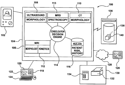
[0027] Figure 1 is a schematic diagram showing a computer-aided detection
(CAD)
system;
[0028] Figure 2 is a block diagram of major functional components of a CAD
application program of the CAD system shown in Figure 1;
CA 02629727 2008-05-14
WO 2007/059615 PCT/CA2006/001910
-8-
[00291 Figure 3 is a flowchart showing steps of a process for analyzing
medical image
data quantitatively and qualitatively implemented by the CAD application
program shown
in Figure 2;
100301 Figure 3A shows another process for analyzing MRS data and ultrasound
images, implemented by the CAD application program;
[0031] Figure 3B is a flowchart illustrating an alternative process that is
implemented
by the CAD application program shown in Figure 2, using results from one
modality as
inputs to another modality;
100321 Figures 4 shows in detail a portion of the process shown in Figure 3;
[0033] Figure 5 illustrates schematically a time sequence of medical images
and a
corresponding contrast variation curve;
[0034) Figure 6 shows general behaviours that can be expected of a contrast
variation
curve;
100351 Figure 7 is a flowchart showing a portion of the process shown in
Figure 3 for
constructing a contrast variation curve shown in Figures 5 and 6;
[00361 Figure 8 is a flowchart showing a portion of the process shown in
Figure 3 for
producing a consolidated result, combining morphological and kinetics
features;
[0037] Figure 9 shows schematically an exemplary screen display, providing to
a user a
side-by-side comparison of analyzed images of two modalities and a
consolidated result;
and
[0038] Figure 10 shows a process modified from that shown in Figure 3 for
processing
images from the same modality, taken at different times.
Detailed Description of Embodiments
100391 The invention relates generally to the field of computer-aided analysis
of
medical images and detection of suspicious abnormalities. In particular, the
invention
relates to a method and system for processing medical images obtained from
multiple
modalities, including analysis of kinetics as well as morphological features.
CA 02629727 2008-05-14
WO 2007/059615 PCT/CA2006/001910
-9-
100401 The invention combines data from multiple modalities, including
kinetics
(quantitative), morphological (qualitative) and biochemical (quantitative)
information to
achieve an optimal discrimination of imaged suspicious abnormalities, such as
imaged
breast lesions. Morphological features of a lesion are generally those
associated with size,
shape, signal distribution within a mass, or border characteristics of the
lesion. They
include features such as whether a lesion is a mass having a round, oval or
lobular shape, a
mass with smooth, irregular or spiculated borders, or a mass having
homogeneous,
peripheral or ductal enhancement. Morphological features can be extracted from
MRI,
ultrasound or x-ray images, or image data from other modalities. Kinetics
features relate to
signal temporal behavior of imaged lesion(s) in a time sequence of images or
image data.
Kinetics features of MRI data generally refer to, but are not limited to, time-
dependent
contrast enhancement of regions in a scanned anatomy volume subsequent to
administering
of contrast agent. A kinetics curve may be type I (continued increase), type
II (plateau) or
type III (washout). Biochemical information can be obtained by analyzing MRS
data, i.e.,
spectroscopic information, to determine the presence and relative
concentration of marker
chemicals (such as choline, creatine, or 31P, among others) in a single voxel
or several
voxels. These information are considered relevant in diagnosing cancer. A
computer
application program is provided for extracting morphological, kinetics and
biochemical
information from medical imaging data and for combining results from
quantitative and
qualitative analysis of the medical imaging data from multiple modalities to
obtain a
consolidated result.
100411 Although a diagnostic assessment may be derived from result of any of a
kinetics, a morphological or biochemical (i.e., spectroscopic) analysis of
image data from a
single modality, combining results from multiple modalities tends to increase
the
confidence level in the assessment obtained, as such consolidated assessments
generally are
derived from a larger data set and therefore tend to be more statistically
reliable. For
example, the analysis of time course kinetics can be performed before or after
the evaluation
of the lesions morphology in post-contrast images. Optionally, the results
from the first
performed analysis are evaluated prior to performing the next analysis. If
results from the
first performed analysis (for example, kinetics analysis) are clearly
suggestive, the next
analysis (for example, morphology or spectroscopic analysis) may not be
necessary. On the
other hand, if the results from the analysis of one mode (for example,
kinetics) are
CA 02629727 2008-05-14
WO 2007/059615 PCT/CA2006/001910
- 10-
indeterminate or suggest benign lesion, a further analysis (for example,
morphology) may
be worthwhile. Further, results from one analysis may be used as inputs to
analysis of
another mode. For example, results of a kinetics analysis generally include
the
identification of a lesion, which may be used to drive the segmentation part
of the
morphology process.
100421 Figure 1 shows a computer-aided detection (CAD) system 100. The CAD
system 100 processes and analyzes images and data obtained from multiple
modalities,
including performing kinetics, morphological and spectroscopic analysis, for
providing
diagnostic assessments based on extracted kinetics, morphological and
spectroscopic
features. The CAD system 100 has a medical imaging device 102. The medical
imaging
device 102 is used by a user to acquire medical images and data by scanning or
imaging a
patient. Different imaging modalities may be configured for use with a CAD
system 100.
For example, the medical images may be ultrasound images, X-ray images, MRI
images,
Computed Tomography (CT) images, Positron Emission Tomography (PET) images,
PET/CT, nuclear, MRS or any images or data from a suitable image or data
acquisition
device.
[0043] Image data acquired by the medical imaging device 102 is provided to a
computer 104 for processing. Although in Figure 1 a stand-alone computer is
shown, the
computer 104 may be any general purpose computer or a dedicated computer. It
may also
be an embedded system, such as an embedded system in an image acquisition
system that
includes an medical imaging device 102.
[0044] A computer program 106, namely a software application for performing
the
functions of a CAD system is hosted by the computer 104. The CAD application
program
106 has a number of components. Corresponding to each modality, there is a
dedicated
component. For example, there is a ultrasound subsystem 108 that corresponds
to the
ultrasound modality. The ultrasound subsystem is dedicated to retrieving,
processing and
analyzing ultrasound image data. Similarly, there is a CT subsystem 110
dedicated to
processing and analyzing CT image data. Corresponding to MRI image data, there
is an
NIlZI subsystem 112. Corresponding to MRS spectroscopic data, there is an MRS
subsystem 113.
CA 02629727 2008-05-14
WO 2007/059615 PCT/CA2006/001910
-11-
[0045] The CAD application program 106 has a consolidation decision engine
114. The
consolidation decision engine 114 receives as its inputs the results from
these modalities,
namely from the ultrasound subsystem 108, the CT subsystem 110, the MRI
subsystem 112,
and the MRS subsystem 113, and computes a consolidation assessment,
incorporating the
results from each of these modalities. The CAD application program 106 may use
rules
built into the application program or stored in a database 116 for making the
consolidated
decision. These rules may be derived from sample images containing benign and
malignant
lesions or established from statistical models, or established by employing
any suitable
methodology.
[0046] A workstation 118 is provided. The workstation 118 provides a user
interface
120 that allows a user of the system 100 to view medical images, to manipulate
the images
and to interact with the system to process the images. The user interface 120
includes a
display 122. The display may be a display screen, or an image protector, or
any other
suitable display devices capable of visually presenting medical images to a
user and
presenting graphical and textural contents to user.
[0047] The workstation 118 displays image data and results generated by the
CAD
application program 106 to a user to facilitate diagnosis of the images by the
user. For
example, images from each modalities as well as features extracted from these
images may
be displayed to the user. They may be displayed side-by-side on the same
display to make
it more convenient for the user to make a diagnosis. Lesions identified in
these medial
images as well as features extracted may also be highlighted. In addition, a
form
conforming to medical standards may be pre-populated, incorporating any
results that are
automatically detected by the system. The preliminary assessment automatically
computed
by the system may also be shown to the user for the user to confirm or modify.
100481 The user interface 120 also includes input devices 124 for the user to
interact
with the system and to identify to the system particular regions of interest
in the displayed
medical image. The input devices 124 may include a keyboard, for example, for
the user to
enter any textual input. A voice recognition module may be provided for voice-
to-text
transcription to allow a user to enter textual descriptions of imaged object
verbally, to enter
other textual inputs without having to type the text, or to issue any computer
program
command. It may also include a mouse or some other pointing device for the
user to
CA 02629727 2008-05-14
WO 2007/059615 PCT/CA2006/001910
-12-
identify a particular pixel or region of the medical image to the system.
Display 122 and
input devices 124 may be physically combined into a single piece of hardware
unit, such as
a touch screen that is capable of both displaying graphic and textual output
and receiving
user input. The user interface 120 may also include a remote user interface,
such as a
remote terminal or a web browser 126, for sharing results with other
radiologists or
physicians over a telecommunication network 128. The telecommunication network
128
may be that implemented with direct cable connection, a local area network
(LAN) or the
Internet. The remote user interface allows a physician to remotely review
images obtained
by an operator from a patient and make any modification in real-time to
results
automatically produced by the system 100. A physician, whether in a room next
door to the
medical imaging device 102 or workstation 118, or in a facility a few thousand
kilometers
away, can make diagnosis through the remote user interface.
[00491 The system 100 also includes a number of output peripherals 130 so that
a user
may reproduce or record results of an analysis session or other output of the
system. For
example, the output peripherals may include a printer 132, either film based
or paper based.
A film-based printer may be used to transfer the medical images, either the
original image
or the processed image to a film for use with more traditional display devices
that require a
filmed image. A paper-based printer may also be used to produce hard copy
reports for
sharing with other physicians or for archiving purposes. In addition, the
output peripherals
130 may include DICOM-compliant devices 134 for transferring or storing
processed
results, namely composite images generated by the system together with
associated reports.
[0050] The system 100 has access to an image archive server 136. The image
archive
server 136 may be part of the system 100. It may also be provided by an
external service
provider, such as a hospital information system. The image archive server 136
has a server
database 138 for storing archived images 140. When the CAD application program
106
requests archived images 140 from the image archive server 136, the image
archive server
136 retrieves the requested image from the server database 138 and sends the
requested
images to the CAD application program 106. As will be understood, the archived
images
are all images already acquired by a medical imaging device. The archived
images can be
images from any supported modalities, such as MRI, CT, or PET. The archived
image data
can also be images combined from different modalities, such as digital
tomosynthesis image
data. The archived images 140 are not necessarily of the same modality as the
medical
CA 02629727 2008-05-14
WO 2007/059615 PCT/CA2006/001910
- 13 -
imaging device 102 that is currently directly connected to the computer 104.
For example,
the computer may be connected to an ultrasound imaging device, while the image
archive
server 136 may contain images acquired previously from a CT imaging device or
an MRI
imaging device. Further, although in Figure 1 there is shown only one image
archive server
136, it will be understood that there may be several image archive servers
connected to the
computer 104. In addition, each image archive server 136 may not necessarily
have only
one database, it may have access to a number of databases, and these databases
may be
physically located at different sites.
100511 System related or generated data are generally stored together with the
archived
images 140. For example, the archived images may be stored along with
annotations made
on the image by a physician during a previous analysis or diagnosis data.
Preferably, the
image archive server 136 supports archiving DICOM-compliant images, as well as
images
of other formats such as JPEG, BITMAP, among others. Annotations, comments,
results of
image processing all can be archived as part of a DICOM-compliant file. Audit
information,
such as user ID, date or time stamp of processed images, and user addition or
modification
of detected features all can be recorded for each archived instance of a
processed image, as
well.
[00521 Figure 2 is a block diagram of major functional components of the CAD
application program 106 of one embodiment. As shown in Figure 2, the CAD
application
program 106 has an image data module 202, a processing module 204 and a
modality
decision engine 206, for retrieving and analyzing image data. As will be
described in detail
below, the image data module 202 retrieves image data from medical imaging
device 102 or
image archive server 136 and pre-processes the image data to extract images or
other data
from the image data for further processing. Images retrieved and pre-processed
by the
image data module 202 are forwarded to the processing module 204. The
processing
module 204 is provided for extracting information that are relevant to
diagnosing disease
from the pre-processed image data. For example, this module may be provided
for
identifying suspected lesions in an image and extracting from the image those
features
associated with the suspected lesions that are considered relevant to
diagnosing disease, i.e.,
discriminating the lesions. The modality decision engine 206 classifies a
lesion based on the
information extracted and computes an assessment of the lesion from the
extracted
CA 02629727 2008-05-14
WO 2007/059615 PCT/CA2006/001910
- 14-
information. Such assessment can be computed, for example, based on a pre-
established set
of rules or using a pre-selected algorithm.
100531 The CAD application program 106 is modular in that each of image data
module
202, processing module 204 and modality decision engine 206 has a component
for a
supported modality. For example, the modality decision engine 206 has as its
ultrasound
component an ultrasound decision engine 208, its MRS component an MRS decision
engine
(not shown), and its MRI component an MRI morphology decision engine 210 and
an MRI
kinetics decision engine 212. As an image or scan data obtained from a
particular modality
is processed by the CAD application program 106, the image or scan data is
processed by
the corresponding modality components of image data module 202, processing
module 204
and modality decision engine 206. Components of image data module 202,
processing
module 204 and modality decision engine 206 of a particular modality form the
subsystem
of that modality. For example, ultrasound components of image data module 202,
processing module 204 and modality decision engine 206 form the ultrasound
subsystem
108. To handle images or data of another modality, a corresponding component
is added to
each of image data module 202, processing module 204 and modality decision
engine 206,
without having to change the overall architecture of the CAD application
program 106.
Each modality requires its own component because, in general, image data
obtained from
one modality typically have certain unique aspects not found in other
modalities. For
example, certain sonographic characteristics associated with ultrasound
images, such as
echo patterns, generally are not exhibited in x-ray images. Similarly,
spectroscopic
processing is generally unique to the MRS modality.
[0054] Figure 2 shows that MRI modality has two components in each process
module
204 and modality decision engine 206, one for processing and extracting
morphological
characteristics associated with a lesion imaged in an MRI scan, and another
component for
processing and extracting kinetics, namely, temporal, characteristics
associated with a time
sequence of MRI scans.
[00551 The CAD application program 106 has a consolidation decision engine
114. The
consolidation decision engine 114 combines all results obtained from each
modality,
together with patient data, to compute a consolidated score for lesions
identified by
individual modalities. The patient data may include, for example, risk profile
of a patient or
CA 02629727 2008-05-14
WO 2007/059615 PCT/CA2006/001910
-15-
the patient's history or both. A risk profile module 214 is provided. The risk
profile
module 214 extracts risk profile information from a database 116, processes
the risk profile
information and provides the results to the consolidation decision engine 114.
Risk profile
information may include presence of specific genes - e.g., breast cancer
susceptibility gene
(also known as BRCA-1). A patient history module 216 is also provided_ The
patient
history module 216 extracts information pertinent to a patient's history,
processes the
history information and provides the processed history information to the
consolidation
decision engine 114. Patient history may include familial history of breast
cancer, previous
diagnosis and treatments of cancer. Patient history information may also
include
information relating to images of the same lesion taken during previous clinic
sessions, for
example, a few months ago. The patient history module 216 can use the
information about
images taken previously and direct the image data module 202 to retrieve these
previously
taken images for comparison with images currently processed.
100561 The consolidation decision engine 114 has several individual
components.
These individual components include a classification module 218, a lesion-type
module
220, a lesion-extent module 222, and a staging assessment module 224. The same
lesion
generally can be seen in multiple modalities. Each of the modules 218, 220,
222, 224 may
include components for processing the image data from each modality. A
composite image
can be generated and displayed to show results from multiple modalities. For
example,
results of MRS modality can be overlaid onto an image of one of the image
modalities and
shown together with the image. The consolidation decision engine 114
correlates results of
analysing the lesion seen in images, including biochemical information on
chemical
composition of the tumor obtained through a multivoxel or single voxel MRS
analysis, from
multiple modalities to produce a consolidated result.
[0057) For example, in one implementation, the classification module 218
combines
results from all modalities to provide a possible classification of the
lesion. For example,
local morphological characteristics, such as local spiculation, local branch
pattern, local
duct extension, detected by all modalities can be combined and compared
against a set of
pre-defined feature list to classify the lesion as belonging to ACR BI-RADS 5
category or
an ACR BI-RADS 4a category. Similarly, the lesion-type module 220 combines
results
from all modalities to derive a possible type of a lesion, such as DCIS or CA.
The lesion-
extent module 222 combines results from all modalities to arrive at an
estimated size and
CA 02629727 2008-05-14
WO 2007/059615 PCT/CA2006/001910
- 16-
outline geometric contour of the lesion. The staging assessment module 224
combines as
inputs the results from all modalities and the consolidated classification,
type and extent,
together with the patient's risk profile and the patient's history
information, to compute or
produce a suggested assessment of lesion stage. The consolidated result, which
includes
classification, type, and extent of a lesion as well as suggested diagnostic
assessment of
lesion stage, is shown to the user through the user interface 120.
[0058] It will be understood other implementations are also possible. For
example, one
may have one ultrasound subsystem for processing ultrasound images. Namely,
one may
have a classification module, a lesion-type module, a lesion-extent module,
and a staging
assessment module devoted to processing ultrasound images. One may have
another MRI
subsystem that have its own classification module, lesion-type module, lesion-
extent
module, and staging assessment module devoted to processing MRI images, or
other
subsystems for other modalities. A consolidation engine will then combine
results from
each modality subsystem to produce a consolidated result. Other
implementations that
provide the processing of multiple modalities but combine the modules
differently are also
possible, as long as all necessary processing, such as classification,
determination of lesion
type and lesion extent etc., is provided for all modalities and a consolidated
result is
obtained from consolidating results from all modalities.
[0059] This consolidated result is subject to user confirmation or
modification. For
example, a user can modify an automatically detected feature in an image from
one of the
multiple modalities. It will be appreciated that any modification to features
detected in one
modality may affect detection result with respect to a lesion at the modality
level, and may
further change the consolidated result. A user may also modify directly a
consolidated result
automatically produced by the consolidation engine. Whatever the modification
is made by
the user, the modification is communicated back to processing module 204,
modality
decision engine 206, or the consolidation decision engine 114, as the case may
be. A
modified consolidated result, including a modified suggested assessment of a
lesion stage, is
re-calculated and presented to the user again for modification or
confirmation. Once
confirmed, a report can be automatically generated, summarizing the results of
the analysis
and assessment of these medical images.
CA 02629727 2008-05-14
WO 2007/059615 PCT/CA2006/001910
-17-
[00601 In operation, a user directs the CAD application program 106 to
retrieve medial
images or data generated by an imaging acquisition device or to retrieve
previously scanned
and archived images or data from image archive server 136 for processing and
analysis.
The user may issue the direction from the user interface 120 provided by the
workstation
118, for example, or a remote user interface such as a web browser 126. Figure
3 shows in
flowchart format a process 300 followed by the CAD application program 106 to
analyze
and process images contained in the image data and generate a consolidated
assessment.
[0061] Figure 3 shows three parallel sub-processes, namely, a patient profile
data
retrieval sub-process 302, an ultrasound sub-process 304, and an MRI sub-
process 306. The
sub-processes are shown as parallel processes. These sub-processes are not
necessarily
executed parallel in time but rather, they are independent of each other.
These sub-
processes can be performed in any time sequence relative to each other,
provided that the
results of the sub-processes are all available prior to the final step,
computing consolidated
assessment (step 308). For example, patient data related to patient risk
profile or history
may be retrieved before, after, or during the process of ultrasound images.
However, as will
be appreciated, in an actual implementation of the process 300, results from
one modality
often can serve as inputs (or at least part of the inputs) to another
modality. For example, if
the MRI sub-process 306 is first applied to a set of MRI data, a lesion
centroid can be
identified in an analysis of signal enhancement in concentrated areas or a
volume. The
lesion centroid so identified can serve as the starting point of a
segmentation process for the
MRI morphology process. Although sub-processes corresponding to two modalities
are
shown, sub-processes corresponding to other modalities, such as CT modality,
can be
added. As these other modalities follow steps similar to that of the
ultrasound modality or
MR.I modality, they are not shown in Figure 3.
[0062] Referring to Figure 3, each of these three sub-process. is now
described. Patient
data retrieval sub-process 302 starts with the risk profile module 214
retrieving risk profile
data of the patient from a database 116 (step 310). The database may be
directly accessible
to the CAD application program 106 as shown in Figure 1, or it may be
necessary to request
the information from a database maintained externally, such as by a hospital
information
system. Next, at step 312, the patient history module retrieve patient history
information,
from the database 116 where the patient's risk profile data is maintained or
from some other
externally maintained database. The risk profile information and the patient
history
CA 02629727 2008-05-14
WO 2007/059615 PCT/CA2006/001910
-18-
information are forwarded to the consolidation decision engine 114 for its use
at step 308, to
compute a consolidated assessment, as will be described below.
[0063] The ultrasound sub-process 304 starts with obtaining ultrasound image
data, step
314. The ultrasound image data may be obtained from the medical imaging device
102.
Alternatively, the CAD application program 106, namely its image data module
202, may
request the ultrasound image data from the image archive server 136.
Generally, the
obtained ultrasound image data contains information in addition to medical
images. At this
step, individual images are also extracted from the image data. An extracted
image is
forwarded to the processing module 204 for image processing.
[0064] At step 316, the ultrasound component of the processing module 204
processes
the image. At this step, the processing module 204 computes, i.e., extracts
and identifies,
physical, texture, morphological as well as sonographic characteristics
associated with an
object of interest in the separated individual images. The object of interest
may be defined
by the boundary of an abnormal region such as a lesion. At step 318, the
ultrasound
decision engine 208 analyzes these characteristics to provide classification,
lesion type
identification, and lesion assessment. Optionally, features extracted and
identified are
shown to a user for confirmation or modification, at a display and
confirmation step 320.
[0065] Figure 4 shows in detail the sub-steps by the CAD application program
106
when processing morphological features in an ultrasound image. The ultrasound
image can
be a 2-dimensional image of an area or a 3-dimensional image of a volume. The
image
processing step 316 starts from a step of selecting a region of interest
("ROI"), step 402. An
ROI is a region in an anatomy that may contain an abnormal object such as a
lesion. An
ROI can be 2-dimensional, when a 2-dimensional image is processed, or 3-
dimensional
(also called "VOI", or "volume of interest"), when an imaged volume is
processed. An ROI
may be identified in any suitable manner. For example, a user can manually
identify an
ROI on a displayed image through the user interface 120. The CAD application
program
106 can extract an ROI already identified from another source, such as an ROI
identified on
a prior exam and now stored in a database. Or, the CAD application program 106
can
perform a morphological analysis of the image to identify an ROI and suggest
it to a user.
In one implementation, the user selects and identifies the ROI to the system
by first
selecting a segmentation "seed point", i.e., a starting point in the
interested region. The user
CA 02629727 2008-05-14
WO 2007/059615 PCT/CA2006/001910
- 19-
may select the segmentation seed point by, for example, using a pointing
device and
selecting the point in the central region of a suspected lesion. The ROI is
then defined by
dragging the cursor away from the seed point so that a circle is formed around
the seed
point. The circle constrains the region into which the segmentation algorithm
operates. The
user releases the pointing device when the ROI is sufficiently large so as to
enclose the
entire suspected lesion.
[0066] Once the ROI is identified, the ROI is segmented at a segmentation step
404 to
delineate the boundary of the suspected lesion. After an ROI is segmented, a
pattern
recognition operation (step 406) is applied to the segmented ROI to identify
and extract
morphological characteristics from the ROI. During the pattern recognition
step 406,
structural characteristics in the ROI are identified and analyzed. They are
classified based
on their morphological and texture patterns or features. Local morphological
characteristics
such as local spiculation, local branch pattern, local duct extension and
local micro-
lobulation are identified and indexed. In addition, pixels in the ROI are
scanned to identify
sonographic characteristics such as echoing patterns. The local morphological
characteristics are combined with a set of sonographic characteristics, pre-
defined by a
standard such as ACR-BIRADS lexicon, to generate a list of features so
identified. At the
pattern recognition step, processing module 204 may also analyze the image to
identify
features such as clustering and contrast of pixels in the segmented ROI or
analyze the image
to incorporate some notion of the domain knowledge such as information of
pixels
surrounding the ROI in order to better identify specific local features.
[0067] Next, at a step of feature extraction (step 408), the processing module
204
extracts from these locally identified patterns certain special features that
are considered
relevant to diagnosing cancer, i.e., discriminating between benign and
malignant lesions.
Some of these features may include shape, orientation, angular margin, lesion
boundary and
calcification. The features may also include those unique to a specific
detection technology.
For example, for an ultrasonic image, the features may include echo patterns
and posterior
acoustic features.
[0068] Next, at a classification step 410, the features and characteristics
extracted and
identified during the image process step 316 (sub-steps 402 to 408) are
combined and
analyzed. Conveniently, the features or characteristics extracted and
identified generally
CA 02629727 2008-05-14
WO 2007/059615 PCT/CA2006/001910
-20-
coincide with a pre-defined set of characteristics. Pre-defined sets of
characteristics and
features are generally developed by the medical profession as being relevant
to diagnosing
disease, such as cancer. Descriptions of these features are generally provided
together with
a definition of these features. One such set of pre-defined characteristics
and lexicon is the
BI-RADS lexicon. At this step, the features extracted and identified are
compared against
the set of the BI-RADS lexicon to assign a statistical likelihood that any
feature in the set
may present in the lesion being analyzed.
100691 Next at step 412, an assessment of the lesion is computed. Rules or
algorithms
can be developed for computing an assessment. The assessment can be computed
from, for
example, the classification and the likelihood of features identified and
classified according
to BI-RADS lexicon. In one implementation, a large collection of medical
images is first
processed. Pattern recognition and feature extraction operations are applied
to each image
in the collection. Features identified are classified and indexed according to
the scheme and
lexicon defined by BI-RADS. Images in the collection are also diagnosed, based
on, for
example, biopsy results. From the results of image processing and known
diagnosis, a
statistical model linking the set of features extracted and a statistical
likelihood of a
diagnosis can be developed. A set of rules for computing an assessment can be
extracted
from the model, which can then be applied to the results of an analyzed image
to produce an
assessment. It will be appreciated that the computation of an assessment is
not limited to
using a statistical model. The assessment may also be computed using a super
vector
machine (SVM) method or may be generated using an Al engine that employs a
more
complicated approach such as a neural network method. Whatever the method
used, an
assessment is computed at this step from the features identified, extracted
and classified.
[0070] Methods and systems directed to extracting morphology features from a
medical
image and providing a suggested assessment of suspicious lesion based on
morphology
features extracted and classified are also described with further detail in co-
pending, co-
owned U.S. application Ser. No. 60/686,397, filed on June 2, 2005, which
application are
incorporated by reference herein in its entirety.
[0071] Returning to Figure 3, the steps of the MRI sub-process 306 are now
described
in detail. As shown in Figure 3, the MRI sub-process 306 starts at a step 322
of obtaining
MRI image data. The MRI image data may be supplied by the MRI medical imaging
CA 02629727 2008-05-14
WO 2007/059615 PCT/CA2006/001910
-21-
device 102, or may be retrieved from image archive server 136. In one
implementation, the
MRI image data are acquired in multiple MRI scans, forming a time sequence of
MRI
image data. From these series of MRI scans, temporal information associated
with
suspicious abnormalities, such as suspected lesions, can be extracted in a
kinetics analysis.
[00721 In general, a medical image is formed by a medical imaging device by
differentiating between specific types of tissues. Increasing the contrast
between the types
of tissues tends to provide better image quality. Administering contrast
enhancement agent
to a patient may selectively affect imaging properties of certain tissue types
and enhance
contrast between normal and tumor tissues and therefore contrast of imaged
lesions.
Gadolinium based contrast agent (e.g., Gd-DTPA) is one such commonly used
contrast
enhancement agent for MRI images. Typically, a benign or a malignant lesion
will exhibit
different temporal contrast-enhancing behavior subsequent to the administering
of a contrast
agent. A series of MRI scans, performed at regular time intervals, such as
every two
minutes, can be performed on a patient after inj ection of contrast
enhancement agent to
capture temporal contrast-enhancing behavior. The series of MRI scans
therefore contain a
time sequence of MRI data. One diagnosing technique is to analyze a contrast
variation
curve constructed from the time sequence of MRI data. Various kinetics
features relating to
a model or diagnosing methodology are extracted from the contrast variation
curve for
further analysis.
[00731 Figure 5 illustrates schematically one such time sequence. Only three
images in
such a time sequence are shown schematically in Figure 5 although more
typically will be
used. The first window 502 illustrates a pre-contrast scan image 504. It shows
a lesion
imaged prior to the contrast enhancement. The lesion shows visible structures
but not any
detail nor its true extent. The second window 506 shows a contrast enhanced
image 508.
The image, because of enhanced contrast, shows in greater detail the imaged
lesion. It also
shows the actual extent of the lesion, thanks to an enhanced contrast between
the tissues of
the lesion and its surrounding normal tissues. The third window 510
illustrates
schematically a time delayed image 512. The lesion, due to the residual
contrast
enhancement effect, is still more visible than that in the pre-contrast scan
image 504;
however it is less visible and shows less detail than that in the contrast
enhanced image 508.
CA 02629727 2008-05-14
WO 2007/059615 PCT/CA2006/001910
-22-
[0074] Also illustrated in Figure 5 is a window showing a contrast variation
curve 514.
The contrast variation curve 514 is a curve showing the contrast variation in
time
subsequent to the administering of a contrast agent. The curve generally shows
an initial
increase of the contrast followed by a decline of the contrast enhancement as
seen in the
MRI images 504, 508, 512 in the time sequence.
[0075] It is believed that in general the time variation characteristics,
namely kinetics of
NIlZI image data and in particular, the characteristics of the contrast
variation curve, can be
an useful aid in diagnosing cancer. Relevant kinetics features generally are
those global or
local criteria that can be derived from contrast variation curves and
considered important
descriptors for or by a statistical model. One such kinetics feature is simply
the shape of a
contrast variation curve. A display similar to that shown in Figure 5 may be
presented to a
user. The CAD application program 106 may analyze the contrast variation curve
514 and
provide an assessment of the imaged object, namely the suspected lesion, to
assist the user
in making a diagnosis.
[0076] Figure 6 shows the general behaviours that can be expected of a
contrast
variation curve. A contrast variation curve generally consists of an uptake
segment 602, a
transition point 604, and a time delayed portion 606. Advantageously, the
contrast variation
curve shown in Figure 6 is normalized, namely shows only the relative
enhancement of
contrast. A normalized curve shows the rate of increase (and decrease) of
percentage of
contrast enhancement. This tends to reduce the variation from patient to
patient.
[00771 The initial enhancement of contrast induced by the contrast enhancement
agent
is shown as an initial rapid increase of contrast, or a steep uptake segment
602. The steeper
the curve, the more rapid the enhancement is. This initial increase is
generally associated
with the increased level of contrast agent within vasculature associated with
a lesion. After
the initial rapid increase of contrast, the rate of increase slows down and
generally exhibits
one of three different types of behaviors, depending on the type of the
lesion. The transition
point 604 on the contrast variation curve marks this slow-down. The first type
is a slower
but continued increase of contrast enhancement. The continuous enhancement 608
is
generally considered indicative of a benign lesion. The second type is
persistent
enhancement, or a plateau 610. The contrast, after an initial rapid increase,
abruptly stops
increasing and maintains a roughly constant elevation of contrast in the
intermediate and
CA 02629727 2008-05-14
WO 2007/059615 PCT/CA2006/001910
-23-
late post-contrast periods. A third type is a slow decline showing a wash-out
segment 612.
The transition point 604 corresponds to a peak enhancement. The contrast,
after an initial
rapid increase, abruptly stops increasing and starts declining in the
intermediate and late
post-contrast periods, producing the wash-out segment 612. The presence of
either the
plateau 610 or the wash-out segment 612 is believed to be indicative of tumor
angiogenesis
and vascular permeability. It is generally believed that the growth and
metastatic potential
of tumors can be directly linked to the extent of surrounding angiogenesis.
Analyzing the
contrast variation curve 514 may therefore provide an additional indicator to
discriminate
between benign and malignant lesions.
100781 The MRI sub-process 306 bifurcates into two branches after step 322 at
which
individual image data of MRI scans are extracted. One branch is similar to
processing
morphological features in individual ultrasound images as described in
connection with
Figure 4, which has the steps of processing image (step 324), analyzing and
assessing lesion
(step 326) and optionally displaying results to a user for confirmation and
modification
(step 328). These steps are generally the same as that described in connection
with the
ultrasound sub-process 304 and will not be described in further detail here.
100791 However, it will be noted that, as MRI data may contain a time sequence
of
multiple scans, the step of processing image (step 324) can incorporate the
temporal
information in a morphological analysis. To illustrate this, consider a pre-
contrast scan and
a post-contrast scan. Subtracting voxel values in the pre-contrast scan from
the
corresponding voxel values in the post-contrast scan tends to emphasize
regions in the
scanned volume that are enhanced, i.e., regions that may correspond to
structures in a
suspicious lesion. As will be appreciated, mathematical operations other than
subtraction
can be performed. Further, a series of mathematical or logical operations may
be applied to
(or between, if logical operations) several, including multiple post-contrast,
scans where
appropriate, in order to assist the morphological analysis.
100801 The other branch of the MRI sub-process 306 includes the steps of
extracting
and processing kinetics data (step 330), classifying lesion and computing an
assessment
based on kinetics features extracted (step 332), and optionally displaying
results to a user
for confirmation and modification (step 334). These steps are described in
great detail
below in reference to Figures 5 to 8.
CA 02629727 2008-05-14
WO 2007/059615 PCT/CA2006/001910
-24-
[00811 MRI image data generally corresponds to a three-dimensional region or
volume,
represented by data points (or "voxels") arranged in a 3-dimensional grid or
lattice. The 3-
D volume represented by the MRI scan can be processed as a unitary volume in a
3-D
processing. Alternatively, such a 3-dimensional scan can be organized into a
stack of
planar "slices". A user can choose to process the stack slice by slice in a
series of 2-D
processes.
100821 Figure 7 is a flowchart showing in detail the kinetics branch of the
MRI sub-
process 306 for constructing a contrast variation curve. These steps
correspond to steps 322
and 330 shown in Figure 3. The first step, step 702, is to obtain MRI data
from either the
medical imaging device 102 or from an image archive server 136. Image data
acquired
from a scan at a first initial time, prior to the administering of contrast-
enhancement agent,
are first extracted (step 704).
[00831 Advantageously, results from the morphological branch of the MRI sub-
process
306 or morphological analysis of the ultrasound sub-process 304 can be re-used
here. The
same lesion identified during the morphological analysis can be selected for
the kinetics
analysis (step 706). If no morphological analysis has been performed and no
ROI has been
identified for the MRI scan, an ROI can be identified manually by a user or
from the time
sequence. For example, the time sequence of MRI scans can be processed to
identify voxels
that have marked increase of signal strength over the time course. The time
delayed
behavior (e.g., plateau or washout) can be analyzed as well. Voxels showing
enhanced
contrast and exhibiting expected time delay behavior are likely within a
centroid
corresponding to a lesion. An ROI enclosing these voxels may be selected
automatically.
The clustering of such voxels can be analyzed to isolate one lesion from
another, or to group
different structural elements belonging to the same lesion together. An ROI
can be defined
that enclose all voxels potentially belonging to a lesion.
[00841 Next, at step 708, morphological operations, including segmentation and
pattern
recognition, are applied to the ROI to delineate a centroid containing the
lesion and to
identify structures in the lesion. Again, results produced by the
morphological branch of the
MRI sub-process can be re-used here. Further, as will be described below, if
the ROI is
identified from an analysis of time-dependent contrast enhancement, the
clustering of
voxels may already provide a good segmentation. Next, at step 710, the
contrast between
CA 02629727 2008-05-14
WO 2007/059615 PCT/CA2006/001910
-25-
the identified morphological features, namely the signal strength of the
lesion relative to the
surrounding structure, is evaluated. In one implementation, signal strengths
of all voxels
within an identified centroid is summed to provide an estimate of the contrast
value of the
suspected lesion. However, other ways of representing contrast enhancement can
be used.
For example, in a model taking into account rim enhancement, total signal
strength can be
the sum of voxels located along the boundary of a lesion. When another
diagnostic
methodology or model is implemented, voxels corresponding to some other
structures may
be summed. In other words, the contrast value can be a sum over voxels in any
specific
subset in the lesion, depending on diagnostic methodology or model implemented
or
supported by the CAD application program.
100851 After a contrast level of the first pre-contrast image is evaluated,
the process
continues with extracting the MRI data of the next scan in the time sequence.
Namely, the
process returns to the image extraction step, step 704. Subsequent to the
image extraction
step, the steps 706 to 710 are repeated for the first post-contrast scan.
First, the same lesion
already identified is re-used here to provide a starting point in ROI
identification. An ROI
enclosing these voxels may be re-used as well. Following the identification of
ROI at step
706, morphological operations are performed to identify and delineate a
centroid containing
the lesion at step 708. Next, the contrast between the lesion and its
surrounding tissues in
this first post-contrast scan is computed at step 710. These steps are
repeated for all MRI
scans in the time sequence until all MRI scans in the time sequence have been
processed
(step 712). At a final step 714, contrast values of the lesion computed from
the series of
images are normalized against the initial contrast value and a contrast
variation curve 514 is
constructed.
[0086] Returning to Figure 3, once a contrast variation curve is constructed,
a
quantitative analysis of the contrast variation curve 514 is performed to
extract temporal,
i.e., kinetics features from the time sequence of images to provide a
classification of the
lesion (step 332). A quantitative analysis of the contrast variation curve 514
generally
includes an analysis and classification of the shape of the kinetics curve,
namely whether
the time delayed portion 606 is a continuous enhancement 608, a plateau 610,
or a wash-out
segment 612, the level of enhancement at the transition point 604, the time to
reach the
transition point 604, i.e., the slope or the initial rate of increase of the
uptake segment 602,
and the rate of decline in the post-contrast period, i.e., the presence or
absence of a wash-out
CA 02629727 2008-05-14
WO 2007/059615 PCT/CA2006/001910
-26-
segment 612 and its rate of decline. The underlying lesion can be classified
based on these
kinetics features. In one implementation, a lesion is simply assigned a score
of 0 if a
continuous enhancement is seen, a score of 1 if a plateau is seen, and a score
of 2 if a wash-
out segment is seen, where 0 indicates a benign lesion and 2 indicates a
malignant lesion.
More sophisticated classification schemes can be implemented by taking into
account of
other features, such as the slope of the uptake segment, the peak value of the
curve, or the
rate of decline. Such a sophisticated scheme generally may be established
using a statistical
model, similar to that described earlier in connection with ultrasound images.
[0087] Referring to Figure 3, as a final step, results from each of these
parallel sub-
processes are forwarded to the consolidation decision engine 114 for making a
consolidated
assessment (step 308). The consolidation decision engine 114 correlates the
features
identified and extracted for the lesion from all modalities. Results from all
modalities are
also combined to provide a consolidated estimate of the extent of the lesion,
to classify the
lesion and to stage the lesion, namely to provide a stage assessment of the
lesion according
to a pre-defined staging scheme.
[0088] As described earlier, the CAD application program 106 is modular.
Although
Figure 3 shows a flowchart implementing two modalities, namely a ultrasound
modality and
an MRI modality, other modalities, namely other sub-processes, can be easily
added to the
CAD application program 106. Any one of the ultrasound or MRI modalities can
also be
replaced or substituted with other modalities as well. For example, in Figure
3A, there is
shown an alternative embodiment that implements an MRS modality. In Figure 3A,
an
MRS sub-process 340 replaces the MRI sub-process 306, while the ultrasound sub-
process
304 is substantially the same as described in reference to Figure 3 and
therefore will not be
further described here.
[0089] Referring to Figure 3A, the MRS sub-process 340 starts with obtaining
MRS
data, step 342. As will be appreciated, the MRS data may be obtained directly
from an
MRS device 102, for example, a procedure performed based on results from other
modalities. Alternatively, the MRS data may be retrieved from an image archive
server
136.
[0090] In general, the MRS data corresponds to a number of MRS measurements.
Each
MRS measurement may be a single spectrum, corresponding to spectroscopic data
obtained
CA 02629727 2008-05-14
WO 2007/059615 PCT/CA2006/001910
-27-
from chemicals in a single voxel. The MRS measurement may also correspond to
spectroscopic data from chemicals in multiple voxels, such as data obtained
from 2DCSI or
3DCSI exams. In a 2DCSI or 3DCSI exam, each measurement corresponds to spectra
of
chemicals from multiple voxels, each of which may be, for example, 1 cm3 to
1.5 cm3 in
volume. A measurement is extracted from the MRS data at step 344 for further
analysis.
[0091] At the next step, the strength or concentration of the marker chemicals
is
identified and computed in a spectroscopic analysis 344. For example, the
spectrum of
choline may be isolated or identified from the spectroscopic data. The peaks
of choline
characteristic frequencies are identified and measured and then converted to
an absolute
measure of concentration of choline in the voxel or as relative strength or
concentration
relative to other chemicals in the voxel. If biochemical information from
multiple marker
chemicals is desirable, the spectroscopic data can be further processed to
isolate or identify
contributions from each of the remaining marker chemicals. Their
concentrations or
relative strengths can also be computed from their respective spectroscopic
data.
[0092] At the next step, the results of the spectroscopic analysis 346, namely
the
concentration or relative strengths of marker chemicals corresponding to each
voxel or
voxels, are displayed. The results can be displayed numerically for each
measurement. The
results can also be plotted as iso-concentration contours to show more visibly
the
distribution of concentration or strength of marker chemical or chemicals.
Advantageously,
the distribution of the concentration or strength also can be converted to a
false color map
and super-imposed on the MRI image.
[0093] As will be appreciated, although the MRS sub-process 340 is described
here as
being performed independent of the ultrasound sub-process 304, advantageously,
the
ultrasound sub-process 304 can be first performed. Results from a
morphological analysis,
in particular, a segmentation process, can help identify a collection of
voxels or the centroid,
that likely represents a lesion. An envelope enclosing the volume or centroid
can be
generated. Subsequently, only MRS data corresponding to the voxels contained
within the
envelope needs to be analyzed. As another example, it may often be the case
that an
analysis of image data from one modality, such as ultrasonic or MRI,
identifies one or more
regions suspicious of cancer, for example, based on an initial assessment from
data from
these modalities alone. The results, however, may not be conclusive. Instead
of performing
CA 02629727 2008-05-14
WO 2007/059615 PCT/CA2006/001910
-28-
an MRS procedure for an entire anatomy region or the same region as in other
modalities,
an MRS procedure may be performed for a much smaller region or regions,
enclosing only
the suspected lesions identified in other modalities. This tends to improve
efficiency.
Likewise, a preliminary result from MRI or MRS analysis may also provide a
starting point
for the data acquisition and analysis in other modalities.
100941 In general, a multimodality system such as the system 100 can take
advantage of
results from one modality and use the results as inputs to another modality to
improve
efficiency. As the same lesion generally can be seen in multiple modalities,
results of
morphological analysis performed in one modality often can be used directly in
another
modality. Figure 3B illustrates in flowchart form an alternative process 350
that uses
results from a morphological analysis as inputs to an MRI kinetic analysis.
100951 As will be remembered, in a morphological analysis, first an ROI is
identified
and then a segmentation process is performed to identify a boundary that
likely separates
tumor from normal tissue. In a 3-dimensional segmentation process, the
boundary is an
envelope enclosing a volume or centroid that likely corresponds to a tumor. As
a first step
of process 350, this segmentation result is first obtained from, for example,
an ultrasound
module (step 352). Next, an envelope enclosing these voxels or the centroid is
generated
(step 354). All voxels contained within the envelope will next be analyzed to
extract
kinetics features.
[00961 The steps of obtaining MRI data and separating them into individual
scans in a
time sequence at different times TO, T1, T2, ... are similar to that described
in reference to
Figure 3 and therefore will not be described here. To extract kinetic
features, images
scanned at different times, such as TO and T1, or TI and T2, are compared.
This can be
implemented by, for example, first subtracting image taken at TO from image
taken at Ti.
Voxels having a positive value then represent voxels that have increasing
contrast while
voxels having a negative value represent voxels that have decreasing contrast.
As an
envelope is already determined from a morphological analysis, only voxels
within the
envelope need to be processed to extract the kinetics information. Subsequent
images or
scans in the time sequence are similarly processed to extract the kinetics
information (step
356). Limiting the processing of kinetics information to those voxels
contained inside the
envelope tends to obliterate the needs of identifying voxels corresponding to
a tumor in a
CA 02629727 2008-05-14
WO 2007/059615 PCT/CA2006/001910
-29-
separate run, for example, by identifying those that exhibit initial up-take
and then plateau
or wash-out behavior. This may also avoid the needs of processing voxels
outside the
envelope. Thus, efficiency as well as accuracy of the multimodality system 100
may be
improved as a result.
100971 Figure 8 is a flowchart showing steps followed by the consolidation
decision
engine 114 in a consolidated scoring process. The consolidation decision
engine 114 first
correlates at step 802 all features provided by all modality decision engines.
For example,
the shape of the lesion as determined by each modality can be correlated at
this step. Each
of the classification module 218, lesion-type module 220 and lesion-extent
module 222 of
the consolidation decision engine 114 combines the results from all modalities
to classify
the lesion at a classification step 804, to determine a lesion type at a type
determination step
806, and to estimate the size of the lesion at an extent determination step
808.
100981 The consolidation decision engine 114 also scores the lesion (step
810), namely,
computes a diagnostic assessment, by incorporating results from all
modalities. As a
consolidated diagnostic assessment is produced based on results from more than
one
modality, confidence level in the assessment is generally increased. A rule-
based process
may be followed to compute a consolidated assessment. For example, features
generally
indicating malignancy in each modality may be assigned a score point. By
summing score
points obtained from all modalities, a total score can be obtained. A stage
assessment can
be assigned to the lesion based on the value of the final total score.
Generally, a stage
assessment based on features seen in one modality confirmed by features seen
in another
modality tends to increase the confidence in the assessment.
[0099] For example, in one implementation, the following scoring scheme is
used when
the consolidation decision engine 114 computes a consolidated stage assessment
based on
results from analysis of only MRI image data:
[00100]
Features Identified Points
Lesion Morphology
Shape round, oval, lobulated 0
irregular I
Margin smooth 0
CA 02629727 2008-05-14
WO 2007/059615 PCT/CA2006/001910
-30-
Features Identified Points
spiculated, irregular I
Mass homogeneous 0
enhancement heterogeneous, rim enhancement 1
Lesion Dynamics
Initial increase < 50% 0
enhancement increase: [50% : 100%] 1
increase > 100% 2
Kinetics continuous enhancement 0
plateau I
washout 2
BI-RADS categories
Total Score total score = 3 3
total score = 4 4
total score: 5 to 8 5
1001011 Similarly, a statistical model may be built for image data obtained
from multiple
modalities, similar to that built for a single modality. For example, with
results of biopsies
known for a pool of image data obtained from multiple modalities, rules can be
constructed
to relate the presence of features seen in each of the multiple modalities to
a possible stage
of the tumor, with a statistical likelihood assigned to the result. This set
of rules can be
applied to the results from all modalities, the lesion type, extent and
classification of the
lesion produced by the consolidation decision engine, to compute a
consolidated staging
assessment of the lesion. As such a combined scoring or assessment takes into
account a
larger set of inputs, the result tends to be more robust. As a general rule,
assessments
computed from more independently obtained data tend to be more statistically
reliable.
Further, assessments computed from results of analyzing image data of a single
modality
may be strongly affected by a missing data point, for example, when an
important descriptor
contributing to the basis function(s) of the statistical model cannot be
computed. With
results from multiple modalities, results from another modality may provide
the required
(and missing) information, and therefore increase the confidence in the
assessment
computed.
CA 02629727 2008-05-14
WO 2007/059615 PCT/CA2006/001910
-31-
[00102] However, it will be appreciated that although combining results from
multiple
modalities tend to improve the reliability and confidence in the assessment,
there are
situations where results from the analysis of one modality may be sufficient.
If results from
one modality are clearly suggestive, to improve performance, analysis by other
modalities
may be optionally skipped. For example, if an MRI kinetics analysis finds a
lesion clearly
cancerous, the MRI morphology analysis or morphology analysis by other
modalities may
be optionally skipped or suspended, unless a user specifically requests a
morphology
analysis. Likewise, if a morphological analysis clearly indicates a cancerous
finding, an
MRI kinetics analysis may be skipped or suspended. The result provided by the
consolidation decision engine will then be the same as that provided by the
particular
modality decision engine in question.
1001031 The results of the consolidation engine are presented to a user for
confirmation
or modification (step 812). The user may be shown images from each of the
modalities
with features extracted superimposed on the images. The features identified by
the CAD
application program 106 may be annotated. Contrast variation curve 514 may be
shown
simultaneously to the user as well. Results identified may be pre-populated in
a standard
report form, following the formats established by standards such as BI-RADS
MRI Lexicon
or any other suitable standards. Figure 9 shows one such possible display,
which shows a
first image 902 obtained from a first modality, a second image 904 from a
second modality,
and a report 906 containing a consolidated result, with extracted features and
a consolidated
assessment pre-populated.
[00104) A user, for example, a physician or a radiologist, may confirm the
results as
computed by the CAD application program 106, or may modify any of the
automatically
detected and evaluated results. An annotation module (not shown) may be
provided for
receiving user inputs. A user may modify or annotate the results displayed
through a user
interface. For example, the user may reclassify a lesion, override the
classification made by
the CAD application program 106, or the user may modify a staging assessment
computed
by the CAD application program 106. The user may also reclassify morphological
or
kinetics features extracted by the CAD application program 106. The CAD
application
program 106 will then recompute as necessary to produce a modified
consolidated decision
and assessment.
CA 02629727 2008-05-14
WO 2007/059615 PCT/CA2006/001910
-32-
[001051 Once the result is confirmed by the user, a report can be generated
(step 814).
The generated report is similar to that generated for each individual
modality, except that
the result is a consolidated decision and assessment. The report contents are
generally by
default based on the data available in the processed images. In other words,
data available
in a result similar to that shown in Figure 9 are reflected in the report. The
report includes
detected and classified MRI, sonographic or CT characteristics, as the case
may be, and the
computed and confirmed assessment, along with any annotations and comments and
user
modifications. Original medical images and the processed counterparts are
included as well.
Finally, the report contains the image findings and assessment of the
radiologists
(preferably, in a format complying with the relevant ACR-BIRADS classification
form or
forms).
1001061 A report may include identification and audit information for
traceability and
auditing purposes. Identification and audit information may include unique
report identifier,
series number, date or time stamp, namely the time and date of the study or
report, patient
identification number, study identification number, user ID, unique report
identifier, user
addition or modification of detected features, among others. Conveniently, a
cryptographic
module may be provided for a radiologist to digitally sign a report. A digital
signature can
be included and recorded for each archived instance, to provide improved audit
capability
and to discourage accidental modification of the reports.
[00107] Preferably, reports are archived as DICOM Secondary Capture.
Annotations,
comments, image processing results such as lesion boundaries and diagnosis
results are
archived as part of a DICOM-compliant file. A user can also save, for example,
a PDF
version of the report locally in a patient's instantiated directory. This
facilitates easy future
reference. If an instance for that composite rendering already exists in the
archive for the
patient, a new instance is created.
[001081 The CAD application program 106 is not limited to analysing image data
taken
during a single imaging session, or clinical visit. Often, it is necessary to
image a patient
several months apart. This may be necessary as part of regular check-ups, or
as part of
follow-up clinical visits after a surgery or treatment of cancer. Images from
the same
modality taken during different visits may need to be analyzed and compared
with each
other. For example, it may be necessary to determine whether a detected benign
lesion has
CA 02629727 2008-05-14
WO 2007/059615 PCT/CA2006/001910
- 33 -
become malignant. Alternatively, it may be necessary to determine whether a
malignant
lesion has become smaller or stopped growing after treatment.
1001091 Figure 10 shows a process for processing images from the same
modality,
acquired at different times. This is a process modified from that shown in
Figure 3. Figure
10 also shows three parallel sub-processes, namely, a patient data retrieval
sub-process 302,
a first morphology sub-process 1004, and a second morphology sub-process 1006.
The sub-
processes are shown as parallel processes as well. In this modified process,
the first and
second morphology sub-processes 1004, 1006 are essentially the same, with one
exception.
At the start of sub-process 1004, image data of the first visit are retrieved
(step 1014). At
the start of sub-process 1006, image data of the second visit are retrieved
(step 1020). The
processing of the image data (step 1016) and lesion classification and
assessment (step
1018) are the same for both sub-processes 1004, 1006, and are essentially the
same as that
described in connection with the ultrasound sub-process 304 as well. The sub-
processes
1004, 1006 therefore will not be described in detail here.
[00110) In the final step 1008, the consolidation decision engine 114 computes
a
consolidated assessment of the lesion in the image data of the second visit,
incorporating
features extracted from image data of first visit. As the image data are
obtained at different
times, the same lesion, even if visible in both image data, tends to be at
different stages and
will need to be matched to imagined patterns seen in images acquired during
the two visits.
The consolidation decision engine 114, when correlates the lesion, will need
to take into
account of the time difference. Time projection of development of the lesion
seen in the
image data of the first visit may be necessary. Once the features of the
lesion in both sets of
image data are correlated, a consolidated assessment can be evaluated as
before. However,
it will be understood that a different model or a different set of rules may
be required to
correlate features identified in a lesion imaged at different times. Results
for each
individual set of image data can also be presented to a user, such as a
radiologist in a side-
by-side comparison. The side-by-side comparison can include results such as
lesion type
and extent as well as its classification. Such a comparison may assist a
physician to assess
either the development of the lesion or the effect of the treatment.
[001111 Various embodiments of the invention have now been described in
detail. Those
skilled in the art will appreciate that numerous modifications, adaptations
and variations may
CA 02629727 2008-05-14
WO 2007/059615 PCT/CA2006/001910
-34-
be made to the embodiments without departing from the scope of the invention.
Since changes
in and or additions to the above-described best mode may be made without
departing from the
nature, spirit or scope of the invention, the invention is not to be limited
to those details but
only by the appended claims.