Note: Descriptions are shown in the official language in which they were submitted.
CA 02630452 2010-08-24
DEV'CES, 'STEMS, AND METHODS FOR OCCLUDING A DEFECT
FIELD OF THE INVENTION
[0002] The present invention relates generally to medical devices, systems,
and
methods, and in particular aspects to medical devices, systems, and methods
for treating
defects.
BACKGROUND OF THE INVENTION
[0003] A variety of defects, or abnormal passages, can occur in a mammalian
body. Such defects may be caused by, for example, an infection, a congenital
defect,
inflammatory bowel disease (such as Crohn's disease), irradiation, trauma,
neoplasia,
childbirth, or a side effect from a surgical procedure.
[0004] Some defects occur as fistulas between the vagina and the bladder
(vesico-
vaginal fistulas) or between the vagina and the urethra (urethro-vaginal
fistulas)., These
fistulas may be caused by trauma during childbirth. Traditional surgery for
these types of
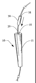
fistulas is complex and not very successful.
[0005] Other fistulas include, but are not limited to, tracheo-esophageal
fistulas,
gastro-cutaneous fistulas, fistulas extending between the vascular and
gastrointestinal
systems, between the small bowel and skin (entero-cutaneous fistulas), between
the
stomach and skin (gastro-cutaneous fistulas), between the colon and skin (colo-
cutaneous
fistulas), between the vagina and bladder (vesico-vaginal fistulas), between
the vagina
and urethra (urethra-vaginal fistulas), any number of anorectal (ano-
cutaneous) fistulas,
such as fistulas that form between the anorectum and vagina (recto-vaginal
fistulas),
between the anorectum and bladder (recto-vesical fistulas), between the
anorectum and
urethra (recto-urethral fistulas), or between the anorectum and prostate
(recto-prostatic
fistulas), or fistulas extending between any other two portions of the body.
Anorectal
fistulas, for example, can result from infection in the anal glands, which are
located
around the circumference of the distal anal canal forming an anatomic landmark
known
1
CA 02630452 2010-08-24
-- a tiedtditt to fkie Ap~5pr cimately 20-30 such glands are found in humans.
Infection in
an anal gland can result in an abscess. This abscess can then track through
soft tissues
(e.g., through or around the sphincter muscles) and into the perianal skin,
where it drains
either spontaneously or surgically. The resulting void through the soft tissue
is known as
a fistula. Fistulas may take various paths, which vary in complexity. The
internal or
inner opening of the fistula, usually located at or near the dentate line, is
known as the
primary opening. The primary opening is usually the high pressure end of a
fistula. Any
external or outer openings, which are usually located in the perianal skin,
are known as
the secondary openings. The secondary openings are usually the low pressure
end of a
fistula.
[0006] Other types of defects include, but are not limited to, defects in
solid
organs, such as bleeding biopsy sites in the liver or perineal sinuses
typically observed in
patients with Crohn's disease; defects in vessels, such as arteries or veins
in which
hemorrhage needs to be arrested or blood flow needs to be diverted from one
area to
another; and viscera containing persistent air leaks from a bronchial stump
after surgical
pneumonectomy or lobectomy.
[0007] Typical techniques for treating a defect such as a fistula involve
draining
infection from the fistula tract and maturing it prior to a definitive closure
or sealing
procedure by inserting a narrow diameter rubber drain, known as a seton,
through the
tract. This is usually accomplished by inserting a fistula probe through the
outer
(secondary) opening and gently guiding it through the fistula, and out through
the inner
(primary) opening. A seton, thread or tie is then affixed to the tip of the
probe, which is
then withdrawn from the tract, leaving the seton in place. The seton may then
be tied as a
loop around the contained tissue and left for several weeks or months.
[0008] One technique for treating a defect is to occlude the defect with an
occluding member, such as, a plug or graft. Examples of such occluding members
and .
related methods are disclosed in U.S. Patent Publication Nos. 20050070759
(Armstrong),
20050159776 (Armstrong), and 20070031508 (Armstrong et al.).
For
example, an occluding member may be pulled through the primary opening
2
CA 02630452 2010-08-24
dffa-fi~,i! l.h=tintil4tbe<bcLluding member is securely lodged within the
fistula. The
occluding member may be further secured within the fistula by the use of
sutures or a
capping member associated with the body of the occluding member.
[0009] Another technique for treating a fistula involves the use of a plug-
like
closure device in combination with a drainage thread or seton, as disclosed in
U.S. Patent Publication No. 20050049626 (Burgard).
In this technique, a closure device is provided with a flexible
application string that can be used to drain secretions or other undesirable
liquids from the
fistula. A rod-like instrument is pushed into the fistula from the outer
opening and is used
to investigate the trajectory of the fistula. After the instrument is pushed
forward enough
to protrude from the inner opening, the application string is pulled through
the fistula
from the inner opening until the closure device "sticks" in the inner opening.
The closure
device is then pushed as far as necessary for it to be tightly secured within
the fistula.
[0010] Still other techniques for treating fistulas are described in U.S.
Patent
Publication No. 20080004657, titled "VOLUMETRIC GRAFTS FOR TREATMENT OF
FISTULAE AND RELATED METHODS AND SYSTEMS" (Cook Biotech
Incorporated), published January 3, 2008.
[00111 The above techniques can be difficult for some physicians to perform,
especially when the defect to be treated is located in an area that is not
easily accessible.
Therefore, there remains a need for simplified procedures and new medical
devices and
systems for treating defects. The present invention addresses these needs.
SUMMARY OF THE INVENTION
[0012] The present invention provides devices, systems, and minimally invasive
methods for occluding fistulas that overcome several shortcomings of the prior
art and
simplify the implantation of an occluding member in a defect of a patient.
[0013] The present invention may be used to occlude any type of defect, or
abnormal bodily passage. For example, the claimed devices, systems, and
methods may
3
CA 02630452 2008-05-20
WO 2007/064819 PCT/US2006/045890
:err.- ;r ti"!i
iL occ'IMa'~ e1ie i-esophageal fistulas, gastro-cutaneous fistulas, anorectal
fistulas, fistulas occurring between the vagina and the urethra or bladder,
fistulas
occurring between the anorectum and vagina, fistulas occurring between the
vascular and
gastrointestinal systems, defects in solid organs, defects in vessels, viscera
containing
persistent air leaks, or any other type of defect.
[0014] In one aspect of the present invention, a medical device for occluding
a
defect is provided. In some embodiments, the medical device comprises an
occluding
member, such as a plug or graft, configured to be placed within a fistula and
to occlude
the fistula. The medical device may further comprise a lumen configured to
receive a
wire guide. In some embodiments, the lumen extends throughout the length of
the
occluding member and is positioned in or near the center of the occluding
member. A
longitudinal slit may be positioned adjacent to the lumen to permit lateral
insertion of the
wire guide. The device may be made of any biocompatible material and may be of
any
suitable shape, dimension, and material for at least partially occluding a
defect. In some
desirable embodiments, the device has a generally conical shape and is made of
a
remodelable extracellular matrix material, such as small intestinal submucosa.
[0015] In another aspect of the present invention, a system for occluding a
fistula
is provided. In some embodiments, the system comprises a placement member
(such as a
wire guide), an occluding member (such as a plug or graft) having a first
lumen, and a
pusher member having a second lumen. Desirably, the occluding member and the
pusher
member are configured to be received on the wire guide to facilitate insertion
of the
occluding member into a defect (such as a fistula). The occluding member may
be of any
suitable shape, dimension, and material for at least partially occluding a
defect, and the
wire guide and pusher member may be of any suitable shape, dimension, and
material for
facilitating the delivery of the occluding member to its desired location.
[0016] In still another aspect of the present invention, a method of occluding
a
defect is provided. In some embodiments, the method comprises the steps of.
(a)
providing a placement member (such as a wire guide), an occluding member (such
as a
plug or graft) having a first lumen, and a pusher member having a second
lumen; (b)
inserting the placement member at least partially into the defect; (c)
advancing the
placement member to a desired location; (d) inserting the placement member
into the first
4
CA 02630452 2008-05-20
WO 2007/064819 PCT/US2006/045890
,= au x :: :<f~'f i, if {C
i i qle i 4{tbf~ilye"o8 1u't ihg l dmber; and (e) advancing the occluding
member to the desired
location by inserting the placement member into the second lumen of the pusher
member
and pushing the occluding member with the pusher member until the occluding
member
reaches the desired location. In some embodiments, the placement member is an
endoscope or a hollow tubular device that may be used to inject contrast
solution into the
defect. The placement member may be inserted into the occluding member by
threading
it through a lumen in the occluding member or by moving it in a lateral
direction through
a slit in the external surface of the occluding member and then moving it into
the lumen.
The method may also include anchoring the occluding member within the defect
by any
suitable means, injecting a rehydrating fluid into the tissues surrounding the
defect to
rehydrate a dehydrated occluding member, and/or trimming any excess portions
of the
occluding member to prevent protrusion from the defect. In some embodiments,
an
endoscope is utilized to assist with visualization and insertion of the
placement member
into the defect. An instrument channel within the endoscope may be used to
facilitate the
delivery of wire guides, catheters, medical devices, and the like into the
defect during the
implantation procedure.
[0017] Additional features and advantages of the present invention will be
apparent to one of ordinary skill in the art from the drawings and detailed
description of
the preferred embodiments below. Moreover, it should be appreciated that
several
aspects of the present invention can be performed with alternative types of
wire guides,
catheters, endoscopes, occluding members, pusher members, and other medical
devices.
BRIEF DESCRIPTION OF THE DRAWINGS
[0018] Figure 1 is a perspective view of one embodiment of a medical device of
the present invention, where the medical device comprises an occluding member
having a
curved, generally conical body, and where a wire guide has been inserted
through a lumen
in the occluding member;
[0019] Figure 2 is a perspective view of another embodiment of a medical
device
of the present invention, where the medical device comprises an occluding
member
having a generally conical body and a central lumen extending throughout the
length of
the occluding member, and where a wire guide has been inserted through a lumen
in the
occluding member;
CA 02630452 2008-05-20
WO 2007/064819 PCT/US2006/045890
r t { } [b i ( "{n ` ~ gig
N-1 -
Tli a perspective view of still another embodiment of the medical
device of the present invention similar to the embodiment of Figure 2, but
including a
longitudinal slit located adjacent to the lumen to facilitate lateral
insertion of a placement
member, such as a wire guide, into the lumen;
[0021] Figure 4 is a side view of yet another embodiment of a medical device
of
the present invention, where the medical device comprises an occluding member
having a
generally cylindrical body, a lumen extending throughout the length of the
occluding
member; and a capping member to assist with anchoring the device within a
defect;
[0022] Figure 5 is a perspective view of the embodiment of Figure 2 in which a
pusher member has been threaded over the wire guide and is being advanced
distally
along the wire guide;
[0023] Figure 6 shows a medical device implanted within an anorectal fistula,
according to one embodiment of the method of the present invention.
DETAILED DESCRIPTION OF THE PREFERRED EMBODIMENT
[0024] While the present invention may be embodied in many different forms,
for
the purpose of promoting an understanding of the principles of the present
invention,
reference will now be made to the embodiments illustrated in the drawings, and
specific
language will be used to describe the same. It will nevertheless be understood
that no
limitation of the scope of the invention is thereby intended. Any alterations
and further
modifications in the described embodiments and any further applications of the
principles
of the present invention as described herein are contemplated as would
normally occur to
one skilled in the art to which the invention relates.
[0025] Turning now to a discussion of particular embodiments of the medical
devices, systems, and methods of the present invention useful for treating
defects,
illustrative medical devices of the invention are configured to at least
partially occlude a
defect within a patient. For example, in some embodiments of the invention, a
medical
device is used to occlude at least the primary opening of a fistula and
potentially one or
more other segments of the fistula, the fistula tract, and/or any secondary
openings. In
this context, the term "fistula tract" is meant to include, but is not limited
to, a void in the
soft tissues extending from a primary fistula opening, whether blind-ending or
leading to
one or more secondary fistula openings.
6
CA 02630452 2010-08-24
[006' Thevd cal`devices, systems, and methods of the present invention may be
used to occlude any type of defect. For example, defects such as anorectal
fistulas,
tracheo-esophageal fistulas, gastro-cutaneous fistulas, or fistulas occurring
between the
vagina and bladder (vesico-vaginal fistulas), between the vagina and urethra
(ureth o-
vaginal fistulas), between the anorectum and vagina (recto-vaginal fistulas),
between the
anorectum and bladder (recto-vesical fistulas), between the anorectum and
urethra (recto-
urethral fistulas), between the anorectum and prostate (recto-prostatic
fistulas), or
between the vascular and gastrointestinal systems, defects in solid organs
(such as
bleeding biopsy sites in the liver or perineal sinuses typically observed in
patients with
Crohn's disease), defects in vessels (such as arteries or veins) in which
hemorrhage needs
to be arrested or blood flow needs to be diverted from one area to another,
and viscera
containing persistent air leaks from a bronchial stump after surgical
pneumonectomy or
lobectomy may be treated with the devices, systems, and methods of the present
invention.
[0027] Generally, the medical devices of the present invention comprise an
occluding member configured for implantation into a defect such as a fistula.
The
occluding member may have any suitable shape, configuration, and dimensions,
such as
those disclosed in U.S. Patent Publication Nos. 20050049626 (Burgard),
20050070759
(Armstrong), 20050159776 (Armstrong), 20080004657 (Cook Biotech Incorporated,
assignee), and 20070031508 (Armstrong et al.).
For example,
the occluding member may be of any suitable dimensions and may have a body
that is
generally convex, bi-convex, concave, bi-concave, S-shaped, straight, curved,
flat,
polygonal, ovoid, conical, cylindrical, elliptical, helical, spherical, or
hemispherical, or it
may have any other configuration capable of being inserted into and secured
within a
defect. In certain embodiments, the occluding member comprises a plug or graft
that is
sufficiently rigid to facilitate delivery into an implantation site, and that
has one or more
lumens extending at least partially through the plug or graft body along its
length. In
7
CA 02630452 2008-05-20
WO 2007/064819 PCT/US2006/045890
s::r t sa. n:, u ; , it, if r{,;;, ti g : It
s{ !e
<<, - stlitie''d 6dim"EnV`,!h 6tcluding member comprises a body having a
central lumen to
facilitate deployment of the occluding member body over a wire guide or other
delivery
device. In some embodiments, the body of the occluding member has portions
that are
tapered and/or curvilinear. In other embodiments, the body of the occluding
member is
curved to conform to the shape of the fistula, thereby facilitating
introduction of the
occluding member, a secure fit of the occluding member within the fistula, and
less
discomfort for the patient.
[0028] The body of the occluding member of the present invention may have any
dimension suitable for implantation within a defect of a patient. In some
embodiments,
the body of the occluding member has a size and shape adapted to extend into
at least a
portion of a fistula tract, and is generally (but not necessarily) of
sufficient dimension to
fill a fistula, or a segment thereof, e.g., the primary fistula opening,
fistula tract, and/or
any secondary fistula openings, either alone or in combination with other
components of
the occluding member and/or other similar or differing medical devices. The
body of the
occluding member may or may not be sized and shaped to fill the entire defect.
[0029] In some embodiments of the present invention, the occluding member
includes a lumen. The lumen may have any shape or configuration suitable for
receiving
a placement member, such as a wire guide, and may be positioned in any
suitable location
within the medical device. Desirably, the lumen is a cylindrical channel that
extends
throughout the length of the occluding member, and is disposed in or near the
center of
the occluding member body. In certain embodiments, the occluding member also
contains a longitudinal slit extending from the exterior surface of the
occluding member
to the lumen. Such a slit may facilitate lateral insertion of the placement
member into the
lumen of the occluding member. The lumen and the longitudinal slit may have
any
suitable dimensions appropriate for use with a placement member. For example,
in
desirable embodiments, the lumen disposed in the occluding member body has an
inner
diameter of about 0.1 mm to about 5 mm. More desirably, the lumen has an inner
diameter of about 0.2 mm to about 2 mm, and even more desirably, the lumen has
an
inner diameter of about 0.5 mm to about 1 mm.
[0030] In addition to an occluding member body, the medical devices of the
present invention may include other components that are integrally
incorporated into the
8
CA 02630452 2010-08-24
~'' mecTie l^ evic as~~a- iitg' unitary construct or configured as separate
components that are
associated with the occluding member body in any suitable manner. For example,
a
capping member may be integral with, attached to, or otherwise associated with
the body
of the occluding member, as described in U.S. Patent Publication No.
20070031508
(Armstrong et al.). The capping member may be used to prevent
unintentional displacement of the occluding member after implantation. In some
embodiments, the capping member is configured to contact portions of an
alimentary
canal wall adjacent to the primary opening of an anorectal fistula, and the
body of the
occluding member is configured to extend into at least a portion of the
fistula tract. In
other embodiments, a second cap (which may be expandable) configured to
contact
portions of the tissue adjacent to a secondary opening is associated with or
attached to the
body of the occluding member before, during, or after implantation. In still
other
embodiments, the medical device of the present invention also includes an
elongated tail,
which may be used to facilitate deployment of the occluding member and to
eliminate the
need for a separate seton placement step in the implantation procedure.
[0031] In certain embodiments of the present invention, the medical device
includes a coupling structure. The coupling structure may have any suitable
configuration and dimension for implantation into a defect of a patient. In
some
embodiments, the coupling structure is configured to engage a delivery device
(e.g., a
pusher member or placement member, such as a wire guide). Desirably, the
coupling
structure is configured to be easily attached to a delivery device and to
remain attached to
the delivery device while force is exerted on the delivery device and attached
medical
device to properly position the medical device within a patient. The coupling
structure
may also be configured for easy detachment from the delivery device after the
medical
device is properly positioned within the patient.
[0032] In certain embodiments of the present invention, the medical device
includes an anchoring adaptation to prevent displacement of the medical device
and/or its
components following implantation of the medical device. For example, the
medical
device may have protrusions on its outer surface to assist in anchoring the
medical device
within a defect, or it may have other suitable anchoring adaptations,
including but not
limited to barbs, hooks, sutures, adhesives, ribs, and the like. Such
anchoring adaptations,
9
CA 02630452 2010-08-24
~+vllile`Ãlritaous Yrr otain embodiments of the invention, are not necessary
to broader
aspects of the invention. Illustratively, certain medical devices are
configured so that a
capping member is used to maintain contact with the tissue adjacent to the
primary
opening of a fistula following implantation, thereby eliminating the need for
other
anchoring adaptations, as disclosed in U.S. Patent Publication No. 20070031508
(Armstrong et al.). In other embodiments of the invention, suitable anchoring
adaptations
may aid or facilitate the maintenance of such contact.
[0033] In some aspects of the invention, a system for occluding a defect is
provided. In certain embodiments, the system comprises an occluding member
(e.g., a
plug or graft) having a lumen, a pusher member having a lumen, and a placement
member
(e.g., a wire guide or similar device). In some embodiments, the lumen of the
occluding
member is located in or near the center of the occluding member. The occluding
member
may also include a longitudinal slit in its external surface, adjacent to the
lumen, to
facilitate lateral insertion of the placement member into the lumen of the
occluding
member. Desirably, the occluding member and the pusher member are configured
to be
received on the placement member to facilitate insertion of the plug into the
defect.
[0034] The placement member may have any suitable shape, configuration, and
dimensions for facilitating delivery of a medical device into a defect of a
patient. In some
embodiments of the system of the present invention, the placement member is a
wire
guide or endoscope. The placement member may be rigid, semi-rigid, flexible,
or any
combination thereof. In certain embodiments, the placement member comprises a
hollow
tubular device that is configured to inject a contrast solution into the
defect to facilitate
visualization and implantation of the medical device.
[0035] The pusher member may have any suitable shape, configuration, and
dimensions for advancing a medical device along a placement member and into a
defect.
In desirable embodiments, the pusher member is sufficiently flexible to
navigate any
curvature present in the defect of a patient. The dimensions of the pusher
member may
vary with the dimensions of the occluding member and the placement member. For
example, in some embodiments, a wire guide having a diameter of about 0.1 mm
to about
1 mm and a length of about 50 cm to about 300 cm is used with a pusher member
having
an inner diameter of about 0.25 mm to about 1.5 mm and a length of about 10 cm
to about
CA 02630452 2010-08-24
.S1 iiL Iii oThg ibbb+dlments, a wire guide having a diameter of about 0.5 mm
and a
length of about 100 cm is used with a pusher member having an inner-diameter
of about
0.75 mm to about 1 min and a length of about 25 cm to about 75 cm.
100361 The body of the occluding member and/or any other components of the
medical device or system of the present invention may be made of any
biocompatible
material suitable for implantation into a mammalian body. Desirably, the
biocompatible
material comprises a biocompatible biological material (e.g., a heterograft,
allograft, or
autograft material) or a biocompatible synthetic material. More desirably, the
material
comprises a tissue ingrowth material, which facilitates incorporation of the
host tissue of
the patient into the body of the occluding member and/or other components of
the
medical device after implantation. A detailed description of and listing of
non-limiting
illustrative examples of suitable materials for use in the present invention
are provided in
U.S. Patent Publication No. 20070031508 (Armstrong et al.). In some
embodiments, a sheet form material that is deformable upon impingement by soft
tissue is
used to form one or more of the components of the medical device. In some
embodiments, the material has a collagenous tissue frame that remains intact
to allow for
ingrowth of host cells and eventual reconstruction of the host tissue itself.
Desirable
remodelable collagenous materials can be provided, for example, by collagenous
materials isolated from a warm-blooded vertebrate, and especially a mammal.
Such
isolated collagenous material can be processed so as to have remodelable,
angiogenic
properties and promote cellular invasion and ingrowth. Remodelable materials
may be
used in this context to promote cellular growth on, around, and/or within
tissue in which a
medical device of the invention is implanted, e.g., around tissue defining a
fistula tract or
an opening to a fistula.
[0037) Suitable remodelable materials include, but are not limited to,
collagenous
extracellular matrix (ECM) materials, which are described more fully in U.S.
Patent
'Publication No. 20070031508 (Armstrong et al.). In some embodiments of the
present invention, naturally-derived ECM materials are used. In other
embodiments,
synthetic remodelable/regenerative ECM materials are used. ECM material used
in the
present invention may be free of additional non-native crosslinking, or may
contain
additional crosslinking. Examples of suitable collagenous materials include,
but are not
11
CA 02630452 2008-05-20
WO 2007/064819 PCT/US2006/045890
5.. ~..n, rdz.c .,. r x ,ra=._ hnq,.,u=s (1;. aw " . ;~" si i{!==:C
'r==M l chit l"tb~,` a=i&fi&l such as submucosa, renal capsule membrane,
dermal collagen,
dura mater, pericardium, serosa, and peritoneum or basement membrane layers,
including
liver basement membrane. Suitable submucosa materials for these purposes
include, for
instance, intestinal submucosa including small intestinal submucosa, stomach
submucosa,
urinary bladder submucosa, and uterine submucosa. Submucosa useful in the
present
invention can be obtained by harvesting such tissue sources and delaminating
the
submucosa from smooth muscle layers, mucosal layers, and/or other layers
occurring in
the tissue source. For additional information as to submucosa useful in the
present
invention, and its isolation and treatment, reference can be made, for
example, to U.S.
Patent Nos. 4,902,508, 5,554,389, 5,993,844, 6,206,931, 6,099,567, and
6,331,319, and
PCT Publication W003002165.
[0038] When formed separately, the components of the medical devices and
systems of the present invention may or may not be comprised of the same
biocompatible
material(s) as the other components of the device or system. In certain
aspects, the
components are formed from separate pieces of material, yet are retained in
association
with one another without the use of any other device or material (e.g.,
sutures, an
adhesive, etc.). For example, the body of the occluding member and the tail
(if present)
may be held together by having at least one member (or any portion thereof)
received
around, through, over, etc., the other member or any portion thereof. In some
embodiments, a single component of the medical device of the present invention
may
comprise one or more types of material. For example, an occluding member body
may be
made of a multilaminate material comprising a plurality of layers of a single
material or
of multiple, different materials, where the layers may be bonded together in
any suitable
manner (e.g., by a bonding agent, cross-linking, or vacuum pressing).
[0039] In some embodiments of the present invention, one or more bioactive
agents are included. As used herein, the phrase "bioactive agent" refers to
any
pharmaceutically active agent that produces an intended therapeutic effect on
the body to
treat or prevent conditions or diseases. Such bioactive agents may be
incorporated into
the medical device, coated onto the medical device, or included in the medical
device (or
portions thereof) in any other suitable manner. For example, a bioactive agent
(or a
bioactive agent combined with another biocompatible material) may be coated
onto the
12
CA 02630452 2008-05-20
WO 2007/064819 PCT/US2006/045890
,==7..,ra u:{{.u .r. t S,n. d e,:r ~_`:.,:n:{; 44"õ
~'' < <E l rc~tt lf' heFindtl d 'IrI~Vi e and configured to release over a
certain period of time.
[0040] Suitable bioactive agents may include one or more bioactive agents
native
to the source of an ECM tissue material. For example, a submucosa or other
remodelable
ECM tissue material may retain one or more growth factors including but not
limited to
basic fibroblast growth factor (FGF-2), transforming growth factor beta (TGF-
beta),
epidermal growth factor (EGF), cartilage derived growth factor (CDGF), and/or
platelet
derived growth factor (PDGF). In addition, submucosa or other ECM materials
when
used in the invention may retain other native bioactive agents including but
not limited to
proteins, glycoproteins, proteoglycans, and glycosaminoglycans. For example,
ECM
materials may include heparin, heparin sulfate, hyaluronic acid, fibronectin,
cytokines,
and the like. Thus, generally speaking, a submucosa or other ECM material may
retain
one or more bioactive components that induce, directly or indirectly, a
cellular response
such as a change in cell morphology, proliferation, growth, protein or gene
expression.
[0041] In addition or as an alternative to the inclusion of such native
bioactive
components, non-native bioactive components such as those synthetically
produced by
recombinant technology or other methods (e.g., genetic material such as DNA),
may be
incorporated into the material used to form the components of the medical
device of the
present invention. These non-native bioactive components may be naturally-
derived or
recoinbinantly produced proteins that correspond to those natively occurring
in an ECM
tissue, but perhaps of a different species. These non-native bioactive
components may
also be drug substances. Illustrative drug substances that may be added to
material layers
include, for example, anti-clotting agents, e.g. heparin, antibiotics, anti-
inflammatory
agents, and anti-proliferative agents, e.g. taxol derivatives such as
paclitaxel. Such non-
native bioactive components can be incorporated into and/or onto a material in
any
suitable manner, such as by surface treatment (e.g., spraying) and/or
impregnation (e.g.,
soaking), just to name a few non-limiting examples.
[0042] Other suitable bioactive agents that may be used in the present
invention
include, but are not limited to: antithrombotics, antiplatelets,
fibrinolytics,
antiproliferative/antimitotic agents, antiplatelet agents,
antiproliferative/antimitotic
alkylating agents, antiproliferative/antimitotic antimetabolites, platinum
coordination
complexes, hormones, anticoagulants, fibrinolytic agents, antimigratory
agents;
13
CA 02630452 2010-08-24
drftisenretEtiry htER- t at iti?inflammatory agents, para-aminophenol
derivatives, indole and
indene acetic acids, immunosuppressives, angiogenic agents, angiotensin
receptor
blockers, nitric oxide and nitric oxide donors, anti-sense oligionucleotides
and
combinations thereof, cell cycle inhibitors, retenoids, cyclin/CDK inhibitors,
endothelial
progenitor cells (EPC), angiopeptin, pimecrolimus, angiopeptin, HMG co-enzyme
reductase inhibitors, metalloproteinase inhibitors, protease inhibitors,
antibodies, and
Liposomal Biphosphate Compounds (BPs). Additional illustrative examples of
suitable
bioactive agents that may be used in the present invention are set forth in
U.S.
Patent Publication No. 20070031508 (Armstrong et al.).
[0043) Medical devices of the present invention may also comprise a variety of
synthetic polymeric materials including but not limited to bioresorbable
and/or non-
bioresorbable plastics. Bioresorbable, or bioabsorbable polymers that may be
used
include, but are not limited to, poly(L-lactic acid), polycaprolactone,
poly(lactide-co-
glycolide), poly(hydroxybutyrate), poly(hydroxybutyrate-co-valerate),
polyethylene
terephthalate, polygalactin, hyaluronic acid, polydioxanone, polyorthoester,
polyanhydride, poly(glycolic acid), poly(D,L-lactic acid), poly(glycolic acid-
co-
trimethylene carbonate), polyhydroxyalkanaates, polyphosphoester,
polyphosphoester
urethane, poly(amino acids), cyanoacrylates, poly(trimethylene carbonate),
poly(iminocarbonate), copoly(ether-esters) (e.g., PEO/PLA), polyalkylene
oxalates, and
polyphosphazenes. These or other bioresorbable materials may be used, for
example,
where only a temporary blocking or closure function is desired, and/or in
combination
with non-bioresorbable materials where only a temporary participation by the
bioresorbable material is desired.
[0044] Non-bioresorbable, or biostable polymers that may be used include, but
are
not limited to, polytetrafluoroethylene (PTFE) (including expanded PTFE),
polyethylene
terephthalate (PET), polyurethanes, silicones, and polyesters and other
polymers such as,
but not limited to, polyolefins, polyisobutylene and ethylene-alphaolefin
copolymers;
acrylic polymers and copolymers, vinyl halide polymers and copolymers, such as
polyvinyl chloride; polyvinyl ethers, such as polyvinyl methyl ether;
polyvinylidene
halides, such as polyvinylidene fluoride and polyvinylidene chloride;
polyacrylonitrile,
14
CA 02630452 2010-08-24
pa1yVifkft. ketdh@s Pdl' V1nyl aromatics, such as'polystyrene; polyvinyl
esters, such as
polyvinyl acetate; copolymers of vinyl monomers with each other and olefins,
such as
ethylene-methyl inethacrylate copolymers, acrylonitrile-styrene copolymers,
ABS resins,
and ethylene-vinyl acetate copolymers; polyamides, such as Nylon 66 and
polycaprolactam; alkyd resins; polycarbonates; polyoxymethylenes; polyimides;
polyethers; epoxy resins; polyurethanes; rayon; and rayon-triacetate.
[0045] Desirably, the biological or synthetic materials used in the present
invention assist in reconstruction of the host tissues, elicit little
immunological reaction,
and have some inherent resistance to infection. Such materials may desirably
allow
incorporation of the medical device into the host tissue of the fistula
(rather than complete
absorption of the medical device into the surrounding tissue), thereby
occluding the
defect in the patient.
[0046] The components of a medical device of the present invention (e.g.,
occluding member body, tail, capping member(s), anchoring adaptations, and/or
coupling
structure), whether formed separately or together as a single unit, can be
constructed in
any suitable manner. In some embodiments, the occluding member body, tail,
capping
member(s), anchoring adaptations and/or coupling structure are formed with a
reconstituted or otherwise reassembled ECM material. Any or all of the
components of
the medical device may be formed by folding or rolling, or otherwise
overlaying one or
more portions of a biocompatible material, such as a biocompatible sheet
material. The
overlaid biocompatible sheet material can be compressed and dried or otherwise
bonded
into a volumetric shape such that a substantially unitary construct is formed.
In some
embodiments, a medical device is constructed by randomly or regularly packing
one or
more pieces of single or multilayer ECM sheet material within a mold and
thereafter
processing the packed material. Occluding member bodies useful in the present
invention
can be prepared, for example, as described in U.S. Patent Publication No.
20080004657
(Cook Biotech Incorporated, assignee).
[0047] With reference now to the Figures, Figure 1 shows one embodiment of a
medical device 10 of the present invention. In this embodiment, the medical
device 10
has a slightly curved, generally conical occluding member body 11 having a
length L, a
proximal end 12, and a distal end 14. As shown, the medical device also
includes a
CA 02630452 2008-05-20
WO 2007/064819 PCT/US2006/045890
``1t`' `~ .u, it meii ~1~ hasitI ed in r near the center of the device 10 and
anchoring members 16
located on the external surface of the device 10. The anchoring members 16
illustrated in
Figure 1 are barbs, but any suitable anchoring mechanism may be used,
including but not
limited to, capping members, hooks, sutures, adhesives, or ribs. In some
embodiments,
no anchoring member is necessary to adequately secure the medical device
within a
defect. In other embodiments, multiple different types of anchoring members
may be
used. The lumen 18 in the medical device 10 of Figure 1 extends throughout the
length L
of the medical device 10, from the proximal end 12 to the distal end 14, and
is adapted to
receive a wire guide 20 therethrough. Although the medical device 10 may be
made of
any biocompatible material, desirable embodiments comprise an extracellular
matrix
material, such as small intestinal submucosa.
[0048] With reference now to Figure 2, an alternative embodiment of a medical
device 10 of the present invention is shown. In this embodiment, the medical
device 10
has a generally conical occluding member body 11 and a central lumen 18
extending from
the proximal end 12 of the occluding member body 11 to the distal end 14 of
the
occluding member body. A wire guide 20 may be inserted into the lumen 18 of
the
occluding member body 11, for example, by placing the distal end 14 of the
occluding
member body 11 over the proximal end of the wire guide 20 and threading the
occluding
member body 11 over the wire guide 20 until the wire guide 20 protrudes from
the
proximal end 12 of the occluding member body 11, as shown in Figure 2.
[0049] With reference now to Figure 3, still another embodiment of a medical
device 10 of the present invention is shown. In this embodiment, the medical
device 10
has a generally conical occluding member body 11, a lumen 18, and a
longitudinal slit 24
extending throughout the length of the occluding member body 11. The
longitudinal slit
24 extends from the external surface of the medical device 10 to the lumen 18,
which is
desirably in or near the center of the device 10. The longitudinal slit 24 is
adapted to
facilitate insertion of a wire guide into the lumen 18 laterally, rather than
threading the
wire guide through the lumen 18, as described above with reference to Figure
2.
[0050] With reference now to Figure 4, yet another embodiment of a medical
device 10 of the present invention is shown. In this embodiment, the medical
device 10
has a generally cylindrical occluding member body 11, a lumen 18 into which a
wire
16
CA 02630452 2010-08-24
uid-et~U~ha b;n i S6ft , and a capping member 22. The capping member 22 may
have
~` mg
any suitable shape and configuration to assist with anchoring the medical
device 10
within a defect, as described in U.S. Patent Publication No. 20070031508
(Armstrong
et al.), for example. In other embodiments, a capping member 22 may not be
necessary to
properly secure the medical device 10 within a defect, or a capping member 22
may be
used in conjunction with another capping member on the opposite end of the
medical
device or with other suitable anchoring mechanisms to properly secure the
medical device
within a defect.
[00511 With reference now to Figure 5, a system for occluding a fistula is
provided. In this embodiment, the system includes a wire guide 20, a medical
device 10
comprising a generally conical occluding member body II having a first lumen
18, and a
generally cylindrical pusher member 30 having a second lumen 38. Desirably,
the
occluding member body 11 and the pusher member 30 are configured to be
received on
the wire guide 20 (or other placement member) to facilitate insertion of the
medical
device 10 into a defect. The occluding member body 11 may be of any suitable
shape,
dimension, and material for at least partially occluding a defect in a
patient. Similarly,
the wire guide 20 and pusher member 30 may be of any suitable shape,
dimension, and
material for delivering the medical device 10 to its desired location. In the
embodiment
of Figure 5, the pusher member 30 comprises a generally tubular device with a
central'
lumen therethrough.
[0052] With reference now to Figure 6, one embodiment of the medical device 10
of the present invention has been implanted into an anorectal fistula 32
located within the
tissues surrounding the rectum 26 of the patient. The generally cylindrical
occluding
member body 11 of this embodiment extends from the primary opening 37 of the
fistula
32 to the secondary opening 36 located in the perianal skin on the buttock 28
of the
patient. In this embodiment, a capping member 22 and an anchoring member 34,
such as
a T-fastener, have been used to further secure the medical device 10 within
the fistula.
The capping member 22 is positioned adjacent to the primary opening 37, and
the T-
fastener 34 is positioned adjacent to the secondary opening 36. In other
embodiments,
only one anchoring member is used. In still other embodiments, no anchoring
member is
necessary to properly secure the medical device 10 within the fistula 32.
17
CA 02630452 2008-05-20
WO 2007/064819 PCT/US2006/045890
fl "' iLxti
ti ritlg d V to a general discussion regarding methods for treating defects
according to the present invention, suitable treatment methods include the
steps of
providing a medical device, such as any of those described herein, and
implanting the
medical device within a patient so that: (i) the medical device at least
partially blocks the
defect, for example at least the primary opening of a fistula, i.e., the
primary opening and
potentially one or more other segments of a fistula, such as the fistula tract
and/or any
secondary openings; (ii) the capping member(s) (if present) contacts portions
of the
tissues adjacent to the defect and/or portions of the tissues surrounding the
openings of
the defect; and (iii) the body of the medical device extends into at least a
portion of the
defect. The medical devices, systems, and methods of the present invention can
be used
to treat any defect in a mammalian body, such as a fistula having a primary
opening in a
wall of an alimentary canal. In some aspects, the invention provides medical
devices and
methods useful for blocking openings anywhere on or within the body of a
patient, for
example, blocking at least the primary opening of a urethro-vaginal fistulas,
vesico-
vaginal fistulas, tracheo-esophageal fistulas, gastro-cutaneous fistulas,
fistulas occurring
between the vascular and gastrointestinal systems, fistulas occurring between
the vagina
and the urethra or bladder, fistulas occurring between the anorectum and
vagina, fistulas
occurring between the vascular and gastrointestinal systems, defects in solid
organs,
defects in vessels, viscera containing persistent air leaks, any number of
anorectal fistulas,
such as recto-vaginal fistula, recto-vesical fistulas, recto-urethral
fistulas, or recto-
prostatic fistulas, or any other type of defect in a patient. Also, inventive
devices and
methods can be used to treat a defect regardless of its size and shape, and in
some forms,
are used to treat defects having a primary opening, secondary opening(s),
and/or fistula
tract with a diameter ranging from about 1 mm to about 20 mm, more typically
from
about 5 mm to about 10 mm.
[00541 Medical devices of the invention can be implanted using any suitable
delivery method or placement technique. Illustratively, an occluding member
body can
be implanted by pushing or pulling the occluding member body into a suitable
position
within a fistula, either with or without the assistance of additional
instrumentation,
including but not limited to, catheters, wire guides, pusher members, probes,
scopes, and
the like. The implantation of the medical device may be facilitated by the use
of visual,
18
CA 02630452 2008-05-20
WO 2007/064819 PCT/US2006/045890
õrr,=, nber ,.e t z, {W ~srf .uus. s;,n{C
L. iP `nd6s pi6i- f lirdrei eo ; i Biological, or CT guidance. The body of the
occluding
member may be secured at one or both ends by means of sutures, capping
members, or
any other suitable method of affixation, before, during, or after implantation
of the
medical device. The use of a capping member on each end of the medical device
may be
desirable to avoid the need for using sutures and piercing the tissues of the
patient to
firmly secure the medical device within the fistula tract. In some
embodiments, at least
one capping member is expandable so that it can be deployed in an un-expanded
position
and then expanded after the body of the medical device is properly positioned
within the
patient. In certain embodiments, a wire guide and catheter are used to
cannulate the
fistula before implantation.
[0055] As shown in Figure 5, one embodiment of the method of the present
invention involves occluding a defect by using a placement member (such as a
wire
guide, endoscope, or hollow tubular member), an occluding member (such as a
graft or
plug) having a lumen therein, and a pusher member (such as those described
herein)
having a lumen therein. In some embodiments, the method of the present
invention
includes the steps of: inserting the placement member at least partially into
the defect,
advancing the placement member to a desired location within the patient,
inserting the
placement member into the lumen of the occluding member, and then advancing
the
occluding member to the desired location by inserting the placement member
into the
lumen of the pusher member and pushing the occluding member with the pusher
member
until the occluding member reaches the desired location within the patient. In
some
embodiments, a plug is pushed at least partially into a fistula with a pusher
member,
guided by a wire guide, by pushing a first end of the plug into the primary
opening of the
fistula and toward the secondary opening of the fistula, where the first end
of the plug has
a first diameter that is less than a second diameter of a second end of the
plug. The plug
may be pushed into the fistula until the second end of the plug becomes
adequately
secured within the primary opening of the fistula. The wire guide and pusher
member
may then be withdrawn from the fistula. In some embodiments, the placement
member
comprises a hollow tubular device, which may also be used to inject a contrast
solution
into the defect to facilitate visualization and implantation of the medical
device.
[0056] In certain embodiments of the method of the present invention, a wire
19
CA 02630452 2008-05-20
WO 2007/064819 PCT/US2006/045890
rrrt,õ tõrM -,e; t'";: U".:: a {;. r ...;u,,~,..<<.- jJ:rxi
i~- ,u~, a gln~ie~rls tit~etec iritCittlt'~imen of a plug by advancing it
through a longitudinal slit in an
external surface of the plug and into the lumen. In other embodiments, the
wire guide or
other placement member is inserted into the occluding member lumen by
threading the
wire guide or other placement member into the proximal end of the medical
device and
out through the distal end of the medical device, without the need for a slit
in the surface
of the device. In still other embodiments, the placement member is used to
create a
lumen through the body of the occluding member by applying sufficient force
for the
placement member to pierce the body of the occluding member, thereby creating
a lumen.
[0057] In some embodiments of the present invention, the occluding member
comprises a plug made from a dehydrated ECM material (such as the Anal Fistula
Plug
device from Cook Surgical, made of lyophilized porcine small bowel submucosa),
and the
plug is advanced to the desired location prior to rehydration. The plug may
then be
rehydrated by injecting a rehydrating fluid (such as saline) into the tissue
surrounding the
defect. Alternatively, the plug may be rehydrated prior to implantation or
rehydrated in
situ by absorption of fluid from surrounding tissues.
[0058] In the embodiment illustrated in Figure 6 (described more fully above),
the
method of the present invention involves occluding an anorectal fistula 32
within a
patient. However, it will be understood that the devices, systems, and methods
of the
present invention may be useful in treating other types of defects as well.
For example,
the present invention may be used to occlude the lumens of vessels, such as
arteries or
veins, to prevent hemorrhage, or to divert blood flow.
[0059] More specifically, the methods of the present invention may be used to
occlude the lumen of a hollow viscus, such as a leak in a bronchial stump
after lobectomy
or peumonectomy. An air leak may develop in a patient from a cut end of a
bronchus
after resectioning of a lung or lobe of a lung, resulting in a persistent
pneumothorax. To
treat such a defect, a wire guide may be inserted into the defect, desirably
with the
assistance of a bronchoscope. The bronchoscope may then be withdrawn, leaving
the
wire guide in place. An occluding member, such as a plug, may then be threaded
onto the
proximal end of the wire guide (or inserted laterally through a longitudinal
slit in the
occluding member) and advanced along the wire guide and toward the site of the
air leak.
Because the distal bronchus is disposed deep within the chest cavity of the
patient, a
CA 02630452 2010-08-24
pciaivr~~rnL~hbeuisitirir~dElr used to push the placement member into the
desired location
within the bronchial defect to stop the air leak.
[0060] In the case of an entero-cutaneous fistula, the distal end of a wire
guide
may be inserted into the fistula tract and advanced toward the defect in the
small bowel
lumen, desirably using endoscopic or radiographic guidance. After the distal
end of the
wire guide is advanced to the desired location, an occluding member, such as a
plug, may
be slidably deployed from the proximal end of the wire guide, down the wire
guide, and
toward the defect. To advance the plug along the wire guide, a pusher member
may be
used. The use of a pusher member is especially desirable when the defect is in
a relative
inaccessible area of the body. The wire guide and pusher member may then be
removed,
leaving the plug implanted within the patient.
[0061] In another embodiment of the method of the present invention, a defect
in a
solid organ is treated. Following a biopsy of the liver (which entails
inserting a needle
through the skin of a patient and taking a small piece of the liver
parenchyma), a
hemorrhage may result. To occlude such a defect, a wire guide may be inserted
through
an access needle, and the distal end of the wire guide inserted into the
depths of the
bleeding biopsy site. An occluding member may then be threaded onto the
proximal end
of the wire guide and advanced down the wire guide and toward the bleeding
biopsy site.
Because the liver is within the peritoneal cavity at a relatively inaccessible
site within the
body, the occluding member is desirably pushed into position by using a pusher
member
until the occluding member reaches a desired position deep within the liver
parenchyma
to occlude the defect.
[0062] Methods of the present invention may include an endoscopic
visualization
(fistuloscopy) step, as disclosed in U.S. Patent Publication No. 20050070759
(Armstrong).: Such endoscopic
visualization can be used, for example, to determine the shape and size of a
defect, which
in turn can be used to select an appropriately sized and shaped medical device
for treating
the defect. Illustratively, a thin flexible endoscope can be inserted into a
secondary
opening of a fistula and advanced under direct vision through the fistula
tract and out
through the primary opening. By performing fistuloscopy of a fistula, the
primary
opening can be accurately identified. Also, cleaning of the fistula can be
performed prior
21
CA 02630452 2008-05-20
WO 2007/064819 PCT/US2006/045890
lurihk., dp+`Ioyih6nt of a medical device of the invention. For example, an
irrigating fluid may be used to remove any inflammatory or necrotic tissue
located within
the fistula prior to implanting the medical device. In certain embodiments,
one or more
antibiotics are applied to the medical device and/or the soft tissues
surrounding the fistula
as an extra precaution or means of treating any residual infection within the
fistula.
[0063] In some embodiments of the method of the present invention, after a
defect
is visualized using an endoscope, the endoscope is then used as a placement
member. In
desirable embodiments, the endoscope is long and thin. The proximal end of the
endoscope may be placed inside the distal end of the occluding member body.
The
occluding member body may then be advanced along the endoscope until the
endoscope
protrudes from the proximal end of the occluding member body. A pusher member
may
then be threaded over the endoscope and used to facilitate placement of the
medical
device in the desired location in the patient.
[0064] The medical devices of the present invention can be modified before,
during, and/or after deployment. Illustratively, the medical device may be
cut, trimmed,
sterilized, and/or treated (e.g., brought into contact, impregnated, coated,
etc.) with one or
more desirable compositions, such as any of those disclosed herein, e.g.,
anticoagulants
(e.g., heparin), growth factors or other desirable property modifiers. In
certain
embodiments, following deployment of a medical device in accordance with the
present
invention, one or more portions of the medical device, for example, material
protruding
from the opening(s) of a defect, are trimmed off or otherwise removed. In
other
embodiments, where multiple or complex defects are present, multiple or
composite
medical devices may be implanted until all defects have been closed.
[0065] In certain embodiments, the medical device is anchored within the
fistula
by threading a securing device having a central lumen, over the tail of the
medical device
(if present) and securing it into position at skin level (e.g., by crimping
it). In some
embodiments, further anchoring of the medical device is achieved by using a
material
such as a small intestinal submucosa heterograft (a freeze-dried material that
requires
rehydration before use) for the medical device and inserting the medical
device into the
tract before the medical device material has been fully expanded by hydration.
In other
embodiments, autologous fibrin glue or other suitable adhesive is used in
conjunction
22
CA 02630452 2010-08-24
vc~t1~` idict~dclc Id supplement the adhesive and occlusive properties of the
disclosed invention (e.g., Symphony PCS, DePuy AcroMed Inc.).
[0066] After the medical device is secured within a defect, such as an
anorectal
fistula, each end of the device may be trimmed to prevent any excess portions
from
protruding from the primary and/or secondary openings of the fistula after the
procedure.
As shown in Figure 6, the portions of the medical device 10 adjacent the
secondary
opening 36 and the primary opening 37 of the fistula have been trimmed to be
flush with
the secondary opening 36 and primary opening 37, respectively. In this
embodiment, a
capping member 22 and an anchoring member 34, such as a T-fastener, have been
used to
further secure the medical device 10 within the fistula. The capping member
may be
permanently attached to the occluding member body 11 or it may be configured
to detach
from the occluding member body 11 after a certain period of time sufficient
for the
occluding member body 11 to become ingrown into the fistula tract 32, as
described in
U.S. Patent Publication No. 20070031508 (Armstrong et al.). The capping
member may be expandable or non-expandable and may be adjustable to various
positions along the body of the occluding member.
[0067]
Any theory, mechanism of operation, proof, or finding stated herein is
meant to further enhance understanding of the present invention, and is not
intended to
limit the present invention in any way to such theory, mechanism of operation,
proof, or
finding. While the invention has been illustrated and described in detail in
the drawings
and foregoing description, the same is to be considered as illustrative and
not restrictive
in character, it being understood that only selected embodiments have been
shown and
described and that all equivalents, changes, and modifications that come
within the spirit
of the inventions as defined herein or by the following claims are desired to
be protected.
23