Note: Descriptions are shown in the official language in which they were submitted.
CA 02630533 2008-05-20
WO 2007/100796 PCT/US2007/005020
CATHETER WITH INTEGRAL BIOSENSOR
Claim of Priority under 35 U.S.C. 119
[0001] The present Application for Patent claims priority to Provisional
Application No. 60/777,030 filed February 27, 2006, and assigned to the
assignee
hereof and hereby expressly incorporated by reference herein.
BACKGROUND
Field
[0002] The invention relates generally to catheters used in medical
applications.
More specifically, the invention relates to a multilumen central venous
catheter
(CVC) having an integral biosensor for detecting a physiological parameter.
Background
[0003] In medical applications, patients in intensive care units (ICUs) or
other
emergency situations are often fitted with invasive appliances such as
catheters so
that vital fluids or medicine may be administered intravenously. A physician
determining a fluid dosage to be provided to a patient intravenously may need
to
know symptoms as quickly as possible that can only be determined through blood
tests. Just how quickly the information is needed depends on the gravity of
the
situation. In some cases, the speed with which a physiological parameter can
be
determined may be the difference between life and death. In those situations,
the
practice of drawing a blood sample and sending it off for laboratory analysis
may be
entirely too slow.
[0004] A more timely method for measuring blood chemistry to ascertain a
physiological parameter of interest may eventually be perfected. One promising
CA 02630533 2008-05-20
WO 2007/100796 PCT/US2007/005020
-2-
area in this field is amperometry, or intravenous amperometric sensing, in
which the
concentration of a material present in a patient's bloodstream may be
determined by
locating, within the circulatory system, an enzyme electrode that produces an
electrical current proportional to the material concentration. If successfully
engineered, this type of sensor, or biosensor, could be monitored continuously
over
many hours, or perhaps even days, using analytical electronics coupled to the
biosensor through a conductive interface.
[0005] Among many problems impeding the development of a practical
intravenous amperometric biosensor is the spatial design constraint posed by
the
circulatory system. The biosensor needs to be small enough to be suspended
within
a blood vessel, and still have sufficient mechanical integrity to withstand
the rigors
of installation. In addition, an attending physician needs to be able to
quickly
position the biosensor in a location that will provide accurate measurements.
[0006] One approach to solving the positioning problem has been proposed in
U.S.
Patent Application Publication 2004/0064086, which is directed to a multilumen
catheter fitted with a sensing element.' This publication, however, provides
little or
no guidance regarding how to install the sensing element within the catheter.
[0007] Installing a biosensor within a catheter raises a number of other
problems.
Any shielding system employed to protect the biosensor from damage during
installation may still expose the biosensor to a continuous flow of venous
blood
when in use. The system may also discourage blood from clotting around the
exposed portion of the biosensor, and allows for a reliable electrical
connection to
external instrumentation to be maintained. In short, a reliable system for in
situ
positioning of an intravenous biosensor has yet to be developed.
CA 02630533 2008-05-20
WO 2007/100796 PCT/US2007/005020
-3-
SUMIVIARY
[000$] The invention discloses a single lumen or multilumen intravenous
catheter
assembly that includes an integral biosensor. The biosensor may be an
amperometric sensor formed on a flex circuit and having an active portion
containing an enzyme electrode that reacts with a substance in blood, such as
glucose, to measure a physiological parameter such as glucose concentration.
The
biosensor may be positioned on the insertion or distal end of the catheter
within or
adjacent to a lumen for exposure to blood when the catheter is installed in a
blood
vessel. Electrical wires secured to the flex circuit may energize the
electrode and
may carry signals indicative of the physiological parameter to an electrical
connector
disposed on the proximal end of the catheter. One or more infusion ports also
located on the proximal end of the catheter may be provided to inject infusate
through another lumen into a patient.
[00091 In one embodiment, the catheter may include an elongated tube that
forms
the insertion portion of the assembly. The biosensor may be exposed to blood
through a sensing port perforating an outer wall of the catheter tube between
its
proximal and distal ends. A lumen may extend through the tube and connect to
the
sensing port. The biosensor may be mounted to a support member or probe that
displaces the active portion from an inner wall of the catheter for protection
from
friction during installation of the biosensor through the lumen. The support
member
or probe may position the biosensor concentrically within the lumen or against
an
inner diameter of the outer wall, so that the active portion is protectively
displaced
from an inner wall of the catheter. The biosensor may be sealed about the
sensing
port to prevent passage of fluid therethrough, or a proximal end of the
sensing port
CA 02630533 2008-05-20
WO 2007/100796 PCT/US2007/005020
-4-
may remain open to allow flushing of the biosensor with saline infused through
the
lumen. Alternatively, the biosensor may be mounted in a recessed area formed
in
the outer wall. The sensing port or recessed area may be placed proximally to
fluid
ejection ports to prevent infusate from affecting intravenous biosensor
measurements.
BRiEF DESCRIPTION OF TIiE DRAWINGS
[0010] The features, objects, and advantages of the invention will become more
apparent from the detailed description set forth below when taken in
conjunction
with the drawings, wherein:
[0011] FIG. 1 is a side view of a multilumen catheter assembly according to an
embodiment of the invention.
[0012] FIG. 2 is a magnified detail of the distal end of the multilumen
catheter of
FIG. 1 according to an embodiment of the invention.
[0013] FIG. 3 is a magnified transparent side view of an intermediate portion
of
the distal end of the catheter of FIG. 1 in which a biosensor is centrally
oriented
within a lumen and exposed through an opening in the outer catheter wall
according
to an embodiment of the invention.
[0014] FIG. 4 is a transparent bottom view of the intermediate portion of FIG.
3
according to an embodiment of the invention.
[0015] FIG. 5 is a magnified cross sectional view of the catheter of FIG. 3
according to an embodiment of the invention.
[0016] FIG. 6 is a magnified transparent side view of an intermediate portion
of
the distal end of the catheter of FIG. 1 in which a biosensor is mounted to an
inner
CA 02630533 2008-05-20
WO 2007/100796 PCT/US2007/005020
-5-
wall of the catheter and exposed through an opening in the outer catheter wall
according to an embodiment of the invention.
[0017] FIG. 7 is a transparent bottom view of the intermediate portion of FIG.
6
according to an embodiment of the invention.
[0018] FIG. 8 is a magnified cross sectional view of the catheter of FIG. 6
according to an embodiment of the invention.
[0019] FIG. 9 is a magnified transparent side view of an intermediate portion
of
the distal end of the catheter of FIG. 1 in which a biosensor is centrally
oriented
within a lumen open at the proximal side of the biosensor to allow for
flushing of the
biosensor according to an embodiment of the invention.
[0020] FIG. 10 is a transparent bottom view of the intermediate portion of
FIG. 9
according to an embodiment of the invention.
[0021] FIG. 11 is a magnified cross sectional view of the catheter of FIG. 9
according to an embodiment of the invention.
[0022] FIG. 12 is a magnified transparent side view of an intermediate portion
of
the distal end of the catheter of FIG. 1 in which a biosensor is mounted to an
outer
wall of the catheter according to an embodiment of the invention.
[0023] FIG. 13 is a transparent bottom view of the intermediate portion of
FIG. 12
according to an embodiment of the invention.
[0024] FIG. 14 is a magnified cross sectional view of the catheter of FIG. 12
according to an embodiment of the invention.
[0025] FIG. 15 is a magnified transparent side view of an intermediate portion
of
the distal end of the catheter of FIG. I in which a biosensor is integrated
into a probe
CA 02630533 2008-05-20
WO 2007/100796 PCT/US2007/005020
-6-
inserted through a lumen to position the biosensor coincident with an opening
in the
outer catheter wa11 according to an embodiment of the invention.
[0026] FIG. 16 is a transparent bottom view of the intennediate portion of
FIG. 15
according to an embodiment of the invention.
[0027] FIG 17 is a magnified cross sectional view of the catheter of FIG. 15
according to an embodiment of the invention.
DETAII.ED DESCRIPTION
j002$1 The invention provides a reliable system for in situ positioning of an
intravenous biosensor. A catheter such as multilumen catheter, a central
venous
catheter (CVC), a peripherally inserted central catheter (PICC), or other
commonly
used peripheral intravenous (IV) line may provide a suitable platform for
effective
intravenous positioning of a biosensor. Although the invention may be employed
using any of these types of devices, for purposes of illustration only, the
invention is
presented with reference to use with a multilumen CVC. One advantage of using
a
CVC as a platform for installing an intravenous biosensor may be its ability
to reach
the largest blood vessels of the body where a biosensor may be exposed to an
abundant flow of blood. Further, certain embodiments of the invention may be
economically employed for use with multilumen catheters. Thus, the invention
is
intended to have universal application to catheters.
[0029] The invention attaches, or integrates, a biosensor within a catheter.
More
specifically, the invention provides a system for reliably mounting a
biosensor to the
catheter or within a lumen of a catheter without increasing the catheter outer
diameter. The invention provides for secure mounting and displacement of the
CA 02630533 2008-05-20
WO 2007/100796 PCT/US2007/005020
-7-
biosensor from an inner wall of the catheter so that it may withstand
mechanical
stress during installation, and after installation receive an unimpeded flow
of blood
for sustained measurement accuracy.
[0030] One embodiment of the invention may employ an amperometric biosensor
manufactured using flex circuit technology. Flex circuits have been used in
medical
devices as microelectrode substrates for in vivo applications. For example,
one flex
circuit design uses a laminate of a conductive foil (e.g., copper) on a
flexible
dielectric substrate (e.g., polyamide). The flex circuit may be formed on the
conductive foil using masking and photolithography techniques. Flex circuits
are
desirable due to their small size, low manufacturing cost, ease in design
integration,
and physical flexibility during transport in applications such as CVC
insertion. In
one embodiment, the invention may employ a flex circuit having a length
between
about 1.00 inches and about 3.00 inches, and having a width between about 0.20
inches and about 0.40 inches.
[0031] A biosensor integrated with a catheter may be formed on a flex circuit
substrate having electrodes mounted thereon, wherein one electrode may be an
enzyme-bearing electrode. In one embodiment, the biosensor may be a glucose
sensor, and the enzyme electrode may be at least partially coated with a
glucose
oxidase enzyme. Under proper conditions, when the enzyme electrode is
energized
and exposed to a flow of blood, oxygen and glucose may react with the enzyme,
resulting in an output of electrical current that is proportional to the
concentration of
glucose in the blood. Energization of the enzyme electrode and detection of
the
resulting electrical signal may be achieved by connecting the electrode to
external
electronics via electrical wires. In addition to glucose monitoring, other
biosensors
CA 02630533 2008-05-20
WO 2007/100796 PCT/US2007/005020
-8-
may be used in the invention, such as sensors that measure electrolyte levels
in
blood or other analytes found in various body fluids.
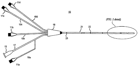
[0032] FIG. I shows integrating a biosensor within a multilumen catheter
assembly. The catheter assembly 10 may include multiple infusion ports 11 a,
11 b,
11 c, I 1 d and one or more electrical connectors 13 at its most proximal end.
A
lumen 15a, 15b, 15c or 15d may connect each infusion port l l a, 11 b, 11 c,
or 11 d,
respectively, to a junction 19. Similarly, the conduit 17 may connect an
electrical
connector 13 to the junction 19, and may terminate at junction 19, or at one
of the
lumens 15a-15d (as shown). Although the particular embodiment shown in FIG. I
is a multilumen catheter having four lumens and one electrical connector,
other
embodiments having other combinations of lumens and connectors are possible
within the scope of the invention, including a single lumen catheter, a
catheter
having multiple electrical connectors, etc. In another embodiment, one of the
lumens and the electrical connector may be reserved for a probe or other
biosensor
mounting device, or one of the lumens may be open at its proximal end and
designated for insertion of the probe or biosensor mounting device. The
details of
the probe and other devices for mounting a biosensor will be further explained
below.
[0033] The junction 19 connects the lumens lla- lld and the conduit 17 to a
narrow elongated tube 21 that forms an intravenous insertion portion of the
catheter
assembly 10. The tube 21 may be typically cylindrical, having a circular or
somewhat oval cross section defining a longitudinal axis extending
therethrough.
The tube 21 may be formed from any material, including synthetic materials
such as
silicone, polyurethane, polyethylene, and the like. Through the junction 19,
each of
CA 02630533 2008-05-20
WO 2007/100796 PCT/US2007/005020
-9-
the lumens lla-I ld extend in separate parallel paths for some distance into
the distal
end of tube 21. One or more support structures 23 within the tube 21 may be
disposed along the length of the catheter to provide rigidity.
[00341 The distal end of the catheter assembly 10 is shown in greater detail
in
FIG. 2. At one or more intermediate locations along the distal end, the tube
21 may
define one or more ports formed through its outer wall. These may include the
intermediate ports 25a, 25b, and 25c, and an end port 25d that may be formed
at the
distal tip of tube 21. Each port 25a-25d may correspond respectively to one of
the
lumens 15a-15d. That is, each lumen may define an independent channel
extending
from one of the infusion ports l la-I ld to one of the tube ports 25a-25d.
[0035] A port 25 exposing an active portion of a biosensor 29 may be referred
to
as a sensing port. A sensing port 25 may perforate an outer wall of catheter
10 to
form a hole that opens into a lumen. In one embodiment, the sensing port 25
opens
into only one lumen. The sensing port 25 as described herein may be generally
oval
or rectangular in shape, having a length between about 5.0 mm and about 15.0
mm,
and having a maximum width between about 1.0 mm and about 3.0 mm. The
sensing port 25 may be formed in a catheter, for example, by skiving an area
of the
outer wall of tube 21.
[0036] In one embodiment, one or more sensing ports 25 may be located on the
tube 21 proximally to an end port. In another embodiment, a catheter may be
configured with a single sensing port that is proximal to all other ports,
such as port
25a of FIG. 3. In operation within a venous location, the most proximal
sensing port
of the catheter may lie advantageously upstream of the distal ports, so that
any
CA 02630533 2008-05-20
WO 2007/100796 PCT/US2007/005020
-10-
infusion fluids introduced into the bloodstream through a distal port are
prevented
from affecting biosensor measurements.
[0037] The embodiment of FIG. 3 shows a magnified transparent side view of an
intermediate portion of the distal end of the tube 21 in the vicinity of the
sensing
port 25. In the orientation shown, a lumen 15 extends longitudinally within
tube 21
along the bottom portion of the catheter. A biosensor 29 may be positioned
within
the lumen 15 such that its active portion 31, i.e. the portion containing an
enzyme
electrode, may be exposed to space outside the tube 21 through the port 25. At
the
proximal end of the biosensor 29, the electrical wires 33 coupled to the
enzyme
electrode extend from the biosensor 29 through the lumen 15. The electrical
wires
33 are coupled to, or provide, a conductive path through the lumen 15 and the
conduit 17 that may terminate at the electrical connector 13. In one
embodiment,
the electrical wires 33 may be bonded to the substrate of the biosensor 29 at
a
proximal location on the substrate having an area of about 0.15 square. inches
to
about 0.30 square inches. A suitable adhesive such as Loctite 401 may be used
to
affect this bond.
[003$] As shown in FIG. 3, the biosensor 29 may be connected or mounted inside
a length of support tubing 35. The support tubing 35 may be formed of material
of a
desired rigidity similar to the tube 21. The support tubing 35 may be inserted
within
the lumen 15 such that it spans the sensing port and positions the active
portion 31
of the biosensor 29 facing radially outward and displaced from an inner wall
of the
catheter.
[0039] FIG. 4 is a bottom view of the intermediate portion of the tube 21 of
FIG.
3. FIG. 5 shows a cross sectional view of the tube 21 corresponding to section
A-A.
CA 02630533 2008-05-20
WO 2007/100796 PCT/US2007/005020
-11-
As shown in these figures, the support tubing 35 may be positioned
concentrically
within the lumen 15, and the biosensor 29 may be mounted concentrically within
the
support tubing 35. With such an arrangement, the biosensor 29 may be
effectively
shielded from damage when the biosensor is positioned within the catheter,
during
which time frictional forces may act between the inner diameter of the lumen
15 and
the outer diameter of the support tubing 35, but not on the active portion 31
of the
biosensor due to its displacement from the inner diameter of the lumen 15.
[0040] After positioning the support tubing 35, to ensure that the biosensor
29
remains firmly anchored at the sensing port 25, an adhesive agent (not shown)
such
as an epoxy may be applied at locations 37 and 39, which correspond to the
proximal and distal ends, respectively, of the sensing port 25. The adhesive
may
bond the biosensor 29 to support the tubing 35, and also bond support tubing
35 to
the inner walls of the lumen 15. The adhesive may also beneficially seal the
lumen
to prevent fluid or other material from entering the catheter interior through
the
15 sensing port 25. Thus, a completed catheter assembly 10 may provide an
integral
biosensor that is protectively centrally oriented within a lumen and exposed
through
a sealed sensing port in the outer catheter wall.
[0041] FIGS. 6, 7 and 8 illustrate another embodiment of a catheter assembly
with
integral biosensor according to an embodiment of the invention. These figures
show
alternative magnified side, bottom and cross sectional views, respectively, of
the
intermediate portion of the tube 21 of FIG. 3. As in a previous embodiment, a
sensing port 25 may be formed at an intermediate location along a distal end
of a
catheter tube 21, and may be located proximally with respect to all other
ports
formed in the outer wall of the tube 21. In this embodiment, as shown in FIG.
6, a
CA 02630533 2008-05-20
WO 2007/100796 PCT/US2007/005020
-12-
biosensor 29 may be mounted directly to an inner diameter of the lumen 15 at
its
furthest radial distance from the longitudinal axis of the tube 21 (or
equivalently, to
an inner diameter of the outer wall of the tube 21) such that its active
portion 31 is
exposed through the sensing port 25 and displaced radially inwardly from the
outer
diameter of the tube 21. In other words, in this configuration the active
portion 31
of biosensor 29 may form an outer diameter of the catheter at the location of
the
sensing port 25 that is inwardly displaced a small distance less than the
outer
diameter of adjacent areas of the outer wall of the tube 21.
[0042] Prior to positioning of the biosensor 29, it may be mounted to a
support
member 43, which may be a tube or rod having a cylindrical or trapezoidal
cross
section. The support member 43 may then be inserted through the lumen 15 until
the active portion 31 of the biosensor 29 is properly exposed through the
sensing
port 25. As shown in the cross sectional view of FIG. 8, the support member 43
may abut an inner radial wall of the lumen 15 and place the biosensor 29 in a
position facing the opposite outer wall.
[0043] One advantage to embodiment of FIG. 6 is that it allows for simplified
sealing of the sensing port. By mounting the biosensor 29 flush against the
inner
wall of the lumen 15, a circumferential interface 41 is created at the border
of the
sensing port 25 and the outwardly facing surface of the biosensor 29. The
interface
41 may be sealed with a single bead of an appropriate sealant or bonding agent
to
prevent fluid and foreign materials from entering the lumen 15 through the
sensing
port 25. Another advantage of this embodiment is that placement of the
biosensor
directly adjacent to the outer diameter of the catheter may provide better
exposure to
blood flow.
CA 02630533 2008-05-20
WO 2007/100796 PCT/US2007/005020
-13-
[0044] FIGS. 9, 10 and 11 illustrate an embodiment of a catheter assembly
according to an embodiment of the invention which allows an integral biosensor
to
be flushed with an IV solution, whether the catheter is withdrawn or in situ.
These
figures show alternative magnified side, bottom and cross sectional views,
respectively, of the intermediate portion of tube 21 of FIG. 3. As in previous
embodiments, a sensing port 25 may be formed at an intermediate location along
a
distal end of a catheter tube 21, and may lie most proximally with respect to
any
other infusion port formed in an outer wall of the tube 21. As in the
embodiment of
FIG. 3, a support tubing 35 may be included to mount and position a biosensor
29 so
that its active portion 31 is exposed through the sensing port 25 and
displaced from
the inner diameter of the lumen 15. In this embodiment, the support tubing 35
may
be positioned such that the proximal end 45 of the biosensor 29 is located
distally
with respect to the proximal end 37 of the sensing port 25. This configuration
allows for a flow 47 of an IV solution (such as saline or other cleansing
solution) to
be injected into the lumen 15 (e.g. through an infusion port 1Ia) and ejected
from
the catheter through the sensing port 25. In this manner, the cleansing fluid
may
advantageously flush the active portion 31 of the biosensor 29 and thereby
remove
clotted blood or other materials from the surface of the biosensor that may
adversely
affect its operation. A sealant may be applied at the distal end 39 of the
sensing port
25 to bond the biosensor 29 to support the tubing 35, and to seal the distal
portion of
the lumen 15.
[0045] FIGS. 12-14 illustrate another embodiment of a catheter with integral
biosensor according to an embodiment of the invention. These figures show an
alternative set of magnified side, bottom and cross sectional views,
respectively, of
CA 02630533 2008-05-20
WO 2007/100796 PCT/US2007/005020
-14-
the intermediate portion of the tube 21 of FIG; 3. Using this arrangement, the
biosensor 29 may be exposed to a flow of blood by mounting it directly to an
outer
wall of the catheter without having to form a sensing port through the tube
21.
[00461 To biosensor may not increase the overall outer diameter of the
catheter
because the biosensor 29 is mounted in a recessed area of the tube 21. The
side
view of FIG. 12 shows one example of a generally rectangular recessed area 49
formed on the outer wall of the catheter between proximal and distal ends of
the tube
21. The recessed area 49 may be located proximally with respect to one or more
intermediate ports formed in the outer wall of the tube 21, and may be the
most
proximal of all such ports. A lumen 15 may extend longitudinally through tube
41
and form an inner wall bordering the recessed area. In one embodiment, the
recessed area 49 may be formed in a manufactured catheter by heating and
pressing
a portion of the tube 21. In another embodiment, the recessed area 49 may be
formed during catheter fabrication by molding.
[0047j A mounting port 51 may be formed through a proximal, substantially
transverse wall of the recessed area 49, as indicated. A biosensor 29, such as
a thin
flex circuit amperometric biosensor, may extend through the mounting port 51
along
the surface of the recessed area 49, such that a portion of the proximal end
37 of the
biosensor 29 remains inside the lumen 15. The portion of the proximal end 37
remaining within the lumen 15 may include at least an area sufficient for
coupling
the wires 33 to the biosensor 29. The distal end 55 of the biosensor 29 may
abut a
substantially transverse distal wall of the recessed area 49. An adhesive or
sealant
53 may then complete the assembly. The sealant 53 may be applied to the area
in
and around the mounting port 51 to provide a seal preventing passage of fluid
CA 02630533 2008-05-20
WO 2007/100796 PCT/US2007/005020
- 15-
therethrough. The sealant 53 may also be applied to the edges and bottom
surface of
the biosensor 29 to securely bond it to the recessed area 49.
[0048] In an alternative embodiment indicated in FIG. 13, a second mounting
port
57 may be formed in the transverse distal wall of the recessed area 49. In
this
option, the distal end of the biosensor 29, indicated by dashed portion 55a,
extends
into the lumen 15 through the second mounting port 57. The sealant 53 may then
be
applied to the second mounting port area to seal the lumen 15 at the location
of the
mounting port 57. This arrangement may provide a stronger and more reliable
means for fastening the biosensor to the catheter.
[0049] As shown in FIG. 14, the mounting arrangement for either option (i.e.
one
or two mounting ports) allows the biosensor to be installed on the outer wal]
of the
catheter without increasing the area of the catheter cross section. This
installation
further protects the biosensor from frictional forces by placing the outermost
surface
of the biosensor at a radial distance from the axis of the tube 21 that is
less than the
radius of the tube's outer diameter.
[0050] Another embodiment of a catheter with integral biosensor is depicted in
FIGS. 15-17. As in previous embodiments, a sensing port 25 may be formed at an
intermediate location along a distal end of a catheter tube 21, which location
may be
proximal to one or more fluid ejection ports. In this embodiment, a biosensor
having an active portion 31 is integrated with a probe 61. The probe 61 may be
a
rod or tubing formed from a flexible substance such as vinyl, urethane, nylon
or
other suitable material. In one embodiment, the probe 61 may be formed from a
material that may be bonded to a flex circuit substrate. The wires 33 for
energizing
CA 02630533 2008-05-20
WO 2007/100796 PCT/US2007/005020
-16-
and sensing of the integral biosensor may extend from the proximal end of the
probe
61 and terminated at a connector 13.
[0051] The flexibility of probe 61 allows it to be inserted into a lumen 15 at
a
proximal location, such as through an infusion port 11a, and moved through
lumen
until it reaches a sensing port 25. A plug 59 may be inserted in the distal
end of
lumen 15, as shown, to stop the progress of the probe 61 so that the active
portion 31
may be accurately positioned at the sensing port 25. A keying configuration 63
may
be formed in the inner wall of the lumen 15 to ensure proper orientation of
the probe
61 within the lumen 15 so that the active portion 31 faces outward through the
sensing port 25 for optimal exposure to blood flow. Thus, during installation,
the
key 63 guides the probe through the lumen 15 in proper orientation to exposes
the
active portion 31 through the sensing port 25 when a distal end of the probe
61
reaches the plug 59.
[0052] As indicated in FIGS. 15 - 17, the active portion 31 may be protected
from
frictional forces by mounting it concentrically with respect to the probe 61
so that
during installation, only the outer diameter of the probe 61 comes into
contact with
the inner wall of the lumen 15. After inserting the probe 61, the assembly may
be
completed by sealing the proximal end 37 and distal end 39 of the sensing port
25
with an appropriate sealant. In one embodiment, where the probe 61 forms a
tight
compression fit against the inner wall of the lumen 15, a sealant may not be
required
at one or both ends 37 and 39.
[0053] The invention has been disclosed in an illustrative manner.
Accordingly,
the terminology employed throughout should be read in an exemplary rather than
a
limitinp- manner. Although minor modifications of the invention will occur to
those
CA 02630533 2008-05-20
WO 2007/100796 PCT/US2007/005020
-17-
well versed in the art, it shall be understood that what is intended to be
circumscribed within the scope of the patent warranted hereon are all such
embodiments that reasonably fall within the scope of the advancement to the
art
hereby contributed, and that that scope shall not be restricted, except in
light of the
appended claims and their equivalents.