Note: Descriptions are shown in the official language in which they were submitted.
CA 02631101 2008-05-26
Ophthalmologic Measuring System and Method for Determining the Biometric
Data of an Eye
The invention at hand concerns an ophthalmologic measuring system for
determining the
biometric data of an eye.
A number of known methods and measuring instruments exist for determining the
biometric data of an eye. For example, it is necessary to determine various
biometric
parameters of the eye prior to an operation to replace the lens of the eye if
there is a
clouding of the lens (cataract). To guarantee the optimal post-procedural
visual acuity,
these parameters must be determined with great accuracy. The appropriate
replacement
lens is selected based upon established formulae and calculation methods.
The most important parameters to be determined are, among others, the axial
length
(distance to the retina), the curvature and power of refraction of the cornea
as well as the
length of the anterior chamber (distance to the eye lens). These measurements
can be
determined successively using various ophthalmologic devices or with the help
of
specially optimized biometric measuring systems.
For the determination of these parameters primarily ultrasound measuring
devices and
optical measuring devices based upon short coherence-light procedures
prevailed.
With the ultrasound devices there are two different designs that function
either based
upon the "A-scan" principle or upon the "B-scan" principle. While the A-scan
provides
only one measurement in the axial direction, there is an additional
measurement in
transverse direction with the B-scan. The ultrasound procedure basically
requires direct
contact with the eye.
1
CA 02631101 2008-05-26
WO 2007/079835 PCT/EP2006/011537
In this context a device for examining the eye, especially the human eye, is
described in
DE 42 35079 C2 that basically has the shape of a truncated cone in a shape
matched to
the eye which contains a probe for the evaluation of acoustic (ultrasound)
signals. The
probe is affixed at an oblique angle to the central axis of the holder and is
suitable for
transmitting as well as for receiving pulsed signals.
The specific disadvantages of the determination of the biometric data of an
eye using
ultrasound devices are, on one hand, the lesser accuracy and, on the other
hand, the
requirement of direct contact with the eye. This way the measurements could be
distorted
through denting of the eyeball. These disadvantages can be reduced through the
use of
the immersion technique where ultrasound waves are directed at the eye through
a funnel
filled with water and placed over the eye, but the major disadvantages of this
measuring
method remain.
These lie, on one hand, in the necessity of direct contact with the eye which
always
carries the risk of transmission of infections and, on the other hand, it is
necessary to
anesthetize the eye for the determination of the data. For the correct
selection of the
replacement lens it must be ascertained that the visual axis of the eye is
appropriately
aligned when determining the biometric data. For this purpose special devices
must be
provided for the ultrasound equipment since the alignment of the visual axis
does not
happen automatically.
Analogous to the ultrasound devices, where images of the structural
transitions can be
reconstructed based upon the acoustic signals, optical images of the
structural transitions
are depicted as two-dimensional depth tomograms. In this regard the OCT
procedure
(OCT = optical coherence tomography) has prevailed as a short coherence-light
procedure where temporal incoherent light is used with the help of an
interferometer for
measuring the distance of reflective and dispersive materials.
2
CA 02631101 2008-05-26
WO 2007/079835 PCT/EP2006/011537
The underlying principle of the OCT procedure is based upon white light
interferometry
and compares the travel time of a signal using an interferometer (in most
cases a
Michelson interferometer). The arm with a known optical length (= reference
arm) is
used as a reference arm for the measuring arm. The interference of the signals
from both
arms yields a pattern from which one can determine the relative optical travel
distance
within an A-scan (individual depth signal). In the one-dimensional scanning
grid
procedure the beam is guided transversally in one or two directions, analogous
to the
ultrasound technique, allowing the recording of a plane B-scan or a three-
dimensional
tomogram (C-scan). This way, the amplitude data of the individual A-scans are
depicted
as logarithmized gray scale or phantom color data. For example, a measuring
time of one
second will be needed for a B-scan consisting of 100 individual A-scans.
The measuring resolution of the OCT procedure is determined by the coherency
length of
the light source used and is typically about 15 m. Due to its special
suitability for
examining optically transparent media the procedure is widespread in the field
of
ophthalmology.
Two different kinds of OCT procedures have prevailed among those used in the
field of
ophthalmology. With the first kind, the reference arm is modified in length to
determine
the measured data and continually measure the intensity of the interference
without
consideration given to the spectrum. This procedure is called "Time Domain"
procedure.
With the other procedure, called "Frequency Domain" procedure, however, the
spectrum
is considered in determining the measurements and the interference of the
individual
spectral components are recorded. Therefore, we refer to a signal within the
time domain,
on one hand, and to a signal within the frequency domain on the other.
3
CA 02631101 2008-05-26
WO 2007/079835 PCT/EP2006/011537
The advantage of the frequency domain lies in the simple and quick
simultaneous
measuring where complete information about the depth can be determined without
requiring movable parts. This increases both the stability and the speed.
The big technological advantage of the OCT is the decoupling of the depth
resolution
from the transversal resolution. In contrast to microscopy, this allows the
recording of the
three-dimensional structure of the item to be examined. The purely reflective
and,
therefore, contact-free measuring makes it possible to generate microscopic
images of
live tissue (in vivo).
Due to the high selectivity of the method very weak signals (less than a
nanowatt) can be
detected and identified to a certain depth. Therefore, the procedure is
suitable for
examining optically sensitive tissue. The use of the OCT procedures is limited
by the
depth penetration of the electromagnetic radiation into the subject to be
examined, which
is dependent upon the wavelength, as well as by the resolution, which depends
upon the
bandwidth.
With the currently customary biometric measuring devices, the measured data
are
processed in the device and suggestions are made as to the exchange lenses to
be used.
These depend upon the formulae used in the calculation and the type of
available lenses
(depending on the manufacturer). It is possible, or necessary, to let the post-
operative
results enter into the calculation formulae via the optimization of constants
in order to
allow for individual influences during the surgery as well as the measuring
technique
actually used. All measured values, data, and formulae are administered,
analyzed, and
saved in data banks and software programs. In part, these solutions are
integrated in
networks and various additional applications can be linked to them.
4
CA 02631101 2008-05-26
WO 2007/079835 PCT/EP2006/011537
With the optical measuring devices based upon short coherence-light
procedures, the
interferometric principle based upon the dual-beam is used. This procedure is
contact-free
and works with the greatest accuracy currently possible. Solutions based upon
this
measuring principle have been described as examples in DE 198 12 297 C2, DE
103 60
570 Al and WO 2004/071286 Al.
The disadvantages pointed out with the ultrasound devices can be avoided with
the
optical procedure. Special mention should be made of the high degree of
accuracy
(interferometer) and patient comfort. However, the disadvantage here is the
fact that 10 to
20 percent of patients cannot be measured because, for example, the scattering
of dense
cataracts attenuates the measuring signal too much and the laser output cannot
be
increased at will due to the limits to be respected around the eye. In these
cases it is also
possible that the patient is no longer able to see the focal point and
measuring becomes
difficult.
Certain pathological changes can cause individual problems with determining
the
measuring data with both procedures. As a result of these negative influences
upon
obtaining the measurements there is an increased risk of making the wrong
decision when
selecting a suitable exchange lens.
The invention at hand is based upon the task of developing a solution which
avoids the
disadvantages of the current state of technology and makes it possible to
determine
biometric measuring data of an eye even under difficult conditions with great
reliability
and accuracy.
According to the invention, the task is accomplished through the
characteristics of the
independent claims. Preferred developments and modifications are the subject
of the
independent claims.
CA 02631101 2008-05-26
WO 2007/079835 PCT/EP2006/011537
The present technical solution is intended to determine the biometric data of
an eye
within the scope of the pre-operative determination of the exchange lens, or
additional
lens, or refractive procedures, where measuring data can be determined even
under
difficult circumstances with great reliability and accuracy. In addition, the
proposed
solution allows the determination of the position of the anterior chamber and
lens of the
eye, the shape of the front of the cornea of the human eye (keratometric
measurement), as
well as the thickness of the cornea (pachymetric measurement).
The invention is described below in more detail through embodiments. The
following
figures will show:
Figure #1: an ophthalmologic measuring system as a coupling of an optical
measuring device based upon ultrasound and one based upon a
short coherence-light process and
Figure #2: an ophthalmologic measuring system where an optical measuring
device based upon ultrasound and one based on short coherence-
light process are integrated.
The ophthalmologic measuring system for determining the biometric data of an
eye, according to the invention, within the scope of the pre-operative
determination of the
exchange lens, or additional lens, or refractive procedure, consists of a
combination of a
measuring device that is based upon ultrasound plus an optical measuring
device and an
evaluating device. The evaluation unit uses measuring data from the optical
and/or the
ultrasound measuring device to determine the biometric data of an eye.
The optical measuring device used here can be a Scheimpflug camera or an
optical
measuring device based upon short coherence-light procedures such as, for
example, an
IOLMaster (Carl Zeiss Meditec AG).
6
CA 02631101 2008-05-26
WO 2007/079835 PCT/EP2006/011537
While a Scheimpflug camera can be used to generate 2-dimensional images of the
front
parts of the eye and to measure distances in this area of the eye, the
IOLMaster is used
for the exact determination of the axial length, the anterior chamber of the
eye, and the
power of refraction of the cornea.
In an advantageous technical embodiment the measuring data obtained by the
evaluating
unit of both measuring devices are used for mutual calibration where
preferably sample
eyes are used. The data transmission required for this is accomplished
preferably via a
data link that connects the evaluating units of both measuring devices.
In another technical embodiment both measuring devices are integrated into one
device
which will make the ophthalmologic measuring device more compact and easier to
handle. This offers the additional advantage that certain systems components,
such as PC,
monitor, as well as input and output units can be used jointly.
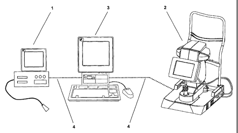
The combination of a measuring device 1, based upon ultrasound shown in Figure
#1
(acoustic image generating procedure to depict the front and/or back areas of
the eye),
and an optical measuring device 2 based upon short coherence-light procedures
(optical
image generating procedure to depict the front and/or back areas of the eye),
represents a
particularly advantageous ophthalmologic measuring system where, preferably,
an
IOLMaster by the Carl Zeiss Meditec AG company is used as measuring device 2.
This
ophthalmologic measuring system allows for a comprehensive examination or the
clarification of unanticipated or unclear results. Preferably the evaluating
unit 3 uses the
measured data to provide and evaluate 2-dimensional or 3-dimensional images of
the
examined eye. The transmission of the measuring data required for this is
handled via
the data transmission line 4 which connects the evaluating unit 3 with the two
measuring
devices 1 and 2.
7
CA 02631101 2008-05-26
WO 2007/079835 PCT/EP2006/011537
By contrast, Figure #2 shows an ophthalmologic measuring system where an
optical
measuring device based upon ultrasound and one based upon short coherence-
light
procedures are integrated.
The biometric data of an eye determined by the ophthalmologic measuring system
can
be passed on in an advantageous manner within the scope of the pre-operative
determination of the exchange, or additional lens, or refractive procedures,
to post-
procedural devices, such as e.g. surgical microscopes.
With this procedure according to the invention for the pre-operative
determination
of the exchange, or additional lens, or refractive procedures, measuring data
from a
measuring device based upon ultrasound and/or an optical device will be
supplied
to an evaluating unit where they are used by the evaluating unit to determine
the
parameters of the lens to be implanted, using known formulae and calculation
methods.
The biometric data generated by the evaluating unit, based upon measuring data
determined by both measuring devices, will be compared with each other. This
offers the
advantage that possible erroneous measurements can be detected and corrected.
In case
there are significant differences between the measured data of the two
measuring devices,
it always makes sense to produce a 2-dimensional image of the eye in order to
be able to
find the cause of the faulty measuring results. Possible reasons for such
differences
could be retinal detachment or staphyloma. Also, in pseudophakic eyes,
artefacts could
appear in the various measuring procedures that could lead to faulty measuring
results if
interpreted incorrectly.
Besides, it is an advantage for increasing the reliability and accuracy to use
the
measuring data of both measuring devices for mutual calibration preferably
using sample
eyes. The measuring data obtained by both measuring devices can also be used
to
optimize the lens constants.
8
CA 02631101 2008-05-26
WO 2007/079835 PCT/EP2006/011537
In yet another embodiment of the procedure, the measuring data of two separate
measuring devices are further processed by the evaluating unit of the
respective
measuring device and the results are then handed off to the other measuring
device via a
data link.
With the solution according to the invention, an ophthalmologic measuring
system and a
process to determine the biometric data of an eye is being provided which can
determine
measuring data with great reliability and accuracy, even under difficult
circumstances.
The combination makes it possible to compensate for the given specific
disadvantages of
the various measuring procedures, at least in part, without losing their
advantages. The
very high accuracy of the optical measuring procedure with the corresponding
contact-
free determination of measuring data is preserved as well as the option to use
ultrasound-
based measuring procedures under difficult circumstances, such as a dense
cataract. A
comparison of the measured data from the two systems can further enhance the
reliability
and accuracy of the measuring data.
The combination of different measuring procedures allows a complete
examination and
assessment of the patient at one single measuring position so that the patient
neither
has to be moved nor must additional measuring appointments be scheduled on
another
day.
Determining a multitude of different biometric data of an eye allows for an
improved
characterization of the patient's eyesight and makes the selection of
replacement or
refractive additional lenses more reliable.
9