Note: Descriptions are shown in the official language in which they were submitted.
CA 02631752 2008-06-02
WO 2007/062128 PCT/US2006/045217
METHODS AND APPARATUS FOR
ATRIOVENTRICULAR VALVE REPAIR
BACKGROUND OF THE INVENTION
[0001] This invention relates generally to medical methods and
apparatus, and more particularly, to methods and apparatus for the
endovascular or
minimally invasive surgical repair of atrioventricular valves of the heart,
including the
mitral valve and the tricuspid valve.
[0002] The heart includes four valves that direct blood through the
two sides of the heart. The mitral valve lies between the left atrium and the
left
ventricle and controls the flow of blood into the left side of the heart. The
valve
includes two leaflets, an anterior leaflet and a posterior leaflet, that close
during
systole. The leaflets are passive in that they open and close in response to
pressure
induced to the leaflets by the pumping of the heart. More specifically, during
a
normal cycle of heart contraction (systole), the mitral valve functions as a
check valve
to prevent the flow of oxygenated blood back into the left atrium. In this
manner,
oxygenated blood is pumped into the aorta through the aortic valve.
[0003] Occasionally, the mitral valve is formed abnormally through a
congenital condition. More often, however, the mitral valve degenerates with
age.
Among the problems that can develop is mitral valve regurgitation in which the
mitral
valve leaflets become unable to close properly during systole, thus enabling
lealcage
to flow through the mitral valve during systole. Over time, regurgitation of
the mitral
valve can adversely affect cardiac function and may compromise a patient's
quality of
life and/or life-span.
[0004] Mitral valve regurgitation can result from a number of
different mechanical defects in the mitral valve. For example, the valve
leaflets, the
valve chordae which connect the leaflets to the papillary muscles, or the
papillary
muscles themselves may become damaged or otherwise dysfunctional. Moreover,
the
-1-
CA 02631752 2008-06-02
WO 2007/062128 PCT/US2006/045217
valve annulus may become damaged or weakened and may limit the ability of the
mitral valve to close adequately during systole.
[0005] Known treatments for mitral valve regurgitation commonly
rely on valve replacement or annuloplasty, or strengthening of the mitral
valve
through surgical repairs and/or implanting a mechanical structure within the
mitral
valve. For example, the most prevalent and widely accepted known techniques to
correct mitral valve regurgitation, repair the mitral valve via open heart
surgery.
During such an invasive surgical procedure, it is known to suture adjacent
segments
of the opposed valve leaflets together in a procedure known as a "bow-tie" or
"edge-
to-edge" surgical technique. Although each of the afore-mentioned treatments
can be
effective, generally known treatments rely on open heart surgery wherein the
patient's
chest is opened and the patient's heart is stopped while the patient is place
on a
cardiopulmonary bypass. The need to open the patient's chest and to place the
patient
on a cardiopulmonary bypass creates inherent risks that may be traumatic to
the
patient.
[0006] Percutaneously treatments are less invasive than the
treatments mentioned above, but such treatments may be less effective and more
difficult to effect repair because of the limited amount of space in and
around the
mitral valve in which to maneuver a repair device or devices. For example,
U.S.
Patent No. 6,875,224 to Grimes describes a percutaneous mitral valve repair
method
in which the opposed leaflets are each immobilized to enable the two leaflets
to be
fastened together. Furthermore, U.S. Patent No. 6,6290,534 to St. Goar et al.
describes a plurality of embodiments for use in endovascular repair of cardiac
valves
in which, in each embodiment, both leaflets are grasped and held firmly in
position
prior to permanent treatment. However, grasping both leaflets while the
patient's
heart is beating may be a time-consuming and laborious task that demands a
coordinated effort on the part of the surgical team. Moreover, to facilitate
grasping
both leaflets percutaneously may require that the patient's heart be
temporarily
stopped or slowed by drugs or other techniques. Slowing and/or stopping the
patient's heart during surgery may increase the risks to the patient.
-2-
CA 02631752 2008-06-02
WO 2007/062128 PCT/US2006/045217
BRIEF DESCRIPTION OF THE INVENTION
[0007] In one aspect, a method of repairing an atrioventricular valve
in a patient is provided. The method comprises accessing the patient's
atrioventricular valve percutaneously, securing a fastening mechanism to a
valve
leaflet, and coupling the valve leaflet, while the patient's heart remains
beating, to at
least one of a ventricular wall adjacent the atrioventricular valve, a
papillary muscle,
at least one valve chordae, and a valve annulus to facilitate reducing leakage
through
the valve.
[0008] In another aspect, a method of repairing a mitral valve in the
heart of a patient is provided. The method coinprises accessing the patient's
mitral
valve percutaneously, securing a first end of a fastening mechanism to a valve
leaflet
of the mitral valve, and coupling a second end of the fastening mechanism to a
cardiac
structure other than a mitral valve leaflet to facilitate reducing lealcage
through the
patient's mitral valve during ventricular systole.
[0009] In a further aspect, a method of enhancing operation of a
patient's heart valve is provided. The method comprises inserting a guide
catheter
along the venous system of the patient to approach the mitral valve, guiding a
fastening mechanism towards one of a mitral valve and a tricuspid valve within
the
patient's heart, and securing a first end of the fastening mechanism to one of
the
mitral valve and the tricuspid valve using one of fusing, gluing, stapling,
clipping,
riveting, anchoring, and suturing. The method also comprises securing a second
end
of the fastening mechanism to a cardiac structure other than a valve leaflet
to facilitate
enhancing operation of the valve during ventricular systole.
[0010] In an additional aspect, a medical kit for use in repairing a
mitral valve is provided. The kit includes a guide catheter and a fastening
mechanism. The guide catheter is configured for insertion along the venous
system of
the patient to approach the mitral valve. The fastening mechanism is
positionable
percutaneously within the patient using the guide catheter. The fastening
mechanism
includes a first end and an opposite second end. The first end is configured
to couple
-3-
CA 02631752 2008-06-02
WO 2007/062128 PCT/US2006/045217
to the mitral valve using one of fusing, gluing, stapling, clipping, riveting,
anchoring,
and suturing. The second end is configured to only couple to a cardiac
structure other
than a valve leaflet to facilitate enhancing operation of the valve during
ventricular
systole.
BRIEF DESCRIPTION OF THE DRAWINGS
[0011] Figure 1 is a cross-sectional view of the left and right
ventricles of a human heart in diastole;
[0012] Figure 2 is an another cross-sectional view of the heart shown
in Figure 1 during systole;
[0013] Figure 3 is an exemplary schematic illustration of a fastening
mechanism that may be used to facilitate repair of a cardiac valve within the
heart
shown in Figures 1 and 2;
[0014] Figure 4 is an enlarged view of a portion of the fastening
mechanism shown in Figure 2 and coupled to a papillary muscle in the heart
shown in
Figures 1 and 2;
[0015] Figure 5 is a schematic view of an alternative embodiment of
a portion of a fastening mechanism that may be used to facilitate repair of a
cardiac
valve within the heart shown in Figures 1 and 2;
[0016] Figure 6 is a schematic view of an another alternative
embodiment of a portion of a fastening mechanism that may be used to
facilitate
repair of a cardiac valve within the heart shown in Figures 1 and 2;
[0017] Figure 7 is a schematic view of a further alternative
embodiment of a portion of a fastening mechanism that may be used to
facilitate
repair of a cardiac valve within the heart shown in Figures 1 and 2; and
[0018] Figure 8 is a flowchart illustrating an exemplary method for
the endovascular repair of a cardiac valve.
-4-
CA 02631752 2008-06-02
WO 2007/062128 PCT/US2006/045217
DETAILED DESCRIPTION OF THE INVENTION
[0019] Figure 1 is a cross-sectional view of the left and right
ventricles 10 and 12, respectively, of a human heart 14 during diastole.
Ventricles 10
and 12 are separated by an interatrial septum 15. Figure 2 is a cross-
sectional view of
heart 14 during systole. The present invention provides methods and apparatus
for the
endovascular repair of cardiac valves, particularly atrioventricular valves
16, which
inhibit baclc-flow of blood from a heart ventricle during contraction
(systole). In
particular, the present invention may be used in repairing, but is not limited
to
repairing, mitral valves 20.
[0020] As used herein, the term "endovascular," refers to
procedure(s) of the present invention that are performed with interventional
tools and
supporting catheters and other equipment introduced to the heart chambers from
the
patient's arterial or venous vasculature remote from the heart. The
interventional tools
and other equipment may be introduced percutaneously, i.e., through an access
sheath,
or may be introduced via a surgical cut down, and then advanced from the
remote
access site through the vasculature until they reach heart 14. As such, the
methods
and apparatus described herein generally do not require penetrations made
directly
through an exterior heart muscle, i.e., myocardium, although there may be some
instances where penetrations will be made interior to the heart, e.g., through
the
interatrial septum to provide for a desired access route. Moreover, as will be
appreciated by one of ordinary skill in the art, the methods and apparatus
described
herein are not limited to use with percutaneous and intravascular techniques,
but
rather the present invention may be used with open surgical procedures as
well.
[0021] The atrioventricular valves 16 are each located at a junction
of the atria and their respective ventricles. The atrioventricular valve 16
extending
between the right atriuin 30 and the right ventricle 12 has three valve
leaflets (cusps)
and is referred to as the tricuspid or right atrioventricular valve 31. The
atrioventricular valve 16 between the left atrium 32 and the left ventricle 10
is a
-5-
CA 02631752 2008-06-02
WO 2007/062128 PCT/US2006/045217
bicuspid valve having only two leaflets or cusps 34 and is generally referred
to as the
mitral valve 20.
[0022] During operation of the heart 14, the valve leaflets 34 open
during diastole when the heart atria fill with blood, allowing the blood to
pass into the
ventricle. During systole, however, the valve leaflets 34 are pushed together
such that
the free edges 36 of the leaflets 34 are closed against each other along a
line of
coaptation to prevent the baclc-flow of blood into the atria. Back flow of
blood or
"regurgitation" through the mitral valve 20 is facilitated to be prevented
when the
leaflets 34 are closed, such that the mitral valve 20 functions as a "check
valve" which
prevents baclc-flow when pressure in the left ventricle 10 is higher than that
in the left
atrium 32,
[0023] The mitral valve leaflets 34 are attached to the surrounding
heart structure along an annular region referred to as the valve annulus 40.
The free
edges 36 of the leaflets 34 are secured to the lower portions of the left
ventricle 10
through tendon-like tissue structures, known as chordae tendineae or chordae
42. The
chordae 42 are attached to the papillary muscles 44 which extend upwardly from
the
lower portions of the left ventricle and interventricular septum 46.
[0024] A number of structural defects in the heart can cause mitral
valve regurgitation. For example, ruptured chordae 42 may cause a valve
leaflet 34 to
prolapse if inadequate tension is induced to the leaflet 34 through the
remaining
unruptured chordae 42. Moreover, and for example, regurgitation may also occur
in
patients suffering from cardiomyopathy, wherein the heart 14 is dilated and
the
increased size prevents the valve leaflet edges 36 from contacting each other
properly,
or in patients who have suffered ischemic heart disease wherein the
functioning of the
papillary muscles 44 may be impaired. Generally during regurgitation the free
edges
36 of the anterior and posterior leaflets 34 do not contact sufficiently along
the line of
coaptation, but rather lealcage may occur through a gap defined between the
leaflets
34.
-6-
CA 02631752 2008-06-02
WO 2007/062128 PCT/US2006/045217
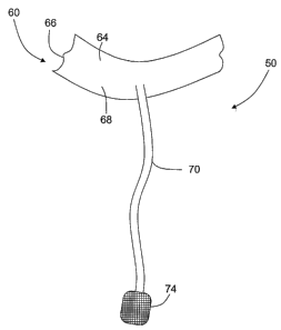
[0025] Figure 3 is an exemplary schematic illustration of a fastening
mechanism 50 that may be used to facilitate repair of an atrioventricular
valve 16
within heart 14 (shown in Figures 1 and 2). Figure 4 is an enlarged view of a
portion
of the fasteiiing mechanism shown in Figure 3 and coupled to a papillary
muscle 44.
Fastening mechanism 50 includes a first attachment end 60 and a second
attachment
end 62. In the exemplary embodiment, first attachment end 60 includes a
generally
deformable clip portion 64 that is sized and shaped to couple to a free edge
36 (shown
in Figures 1 and 2) of a leaflet 34 (shown in Figures 1 and 2). In alternative
embodiments, first attachment end 60 is coupled to leaflet 34 without using
clip
portion 64.
[0026] Overall dimensions of, and material properties used in
fabricating, clip portion 64 are variably selected based on the leaflet 34
being
repaired. In the exemplary embodiment, portion 64 is pinched or crimped
against a
leaflet free edge 36 to facilitate repair of the valve as described in more
detail below.
More specifically, in this embodiment, fastening mechanism 50 is coupled to
valve 16
such that an outer surface of leaflet free edge 36 is grasped without
mechanism 50
penetrating the leaflet tissue. Specifically, in the exemplary embodiment, the
leaflet
free edge 36 is crimped between opposing sides 66 and 68 of portion 64. In
another
embodiment, portion 64 is coupled to a free edge 36 using any suitable means
that
enables portion 64 to remain coupled to leaflet free edge 36, such as, but not
limited
to, with gluing, stapling, suturing, fusing, riveting, external clips, or any
combination
thereof.
[0027] Alternatively, first attachment end 60 may be secured to
leaflet 34 through atraumatic partial, or full penetration, or piercing of
leaflet 34. For
example, first attachtnent end 60 and/or portion 64 may include attachment
prongs
that extend from clip portion 64 and that are configured to pinch, partially
penetrate,
or pierce the leaflet 34. In one alternative embodiment, first attachment end
60 may
be inserted from a first side of the leaflet 34, through leaflet 34, and
outward from an
opposite second side of the leaflet 34. In such an embodiment, in use first
attachment
end 60 is coupled to, and secured against the second side of the leaflet. In
another
-7-
CA 02631752 2008-06-02
WO 2007/062128 PCT/US2006/045217
alternative embodiment, first attachment end is inserted only partially
through the
leaflet 34, and tlius is secured to leaflet tissue intermediate the first and
second sides
of the leaflet.
[0028] In anotlier alternative embodiinent, first attachment end 60 is
attached to leaflet 34 using any suitable means that will enable fastening
mechanism
50 to function as described herein, such as, but not limited to, an adhesive
process, a
riveting process, a suturing process, a stapling process, or any coinbination
thereof.
In a further alternative embodiment, a threaded loclcing member or any other
suitable
mechanical coupling, may be used to secure first attachtnent end 60 to the
leaflet. In
another alternative embodiment, attachment end 60 may be fused directly to the
leaflet 34 using a known fusion process in which laser, RF, microwave or
ultrasonic
energy, for example, is applied at specified coaptation points.
[0029] In the exemplary embodiment, clip portion 64 is fabricated
from a formable material that is coated in a protective cloth-like material.
Clip
portion 64 may be fabricated from any suitable biocompatible material that
enables
fastening mechanism 50 to function as described herein, such as, but not
limited to,
titanium alloys, platinum alloys, stainless steel, or any combination thereof.
In the
exeinplary embodiment, clip portion 64 is coated with a fabric material such
as, but
not limited to; a DACRONO material, a TEFLON material, a GORE-TEX , or any
material or combination thereof that enables clip portion 64 to function as
described
herein. In one embodiment, clip portion 64 is covered by a material that
encourages
tissue in-growth.
[0030] In the exemplary embodiment, a tensioning member 70,
extends from clip portion 64 to second attachment end 62. The overall size,
shape,
and material used in member 70 is variably selected depending on the
application.
For example, in one embodiment, member 70 is fabricated from a mesh material.
The
relative location of member 70 with respect to clip portion 64 is variably
selected
based on the amount of tension to be induced, the desired locations for the
tension to
be induced, and based on the leaflet 34 being repaired.
-8-
CA 02631752 2008-06-02
WO 2007/062128 PCT/US2006/045217
[0031] In the exemplary embodiment, tensioning member 70
includes an attachment pad 74. The overall size, shape, thiclcness, and
material used
in fabricating pad 74, as well as the number and location of pad 74, are
variably
selected based on the intended use of fastening mechanism 50. Alternatively,
fastening mechanism 50 includes more than one tensioning member 70. In another
alternative embodiment, fastening mechanism 50 may include a single tensioning
member 70 that includes a forked or bifurcated end that includes two pads 74.
In yet
another alternative embodiment, fastening tensioning member 70 does not
include pad
74. In a further alternative embodiment, fastening mechanism 50 includes at
least one
tensioning member that is fonned with a looped end that is sized to
circumscribe the
cardiac structure to which it is attached, and is cinchable to facilitate
securing
fastening mechanism 50 to the papillary muscle 44. Tensioning member 70
facilitates
inducing tension to the leaflet 34 being repaired, and pad 74 facilitates
distributing
loading across the papillary muscle 44. Moreover, pad 74 is sized for
placement
along an external surface of papillary muscle 44 when fastening mechanism 50
is
coupled to the papillary muscle 44.
[0032] In the exemplary embodiment, tensioning member 70 and pad
74 are formed integrally together. Alternatively, pad 74 may be securely
coupled to
member 70 using any of a plurality of known coupling means. In the exemplary
embodiment, member 70 is coupled to papillary muscle 44 using a fastener (not
shown) that is inserted at least partially through papillary muscle 44. In one
embodiment, the fastener has a tack-like configuration. In another embodiment,
the
fastener is mechanically coupled to the papillary muscle 44 using, for
example, a
suitable threaded coupling. In a further embodiment, at least one of a pair of
interlocking fasteners is inserted through a pad 74 prior to insertion through
the
papillary muscle 44 and prior to the two fasteners being interlocked. In
another
embodiment, pad 74 is coupled in position against the papillary muscle 44 by a
cinch-
type fastener that circumscribes the papillary muscle 44 when securely
cinched. In
another alternative embodiment, pad 74 is coupled directly to the papillary
muscle 44
using any suitable means that will enable fastening mechanism 50 to function
as
-9-
CA 02631752 2008-06-02
WO 2007/062128 PCT/US2006/045217
described herein, such as, but not limited to, an adhesive process, a riveting
process, a
suturing process, a coil or corkscrew device, a stapling process, external
clips, or any
combination thereof. In a further alternative embodiment, a threaded locking
member
and a self-locking or spin-lock ratcheting fastener may be used to secure
member 70
to the papillary muscle 44. In yet a further alternative embodiment, pad 74,
and/or
tensioning member 70 is coupled to the papillary muscle 44 using a flat ribbon
that
has been heat-set in the shape of double loops.
[0033] Pad 74 and member 70 may be fabricated from any material
that enables pad 74 and member 70 to function as described herein. For
example, pad
74 and member 70 may be fabricated from, but are not limited to being
fabricated
from, a DACRON material, a TEFLON material, a GORE-TEX , or any material
or combination. In addition, depending on the application, pad 74 and member
70
may be fabricated from, but are not limited to being fabricated from a
superelastic
material or a shaped memory alloy (SMA) material, such as, but not limited, to
Nitinol , stainless steel, plastic, or any of several known shaped memory
alloys
(SMA) that have properties that develop a shaped memory effect (SME). In one
embodiment, pad 74 is fabricated from a material that encourages tissue in-
growth.
[0034] During use, to repair a mitral valve 20 using fastening
mechanism 50, first attachment end 60 is coupled securely to mitral valve 20
and
second attachment end 62 is coupled to a cardiac structure, such as the
papillary
muscle 44. Alternatively, second attachment end 62 may be coupled to any
cardiac
structure other than a mitral valve leaflet 34 such as, but not limited to, a
ventricular
wall 46 adjacent the atrioventricular valve 30, a valve chordae 42, either
intact or
ruptured, a valve annulus 36, an interatrial septum 15 or any combination
thereof. In
the exemplary embodiment, second attachment end 62 is coupled to the papillary
muscle 44. More specifically, when end 62 is firmly secured to the papillary
muscle
44, pad 74 is retained tightly against the exterior surface of the papillary
muscle 44.
As such, loading induced to the papillary muscle from fastening mechanism 50
is
distributed across pad 74.
-10-
CA 02631752 2008-06-02
WO 2007/062128 PCT/US2006/045217
[0035] In the exemplary embodiment, overall dimensions and
material properties of member 70 are variably selected to facilitate inducing
a desired
tension to leaflet 34 and to facilitate improving the ability of the
atrioventricular valve
16 to close against the elevated pressures within the ventricle during
systole. More
specifically, member 70 is variably selected to facilitate modifying operation
of the
leaflet 34 such that the free ends 36 of the opposed leaflets 34 again contact
each
other during systole along the line of coaptation to prevent the back-flow or
regurgitation of blood through the mitral valve 20 into the atria.
[0036] Figure 5 is a schematic view of an alternative embodiinent of
a portion of a fastening mechanism 100 that may be used to facilitate repair
of a
cardiac valve 16 (shown in Figures 1 and 2). Fastening inechanism 100 is
substantially similar to fastening mechanism 50 (shown in Figures 3 and 4)
and,
components of fastening mechanism 100 that are identical to components of
fastening
mechanism 50 are identified in Figure 5 using the same reference numerals used
in
Figures 3 and 4. Accordingly, fastening mechanism 100 includes first
attachment end
60, second attachment end 62 (shown in Figures 3 and 4), and at least one
tensioning
member 110 extending therebetween. In the exemplary embodiment, tensioning
member 110 includes an anchor member 112. It should be noted that althougli
attachment end 60 is illustrated, the anchor member 112 may also be included
at
attachment end 62 and/or end 60, or at any suitable location between ends 60
and 62
depending on the application.
[0037] Tensioning member 110 is substantially similar to tensioning
member 70 and as such, facilitates inducing tension to the leaflet 34 (shown
in
Figures 1 and 2) being repaired. In the exemplary embodiment, tensioning
member
110 and anchor member 112 are formed integrally together. Alternatively,
anchor
member 112 may be securely coupled to tensioning member 110 using any of a
plurality of known coupling means. In the exemplary embodiment, member 110 is
coupled to leaflet 34 using anchor member 112, or any other cardiac structure
other
than a mitral valve leaflet 34, such as, but not limited to, a ventricular
wall 46 (shown
in Figures 1 and 2) adjacent the atrioventricular valve 30 (shown in Figures 1
and 2),
-11-
CA 02631752 2008-06-02
WO 2007/062128 PCT/US2006/045217
a valve chordae 42 (shown in Figures 1 and 2), either intact or ruptured, a
valve
annulus 36 (shown in Figures 1 and 2), an interatrial septum 15 (shown in
Figures 1
and 2), a papillary muscle 44 (shown in Figures 1, 2, and 4), or any
combination
thereof.
[0038] In the exeinplary embodiment, anchor member 112 has a
distal end 114 that is pointed and is self-piercing that facilitates
transmural attachment
to a ventricular wall. Accordingly, the anchor member distal end 114 may be
fabricated of any material having sufficient rigidity to pierce, and/or at
least partially
penetrate, through a portion of the cardiac component to which it is intended
to be
attached. For example, the distal end 114 may be fabricated from, but is not
limited
to being fabricated from, stainless steel, titanium, various shaped memory or
superelastic materials, metal alloys, various polymers, and combinations
thereof.
Moreover, the geometries, tip sharpness, and dimensions of anchor member 112
are
variably selected to ensure a desired ainount of piercing, if any, occurs. In
an
alternative embodiment, the anchor member distal end 114 does not actually
pierce
the cardiac structure, but rather is positioned in a desired position by a
surgical
instrument, such as, but not limited to a piercing catheter or a needle.
[0039] In the exemplary embodiment, anchor member 112 includes a
plurality of anchoring arins 120 that are biased outwardly from tensioning
member
110. Alternatively, anchor member 112 may include, but is not limited to
including, a
plurality of penetrating and/or non-penetrating petals, wings, propellers,
coils, arms,
ribbons, tubes, loops, grappling hooks, barbs, or clips, that are extend
outwardly from
tensioning member 110 to enable fastening mechanism 100 to function as
described
herein. Moreover, in other embodiments, anchor member 112 may include
expandable arms that expand outwardly from a compressed state. For example, in
one embodiment, the arms 120 function similarly to an umbrella and include a
pleated, supported material member that is biased outwardly, as described
herein.
Furthermore, the cross-sectional shape of arms 120 is illustrated as exemplary
only.
Rather, anchor member 112, arms 120, and tensioning member 110 may be
fabricated
-12-
CA 02631752 2008-06-02
WO 2007/062128 PCT/US2006/045217
with any cross-sectional shape that enables fastening mechanism 100 to
function as
described herein.
[0040] In the exemplary embodiment, arins 120 are biased outwardly
such as is possible using pre-shaped, resilient metallic rods, for example.
Alternatively, the arms 120 may be fabricated from any suitable material and
in any
suitable manner that enables arms 120 to function as described herein. For
example,
arms 120 may be fabricated from, but are not limited to being fabricated from
Nitinol , stainless steel, plastic, superelastic alloys, polymers, or any of
several
known shaped memory alloys (SMA) that have properties that develop a shaped
memory effect (SME). Moreover, arms 120 may be fabricated from, but are not
limited to being fabricated from, a DACRON material, a TEFLON material, a
GORE-TEX , or any material or combination. In one embodiment, arms 120 are
fabricated from a material that encourages tissue in-growth.
[0041] During installation, after distal end 114 has penetrated at least
partially through the cardiac component to which it is being attached, arms
120 are
advanced through the penetration or opening and are displaced outwardly. More
specifically, as tensioning member 100 is withdrawn or retracted from the
opening in
an opposite direction to that of insertion within the opening, because arms
120 are
biased outwardly from tensioning member 100. More specifically, the biasing of
the
arms 120 causes the arms 120 to contact the surface of the cardiac component
radially
outward from the opening, such that the arms 120 are not retractable through
the
opening as tensioning member 100 is withdrawn from the opening. Rather, as
tensioning member 100 is withdrawn from the opening, anchor member 112 is
secured against a tissue surface of the cardiac component.
[0042] Figure 6 is a schematic view of an alternative embodiment of
a portion of a fastening mechanism 150 that may be used to facilitate repair
of a
cardiac valve 16 (shown in Figures 1 and 2). Fastening mechanism 150 is
substantially similar to fastening mechanisms 50 and 100 (shown in Figures 3
and 4,
and 5, respectively) and, components of fastening mechanism 150 that are
identical to
-13-
CA 02631752 2008-06-02
WO 2007/062128 PCT/US2006/045217
components of fastening mechanism 50 and 100 are identified in Figure 6 using
the
same reference numerals used in Figures 3-5. Accordingly, fastening mechanism
150
includes first attachment end 60 (shown in Figures 3-5), second attachment end
62
(shown in Figures 3 and 4), and at least one tensioning member 152 extending
therebetween. In the exemplary embodiment, tensioning member 152 includes an
anchor member 156. It should be noted that although attachment end 62 is
illustrated,
the anchor member 156 may also be included at attachment end 60 and/or end 62,
or
at any suitable location between ends 60 and 62 depending on the application.
[0043] Tensioning member 152 is substantially similar to tensioning
member 70, and/or tensioning meinber 110, and as such, facilitates inducing
tension
to the leaflet 34 (shown in Figures 1 and 2) being repaired. In the exemplary
einbodiment, tensioning member 152 and anchor member 156 are formed integrally
together. Alternatively, anchor member 156 may be securely coupled to
tensioning
member 152 using any of a plurality of known coupling means. In the exemplary
embodiment, member 152 is coupled to leaflet 34 using anchor member 156, or
any
other cardiac structure other than a mitral valve leaflet 34, such as, but not
limited to,
a ventricular wall 46 (shown in Figures 1 and 2) adjacent the atrioventricular
valve 30
(shown in Figures 1 and 2), a valve chordae 42 (shown in Figures 1 and 2),
either
intact or ruptured, a valve annulus 36 (shown in Figures 1 and 2), an
interatrial
septum 15 (shown in Figures 1 and 2), a papillary inuscle 44 (shown in Figures
1, 2,
and 4), or any coinbination thereof.
[0044] In the exemplary embodiment, anchor member 156 is formed
with a corlc-screw or coil configuration and has a distal end 160 that is
pointed and is
self-piercing. Accordingly, the anchor member 156 may be fabricated of any
material
having sufficient rigidity to pierce, and/or at least partially penetrate,
through a
portion of the cardiac component to which it is intended to be attached. For
example,
the distal end 156 may be fabricated from, but is not limited to being
fabricated from,
stainless steel, titanium, various shape memory or superelastic materials,
metal alloys,
various polymers, and combinations thereof. In an alternative embodiment, the
anchor member 156 is not self-tapping, but rather is threadably coupled within
a
-14-
CA 02631752 2008-06-02
WO 2007/062128 PCT/US2006/045217
starter hole formed a surgical instrument, such as, but not limited to a
piercing
catheter or a needle.
[0045] In one embodiment, anchor member 156 may be formed from
a shape memory wire that is annealed or heat-set in a straight configuration
and then
coiled. In such an embodiment, anchor member 156 may be processed to have
different properties by varying the diameter and tension therein along its
length. For
example, when anchor member 156 is heated to a pre-determined temperature,
such as
with RF energy, a designated portion of anchor member 156 will become a
randomly
oriented mass of material having self-locking struts to prevent
disentanglement.
When the anchor member 156 is heated to a different pre-determined
temperature, a
full entanglement of occurs such that anchor member 156 is compressed
together.
[0046] In an alternative embodiment, anchor member 156 includes a
plurality of tines or arms that are biased outwardly from member 156, and more
particularly from tip 160. In such an embodiment, the arms facilitate securing
the
anchor member 156 in position within the cardiac structure to which it is
embedded.
Moreover, in other embodiments, anchor meinber 156 may include expandable arms
that expand outwardly from a compressed state. Alternatively, anchor member
156
may include other self-locking struts that facilitate preventing member 156
from
backing out of the cardiac structure to which it is threadalby coupled.
Furthermore,
the cross-sectional shape of anchor member 156 is illustrated as exemplary
only.
Rather, anchor member 156 and tensioning member 152 may be fabricated with any
cross-sectional shape, dimensions, or material that enables fastening
mechanism 150
to function as described herein. For example, anchor member 156 may be formed
with, but is not limited to being formed with, a self-tapping screw
configuration, a
mesh configuration, or with a helical configuration.
[0047] Moreover, in another embodiment, anchor member 156 is
formed with a coiled configuration having a helical filament that includes a
secondary
helical structure that includes, for example, a plurality of loops. In such an
embodiment, anchor member 156 may include an inner element fabricated from a
-15-
CA 02631752 2008-06-02
WO 2007/062128 PCT/US2006/045217
shaped memory material and an outer element that is substantially
concentrically
aligned with respect to the inner element, and is fabricated from a second
material,
such as a radiopaque material or a heat-activated material. Furthermore, in
other
embodiments, to facilitate endovascular orientation, the coil inay be
fabricated with a
stacked coil configuration in which no space is defined between adjacent
windings of
the coil, but rather, the coil assumes a coil configuration when heated to a
pre-
determined temperature as it is deployed.
[0048] Figure 7 is a schematic view of an alternative embodiment of
a portion of a tensioning member 200 that may be used to facilitate repair of
a cardiac
valve 16 (shown in Figures 1 and 2). Tensioning member 200 extends between
first
and second attachment ends 60 and 62 (shown in Figure 3 and 4) and in the
exemplary embodiment, includes at least two anchoring loops 202 and 204, and
an
adjustinent mechanism 206 extending between loops 202 and 204. In the
exemplary
einbodiinent, loops 202 and 204 are each formed integrally with respective
attachment
ends 60 and 62. In another embodiment, loops 202 and 204 are coupled to ends
60
and 62 using any suitable coupling means.
[0049] In the exemplary embodiment, adjustment mechanism 206
enables each attaclunent end 60 and 62 to be coupled to a leaflet 34 (shown in
Figures
1 and 2) and to any other cardiac structure other than a mitral valve leaflet
34, without
tension being induced to either end 60 or 62. Moreover, once ends 60 and 62
are
coupled to the leaflet 34 and the cardiac structure, adjustment mechanism 206
enables
a pre-determined tension to be induced between the leaflet 34 and the cardiac
structure.
[0050] In the exemplary embodiment, adjustment mechanism 206
functions similarly to a drawstring and includes a locking mechanism 220 that
facilitates maintaining a desired tension between the leaflet 34 and the
cardiac
structure. More specifically, after ends 60 and 62 have each been securely
coupled to
the leaflet and the cardiac structure, as adjustment loop 222 is pulled away
from eiids
60 and 62, adjustment mechanism 206 is drawn radially inward between ends 60
and
-16-
CA 02631752 2008-06-02
WO 2007/062128 PCT/US2006/045217
62, inducing tension between the leaflet 34 and the cardiac structure, and
locking
mechanism 220 is coupled to adjustment loop 222 to facilitate ensuring that
ends 60
a.nd 62 are maintained in their relative position such that the tension
induced between
ends 60 and 62 is maintained. In an alternative embodiment, adjustment
mechanism
206 does not include locking mechanism 222, but rather any suitable method of
maintaining the tension between ends 60 and 62 may be utilized, such as, but
not
limited to, self-loclcing twist fastener devices or swivel fasteners.
Moreover, in a
further embodiment, adjustment mechanism 206 does not include locking
mechanism
220, but rather the tension induced by the placement of loop 222 is maintained
by a
knot tied in position adjacent loop 222.
[0051] In alternative embodiments, other adjustment mechanisms
other than mechanism 206 may be used, such as, but not limited to, the
installation of
a spreader bar mechanism within at least one loop of a daisy chained tension
member,
the use of a turnbuckle-type mechanism, and/or the use of tensioning member
that is
shortened as it is twisted, such as would be possible with a tourniquet-type
attachment. Moreover, in furtller alternative embodiments, at least a portion
of
adjustment mechanism 206 is fabricated from a shaped metal alloy that is
formed into
a component that wlien coupled within a fastener assembly either constricts or
bows
outwardly to induce tension between the ends 60 and 62.
[0052] Figure 8 is a flowchart illustrating an exemplary method for
the endovascular repair of a cardiac valve. Initially, the mitral valve, or
other
atrioventricular valve being repaired is accessed percutaneously 300.
Depending on
the point of vascular access, the approach to the mitral valve may be
"antegrade" and
require entry 'into the left atrium by crossing the interatrial septum.
Alternatively,
approach to the mitral valve can be "retrograde" wherein the left ventricle is
entered
through the aortic valve. Once access 300 is achieved, the interventional
tools and
supporting catheter(s) will be positioned 302 endovascularly adjacent the
valve being
repaired. As will be appreciated by one of ordinary skill in the art, the
present
invention may be used with open surgical techniques wherein the heart is
stopped and
the heart valve accessed through the myocardial tissue.
-17-
CA 02631752 2008-06-02
WO 2007/062128 PCT/US2006/045217
[0053] The interventional tools used for performing the valve repairs
may be specifically designed for use with the present invention, or existing
tools may
be modified to accommodate the present invention. For example, in one
embodiment,
a 1 catheter is used to position or guide a plurality of smaller catheters in
which the
1 catheter is used to accomplish general positioning of the device relative
to the
valve being repaired, and the smaller catheters facilitate the more precise
positioning
necessary to repair the valve in accordance with the present invention. In
other
embodiments, a guide catheter, a needle bearing catheter, an introducer, or a
similar
device may be used.
[0054] Once positioned 302, the leaflet to be repaired is captured 310
aid the first attachinent end of the fastening mechanism is securely coupled
to the
leaflet 312. Specifically, as described above, the fastening mechanism may be
coupled to the leaflet in a plurality of manners, but in each case, the first
attachment
end of the mechanism is securely coupled to the valve leaflet in need of
repair. The
leaflet may be captured 310 using any of a plurality of known methods,
including, but
not limited to using grasping pins, articulated graspers, vacuum-assisted
graspers, or
any other suitable method.
[0055] The second attachment end of the fastening mechanism is
then securely coupled 330 to a cardiac structure other than a mitral valve
leaflet. The
tension induced 332 to the mitral valve leaflet is selected to substantially
simulate the
same tension, operation, and functionality of a natural chordae member coupled
to the
leaflet. In at least some embodiments, tension induced to the mitral valve
leaflet is
adjustable via adjustments of the tensioning member.
[0056] After repairing the valve leaflet, flow through the valve can
be observed by conventional cardiac imaging techniques, such as trans-
esophegeal
echocardiography (TEE), intracardiac echocardiography (ICE) or other
ultrasonic
imaging technique, fluoroscopy, angioscopy, catheter based magnetic resonance
imaging (MRI), computed tomography (CT) and the like. By observing the flow
through the repaired valves, it can be determined whether or not back flow or
-18-
CA 02631752 2008-06-02
WO 2007/062128 PCT/US2006/045217
regurgitation has ceased, or whether the tension induced to the leaflet
requires
adjustment.
[0057] Exemplary embodiments of methods and fastener
mechanisms for use in repairing atrioventricular valves are described above in
detail.
Although the methods are herein described and illustrated in association with
the
above-described atrioventricular valve, it should be understood that the
present
invention may be used with any atrioventricular valve. More specifically, the
fastener
mechanisms and methods of repair are not limited to the specific embodiments
described herein, but rather, aspects of each fastener mechanism and/or method
of
repair may be utilized independently and separately from other fastener
mechanisms
and/or repair metlzods.
[0058] While the invention has been described in terms of various
specific embodiments, those skilled in the art will recognize that the
invention can be
practiced with modification within the spirit and scope of the claims.
-19-