Note: Descriptions are shown in the official language in which they were submitted.
CA 02632322 2013-02-20
1
Disc implant
Field of invention
The present invention relates to the field of spine implants. The implant of
the
invention provides fusion with the body of the vertebra and stabilisation of
the spine
in an anatomic correct position. The invention relates to an improved disc
implant
for total disc replacement, comprising two inter-vertebral elements which are
flexibly
connected via coupling means. Following surgery, the relative movability of
said two
inter-vertebral elements is decreased overtime, as bone ingrowth occurring
around
the implant and specifically through osseointegrative sections will gradually
degrease the movability of the elements relative to each other. Eventually
fixation of
the elements will occurred in a position affected by the movement of the
patient, and
thereby in a position more acceptable to the patient.
Background of invention
Back pain is major problem in the adult population. The pain may have multiple
causes, whereof some may require surgery. Lower back pain may be caused by
displacement of the vertebrate bodies and the intermediate discs in the lumbar
region of the spine and particular L4 ¨ L5 and L5-S1 are vulnerable. For
patients
with severe pain that doesn't respond to conservative treatment, fusion
surgery may
be an option. Spinal fusion surgery (fusing one vertebra to another) is often
done to
decrease motion at a painful motion segment to reduce associated pain at that
segment. This abnormal and painful motion can be caused by painful discs
(discogenic pain or degenerative disc disease), abnormal slippage and motion
of the
vertebra (spondylolisthesis or spondylolysis), or other degenerative spinal
conditions, including but not limited to facet joint degeneration. In
addition, a spine
fusion may be indicated for any condition that causes excessive instability of
the
spine, such as certain fractures, infections, tumors, and spinal deformity
(such as
scoliosis).
CA 02632322 2008-06-03
WO 2007/065443 2 PCT/DK2006/000699
Several treatment methods are known but further improvements are desired as
the
different methods all are associated with disadvantages.
During posterolateral spine fusion (PLF) surgery a graft is laid out in the
posterolateral portion of the spine. Interbody surgeries may be performed
either
from the front or from the back and are thus described as Posterior lumbar
interbody
fusion (PLIF), Transforaminal lumbar interbody fusion (TLIF) and Anterior
lumbar
interbody fusion (ALIF). The different types of operation include removing the
disc
between two vertebrae and inserting bone into the space created between the
two
vertebral bodies. Posterior surgery leads to acceptable results and is claimed
to
further improve outcome by adding anterior column support as can be achieved
by
ALIF, TLIF or PLIF. The combined fusion procedures are generally defined as
Circumferential fusion. These types of operations, where posterior
stabilisation is
needed, are unfortunately associated with a long recovery compared to
exclusively
anterior surgery.
In a further used technique the invertebra disc is replaced by an implant
attached to
the vertebra bodies above and below. Following surgery bone tissue grow around
the implant and thereby fusion with the vertebra bodies is obtained.
The position of the vertebra bodies is decided during surgery by the fixation
used or
partly by the design of the implant used. Currently three types of Total Disc
Replacement (TDR) implants have been used. Unconstrained designs appear to
have some advantages as they are more likely to provide a physiologic mobile
instantaneous axis of rotation (IAR), thus displaying a greater range of
motion in
vivo. Their lack of constraint may prevent excessive facet joint or
capsuloligamentous loads in the extremes of flexion and extension.
Furthermore,
since the IAR is mobile, they may be less sensitive to small errors in implant
placement. On the other hand, constrained devices appear to have an advantage
in
protection of the posterior elements from shear loading. Spinal shear loads of
considerable magnitude occur during activities of daily living. A third group
of
implants are characterised as semi constrained implants including Prodisc,
Maverick
and Flexicore and are currently in use.
CA 02632322 2008-06-03
WO 2007/065443 3 PCT/DI(2006/000699
In general the position of the disc implants is determined during surgery as
the
fusion requires stabilization until bone growth has occurred which may often
take
several months (3-6 months). If the position is not correct the surgery may be
inefficient or may even result in secondary effects caused by stress of the
neighbouring discs. Subsequent surgeries are complicated by the previous
surgery.
Summary of invention
The present invention provides a disc implant for use in spine surgery and
methods
of spine surgery wherein said disc implant is used. The disc implants
according to
the invention enables fixation of the elements overtime, as an initial
relative motion
of the elements of the disc implant is lost over time by bone ingrowth and
following
fixation of the disc elements relative to each other.
The lack of success of operation may in several cases be attributed to
fusion/fixation
of implants in a suboptimal position. This may be due to the fact that the
position of
fusion/fixation is determined during surgery where the back is in a position
different
from the position employed during the awake hours when the patient is
predominantly in a standing or seated position.
To account for this, the disc implant according to the invention allows
relative motion
of the elements of the disc implant. Meaning that in a period following
surgery the
elements of the disc implant will be movable in relation to each other, but
also that
the implant due to the stimulatory effect on bone growth will be fixed by bone
ingrowth within a suitable period. This period of temporal movability allow
the
fixation to occur in a position affected by the life/motion of the patient,
thus the
position of fixation will be closer to the natural position of the patient and
thus the
likelihood of a successful recovery is increased.
An aspect of the invention relates to a disc implant for total disc
replacement
comprising;
- a first inter-vertebral element having a first outer fusion surface and
an
internal coupling surface,
- a second inter-vertebral element having a second outer fusion surface and
an internal coupling surface,
- coupling means for connecting said first and second inter vertebral
elements,
CA 02632322 2008-06-03
WO 2007/065443 4 PCT/DI(2006/000699
- each element comprising osseointegrative sections enabling
fixation of the
first and second elements relative to each other overtime,
- wherein said first and second elements of the implant remain
relatively
movable for at least 1 day after insertion and the implant is converted into a
fixed implant less than 12 month after insertion.
In one preferred embodiment the implant is 75 % fixed after 1 month.
In order to enable and direct bone ingrowth it may be preferred that the
osseointegrative sections of the first and second inter-vertebral element
comprise
openings or incisions. More preferred are embodiments of the invention where
the
openings of the first and second inter-vertebral element oppose each other
when the
elements are engaged with each other via the coupling means. Such an
arrangement is optimal for fixation of the elements of the implant over time
following
insertion.
In order to have a disc implant of sufficient stability or tolerability the
elements of the
disc implant is preferably made of ceramic, polymers, and/or metals.
In one preferred embodiment the disc implant comprise openings filled with a
suitable material, such as auto or allograft of bone, or a bioceramic
material, which
may allow and stimulate bone ingrowth. The bioceramic material may be selected
from the group of: hydroxyapatite, tricalcium phosphate, or mixtures of the
two.
In one embodiment the disc implant may comprise at least a partial coating,
for
protection of for stimulating bone fusion and/or bone ingrowth by inclusion of
osteoinductive or osteogenic agents in the coating.
The disc implant according to the invention may be for anterior insertion,
posterior,
insertion or transforaminal lumbar interbody fusion
An aspect of the present invention regards the ability of the disc implant to
be
supported by a posterior stabilisation means.
CA 02632322 2008-06-03
WO 2007/065443 5 PCT/DK2006/000699
In a further aspect the invention relates to a method of treatment an
individual in
need thereof comprising;
- insertion of a disc implant, wherein a first and second element of
said disc
implant remains relatively movable for at least 1 day after insertion and is
converted into a fixed implant less than 18 month after insertion.
The inserted disc implant may comprise any of the features described for the
disc
implant according to the invention. The method of the invention relates to
anterior,
posterior insertion or transforaminal lumbar interbody fusion.
Description of Drawings
Figure 1
Implants according to the invention with openings formed by straight channels.
Figure 2
Implants according to the invention with openings formed by channels with
changing
diameter and a void volume.
Figure 3
Implants according to the invention with incision or openings and incisions
Figure 4
Implants according to the invention with large openings or filled openings.
Figure 5
Implants according to the invention for transforaminal lumbar interbody
fusion.
Detailed description of the invention
The present invention relates to a disc implant for total disc replacement
capable of
stabilizing the spine. The disc implant stimulates fusion with the
neighbouring
vertebrate bodies and fixation over time of the disc implant in a
physiologically
acceptable position. The disc implant according to the invention may be used
for
insertion in the lumbar spine region.
CA 02632322 2008-06-03
WO 2007/065443 6 PCT/DK2006/000699
Disc implant
The disc implant according to the invention relates to a disc implant for
total disc
replacement. The implant generally comprises two elements, which are coupled
together forming the disc implant. The top and bottom surface of the implant,
when
viewed as positioned in a standing individual, are referred to at as first and
second
outer fusion surfaces. The opposing surfaces of the two elements are described
as
internal coupling surfaces as means for coupling of the elements are
conveniently
located on this surface. The coupling means serve to connect the first and
second
inter vertebral elements. The coupling of the inter-vertebral elements
regulates the
movement of said first and second inter-vertebral element relative to each
other.
Thus coupling of said two inter vertebral elements does not firmly position
the
elements relative to each other. Minor movements of the elements in at least
on
direction should be possible when said elements are coupled.
Each first and second inter-vertebral element may be stabilised to the
adjacent
vertebral discs after insertion by suitable means until fusion with vertebral
discs is
obtained.
A fixed implant herein describes an implant wherein the elements of said
implant are
not movable relative to each other. Fusion of an implant, with neighbouring
discs,
occurs at the outer surface of the disc implant.
As described herein below the invention relates to the temporal nature of the
movability of the first and second inter-vertebral elements relative to each
other of
the disc implant. Thus a first and second element of an implant according to
the
invention remains relatively movable for at least 1 day after insertion and is
converted into a fixed implant less than 12 month after insertion.
An aspect of the invention relates to a disc implant comprising;
- a first inter-vertebral element having a first outer fusion surface and a
first
internal coupling surface,
- a second inter-vertebral element having a second outer fusion surface and
a
second internal coupling surface,
- coupling means for connecting said first and second inter vertebral
elements
CA 02632322 2008-06-03
WO 2007/065443 7 PCT/DK2006/000699
- each element comprising osseointegrative sections enabling
fixation of the
first and second elements relative to each other overtime,
- wherein the first and second elements of the implant remains
relatively
movable for at least 1 day after insertion and the implant is converted into a
fixed implant less than 18 month after insertion.
Shape
The disc implant according to the invention may have any shape that enables
transient stabilization and stimulates long term fixation by fusion and bone
ingrowth.
The shape of the disc implant, as seen from the top, may be such a round,
circular,
oval or oblate shape. In a preferred embodiment the disc has a concave portion
providing a more anatomically acceptable shape to the disk. The implant may
have
a circumference with a kidney shape, wherein the concave portion is position
to the
back of the disc implant. The concave portion may be less than half of the
outer
circumference of the cross section of the disc implant, such as less than a
1/3 or
such as less than a 1/4 of the outer circumference of the cross section of the
disc
implant. Embodiments having a concave portion are shown in figure 1-5.
The disc implant may be designed for posterior or anterior surgery, preferable
anterior surgery, which may lead to a shorter recovering period after surgery.
Alternatively, the implant may be designed for transforaminal lumbar interbody
fusion (figure 5).
The implant may further be equipped with keels positioned on the first and
second
outer fusion surface prevention rotation of the implant (see figure 2).
Coupling means
The coupling means of the first and second inter-vertebral elements should
allow
minor movements of the first and second inter-vertebral elements relative to
each
other. The coupling means are preferably located on the internal coupling
surfaces
of the first and second inter-vertebral elements.
The coupling means may be curved surfaces suited for engaging the two
elements.
CA 02632322 2008-06-03
WO 2007/065443 8 PCT/DK2006/000699
The first internal coupling surface may comprise a protuberance and the second
internal coupling surface a concave indentation/depression suited for
receiving said
protuberance of the first internal coupling surface. Coupling means may thus
be
formed by a flange position at the first internal coupling surface and a slot
for
receiving such projection positioned at the second internal coupling surface.
Alternative coupling means may be characterised as a "ball and socket
arrangement". It is to be understood the position of the coupling means may be
switched, thus said flange and said slot may be positioned on either of the
elements.
In further embodiment said couplings means may include a third element such as
a
ball or plate to be position in between said first and second inter-vertebral
elements
both having suitable slots for receiving such ball or plate.
The area/volume formed by the internal coupling surface of the inter-vertebral
element may be referred to as the coupling zone of the implant.
In order to obtain temporal movability of the disc implant, coupling of said
first and
second inter-vertebral elements does not result in formation of a rigid disc
implant.
As illustrated in figures the coupling of the internal surfaces leaves some
room for
movement of the first at second element relative to each other in at least one
direction.
Size
In one embodiment the circumference of the disc implant is smaller than the
circumference of the corpus, particularly the basis of the corpus should
protrude
relative to the implant at the front of basis. It is preferred that the corpus
protrude at
least 0.2 mm, such as 0.4 mm, such as 0.6 mm past the edge of the implant.
More
preferably the distance from the edge of circumference of the implant to the
edge of
the corpus is at the most 5, such as 2 mm, such as at 1.5, such as 1.0 mm.
Such arrangement may provide stimulation of bone growth at the side of the
disc
implant and following fixation of the inter-vertebral elements (se below),
when bone
tissue join at the edge of the internal surfaces of the elements.
CA 02632322 2008-06-03
WO 2007/065443 9 PCT/DK2006/000699
Material
The disc implant according to the invention may be of any material suitable
for
implantation. Thus the implant may be constructed from one or more materials
selected from but not limited to the group of ceramic, polymers, and metals.
Preferred are metals and ceramics. The material(s) may be in states of glassy,
rubbery, semi-crystalline, or crystalline, before and/or after processing into
the
implant.
In one embodiment the implant is constructed of metal or metal alloys,
selected from
the group of but not limited to stainless steel, cobalt-chromium, titanium
(Ti), titanium
alloys, shape memory alloys, e.g. NiTi, Tantalum (Ta), niobium (Nb), zirconium
(Zr)
and platinum (Pt). Preferred metals and metal alloys are titanium, tantalum,
titanium
alloys, and cobalt-chromium and alloys thereof. Cobalt-chromium may be e.g.
CoCrMo alloy. Titanium alloys may be e.g. Ti6AI4V. Stainless steel may be e.g.
austenitic stainless steels, especially Types 316 and 316L and Ni-free
stainless
steel.
Metals such as transition metals may be used for the disc implant. Particular
tantalum (Ta) which is corrosion-resistant is considered. Tantalum is very
useful for
implants because it is totally immune to the action of body liquids and is non-
irritating. A second transition metal, titanium, which likewise is very
corrosion
resistant has a high stiffness and is physiologically inert is preferred.
Titanium and
tantalum has the unusual ability to osseointegrate. Furthermore the position
of disc
implants of these metals is easily analyzed by conventional photo diagnostic
methods.
The ceramic may be selected from the group of but not limited to bioinert
ceramics
(alumina (A1203), partially stabilized zirconia (Zr02), silicon nitride
(Si3N4)), bioactive
ceramics (Hydroxyapatite (Ca1o(PO4)6(OH)2) and bioglasses), and resorbable
ceramics (Calcium phosphate ceramics, e.g., tri-calcium phosphate, Ca3(PO4)2).
Apatite is a group of phosphate minerals, usually referring to:
hydroxylapatite,
fluorapatite, and chlorapatite, named for high concentrations of OH-, F-, or
Cl- ions,
respectively, in the crystal lattice. Hydroxylapatite is the major component
of tooth
enamel, and a large component of bone material. Hydroxylapatite is a naturally
CA 02632322 2008-06-03
WO 2007/065443 10 PCT/DK2006/000699
occurring form of calcium apatite with the formula Ca5(PO4)3(OH), but is
usually
written Ca10(PO4)6(OH)2 to denote that the crystal unit cell comprises two
molecules.
Hydroxylapaptite is easily accepted by the recipient, and provides substantial
stimulation of bone in-growth.
Most of the calcium phosphate ceramics are crystalline substances. The
crystals are
subjected to heat treatment at high temperatures, and sintered to produce a
bioceramic material. Chemically, they are hydroxyapatite, tricalcium
phosphate, or
mixtures of the two. They are supplied as powders, granules, porous or non-
porous
blocks.
Tricalcium phosphate is more porous than hydroxyapatite, and is biodegraded
ten to
twenty times faster. The sintering temperature also has an influence on the
behavior
of the finished product: Depending on manufacturing conditions, tricalcium
phosphate will be totally resorbed within a few months, or take several years
to be
removed by bioresorption. In the body, it is partially converted to
hydroxyapatite,
which is biodegraded more slowly
In one embodiment artificial bone material, such as resorbable ceramic
granules,
resorbable tricalcium phosphate (TCP) ceramic granules, is preferred. Other
preferred ceramics are alumina and zirconia.
The implant may further be made of Glassy and pyrolytic carbon which is highly
efficient for stimulating bone fusion.
The polymer may be selected from the group of but not limited to polylactides
(PLA),
polyglycolides (PGA), polyanhyd rides, polyorthoesters, poly(D,L-lactic acid),
poly(lactide-co-glycolide) (PLGA), poly-D,L-lactic acid-poly(ethylene glycol),
polyphosphates, poly(2-hydroxy ethyl methacrylate), poly(N-vinyl pyrrolidone),
poly(methyl methacrylate), poly(vinyl alcohol), poly(acrylic acid),
polyacrylamide,
poly(ethylene-co-vinyl acetate), and poly(methacrylic acid), Preferred
polymers are
PLA, PGA, and PLGA.
The implant may be made of one or more suitable materials. In one embodiment
the
implant is made of at least one of the materials mentioned above. In further
CA 02632322 2008-06-03
WO 2007/065443 11 PCT/DK2006/000699
embodiments the implant is made of at least two different materials. Either
material
may constitute such as between 1 and 90 percent of the total volume of the
entire
implant. One material may constitute 1-10% such as 10-20%, e.g. 20-30%, such
as
30-40%, e.g. 40-50%, such as 50-60%, e.g. 60-70%, such as 70-80%, e.g. 80-90%
of the total volume of the entire implant. The elements of the implant may
comprise
a central core of a metal surrounded by a layer of resorbable ceramic material
The resilience of the material of the disc implant is preferably of an order
similar to
the resilience of bone.
One or more elements or part of elements may be covered by a coating layer of
a
particular material in order to optimize function.
Coating
Coating of the implant can be performed to protect the implant from body
fluids
including blood at the time of implanting as well as in a period followed
implanting. A
coating may alternatively or in addition be used for controlling bone growth
in the
vicinity of the implant by including suitable compounds.
In one embodiment the implant as described herein may be coated on the outer
fusion surface, the internal coupling surfaces or the internal surface of the
openings
of the elements or any part of each surface or any combination of surfaces. In
a
preferred embodiment the internal surface of the openings is coated.
The coating comprises at least one layer of a coating material. The coating
material
may be selected from any suitable material. The said coating may include
osteoinductive and/or osteogenic agent(s) as described here below. The coating
may further comprise antibiotics.
By 'coated' is meant that the said coating material may be situated only on
the
outside of the coated surface. The thickness of the said coating may be such
as less
than 1 mm, 0.5 mm, such as 0.25.
The thickness of said coating may also at different surface points of the
implant.
CA 02632322 2008-06-03
WO 2007/065443 12 PCT/DK2006/000699
The coating of one or more of the disc implants according to the invention may
be
performed by dipping the elements into a solution of or with the coating
material for
a predetermined time. The said coating material may also be sprayed onto the
implant; another possibility is to apply the said coating by brushing.
Coating material
In one embodiment the protective coating comprises material selected from the
group of polylactides (PLA), polyglycolides (PGA), polyanhydrides,
polyorthoesters,
poly(D,L-lactic acid), poly(lacide-co-glycolide) (PLGA), poly-D,L-lactic acid-
polyyethylene glycol, polyphosphates, poly(lactide-co-glycolide) composited
with
gelatine sponge, poly(2-hydroxy ethyl methacrylate), poly(N-vinyl
pyrrolidone),
ethylene vinyl acetate (EVA), poly(methyl methacrylate), poly(vinyl alcohol),
poly(acrylic acid), polyacrylamide, poly(ethylene-co-vinyl acetate),
poly(ethylene
glycol), poly(methacrylic acid), Homopolymers of L-PLA and poly-caprolactone
(PCL), Poly(orthoesters), like poly(anhydrides) and Pseudo-poly(amino acids).
In a second embodiment, said coating contains biologically active components,
e.g.
osteoinductive and/or osteogenic agent(s) or antibiotics. As examples, the
inclusion
of osteoinductive and/or osteogenic agents in said coating may induce early
osteogenic processes, e.g. chemotaxis of specific cell classes, while the
inclusion of
antibiotics may reduce or prevent microbial infection.
Osteoinductive and/or osteogenic agents which also can be denoted as 'growth
factors' are proteins that bind to receptors on the cell surface, with the
primary result
of activating cell migration, cellular proliferation and/or differentiation.
Many
osteoinductive and/or osteogenic agents are quite versatile, stimulating
cellular
division in numerous different cell types, while others are specific to a
particular cell-
type.
Materials that are considered osteo-inductive contain morphogens, such as Bone
Morphogenetic Proteins. Morphogens initiate tissue and organ system
development
by stimulating undifferentiated cells to convert phenotypically.
Suitable growth factors which may be used include, but are not limited to,
tissue
growth enhancing substances such as growth and differentiation factors include
CA 02632322 2008-06-03
WO 2007/065443 13 PCT/DK2006/000699
platelet-derived growth factor (PDGF), transforming growth factor (TGF),
acidic and
basic fibroblast growth factor (FGF), insulin-like growth factor (IGF), bone
morphogenetic proteins (BMPs) and combinations thereof.
In one embodiment the osteoinductive and/or osteogenic agent is selected from
the
group of Bone Growth Factors: platelet-derived growth factor (PDGF) (PDGF-AA, -
AB, -BB), insulin-like growth factors I and ll (IGF-I, IGF-II), fibroblast
growth factors
(FGFs) (acidic FGF ¨ aFGF, basic FGF ¨ bFGF), transforming growth factor beta
(TGF-B) (TGF-B (TGF-Bs 1, 2, 3, 4, and 5)), osteoinduction and bone
morphogenetic protein (BMP) (BMP-1, BMP-2, BMP-3, BMP-4, BMP-5, BMP-6,
BMP-7, BMP-8, BMP-9, BMP-10, BMP-11, BMP-12), Epidermal Growth Factor
(EGF), Cementum-Derived Growth Factor (CGF), Parathyroid Hormone-Related
Protein (PTHrP). Preferred growth factors or osteoinductive and/or osteogenic
agents are the Bone Morphogenetic Proteins (BMP-1, BMP-2, BMP-3, BMP-4,
BMP-5, BMP-6, BMP-7, BMP-8, BMP-9, BMP-10, BMP-11, BMP-12) and Platelet-
derived Growth Factors (PDGF) (PDGF-AA, -AB, -BB).
A coating may comprises at least one osteoinductive and/or osteogenic agent,
such
as 2 agents, e.g. 3 agents, such as 4 agents, e.g. 5 agents, such as 6 agents,
e.g. 7
agents, such as 8 agents, e.g. 9 agents, such as 10 agents. Preferred is when
a
coating comprises 1, 2 or 3 osteoinductive and/or osteogenic agents. More
preferred
are 1 or 2 osteoinductive and/or osteogenic agents.
One or more layers of the said coating mater may be placed on the implant. In
case
of two or more layers, these layers may be equal or different in composition
and one
or more layers may contain osteoinductive and/or osteogenic agent(s) or other
biologically active components.
Alternatively these osteoinductive and/or osteogenic agents may be comprised
by
one or more of the materials forming the elements of disc implant, thus the
implant
may be design for secretion of one or more of said osteoinductive and/or
osteogenic
agents, whereby stimulation of bone growth is directed by the elements of the
disc
implant. The disc implant preferably encourages bone formation while
inhibiting
osteoclast activity and bone resorption.
CA 02632322 2008-06-03
WO 2007/065443 14 PCT/DK2006/000699
Osseointeorative section
The first and second inter-vertebral elements of the invention may according
to the
invention comprise osseointegrative sections. Such sections having the
capacity of
stimulating and directing bone growth. The inter-vertebral elements may
stimulate
bone growth for fusion of each element to the neighbouring vertebral elements.
The
inter-vertebral elements according to the invention, further direct bone
ingrowth for
fixation over time of the elements relative to each other. Hereby the temporal
movability of the first and second elements of the disc implant is displaced
by
fixation of the first and second inter-vertebral elements within a period of
time after
insertion. Thus the inclusion of osseointegrative sections enables fixation of
the first
and second elements relative to each other over time.
A fixed implant herein describes an implant wherein the elements of said
implant are
not movable relative to each other, while fusion of an implant, with
neighbouring
discs, occurs at the outer surface of the disc implant.
The inner and outer surface of the first and second inter-vertebral elements
may
comprise osseointegrative sections designed for optimisation of bone ingrowth
according to the invention. As described here below, the osseointegrative
sections
may be openings, such as holes and incisions in the surface of the elements,
which
provide entry points for bone ingrowth. The osseointegrative sections may
comprise
suitable osteoinductive and/or osteogenic agents, and/or osteoinductive and/or
osteogenic materials, and are as such referred to as filled.
In a preferred embodiment the disc implant for total disc replacement
according to
the invention comprises;
- a first inter-vertebral element having a first outer fusion
surface and an
internal coupling surface,
- a second inter-vertebral element having a second outer fusion
surface and
an internal coupling surface,
- coupling means for connecting said first and second inter
vertebral elements,
- each element comprising osseointegrative sections enabling
fixation of the
first and second elements relative to each other overtime,
CA 02632322 2008-06-03
WO 2007/065443 15 PCT/DK2006/000699
- wherein said first and second elements of the implant remain
relatively
movable for at least 1 day after insertion and the implant is converted into a
fixed implant less than 18 month after insertion.
Openings
In one embodiment the inter-vertebral elements comprise one or more openings
suitable for bone ingrowth, such openings being sufficiently large to allow
entrance
and sustain the viability of osteoblasts and osteogenic cells. The openings
proceed
through the inter-vertebral elements of the invention and allows ingrowth of
bone
through the elements. The openings may have any shape or size compatible with
the elements of the disk implant. The figures herein show embodiments
comprising
a plurality of holes of different sizes and shapes (figure 1-5).
The openings may constitute straight channels through the element. In some
embodiments the diameter of the opening vary through the element as seen in
figure 2a and 2b, displaying examples wherein the diameter of the opening
channels
are expanded with an internal void in the element.
The area of the fusion surface or the internal coupling surface occupied by
the
openings should be at least 5 %, such as 10 %, such as at least 15 % in order
to
stimulate sufficient ingrowth of bone. In preferred embodiments, the area
covered by
the openings/holes is preferably 10-40 %, 20-35 %.
The openings and the internal void volume may constitute 10-90 % of the bulk
volume of the disk implant elements, such as 20-80 %, preferably 30-70 %, more
preferred 40-60 %, most preferably 30-60% of the bulk volume of the elements.
When referring the bulk volume of the elements the volume of the coupling zone
is
not included, but merely the approximate volume of the individual elements
including
the volume of said openings and internal void volume if present.
In a preferred embodiment the one or more openings of the first and second
inter-
vertebral elements are opposing each other when the elements are engaged with
each other via the coupling means. Such an arrangement is illustrated in
figure 1, 2,
3 and 4. This arrangement provide optimal conditions for promoting bone
ingrowth
CA 02632322 2008-06-03
WO 2007/065443 16 PCT/DK2006/000699
though both inter-vertebral elements e.g. fusion of the disk implant at each
outer
surface and following fixation (se below) of the disc implant elements
relative to
each other when bone tissue is formed in the coupling zone formed by the
internal
surface of the inter-vertebral elements.
Minor openings on the surface of the element may be denoted pores, which
affect
the capabilities of the implant to stimulate bone growth at the surfaces. The
level of
porosity, pore size distribution, pore morphology, and the degree of pore
interconnectivity of implants significantly influences the extent of bone
growht. The
optimum pore volume to encourage osteoinduction is 150-500.
The surfaces of each element may further be rough, rugged or granular as
depicted
in figure 3b.
Incisions
Alternative or in combination with openings the elements may have incisions of
any
shape of the outer circumference (figure 3 a). Incisions and openings may
further be
combined (figure 3 b).
These openings and incision may stimulate osteoconduction, by providing a
scaffold
for the cells to move into and create new bone.
Filling
As seen above the elements of the disc implant may be made of one or more
different material. In one embodiment a filling may be located in the
openings/incision of the disc implant whereby a filled implant is obtained;
such filling
may comprise material suitable for directing and/or stimulating ostegenic
activity and
or inhibition of bone resorbtion. Auto or allograft of bone can be used.
Artificial bone
materials as ceramic materials are preferred. Resorbable materials, such as
resorbable ceramic granules are more preferred, allowing bone formation in the
openings within a suitable time. The implant may according to the invention be
filled
with resorbable materials, such as resorbable ceramic granules, which by
suitable
packaging may aid timing and extent of bone ingrowth.
CA 02632322 2008-06-03
WO 2007/065443 17 PCT/DK2006/000699
In further embodiment the filling may comprise osteoinductive and/or
osteogenic
agent(s) as described in relation to coatings.
Temporal movability
The disc implant according to the invention may fuse with the surrounding
vertebrae,
particularly the outer fusion surface of the inter-vertebral elements are
suited for
fusion with the neighbouring bones.
The characteristics and arrangement of the first and second inter-vertebral
elements
according to the invention provides a temporal movability of the elements
relative to
each other. The elements of the disc implant according to the invention are
constructed to stimulate osteoconduction - i.e. the channeling of bone growth
through the implant. This bone ingrowth leads to fixation of the first and
second
element relatively to each other, over time and thereby displaces the temporal
movability of the first and second element of the disc implant.
The temporal movability of the first and second element is displaced by
fixation of
the disc implant in a physiologically acceptable position, as the implant
during the
days to weeks following insertion will adapt to a position affected by the
posture of
the recipient and thus fixation by bone ingrowth of the implant will occur at
this
position and not in a position determined during the surgical procedure
inserting the
implant.
In an embodiment the fixation of the first and second element, relative to
each other
leading to the formation of a fixed implant, is caused by bone ingrowth, said
ingrowth occur preferably predominantly through the osseointegrative section
of the
elements of the disc implant.
In an embodiment the elements of the first and second element of the disc
implant
remain relatively movable for more than 8 hours, such as more than 16 hours,
and
preferably more than 24 hours. It is more preferred that the elements of the
disc
implant remain relatively movable for at least 1 day, such as 2 days or such
as at
least 3 days or more preferred more than 4 or 5 days. In particular
embodiments the
disc implant elements retain movability for 1- 90 days, 3-30 days, such as 25
days
or 20 days after insertion.
CA 02632322 2008-06-03
WO 2007/065443 18 PCT/DK2006/000699
In an embodiment the disc implant is converted to a fixed implant, wherein the
relative movability of the first and second element of the disc implant are
fixed
relative to each other less than 18 or preferably less than 12 more preferably
less
than 8 or more preferably less than 6 months after insertion. Preferably the
elements
are fixed relative to each other within 3-12 months, such as more preferably
within
5-10 months most preferably within 6-9 months after insertion.
The elements of the disc implant according to the invention are temporally
moderately movable relative to each other in at least one direction.
As the fixation of the disc implant is a gradual process the degree of
fixation or
movability may be evaluated after implantation. It is further considered that
the
process of fixation will occur with different kinetics in different subjects.
The disc implant according to the invention is at least 65 %, 70 %, preferably
75 %,
more preferably 80 % or such as 85 % fixed after one week. In a preferred
embodiment the disc implant is at least 90 %, such as 92 %, 95 % fixed after 1
month.
It is an object of the present invention, that the device can be combined with
a
posterior stabilisation means. The posterior stabilisation can be in form of
flexible
(dynamic), semi-rigid or rigid implants, such as pedicle screws or facet joint
screws
or any other fixation/stabilisation method known in the art.
Method of treatment
Individual suffering from lower back pain resulting from spine injury of other
disease
may obtain relief by an insertion of a disc implant. Back pain may be
associated with
disease such as Degenerated disk diseases and Central herniated disc.
An aspect of the present invention relates to a method of treatment an
individual in
need thereof comprising:
- insertion of a disc implant
CA 02632322 2008-06-03
WO 2007/065443 19
PCT/DK2006/000699
-
wherein a first and a second element of said disc implant remains relatively
movable for at least 1 day after insertion and said implant is converted into
a
fixed implant less than 18 month after insertion.
In an embodiment the method includes insertion of a disc implant as described
herein.
In further embodiments the method is for anterior, posterior and or
transforaminal
insertion.
The method of the invention for insertion of a disc implant may further be
combined
with posterior stabilisation means.
Detailed description of the drawings
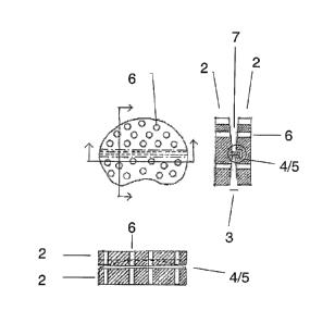
Wording used in figures
1. Disc implant
2. Disk element
3. Coupling zone
4. Coupling means
5. Protrusion/slot arrangement
6. Openings
7. Convex relation ship
8. Internal void volume
9. Keels
10. Incisions
11. Outer surface
12. Filled openings
Figure 1.
The figure shows examples of a kidney shaped disc implants according to the
invention. Figure la. Graphic illustration of the disc implant (1) viewed from
above,
and cross sections orthogonal to each other showing the openings e.g. straight
channels (6) of this embodiment. The convex relation ship (7) of the two
elements
(2) is illustrated as well as the coupling means (4) of the disc implant
provided by a
curved protrusion engaged in a slot of the opposing element (5). The figure
further
CA 02632322 2008-06-03
WO 2007/065443 20 PCT/DK2006/000699
illustrates the opposing position of the opening channels of the elements.
Figure 1b
is an embodiment of the invention depicted as the embodiment of Figure la,
wherein the coupling means (4) are arranged across the shortest "diameter" of
the
elements. Figure lc shows an implant with few openings (6) than the implant of
Figure 1a, wherein the coupling means (4) are formed by a protrusion/slot
arrangement (5) which does not extend across the entire length of the
elements.
Figure 2
Further examples of disc implants according to the invention comprising
openings
with an internal void volume are shown. The diameter of the channel through
the
elements varies and a void volume (8) is seen in both 2a and 2b. The
embodiment
depicted in 2a is differentiated from the embodiment shown in figure 2b by the
number and position of the openings (6). A further difference is seen by the
localisation of the coupling means (4). The figures further shows keels (9)
positioned
on the first and second outer fusion surfaces prevention rotation of the
implant.
Figure 3
Two embodiments according to the invention are shown comprising incisions (10)
(3a) and opening (6) and incisions (10) (3b). Different coupling means (4) are
further
illustrated by a long and narrow projection in 3a and a small circular
protuberance in
3b engaged in suitable slots/depressions of the opposing surface. 3b further
illustrates a disc implant with a rugged outer surface (11).
Figure 4.
As an alternative to relatively many minor openings, the disc elements may as
shown in figure 4a comprise few larger openings (6). In a further embodiment
the
openings are filled (12) with a suitable material, such as artificial bone
(figure 4b).
Figure 5.
The shape of the disc elements may be optimised for different surgical
procedures
as seen in figure 5 displaying a disc implant for Transforaminal Lateral
Interbody
Fusion. The figure is further an example of how the volume occupied by the
openings (6) may be optimised, as the elements are merely frames including
coupling means (4).