Note: Descriptions are shown in the official language in which they were submitted.
CA 02632841 2008-06-09
WO 2007/149595 PCT/US2007/060306
INTERVENTIONAL DEPLOYMENT AND IMAGING SYSTEM
BACKGROUND OF THE INVENTION
[0001] 1. Field of the Invention. The present invention relates generally to
medical
devices and methods. More particularly, the invention relates to systems which
provide for
removably positioning an imaging core and an interventional core in a sheath
and to methods
for using such systems.
[0002] Treatment of the female reproductive tract for dysfunctional uterine
bleeding and
fibroids remain unmet clinical needs. Fibroids are benign tumors of the
uterine myometria
(muscle) and are the most common tumor of the female pelvis. Fibroid tumors
affect up to
30% of women of childbearing age and can cause significant symptoms such as
discomfort,
pelvic pain, menorrhagia (excessive menstrual bleeding), pressure, anemia,
compression,
infertility and miscarriage. Fibroids may be located in the myometrium
(intramural), adjacent
to the endometrium (submucosal) or in the outer layer of the uterus
(subserosal). Most
commonly fibroids are a smooth muscle overgrowth that arise intramurally and
can grow to
be several centimeters in diameter.
[0003] Current treatments for fibroids include both pharmacological therapies
and surgical
interventions. Pharmacological treatment includes the administration of
medications such as
NSAIDS, estrogen-progesterone combinations, and GnRH analogues. All
medications are
relatively ineffective and are palliative rather than curative. Hysterectomy
(surgical removal
of the uterus) is another common treatment for fibroids. While effective,
hysterectomy has
many undesirable side effects such as loss of fertility, the need for open
surgery, sexual
dysfunction, and long recovery time. There is also significant morbidity
(sepsis, hemorrhage,
peritonitis, bowel and bladder injury), mortality and cost associated with
hysterectomy.
Surgical myomectomy, in which fibroids are removed, is an open surgical
procedure
requiring laparotomy and general anesthesia. Often these procedures result in
significant
blood loss and remove a portion of the fibroid mass.
[0004] To overcome at least some of the problems associated with open surgical
procedures, laparoscopic myomectomy was pioneered in the early 1990's.
However,
laparoscopic myomectomy remains technically challenging, requiring
laparoscopic suturing
1
CA 02632841 2008-06-09
WO 2007/149595 PCT/US2007/060306
which limits its performance to only the most skilled of laparoscopic
gynecologists. Other
minimally invasive treatments for uterine fibroids include hysteroscopy,
uterine artery
ablation, endometrial ablation, and myolysis.
[0005] Hysteroscopy utilizes a thin fiber optic camera to image the uterus and
an
attachment to destroy tissue. Hysteroscopic resection is a surgical technique
that uses a
variety of devices (loops, roller balls, bipolar electrodes) to ablate or
resect uterine tissue.
The uterus needs to be filled with fluid for better viewing and thus has
potential side effects
of fluid overload. Hysteroscopic ablation is limited by its visualization
technique and is thus
only appropriate for those fibroids that are submucosal and/or protrude into
the uterine cavity.
[0006] Uterine artery embolization was introduced in the early 1990's and is
performed
through a groin incision by injecting small particles into the uterine artery
to selectively block
the blood supply to fibroids. Complications include pelvic infection,
premature menopause
and severe pelvic pain. In addition, long term MRI data suggest that
incomplete fibroid
infarction may result in regrowth of infarcted fibroid tissue and symptomatic
recurrence.
[0007] Endometrial ablation is primarily a procedure for dysfunctional (or
abnormal)
uterine bleeding and may be used at times for fibroids. Endometrial ablation
relies on various
energy sources such as cryo energy, microwave energy and radiofrequency
energy.
Endometrial ablation destroys the endometrial tissue lining the uterus but
does not
specifically treat fibroids. This technique is also not for women who desire
future
childbearing. Endometrial ablation remains an excellent therapy for
dysfunctional uterine
bleeding but is limited in its ability to treat fibroids.
[0008] Myolysis was first performed in the 1980's using lasers or RF energy to
coagulate
tissue, denature proteins and necrose myometrium with laparoscopic
visualization.
Laparoscopic myolysis can be an alternative to myomectomy, as the fibroids are
coagulated
and then undergo coagulative necrosis resulting in a dramatic decrease in
size. As with all
laparoscopic techniques, myolysis is limited by the fact that it can only see
(and therefore
treat) subserosal fibroids.
[0009] Needle myolysis uses a laparoscope to introduce one or more needles
into a fibroid
tumor under visual control. Bipolar Radio Frequency ("RF") current is then
delivered
between two adjacent needles, or unipolar current between a single needle and
a distant
dispersive electrode affixed to the thigh or back. The aim of needle myolysis
is to coagulate a
significant volume of the tumor and thereby cause it to shrink substantially.
The traditional
2
CA 02632841 2008-06-09
WO 2007/149595 PCT/US2007/060306
technique is to make multiple passes through different areas of the tumor
using the
coagulating needle to destroy many cylindrical cores of abnormal tissue.
However, the
desirability of multiple passes is mitigated by the risk of adhesion
formation, which is
thought to increase with increasing amounts of injured uterine serosa, and by
the operative
time and skill required.
[0010] To overcome the limitations of current techniques, it would be
desirable to provide
a minimally invasive approach to selectively eradicate fibroid tumors within
the uterus. It
would be particularly desirable to provide a treatment device that combines
imaging and
ablation in one simple hand held unit or assembly. It would be further
desirable if the method
and apparatus could locate and treat all types of fibroids in the uterus in a
safe and effective
manner with minimum risk and discomfort for the patient. It would be still
further desirable
if the devices could employ multiple interchangeable components both to permit
selective
sterilization or reuse of the devices and to permit the system to be
configured individually for
patients having different anatomies and needs. It would be still further
desirable if such
devices could be used in and for procedures used in other parts of the anatomy
in addition to
the uterus. At least some of the objectives will be met by the inventions
described below.
BRIEF SUMMARY OF THE INVENTION
[0011] The present invention provides a needle deployment and imaging system
which
allows for needle deployment into solid tissue under direct, usually real-
time, visualization.
Typically, the needle will be deployed from within a natural or created body
cavity or body
lumen. Exemplary body cavities include the uterus, the esophagus, the stomach,
the bladder,
the colon, and the like. Exemplary body lumens include the ureter, the
urethra, fallopian
tubes, and the like. Created body cavities include insufflated regions in the
abdomen, the
thoracic cavity, regions around joints (for arthroscopic procedures), and the
like. The present
invention will generally not find use with procedures in blood vessels or
other regions of the
vasculature. Thus, while the following description will be directed
particularly at procedures
within the uterus for detecting and treating uterine fibroids, the scope of
the present invention
is not intended to be so limited.
[0012] Needle deployment and imaging systems according to the present
invention will
comprise a sheath, an imaging core, and an interventional core. The sheath
will have a
proximal end, a distal end, a first axial passage, and a second axial passage
which is
preferably isolated from the first axial passage. By "isolated" it is meant
that the passages are
3
CA 02632841 2008-06-09
WO 2007/149595 PCT/US2007/060306
separated by an internal wall or barrier that prevents cross-contamination
from one passage to
the other. The axial passages will typically extend the entire length of the
sheath from the
proximal to distal end and may be open at least the proximal end(s), often at
both ends. In
some instances, however, the passage(s) may be shorter and/or may be plugged
or otherwise
sealed at one end (usually the distal end) to isolate that end of the passage
from the external
environment. The sheath will usually be flexible, sometimes being deflectable
or steerable,
but in other instances may be rigid along all or a portion of its length. In
some instances, at
least a portion of the sheath at or near the distal end will be open or
visually transparent to
permit imaging from within the second axial passage. For example, at least a
portion of the
sheath may be composed of an ultrasonically translucent material to permit
ultrasonic or
optical coherence tomographic (OCT) imaging through a wall of the sheath. In
other
instances, the second axial passage may be open (e.g., provided with an open
port or
aperture) at its distal end to permit the imaging core to be advanced beyond
the distal end of
the sheath. Typically, the second axial or passage will be open at its distal
end to permit
advancement of a needle or other interventional element therethrough. In other
instances,
however, the second axial passage may be closed or covered by a penetrable
septum or cover.
[0013] The imaging core may be adapted for any conventional form of medical
imaging,
such as ultrasonic imaging, optical coherence tomographic imaging (OCT),
direct optic
visualization (e.g., using optical fibers for image transmission or using in
situ charged
coupled devices (CCD's) or other imaging elements for in situ visualization),
or the like. The
imaging core will be disposed in the first axial passage, usually being
removably disposed so
that it may be removed and replaced within the sheath to permit sterilization
and reuse of the
imaging core. The imaging core will usually be flexible and in some instances
may be
deflectable or steerable, either in place of a steerable sheath or in addition
to a steerable
sheath.
[0014] The interventional core may be replaceably, translatably, or fixedly
disposed in the
second axial passage. In most cases, the interventional core will typically be
advanceable or
otherwise deployable from the sheath in order to effect the desired
therapeutic or diagnostic
procedure. In the specific embodiments described below, the interventional
core will
typically be a needle which is reciprocatably disposed within a second axial
passage having
an open distal end (and/or other lateral opening) to permit deployment and
penetration into
adjacent solid tissue. In those cases, the needle may be a radiofrequency (RF)
electrode, a
microwave antenna, a cryogenic probe, or other energy delivery or mediating
element
4
CA 02632841 2008-06-09
WO 2007/149595 PCT/US2007/060306
intended for ablating or otherwise treating the tissue. In other cases, the
needle could be a
hollow core needle intended for sampling, biopsy, or otherwise perfonming a
diagnostic
procedure, or for delivering a therapeutic agent or drug into the tissue.
[0015] In the most common embodiments, the imaging core will be removable and
replaceable and the treatment core will be non-removably or at least non-
replaceably coupled
within the second passage (although being adapted for reciprocatable
deployment is
described above.) In such cases, the needle deployment and imaging system may
be used for
a therapeutic or diagnostic procedure and removed from the patient after the
procedure is
complete. The removable imaging core may then be removed and sterilized. The
sheath and
interventional core will then typically be disposed, although in other
instances it's possible
that they could be sterilized and reused.
[0016] In other embodiments, the imaging core may be fixed within the first
axial passage
while the interventional core is removable and replaceable within the sheath.
In those
instances, after the needle deployment and imaging system has been used in a
procedure, the
system will be extracted from the patient and the interventional core removed
from the
sheath. The removable interventional core will usually be sterilized for
reuse, while the
combination of the sheath and the imaging core will be disposed of or
separately sterilized for
reuse.
[0017] In still further embodiments, both the imaging core and the
interventional core may
be removable and replaceable within the respective axial passages of the
sheath. After use of
such needle deployment and imaging systems, both the imaging core and the
interventional
core may be removed from the respective axial passage. Each of the sheath,
imaging core,
and interventional core may then be disposed and/or sterilized for reuse as
determined by the
treating physician or medical facility at that time. Most commonly, at least
the sheath would
be disposed of, while either or both of the imaging core and the
interventional core might be
sterilized for reuse.
[0018] The geometries of the imaging core and the interventional core may be
varied in
accordance with the intended use of the needle deployment and imaging system.
Usually, the
imaging core will be adapted for lateral imaging and will be positionable
within the first axial
passage for side-viewing from the sheath. The first axial passage may be
entirely sealed to
the exterior with an ultrasonically, optically, or other visually transparent
window formed in a
wall of the sheath to permit imaging by the imaging core. The use of a sealed
first axial
5
CA 02632841 2008-06-09
WO 2007/149595 PCT/US2007/060306
passage is frequently preferred since it isolates the imaging core from the
body cavity or
lumen being treated or diagnosed. Alternatively, an open window could be
formed within the
wall of the sheath to permit imaging by the imaging core. Still further
alternatively, the
imaging core may be adapted to extend distally beyond an opening in the first
axial passage
at the distal end of the sheath to permit imaging. In such cases where the
imaging core is
distally extendable, at least a distal end of the imaging core will frequently
be adapted for
deflection or steering.
[0019] In still other embodiments of the present invention, the imaging core
may be
adapted to image in a distally forward direction. As with the lateral imaging
embodiments,
the sheath may be composed at least partially of an ultrasonically or
otherwise visually
transparent material and/or an opening may be formed in the sheath to permit
imaging
therethrough.
[0020] In the preferred case of ultrasonic imaging cores, the ultrasonic
transducers may be
arranged in a phased array, for example either a linear (typically axially
aligned) phased array
or a circumferential phased array. Alternatively the ultrasonic imaging
element may
comprise one or more independent elements, such as parabolic or other shaped
imaging
elements. In still further embodiments, the ultrasonic imaging transducers may
be arranged
in a rotating mechanism to permit rotational scanning.
[00211 A particular advantage of the present invention will be the ability to
selectively
position both the imaging core and the interventional core within the body
cavity or lumen
being treated or diagnosed. The positioning capabilities may come from the
sheath, the
imaging core, and/or less frequently from the interventional core. In some
embodiments, the
sheath will either be steerable or deflectable, often using single or multiple
tensioning wires
for selective deflection of the distal end. Alternatively, the sheath may be
provides as a kit
including a plurality of sheaths having different distal end geometries
intended for particular
anatomies and anatomical access routes. In such instances, the systems will
comprise at least
a first and second sheath, often including third, fourth, fifth, or additional
sheaths, each
having a unique distal end geometry.
[0022] In still other systems, the imaging core may itself be steerable or
deflectable, again
typically being provided by one or more pull or tensioning wires. Such
steerable or
deflectable imaging cores may be deployed or manipulated within the first
axial passage of
the sheath so that deflection of the core will in turn deflect a distal
portion of the sheath.
6
CA 02632841 2008-06-09
WO 2007/149595 PCT/US2007/060306
Alternatively or additionally, the imaging cores may be deployable beyond the
distal end of
the sheath so that the core may be deflected and reoriented outside the
sheath. Such
deployment and actuation of the steerable imaging cores will be particularly
useful with
sheaths having rigid, non-bendable structures, although they could also be
used with sheaths
having steering mechanisms.
[0023] The interventional cores will typically comprise a needle or other
tissue-penetrating
element. Typically, the interventional cores will have a tissue-penetrating
element at or near
their distal ends, such as a sharpened distal tip, an RF cutting element at
the tip, a removable
stylet having a sharpened tip, or the like. In any event, the distal end will
usually be adapted
so that it will self-penetrate into the tissue as it is advanced from the
sheath. The direction of
advancement will be coordinated with the imaging field of the imaging core so
that the
penetration of the interventional core into tissue can be viewed by the
practitioner, usually in
real time. In the exemplary embodiments, a therapeutic needle advanced from
the sheath can
be viewed as it enters the uterine wall to treat a uterine fibroid. Such
tissue-penetrating
elements may be adapted to reciprocate through a side port in the sheath to
extend laterally or
may be adapted to reciprocate through a distal port in the sheath to extend
axially.
BRIEF DESCRIPTION OF THE DRAWINGS
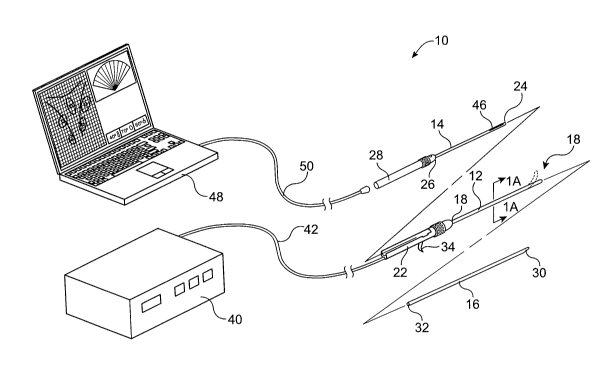
[0024] Fig. 1 is an exemplary illustration of a needle deployment and imaging
device and
system constructed in accordance with the principles of the present invention.
[0025] Fig. IA is a cross-sectional view of the device taken along line lA-lA
in Fig. 1.
[0026] Figs. 2A and 2B illustrate insertion of a first embodiment of steerable
imaging core
into a sheath in accordance with the principles of the present invention.
[0027] Fig. 3 illustrates an alternative embodiment of a non-steerable imaging
core within a
sheath in accordance with the principles of the present invention.
[0028] Fig. 4 illustrates a still further embodiment of a non-steerable
imaging core within a
non-steerable sheath in accordance with the principles of the present
invention.
[0029] Figs. 5A-5C illustrate insertion of an imaging core into a sheath where
both the
imaging core and an interventional core extend axially from a distal end of
the sheath.
[0030] Figs. 6A-6D illustrate sheaths having pre-shaped distal ends intended
for
intrauterine treatments.
7
CA 02632841 2008-06-09
WO 2007/149595 PCT/US2007/060306
[0031] Figs. 7A and 7B illustrate a still further embodiment of a sheath
having an open
window near its distal end for deployment of a pair of needle-type
interventional cores.
[0032] Figs. 8A-8C illustrate use of a needle deployment and imaging system
for delivering
occlusive elements to a uterine artery in accordance with the principles of
the present
invention.
[0033] Fig. 9 illustrates the image viewable with the system of Figs. 8A-8C.
DETAILED DESCRIPTION OF THE INVENTION
[0034] As seen in Fig. 1, a needle deployment and imaging system 10 comprises
a sheath
12, an imaging core 14, and an interventional core 16. The sheath 12 has a
distal end 18, a
proximal end 20, and a handle structure 22 disposed at the proximal end.
Imaging core 14
also has a distal end 24, a proximal end 26, and a handle structure 28
disposed at the proximal
end. Similarly, the interventional core 16 has distal end 30, a proximal end
32, and will will
usually have a trigger 34 or other deployment mechanism on the sheath 12 in
order to axially
reciprocate the needle from the proximal end 18 of the sheath.
[0035] As shown in Fig. 1, the interventional core 16 is a tissue-penetrating
electrode
where the distal end 30 is typically sharpened or otherwise adapted for
penetrating or
otherwise entering solid tissue when it is advanced from the sheath 12. The
interventional
core 16 will be connectable to an RF power supply 40 via a connecting cable 42
which
attaches to the interventional core through the handle structure 22 of the
sheath 12. A switch,
foot pedal, or other trigger (not shown) is provided on or with the RF power
supply 40 in
order to initiate delivery of RF energy through the interventional core 16 in
a generally
conventional manner. The radiofrequency energy provided can be monopolar,
bipolar, or
combinations thereof as are well-known in the art of RF tissue ablation. In
addition, the core
16 may have a lumen or other delivery means for saline infusion or "wet RF" as
is well
known to those skilled in the art.
[0036] The imaging core 14 typically comprises an ultrasonic imaging
transducer or
transducer array 46 near its distal end 24. The ultrasonic transducer or array
46 is
connectable to an imaging monitor 48, illustrated in the form of a laptop
computer, via a
cable 50 attachable to the handle structure 28 of the imaging core 14. It will
be appreciated
that in other embodiments, the imaging monitor 48 could be combined with the
RF power
supply 40 in a single unit providing for both interventional control and image
monitoring.
8
CA 02632841 2008-06-09
WO 2007/149595 PCT/US2007/060306
[0037] In the embodiment of Fig. 1, the imaging core 14 is removably placed in
a first axial
passage 56 extending through the sheath 12, as shown in Fig. 1 A. The
interventional core 16
is to be disposed within a second axial passage 58 which is usually formed in
parallel in the
sheath body 12, as also shown in Fig. lA. In the embodiment of Fig. 1, the
interventional
core 16 will be reciprocatable within the second axial passage 58 to extend
either axially or
laterally from the distal end of the sheath 12. Usually, the proximal end of
the interventional
core 16 will be non-removeably coupled to the trigger 34 or other advancement
component of
the sheath so that the interventional core is not intended to be removable and
replaceable.
[0038] Referring now to Figs. 2A and 2B, the imaging core 14 is loaded into
the first axial
passage 56 of the sheath 12 by advancing the core through a cradle 60 formed
in the handle
structure 22. When fully inserted, the handle structure 28 is fully received
into cradle 60, as
shown in Fig. 2B. In this embodiment, the first axial passage 60 is sealed
over its entire
length within the sheath 12 and an acoustically transparent window 62 is
formed near the
distal end 18 of the sheath. The distal end 18 of the sheath 12 may be
deflected by rotating a
control knob 64 on the handle structure 22, as shown by arrow 66 in Fig. 2A.
In other
embodiments, the sheath 12 will be rigid and non-steerable. The distal end 30
of the
interventional core 16 may be advanced from the distal end 18 of the sheath by
pulling a
trigger 68 on the handle structure 22 of the sheath 12, as shown in Fig. 2B.
The distal end 24
of the imaging core 14 is optionally deflectable using a control knob 72 on
the handle
structure 28, although deflection of the imaging core is not illustrated in
the figures.
[0039] While it will often be desirable to provide deflection or steering
capability in at least
one of the sheath 12 and imaging core 14, it will not be necessary to provide
such steering in
either or both the sheath and imaging core as shown in the embodiment of Fig.
1. For
example, as shown in Fig. 3, the handle structure 22 may have the control knob
64 for
deflecting the distal end of sheath 12, while the handle structure 28 may be
free of control
knobs and the imaging core 14 may not be steerable. Alternatively, the sheath
12 may be
non-steerable, with the imaging core 14 being steerable, as shown in Fig. 4.
In still other
embodiments, neither the sheath nor the imaging core may be steerable, e.g.,
when the sheath
has a permanently deflected distal end or is provided as a kit including
multiple sheaths, as
illustrated in Fig. 6A-6D, and described in detail below.
[0040] Deployment of the imaging core 14 and interventional core 16 from
and/or within
the sheath 12 may be accomplished in a variety of ways. 'In the embodiment of
Fig. 1, 2A,
9
CA 02632841 2008-06-09
WO 2007/149595 PCT/US2007/060306
and 2B, the first passage 56 is sealed (typically with an acoustically
transparent window 61 so
that the imaging core 14 remains within the passage and is never extended from
the sheath.
The interventional core 16, in contrast, is extended through a port 63 in the
side of the sheath
12 in a lateral direction, as best shown in Fig. 2B.
[0041] Referring now to Figs. 5A and 5B, an embodiment 100 of the needle
deployment
and imaging system of the present invention includes sheath 12, imaging core
14, and
interventional core 16 which are generally the same as described previously
with reference to
Figs. 1, 2A and 2B, except for the distal end deployment configurations. As
shown in Fig.
5A, imaging core 14 is loaded into the sheath 12 as described above, and the
only significant
difference with the prior embodiment is that the sheath 12 does not
necessarily include an
acoustically or optically transparent window at its distal end. Instead as
best shown in Fig.
5B, both the distal end 30 of the interventional core 16 and the distal end 24
of the imaging
core 14 are extendable through ports in the distal end of the sheath 12.
Moreover, the distal
end 24 of the imaging core 14 is deflectable using the control knob 72 of the
handle structure
28, as shown in broken line. The distal end of the sheath 12 will also be
steerable, and the
embodiment of the needle deployment and imaging system 100 will allow access
to a variety
of tissue surfaces within the uterine or other body cavities by steering of
the sheath,
deflection of the imaging core, and rotation of the imaging core relative to
the sheath.
[0042] Referring now to Figs. 6A-6D, a sheath kit will comprise a plurality of
individual
sheaths 12A, 12B, 12C, and 12D, and optionally still further sheaths. The
distal end of each
sheath 12A-12D will have a different shape which is permanently set into the
sheath. While
the sheath bodies may still retain a certain degree of flexibility, they will
have sufficient
rigidity in order to retain the pre-set shape. The simplest shape will be
generally linear, as
shown in Fig. 6A. The sheath 12B of Fig. 6B will have a gentle curve to
facilitate access to
the front and back of the uterus. The distal tip of the sheath 12C as
illustrated in Fig. 6C will
have a generally L-shaped deflection which permits access to the sidewalls of
the uterus.
Finally, the sheath 12D of Fig. 6D will have a curve intermediate those of
sheaths 12B and
12C to allow access to the fundus of the uterus. Still further geometries may
be useful for
access and interventions within the uterus and other body cavities and lumens.
[0043] Referring now to Fig. 7A and 7B, it will be appreciated that a wide
variety of needle
geometries and sheath configurations may be utilized. For example, a sheath
112 may have a
Notch-like opening 113 near its distal end 118 to permit one or more tissue-
penetrating
CA 02632841 2008-06-09
WO 2007/149595 PCT/US2007/060306
needles 116A and 11 6B to be advanced forwardly and laterally relative to the
access of the
sheath, as shown in Fig. 7A. The needles 116A and 116B will be reciprocatably
received
within axial passages 158A and 158B through the sheath 112, as best shown in
Fig. 7B. An
additional axial passage 156 will be provided for an imaging core (not shown)
which will be
advanced through the sheath 112 and be positioned within the open notch 113
for imaging of
the needles 116A and 11 6B as they are being deployed in solid tissue.
[0044] Referring now to Figs. 8A-8C and Fig. 9, the needle deployment and
imaging
systems of the present invention can be used for delivering substances,
energy, or a variety of
other interventional modes to treat uterine fibroids UF in the wall of the
uterus U. As shown
in Fig. 8A, a hollow delivery needle 200 may be advanced from the distal end
of the sheath
212 under ultrasonic imaging provided by an array 246. The needle 200 may be
deployed in
an artery A supplying blood to the uterine fibroid F, and occlusive elements
delivered to the
artery in order to occlude the artery to deprive the fibroid of blood.
[0045] As shown in Figs. 8B and 8C, the needles 200 could also be configured
to enter into
tissue adjacent the fibroid UF as shown in Fig. 8B, or within the fibroid UF
as shown in
Fig. 8C. A variety of materials, such as markers, dyes, fluid blebs, air
blebs, solid materials,
biodegradable materials, and the like, could also be delivered using these
needles in any of
the illustrated configurations. As shown in Fig. 9, the location of the needle
200 within the
field of view 250 can be observed in real time using the ultrasonic imaging
array 246.
[0046] While the above is a complete description of the preferred embodiments
of the
invention, various alternatives, modifications, and equivalents may be used.
Therefore, the
above description should not be taken as limiting the scope of the invention
which is defined
by the appended claims.
11