Note: Descriptions are shown in the official language in which they were submitted.
CA 02632856 2008-05-26
FLUID SAMPLE TESTING SYSTEM
jMTRODUCTION
This application is a divisional application of Canadian Application Serial
No.
2,301,153 filed February 17, 2000.
The present invention is directed to a system for testing a fluid sample, and,
more
particularly, to a fluid sample testing system having improved automation,
safety and
efficiency.
BACKGROUND
Collection, transportation and pretreatment of fluid samples, such as blood
samples, are currently done generally in a manual fashion. Blood is commonly
collected
in test tubes and samples from these test tubes are deposited in reaction
chambers for
testing. These tubes can be placed in an automated testing machine to perform
testing
using various assays. This process can be expensive, time consuming, and may
lead to
human error, possibly leading to false test results. Current automated testing
systems
require large capital investment; incur high costs for reagents, disposables,
operation,
maintenance, service and training; and do not provide required sample
pretreatment.
SLIIHIRY
The present invention provides a sample testing system which reduces or wholly
overcomes some or all of the aforesaid difficulties inherent in prior known
devices.
Particular aspects and advantages of the invention will be apparent to those
skilled in the
art, that is, those who are knowledgeable or experienced in this field of
technology, in
view of the following disclosure of the invention and detailed description of
certain
preferred embodiments.
The principles of the invention may be used to advantage to provide a sample
testing system which is highly automated, thereby increasing efficiency,
reducing costs,
and increasing safety due to reduced handling of samples. A sample can be
collected in
a chamber which is then divided into a plurality of sealed segments. A reagent
can be
added to a segment and the segment can be inspected to detect a condition of
the sample.
CA 02632856 2008-05-26
WO 99/67646 PCT/US99/14105
-2-
In accordance with a first aspect. a sample testing system has a chamber
sealing
apparatus to form a plurality of seals defining a plurality of fluid-tight
segments of the
chamber. A reagent injector cart:ridge actuator is adapted to receive a
reagent injector
cartridge having at least one needle in fluid communication with a reagent
reservoir, and
to move a reagent injector cartridge to inject a quantity of reagent into a
segment of a
chamber. A sensor generates an output signal corresponding to a condition of a
fluid
sample material within a segment of a chamber.
In accordance with another aspect, a sample testing system has a tube sealing
apparatus having a tube compression and sealing member to laterally sea] a
flexible
plastic tube containing a fluid sample material, whereby a fluid-tight tubule
containing
a portion of the fluid sample material can be forrned between axially spaced
lateral seals.
A reagent injector cartridge actuator is adapted to receive a reagent injector
cartridge
having at least one needle in fluid communication with a reagent reservoir,
and to move
a reagent injector cartridge to inject a quantity of reagent into a tubule. A
flow control
device has a contact member movable into contact with a tubule to effect
mechanically
induced fluid flow within a fluid passageway in the tubule. An inspection
system has a
light detector to receive light passed through a tubule and to generate an
output signal
corresponding to a condition of the fluid sample material within a tubule.
In accordance with another aspect, a sample testing system has a tube sealing
apparatus having a tube compression and sealing member to laterally seal a
flexible
plastic tube containing a fluid santple material, whereby a fluid-tight tubule
containing
a portion of the fluid sample material can be fotmed between axially spaced
lateral seals.
A reagent injector has at least one rieedle in fluid cotnmunication with a
reagent reservoir,
and a needle actuator to insert the needle into a tubule and inject a quantity
of reagent into
a tubule. A flow control device has a contact member movable into contact with
a tubule
to effect mechanically induced fluid flow within a fluid passageway in the
tubule. An
inspection system has a light detector to receive light passed through a
tubule and to
generate an output signal cotresportding to a condition of the fluid sample
material within
a tubule.
In accordance with another aspect, a reagent cartridge has a housing and at
least
one reservoir in the housing. At least one needle in the housing is in fluid
CA 02632856 2008-05-26
-~-
conununication with one of the reagent reservoirs. A needle actuator inserts
the needle
into a tubule and injects a quantity of reagent.
In accordance with yet another aspect, a sample testing tubule has a length of
flexible plastic tube having fluid-tight lateral seals at axially spaced
locations to define
a fluid-tight fluid sample chamber between the lateral seals containing a
fluid sample
material. A self-sealing injection channel is formed in the tubule, the
injection channel
being normally substantially free of fluid sample material and capable of
fluid
communication with the fluid sample material in the tubule.
In accordance with another aspect, a method of performing a sample assay
includes the following steps: collecting a sample of fluid material into a
length of
substantially transparent, flexible, heat-sealable, plastic tube; inserting
the tube into a
sample testing machine having a tube sealing apparatus, a reagent injector
having at least
one needle in fluid communication with a reagent reservoir and a needle
actuator to insert
the needle into a tubule and inject a quantity of reagent, a flow control
device having a
contact member movable into contact with a tubule to effect mechanically
induced fluid
flow within the tubule, and an inspection system having a light detector to
receive light
passed through a tubule and to generate an output signal corresponding to a
condition of
the sample material within a tubule; actuating the tube sealing apparatus to
seal lengths
of the tube into tubules; actuating the needle actuator to insert the needle
into a selected
tubule and inject reagent to form a mixture of sample material and reagent in
the selected
tubule; actuating the flow control device to mix the mixture of sample
material and
reagent; and actuating the inspection system to inspect the mixture and to
generate an
output signal corresponding to a condition of the mixture.
In accordance with another aspect, there is provided a sample testing system
comprising, in combination:
a chamber sealing apparatus to fotrn a plurality of seals defining a plurality
of fluid-
tight segments of the chamber;
a reagent injector cartridge actuator adapted to receive a reagent injector
cartridge
having at least one needle in fluid communication with a reagent reservoir,
and to move a
reagent injector cartridge to inject a quantity of reagent into a segment of a
chamber;
CA 02632856 2008-05-26
3a
a flow control device comprising at least one contact member movable into
contact
with a segment of a chamber to effect mechanically induced fluid flow within a
flow
passageway in a segment of a chamber; and
a sensor to generate an output signal corresponding to a condition of a fluid
sample
material within a segment of a chamber.
In accordance with another aspect, there is provided a sample testing system
comprising, in combination:
tube sealing apparatus having a tube compression and sealing member to
laterally seal
a flexible plastic tube containing a fluid sample material, whereby a fluid-
tight tubule
containing a portion of the fluid sample material can be formed between
axially spaced lateral
seals; the tube sealing apparatus comprising a first sealing head comprising
the tube
compression and sealing member; and a second sealing head, at least one of the
first and
second sealing heads being movable towaxd the other of the sealing heads to
compress a
section of flexible plastic tube positioned between the first and second
sealing heads to create
a sample-free zone in the tube, wherein the tube compression and sealing
member is
operatively connected to a power source to heat a sealing zone of a tube
located in the sample-
free zone to form a fluid-tight lateral seal in the tube;
a reagent injector cartridge actuator adapted to receive a reagent injector
cartridge
having at least one needle in fluid commuriication with a reagent reservoir,
and to move a
reagent injector cartridge to inject a quantity of reagent into a fluid-tight
tubule; and
a sensor to generate an output signal corresponding to a condition of a fluid
sample
material within a fluid-tight tubule.
In accordance with another aspect, there is provided a sample testing system
comprising, in combination:
a chamber sealing apparatus to forrn a plurality of seals defining a plurality
of fluid-
tight segments of the chamber; ,
a reagent injector cartridge actuator adapted to receive a reagent injector
cartridge
having at least one needle in fluid communication with a reagent reservoir,
and to move a
reagent injector cartridge to inject a quantity of reagent into a segment of a
chamber;
a sensor to generate an output signal corresponding to a condition of a fluid
sample
material within a segment of a chamber;
CA 02632856 2008-05-26
3b
a pair of electrodes adapted to have a predetermined voltage difference, and
an electrode actuator to insert the pair of electrodes into a segment of a
chamber,
wherein the sensor is responsive to electrophoretic light emitted from within
a segment of a
chamber.
In accordance with another aspect, there is provided a sample testing system
comprising, in combination:
a chamber sealing apparatus to form a plurality of seals defining a plurality
of fluid-
tight segments of the chamber;
a segment of the chamber including a coating on an outside surface of the
segment to
increase the transmission of light through the segment;
a reagent injector cartridge actuator adapted to receive a reagent injector
cartridge
having at least one needle in fluid communication with a reagent reservoir,
and to move a
reagent injector cartridge to inject a quantity of reagent into a segment of a
chamber; and
a sensor to generate an output signal corresponding to a condition of a fluid
sample
material within a segment of a chamber.
In accordance with another aspect, there is provided a sample testing system
comprising, in combination:
a chamber sealing apparatus to form a plurality of seals defining a plurality
of fluid-
tight segments of the chamber;
a reagent injector cartridge actuator adapted to receive a reagent injector
cartridge
having at least one needle in fluid communication with a reagent reservoir,
and to move a
reagent injector cartridge to inject a quantity of reagent into a segment of a
chamber;
an incubation chamber to retain a segment of a chamber for a predetermined
period of
time; and
a sensor to generate an output signal corresponding to a condition of a fluid
sample
material within a segment of a chamber.
CA 02632856 2008-05-26
3c
From the foregoing disclosure, it will be readily apparent to those skilled in
the art,
that is, those who are knowledgeable or experienced in this area of
technology, that the
present invention provides a significant technological advance. Preferred
embodiments of the
fluid sample testing system of the present invention can provide increased
efficiency, reduced
costs, and increase safety. These and additional features and advantages of
the invention
disclosed here will be further understood from the following detailed
disclosure of certain
preferred embodiments.
CA 02632856 2008-05-26
WO 99/67646 PCT/US99/14105
-4-
BRIEF DESCI3IPTION OF THE DRAWIN S
Certain preferred embodirnents are described in detail below with reference to
the
appended drawings wherein:
Fig. 1 is a partially schernatic perspective view of a sample testing system
in
accordance with a preferred embodiment of the present invention;
Fig. 2 is a schematic representation of the components of the sample testing
system of Fig. 1;
Fig. 3 is a schematic perspective view, partially in phantom, of a tube
sealing
apparatus of the testing system of Fig. 1;
Fig. 4 is a schematic elevation view, shown partially cut away, of a tube
being
compressed by the tube sealing apparatus of Fig. 3;
Fig. 5 is a schematic elevation view, shown partially cut away, of a tube
being
sealed by the tube sealing appara-tus of Fig. 3;
Fig. 6 is a schematic plan view of a sealing head of the tube sealing
apparatus of
Fig. 3;
Fig. 7 is a schematic plan view of a plurality of tubules formed in a length
of tube
by the tube sealing apparatus of Fig. 3;
Fig. 8 is a schematic plan view of an alternative embodiment of a sealing head
of the tube sealing apparatus of Fig. 3;
Fig. 9 is a schematic plan view of another alternative embodiment of a sealing
head of the tube sealing apparatus of Fig. 3;
Fig. 10 is a schematic section view of a reagent cartridge suitable for use in
the
sample testing system of Fig. 1;
Fig.. 11 is a schematic section view of an alternative embodiment of a reagent
cartridge for the sample testing system of Fig. 1;
Fig. 12 is a schematic section view of the reagent cartridge of Fig. 11 shown
injecting reagent into a tubule;
Fig. 13 is a schematic section view of another alternative embodiment of a
reagent
cartridge of the sample testing system of Fig. 1;
Fig. 14 is a schematic section view of yet another alternative embodiment of a
reagent cartridge of the sample testing system of Fig. 1;
CA 02632856 2008-05-26
WO 99/67646 PCTIUS99/14105
-5-
Fig. 15 is a schematic elevation view of a flow control device and inspection
system of the sample testing system of Fig. 1;
Fig. 16 is a schematic elevation view of an alternative embodiment of the flow
control device of the sample testing system of Fig. 1;
Fig. 17 is a schematic elevation view of another alternative embodiment of the
flow control device of the sample testing system of Fig. 1;
Fig. 18 is a schematic elevation view of yet another alternative embodiment of
the
flow control device of the samp;le testing system of Fig. 1;
Fig. 19 is a schematic elevation view of an alternative embodiment of the
inspection system of the sample testing system of Fig. 1;
Fig. 20 is a schematic elevation view of another altemative embodiment of the
inspection system of the sample testing system of Fig. 1;
Fig. 21 is a schematic elevation view of a coating being applied to a tubule
of the
present invention;
Fig. 22 is a schematic perspective view of a reagent cartridge and a tube
divided
into tubules, suitable for the sarnple testing system of Fig. 1;
Fig. 23 is a schematic perspective view of one preferred embodiment of a tube
of
the present invention and a drawing device into which the tube is placed;
Fig. 24 is a schematic elevation view of an alterrrative embodiment of the
tube
sealing apparatus of Fig. 1; and
Fig. 25 is a schematic plan view of an alternative embodiment of a tubule of
the
present invention, shown with a pressure gate between compartments of the
tubule.
The figures referred to above are not drawn necessarily to scale and should be
understood to present a representation of the invention, illustrative of the
principles
involved. Some features of the sample testing system depicted in the drawings
have been
enlarged or distorted relative to others to facilitate explanation and
understanding. The
same reference numbers are used in the drawings for siniilar or identical
components and
features shown in various alternative embodiments. Sample testing system as
disclosed
herein, will have configurations and components determined, in part, by the
intended
application and environment in which they are used.
CA 02632856 2008-05-26
WO 99/67646 PCT/US99/14105
-6-
DETAILED DESCRIPTION OF CERTAIN PREFERRED EMBODIMENTS
The present invention has many uses which will become readily apparent to
those
skilled in the art, given the benefit of this disclosure. Sample material to
tested may be.
e.g., blood, cell suspensions, biofluids or other fluids. Exemplary tests to
be performed
on fluid samples include clinical diagnosis, therapeutic monitoring, and
screening of
chemical compounds for discovery of new drugs. The following discussion will
discuss
blood testing specifically for purposes of illustration.
The present invention provides for a chamber containing a fluid sample to be
divided into a plurality of segments, with fluid-tight seals separating
adjacent segments
from one another. It is considered to be a highly advantageous feature of
certain
preferred embodiments that a chamber into which a fluid sample is drawn, e.g..
a tube
into which a patient's blood is drawn. can itself then also be the testing or
reaction
chamber within which that blood or other fluid sample is tested, without ever
having to
remove the blood or fluid sample from the chamber.
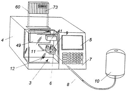
Referring to Fig. 1, a testing machine according to the present invention is
shown
generally by the reference numeral 2. Testing machine 2 comprises a housing 4
having
an entry port 6 on a front side thereof for receiving a chamber containing a
fluid sample.
In the illustrated embodiment, the chamber is a tube 8 from a blood bag 10.
Tube 8 is
preferably a flexible, thermoplastic, substantially transparent tube having an
inner
diameter of approximately 1 mm to 5 mm, preferably approximately 3-4 mm. Tube
8
may be formed of polyvinylchloride (PVC) or other suitable material. A control
panel
7 is located on the front of housing 4 to receive information, such as
information read
from bar code labels or keyed data, and a monitor 5 displays operating
information, such
as the results of testing. A tube sealing apparatus 12, described in greater
detail below,
is contained within housing 4 for sealing portions of tube 8 into tubules 14.
Reagent
cartridge 60 is loaded into a reagent cartridge actuator 49 in housing 4, with
reagent from
reservoirs 16 contained within reagent cartridge 60 being added to tubules 14
(described
in greater detail below). A sensor 41 in housing 4 reads a bar code label 73
(seen in Fig.
22) on reagent cartridge 60 which provides information identifying the
particular reagent
or reagents in reagent cartridge 60 as well as information regarding test
procedures
associated with the particular reagent or reagents. Mixing device or flow
control device
CA 02632856 2008-05-26
WO 99/67646 PCT/US99/14105
-7-
18, seen in Fig. 2 and described iin greater detail below, is also contained
within housing
4 for creating a fluid passageway to allow the flow of cells within tubule 14.
Computerized microscopic inspection system 20 is mounted in housing 4 to view
and
analyze the flow of cells within tubule 14. In certain prefetred embodiments,
multiple
testing machines 2 may be connected to computer analysis and system control
components of inspection system 20, either directly, or via a computer
network. In
certain preferred embodiments, flow control device 18 may not be present, or
may not be
employed if present. In such an alternative embodiment, inspection system 20
inspects
a sample within tubule 14 without a flow of cells within the sample being
created.
A tube advancement system 3 is provided to support and control forward
movement of tube 8 through testing machine 2. Suitable tube advancement
systems will
become readily apparent to those skilled in the art, given the benefit of this
disclosure.
In the embodiment illustrated in Fig. 2, tube advancement system 3 comprises a
pair of
rotating wheels 22 which rotate in opposite directions to advance the tube. At
least one
wheel 22 is connected to and driven by output shaft 23 of a motor which is not
shown.
Tube 8 is inserted between rotating wheels 22 and advanced into tube sealing
apparatus
12. The volume of sample witl-iin each tubule 14 is controlled by compressing
tube 8.
Specifically, upper plunger 9 and lower plunger 11 are spaced apart from one
another and
movable toward one another to partially compress a tubule 14 positioned
therebetween
prior to it being sealed. An upper, or first sealing head 24 and a lower, or
second sealing
head 26 compress a portion of tube 8 and then use radio frequency energy to
seal tube 8,
forming lateral seals 13 between adjacent tubules 14. Lateral seals, as used
herein, refer
to seals which separate axially adjacent portions of tube 8. In a preferred
embodiment,
the lateral seals extend substantially perpendicular to a longitudinal axis of
tube 8. Seals
13 are fluid-tight seals, that is, seals 13, under normal operating
conditions, prevent the
flow of fluid through the seal. Each tubule 14 contains a sample of blood. The
length
of each tubule 14 is preferably approximately 3 to 15 mm, and more preferably
about 5
to 10 mm. Reagent is added to tubule 14 via needle 15 of injector 17.
Tubules 14 then advance to one of an incubation chamber 19. a centrifuge 35,
or
flow control device 18. Flow control device 18 forms a pair of reservoir zones
in tubule
14 with a thin fluid passageway extending between the reservoirs. Light from
light
CA 02632856 2008-05-26
WO 99/67646 PCT/US99/14105
-8-
source 28 is projected through the tubule 14 in flow control device 18. A
camera with
a microscopic lens 30 captures images of blood cell aggregates flowing from
one
reservoir zone to the other through the thin passageway. It sends the images
to a frame
grabber 32, which in turn sends the images to programmable control system or
computer
34 for analysis. The results of the testing done in computer 34 may be
transmitted to
display 7, seen in Fig. 1, for reading by an operator. In other preferred
embodiments, the
results of the testing may be stored for later retrieval, or forwarded to
another computer
or other device, e.g. a printer for preparing a hard copy of the results.
Centrifuge 35 is provided to separate components of the sample in a length of
tube 8 in a known fashion. A length of tube 8, typically longer than a typical
tubule 14,
is conveyed to centrifuge 35 via suitable conveying means. Once the components
of the
sample in the lengtli of tube 8 have been separated, the length of tube is
sealed into
tubules 14 providing a fluid-tighl: seal between the different components. The
length of
tube is sealed either by a tube sealing apparatus at centrifuge 35, or it may
be advanced
to tube sealer 12 by suitable conveying means for sealing. Centrifuge 35 may
also be
used during testing in order to perform certain assays.
In certain preferred embodiments, selected tubules 14 may be stored in
incubation
chamber 19 prior to advancing to flow control device 18. Incubation chamber 19
may
provide temperature control of tubules 14, and may allow the addition of a
second reagent
to tubules 14. Temperature controlling means 21 is connected to incubation
chamber 19
to heat and/or cool incubation chamber 19. It is to be appreciated that the
temperature
of tubules 14 may be controlled directly, such as with a temperature sensor
detecting the
temperature of tubules 14 and maintaining a desired setpoint temperature.
Alternatively,
the temperature of the tubules could be controlled indirectly by sensing and
controlling
the temperature of incubation chamber 19. Temperature controlling means 21 may
include a heating element and may also include a cooling device. Other
suitable
temperature controlling means will become readily apparent to those skilled in
the art
given the benefit of this disclosure.
Turning now to Fig. 3, tube sealing apparatus 12 will be shown in greater
detail.
Tube sealing apparatus 12 compiises upper, or first sealing head 24 and lower,
or second
sealing head 26. Upper sealing head 24 has conductors 36 extending from an
upper
CA 02632856 2008-05-26
WO 99/67646 PCT/US99/14105
-9-
surface 38 to a lower sealing surface 40. Lower sealing head 26 also has
conductors 36
extending from an upper sealing surface 42 to a lower surface 44. Conductors
36 are
connected by cables 45 to a power source 46 which creates a radio frequency
(RF)
electrical field between the conductors 36 of upper sealing head 24 and lower
sealing
head 26 which heat seals tube 8. Conductors 36 are preferably formed of a
material
having high electrical and heat conductivity. Suitable materials for conductor
36 are, for
example, metals such as copper. Other suitable materials for the sealing heads
will
become readily apparent to those skilled in the art, given the benefit of this
disclosure.
Upper sealing head 24 and lower sealing head 26 are preferably formed of a
substantially
rigid insulating material having high heat conductivity. Suitable materials
for the sealing
heads include plastics such as nylon. Other suitable materials for the sealing
heads will
become readily apparent to those skilled in the art, given the benefit of this
disclosure.
Resilient pads 48 are preferably lcicated at the outer edges of lower sealing
surface 40 and
upper sealing surface 42. Resilient pads 48 may be formed of rubber, silicone
rubbers,
teflon, fluoropolymers, or any other suitable resilient material. In certain
preferred
embodiments, a central bar 50 may be located between a pair of conductors 36.
As seen
in Fig. 4, both upper sealing head 24 and lower sealing head 26 have a central
bar 50. It
is to be appreciated that in certain preferred embodiments, only upper sealing
head 24
may have a central bar 50, while lower sealing head 26 has a single conductor
36.
As seen in Fig. 4, tube 8, containing fluid sample 51, e.g., whole blood, is
passed
between upper sealing head 24 and lower sealing head 26. The volume of a
portion of
tube 8, or tubule 14, is adjusted by compressing upper bar 9 and lower bar 11
together
about tubule 14. In certain preferred embodiments, the volume of tubule 14 is
approximately 201il.. The tubule 14 may contain, for example, approximately 5
l of
whole blood or approximately 15 1 of plasma. Upper and lower sealing heads 24,
26 are
then squeezed together under pressure, compressing a portion of tube 8 and
pushing fluid
sample 51 outwardly in the direction of arrows A. As sealing heads 24, 26
compress tube
8, a sample free zone 52 is created, that is, a zone is created within tube 8
which is
substantially free of any fluid sample 51. The pressure must be sufficient to
squeeze
fluid sample 51 out of sample free zone 52 as well as sufficient to prevent
pressure in
tubule 14 from forcing fluid sample 51 back into sample free zone 52,
especially during
CA 02632856 2008-05-26
WO 99/67646 PCT/US99/14105
- 10-
sealing. The required pressure forcing sealing heads 24. 26 together is
dependent on the
material of tube 8, as well as its diameter and wall thickness. In certain
preferred
embodiments, fluid sample 51 is approximately 2mm away from conductors 36
which
provide the sealing of tubule 14.
As seen in Fig. 5, central bar 50 is then raised, releasing the pressure in a
central
area of sample free zone 52 and creating an injection channel 54 which is also
free of
fluid sample 51. Power source 46 then supplies RF power through cables 45 to
conductors 36 which seals tube 8 forming seal 13. In certain preferred
embodiments, the
frequency of the RF power supplied is approximately 40 MHz. The RF power is
supplied for a time period typically less than one second. The power and
duration of the
supplied RF energy may vary based on the size of tube 8 and the material of
which it is
constructed. Upper sealing member 24 is then raised, tube 8 is advanced to the
left as
seen in Fig. 4, and tube 8 is sealed again, forming a tubule 14 between seals
13. By
creating sample free zone 52, fluid sample 51 is kept a safe distance from
conductors 36
when the RF power is applied, thereby reducing negative effects on fluid
sample 51 from
the RF power and the heat it gerierates.
In the embodiment illustrated in Fig. 4, lower sealing head 26 is fixed and
upper
sealing head 24 moves downwardly in the direction of arrows B toward lower
sealing
head 26. In other preferred embodiments, upper sealing head 24 may be fixed
with lower
sealing head 26 moving toward upper sealing head 24, or both upper and lower
sealing
heads 24, 26 may move toward one another.
In the embodiment illustrated in Figs. 4, 5, lower sealing surface 40 and
upper
sealing surface 42 have a substantially convex profile. Thus when sealing
heads 24, 26
are brought together, tube 8 is compressed a maximum amount in the central
area of
heads 24, 26, that is, in sample free zone 52, and compresses to a lesser
extent outside of
sample free zone 52.
In certain preferred embodiments, as seen in Fig. 6, central bar 50 has an L
shaped, or inverted L shaped profile. In the embodiment illustrated, central
bar 50 of first
sealing head 24 has an inverted I, shape and central bar 50 of second sealing
head 26 has
an L shape. Conductor 36 is foimed of conductor element 36A and conductor
element
36B, spaced apart by central bar 50. Conductor element 36A extends along the
long leg
CA 02632856 2008-05-26
WO 99/67646 PCT/US99/14105
-11-
of central bar 50 and tenninates at its short leg. Conductor element 36 B
extends along
the length of the long leg of central bar 50. Lines W represent the width of a
tube 8
which is sealed by sealing heads 24, 26. It can be seen that the sealing heads
extend
beyond the edge of the tube such that the seal, when formed, extends across
the entire
width of the tube. When the RF power is applied, as seen in Fig. 7, seal 13,
comprising
first portion 13A and second portion 13B is formed only in the areas where
conductor
elements 36A, 36B lie, creating L. shaped injection channe154 which is capable
of being
in fluid communication with tubule 14. However, tension in the area of seal 13
prevents
fluid sample 51 from entering injection channel 54. Reagent is added to
injection channel
54 through needle 15, seen in Fig. 2 and described in greater detail below.
The amount
of reagent added to tubule 14 is preferably approximately 1-15 1 depending on
the assay
being performed. By maintaining injection channel 54 free of fluid sample 51,
any
leakage from tubule 14 is prevented when a needle punctures the side wall of
the tube to
inject reagent into the tubule through injection channel 54. In certain
preferred
embodiments, the needle puncture in injection channel 54 has been found to be
able to
withstand pressure of up to approximately 3 atm. without leaking.
The specific configuration of injection channel 54 is not critical, except
that it
must be sufficiently large to receive the reagent injection needle. Also, in
accordance
with a highly advantageous aspect, indicated above, it is sufficiently small
so as to be
self-sealing. That is, the bore, length, and configuration of the injection
channel are such
that the passageway is normally substantially devoid of fluid sample. Given
the benefit
of this disclosure of the general aDncept and principles of the injection
channel, it will be
within the ability of those skilled in the art to select suitable dimensions
and
configurations for the injection channel, taking into account the size, wall
thickness and
resiliency of the flexible plastic tube. Thus, while the injection channel is
normally
closed or collapsed so as to be devoid of fluid sample, it still provides
fluid
communication into the main fluid chamber within the tubule. That is, reagent
or other
fluid injected into the injection channel under suitable injection pressure
passes through
the injection channel to the main chamber. Once the injection needle is
withdrawn,
however, the injection channel retums to its closed or collapsed condition
such that
CA 02632856 2008-05-26
WO 99/67646 PCTIUS99/14105
-12-
leakage does not occur during normal operating conditions through the hole in
the wall
formed at the end of the passageway by the needle.
In another preferred embodiment, seen in Fig. 8, central bar 50' has a T
shaped
profile with conductor 36 comprising conductor elements 36B, 36C, and 36D. In
yet
another preferred embodiment, seen in Fig. 9, conductor 36 is fonned of a
single
conductor element 36E. ln this embodiment, a single lateral seal 13 is formed
across tube
8. Alternatively, tube 8 or tube sealing apparatus 12 can be repositioned
after a first seal
13A is formed, creating a second seal 13B as seen in Fig. 7 to form an
injection channel
54.
As shown in Fig. 2, needle l5 is inserted into tubule 14, preferably into
injection
channel 54, to add reagent to fluid sample 51 into tubule 14. In a preferred
embodiment,
the reagent is added through injection channel 54 prior to upper and lower
sealing heads
24, 26 being fully released. In other preferred embodiments, the reagent is
added just
prior to the tubule 14 entering flow control device 18, so that the inspection
of the sample
is done soon after the reagent has been added. Reagent can be drawn from
reservoir 16
by releasing upper and lower bars 9, 11, creating vacuum pressure within
tubule 14 and
drawing reagent into tubule 14. Central bar 50 may then be depressed, forcing
any
reagent remaining in injection channel 54 into tubule 14.
As seen in Fig. 24, tube sealing apparatus 55 may comprise a pair of rotatable
wheels 57 having a plurality of circumferentially disposed teeth 59. The outer
surface of
each tooth 59 is substantially planar or curvoplanar. A conductor 61 operably
connected
to power source 46 by cables (not shown) is located within each tooth 59. The
surface
63 of wheels 57 extending between teeth 59 is substantially concave. Wheels 57
rotate
in opposite directions to progress tube 8 through tube sealing apparatus 55,
with surfaces
63 preferably being configured to compress each portion of tube 8 between the
seals to
a desired volume. As an opposed pair of teeth 59 meet, radio frequency energy
or heat,
etc. is transmitted through conductors 61, forming seal 13 in the manner
described above.
In other preferred embodiments, sealing of the chamber or tube 8 can be
accomplished by other suitable sealing means. Examples of other sealing means
include,
for example, mechanical clamps, a fold lock, ultrasound fusion, and direct
application of
heat to the tube. Tube 8 may, in certain preferred embodiments, be a heat
shrinkable tube
CA 02632856 2008-05-26
-13-
and the tube sealing apparatus may be a device for applying focused heat to
each of the
seal locations along the length of the tube.
In another preferred embodiment, shown in Fig. 10, reagent reservoir 16 may be
contained in a reagent cartridge 60 having housing 62. Bladder 64 is disposed
within
housing 62 and is secured to an inner wall of housing 62 by ring 66. Reagent
is thus
contained within bladder 64. Needle 15 extends from housing 62 and is
preferably
covered by resilient cover 68. Vent 70 is provided in an upper surface of
housing 62 and
a filler plug 71 is provided in housing 62 for adding reagent. In certain
preferred
embodiments, magnetic stirrer 72 is positioned in reservoir 16 on a bottom
surface of
housing 62. A magnetic field generator 74 positioned outside liousing 62
creates rotation
of magnetic stirrer 72, mixing the reagent, e.g. a cell suspension, prior to
injection into
tubule 14. The reagent may also be mixed by other means such as shaking. Tube
76 of
piezoelectric material surrounds needle 15 and serves as a drop generator as
described
more fully in U.S. Patent No. 4,329,698. Multiple reservoirs 16 of reagent may
be
contained within reagent cartridge 60, allowing different reagents to be added
to different
tubules 14 as they pass through testing machine 2.
testing machine 2.
One preferred embodiment is shown in Fig. 22. In the illustrated embodiment,
reagent cartridge 60 contains 12 reservoirs of different reagents, each
reservoir having its
own needle 15, and each reagent being used for a specific test. A bar code
labe173 on
reagent cartridge 60 provides information to identify particular reagents
contained therein
and test procedure necessary for programming the sample test system. Tubules
14 are
moved in an axial direction, preferably in step-wise fashion, past reagent
cartridge 60.
Reagent cartridge 60 is movable in a direction transverse to a longitudinal
axis of the
tubules in order to position the proper needle 15 corresponding to a desired
reagent, at
the injection channel of each tubule in turn. Once reagent cartridge 60 is
properly
positioned, needle 15 is injected into tubule 14 to inject the desired
reagent.
Another preferred embodiment is shown in Fig. 11, where reagent cartridge 60A
has housing 62A with an adapter 78 located on an upper surface of housing 62A
to
receive air nozzle 80. In use, as seen in Fig. 12, needle 15 extends through
resilient cover
68 and penetrates the wall of tubule 14. In the preferred embodiment
illustrated, needle
CA 02632856 2008-05-26
WO 99167646 PCT/US99/14105
-14-
15 extends into injection channe154. Air pressure is introduced onto bladder
64 through
air nozzle 80, causing reagent from reservoir 16 to be forced into tubule 14.
In the
embodiment illustrated, needle 15 is fixed with respect to reagent cartridge
60A, and the
entire reagent cartridge 60A is moved vertically by actuator 49 (seen in Fig.
1) in order
to inject needle 15 into tubule 14. In other preferred embodiments. needle 15
may be
independent of reagent cartridge 60A such that only needle 15 moves in order
to inject
reagent into tubule 14.
Another preferred embodiment is shown in Fig. 13, where reagent cartridge 60B
comprises housing 62B having piston 82 disposed therein above reservoir 16
containing
reagent. A pair of resilient annular rings 84 are positioned between piston 82
and an
inner wall of housing 62B, providing a seal between piston 82 and housing 62B.
Shaft
86 is in contact with the upper surface of piston 82 and pressure is
introduced into
reservoir 16 as shaft 86 causes piston 82 to be lowered. The pressure in
reservoir 16
forces reagent through needle 15 into tubule 14.
Yet another embodiment is shown in Fig. 14, where reagent cartridge 60C
comprises housing 62C having resilient sac 88 forming reservoir 16 therein.
Shaft 86
engages an outer surface of sac 88, introducing pressure into reservoir 16 in
order to force
reagent through needle 15.
In other preferred embodiments, multiple reagent cartridges, each having a
single
reservoir or reagent, may be chained together with a flexible connector such
that a large
number of reagent cartridges may be connected together. The connected reagent
cartridges can then, for example, be rolled up to facilitate storage and
delivery.
In certain preferred embodiments, a reagent cartridge with multiple needles in
fluid communication with a single, or corresponding multiple reservoirs, may
be used to
inject, or deposit reagent simultaneously, or sequentially, into multiple
different tubules.
The reagent cartridge may also be used to inject or deposit reagent into other
chambers
or containers. For example, a reagent cartridge with multiple needles in fluid
communication with a single, or corresponding multiple reservoirs, can be used
to
simultaneously, or sequentially, inject or deposit reagent into a plurality of
containers,
such as the recesses of a ninety-six well microplate.
CA 02632856 2008-05-26
WO 99/67646 PCT/US99114105
-15-
Flow control device 18 is seen in Fig. 15 and comprises transparent base
member
90 upon which tubule 14 is placed. Transparent central plunger 92 is
positioned above
tubule 14 and lowered onto tubule 14 such that tubule 14 is sandwiched between
central
plunger 92 and base member 90, creating first and second reservoir zones 94,
96 in tubule
14, with a narrow flow passage 98 extending therebetween through which a thin
layer of
sample flows. A first outer plunger 100 is positioned above first reservoir
zone 94 and
a second outer plunger 102 is positioned above second reservoir zone 96. First
and
second outer plungers 100, 102 are alternately raised and lowered (shown by
arrows D),
engaging and disengaging tubule 14, creating a flow of fluid sample 51 back
and forth
through narrow flow passage 98. By sensing,the pressure needed to cause the
flow of
fluid sample 51 through passage 98, the specific molecular binding strength
between cells
or particles in the sample can be determined. The number of particles or cells
in the
sample can be counted, and cell properties such as size and light intensity
can be
measured. In a preferred embodiment, the height of, or gap created by, flow
passage 98
is approximately l0 m to 100 m, depending on the assay performed. Through such
a
narrow passageway, the flow of fluid sample 51 can be analyzed by computerized
microscopic inspection system 20. Light from light source 28, shown by arrows
C, is
projected through central plunger 92 and passage 98. Images of fluid sample 51
as it
flows through passage 98 are captured by camera with microscopic lens 30 which
then
transfers the images through frame grabber 32 to computer 34 (seen in Fig. 2)
for analysis
through known signal processing algorithms. It is to be appreciated that
operation of
flow control device 18 may, in certain preferred embodiments, include portions
of time
where no flow is generated through passage 98, and camera 30 may capture
images of
fluid sample 51 during these non-flow periods. Camera 30 is, in certain
preferred
embodiments, a charged-coupled device (CCD) camera. Cell interaction kinetics
can be
analyzed by computer 34 by monitoring cell motion and/or location as well as
optical
properties of the cells such as light scattering.
Cell-cell interaction occurs in tubule 14 when any of certain known reagents
are
added to a blood sample. Molecular interactions occur when the reagent is
added to the
sample. Aggregates may be formed in the sample, and the size and distribution
of the
aggregates varies depending on the type of reagent added to fluid sample 51,
the shear
CA 02632856 2008-05-26
WO 99/67646 PCT/US99/14105
-16-
flow of the sample, and the time period elapsed after injection of the
reagent. In a known
fashion, the size and quantity of aggregates passing through flow passage 98
allows
various types of screening or analysis to be performed on fluid sample 51. For
example,
immunodiagnosis such as blood typing, antibody screening and infectious
disease testing
can be performed using the present invention by selecting suitable known
reagents to be
injected into one or more tubules. Specifically, blood forward typing can be
performed
by adding a related antibody as the reagent to fluid sample 51 comprising
whole blood.
Blood reverse typing can be performed by adding a cell suspension as the
reagent to fluid
sample 51 comprising plasma. Blood reverse typing can also be performed by
adding cell
suspension as the reagent to fluid sample 51 comprising whole blood.
Hematology tests
for blood components such as red and white blood cell counts, coagulation and
aggregation time testing, and platelet function tests can be performed as
well. The
reagent may comprise anti-analyte coated beads in order to detect specific
analyte in the
sample. Other tests such as nucleic acid amplification and DNA analysis may
also be
performed in the manner disclosed here. Blood chemistry analysis can detect,
for
example, sugar levels, cholesterol levels, etc. Drug compound testing can also
be
performed using the present invention. Other testing which can be performed
using the
present invention will become readily apparent to those skilled in the art,
given the
benefit of this disclosure.
The present invention provides many advantages. A testing machine can be used
cost effectively for many different tests and groups of tests. The testing
machine has high
throughput and low complexity for ease of operation. Bio-safety is increased
due to
reduced handling of samples such as blood.
Computer 34, in certain preferred embodiments, may be operably connected to
tube advancing system 3, tube sealing apparatus 12, flow control device 18,
incubation
chamber 19, centrifuge 35, and inspection system 20 by cables (not shown).
Computer
34 can provide control and coordination of the operating parameters of the
components
of testing machine 2 in a known fashion, and further description of the
control of the
components of testing machine 2 need not be provided here.
In another preferred embodiment, shown in Fig. 16, flow control device 18A
comprises transparent cylindrical plunger 92A having a longitudinal axis L and
a beveled
CA 02632856 2008-05-26
WO 99/67646 PCT/US99/14105
-17-
surface 104 formed on lower surface 106 of plunger 92A. A reservoir 94A is
formed
beneath beveled surface 104 and passage 98A is formed beneath lower surface
106. As
plunger 92A is rotated about longitudinal axis L, flow through passage 98A can
be
observed in the same manner described above.
Another preferred embodiment is shown in Fig. 17, where flow control device
18B comprises transparent plunger 92B having first and second beveled surfaces
108,
110 formed on a lower surface thereof. First and second reservoirs 94B, 96B
are formed
beneath beveled surfaces 108, 100, respectively, with narrow passage 98B
extending
therebetween. As plunger 92B is rocked back and forth, fluid sample 51 passes
back and
forth from first reservoir 94B to second reservoir 96B through passage 98B.
The flow
of fluid sample 51 is observed by camera 30 as described above.
Yet another embodiment is shown in Fig. 18, where flow control device 18C
comprises transparent plunger 92C whose lower surface 112 has an arcuate
profile. The
arcuate profile of lower surface 112 creates a narrow flow passage 98C
extending
between a first reservoir 94C and a second reservoir 96C. Plunger 92C is
rolled back
and forth, forcing fluid sample 51 back and forth from first reservoir 94C to
second
reservoir 96C through flow passage 98C. The flow of fluid sample 51 through
flow
pa., -qge 98C is observed by camera 30 as described above.
In certain preferred embodiments, as seen in Fig. 19, a first electrode 120
and a
second electrode 122 are insertfsd into tubule 14 and are connected by cables
124 to
voltage source 126 which creates a voltage difference between first and second
electrodes
120, 122. Red blood cells in fluid sample 51 within tubule 14 are negatively
charged so
that by electrophoresis they are attracted to the positively charged electrode
122. An
electrochemiluminescent reagent is added to tubule 14 by reagent cartridge 60
or other
suitable means, creating an electrochemiluminescent reaction near the surface
of
electrode 122 which causes a partiaular light to be emitted (shown by arrows
E) from
electrode 122 based on the type of reagent added to tubule 14. Sensor 128
receives the
transmitted light and generates a corresponding electrical signal which is
sent to computer
34 for analysis, display, recording, etc. In other preferred embodiments, a
current is
passed by first and second electrodes 120, 122 through the sample. In this
embodiment,
CA 02632856 2008-05-26
WO 99/67646 PCT/US99/14105
-18-
certain electrochemical properties of the sample can be measured by analyzing
the
voltage difference between the first and second electrodes 120, 122.
Another preferred embodiment is shown in Fig. 20. First and second electrodes
130, 132 are inserted into tubule 14. Second electrode 132 is a fiberoptic
sensor. As
described above with respect to Fig. 19, an electrochemiluminescent reaction
occurs near
the surface of electrode 132 causing light to be generated. The light travels
through
fiberoptic electrode 132 to a fiber optic sensor, or reader 134 which captures
and
interprets the information provided by the type of light generated. Second
electrode 132
preferably has a diameter between approximately 0.4mm and 1 mm. Second
electrode
132 is formed of a material or is coated with a material suitable for
providing sufficient
conductivity.
In certain preferred embodiments, a coating may be deposited on tubule 14 to
increase visibility through the wall of tubule 14. As seen in Fig. 21, a
coating material
140 is transferred through conduit 142 from coating supply 144 and deposited
on the
outer surface of tubule 14. If the walls of tubule 14 are translucent, the
addition of
coating 140 to the outer surface of tubule 14 can make the walls of tubule 14
substantially
transparent, increasing the effectiveness of viewing the flow of fluid sample
51 through
flow passage 98. Coating 140 preferably has the same optical refractive index
as that of
the walls of tubule 14. Suitable materials for coating 140 are dependent on
the material
of tubule 14 and include, for example, oil.
Suitable methods for filling a tube with a sample will be apparent to those
skilled
in the art, given the benefit of this disclosure. Exemplary methods include
injecting
sample fluid into one end of a tube or drawing sample into a tube by creating
a vacuum
in the tube. A suitable tube 150 is shown in Fig. 23, having a self-sealing
head 152 at a
first end thereof for needle penetration. Tube 150 may have a label 154 to
assist in
identifying the source of the sample, e.g., a patient's name when the sample
is blood.
Label 154 may be, e.g., a bar code label. Tube 150 is inserted into a tube-
like drawing
device 156 through an aperture 158 at a first end of drawing device 156. To
draw a
sample into tube 150, the tube-like drawing device 156 is plugged into a
needle holder
commonly used for drawing blood into a vacuum tube, and slide handle 160 is
moved
downwardly along drawing device 156. A pair of opposed rollers (not shown)
within
CA 02632856 2008-05-26
WO 99/67646 PCT/US99/14105
-19-
drawing device 156 and operably connected to slide handle 160 compress a
portion of,
and roll downwardly along, tube 150, pumping or drawing a sample of blood into
tube
150.
In some cases a multiple stage reaction within a segment of a chamber may be
desired. In one embodiment, the reagent is injected through an injection
channel in the
segment, reacted with the contents therein, and then, later, a second reagent
is added and
reacted with the contents. In an alternative preferred embodiment. the segment
may be
formed with a pressure gate, separating the volume of the segment into two
compartments between which there is fluid communication onlv at pressure
levels
achieved by application of exteraal pressure. Pressure for moving sample
material from
one compartment into an adjacent compartment may be applied. e.g., by hand or
by
automatic mechanical pressure devices such as those shown in Figs. 2, 4, 5 and
adapted
to apply pressure to a single compartment.
One preferred example is shown in Fig. 25, where a segment or tubule 168 is
separated by a seal 170 into first compartment 172 and second compartment 174.
Seal
170 is formed in a manner as described above with respect to seal 13. Seal 170
forms a
pressure gate 176, which, under normal operating conditions, provides a fluid-
tight seal
between first and second sub-segments or compartments 172, 174. In a preferred
embodiment, pressure gate 176 opens upon application of pressure greater than
a certain
value, for example, approximately 2 atm. When extemal pressure is applied to
one of the
compartments, pressure gate 176 opens, allowing fluid to flow from the high
pressure
compartment to the low pressure compartment. One preferred application is in a
two
stage antibody screening wherein first compartment 172 of tubule 168 is pre-
filled with
plasma. A first reagent is injected through injection channe154 into second
compartment
174. External pressure is then applied to second compartment 174, forcing the
first
reagent into first compartment 172. A second reagent is added to second
compartment
174 through injection channel 54. Tubule 168 is then conveyed by suitable
means to
incubation chamber 19 for a predetermined time period of incubation. Tubule
168 is then
conveyed by suitable means to centrifuge 35 where tubule 168 is spun such that
the cells
of the first reagent accumulate proximate pressure gate 176. In certain
preferred
embodiments, the second reagerit may be added after tubule 168 has been
incubated in
CA 02632856 2008-05-26
WO 99/67646 PCT/US99/14105
-20-
incubation chamber 19 or spun in centrifuge 35. External pressure is applied
to first
compartment 172 such that cells of the first reagent are passed to second
compartment
174. Tubule 168 is then conveyed to flow control device 18 and inspected by
inspection
system 20 in the manner described above.
In light of the foregoing disclosure of the invention and description of the
preferred embodiments, those skilled in this area of technology will readily
understand
that various modifications and adaptations can be made without departing from
the true
scope and spirit of the invention. All such modifications and adaptations are
intended to
be covered by the following claims.