Note: Descriptions are shown in the official language in which they were submitted.
CA 02633775 2008-06-18
WO 2007/079184 PCT/US2006/049492
CO-CULTURE OF PLACENTAL STEM CELLS
AND STEM CELLS FROM A SECOND SOURCE
[0001] This application claims benefit of U.S. Provisional Application No.
60/754,692, filed
December 29, 2006, the disclosures of each of which are hereby incorporated by
reference
herein.
1. INTRODUCTION
[0002] The present invention provides in vitro and in vivo methods for
optimizing the ratio of
a placenta-derived stem cell population to a stem and/or progenitor cell
population from a
second source to create a combined stem cell population having improved
engraftment
potential over populations of placental stem cells, or stem cells from the
second source,
alone. The present invention also provides combined stem cell populations
comprising
placenta-derived stem cells and stem or progenitor cells derived from a second
source,
wherein the combination shows improved engraftment as compared to placental
stem cells or
the stem cells from a second source, alone. In accordance with the present
invention,
placenta-derived stem cells may be combined with, e.g., umbilical cord blood-
derived stem or
progenitor cells, fetal or neonatal stem cells or progenitor cells, adult stem
cells or progenitor
cells, hematopoietic stem cells or progenitor cells, stem or progenitor cells
derived from bone
marrow, etc. The combined stem cell populations may be transplanted into an
individual in
need of a transplantation of stem cells, for example, an individual who has
undergone
myeloablative therapy and requires re-establishment of an immune and
hematopoietic
system, or an individual having a disease, disorder or condition treatable by
the introduction
to said individual of stem cells. The combined stem cell populations may be
used to treat any
condition that would benefit from administration of stem cells, including
blood disorders
such as anemia, neurological disorders, immune disorders, and the like.
2. BACKGROUND OF THE INVENTION
[0003] Human stem cells are totipotential, pluripotential or multipotential
precursor cells
capable of generating a variety of mature human cell lineages. Stem cells can
be employed to
repopulate many, if not all, tissues and restore physiologic and anatomic
functionality. For
example, cell populations containing stem cells have been used in transplants
to restore
partial or full hematopoietic function in patients who have undergone ablative
therapy.
[0004] Recently, Hariri has reported the isolation of stem cells from
mammalian placentas,
and the characterization of those stem cells. See Hariri, U.S. Application
Publication No.
2002/0123141 "Method of Collecting Placental Stem Cells," Hariri, U.S.
Application
CA 02633775 2008-06-18
WO 2007/079184 PCT/US2006/049492
Publication No. 2002/0160510 "Renovation and Repopulation of Decellularized
Tissues and
Cadaveric Organs by Stem Cells," Hariri, U.S. Application Publication No.
2003/0032179
"Post-partum Mammalian Placenta, Its Use and Placental Stem Cells Therefrom,"
and Hariri,
U.S. Application Publication No. 2003/0180269 "Embryonic-like Stem Cells
Derived From
Post-partum Mammalian Placenta, and Uses and Methods of Treatment Using Said
Cells".
[0005] Many different types of mammalian stem cells have been characterized.
See, e.g.,
Caplan et al., U.S. Patent No. 5,486,359 (human mesenchymal stem cells); Hu et
al.; WO
00/73421 (methods of isolation, cryopreservation, and therapeutic use of human
amniotic
epithelial cells); Boyse et al., U.S. Patent No. 5,004,681 (fetal and neonatal
hematopoietic
stem and progenitor cells); Boyse et al., U.S. 5,192,553 (same); Beltrami et
al., Cell
114(6):763-766 (2003) (cardiac stem cells); Forbes et al., J. Pathol.
197(4):510-518 (2002)
(hepatic stem cells).
[0006] The success of transplantation of stem cells is significantly related
to the numbers of
engraftable cells administered. The number of engraftable cells in, for
example, a unit of
cord blood, and the amount of cord blood, that may be obtained from a single
donor can vary
by two orders of magnitude. See, e.g., Gluckman, Hematolog,y, American Society
of
Hematology Education Program Book, 1-14 (1998). Therefore, a need exists for a
method
for improvement of the engraftment potential of units of cord blood, cord
blood-derived
nucleated cells, or other stem cells, especially prior to transplantation.
3. SUMMARY OF THE INVENTION
[00071 The present invention provides a method of determining ratios of
placenta-derived
stem cells to stem cells from a second source to produce stem cell populations
that produce
greater numbers of colony-forming units, or improved engraftment in vivo,
compared to
placental stem cells or stem cells from a second source, alone. The present
invention
provides methods for enhancing and/or accelerating the engraftment potential
of cultures or
units of stem cells, progenitor cells, or tissues containing stem or
progenitor cells, e.g., cord
blood, and combinations thereof. In particular, the invention provides methods
and
compositions for enhancing and/or accelerating the engraftment potential of a
combination of
placental stem cells and stem cells from a second source, e.g., umbilical cord
blood or
placental blood, or of stem cells derived therefrom. Such populations are
referred to herein
as "combined stem cell populations". The invention further provides in vivo
uses for the
combined stem cell populations. In a preferred embodiment, the placental stem
cells are
placental stem cells contained within a population of cells obtained from
placental perfusate.
2
CA 02633775 2008-06-18
WO 2007/079184 PCT/US2006/049492
[0008] In one embodiment, the invention provides a method of identifying a
ratio of placental
stem cells to stem cells from a second source, comprising identifying a ratio
of placental stem
cells to stem cells from a second source in a total number of cells that, when
said placental
stem cells and stem cells from a second source are cultured together for a
time and under
conditions sufficient to allow the formation of colony-forming units, produces
a greater
number of colony-forming units than a number of placental stem cells or a
number of stem
cells from a second source, equivalent to said total number of cells, alone,
thereby identifying
said combination as a combined stem cell population. In a specific embodiment,
said
combined stem cell population improves engraftment in an individual in need of
stem cells
when said combined stem cell population is transplanted into said individual,
compared to the
transplantation of a number of placental stem cells equivalent to said number
of cells, or stem
cells from a second source equivalent to said number of cells, alone.
[0009] In another embodiment, the invention provides a method of identifying a
combined
stem cell population comprising contacting in vitro placental stem cells with
stem cells from
a second source in a plurality of ratios, for a time and under conditions that
allow the
formation of colony-forming units, and identifying a ratio within said
plurality of ratios that
produces the greatest number of colony-forming units, wherein said placental
stem cells and
said stem cells from a second source, when combined in said ratio, are
identified as a
combined stem cell population. In a specific embodiment, said combined stem
cell
population improves engraftment in an individual in need of stem cells when
said combined
stem cell population is transplanted into said individual.
[0010] In more specific embodiments, said combined stem cell population
improves
engraftment in an individual in need of stem cells at least, or at, 1, 2, 3,
4, 5, 6, 7, 8, 9, 10, 11,
12, 13, 14, 15, 16, 17, 18, 19, 20 or 21 days post-transplant. In another more
specific
embodiment, said combined stem cell population improves engraftment in an
individual in
need of stem cells at least, or at, more than 21 days post-transplant. In
specific embodiments,
said combined stem cell population improves engraftment in an individual in
need of stem
cells at least, or at, more than 25, 30, 35, 40, 45, 50, 55 weeks, or 1 year
or longer post-
transplant.
[0011] In another specific embodiment, said contacting comprises culturing
said placental
stem cells and said stem cells from a second source in the same physical
space. In another
specific embodiment, said contacting comprises culturing said placental stem
cells and said
stem cells from a second source in separate physical spaces in shared culture
medium.
3
CA 02633775 2008-06-18
WO 2007/079184 PCT/US2006/049492
[0012] In another embodiment, said stem cells from a second source are stem
cells derived
from cord blood. In another embodiment, placental stem cells comprise CD34""
cells, for
example, CD34+CD38* cells and/or CD34"'CD38- cells. In another embodiment,
placental
stem cells comprise cells that express one or more of markers CD 10, CD29,
CD44, CD54,
CD90, CD73 or CD105, and lack one or more of markers CD34, CD38, CD45, SSEA3
and
SSEA4. In another embodiment, placental stem cells comprise cells that are
positive for
CD10, CD29, CD44, CD54, CD90, CD73 or CD 105, and negative for CD34, CD38,
CD45,
SSEA3 and SSEA4. In another embodiment, placental stem cells comprise cells
that
comprise one or more of markers CD10, CD29, CD44, CD54, CD90, CD73 and CD 105,
and
lack one or more of markers CD34, CD38, CD45, SSEA3 and SSEA4. In another
embodiment, placental stem cells comprise cells that are positive for CD 10,
CD29, CD44,
CD54, CD90, CD73 and CD105, and negative for CD34, CD38, CD45, SSEA3 and
SSEA4.
In another embodiment, said placental stem cells comprise CD34- cells. In a
specific
embodiment, said placental stem cells are CD34-CD38- placental stem cells. In
another
embodiment, said placental stem cells are OCT-4+ or ABC-p+. In a more specific
embodiment, said placental stem cells are OCT-4+ and ABC-p+ . In another
embodiment, said
placental stem cells comprise cells that are positive for CD 10, CD29, CD33,
CD44, CD73,
CD105, CD117, and CD133, and negative for CD34 or CD45. In a more specific
embodiment, said placental stem cells comprise cells that are HLA-ABC+. In a
more specific
embodiment, said placental stem cells comprise cells that are HLA-ABC-. In a
more specific
embodiment, said placental stem cells comprise cells that are HLA-DR+. In a
more specific
embodiment, said placental stem cells comprise cells that are HLA-DR . In
another specific
embodiment, the placental stem cells comprise cells that are CD200* and HLA-
G+. In
another specific embodiment, the placental stem cells comprise cells that are
CD73+, CD 105+
and CD200+. In another specific embodiment, the placental stem cells comprise
cells that are
CD200+ and OCT-4''. In another specific embodiment, the placental stem cells
comprise
cells that are CD73+, CD 105+ and facilitate the formation of embryoid-like
bodies in a
population of isolated placental cells comprising said stem cells, when said
population is
cultured under conditions that allow the formation of embryoid-like bodies. In
another
specific embodiment, the placental stem cells comprise cells that are CD73+,
CD105{ and
HLA-G'. In another specific embodiment, the placental stem cells comprise
cells that are
OCT-4+ and facilitate the formation of embryoid-like bodies in a population of
isolated
placental cells comprising said stem cells, when said population is cultured
under conditions
that allow the formation of embryoid-like bodies.
4
CA 02633775 2008-06-18
WO 2007/079184 PCT/US2006/049492
[0013] In a specific embodiment, said placental stem cells are obtained from a
single
placenta. In another specific embodiment, said placental stem cells are
obtained from a
plurality of placentas: In another specific embodiment, said placental stem
cells are obtained
from placental perfusate. In another specific embodiment, said placental stem
cells are
obtained from said placenta by perfusion of said placenta with a perfusion
solution. In a
more specific embodiment, said perfusion solution comprises a protease or a
mucolytic
enzyme. In another specific embodiment, said placental stem cells are obtained
by physical
disruption of the placenta, or a part of the placenta. In a more specific
embodiment, said
physical disruption comprises contacting said placenta with a protease or
mucolytic enzyme.
In an even more specific embodiment, said protease is a collagenase (e.g.,
collagenase I,
collagenase IV), trypsin (e.g., trypsin-EDTA), elastase, dispase, or a
combination thereof. In
another even more specific embodiment, said mucolytic enzyme is hyaluronidase.
[0014] In another specific embodiment, said stem cells from a second source
are cord blood-
derived stem cells. In a more specific embodiment, said cord blood-derived
cells are
hematopoietic stem cells. In another more specific embodiment, said cord blood-
derived
cells are non-hematopoietic stem cells. In another specific embodiment, said
placental stem
cells and stem cells from a second source are combined in suspension. In
another specific
embodiment, the method additionally comprises adding to said combination a
bioactive
molecule. In a more specific embodiment, said bioactive molecule is a cytokine
or growth
factor.
[0015] The present invention also provides a combined stem cell population
comprising a
number of cells in vitro, said number of cells comprising placental stem cells
and stem cells
from a second source, wherein said combined stem cell population, when
cultured for a time
and under conditions that allow the formation of colony-forming units,
produces more
colony-forming units than a number of placental stem cells equivalent to the
number of cells
in the combined stem cell population or a number of stem cells from a second
source
equivalent to the number of cells in the combined stem cell population, alone.
The present
invention further provides a combined stem cell population comprising a number
of placental
stem cells and stem cells from a second source in vitro, wherein
transplantation of said
combined stem cell population enhances engraftment of said stem cells compared
to
transplantation of a number of said placental stem cells equivalent to the
number of cells in
the combined stem cell population or a number of stem cells from a second
source equivalent
to the number of cells in the combined stem cell population, alone. In another
specific
embodiment, the combined stem cell "population comprises said placental stem
cells and said
CA 02633775 2008-06-18
WO 2007/079184 PCT/US2006/049492
stem cells from a second source in a ratio, out a plurality of ratios, that,
when cultured under
conditions allowing the formation of colony forming units, produces the most
colony forming
units. In a specific embodiment, said stem cells from a second source are cord
blood stem
cells, bone marrow stem cells, hematopoietic stem cells, or mesenchymal stem
cells. In a
more specific embodiment, said hematopoietic stem cells are cord blood
hematopoietic stem
cells. In another more specific embodiment, said hematopoietic stem cells are
CD34+ cells.
In another specific embodiment, said placental stem cells comprise CD34+
cells. In another
specific embodiment, said placental stem cells comprise CD34- cells. In
another specific
embodiment, said placental stem cells comprise cells that are OCT4+ or ABC-p+.
In another
specific embodiment, said placental stem cells comprise cells that are CD34''
and cells that
are OCT4+ or ABC-p+. In another specific embodiment, said placental stem cells
are
contained within placental perfusate substantially lacking red blood cells and
cellular debris.
In another specific embodiment, the placental stem cells comprise, or are,
placental stem cells
isolated from placental perfusate. In another specific embodiment, the
placental stem cells
are contained within total nucleated cells from placental perfusate. In
another specific
embodiment, said placental stem cells are contained within a population of
cells obtained
from placental perfusate. In another specific embodiment, said composition
comprises
placental cells isolated from enzyme-digested placental tissue. In another
specific
embodiment, said placental stem cells and said stem cells from a second source
are obtained
from the same individual. In another specific embodiment, said placental stem
cells and said
stem cells from a second source are obtained from different individuals. In
another specific
embodiment, said placental stem cells are derived from a plurality of
placentas. In another
specific embodiment, said stem cells from a second source are obtained from a
plurality of
individuals.
[0016] In another embodiment, placental stem cells in said combined stem cell
population
comprise CD34+ cells, for example, CD34+CD38+ cells and/or CD34+CD38- cells.
In
another embodiment, placental stem cells comprise cells that express one or
more of markers
CD 10, CD29, CD44, CD54, CD90, CD73 or CD 105, and lack one or more of markers
CD34,
CD38, CD45, SSEA3 and SSEA4. In another embodiment, placental stem cells
comprise
cells that are positive for CD10, CD29, CD44, CD54, CD90, CD73 or CD105, and
negative
for CD34, CD38, CD45, SSEA3 and SSEA4. In another embodiment, placental stem
cells
comprise cells that comprise one or more of markers CD 10, CD29, CD44, CD54,
CD90,
CD73 and CD105, and lack one or more of markers CD34, CD38, CD45, SSEA3 and
SSEA4. In another embodiment, placental stem cells comprise cells that are
positive for
6
CA 02633775 2008-06-18
WO 2007/079184 PCT/US2006/049492
CD10, CD29, CD44, CD54, CD90, CD73 and CD105, and negative for CD34, CD38,
CD45,
SSEA3 and SSEA4. In another embodiment, said placental stem cells comprise
CD34- cells.
In a specific embodiment, said placental stem cells are CD34-CD38- placental
stem cells. In
another embodiment, said placental stem cells are OCT-4+ or ABC-p+. In a more
specific
embodiment, said placental stem cells are OCT-4+ and ABC-p+. In another
embodiment, said
placental stem cells comprise cells that are positive for CD 10, CD29, CD33,
CD44, CD73,
CD 105, CD 117, and CD133, and negative for CD34 or CD45. In a more specific
embodiment, said placental stem cells comprise cells that are HLA-ABC+. In a
more specific
embodiment, said placental stem cells comprise cells that are HLA-ABC-. In a
more specific
embodiment, said placental stem cells comprise cells that are HLA-DR''. In a
more specific
embodiment, said placental stem cells comprise cells that are HLA-DR . In
another specific
embodiment, the placental stem cells comprise cells that are CD200+ and HLA-
G+. In
another specific embodiment, the placental stem cells comprise cells that are
CD73+, CD105+
and CD200+. In another specific embodiment, the placental stem cells comprise
cells that are
CD200+ and OCT-4+. In another specific embodiment, the placental stem cells
comprise
cells that are CD73+, CD 105+ and facilitate the formation of embryoid-like
bodies in a
population of isolated placental cells comprising said stem cells, when said
population is
cultured under conditions that allow the formation of embryoid-like bodies. In
another
specific embodiment, the placental stem cells comprise cells that are CD73+,
CD105+ and
HLA-G+. In another specific embodiment, the placental stem cells comprise
cells that are
OCT-4+ and facilitate the formation of embryoid-like bodies in a population of
isolated
placental cells comprising said stem cells, when said population is cultured
under conditions
that allow the formation of embryoid-like bodies.
[0017] In another embodiment, placental stem cells, or stem cells from a
second source, in
said combined stem cell population comprise CD34+ cells that are positive for
aldehyde
dehydrogenase (ALDH). Such cells demonstrate detectable levels of ALDH
activity in an
ALDH assay. Thus, in various embodiments, a combined stem cell population of
the
invention comprises CD34+ stem cells, where at least about 5%, 10%, 15%, 20%,
25 10, 30%,
35%, 40%, 45%, 50%, 55%, 60%, 65%, 70%, 75%, 80%, 85%, 90%, or at least 95% of
the
CD34+ stem cells are ALDH+.
[0018] The present invention also provides pharmaceutical compositions that
comprise
combined stem cell populations, e.g., placental perfusate, placental enzymatic
digestate, or
placental stem cells derived therefrom, combined with umbilical cord blood or
umbilical cord
blood-derived stem cells, in a pharmaceutically-acceptable carrier. In various
specific
7
CA 02633775 2008-06-18
WO 2007/079184 PCT/US2006/049492
embodiments, the placental stem cells in said combined stem cell population
can be derived
from a single donor, or from a plurality of donors; the stem cells from a
second source may
be derived from a single donor, or from a plurality of donors; or both the
placental stem cells
and the stem cells from a second source may be derived from single donor, or
from a plurality
of donors. The combined stem cell populations useful in the methods of the
invention may
comprise stem cell populations that are partially or completely non-HLA
matched to an
intended recipient, as well as stem or progenitor cell populations that are
completely HLA-
matched to an intended recipient.
[0019] Combined stem cell populations, e.g., umbilical cord blood supplemented
with
placental perfusate or placental perfusate-derived stem and/or progenitor
cells in an optimum
ratio, have a multitude of uses, including prophylactic, therapeutic and
diagnostic uses. In
one embodiment of the invention, the combined stem cell populations comprising
placental
stem cells and stem cells from a second source are used to renovate and
repopulate tissues
and organs, thereby replacing or repairing diseased tissues, organs or
portions thereof. In
another embodiment, the combination stem cell populations comprising placental
stem cells
and stem cells from a second source are used to promote re-establishment of
hematopoiesis in
individuals that have undergone partial or complete myeloablation. In another
embodiment,
the combination stem cell populations are used to promote re-establishment of
hematopoiesis
in an individual that has been exposed to a lethal or sub-lethal dose of
radiation.
[0020] The present invention also provides methods of transplantation, and of
treating an
individual in need thereof, by administration of a combined stem cell
population, comprising
transplanting to said individual a number of placental stem cells and stem
cells from a second
source in a ratio, wherein said combined stem cell population exhibits
improved engraftment
as compared to transplanting a number of placental stem cells equivalent to
the number of
cells in the combined stem cell population or a number of stem cells from a
second source
equivalent to the number of cells in the combined stem cell population, alone.
In more
specific embodiments, transplantation of said combined stem cell population
improves
engraftment in an individual in need of stem cells at least, or at, 1, 2, 3,
4, 5, 6, 7, 8, 9, 10, 11,
12, 13, 14, 15, 16, 17, 18, 19, 20 or 21 days post-transplant, compared to
transplantation of a
number of placental stem cells equivalent to the number of cells in the
combined stem cell
population or stem cells from a second source equivalent to the number of
cells in the
combined stem cell population, alone. In another more specific embodiment,
said combined
stem cell population improves engraftment in an individual in need of stem
cells more than
21 days post-transplant.
8
CA 02633775 2008-06-18
WO 2007/079184 PCT/US2006/049492
[0021] In a more specific embodiment, said ratio is a ratio in a total number
of cells that
produces in vitro more colony-forming units than either a number of placental
stem cells or
stem cells from a second source, equivalent to said total number of cells,
alone, under
conditions that allow the formation of colony-forming units. In another more
specific
embodiment, said ratio is the ratio in a plurality of ratios of placental stem
cells and stem
cells from a second source that, when combined in vitro under conditions that
allow the
formation of colony-forming units, produces the greatest number of colony-
forming units.
That is, if X is the number of placental stem cells plus the stem cells from a
second source, in
such an embodiment, the ratio of placental stem cells to stem cells from a
second source
produces in vitro more colony-forming units than either X placental stem cells
alone, or X
stem cells from a second source, alone.
[0022] The invention further provides for the assembly of a bank of HLA-
characterized
placenta-derived stem cells for use in producing combined stem cell
populations of the
invention. In one embodiment, the invention provides a stem cell bank
comprising a plurality
of units of placenta-derived stem cells, wherein said placenta-derived stem
cells are identified
by at least one HLA marker. In a specific embodiment, said placenta-derived
stem cells are
isolated from placental perfusate. In another specific embodiment, said
placenta-derived
stem cells are contained within a population of nucleated cells isolated from
placental
perfusate. In another specific embodiment, said placenta-derived stem cells
are CD34+ stem
cells. In another specific embodiment, said placenta-derived stem cells are
positive for CD73
or CD105, or are bound by antibodies SH2, SH3 or SH4. In another specific
embodiment,
said stem cell bank additionally comprises a plurality of units of placental
blood or umbilical
cord blood. In another specific embodiment, at least one unit of said
plurality of units of
placental blood or umbilical cord blood is identified by an HLA marker shared
by one of said
plurality of units of placenta-derived stem cells. In another specific
embodiment, a majority
of units within said plurality of units of placental blood or umbilical cord
blood is identified
by an HLA marker shared by a majority of units within said plurality of units
of placenta-
derived stem cells.
3.1 DEFINITIONS
[0023] As used herein, the term "exsanguinated" or "exsanguination," when used
with
respect to the placenta, refers to the removal and/or draining of
substantially all cord blood
from the placenta.
9
CA 02633775 2008-06-18
WO 2007/079184 PCT/US2006/049492
[0024] As used herein, "passage," with respect to cell culture, means the
aliquoting of a
plurality of cells from one culture into a separate container to start a new
culture of cells.
Typically, passaging comprises the aliquoting of, e.g., 104-105 cells from one
culture in one
container into fresh medium in a separate container. Cells are typically
passaged when a
culture of cells approaches confluency, that is, when a monolayer of adherent
cells forms a
single layer over the entire area available for growth.
[0025] As used herein, the term "perfuse" or "perfusion" refers to the act of
passing a fluid
through the vasculature of a placenta with a force sufficient to collect a
plurality of placental
cells. As used herein, the term "placental perfusate" refers to the fluid
collected following its
passage through a placenta, including cells that have been collected from the
placenta during
perfusion.
[0026] As used herein, the terms "placental blood" and "umbilical cord blood"
are
equivalent.
[0027] As used herein, the terms "placental stem cell" and "placenta-derived
stem cell" are
equivalent.
[0028] As used herein, the term "placental stem cell" refers to a stem cell
that is obtained
from or derived from a mammalian placenta, or a portion thereof (e.g., amnion,
chorion, and
the like) regardless of morphology, cell surface markers, etc., but does not
encompass a
trophoblast. The phrase encompasses a stem cell obtained directly from a
placenta, e.g., as
part of a population of placental cells in placental perfusate or digested
placental tissue
(digestate), or a stem cell that is part of a population of placental cells
that has been expanded
and/or passaged one or more times. The term does not, however, encompass stem
cells
derived solely from another tissue, e.g., placental blood or umbilical cord
blood. The
placenta comprises stem cell populations having, and distinguishable from each
other by, for
example, distinct sets of markers.
[0029] As used herein, the term "positive," in reference to a stem cell
marker, means that the
marker is present in a detectably higher amount, or detectably higher level,
than the amount
or level of said marker in a reference non-stem cell, e.g., a fibroblast. More
generally, a cell
is "positive"' for a marker when the cell can be differentiated from one or
more other cell
types on the basis of the presence of that marker in or on the cell.
[0030] As used herein, "stem cell from a second source" means any mammalian
stem cell
(including progenitor cells) from a source other than a mammalian placenta.
[0031] As used herein, the term "stem cell" encompasses stem cells and
progenitor cells.
CA 02633775 2008-06-18
WO 2007/079184 PCT/US2006/049492
[0032] As used herein, the term "unit," when applied to cord blood or
placental blood,
indicates a single collection of blood from a single donor, or the nucleated
cells, or the stem
cells, obtainable from such a collection. Typically, the volume of blood from
a single donor
ranges from about 50 to about 150 ml of blood. The term "unit," when applied
to placental
perfusate, means the volume of perfusion fluid used to collect placental stem
and progenitor
cells from a single placenta, or the nucleated cells, or the stem cells,
obtainable from such a
volume of perfusion solution. The volume of placental perfusate in a unit is
typically from
about 100-500 ml to about 1000 ml.
4. BRIEF DESCRIPTION OF THE FIGURES
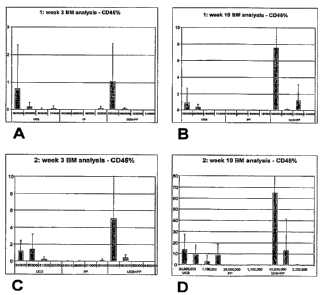
[0033] FIGS. 1 A-1 D: Summary of FACS analysis of engrafted human cells in
mice bone
marrow using CD45 antibodies in two independent experiments. (A): First
experiment,
CD45+ cells present in bone marrow at 3 weeks for umbilical cord blood cells
only (UCB),
placental perfusate cells only (PP) or umbilical cord cells combined with
placental perfusate
cells (UCB+PP). X-axis: numbers of cells per transplantation. (B): First
experiment, CD45+
cells at 10 weeks post-transfusion. (C): Second experiment, CD45+ cells in
bone marrow at
3 weeks post-transfusion. (D): Second experiment, CD45+ cells in bone marrow
at 10 weeks
post-transfusion.
[0034] FIG. 2: FACS analysis of engrafted human cells expressing lymphomyeloid
cell
markers in NOD/S CID mice. Co-expression of CD45+ with CD19 (left bar in each
category);
CD33 (middle bar); or CD7 (right bar). X-axis: numbers of cells per
transplantation. UCB =
transplantation of umbilical cord blood cells only; PP = transplantation of
placental perfusate
cells only. UCB+PP = transplantation of umbilical cord cells combined with
placental
perfusate cells.
5. DETAILED DESCRIPTION OF THE INVENTION
[0035] The present invention provides combinations of (1) placental stem
cells, e.g.,
placental stem cells in human placental perfusate, placental stem cells in
placental enzymatic
digestate, isolated placental stem and/or progenitor cells, and the like; and
(2) stem cells from
a second source, in a total number of cells, wherein the placental stem cells
and stem cells
from the second source are present in the combination in a ratio that produces
a greater
number of colony-forming units compared to a number of colony-forming units
produced by
placental stem cells or by stem cells from a second source, equivalent to said
total number of
cells, alone. The invention further provides combinations of placental stem
cells and stem
11
CA 02633775 2008-06-18
WO 2007/079184 PCT/US2006/049492
cells from a second source that enhance engraftment in vivo compared to the
number of
colony-forming units produced by a number of placental stem cells equivalent
to the number
of cells in said combination, or a number of stem cells from a second source
equivalent to the
number of cells in said combination, alone. The present invention further
provides methods
of identifying such ratios, and such combinations, and methods of using the
combined stem
cell populations.
5.1 OPTIMIZING COMBINATIONS OF PLACENTAL STEM CELLS
AND STEM CELLS FROM A SECOND SOURCE
5.1.1 In Vitro Assay
[0036] The invention provides in vitro co-culture methods for identifying a
combination of
placental stem cells and stem cells from a second source that has improved
engraftment
potential as compared to a number of either placental stem cells or stem or
progenitor cells
from a second source, equivalent to the number of cells in said combination,
alone. The in
vitro co-culture assay thus identifies ratios of placental stem cells to stem
cells from a second
source that improve the number of colony-forming units, and engraftment, in a
non-cell
number-dependent manner.
[0037] In one embodiment, for example, the invention provides a method of
identifying a
ratio of placental stem cells to stem cells from a second source, comprising
identifying a ratio
of placental stem cells to stem cells from a second source in a total number
of cells that, when
said placental stem cells and stem cells from a second source are cultured
together for a time
and under conditions that allow the formation of colony-forming units,
produces a greater
number of colony-forming units than a number of placental stem cells or stem
cells from a
second source, equivalent to the number of cells in said total number of
cells, alone. In
another embodiment, where several ratios are compared, the invention provides
a method of
identifying a ratio of placental stem cells and stem cells or progenitor cells
from a second
source in a total number of cells, comprising contacting a population of said
placental stem
cells in vitro with a population of said stem cells from a second source in a
plurality of ratios
for a time and under conditions sufficient to allow the formation of colony-
forming units, and
identifying a ratio within said plurality of ratios that yields the greatest
number of colony-
forming units. In a specific embodiment, said ratio improves engraftment into
a recipient as
compared to engraftment by a number of placental stem cells or stem cells from
a second
source, equivalent to the number of cells in said total number of cells,
alone. In more specific
embodiments, said combined stem cell population improves engraftment in an
individual in
12
CA 02633775 2008-06-18
WO 2007/079184 PCT/US2006/049492
need of stem cells for at least 1, 2, 3, 4, 5, 6, 7, 8, 9, 10, 11, 12, 13,'14,
15, 16, 17, 18, 19, 20
or 21 days post-transplant. In another more specific embodiment, said combined
stem cell
population improves engraftment in an individual in need of stem cells at a
time more than 21
days post-transplant.
5.1.1.1 Placenta-Derived Stem Cells
[0038] Placenta-derived stem cells useful in the methods and compositions of
the invention
include, for example, embryonic-like cells, pluripotent cells, multipotent
cells, committed
progenitor cells, hematopoietic progenitor cells, and mesenchymal-like stem
cells from
placenta. In one embodiment, the placenta-derive stem cells are contained
within, or are
derived from, placental perfusate.
[0039] Placenta-derived stem cells used in the methods of the invention can be
derived from
a single placenta, or from a plurality of placentas, and may be obtained by
any method.
Placenta-derived stem cells can be obtained by, for example, perfusion, as
disclosed in U.S.
Application Publication Nos. 2002/0123141 and 2003/0032179, the disclosures of
each of
which are incorporated herein by reference. Such perfusion can be perfusion by
the pan
method, wherein perfusion liquid is forced through the placental vasculature
and perfusion
fluid that exudes from the placenta, typically the maternal side, is collected
in a pan
containing the placenta. Perfusion can also be a closed-circuit perfusion,
wherein perfusion
fluid is passed through, and collected from, only the fetal vasculature of the
placenta. In a
specific embodiment, such perfusion can be continuous, that is, perfusion
fluid that has been
passed through the placenta, and which comprises a plurality of placental
cells, is passed
through a second time, or a plurality of times, prior to isolation of
placental cells.
[0040] Placenta-derived stem cells may also be obtained by physical or
enzymatic disruption
of the placenta using, e.g., proteases and/or other tissue-disruptive enzymes
to disrupt the
multicellular structure of the placenta. Such proteases may include neutral
proteases or
metalloproteases, e.g., collagenase, dispase, trypsin, elastase, and the like.
Placental stem
cells may also be obtained by physical disruption of the placenta using, e.g.,
mucolytic
enzymes, for example, hyaluronidase.
[0041] The isolated perfused placenta of the invention provides a source of
large quantities of
stem cells enriched for CD34+ stem cells, e.g., CD34+CD38- stem cells, e.g.,
CD34+, CD38-,
liri stem cells, and CD34- stem cells, e.g., CD34-CD38+ stem cells. The first
collection of
blood from the placenta is referred to as cord blood which contains
predominantly
CD34+CD38+ hematopoietic progenitor cells. Within the first twenty-four hours
of post-
13
CA 02633775 2008-06-18
WO 2007/079184 PCT/US2006/049492
partum perfusion, high numbers (e.g., 1 x 10⁵ to about 2 x 10⁷) of CD34+CD38-
hematopoietic progenitor cells may be isolated from the placenta, along with
high
concentrations of CD34-CD38+ cells. After about twenty-four hours of
perfusion, high
numbers (e.g., 1-10 million) of CD34-CD38- cells can be isolated from the
placenta along
with the aforementioned cells. An isolated placenta that has been perfused for
twenty-four
hours or more provides a source of large quantities of stem cells enriched for
CD34-CD38-
stem cells.
[0042] In another embodiment, the combined stem cell populations of the
invention comprise
CD34+ placental stem cells that are positive for aldehyde dehydrogenase
(ALDH). Such cells
demonstrate detectable levels of ALDH activity in an ALDH assay. Such assays
are known
in the art (see, e.g., Bostian and Betts, Biochem. J., 173, 787, (1978)). In a
specific
embodiment, said ALDH assay uses ALDEFLUOR (Aldagen, Inc., Ashland, Oregon)
as a
marker of aldehyde dehydrogenase activity. Thus, in various embodiments, a
combined stem
cell population of the invention comprises CD34+ stem cells, where at least
about 5%, 10%,
15 !0, 20%, 25 fo, 30%, 35%, 40%, 45 .' 0, 50%, 55%, 60 Oo, 65 fo, 70%, 75%,
80%, 85%, 90%,
or at least 95% of the CD34+ stem cells are ALDH+.
[0043] At least one class of human placental stem cells has characteristics of
embryonic stem
or germ cells. For example, stem cells of this class are SSEA3- (stage-
specific embryonic
antigen 3), SSEA4-, OCT-4+ (a stem cell transcription factor) and ABC-p+ (ATP-
binding
cassette (ABC) transporter protein), a marker profile exhibited by pluripotent
stem cells that
have not yet undergone differentiation. Thus, the methods and compositions of
the invention
can use or comprise non-embryonic, placental stem cells that are, e.g., SSEAY,
SSEA4-,
OCT-4+ or ABC-p+. Preferably, the placental stem cells are OCT-4+ABC-p+, and,
even more
preferably, are SSEA3-SSEA4-OCT-4+ABC-p*. In another embodiment, the invention
encompasses the use of placental stem cells positive for at least one of CD
10, CD29, CD44,
CD54, CD90, CD73 or CD105, or negative for at least one of CD34, CD38, or
CD45. In
another embodiment, the methods and compositions of the invention can use or
comprise
placental stem cells having or positive for CD 10, CD29, CD44, CD54, CD90,
CD73 or
CD 105, and lacking or negative for CD34, CD3 8, or CD45. In another
embodiment, the
methods and compositions of the invention can use or comprise placental stem
cells positive
for at least one of CD 10, CD29, CD44, CD54, CD90, CD73 or CD 105, or negative
for at
least one of CD34, CD38, or CD45. In another embodiment, the invention
encompasses the
use of placental stem cells having or positive for CD 10, CD29, CD44, CD54,
CD90, CD73 or
CD 105, and lacking or negative for CD34, CD3 8, or CD45.
14
CA 02633775 2008-06-18
WO 2007/079184 PCT/US2006/049492
[0044] In one embodiment, placental stem cells used in the methods and
compositions of the
invention are identified by the presence of the markers CD10, CD29, CD44,
CD54, CD90,
CD 105 (SH2), CD73 (SH3, SH4), OCT-4, and/or ABC-p, and/or the absence of the
markers
CD34, CD38, CD45, SSEA3, or SSEA4. In a specific embodiment, the placental
stem cells
are CD10+, CD29+, CD34-, CD38-, CD44+, CD45-, CD54+, CD73+, CD90+, CD105*,
SH2+,
SH3+, SH4+, SSEA3-, SSEA4-, OCT-4+, and ABC-p+. In another specific
embodiment, the
placental stem cells are CD200+ and HLA-G+. In this context, "SH2+", "SH3+"
and "SH4+"
mean that a stem cell is bound by antibody SH2, SH3, or SH4, respectively. In
another
specific embodiment, the placental stem cells are CD73+, CD105+ and CD200*. In
another
specific embodiment, the placental stem cells are CD200+ and OCT-4+. In
another specific
embodiment, the placental stem cells are CD73+, CD 105+ and facilitate the
formation of
embryoid-like bodies in a population of isolated placental cells comprising
said stem cells,
when said population is cultured under conditions that allow the formation of
embryoid-like
bodies. In another specific embodiment, the placental stem cells are CD73{, CD
105+ and
HLA-G+. In another specific embodiment, the placental stem cells are OCT-4+
and facilitate
the formation of embryoid-like bodies in a population of isolated placental
cells comprising
said stem cells, when said population is cultured under conditions that allow
the formation of
embryoid-like bodies. As used herein, "embryoid-like bodies" refers to three-
dimensional
clusters of differentiating, and differentiated, cells that emerge from the
adherent stem cell
layer.
[0045] In another embodiment, the human placental stem cells do not express
MHC Class 2
antigens.
[0046] Populations of placental perfusate-derived stem cells, in one
embodiment, comprise
trophoblasts.
[0100] Cell markers, e.g., stem cell markers and cell surface markers, can be
routinely
determined according to methods well known in the art, e.g. by flow cytometry
or
fluorescence-activated cell sorting (FACS) analysis by washing and staining
with an anti-cell
surface marker antibody labeled with an appropriate fluorophore. For example,
to determine
the presence of CD34 or CD38, cells may be washed in PBS and then double-
stained with
anti-CD34 phycoerythrin and anti-CD38 fluorescein isothiocyanate (Becton
Dickinson,
Mountain View, CA). The cells would then be analyzed using a standard flow
cytometer.
Alternatively, intra-cellular markers can also be examined via standard
methodology.
Antibody/fluorophore combinations to specific markers include, but are not
limited to,
fluorescein isothiocyanate (FITC) conjugated monoclonal antibodies against HLA-
G
CA 02633775 2008-06-18
WO 2007/079184 PCT/US2006/049492
(available from Serotec, Raleigh, North Carolina), CD10 (available from BD
Immunocytometry Systems, San Jose, California), CD44 (available from BD
Biosciences
Pharmingen, San Jose, California), and CD105 (available from R&D Systems Inc.,
Minneapolis, Minnesota); phycoerythrin (PE) conjugated monoclonal antibodies
against
CD44, CD200, CD 117, and CD 13 (BD Biosciences Pharmingen); phycoerythrin-Cy7
(PE
Cy7) conjugated monoclonal antibodies against CD33 and CD10 (BD Biosciences
Pharmingen); allophycocyanin (APC) conjugated streptavidin and monoclonal
antibodies
against CD38 (BD Biosciences Pharmingen); and Biotinylated CD90 (BD
Biosciences
Pharmingen). Other antibody/label combinations that can be used include, but
are not limited
to, CD133-APC (Miltenyi), KDR-Biotin (CD309, Abeam), CytokeratinK-Fitc (Sigma
or
Dako), HLA ABC-Fitc (BD), HLA DRDQDP-PE (BD), (3-2-microglobulin-PE (BD), CD80-
PE (BD) and CD86-APC (BD), CD45-PerCP (peridin chlorophyll protein); CD44-PE;
CD 19-
PE; CD10-F (fluorescein); HLA-G-F and 7-amino-actinomycin-D (7-AAD); HLA-ABC-
F;
and the like.
[0101] Placental stem cells, e.g., placental stem cells contained in placental
perfusate, can be
used immediately after collection, or can be cultured for a period of time
prior to assaying or
administration to an individual in a combined stem cell population. For
example, in one
embodiment, the stem cells can be cultured in medium comprising Notch agonist,
e.g., a
deletion form of a Notch protein consisting essentially of the intracellular
domain of the
Notch protein, or a Delta protein. See U.S. 2004/0067583.
5.1.1.2 Stem Cells From a Second Source
[0047] The methods and compositions described herein use placental stem cells
in
combination with stem cells from a second source, that is, stem cells from any
source other
than a mammalian placenta. Stem cells from a second source can comprise one or
more
types of stem cells, such as embryonic stem cells, embryonic germ cells, adult
stem cells,
mesenchymal stem cells, hematopoietic stem cells, non-hematopoietic stem
cells, bone
marrow-derived stem cells, neural stem cells, cardiac stem cells, ocular stem
cells, epithelial
stem cells, endothelial stem cells, hepatic stem cells, pulmonary stem cells,
muscle stem cells,
intestinal stem cells, and the like. Stem cells from a second source can be
stem cells isolated
from the second, non-placental source, or can be tissue comprising the stem
cells. As for the
placenta, stem cells can be isolated by perfusion of the organ(s) comprising
the stem cells, or
by tissue disruption and/or enzymatic digestion of the organ(s) comprising the
stem cells.
16
CA 02633775 2008-06-18
WO 2007/079184 PCT/US2006/049492
Stem cells from a second source can be, e.g., stem cells derived solely from
umbilical cord,
or solely from amniotic fluid.
[0048] Stem cells from a second source may be obtained by providing a sample
of a relevant
tissue, and isolating stem cells from the tissue using one or more cell
surface markers. For
example, hematopoietic stem cells may be obtained from blood (e.g., peripheral
blood,
placental blood, umbilical cord blood) or from bone marrow by obtaining a
sample of blood
or bone marrow, isolating mononuclear cells from the blood or bone marrow, and
separating
CD34+ cells from the isolated mononuclear cells. Such separation may be
accomplished by
methods routine in the art, e.g. using apherisis, followed by separation using
magnetic beads
or a column comprising one or more antibodies to the cell surface marker,
e.g., CD34 or
CD200; fluorescence-activated cell sorting (FACS), and the like. For blood,
the stem cells
can be provided in a population of total nucleated cells (TNC) from the blood,
e.g., total
nucleated cells from peripheral blood, placental blood, umbilical cord blood,
and the like.
[0049] Stem cells from other tissues may be isolated in a similar manner.
Mesenchymal stem
cells may be isolated from, e.g., bone marrow by isolation of cells positive
for CD73, CD105
and/or CD45 (see, e.g., U.S. Patent No. 6,387,367). Ocular (limbal) stem cells
may be
obtained from the cornea by obtaining corneal cells and isolating SSEA-4+
cells (see, e.g.,
U.S. Application Publication No. 2005/0186672). Hepatic stem cells may be
obtained from
liver, particularly fetal liver, samples, by selecting cells expressing CD14,
CD34, CD38,
ICAM, CD45, CD 117, glycophorin A, connexin 32, osteopontin, bone
sialoprotein, collagen
I, collagen II, collagen III, collagen IV, or combinations thereof (see, e.g.,
U.S. Application
Publication No. 2005/0148072). Muscle stem cells may be obtained from muscle
tissue by
selecting CD34+CD45- cells that do not express other hematopoietic cell
markers (see, e.g.,
U.S. Application Publication No. 2005/0079606). Cardiac stem cells may be
isolated from
cardiac tissue by selecting c-kit CD31+CD38+ cells (see, e.g., U.S.
Application Publication
No. 2004/0126879). Isolation of stem cells may be accomplished using other
known
characteristics or markers, as well.
[0050] In one embodiment, said stem cells from a second source are cord blood
stem cells.
In specific embodiments, the cord blood stem cells are CD34+ stem cells, e.g.,
CD34+, CD38+
stem cells, CD34+, CD38' stem cells, CD34+, CD38-, liri stem cells, and the
like. In a
specific embodiment, the CD34+ stem cells from a second source are ALDH+. Cord
blood
itself, or stem and/or progenitor cells obtained from cord blood, can be used
in the methods of
the invention. In a specific embodiment, said cord blood-derived cells
comprise
hematopoietic stem cells, where the combined stern cell population is to be
used for
17
CA 02633775 2008-06-18
WO 2007/079184 PCT/US2006/049492
hematopoietic engraftment. The stem cells from a second source may be derived
from a
single donor, or from a plurality of donors in equal or unequal amounts. Stem
cells from a
plurality of second (that is, non-placental) sources may be combined with
placental stem
cells, and used for the methods and compositions of the present invention.
[0051] Stem cells from a second source, e.g., hematopoietic stem cells from a
second source,
can be used immediately after collection, or can be cultured for a period of
time prior to
assaying or administration to an individual in a combined stem cell
population. For example,
in one embodiment, the stem cells can be cultured in medium comprising Notch
agonist, e.g.,
a deletion form of a Notch protein consisting essentially of the intracellular
domain of the
Notch protein, or a Delta protein. See U.S. 2004/0067583
5.1.1.3 Assay Parameters
[00521 Once a population of placental stem cells and a population of stem
cells from a
second source are obtained, the cells can be combined in an in vitro co-
culture, or colony-
forming, assay to determine if the number of stem cells in a particular
combination produces
more colony-forming units than a number of placental stem cells or stem cells
from a second
source, equivalent to the number of cells in said combination, alone. Any such
combination
of placental stem cells and stem cells from a second source in a ratio that
produces more
colony forming units than either placental stem cells or stem cells from a
second source
alone, for equivalent numbers of cells, is identified as a combined stem cell
population of the
invention.
[00531 The identification of a combined stem cell population can use any
colony forming
unit assay commonly used and known in the art, provided the assay allows for
the
proliferation and differentiation of stem cells from placenta and from a
second source, for
example, colony forming assays provided by StemCell Technologies, Inc. Such an
assay
may use, e.g., MESENCULTTM medium (Stem Cell Technologies, Inc., Vancouver
British
Columbia). The identification of combined stem cell populations can use cells
that are
freshly-prepared, or thawed from frozen stocks, or both. Preferably, both the
placental stem
cells and stem cells from a second source are in suspension when combined for
co-culture.
Placental stem cells, and stem cells from a second source, may be assessed for
viability,
proliferation potential, and longevity using standard techniques known in the
art, such as
trypan blue exclusion assay, fluorescein diacetate uptake assay, propidium
iodide uptake
assay (to assess viability); and thymidine uptake assay, MTT (3-(4,5-
dimethylthiazol-2-yl)-
2,5-diphenyltetrazolium bromide) cell proliferation assay (to assess
proliferation). Longevity
18
CA 02633775 2008-06-18
WO 2007/079184 PCT/US2006/049492
may be determined by methods well known in the art, such as by determining the
maximum
number of population doublings in an extended culture.
[0054] In one embodiment of the in vitro method, a colony forming unit assay
using
placental stem cells and cord blood-derived stem cells is performed as
follows. Fresh or
thawed HLA/donor matched placental perfusate and cord blood units are
obtained, and the
number of total nucleated cells in each is determined with a hemacytometer.
Where thawed
units are used, cord blood samples can be hetastarch-separated, and placental
perfusate units
are preferably Ficoll-separated. Small samples of nucleated cells from each
source are
seeded together in suspension in two or more ratios in a co-culture, and
expanded. The co-
culture can be performed in, e.g., triplicate for one or more ratios of
placental stem cells to
stem cells from a second source, in, for example, 35 mm dishes in an
appropriate cell culture
medium (e.g., RPMI 1640 medium supplemented with 2-10% fetal calf serum and,
optionally, 1% Stemspan cytokine cocktail; Methocult GF+ H4435 medium, etc.).
Hematopoietic stem cells may be expanded in culture medium comprising GM-CSF,
IL-3,
IL-6, SCF and flt-3 ligand.
[0055] The container used for the co-culture assay is preferably appropriate
for tissue culture
of stem cells. For example, co-cultures may be performed in glass or plastic
Petri dishes, 16-
well plates, 32-well plates, 96-well plates, 128-well plates, and the like.
Typically, the total
number of nucleated cells from each source in each co-culture varies from 1 x
104 to I x 106.
Cells may also be co-cultured in a micropatterned configuration. See U.S.
Patent No.
6,221,663.
[0056] When determining the ratio of placental stem cells to stem cells from a
second source
in a cell population that comprises a number of placental stem cells and stem
cells from a
second source, the preferred ratio is any ratio that generates more colony
forming units than
that generated by said number of placental stem cells or said number of stem
cells from a
second source under the same conditions. More preferably, the ratio is a ratio
that generates a
higher number of colony-forming units than all other ratios tested.
Statistical significance
between ratios tested is desirable, but not necessary. The higher number of
colony-forming
units may be attributable to, or be derived from, both placental stem cells
and stem cells from
a second source; from predominantly or only the placental stem cells; or
predominantly or
only the stem cells from a second source.
[0057] The combined stem cell population is cultured for a time sufficient for
colony forming
units to form, typically 10-20 days. Cell culture during expansion follows
standard protocols
known in the art of stem or progenitor cell culture, and includes, for
example, daily or semi-
19
CA 02633775 2008-06-18
WO 2007/079184 PCT/US2006/049492
daily changes of medium; culture at about 37 C at 5% CO2 in a humidified
incubator, and the
like. After 10-20 days, the number and morphology of colony forming units in
the co-culture
is determined (e.g., for hematopoietic stem cells, the number of CFU-GM, CFU-
L, CFU-M,
CFU-G, CFU-DC, CFU-GEMM, CFU-E).
[0058] In a specific example of the co-culture assay, nucleated cells from
placenta perfusate,
and nucleated cells from cord blood are combined a ratio of 1:1, 1:3 and 3:1
(where 1 equals,
e.g., 1 x 10⁵ cells) in Methocult GF+ H4435 medium. The co-culture is then
expanded in
tissue culture for about 14 days. The morphology of the co-cultured cells, and
the number of
colony forming units, is determined. The ratio of the nucleated cell samples
from the two
sources that provides the highest number of colony-forming units is designated
an optimum
ratio, and the two units, or stem and/or progenitor cells from one or both of
the units, are
combined in the optimum ratio for administration to a recipient in need of a
stem cell
transplant. Such an optimum ratio provides superior engraftment in vivo over
the
administration of either unit, or stem and/or progenitor cells from either
unit, alone, where
equivalent numbers of cells are administered.
[0059] The placental stem cells and stem cells from a second source are
contacted with each
other during the co-culturing, either directly or indirectly. At a minimum,
this comprises
contacting one of the types of stem cells with culture medium in which the
other type of stem
cell has cultured for a period of time, e.g., contacting one of the types of
stem cells with
medium that has been conditioned by the other type of stem cell. For example,
the placental
stem cells, and stem cells from a second source may be cultured together in
the same physical
space during culture for colony-forming unit formation, e.g., in the same
culture dish or well
in a multi-well plate. The placental stem cells and stem cells from a second
source may also
be contacted with each other by culturing in separate physical spaces, but in
common culture
medium (e.g., separated by a membrane, or in two wells of a multiwell plate
wherein culture
medium may move actively or passively between the wells, but cells cannot
mix). In another
embodiment, placental stem cells and stem cells from a second source may be
cultured in
separate physical spaces with no common culture medium, and the stem cells
brought into
contact with each other by an exchange of part or all of the culture medium
from one stem
cell culture with that of the other. In another embodiment, the cells in the
co-culture are
cultured in a manner that physically separates the cells, but allows
biomolecules to diffuse
between the two cultures. See, e.g., U.S. Patent No. 5,665,596 "Device for
Cell Co-culture
and Method for Its Use in Culturing Cells". Where the stem cell cultures are
separate, the
CA 02633775 2008-06-18
WO 2007/079184 PCT/US2006/049492
number of colony-forming units in the separate, paired cultures is totaled for
each replicate of
ratio, and an optimum ratio determined, as above.
[0060] In another embodiment of the method, a bioactive molecule is added to
the placental
stem cells and stem cells from a second source during the assay, and a ratio
of placental stem
cells to stem cells from a second source is identified that, for a total
number of cells, results in
more colony-forming units, or enhanced engraftment, compared to a number of
placental
stem cells or stem cells from a second source, equivalent to said total number
of cells in said
combination, alone. Such a bioactive molecule may be a small organic molecule
of less than
50 kDa, 30 kDa, 20 kDa, 10 kDa, 5 kDa, 3kDa, 2 kDa, I kDa, 500 Da, 300 Da, -
200 Da, 100
Da or smaller. In a specific embodiment, said small organic molecule is
synthetic or non-
natural, that is, not derived from a natural source. In another specific
embodiment, said
bioactive molecule is a cytokine or growth factor. Bioactive molecules that
can be added to
the co-culture include differentiation-inducing agents such as, but are not
limited to, Caz+,
EGF, a-FGF, 0-FGF, PDGF, keratinocyte growth factor (KGF), TGF-0, cytokines
(e.g., IL-
la, IL-10, IFN-y, TFN), retinoic acid, transferrin, hormones (e.g., androgen,
estrogen,
insulin, prolactin, triiodothyronine, hydrocortisone, dexamethasone), sodium
butyrate, TPA,
DMSO, NMF, DMF, matrix elements (e.g., collagen, laminin, heparan sulfate,
MATRIGELT"'), or combinations thereof. Bioactive molecules that are
differentiation
suppressants may also be added, such as, but not limited to, human Delta-1 and
human
Serrate-1 polypeptides (see, Sakano et al., U.S. Patent No. 6,337,387 entitled
"Differentiation-suppressive polypeptide", issued January 8, 2002), leukemia
inhibitory
factor (LIF), and stem cell factor.
[0061] Where a bioactive molecule is added to the co-culture, the co-culture
assay may be
used to identify a positive effector of engraftment. In one embodiment,
therefore, the
invention provides a method of identifying a bioactive molecule that is a
positive effector of
engraftment comprising contacting a combined stem cell population with said
bioactive
molecule, wherein said bioactive molecule is identified as a positive effector
of engraftment
if engraftment by said combined stem cell population is detectably enhanced
compared to
engraftment by a combined stem cell population not contacted with said
bioactive molecule.
In another embodiment, the invention provides a method of identifying a
positive effector of
engraftment comprising combining placental stem cells and stem cells from a
second source
in vitro in one or more ratios in the presence of said bioactive molecule;
culturing said
placental stem cells and stem cells from a second source for a time sufficient
for colony
forming units to form; determining the number of colony-forming units for each
of said one
21
CA 02633775 2008-06-18
WO 2007/079184 PCT/US2006/049492
or more ratios; and determining, for at least one of said one or more ratios,
whether the
number of colony forming units in the presence of said bioactive molecule is
greater than the
number of colony forming units in the absence of said bioactive molecule, and,
if so,
identifying said bioactive molecule as a positive effector of engraftment.
[0062] The in vitro assay may be performed on any placental stem cell
population and stem
cell population from a second source to determine an optimum ratio for
engraftment. In this
aspect, the in vitro co-culture assay can be used as a standard, routine
procedure to
characterize stem cell populations prior to transplantation.
5.1.2 In Vivo Assay
[0063] The results of the above in vitro assay may be confirmed using an in
vivo engraftment
assay. The in vivo assay may also be performed in the absence of the in vitro
assay to
determine an optimum ratio of placental stem cells, and stem cells from a
second source, to
maximize engraftment.
[0064] In one embodiment of the in vivo assay, placental stem cells and stem
cells from a
second source are transplanted into a plurality of model animals and given
sufficient time to
engraft (typically 6-10 weeks). The animals are subsequently sacrificed, and
the degree of
engraftment in each animal is determined for at least one tissue. Thus, in one
embodiment,
the invention provides a method of identifying a ratio of placental stem cells
and stem cells or
progenitor cells from a second source for engraftment into a recipient,
comprising identifying
a ratio of placental stem cells to stem cells from a second source in a total
number of cells
that, when transplanted into an animal, results in enhanced engraftment
compared to
transplantation of a number of placental stem cells or stem cells from a
second source,
equivalent to the number of cells in said total number of cells, alone. In
another embodiment,
said identifying a ratio of placental stem cells to stem cells from a second
source comprises
transplanting a number of placental stem cells and stem cells from a second
source in a
plurality of animals, in a plurality of ratios; determining the number of
engrafted cells in at
least one tissue of said animals for each of said plurality of ratios; and
identifying the ratio in
said plurality of ratios that yields the highest number of engrafted cells.
[0065] As in the in vitro assay, the placental stem cells can be placental
stem cells obtained
by any means or present in any usable form. For example, the placental stem
cells may be
contained in placental perfusate, or may be contained within isolated total
nucleated cells
from the placental perfusate, or may be a population of stem cells isolated
from the total
nucleated cells, or may be placental stem cells contained within enzyme-
digested placental
22
CA 02633775 2008-06-18
WO 2007/079184 PCT/US2006/049492
tissue, or may be placental stem cells isolated from enzyme-digested placental
tissue, or may
be placental stem cells that have been expanded and/or passaged in culture,
etc.
[0066] Any standard model animal may be used in the in vivo co-culture assay.
Preferably,
the model animal is one in which engraftment of xenografts may be readily
accomplished.
Small mammals such as standard laboratory rodents such as mice and rats are
preferred
because they require fewer administered stem cells to show engraftment. It is
highly
preferable that the model animal be immune-compromised. Animal models that may
be used
in the in vivo assay include, but are not limited to, NOD/SCID (non-obese
diabetic /severe
combined immune deficiency) mice (see Hogan et a1., Blood 90(l):85-96 (1997));
beige/nude/x-linked immunodeficiency (BNX) mice (see, e.g., Kamal-Reid et al.,
Science
242:1706 (1988)); SCID mice (see, e.g., Kamal-Reid et al., Science 246:1597
(1989).
Engraftment may be accomplished in other animal models, such as sheep fetuse's
(see, e.g.,
Shimizu et al., Blood 91(10):3688-3692 (1998); Zanjani et al., Int'l J.
Hemato163(3):179-
182 (1996)).
[00671 The determination of the number of engrafted cells in tissues from the
recipient
animal may be accomplished by any means known in the art. For example,
detection of
engrafted cells may be accomplished by detection of engrafted cell-specific
nucleic acids,
e.g., by the polymerase chain reaction, or by detection of proteins specific
for engrafted cells,
e.g., by immunohistochemstry. Identification of engraftment in vivo may be
determined
through the use of a sample, e.g., biopsy specimen, taken at one or more
locations on, and at
one or more post-transplantation times from, a recipient.
[0068] In one embodiment, demonstration of engraftment of placental stem cells
and/or cord
blood-derived stem cells can be accomplished by taking a biopsy (e.g., bone
marrow aspirate
or peripheral blood sample) and performing PCR to determine whether any non-
recipient
genetic markers are present, which would indicate engraftment. In another
embodiment,
identification of engrafted cells is accomplished by selection of one or more
antibodies that
recognize markers expressed by the engrafted cells. In a specific embodiment,
the engrafted
cells are human, and the one or more antibodies specifically recognize one or
more human
cell markers. Antibodies can be used to detect the markers by any art-accepted
method, e.g.,
immunohistochemical methods. For example, determination of the presence of a
cell surface
marker can comprise sacrifice of a non-human host animal, obtaining a desired
tissue, fixing
and embedding the tissue in paraffin or a similar matrix; thin sectioning the
tissue, optionally
followed by staining; and contacting the tissue with one or more antibodies
that recognize the
marker. In the same manner, one may use antibodies that recognize markers
expressed by
23
CA 02633775 2008-06-18
WO 2007/079184 PCT/US2006/049492
cells into which the engrafted stem cells can differentiate. For example,
placental stem cells
or cord blood-derived stem cells differentiate into cells that express CD45
and vimentin; thus,
antibodies to CD45 and vimentin may be used to determine the number of
engrafting, and
differentiating, stem cells. Antibodies that recognize, e.g., human cell
surface markers in
preference to host cell markers, e.g., mouse cell surface markers, are well-
known in the art.
[00691 In a non-limiting example of the in vivo method, a plurality of model
animals, e.g., a
plurality of mice of the species Mus musculus, are transplanted with human
placental stem
cells and, e.g., human nucleated cells isolated from cord blood, including
hematopoietic stem
cells, in a plurality of ratios. After several days to several weeks (i.e.,
sufficient time to allow
engraftment), the host animals are sacrificed, and tissues (e.g., spleen,
lung, etc.) are
examined to determine the approximate number of human cells that have
engrafted, as
evidenced by the number of cells staining for CD45 and/or vimentin. CD45 is a
marker
specific for leukocytes, including T- and B-lymphocytes, granulocytes,
monocytes and
macrophages. Certain CD45 antibodies, such as clone T29/33 (BioDesign, Saco,
Maine), do
not cross-react with mouse antigens. Vimentin is a marker for mesenchymal
cells, such as
fibroblasts, smooth muscle cells, lipocytes, Schwann cells, vascular
endothelial cells, and the
like. Certain vimentin antibodies, such as clone V9 (BioDesign, Saco, Maine),
do not cross-
react with mouse antigens. Staining with antibodies to these two markers,
therefore, can
establish generally the extent of engraftment of placental stem cells, and
stem cells from a
second source, in a variety of tissues. This example is not limiting;
different antibodies may
be used to determine the extent of engraftment of other cell types. In a long-
term
engraftment model, bone marrow cells isolated from a primary engrafted animal,
e.g., a
mouse, can be transplanted into a second engraftment model animal. Assays for
secondary
engraftment are as listed above and include methods well known to those of
skill in the art.
5.2 COMBINED STEM CELL POPULATIONS
[0070] The invention further provides combined stem cell compositions
comprising placental
stem cells, e.g., cells from placental perfusate, e.g., nucleated cells from
placental perfusate,
comprising placental stem cells and stem cells from a second source that, for
a particular
number of cells, results in a greater number of colony-forming units in a
colony-forming unit
assay, or enhanced engraftment in a transplant recipient, than the number of
either placental
stem cells or stem cells from a second source, alone. Combined stem cell
populations
identified by the above methods represent engraftment-enhanced combinations of
stem cells
24
CA 02633775 2008-06-18
WO 2007/079184 PCT/US2006/049492
based on the characteristics of the stem cell sources, that is, the number of
engraftable cells
contained in, e.g., a unit of placenta perfusate, a unit of cord blood, etc.
[00711 Thus, in one embodiment, the invention encompasses a combined stem cell
composition comprising a number of placental stem cells and stem cells from a
second source
in a ratio, wherein the stem cells from the composition show improved
engraftment compared
to a number of either the placental stem cells or the stem cells from a second
source,
equivalent to the number of cells in said composition, alone. In a specific
embodiment, the
ratio is identified by combining placental stem cells and stem cells from a
second source in
vitro in a plurality of ratios for a time and under conditions sufficient to
allow the formation
of colony-forming units; and identifying a ratio in said plurality of ratios
that yields the
highest number of colony forming units. In a more specific embodiment, said
stem or
progenitor cells from a second source are cord blood stem or progenitor cells,
bone marrow
stem or progenitor cells, hematopoietic stem or progenitor cells, or
mesenchymal stem or
progenitor cells. In another more specific embodiment, said stem cells or
progenitor cells
from a second source are hematopoietic progenitor cells. In an even more
specific
embodiment, said hematopoietic stem cells are cord blood hematopoietic stem
cells. In
another even more specific embodiment, said hematopoietic cells are CD34+
cells.
[0072] In another more specific embodiment, said placental stem cells comprise
CD34+ cells,
for example, CD34+CD38+ cells and/or CD34+CD38- cells. In a specific
embodiment, said
CD34+CD38- cells comprise CD34+CD38-liri stem cells. In another specific
embodiment,
said CD34+ placental stem cells comprise cells that are ALDH+, that is, CD34+,
ALDH+
placental stem cells.
[0073] In another more specific embodiment, said placental stem cells are OCT-
4+ or ABC-
p+. In another more specific embodiment, said placental stem cells comprise
cells that are
OCT4+ABC-p*. In another more specific embodiment, said placental stem cells
comprise
cells that are CD34+ and cells that are OCT4+ABC-p{. In another more specific
embodiment,
said placental stem cells are contained within placental perfusate
substantially lacking red
blood cells and cellular debris. In another more specific embodiment, said
composition
comprises placental stem cells isolated from placental perfusate.
[0074] In another embodiment, placental stem cells comprise cells that express
one or more
of markers CD10, CD29, CD44, CD54, CD90, CD73 or CD105, and lack one or more
of
markers CD34, CD38, CD45, SSEA3 and SSEA4. In another embodiment, placental
stem
cells comprise cells that are positive for CD 10, CD29, CD44, CD54, CD90, CD73
or CD 105,
and negative for CD34, CD38, CD45, SSEA3 and SSEA4. In another embodiment,
placental
CA 02633775 2008-06-18
WO 2007/079184 PCT/US2006/049492
stem cells comprise cells that comprise one or more of markers CD10, CD29,
CD44, CD54,
CD90, CD73 and CD105, and lack one or more of markers CD34, CD3S, CD45, SSEA3
and
SSEA4. In another embodiment, placental stem cells comprise cells that are
positive for
CD 10, CD29, CD44, CD54, CD90, CD73 and CD 105, and negative for CD34, CD38,
CD45,
SSEA3 and SSEA4. In another embodiment, said placental stem cells comprise
CD34- cells.
In a specific embodiment, said placental stem cells are CD34'CD38- placental
stem cells. In
another embodiment, said placental stem cells comprise cells that are positive
for at least one
of CD 10, CD29, CD33, CD44, CD73, CD 105, CD 117, and CD 13 3, and negative
for at least
one of CD34 or CD45. In another embodiment, said placental stem cells comprise
cells that
are positive for CDIO, CD29, CD33, CD44, CD73, CD105, CD117, and CD133, and
negative for CD34 or CD45. In a more specific embodiment, said placental stem
cells
comprise cells that are HLA-ABC+. In a more specific embodiment, said
placental stem cells
comprise cells that are HLA-ABC-. In a more specific embodiment, said
placental stem cells
comprise cells that are HLA-DR*. In a more specific embodiment, said placental
stem cells
comprise cells that are HLA-DR . In another specific embodiment, said
placental stem cells
comprise cells that are CD200+ or HLA-G+. In another specific embodiment, the
placental
stem cells comprise cells that are CD200+ and HLA-G{. In another specific
embodiment, the
placental stem cells comprise cells that are CD73+, CD105+ and CD200+. In
another specific
embodiment, the placental stem cells comprise cells that are CD200+ and OCT-
4+. In another
specific embodiment, the placental stem cells comprise cells that are CD73+,
CD105+ and
facilitate the formation of embryoid-like bodies in a population of isolated
placental cells
comprising said stem cells, when said population is cultured under conditions
that allow the
formation of embryoid-like bodies. In another specific embodiment, the
placental stem cells
comprise cells that are CD73+, CD105+ and HLA-G+. In another specific
embodiment, the
placental stem cells comprise cells that are OCT-4+ and facilitate the
formation of embryoid-
like bodies in a population of isolated placental cells comprising said stem
cells, when said
population is cultured under conditions that allow the forrnation of embryoid-
like bodies.
[00751 In another embodiment, said stem cells from a second source are stem
cells derived
from cord blood.
[0076] In the combined stem cell populations of the invention, the placental
stem cells and
the stem cells from a second source may be identically-HLA-matched, that is,
they may be
derived from the same individual. In another embodiment, the placental stem
cells and the
stem cells from a second source may be HLA-mismatched, that is, they may be
derived from
different individuals. For combined stem cell populations comprising cord
blood or cord
26
CA 02633775 2008-06-18
WO 2007/079184 PCT/US2006/049492
blood-derived stem cells, the combination may also comprise stem cells that
are either HLA-
matched, partially HLA-matched, or HLA-mismatched to an intended recipient.
For
combined stem cell populations comprising non-cord blood stem cells, it is
preferred that at
least the stem cells from a second source be HLA-matched or partially HLA-
matched to the
intended recipient.
[0077] In various embodiments, the ratio of placental stem cells to stem cells
from a second
source can be about 100,000,000:1, 50,000,000:1, 20,000,000:1, 10,000,000:1,
5,000,000:1,
2,000,000:1, 1,000,000:1, 500,000:1, 200,000:1, 1.00,000:1, 50,000:1,
20,000:1, 10,000:1,
5,000:1, 2,000:1, 1,000:1, 500:1, 200:1, 100:1, 50:1, 20:1, 10:1, 5:1, 2:1,
1:1; 1:2; 1:5; 1:10;
1:100; 1:200; 1:500; 1:1,000; 1:2,000; 1:5,000; 1:10,000; 1:20,000; 1:50,000;
1:100,000;
1:500,000; 1:1,000,000; 1:2,000,000; 1:5,000,000; 1:10,000,000; 1:20,000,000;
1:50,000,000; or about 1:100,000,000, comparing numbers of total nucleated
cells in each
population, or comparing total numbers of stem cells in each population. In a
preferred
embodiment, the ratio of placental stem cells to stem cells from a second
source can be about
1:10 to about 10:1. In other preferred embodiments, the ratio of placental
stem cells to stem
cells from a second source can be about 3:1 to about 1:3.
[00781 The combined stem cell populations of the invention can comprise a
therapeutically-
effective amount of placental stem cells, stem cells from a second source, or
both. The
combined stem cell populations of the invention, and pharmaceutical
compositions
comprising a combined stem cell population, can comprise at least 1 x 10⁴ 5 x
104, 1 x 10⁵, 5
x105a 1x10⁶ 5x10⁶a 1x10⁷a 5x10⁷a 1x108,5x108,1x109,5x109a 1x10'0a 5 x 10¹⁰, or
a
1 x 101 I placental stem cells, stem cells from a second source, or both, or
no more than 1 x
104,5x104,1x105,5x105,1x106,5x106,1x107,5x107,1x108,5x108,1x109,5x
109, 1 x 10¹⁰, 5 x 101fl, or 1 x 10¹¹ placental stem cells, stem cells from a
second source, or
both.
[0079] In other embodiments, said combined stem cell population improves
engraftment in
an individual in need of stem cells at least, or at, 1, 2, 3, 4, 5, 6, 7, 8,
9, 10, 11, 12, 13, 14, 15,
16, 17, 18, 19, 20 or 21 days post-transplant. In another more specific
embodiment, said
combined stem cell population improves engraftment in an individual in need of
stem cells at
least, or at, more than 21 days post-transplant. In specific embodiments, said
combined stem
cell population improves engraftment in an individual in need of stem cells at
least, or at,
more than 25, 30, 35, 40, 45, 50, 55 weeks, or 1 year or longer post-
transplant.
[0080J The combined stem cell populations of the invention can be preserved,
for example,
cryopreserved for later use. Methods for cryopreservation of cells, such as
stem cells, are
27
CA 02633775 2008-06-18
WO 2007/079184 PCT/US2006/049492
well known in the art, for example, cryopreservation using the methods of
Boyse et al. (U.S.
Patent No. 5,192,553, issued March 9, 1993) or Hu et al. (WO 00/73421,
published
December 7, 2000). Placenta-derived stem cells, and stem cells from a second
source, which
make up a combined stem cell population, can be combined prior to
cryopreservation, or can
be cryopreserved separately, and combined in the appropriate ratio upon
thawing, e.g., within
hours of use.
[00811 The combined stem cell populations of the invention can be prepared in
a form that is
easily administrable to an individual. For example, a combined stem cell
population can be
contained within a container suitable for medical use. Such a container can
be, for example,
a sterile plastic bag, flask, jar, or other container from which the combined
stem cell
population can be easily dispensed. Preferably, the container is a container
that allows, or
facilitates, intravenous administration of a combined stem cell population.
The container,
e.g., bag, can hold the placenta-derived stem cells and stem cells from a
second source
together, e.g., as a mixed cell population, or can hold the two stem cell
populations
separately. In the latter embodiment, the bag preferably comprises multiple
lumens or
compartments that are interconnected to allow mixing of the placenta-derived
stem cells and
stem cells from a second source prior to, or during, administration. The
container is
preferably one that allows for cryopreservation of the combined stem cell
population. The
combined stem cell population in said container can comprise placenta-derived
stem cells,
stem cells from a second source, or both, that have been passaged at least, or
at most, 1, 2, 3,
4, 5, 6, 7, 8, 9, 10, 11, 12, 13, 14, 15, 16, 17, 18, 19, 20 times, or 25, 30,
35, 40 or more times.
[0082] The invention also provides for combined stem cell populations that
comprise, e.g.,
that are stored or maintained as, separate stem cell populations, e.g., a
population of placenta-
derived stem cells and a population of stem cells from a second source, in
combination with
information on combining the two populations in an appropriate ratio prior to
use, e.g., prior
to administration to an individual in need of stem cells. In this embodiment,
a combined stem
cell population would comprise a population of placenta-derived stem cells in
a first
container, a population of stem cells from a second source in a second
container, and
instructions for combining the two populations either before or during
administration to an
individual in need of stem cells.
[0083] Thus, in one embodiment, the invention provides a composition
comprising a
combined stem cell population in a container, wherein said combined stem cell
population
comprises placenta-derived stem cells and stem cells from a second source. In
a specific
embodiment, the container is a bag, flask, or jar. In a more specific
embodiment, said
28
CA 02633775 2008-06-18
WO 2007/079184 PCT/US2006/049492
placenta-derived stem cells and said stem cells from a second source are
contained together in
said bag. In another more specific embodiment, said placenta-derived stem
cells and said
stem cells from a second source are contained separately within said bag. In
another specific
embodiment, the composition comprises one or more compounds that facilitate
cryopreservation of the combined stem cell population. In another specific
embodiment, said
combined stem cell population is contained within a physiologically-acceptable
aqueous
solution. In a more specific embodiment, said physiologically-acceptable
aqueous solution is
a 0.9% NaCI solution. In another more specific embodiment, said bag is a
sterile plastic bag.
In a more specific embodiment, said bag allows or facilitates intravenous
administration of
said combined stem cell population. In another specific embodiment, said
combined stem
cell population comprises placental cells that are HLA-matched to said stem
cells from a
second source. In another specific embodiment, said combined stem cell
population
comprises placental cells that are at least partially HLA-mismatched to said
stem cells from a
second source. In another specific embodiment, said placenta-derived stem
cells are derived
from a plurality of donors. In another specific embodiment, said stem cells
from a second
source are derived from a plurality of donors.
100841 Combined stem cell populations can be cultured for a period of time
prior to
administration to an individual. For example, in one embodiment, the stem
cells in a
combined stem cell population can be cultured in medium comprising Notch
agonist, e.g., a
deletion form of a Notch protein consisting essentially of the intracellular
domain of the
Notch protein, or a Delta protein. See U.S. 2004/0067583
5.3 PHARMACEUTICAL COMPOSITIONS
[0085] The present invention encompasses pharmaceutical compositions that
comprise
combined stem cell populations of the invention, and a pharmaceutically-
acceptable carrier.
[0086] In accordance with this embodiment, the combined stem cell populations
of the
invention may be formulated as an injectable (e.g., WO 96/39101, incorporated
herein by
reference in its entirety). In another embodiment, the combined stem cell
populations of the
present invention may be formulated using polymerizable or cross linking
hydrogels as
described, e.g., in U.S. Patent Nos. 5,709,854; 5,516,532; 5,654,381.
[0087] In another embodiment, the invention provides for the maintenance of
each stem cell
population of the combined stem cell populations, prior to administration to
an individual, as
separate pharmaceutical compositions to be administered sequentially or
jointly to create the
combined stem cell population in vivo. Each component may be stored and/or
used in a
29
CA 02633775 2008-06-18
WO 2007/079184 PCT/US2006/049492
separate container, e.g., one bag (e.g., blood storage bag from Baxter, Becton-
Dickinson,
Medcep, National Hospital Products, Terumo, etc.) or separate syringe, which
contains a
single type of cell or cell population. In a specific embodiment, cord blood,
or cord blood-
derived nucleated or stem cells, are contained in one bag, and placental
perftisate, or placental
stem cells from placental perfusate, are contained in a second bag.
[0088] A population of placental stem cells can be enriched. In a specific
embodiment, a
population of cells comprising placental stem cells is enriched by removal of
red blood cells
and/or granulocytes according to standard methods, so that the remaining
population of
nucleated cells is enriched for placental stem cells relative to other cell
types in placental
perfusate. Such an enriched population of placental stem cells may be used
unfrozen, or may
be frozen for later use. If the population of cells is to be frozen, a
standard cryopreservative
(e.g., DMSO, glycerol, EPILIFETM Cell Freezing Medium (Cascade Biologics)) is
added to
the enriched population of cells before it is frozen.
[0089] The pharmaceutical compositions of the invention may comprise one or
more agents
that induce cell differentiation. In certain embodiments, an agent that
induces differentiation
includes, but is not limited to, Ca2}, EGF, a-FGF, 0-FGF, PDGF, keratinocyte
growth factor
(KGF), TGF-(3, cytokines (e.g., IL-la, IL-1~, IFN-y, TFN), retinoic acid,
transferrin,
hormones (e.g., androgen, estrogen, insulin, prolactin, triiodothyroxine,
hydrocortisone,
dexamethasone), sodium butyrate, TPA, DMSO, NMF, DMF, matrix elements (e.g.,
collagen, laminin, heparan sulfate, MATRIGELTM), or combinations thereof.
[0090] In another embodiment, the pharmaceutical composition of the invention
may
comprise one or more agents that suppress cellular differentiation. In certain
embodiments,
an agent that suppresses differentiation includes, but is not limited to,
human Delta-1 and
human Serrate-1 polypeptides (see, Sakano et al., U.S. Patent No. 6,337,387),
leukemia
inhibitory factor (LIF), stem cell factor, or combinations thereof.
[0091] The pharmaceutical compositions of the present invention may be treated
prior to
administration to an individual with a compound that modulates the activity of
TNF-a. Such
compounds are disclosed in detail in, e.g., U.S. Application Publication No.
2003/0235909,
which disclosure is incorporated herein in its entirety. Preferred compounds
are referred to as
IMiDs (immunomodulatory compounds) and SeICIDs (Selective Cytokine Inhibitory
Drugs),
and particularly preferred compounds are available under the trade names
ACTIMIDTM,
REVIMIDTM and REVLIMIDTM.
CA 02633775 2008-06-18
WO 2007/079184 PCT/US2006/049492
5.4 METHODS OF TRANSPLANTING STEM CELLS
5.4.1 TRANSPLANTATION METHODS
[0092] The above method of identifying combined stem cell populations (see
Section 5.1)
may be performed on paired units of, for example, placental perfusate or
placental stem cells,
and stem cells from a second source, e.g., cord blood, cord blood stem cells,
and the like, to
produce combined stem cell populations for the treatment of an individual in
need of stem
cells. In one embodiment, the individual is contacted with one or more
combined stem cell
populations. In a specific embodiment, said contacting is the introduction,
e.g.,
transplantation, of said combined stem cell population into said individual.
Thus, the method
of producing combined stem cell populations may be performed as a first step
in a procedure
for introducing stem cells into any individual needing stem cells. Such'a
procedure can
comprise use of pharmaceutical compositions comprising the combined stem cell
populations, as described above.
[0093) In a specific embodiment, a population of placental stem cells of the
invention is
combined with a population of stem cells from a second source prior to
administration to an
individual in need thereof in a ratio that provides improved or enhanced
engraftment over a
number of said placental stem cells or said stem cells from a second source,
equivalent to said
total number of cells, alone. In another specific embodiment, a population of
placental stem
cells of the invention is combined with a population of stem cells from a
second source
during, or simultaneously with, administration to a patient in need thereof,
in an optimum
ratio, wherein said ratio is identified by identifying a ratio of placental
stem cells to stem cells
from.a second source, in a plurality of ratios, that yields the highest number
of said colony-
forming units when said placental stem cells and stem cells from a second
source are cultured
for a time and under conditions sufficient to allow the formation of colony-
forming units. In
another specific embodiment, a population of placental stem cells of the
invention and a
population of umbilical cord blood cells are administered sequentially to a
patient in need
thereof to a final optimum ratio. In one embodiment, the population of
placental stem cells is
administered first and the population of stem cells from a second source is
administered
second. In another embodiment, the population of stem cells from a second
source is
administered first and the population of placental stem cells is administered
second.
[00941 In a specif c embodiment, said combined stem cell population is
contained within one
bag or container. In another embodiment, the invention provides for use in
transplantation of
a population of placental stem cells, and stem cells from a second source,
that are contained
within separate bags or containers. In certain embodiments, stem cell
populations contained
31
CA 02633775 2008-06-18
WO 2007/079184 PCT/US2006/049492
in two bags may be mixed prior, in particular immediately prior, to or at the
time of
administration to a patient in need thereof. In other embodiments, the
contents of each bag
may be administered separately to a patient, wherein two cell populations are
used
adjunctively in vivo.
[00951 Combined populations of placental stem cells, and stem cells from a
second source,
e.g., cord blood-derived stem or progenitor cells, or cord blood, including
banked or
cryopreserved cord blood may be mixed, prior to transplantation, by any
medically-
acceptable means. In one embodiment, the two populations are physically mixed.
In another
embodiment of the method, said placental stem ceIls and stem cells from a
second source are
mixed immediately prior to (i.e., within 1, 2, 3, 4, 5, 7, 10 minutes of)
administration to said
individual. In another embodiment, said placental stem cells and stem cells
from a second
source are mixed at a point in time more than five minutes prior to
administration to said
individual. In another embodiment of the method, the placental stem cells,
and/or stem cells
from a second source, are cryopreserved and thawed prior to administration to
said
individual. In another embodiment, said placental stem cells and stem cells
from a second
source are mixed to form a combined stem cell population at a point in time
more than 1, 2, 3,
4, 5, 6, 7, 8, 9, 10, 11, 12, 13, 14, 15, 16, 17, 18, 19, 20, 21, 22, 23, or
24 hours prior to
administration to said individual, wherein either or both of said placental
stem cells and stem
cells from a second source have been cryopreserved and thawed prior to said
administration.
In another embodiment, the combined stem cell populations may be administered
more than
once.
[0096] In another embodiment, the stem cells contained within the combined
stem cell
population are preconditioned prior to transplantation. In a preferred
embodiment,
preconditioning comprises storing the cells in a gas-permeable container
generally for a
period of time at about -5 C to about 23 C, about 0 C to about 10 C, or
preferably about 4 C
to about 5 C. The cells may be stored between 18 hours and 21 days, between 48
hours and
days, preferably between 3-5 days. The cells may be cryopreserved prior to
preconditioning or, may be preconditioned immediately prior to administration.
100971 Once an appropriate ratio of placental stem cells to stem cells from a
second source is
established, either or both of the placental stem cells, or stem cells from a
second source, may
be differentiated prior to introduction to an individual in need of stem
cells. For example, for
introduction for the purpose of hematopoietic engraftment, the stem cells may
be
differentiated to cells in the hematopoietic lineage. The combination of stem
cells and
differentiated cells, or combination of cells differentiated from both sources
of stem cells, is
32
CA 02633775 2008-06-18
WO 2007/079184 PCT/US2006/049492
encompassed within the term "combined stem cell population." Thus, the
invention provides
a method of introducing stem cells into an individual comprising determining a
ratio of
placental stem cells and stem cells from a second source in a total number of
cells, wherein
the ratio improves engraftment as compared to introduction of a number of
placental stem
cells or stem cells from a second source, equivalent to said total number of
cells, alone;
differentiating one or both of said placental stem cells or stem cells from a
second source into
cells of another cell type; and introducing said stem cells and/or
differentiated cells to an
individual. In certain embodiments of the invention, the method of
transplantation of
combined stem cell populations comprises (a) induction of differentiation of
placental stem
cells, (b) mixing the placental stem cells with a population of stem cells
from a second
source, e.g., cord blood stem cells, to form a combined cell population, and
(c) administration
of the combined cell population to an individual in need thereof. In another
embodiment the
method of transplantation comprises (a) induction of differentiation of stem
cells from a
second source; (b) mixing the differentiated cells with placental stem cells
to form a
combined cell population; and (c) administration of the combined cell
population to an
individual in need thereof. In another embodiment of the invention, the method
of
transplantation of combined stem cell populations comprises (a) mixing
placental stem cells
with a population of cord blood cells; (b) induction of differentiation of the
mixture of the
cord blood cells and placental stem cells and (c) administration of the
mixture to a patient in
need thereof.
[00981 The combined stem cell populations of the invention may be transplanted
into a
patient in any pharmaceutically or medically acceptable manner, including by
injection, e.g.,
intravenous injection, intramuscular injection, intraperitoneal injection,
intraocular injection,
direct injection into a particular tissue, transfusion, etc. For example,
combined stem cell
populations, e.g., placental stem cells in combination with cord blood-derived
stem cells)
may be transplanted by intravenous infusion. In another embodiment, a combined
stem cell
population comprising placental stem cells and cardiac stem cells, in
suspension, may be
injected directly into cardiac tissue, e.g., an ischemic area in a heart. The
combined stem cell
populations may comprise, or be suspended in, any pharmaceutically-acceptable
carrier. The
combined stem cell populations may be carried, stored, or transported in any
pharmaceutically or medically acceptable container, for example, a blood bag,
transfer bag,
plastic tube or vial.
[00991 After transplantation, engraftment in a human recipient may be assessed
using, e.g.,
nucleic acid or protein detection or analytical methods. For example, the
polymerase chain
33
CA 02633775 2008-06-18
WO 2007/079184 PCT/US2006/049492
reaction (PCR), STR, SSCP, RFLP analysis, AFLP analysis, and the like, may be
used to
identify engrafted cell-specific nucleotide sequences in a tissue sample from
the recipient.
Such nucleic acid detection and analysis methods are well-known in the art. In
one
embodiment, engraftment may be determined by the appearance of engrafted cell-
specific
nucleic acids in a tissue sample from a recipient, which are distinguishable
from background.
The tissue sample analyzed may be, for example, a biopsy (e.g., bone marrow
aspirate) or a
blood sample.
[0100] In one embodiment, a sample of peripheral blood is taken from a patient
immediately
prior to a medical procedure, e.g., myeloablation. After the procedure, a
combined stem cell
population of the invention is administered to the patient. At least once post-
administration, a
second sample of peripheral blood is taken. An STR profile is obtained for
both samples,
e.g., using PCR primers for markers (alleles) available from, e.g., LabCorp
(Laboratory
Corporation of America). A difference in the number or characteristics of the
markers
(alleles) post-administration indicates that engraftment has taken place.
[0101] Engraftment can also be demonstrated by detection of re-emergence of
neutrophils.
[0102] In another example, engrafted cell-specific markers may be detected in
a tissue
sample from the recipient using antibodies directed to markers specific to
either the
transplanted stem cells, or cells into which the transplanted stem cells would
be expected to
differentiate. In one embodiment, engraftment of a combination of placental
stem cells and
cord blood-derived stem cells may be assessed by FACS analysis to determine
the presence
of CD45+, CD19+, CD33+, CD7+ and/or CD3+ cells by adding the appropriate
antibody and
allowing binding; washing (e.g., with PBS); fixing the cells (e.g., with 1%
paraformaldehyde); and analyzing on an appropriate FACS apparatus (e.g., a
FACSCalibur
flow cytometer (Becton Dickinson)). In another embodiment, engraftment of a
combination
of placental stem cells and cord blood-derived stem cells may be assessed by
FACS analysis
to determine the presence of CD200+ or HLA-G+ cells. Where placental stem
cells and/or
stem cells from a second source are from an individual of a different sex than
a recipient, e.g.,
male donor and female recipient, engraftment can be determined by detection of
sex-specific
markers, e.g., Y-chromosome-specific markers. Placental stem cells and/or stem
cells from a
second source may also be genetically modified to express a unique marker or
nucleic acid
sequence that facilitates identification, e.g., an RFLP marker, expression of
0-galactosidase
or green fluorescent protein, or the like.
[0103] The degree of engraftment may.be assessed by any means known in the
art. In one
embodiment, the degree of engraftment is assessed by a grading system as
follows, which
34
CA 02633775 2008-06-18
WO 2007/079184 PCT/US2006/049492
uses a thin section of fixed and antibody-bound tissue from the transplant
recipient. In this
example grading system, engraftment is graded as follows: 0 = no positive
cells (that is, no
cells bound by an antibody specific to an engrafted cell); 0.5 = one or two
positive cells,
perhaps positive, but difficult to differentiate from background or non-
specific staining; 1=
2-20 scattered positive cells; 2 = approximately 20-100 scattered or clustered
positive cells
throughout the tissue; 3 = more than 100 positive cells comprising less than
50% of the
tissue; 4 = more than 50% of cells are positive. In specific embodiments,
engraftment is
determined where greater than 0.5%, 1 fo, 2%, 3%, 4%, 5%, 7.5%, 10%, 15%, 20%
or greater
of the cells are positively stained.
[0104] In another embodiment, the degree of engraftment is determined by
analysis of the
gain of one or more biological functions carried out by the engrafted cells.
For example,
where a recipient, who has undergone myeloablative therapy, receives a
transplant of a
combined stem cell population comprising placental stem cells and cord blood-
derived stem
cells, the degree of engraftment may be determined by the degree to which
normal
hematopoiesis, blood cell populations and blood function return to normal.
[0105] Where the combined stem cell population in whole or in part is HLA-
mismatched to
an intended recipient, it may be necessary to treat the recipient to reduce
immunological
rejection of the donor cells. Methods for reducing immunological rejection are
disclosed in,
e.g., U.S. Patent Nos. 5,800,539 and 5,806,529, both of which are incorporated
herein by
reference.
5.4.2 DOSAGES
[0106] Typically, a patient receiving a stem cell infusion, for example for a
bone marrow
transplantation, receives one unit of nucleated cells, where a unit is
approximately 1 x 10⁹
nucleated cells (corresponding to 1-2 X 10⁶ CD34+ stem cells). Transplantation
of a
combined stem cell population into an individual comprises, in various
embodiments,
transplantation of at least one hundred thousand, 1 million, 10 million, 100-
200 million, I
billion, 3 billion, 5 billion, 10 billion, 15 billion, 20 billion, 30 billion,
40 billion, 50 billion or
more, or, alternatively, 3, 5, 10, 20, 30, 40, or 50 units or more, of total
nucleated cells, from
both the placental stem cell population and the stem cell population from a
second source.
Transplantation of a combined stem cell population into an individual
comprises, in other
embodiments, transplantation of at least 10-20 million, 100 million, 300
million, 500 million,
1 billion, 1.5 billion, 2 billion, 3 billion, 4 billion, 5 billion, 6 billion,
7 billion, 8 billion, 9
billion, 10 billion or more stem cells. In another embodiment, the number of
nucleated cells
CA 02633775 2008-06-18
WO 2007/079184 PCT/US2006/049492
administered to an individual is at least five times the number of cells
normally administered
in a bone marrow replacement. In another specific embodiment of the method,
the number of
nucleated cells administered to an individual is at least ten times the number
of cells normally
administered in a bone marrow replacement. In another specific embodiment, the
number of
nucleated cells administered to an individual is at least fifteen times the
number of cells
normally administered in a bone marrow replacement. In another embodiment of
the method,
the total number of nucleated cells, which includes stem cells, administered
to an individual
is between 1-1000 x 10g per kilogram of body weight.
5.5 METHODS OF TREATMENT USING COMBINED STEM CELL
POPULATIONS
[0107] The combined stem cell populations of the invention can be used to
treat an individual
in need of engraftable stem cells. Such an individual, for example, may
require a
transplantation of stem cells to effect hematopoietic reconstitution. In
various other
embodiments, the combined stem cell populations may be used to treat an
individual having a
blood cancer, a lysosomal storage disease, an inflammatory disorder, or an
autoimmune
disorder. In other embodiments, the combined stem cell populations may be used
to facilitate
organ regeneration or repair, or may be used as a transgene carrier.
[0108] Thus, in one embodiment, the invention provides a method of treating an
individual,
comprising contacting (e.g., administering to) an individual with a combined
stem cell
population of the invention. In another embodiment, the invention provides a
method of
treating an individual comprising identifying a combined stem cell population,
and contacting
said individual with said combined stem cell population. In a specific
embodiment, the
combined stem cell populations comprise placental stem cells and stem cells
from a second
source in a ration, in a total number of cells, that improves or enhances
engraftment
compared to a number of placental stem cells or stem cells from a second
source, equivalent
to said total number of cells, alone. In another embodiment, the invention
provides a method
of treating an individual, comprising introducing to said individual a
composition comprising
placental stem cells and stem cells from a second source in a ratio, wherein
said ratio is
selected by identifying a ratio in a plurality of ratios of numbers of
placental stem cells to
stem cells from a second source that, when cultured in vitro for a time and
under conditions
sufficient to allow the formation of colony-forming units, produces the
greatest number of
colony forming units, the numbers of cells in the colony-forming unit being
equivalent in
each condition, wherein said individual has a disease, disorder or condition
treatable with
36
CA 02633775 2008-06-18
WO 2007/079184 PCT/US2006/049492
stem cells. In a specific embodiment, said stem cells from a second source are
umbilical cord
blood or placental blood stem cells. In another specific embodiment, said stem
cells from a
second source are hematopoietic stem cells. In another specific embodiment,
said stem cells
from a second source are bone marrow-derived stem cells. In another specific
embodiment,
said treating is prophylactic. In another specific embodiment, said treating
is therapeutic. In
various embodiments, said disease, disorder or condition is one of the
diseases, disorders or
conditions listed below. The list of diseases, disorders, and conditions
provided herein is not
intended to be limiting.
[0109] One use of combined stem cell populations, particularly stem cell
populations
comprising placental stem cells and umbilical cord blood, or umbilical cord
blood-derived
stem cells, is hematopoietic reconstitution in, e.g., patients who have
undergone partial or
complete myeloablative therapy as part of an anticancer regimen. Typically
bone marrow
stem cells are transplanted to effect hematopoietic reconstitution, at a
dosage of
approximately 1 x 10$ to 2 x 10g bone marrow mononuclear cells per kilogram of
patient
weight must be infused for engraftment in a bone marrow transplantation, or
about 1-8 X 10⁶
CD34+ stem cells (i.e., about 70 ml of marrow for a 70 kg donor).
Hematopoietic
reconstitution may be accomplished by introduction to an individual of an
equivalent number
of total nucleated cells in a combined stem cell population comprising, e.g.,
placental stem
cells and stem cells from a second source, e,g, placental blood or cord blood.
[0110] Placental stem cells and stem cells from a second source can be fully
or partially
immunologically matched to a recipient, or can be from a completely unrelated
individual. In
one embodiment, individuals receiving a combined stem cell population receive
>_3.5 x 107
total nucleated cells (TNC), e.g., from umbilical cord blood, per kg body
weight for 5/6 HLA
matched cells, or >5.0 x 10' total nucleated oells (TNC)/kg body weight for
4/6 HLA
matched cells. Infusion of TNC, e.g., from UCB, is followed, e.g.,
immediately, by an
infusion of about 5 to about 30 x 106 TNC from placental perfusate per kg body
weight. An
individual can receive a single of such doses, or multiple such doses.
[01111 In one embodiment, therefore, combined stem cell populations comprising
hematopoietic stem cells can be used to treat patients having a blood cancer,
such as a
lymphoma, leukemia (such as chronic or acute myelogenous leukemia, acute
lymphocytic
leukemia, Hodgkin's disease, etc.), myelodysplasia, myelodysplastic syndrome,
and the like.
In another embodiment, the disease, disorder or condition is chronic
granulomatous disease.
10112] Because hematopoietic reconstitution can be used in the treatment of
anemias, the
present invention further encompasses the treatment of an individual with a
stem cell
37
CA 02633775 2008-06-18
WO 2007/079184 PCT/US2006/049492
combination of the invention, wherein the individual has an anemia or disorder
of the blood
hemoglobin. The anemia or disorder may be natural (e.g., caused by genetics or
disease), or
may be artificially-induced (e.g., by accidental or deliberate poisoning,
chemotherapy, and
the like). In another embodiment, the disease or disorder is a marrow failure
syndrome (e.g.,
aplastic anemia, Kostmann syndrome, Diamond-Blackfan anemia, amegakaryocytic
thrombocytopenia, and the like), a bone marrow disorder or a
hematopoieticdisease or
disorder.
[0113] In another embodiment, the combined stem cell populations of the
invention can be
introduced into a damaged organ for organ neogenesis and repair of injury in
vivo. Such
injury may be due to conditions and disorders including, but not limited to,
myocardial
infarction, seizure disorder, multiple sclerosis, stroke, hypotension, cardiac
arrest, ischemia,
inflamrnation, age-related loss of cognitive ftmction, cerebral palsy,
neurodegenerative
disease, Alzheimer's disease, Parkinson's disease, Leigh disease, AIDS
dementia, memory
loss, amyotrophic lateral sclerosis, ischemic renal disease, brain or spinal
cord trauma, heart-
lung bypass, glaucoma, retinal ischemia, or retinal trauma.
[0114] In other embodiments, the disease, disorder or condition treatable
using the combined
stem cell populations include, but are not limited to lysosomal storage
diseases, such as Tay-
Sachs, Niemann-Pick, Fabry's, Gaucher's disease (e.g., glucocerebrosidase
deficiency),
Hunter's, and Hurler's syndromes, Maroteaux-Lamy syndrome, fucosidosis
(fucosidase
deficiency), Batten disease (CLN3), as well as other gangliosidoses,
mucopolysaccharidoses,
and glycogenoses.
[0102] The combined stem cell populations can also be used to treat severe
combined
immunodeficiency disease, including, but not limited to, combined
immunodeficiency
disease (e.g., Wiskott-Aldrich syndrome, severe DiGeorge syndrome, and the
like).
[0115] In other embodiments, combined stem cell populations may be used as
autologous or
heterologous transgene carriers in gene therapy to correct, for example,
inborn errors of
metabolism, adrenoleukodystrophy (e.g., co-A ligase deficiency), metachromatic
leukodystrophy (arylsulfatase A deficiency) (e.g., symptomatic, or
presymptomatic late
infantile or juvenile forms), globoid cell leukodystrophy (Krabbe's disease;
galactocerebrosidase deficiency), acid lipase deficiency (Wolman disease),
cystic fibrosis,
glycogen storage disease, hypothyroidism, sickle cell anemia, thalassemia
(e.g., beta
thalassemia), Pearson syndrome, Pompe's disease, phenylketonuria (PKU),
porphyrias,
maple syrup urine disease, homocystinuria, mucoplysaccharidosis, chronic
granulomatous
38
CA 02633775 2008-06-18
WO 2007/079184 PCT/US2006/049492
disease and tyrosinemia and Tay-Sachs disease or to treat solid tumors or
other pathological
conditions.
[0116] In other embodiments, the disease, disorder or condition is a disease,
disorder or
condition requiring replacement or repair of one or more tissues. For example,
the combined
stem cell populations of the invention can be used in therapeutic
transplantation protocols,
e.g., to augment or replace stem or progenitor cells of the liver, pancreas,
kidney, lung,
nervous system, muscular system, bone, bone marrow, thymus, spleen, mucosal
tissue,
gonads, or hair. The combined stem cell populations of the invention can also
be used for
augmentation, repair or replacement of, e.g., cartilage, tendon, or ligaments.
For example, in
certain embodiments, prostheses (e.g., hip prostheses) are coated with
replacement cartilage
tissue constructs grown from combined stem cell populations of the invention.
In other
embodiments, joints (e.g., knee) are reconstructed with cartilage tissue
constructs grown
from combined stem cell populations. Cartilage tissue constructs can also be
employed in
major reconstructive surgery for different types of joints (for protocols, see
e.g., Resnick, D.,
and Niwayama, G., eds., 1988, DIAGNOSIS OF BONE AND JOINT DISORDERS, 2D ED.,
W. B.
Saunders Co.). The combined stem cell populations of the invention can be used
to repair
damage of tissues and organs resulting from trauma, metabolic disorders, or
disease. In one
embodiment, a patient can be administered a combined stem cell population to
regenerate or
restore tissues or organs which have been damaged as a consequence of disease,
e.g., to
repair heart tissue following myocardial infarction.
[0117] In another embodiment, the combined stem cell populations of the
invention may be
used to treat an individual who has received a lethal or sub-lethal dose of
radiation. Such
radiation may be accidentally received, for example in a nuclear incident,
whether work- or
aggression-related, or therapeutic, for example, as part of a medical
procedure. The
particular type of radiation (e.g., alpha, beta, gamma) is not critical. The
combined stem cell
populations of the invention may be used to ameliorate one or more symptoms of
radiation
sickness, for example, nausea, loss of appetite, lethargy, dyspnea, decreased
white blood cell
count, chronic anemia, fatigue, weakness, paleness, difficulty breathing,
feelings of malaise,
and the like, whether such symptoms are indicative of recoverable or fatal
radiation sickness.
In another embodiment, the individual has one or more symptoms associated with
acute
radiation syndrome (ARS). The combined stem cell populations of the invention
may also be
used to partially or fully reconstitute the hematopoietic system of an
individual that has
received a lethal or sub-lethal dose of radiation, such that the individual
becomes partially or
fully chimeric. Such chimerism may be temporary or permanent (e.g., may
persist for 1, 2, 3
39
CA 02633775 2008-06-18
WO 2007/079184 PCT/US2006/049492
weeks, or 1, 2, 3, 4, 5, 6, 7, 8, 9, 10, 11 months or longer). In a preferred
embodiment, a
combined stem cell population of the invention is provided to the individual
within the first
24 hours after exposure. The individual may be administered a combined stem
cell
population within the first hour, 2 hours, 3 hours, 4 hours, 5 hours, 6 hours,
9 hours, 12 hours,
15 hours, 18 hours, or 21 hours after exposure to radiation. A combined stem
cell population
of the invention may also be administered within 2 days, 3 days, 4 days, 5
days, 6 days, one
week, 2 weeks, 3 weeks, 4 weeks or 5 weeks after exposure to radiation.
[0118] The combined stem cell populations are expected to have an anti-
inflammatory effect
when administered to an individual experiencing inflammation. In a preferred
embodiment,
the combined stem cell populations of the invention may be used to treat any
disease,
condition or disorder resulting from, or associated with, inflammation. The
inflammation
may be present in any organ or tissue, for example, muscle; nervous system,
including the
brain, spinal cord and peripheral nervous system; vascular tissues, including
cardiac tissue;
pancreas; intestine or other organs of the digestive tract; lung; kidney;
liver; reproductive
organs; endothelial tissue, or endodermal tissue.
[0119] The combined stem cell populations may also be used to treat autoimmune
or immune
system-related disorders, including those associated with inflammation. Thus,
in certain
embodiments, the invention provides a method of treating an individual having
an
autoimmune disease or condition, comprising administering to such individual a
therapeutically effective amount of the cells or supplemented cell populations
of the
invention, wherein said disease or disorder can be, but is not limited to,
diabetes, amyotrophic
lateral sclerosis, myasthenia gravis, diabetic neuropathy or lupus. In related
embodiments,
the combined stem cell populations of the invention may be used to treat
immune-related
disorders, such as chronic or acute allergies.
[0120] Combined stem cell populations may also be administered to a nominally
healthy
individual to increase the individual's overall health and well-being.
[0121] Therapeutic or prophylactic treatment of an individual with combined
stem cell
populations may be considered effective if the disease, disorder or condition
is measurably
improved in any way. Such improvement may be shown by a number of indicators.
Measurable indicators include, for example, detectable changes in a
physiological condition
or set of physiological conditions associated with a particular disease,
disorder or condition
(including, but not limited to, blood pressure, heart rate, respiratory rate,
counts of various
blood cell types, levels in the blood of certain proteins, carbohydrates,
lipids or cytokines or
modulation expression of genetic markers associated with the disease, disorder
or condition).
CA 02633775 2008-06-18
WO 2007/079184 PCT/US2006/049492
Treatment of an individual with the stem cells or supplemented cell
populations of the
invention would be considered effective if any one of such indicators responds
to such
treatment by changing to a value that is within, or closer to, the normal
value. The normal
value may be established by normal ranges that are known in the art for
various indicators, or
by comparison to such values in a control. Introduction of a combined stem
cell population
of the invention for the purposes of engraftment, e.g., hematopoietic
engraftment, would be
considered successful if the individual to whom the combined stem cell
population is
introduced exhibits any indications of engraftment (e.g., markers of engrafted
cells appearing
in biopsy or tissue samples, or blood sample; detection of one or more
biochemical functions
performed by the engrafted cells, etc.). In medical science, the efficacy of a
treatment is also
often characterized in terms of an individual's impressions and subjective
feeling of the
individual's state of health. Improvement therefore may also be characterized
by subjective
indicators, such as the individual's subjective feeling of improvement,
increased well-being,
increased state of health, improved level of energy, or the like, after
administration of the
stem cells or supplemented cell populations of the invention.
5.6 STEM CELL BANK
[0122J The methods described above, particularly the in vitro method (see
Section 5.1.1) may
be performed on individual units of, for example, placental perfusate,
placental stem cells,
cord blood, cord blood stem cells, and the like, to produce combined stem cell
populations for
the treatment of an individual in need of stem cells. As such, the assay may
be used as part of
a method of stem cell banking or blood banking, including a cord blood
banking, wherein
providing stem cells is at least a part of said banking. The assay may be
performed on each
of a plurality of units of placental stem cells, and stem cells from a second
source, used or
provided by a blood bank, stem cell registry, or similar operation.
[0123] For example, in one embodiment, the invention provides a method of stem
cell
banking comprising providing a plurality of units of combined stem cell
populations
comprising a number of placental stem cells and stem cells from a second
source, wherein
said combined stem cell populations exhibit improved or enhanced engraftment
compared to
a number of said placental stem cells or of said stem cells from a second
source, equivalent to
the number of cells in said combined stem cell population, alone. In a
specific embodiment,
said combined stem cell populations are generated by a method comprising
providing a
plurality of units of placental stem cells; providing a second plurality of
stem cells from a
second source; matching each said units of placental stem cells with a unit of
stem cells from
41
CA 02633775 2008-06-18
WO 2007/079184 PCT/US2006/049492
a second source; and identifying a ratio of said placental stem cells to said
stem cells from a
second source in a total number of cells that, when combined for a time and
under conditions
sufficient to allow the formation of colony-forming units, produces a greater
number of
colony-forming units than a number of said placental stem cells or of said
stem cells from a
second source, equivalent to said total number of cells, alone. In a specific
embodiment, said
stem cells from a second source are cord blood or placental blood stem cells.
In another
specific embodiment, said stem cells from a second source are peripheral blood
stem cells. In
another specific embodiment, said stem cells from a second source are bone
marrow stem
cells. In another specific embodiment, said placental stem cells and said stem
cells from a
second source are randomly matched. In another specific embodiment, said
placental stem
cells and said stem cells from a second source are matched based on a
characteristic of said
unit of placental stem cells and of said unit of stem cells from a second
source. In a more
specific embodiment, said characteristic is the number of total nucleated
cells in said unit of
placental stem cells and in said unit of stem cells from a second source. In
another more
specific embodiment, said characteristic is the number of stem cells in said
unit of placental
stem cells and in said unit of stem cells from a second source. In another
more specific
embodiment, said characteristic is an immunological marker displayed by said
placental stem
cells and by said stem cells from a second source.
[0124] The invention further provides a bank of placenta-derived stem cells,
e.g., a bank of
units of placenta-derived stem cells and stem cells from a second source,
wherein a number
of said placenta-derived stem cells and stem cells from a second source are
provided together
in a ratio that produces more colony-forming units in a total number of cells,
under conditions
that allow the formation of colony-forming units, than a number of placental
stem cells or
said number of stem cells from a second source, equivalent to said total
number of cells,
alone. In a preferred embodiment, the bank comprises a plurality of units of
placenta-derived
stem cells that are matched, or otherwise identified as combinable with, one
or more units of
stem cells from a second source in ratios, specific to the respective units,
that, when the units
are combined, show greater numbers of colony-forming units in a colony-forming
unit assay,
or improved engraftment when transplanted into a recipient, as compared to an
equivalent
number of placenta-derived stem cells or stem cells from a second source,
alone. The bank
can comprise separate, matched units of placenta-derived stem cells and stem
cells from a
second source, or units of combined stem cell populations.
[01251 Placenta-derived stem cells contained within such a bank, or within
units of combined
stem cell populations within such a bank, can be, for example, cells contained
within
42
CA 02633775 2008-06-18
WO 2007/079184 PCT/US2006/049492
perfusate obtained directly from a placenta, placenta-derived stem cells
isolated from
placental perfusate or enzymatic digestion of placenta and contained within a
nucleated cell
fraction, a population of placenta-derived stem cells isolated from the
remainder of placenta
cells according to, e.g., one or more cell surface markers, or a population of
stem cells
cultured and/or expanded from any of the foregoing. Stem cells from a second
source can be
contained within a tissue homogenate or other collection of tissue-specific
cells, e.g., whole
umbilical cord blood or placental blood, stem cells isolated from the second
source to any
degree, or stem cells cultured and/or expanded from any of the foregoing.
[0126] Preferably, the placental stem cells and stem cells from a second
source are derived
from the same individual. In a specific embodiment, said stem cells from a
second source are
cord blood and/or placental blood stem cells from the placenta from which the
placental stem
cells are obtained or derived. In a preferred embodiment, the bank comprises a
plurality of
units of combined stem cell populations comprising placental stem cells and
stem cells from
umbilical cord blood or placental blood units, from the same individual, in
ratios, specific to
the respective units, that produce greater numbers of colony-forming units in
a colony-
forming unit assay, or improved engraftment when transplanted into a
recipient, for a total
number of cells, compared to a number of placenta-derived stem cells or stem
cells from a
second source, equivalent to said total number of cells, alone.
[0127] Preferably, placenta-derived stem cells in the stem cell bank are
characterized by at
least one HLA marker. In a preferred embodiment, the bank comprises a
plurality of units of
HLA-characterized placenta-derived stem cells. In one embodiment, the
invention provides a
stem cell bank comprising a plurality of units of placenta-derived stem cells,
wherein said
placenta-derived stem cells are identified by at least one HLA marker. In a
specific
embodiment, said placenta-derived stem cells are isolated from placental
perfusate. In
another specific embodiment, said placenta-derived stem cells are contained
within a
population of nucleated cells isolated from placental perfusate. In another
specific
embodiment, said placenta-derived stem cells are CD34+ stem cells. In another
specific
embodiment, said placenta-derived stem cells are positive for CD 105 or CD73,
or bind
antibodies SH2, SH3 and/or SH4. In another specific embodiment, said placenta-
derived
stem cells are positive for OCT-4 and/or HLA-G.
[0128] In one embodiment, the stem cell bank of the invention comprises a
plurality of units
of blood or blood-derived stem cells, e.g., placental blood or umbilical cord
blood, or stem
cells obtained from umbilical cord or placental blood. Preferably, at least
one, and preferably
a majority, of the units of blood or blood-derived stem cells contained within
the stem cell
43
CA 02633775 2008-06-18
WO 2007/079184 PCT/US2006/049492
bank are, or can be, HLA-matched to at least one, or preferably a majority, of
the units of
placenta-derived stem cells contained within the bank. Thus, in another
specific
embodiment, said stem cell bank additionally comprises a plurality of units of
blood or
blood-derived stem cells. In another specific embodiment, at least one unit of
said plurality
of units of blood or blood-derived stem cells is identified by at least one
HLA marker shared
by one of said plurality of units of placenta-derived stem cells. In another
specific
embodiment, a majority of units within said plurality of units of placental
blood or umbilical
cord blood is identified by an HLA marker shared by a majority of units within
said plurality
of units of placenta-derived stem cells.
[0129] The units of placenta-derived stem cells and units of blood-derived
stem cells
contained within the stem cell bank are preferably indexed and cross-matched
for easy
identification and combination to introduce into a specific individual. For
example, a specific
individual having a particular HLA marker, or HLA marker profile, can be
matched to one or
more units of placenta-derived stem cells and, preferably, one or more units
of blood-derived
stem cells, e.g., umbilical cord blood or placental blood. Preferably, the
placenta-derived
stem cells and blood stem cells are combined to form a combined stem cell
population of the
invention prior to administration to said individual. Such a combined stem
cell population
may be produced according to the methods described elsewhere herein.
[0130] The stern cell bank may comprise placenta-derived stem cells and/or
matched units of
blood obtained from any number of individuals. In various embodiments, the
stem cell bank
of the invention may comprise units of placental stem cells and/or units of
blood, e.g.,
placental blood and/or umbilical cord blood, obtained from at least 10, 20,
30, 40, 50, 60, 70,
80, 90, 100, 200, 300, 400, 500, 600, 700, 800, 900, 1000, 2000, 3000, 4000,
5000, 6000,
7000, 8000, 9000, 10000, 20000, 30000, 40000, 50000, 60000, 70000, 80000,
90000,
100000, 200000, 300000, 400000, 500000, 600000, 700000, 700000, 800000, 900000
or
1000000, or more, individuals.
5.7 KITS
[0131] The invention further provides kits that can be used to identify and/or
prepare the
combined stem cell populations of the invention. Such kits enable the user to
determine an
appropriate ratio of placental stem cells and stem cells from a second source
to use to prepare
a combined stem cell population. Such kits can be used to prepare combined
stem cell
populations that reflect the physiological status of the individual unit or
units of placental
stem cells, and stem cells from a second source, used to make the combined
stem cell
44
CA 02633775 2008-06-18
WO 2007/079184 PCT/US2006/049492
populations. In particular, such kits enable a user to perform a colony-
forming unit assay
using placental stem cells and stem cells from a second source.
[0132] Thus, in one embodiment, the invention provides a kit comprising, in a
sealed
container, a population of placental stem cells and a plurality of containers
suitable for
performing a colony-forming assay. In a specific embodiment, said plurality of
containers is
a plurality of wells in a tissue culture plate. Said plate may comprise at
least 8, at least 12, at
least 24, at least 48, at least 96, or at least 128 wells.
[0133] In another specific embodiment, said kit comprises a set of
instructions for the co-
culture of placental stem cells and stem cells from a second source. In a more
specific
embodiment, said instructions comprise instructions for culturing said
placental stem cells
and said stem cells from a second source for the production of colony-forming
units. In
another more specific embodiment, said instructions comprise instructions for
co-culturing
said placental stem cells and said stem cells from a second source in a
plurality of ratios, and
for selecting one of said plurality of ratios.
[0134] In another specific embodiment, said kit comprises one or more
containers of medium
suitable for the isolation of stem cells. In another specific embodiment, said
kit contains one
or more containers of medium suitable for the culture and/or differentiation
of stem cells into
colony-forming units. In a more specific embodiment, said medium is a
methylcellulose-
based or starch-based medium. In another more specific embodiment, said medium
is a
culture medium suitable for culturing stem cells. In an even more specific
embodiment, said
medium is Methocult GF+ H4435 medium, RPMI 1640 medium supplemented with 2%
fetal
calf serum and 1 fo Stemspan CC 100 cytokine cocktail, Dulbecco's Modified
Eagle's
Medium (DMEM) or Iscove's Modified Dulbecco's Medium (IMDM.).
[0135] In other specific embodiments, the kit comprises a scoring grid,
wherein said scoring
grid facilitates the counting of colony-forming units. In another more
specific embodiment,
said.kit comprises a hemacytometer.
[0136] In another specific embodiment, the kit comprises a container suitable
for combining
and.storing placental stem cells and stem cells from a second source in a
ratio identified as
described above. In more specific embodiments, said container is a blood bag.
[0137] In various other embodiments, the kit comprises one or more of a
disposable (e.g.,
gloves, towelettes, and the like); a log for recording results; labels for
containers, etc.
[0138] In another specific embodiment, said kit comprises statistical software
for
determining which of a plurality of ratios of placental stem cells to stem
cells from a second
CA 02633775 2008-06-18
WO 2007/079184 PCT/US2006/049492
source yields a significantly higher number of colony-forming units than any
other of said
plurality of ratios.
6. EXAMPLES
6.1 EXAMPLE 1: In Vitro Colony Forming Unit Assay
[0139] Total nucleated cells are isolated from a unit of cord blood by
Hetastarch separation.
Total nucleated placental cells are obtained from 750 milliliters of placental
perfusate by
Ficoll separation. The total nucleated cells from placenta and cord blood are
combined in
triplicate in 35 mm culture dishes in Methocult GF+ H4435 medium (Stem Cell
Technologies, Vancouver, Canada), or RPMI 1640 medium supplemented with 2%
fetal calf
serum and 1 1o Stemspan CC 100 cytokine cocktail (Stem Cell Technologies,
Vancouver,
Canada). Cells are combined in at least two ratios (e.g., 2 x 10⁵:2 x 10⁵; 1 x
105:3 x 10⁵; 3 x
105:1 x 10⁵), and are cultured for 14 days. The morphology of the cells is
then examined
under phase contrast microscope, and the total number of colony-forming units
(e.g., CFU-
GM, CFU-L, CFU-M, CFU-G, CFU-DC, CFU-GEMM, CFU-E) are recorded. A
determination is then made as to which ratio produces the highest number of
colony-forrning
units.
6.2 EXAMPLE 2: Co-Culture Assay Using Hematopoietic Stem Cells
[0140] Ten HLA/donor matched placental perfusate and cord blood units were
thawed and
total nucleated cells (TNC) were counted on a Cell-Dyn 1700 (Abbott
Laboratories, Abbott
Park, IL). The CFU assays of the co-culture experiments were studied in
triplicate in 35 mm
dishes in MethoCult GF+ H4435 Medium (StemCell Technologies, Vancouver,
Canada).
Mononuclear cells were seeded as follows: placental perfusate-derived stem
cells (PP) alone
at 50 (low seeding group), 250 (medium seeding group) and 5000 (high seeding
group) x
103/mL/dish; cord blood-derived stem cells (CB) alone at 50 x 103/mL/dish; and
placental
perfusate-derived stem cells and cord blood-derived stem cells in co-culture,
with 50 x
103/mL/dish cord-blood-derived cells in combination with 50, 250 and 5000 x
103/mL/dish
placental perfusate-derived stem cells. Colony forming unit assays were read
on day 14 after
seeding. The increase in the number of total colony-forming units was
calculated based on
the formula: % increase in total colony-forming units = CB/PP -
(CB+PP)/(CB/PP) x 100.
[0141] A total of 10 matched CB and PP samples were co-cultured. In 4 of the
10 co-
cultures, total CFU activity increased in counts per dish as compared to CB or
PP culture
alone. The percentage increase of total colony-forming unit activity varied
from 7.1% to
46
CA 02633775 2008-06-18
WO 2007/079184 PCT/US2006/049492
43.1% in the low seeding group, 14.9% to 42.1% in the medium seeding group,
and 24.4% to
43.8% in the high seeding group.
6.3 EXAMPLE 3: Pre-clinical Studies to Evaluate the Hematopoietic
Reconstitution Activity of Human Placenta Perfusate and Umbilical Cord
Blood by Accessing SCID-Repopulating Cell Activity
6.3.1 Introduction
[01421 The stem cell properties of cells present in the UCB and HPSC were
evaluated using
quantitative assessments in a xenogeneic transplant model using
immunodeficient
NOD/SCID mice. Reported herein are results of the initial experiments to
determine the
frequency and absolute number of NOD/SCID repopulating cells in UCB and HPSC
by
limiting dilution transplant studies.
6.3.2 Materials and Methods
6.3.2.1 Collection and Cryopreservation of UCB Units
[0143] Briefly, after informed consent of the mother was obtained, the UCB
were harvested
at hospital in a triple-bag system containing citrate/phosphate/dextrose
solution. The units
were stored and processed at room temperature within 48 hours of blood
collection. A
Hetastarch-based method was used to perform volume reduction and RBC
depletion. The
final TNC were frozen in a cryobag containing 40 mL of 10% DMSO and autologous
plasma
in LN2 tank in vapor phase.
6.3.2.2 Placental Perfusion and Cryopreservation of HPSC Units
[0144] Placental stem cells were collected by placental perfusion according to
the methods
disclosed in United States Application Publication Nos. 2002/0123141 and
2003/0032179,
each of which is incorporated herein by reference in its entirety. Briefly,
placentas from the
umbilical cord blood donors were drained of umbilical cord and perfused with
0.9% NaCI
solution at controlled pressure. A total of 750 mL perfusate was obtained. The
cells were
concentrated and separated by gradient separation (Ficoll-Hypaque) to deplete
RBC and cell
debris.
6.3.2.3 Cell Counting and Viability
[0145] Cell counts were performed with automated cell analyzers (Cell-Dyn 1700
or Cell-
Dyn3200, Abbott; Wiesbaden, Germany) and by manual counting. The viability of
the cells
was determined using trypan blue exclusion.
47
CA 02633775 2008-06-18
WO 2007/079184 PCT/US2006/049492
6.3.2.4 Phenotyping of Umbilical Cord Blood (UCB) and Placental
Perfusate (HPSC) Units
[0146] Single donor matching units of UCB and HPSC were maintained in liquid
nitrogen
until the time of use. On the day of transplant, the units were thawed. The
viability of
freshly thawed UCB and HPSC units was determined using trypan blue. Post-thaw
recovery
of total nucleated cell counts was also performed. Prior to transplant, an
aliquot (0.5 ml) of
freshly thawed cells was then used for FACS analysis for the following cell
surface markers:
CD34, CD38, CD33, CD14, CD7, CD3, CD56, CD10 and CD 19.
6.3.2.5 Limiting Dilution NOD/SCID Repopulating Cell Assay
[0147] Quantitative studies using limiting dilution SCID repopulating cell
(SRC) assays were
carried out using NOD/SCID mice at 8-10 weeks of age. The mice were irradiated
at 325-
350 cGy with irradiation from a-linear accelerator at an exposure rate of 20
cGy/min prior to
transplantation. Mice were then transplanted intravenously via the lateral
tail vein with 200gi
of cells from cord blood or placental perfusate or the combination of cord
blood and placental
perfusate. Transplants comprised approximately 2-10 x 105 stem cells per kg
(non-expanded)
or 1-2 x 10⁶ stem cells per kg (expanded). Four cell' doses were used in order
to calculate the
frequency of repopulating cells and 6 mice per group were transplanted. Mice
were then
analyzed for human cell engraftment at three weeks post transplant and at 10
weeks post
transplant. Cells were obtained from 25 1 of aspirated bone marrow harvested
from the
femur and then analyzed by FACS analysis for human lympho-myeloid engraftment.
For
each aspiration a tuberculin syringe with a 28-gauge needle was prepared
containing
approximately 30-40 uL of PBS. At 10 weeks, mice were sacrificed and cells
harvested from
both femurs, both tibiae, and the thymus for engraftment analysis.
Additionally, in the
second experiment carried out, necropsy was performed for all mice in the
highest cell dose
group and tissues collected for histology and presence of human cells. The
tissues collected
included: spleen, liver, lung, brain, heart, skeletal muscle, kidney and
thymus. Engraftment
was defined as >0.5% CD45+ cells.
6.3.3 Results
6.3.3.1 TNC, Viability and TNC Recovery of HPSC and UCB Units
Used in Experiments
[0148] Two matching units of HPSC and their matching UCB units were used
independently
for each experiment. Table 1 shows the TNC and post-thaw viability and TNC
recovery rate
of these cells. The two UCB units have TNC of 1237 x 106 and 778 x 106 and
viability of
48
CA 02633775 2008-06-18
WO 2007/079184 PCT/US2006/049492
82% and 83%, respectively. HPSC units have a relatively lower TNC counts
(752.5 x 10⁶
and 661.5 x 10⁶) and viability (67% and 66% respectively) compared to the UCB
units.
Table-1. TNC and viability of two matching placenta and UCB units used in
NOD/SCID BTM en ra tment ex eriments
Exp-1 UCB Ex -1 PP Exp-2 UCB Exp-2 PP
Prefreeze TNC x10 1237 752.5 778.0 661.5
Post-thaw Viability 83% 67% 82% 66%
Post-thaw TNC Recovery 99% 53% >100% >100%
6.3.3.2 Phenotypic Analysis of UCB and HPSC
[01491 Table 2 outlines the results of the phenotypic analysis of the cord
blood and placental
perfusate cells prior to transplant. As expected, there was variability
between the cord blood
donors in experiment 1 and 2 with respect to the percent of CD34+ cells, with
0.56% of the
cells being CD34+ in experiment 1 versus 1.67% CD34+ cells in experiment 2.
These
differences reflect the natural variability in TNC and numbers of CD34+ cells
in umbilical
cord blood between donors. In either case, the percent of CD34+ cells was
greater in the cord
blood than the placental perfusate. The cord blood was lower in the myeloid
markers (CD33
and CD14), but higher in lymphoid markers (CD3 and CD7). A significantly
higher number
of cells in HPSC express CD10 than in cord blood.
Table 2. EICS analysis of matching UCB and HPSC units used in the NOD/SCID
mice BTMengraftment experiments
Exp-1 UCB Exp-1 PP Exp-2 UCB Exp-2 PP
CD34+ 0.56% 0.28% 1.67% 0.46%
CD34+CD38+ 0.56% 0.28% 1.67% 0.46%
CD33+ 26.0% 60% 28.00% 76.00%
CD14+ 17.0% 46% 22.40% 58.40%
CD7+ 38.5% 10.5% 63.00% 18.00%
CD3+ 35.2% 11.8% 72.00% 29.00%
CD56+ 7.7% 3.7% 16.50% 12.00%
CD10+ 16.8% 53.0% 9.50% 59.00%
CD19+ 15.6% 11.0% 8.80% 13.00%
6.3.3.3 Engraftment of Human Cells in NOD/SCID Mice
[0150] Table 3 shows the cell doses of TNC infused to NOD/SCID mice in two
independent
experiments (Experiment 1 and 2). In both cases, equivalent numbers of CD34+
cells from
UCB or HPSC were used in all mice received the UCB or HPSC. The TNC cell doses
49
CA 02633775 2008-06-18
WO 2007/079184 PCT/US2006/049492
required for that number of CD34+ cells were infused to the mice accordingly.
Six mice
were used per dosage group.
Table 3 Cell dose of TNC transplantation in NOD/SCID mice
A. Experiment 1
na + tv 3 i~j s wr r.yy,= il , G r+ 1 t,a
CD34 TNC/mouse ; r'~~ 5 ~~,. f~, ; iV1~ce/~rp=
S '~lP ~, i~~~Fi ~l.~.sr r H= C h~~,~d:1
equivalent llCBtr ~~ P g~ j~r UCB+PP
~y
' ..= ;.....:1.. r.r .S,
1.5x10⁵ 15x106 15x106 30x106 6
3x10⁴ 3x10⁶ 3x10⁶ 6x10⁶ 6
6x10³ 6x10⁵ 6x10⁵ 1.2x10⁶ 6
1.2x10³ 1.2x10⁵ 1.2x10⁵ 2.4x10⁵ 6
B. Experiment 2
,, .
CD34=. ' T.NC/mouse: "IVlice/grp
equivalent ..UCB':.. ' ' PP'..'.:;:=r: UCB+PP.'_ :::: -.,=:
2.4x10⁵ 24 x 106 24x106 48x106 6
8x10⁴ 8x10⁶ 8x10⁶ 16x106 6
1.1x10⁴ 1.1x10⁶ 1.1x10⁶ 2.4x10⁶ 6
8.9x10³ 8.9x10⁵ 8.9x10⁵ 1.8x10⁶ 6
[01511 FIG 1 shows the summary of FACS analysis of engrafted human cells in
mice bone
marrow using CD45 antibodies in two independent experiments. At week 3 and
week 10,
mice bone marrow aspirates were analyzed for the presence of human CD45+ cells
by FACS.
Very low or undetectable numbers of human CD45+ cells were found in mice
receiving
placental cells alone in both time points in any cell doses. In contrast, at
both time points,
human cell engraftment was seen in mice transplanted with cord blood alone and
with the
combination of cord blood and placental perfusate. At 3 weeks, there was no
significant
difference seen in the level of human engraftment between the cord blood and
combined cord
blood and placental perfusate. However, at 10 weeks, the degree of human cell
engraftment
was significantly enhanced in mice receiving both cord blood cells and
placental perfusate (p
= 0.3 in experiment I and p= 0.0002 in experiment 2), as compared to
engraftment in mice
receiving umbilical cord blood stem cells or placental stem cells alone,
indicating that
placental stem cells enhance engraftment of the stem cells from a second
source, e.g.,
umbilical cord blood.
CA 02633775 2008-06-18
WO 2007/079184 PCT/US2006/049492
[0152] To determine if the human engraftment cell included lymphomyeloid
lineages, FACS
analysis was also used to analyze co-expression of CD l9, CD33 and CD7 in
CD45+ cells
from mouse bone marrow. The results from this experiment are shown in FIG 2.
These
results show that the marrow of mice receiving both UCB and UCB+HPSC contained
engrafted lymphoid and myeloid cells.
6.3.3.4 SCID Repopulating Cell (SRC) Frequency
[01531 The SCID Repopulating Cell frequency is the ratio of primitive
hematopoietic stem
cells, able to engraft and repopulate the hematopoietic system of an
individual, to the total
number of cells transplanted. The ratio provides an indication of the relative
ability of a cell
population to provide engraftable cells to, for example, an irradiated
individual. Table 4 lists
the SRC calculations from experiment 2 (see above). These numbers were
calculated by
limiting dilution transplants and application of the L-Calc software from
StemCell
Technologies. These studies did not demonstrate an enhancement of the SRC
frequency, but
as noted above, did show significant enhancement of overall human engraftment
upon co-
infusion of cord blood and placental perfusate. Thus, the data, in this
instance, indicate that
co-infusion of the placental perfusate with the UCB enhances stem cell
engraftment, rather
than increasing the overall number of stem cells.
Table 4. Estimation of SRC frequency from UCB and HPSC
Frequency Range
WK-3 WK-3
UCB 1/17,791,258 12,060,000 to 26,245,000
PP NA NA
UCB+PP 1/28,728,138 19,782,000 to 41,719,000
WK-10 WK-10
UCB 1/2,859,018 1,867,000 to 4,376,000
PP NA NA
UCB+PP 1/7,864,065 5,186,000 to 11,923,000
6.3.3.5 Engraftment of Human Cells in Non-Bone Marrow Tissues
[0154] To determine if human cells from UCB, HPSC or UCB+HPSC are engrafted in
mouse tissues other than the bone marrow, the presence of human cells in
experimental
mouse thymus was determined by FACS analysis, and immunohistochemical staining
was
performed on mouse spleen tissue.
[0155] In experiment 1, FACS analysis of cells from mouse thymus showed that
one mouse
out of six co-infused with UCB and HPSC showed 0.8% human CD45+ cells. In
experiment
2, one mouse out of six infused with UCB (dose 2) showed 8% of CD45+ cells,
but no CD3+
51
CA 02633775 2008-06-18
WO 2007/079184 PCT/US2006/049492
or CD7+ cells. However, in the UCB+HPSC group, all six mice showed human
engraftment
with 3-23% CD45+ cells and one of these mice has shown CD3/CD7 positive cells.
[01561 Thin sections of the mice spleen were examined to detect the presence
of human cells
by staining with anti-vimentin and anti-CD45 antibodies that recognize human
but not mouse
proteins. Smooth muscle actin antibodies that recognize both human and mouse
proteins
were used as a positive control and IgG 1 and IgG2a isotypes were used as
negative controls.
The results of the staining from each engraftment group are shown in Table 5.
Table 5. Detection of human cells in the spleen of NOD/SCID mice by
immunohistochemstry
Mouse Product Vimentin CD45 Smooth IgGi IgG2a
Number muscle
actin
304 UCB-1 & 3+ 2+ 2+ - -
PP-1
305 UCB-1 & 2+ 1+ (few) 2+ - -
PP-1
306 UCB-1 & 3+ 2+ 2+ - -
PP-1
307 UCB-1 & 3+ 2+ 2+ - -
PP-1
308 UCB-1 & 3+ 2+ 2+ - -
PP-1
350 PP-1 - - 2+ - -
351 PP-1 - - 2+ - -
352 PP-1 - - 2+ - -
353 PP-1 - - 2+ - -
354 PP-1 + (very - 2+ - -
few)
355 PP-1 - - 2+ - -
370 UCB-1 2+ - 2+ - -
371 UCB-1 1+ (few) - 2+ - -
372 UCB-1 2+ 1+ (few) 2+ - -
373 UCB-1 + (very - 2+ - -
few)
374 UCB-1 1+ (few) - 2+ - -
375 UCB-1 1+ (few) - 2+ - -
Human NA 3+ 3+ 2+ - -
tonsil
[01571 Cells in the mouse spleen expressing human vimentin, a mesenchymal cell
marker,
were detectable in all mice receiving UCB cells alone. Vimentin staining was
barely
detectable in the mice receiving HPSC alone. However, significantly higher
levels of
vimentin staining were detected in the mice receiving both UCB and HPSC cells.
Similar
52
CA 02633775 2008-06-18
WO 2007/079184 PCT/US2006/049492
results were found when the spleen tissue was stained with antibodies to CD45,
a
hematopoietic cell marker. Smooth muscle actin (positive control) staining of
the mouse
spleen showed a uniform level of staining on all tissues. The isotype negative
control
antibodies did not stain the tissues.
6.3.4 Discussion
[01581 In these experiments, co-infusion of placental cells with cord blood
cells from the
same donor was shown to enhance the level of human stem cell engraftment in
mice at 10
weeks over infusion of cord blood or placental cells alone. The enhanced human
cell
engraftment in NOD/SCID mice was also found in tissues including thymus and
spleen. The
engrafted cells are shown to include both myeloid and lymphoid cells.
Engrafted human cells
stained positive with vimentin in mouse spleens, indicating that the
engraftment of human
stem cells is enhanced by the UCB-HPSC co-infusion.
6.4 EXAMPLE 4: ENGRAFTMENT IN NOD/SCID MICE
[0159] A dose range pilot study was performed in which combinations of human
umbilical
cord blood cells and placental cells were administered to sub-lethally-
irradiated NOD/SCID
mice in different ratios, and in which the degree of engraftment of, and
repopulation by,
human cells was determined.
[0160] Six groups of NOD/SCID mice, a model of human transplant engraftment,
were
sublethally irradiated at 400 cGy and dosed intravenously with one of three
doses of cord
blood cells and placental cells, based on the number of live total nucleated
cells, at either a
3:1 or 1:1 ratio of cord blood cells to placental cells. FACS analysis was
performed on the
cells following combination and injection. Mice were monitored for engraftment
of human
cells by blood and bone marrow sampling at 10 weeks after administration.
[0161] Mice were administered one of the combinations of cord blood calls and
placental
cells shown in Table 6:
Table 6: Combinations of umbilical cord cells and placental cells administered
to
NOD/SCID mice
Quantitative SCR Assay in NOD/SCID Mice
With Cord Blood Cells and Placental Cells*
Group Cell Ratio Cord Blood Placental Total Cells Dose Numer of
Cells (Live Cells (Live TNC) Volume Mice
TNC) (Live TNC) (Males)
Subset A:
53
CA 02633775 2008-06-18
WO 2007/079184 PCT/US2006/049492
1 1:1 4.5x10 4.5x10⁶ 9x10 200 10
2 1:1 9 x 10 9 x 10⁶ 18 x 10 200 10
3 1:1 18 x 10 18 x 106 36 x 10 200 10
Subset B:
1 3:1 4.5x10 1.5x10⁶ 6x10 200 10
2 3:1. 9x10 3x10⁶ 12x10 200 10
3 3:1 18 x l0 6 x 10⁶ 24 x 10 200 10
* Cell ratio subsets contained single units at each dose level (i.e., groups 1
and 4 used the
same unit, groups 2 and 5 used the same unit, and groups 3 and 6 used the same
unit). At the
highest does, pooling was required.
Materials and Methods
[0162] Animals were handled in accordance with DHHS Publication No. (NIH) 86-
23
(Revised, 1985) and the U.S. Department of Agriculture through the Animal
Welfare Act (7
U.S.C. 2131), 1985 and Animal Welfare Standards incorporated in 9 C.F.R. Part
3, 1991.
[0163] NOD/SCID male mice (Taconic Laboratories, Germantown, New York), all
between
7 and 10 weeks old, were sub-lethally irradiated at 400 cGy using a 137Cesium
source at a rate
of about 171 cGy/min. Unanesthetized animals were placed into a Mark I Model
68A
Cesium irradiator for the 2.34 minute irradiation interval.
[0164] Human placental cells and umbilical cord blood cells were isolated by
positive
pressure collection (PPC) or negative pressure collection (NPC), and were
cryopreserved
prior to administration. Cells were thawed in a 37 C water bath and diluted,
then stored on
wet ice. The diluent for the cells comprised 5% dextran (Baxter) and 2.5%
human serum
albumin (Bayer). Cells were counted and assayed for viability. Cells were
administered in a
single dose through the tail vein of each mouse. The mice were housed under
standard
conditions and sacrificed at 10 weeks post-irradiation to analyze engraftment.
[0165] FACS analysis of marrow and thymus was performed for evidence of human
cell
engraftment, by assessment for frequency of CD45+ cells, as well as frequency
of CD34+,
CD38 CD19+, CD33+, CD7+ and CD3+ cells. Cells were counted and about 500,000
cells
were stained per well, at two wells per sample. Mouse Fe block (purified mouse
IgG) was
added at 1 pg per million cells, to reduce non-specific binding. Antibodies
were added at
about 1 g per million cells. One well contained antibodies for CD45, CD34,
CD38 and
CD19, and the second contained antibodies for CD45, CD33, CD7 and CD3. Isotype
controls for each antibody were also used, at about 1 g per million cells.
Following
antibody staining, the cells were incubated for 30 minutes at 2-8 C, washed
three times with
phosphate buffered saline, 1% bovine serum albumin and 0.05% sodium azide,
fixed with 1%
paraformaldehyde, and stored in the dark at 2-8 C until analysis. Samples were
analyzed by
54
CA 02633775 2008-06-18
WO 2007/079184 PCT/US2006/049492
a Becton-Dickinson FACSCalibur with forward- and side-scatter gates set to
exclude debris
and clumps. Optimal voltage settings and compensations were determined by
isotype
controls.
[0166] Vimentin immunostaining was performed on paraffin sections of mouse
sternum
using a human-specific vimentin antibody. Scoring was performed semi-
quantitatively using
the following scale:
Score = 0: No positive cells
Score = 0.5: One or two positive cells, likely positive but cannot be ruled
out as
background
Score = 1: 2-20 scattered positive cells
Score = 2: Approximately 20-100 scattered or clustered positive cells
throughout
the tissue
Score = 3: More than 100 positive cells, but making up less than 50% of tissue
Score = 4: More than 50% of marrow cells are positive.
Results:
[0167] Repopulation data are summarized in Table 7, and FACS data is
summarized in Table
8. Repopulation was evident to some degree in all animal groups, and the
effect appeared to
be dose-dependent. The mean percentage of cells positive for each marker was
compared at
the two different ratios. CD7, CD33, and CD34 showed statistically significant
differences
between the two ratios, with the 1:1 ratio showing a lower percentage of
positive cells than
the 3:1 ratio
[0168] Vimentin staining. Almost all of the sternum sections were composed of
5-6 marrow
cavities roughly rectangular in shape, showing some variation in size and
shape and
surrounded by bony and cartilage tissue. All vimentin positive cells were seen
within the
bone marrow along with outer erythroid and myeloid precursors in various
stages or
maturation. No vimentin positive cells were observed in the negative control.
Each marrow
cavity was scored individually. Generally, the vimentin score correlated well
with the dose
of cells injected. Both Groups 3 and 6, having the highest number of stem
cells, had similar
high scores of 3.4. At low and medium dose levels, there was a slight
difference between the
groups injected with the same number of cells. For example, the mean score for
Group 4 (3:1
ratio cord blood cells to placental cells) was slightly higher than Group
1(1:1 ratio), and the
mean score for Group 5 (3:1 ratio) was slightly higher than group 2(1:1).
Conclusions
CA 02633775 2008-06-18
WO 2007/079184 PCT/US2006/049492
(0169] Flow cytometry and immunohistochemical evaluations of bone marrow
demonstrated
substantial repopulation in a cell dose-dependent manner. Differences between
the two cell
ratios rose to the level of statistical significance for CD7, CD33 and CD34
engraftment.
Where significant differences existed, animal receiving 3:1 cord blood to
placental cell ratio
had a higher degree of repopulation than animals treated with a 1:1 ratio.
Table 7: Bone Marrow Repopulation
Group Tibia Femur
1 9/10 6/10
2 6/7 5/7
3 9/9 9/9
4 4/10 5/10
7/7 7/7
6 9/9 9/9
Numerator indicates the number of animals per group in which the percentage of
CD45+ cells was greater than or equal to 0.5%. Denominator indicates the
number of
animals per group in which flow cytometry was performed.
56
CA 02633775 2008-06-18
WO 2007/079184 PCT/US2006/049492
o o =-~ ~t ~t cv ~,
0 o c o o ~,
G N d' d' --~
Q O O O O O --
--
-
--
c O
U o co 0 0 0 0 '+
...
0 0 0 0 0 0
H o 0 o ci o 0
Gs.
N
o a" O C7 \O CV N O .~
U3
z Q 0 .-+ ( y o
--~ ~D N d- O O
N cV ho
C'r; M kn
-= U2
~
Q w 6 G
C!~ w
tn ~O O v
C t~ cr,,- tn
~Q =- + N '~ =-- po O v
O "O N t-: M fl
01
-- w ,- ~ oo m ev 'O Z
-; M o ~.~,., .
U1 ~ N
M
-,
~.=' d~' op c,~ oo ~,., .-~ ~p H
~ p t~ O ~ d
N o "~ op - rn cd
Ew+ N 06 tn u
>
C6] M =-+ Q
U
~ II
Q" O O) N --~ n
fs+ C O ~D -+ cy
O
C "1
Q O C l~ O l-1 <:, f0
N ~
O r-; a1 N \D
cj
66 ~ .C Q)
, 4? C]~" "' U
=-- N c ~f ~n ~p ~ 4's~.~
(~ ~
CA 02633775 2008-06-18
WO 2007/079184 PCT/US2006/049492
6.5 EXAMPLE 5: HEMATOPOIETIC RECONSTITUTION IN NOD/SCID
MICE
[0170] Colony formation assays performed with CD34+ placenta-derived stem
cells
(HPDSC) have demonstrated the presence of functional hematopoietic stem and
progenitor
cells in placental perfusate. In addition, there is data suggesting that HPDSC
contains other
novel stem cell populations with more immature characteristics compared to
umbilical cord
blood (UCB).
[0171] An experiment was carried out to determine the engraftment potential of
placental
perfusate stem cells in a xenogenic transplant model using immunodeficient
NOD/SCID
mice. The first part of the study evaluated the engraftment potential of
placental perfusate
cells and umbilical cord blood cells alone or in combination, with control
groups receiving
purified CD34+ cells. The second part of the study evaluated whether the
influence of
placental perfusate stem cells on enhanced engraftment is due to increased
numbers of
repopulating cells or the presence of facilitator cells. In this experiment,
three groups of mice
received either UCB alone, HPDSC alone, or a combination of HPDSC and UCB.
Mice
received the same number of CD34+ cells in each group. Separate groups of mice
also
received a combination of HPDSC and UCB, in which either the HPDSC or UCB
cells had
been irradiated to prevent repopulating ability, but to preserve any
facilitator effect. Because
there is known variability in the SCID repopulating ability between individual
units of cord
blood and placental perfusate, multiple pooled units were used in these
experiments.
Methods and Experimental Design
[0172] Male NOD/SCID mice 8-10 weeks old were obtained from Jackson
Laboratory. Mice
were handled aseptically and housed in micro isolator cages in accordance with
standard
laboratory practice. Mice were offered water and food ad libitum.
[0173] Frozen bags of HPDSC and frozen bags of human umbilical cord blood
(UCB) were
supplied by Celgene Cellular Therapeutics.
[0174] On the day of transplantation, HPDSC and UCB units were removed from
liquid
nitrogen and thawed. After washing, total nucleated cells number (TNC) and
viability was
determined for each unit. In the second repopulation study part, HPDSC and UCB
cells were
thawed and prepared in a manner similar to that of the first part, except that
HPDSC or UCB
cells were irradiated in some of the dose groups. Combinations of HPDSC and
UCB cell
preparations were prepared by mixing appropriate amounts of HPDSC and UCB
cells.
58
CA 02633775 2008-06-18
WO 2007/079184 PCT/US2006/049492
[0175] Cell counts were performed with automated cell analyzers (Cell-Dyne
1700 or Cell
Dyne 320, Abbot; Wiesbaden, Germany). The viability of cell preparation was
determined by
Trypan Blue exclusion method.
[0176] SCID repopulating cell assays were carried out in NOD/ SCID mice at 8-
10 weeks of
age. The mice were irradiated at 325 - 350 cGy with irradiation from linear
accelerator at an
exposure rate of 20 cGy/min prior to transplantation. Mice were then
transplanted
intravenously via lateral tail vain with 200 L of UCB or human placental
perfusate cells or
combination of UCB and human placental perfusate cells. Engraftment analysis
was
performed at 4 weeks and 12 weeks after transplant.
[0177] Analysis of engraftment of human cells was performed by flow cytometric
analysis.
In brief, samples were stained with an antibody, fixed with 1%
paraformaldehyde, and
analyzed by using Becton Dickson FACSCalibur for human CD45 and panel of other
lineage
cell surface markers, including CD34, CD7, CD33, CD10, CD7 and CD3. Optimal
voltage
settings and compensastions were determined by isotype control. Four weeks
engraftment
analysis was performed with bone marrow aspirate obtained from anesthetized
mice and at 12
weeks animals were sacrificed and bone marrow cells were flushed from femurs
and tibias.
Mice were considered engrafted if the percentage of human CD45 was >0.5%.
[0178] Experimental Design. On the day of transplantation animals were
irradiated and
randomized into different treatment groups, as shown in Tables 9 and 10:
Table 9:
Mice/Group Dose volume CD34#/mouse Repopul
Group ACTUAL ation
study
UCB 13 200 1 x 10 Part (A)
HPDSC 13 200 5.1 x 10⁴
UCB+HPDSC 13 200 1 x 10
+
5.1x10⁴
Control Hi 6 200 2.5 x 10
Control Lo 5 200 1.25 x 10
59
CA 02633775 2008-06-18
WO 2007/079184 PCT/US2006/049492
Mice/ CD34#/mouse - CD34#/mouse TNC/mousi
Group Group Estimated ACTUAL ACTUAL(1 i
UCB 15 3.0 x 10⁵ CD34/mouse 2.7 x 10⁵ CD34/mouse 28.4
PP1 15 3.0 x lOs CD34/mouse 2.6 x 10⁵ CD34/mouse 17.72
UCB+PP1 3.0 x 10⁵ CD34/mouse 2.7 x 10⁵ CD34/mouse
(same total CD34 cell dose 15 (1.5+1.5) (1.4+1.3) 14.8 + 8.8E
UCB'rr+PP1 3.0 x 10⁵ CD34/mouse 2.7 x 10⁵ CD34/mouse
(same total CD34 cell dose) 15 (1.5-1-1.5) (1.4+1.3) 14.8 + 8.8E
UCB+PP1ir" 3.0 x 10⁵ CD34/mouse 2.7 x 10⁵ CD34/mouse
(same total CD34 cell dose) 15 (1.5+1.5) (1.4+1.3) 14.8 + 8.SE
Control 5 3.0 x 10⁵ CD34/mouse
Table 10: Repopulation study Part (B)
Irr = irradiated cells
Results
[0179] Post thaw cell viability. Post thaw cell viability of UCB and HPDSC was
more than
70% for the units used in repopulation study.
[0180] Human Cell Engraftment in NOD/SCID Mice. Human cell engraftment (>0.5%
CD45) was observed in all groups 4 weeks post infusion, including HPDSC alone,
with 2 out
of 6 mice positive for engraftment in the UCB group (mean CD45% of 0.62%), 2
out of 8
mice in the HPDSC group (mean CD45% of 0.52%), and 8 out of 9 mice in the
group that
received both UCB and HPDSC (mean CD45% of 2.84%). There was a significant
increase
in human engraftment observed when comparing either the HPDSC group alone to
the
UCB+HPDSC group (p=0.006) and the UCB group to the UCB+HPDSC group (p=0.02).
At 12 weeks post transplant, sustained engraftment in the HPDSC group alone
was not
observed with only 1 out of 8 animals engrafted at >0.5% CD45. In contrast,
although there
was no statistical difference observed in the overall level of human
engraftment between
mice that received UCB alone versus the UCB+HPDSC group (mean CD45% of 15.1%
and
13.1%, respectively; p=0.82), only 3 out of 6 mice were engrafted in the UCB
group as
compared to 9 out of 9 mice in the UCB+HPDSC group. Mice engrafted with human
cells
CA 02633775 2008-06-18
WO 2007/079184 PCT/US2006/049492
also showed engraftment of lymphomyeloyid and other lineage cell types (Tables
11 and 12).
These data indicate that co-infusion of HPDSC and UCB results in significant
enhancement
of both short-and longer-term human engraftment as compared to HPDSC or UCB
alone.
Table 11: Percent engraftment of lymphomyeloid and other lineage markers cells
in bone
marrow of NOD/SCID mice after 4 weeks of intravenous transplantation of human
placenta
derived stem cells and umbilical cord blood alone or in combination
Percent UCB HPDSC UCB+HPDSC Control Control(Low)
human cell (Hi)
Engraftment
CD45 0.62 0.92 0.52 0.76 2.84+1.89 1.33 0.90 0.09 0.14
CD33 0.51 0.78 0.43 0.67 2.41 1.52 0.91 0.68 0.02f.05
CD19 0.17+0.24 0. l 5f0.31 0.76 1.0 0.44=L0.43 0.04 0.08
Table 12: Percent engraftment of lymphomyeloid and other lineage markers cells
in bone
marrow of NOD/SCID mice after 12 weeks of intravenous transplantation of human
placenta
derived stem cells and umbilical cord blood alone or in combination.
Percent UCB HPDSC UCB+HPDSC Control Control(Low)
human cell (Hi)
Engraftment
CD45 15.0+22.09 0.65 1.51 13.09 11.56 11.3 8.11 0.04+0.04
CD34 3.67=L5.55 0.21 0.54 3.21 3.41 2.76 2.08 0.01 0.01
CD33 6.33+9.18 0.40-+1.04 5.61+5.19 4.18=L3.56 0.01t0.01
CD19 9.96 16.66 0.30 0.70 8.52+9.97 8.02+5.67 0.02 0.03
CD10 12.02t17.51 0.26~:0.59 8.74f10.04 8.28+5.60 0.02t0.02
CD7 1.05 1.65 0.21+0.32 1.01+1.36 1.05+0.78 0.01+0.01
CD3 0.14 0.15 0.03 0.03 0.10 0.07 0.10+0.06 0.02 0.01
[0181] Facilitator effect. Enhanced human engraftment was seen in the UCB +
HPDSC
group as compared to the UCB or HPDSC group alone (Tables 13 and 14).
Furthermore,
although the group of mice that received irradiated HPDSC with UCB received
half the
number of functional CD34 cells per mice than the group of mice that received
CB alone or
CB + PP1, there was equivalent human engraftment in this group, suggesting a
facilitator
function of the HPDSC.
[0182] Delayed engraftment following cord blood transplantation remains a
significant
clinical problem, even in the case of double unit myeloablative cord blood
transplantation,
where the median time to neutrophil engraftment is about 23 days. These
results also suggest
clinical investigation of co-infusion of HPDSC with either single or double
cord blood units
for transplantation as a potential method to facilitate more rapid
engraftment.
61
CA 02633775 2008-06-18
WO 2007/079184 PCT/US2006/049492
Table 13: Percent engraftment of lymphomyeloid and other lineage markers cells
in bone
marrow of NOD/SCID mice after 4 weeks of intravenous transplantation of human
placenta
derived stem cells and unlbilical cord blood alone or in combination
Percent UCB HPDSC UCB+ UCBirr+ UCB+ Control
human HPDSC HPDSC HPDSC irr
cell
Engraftme
nt
CD45 11.0=L11. 0.69 0. 6.84+6.61 0.31 0.48 16.48f 19.62 15.8 3+
52 70
11.25
CD34 5.45 5.5 0.34+0. 3.72+ 3.82 0.08+0.13 9.43 13.99 6.90f
0 34 5.32
CD33 5.94+5.5 0.52 0. 5.17+5.26 0.2610.44 11.79+16.62 6.41+
9 59 5.10
CD19 5.65 8.0 0.09 0. 2.06+1.92 0.06 0.10 5.75+5.86 8.23 5.52
7 10
62
CA 02633775 2008-06-18
WO 2007/079184 PCT/US2006/049492
Table 14: Percent engraftment of lymphomyeloid and other lineage markers cells
in bone
marrow of NOD/SCID mice after 12 weeks of intravenous transplantation of human
placenta
derived stem cells and umbilical cord blood alone or in combination
Percent UCB HPDSC UCB+HPD UCBirr UCB+HPD Control
human SC + SC irr
cell HPDSC
Engraftme
nt
CD45 41.86t28. 15.10t23. 48.29+28.18 0.68 0. 51.62 29.91 33.37+19.
70 75 66 18
CD34 8.78f6.20 2.48+3.35 9.32 7.70 0.08 0. 8.20 6.38 10.29+5.7
09 7
CD33 7.98 5.12 2.66+4.01 8.32+6.29 0.17+0. 6.16 4.35 4.23f2.72
CD19 36.99+25. 13.48 21. 41.25 0.49 0.49+0. 47.04t27.07 28.31 16.
69 35 69 69
6.6 EXAMPLE 6: TREATMENT OF AMYOTROPHIC LATERAL
SCLEROSIS USING A COMBINED STEM CELL POPULATION
[01831 Amyotrophic Lateral Sclerosis (ALS), also called Lou Gehrig's disease,
is a fatal
neurodegenerative disease affecting motor neurons of the cortex, brain stem
and spinal cord.
ALS affects as many as 20,000 Americans with 5,000 new cases occurring in the
US each
year. The majority of ALS cases are sporadic (S-ALS) while - 5-10% are
hereditary
(familial - F-ALS). ALS occurs when specific nerve cells in the brain and
spinal cord that
control voluntary movement gradually degenerate. The cardinal feature of ALS
is the loss of
spinal motor neurons which causes the muscles under their control to weaken
and waste away
leading to paralysis. ALS manifests itself in different ways, depending on
which muscles
weaken first. ALS strikes in mid-life with men being one-and-a-half times more
likely to
have the disease as women. ALS is usually fatal within five years after
diagnosis.
[01841 ALS has both familial and sporadic forms, and the familial forms have
now been
linked to several distinct genetic loci. Only about 5-10% of ALS cases are
familial. Of these,
15-20% are due to mutations in the gene encoding Cu/Zn superoxide dismutase
1(SOD1).
63
CA 02633775 2008-06-18
WO 2007/079184 PCT/US2006/049492
These appear to be "gain-of-function" mutations that confer toxic properties
on the enzyme.
The discovery of SOD mutations as a cause for ALS has paved the way for some
progress in
the understanding of the disease; animal models for the disease are now
available and
hypotheses are being developed and tested concerning the molecular events
leading to cell
death.
[0185] Presented below is an example method of treating an individual having
ALS with A
combined stem cell population. The method involves intravenous infusion
through a
peripheral, temporary angiocatheter.
[0186] An individual having ALS is first assessed by the performance of
standard laboratory
analyses. Such analyses may include a metabolic profile; CBC with
differential; lipid profile;
fibrinogen level; ABO rH typing of the blood; liver function tests; and
determination of
BUN/creatine levels. Individuals are instructed the day prior to the
transplant to take the
following medications: diphenhydramine (BENADRYLTM), 25 mg t.i.d, and
prednisone, 10
mg.
[0187] A combined stem cell population is produced from a unit of placental
perfusate and a
matched unit of cord blood (that is, the perfusate is taken from the same
placenta from which
the cord blood is obtained). Total nucleated cell populations from the
perfusate and the cord
blood are isolated, and samples of each are tested in vitro in a plurality of
ratios to determine
the ratio that produces the highest number of colony-forming units. The two
populations are
combined in approximately that ratio to create a combined stem cell
population. This stem
cell population is maintained for approximately two days prior to
transplantation at a
temperature of about 5 C.
[0188] The individual is transplanted at an outpatient clinical center that
has all facilities
necessary for intravenous infusion, physiological monitoring and physical
observation.
Approximately one hour prior to transplantation, the individual receives
diphenhydramine
(BENADRYLTM), 25 mg x 1 P.O., and prednisone, 10 mg x 1 P.O. This is
precautionary,
and is meant to reduce the likelihood of an acute allergic reaction. At the
time of transfusion,
an 18 G indwelling peripheral venous line is placed into one of the
individual's extremities,
and is maintained open by infusion of D5 V2 normal saline + 20 mEq KCI at a
TKO rate. The
individual is examined prior to transplantation, specifically to note heart
rate, respiratory rate,
temperature. Other monitoring may be performed, such as an electrocardiogram
and blood
pressure measurement.
[0189] The combined stem cell population is then infused at a rate of
approximately 1-2 x
109 total nucleated cells per hour in a total delivered fluid volume of 60 ml.
Based upon data
64
CA 02633775 2008-06-18
WO 2007/079184 PCT/US2006/049492
from pre-clinical studies in mice, a total of 2.0-2.5 x 10⁸ cells per kilogram
of body weight
should be administered. For example, a 70 kilogram individual would receive
approximately
14-18 x 109 total nucleated cells. The individual should be monitored for
signs of allergic
response or hypersensitivity, which are signals for immediate cessation of
infusion.
[0190] Post-infusion, the individual should be monitored in a recumbent
position for at least
60 minutes, whereupon he or she may resume normal activities.
[0191] The present invention is not to be limited in scope by the specific
embodiments
described herein. Indeed, various modifications of the invention in addition
to those
described herein will become apparent to those skilled in the art from the
foregoing
description. Such modifications are intended to fall within the scope of the
appended claims.
[0192] All references cited herein are incorporated herein by reference in
their entirety and
for all purposes to the same extent as if each individual publication, patent
or patent
application was specifically and individually indicated to be incorporated by
reference in its
entirety for all purposes.
[0193] The citation of any publication is for its disclosure prior to the
filing date and should
not be construed as an admission that the present invention is not entitled to
antedate such
publication by virtue of prior invention.