Note: Descriptions are shown in the official language in which they were submitted.
CA 02633855 2008-06-10
WO 2007/079410 PCT/US2006/062725
EMBOLUS BLOOD CLOT FILTER DELIVERY SYSTEM
INVENTORS: ALEXANDER GERMANOVICH KASHKAROV
ANDRZEJ J. CHANDUSZKO
CA 02633855 2008-06-10
WO 2007/079410 PCT/US2006/062725
Title: Embolus Blood Clot Filter Delivery System
Priority Data and Incorporation by Reference
[0001] This application claims benefit of priority to U.S. Provisional Patent
Application No. 60/754,636, filed December 30, 2005 which is incorporated by
reference in
its entirety. This invention is related to the subject matter shown and
described in the
following: (i) PCT International Application No. , filed December 29, 2006,
having Attorney Docket No. 14673-007W0, entitled "Removable Blood Clot Filter
with
Edge For Cutting Through the Endothelium" and claiming the benefit of priority
to U.S.
Provisional Patent Application No. 60/754,600, filed December 30, 2005; (ii)
PCT
International Application No. , filed December 29, 2006, having Attomey
Docket No. 14673-004W0, entitled "Embolus Blood Clot Filter with Post Delivery
Actuation," and claiming the benefit of priority to U.S. Provisional Patent
Application No.
60/754,633, filed December 30, 2005; (iii) PCT International Application No.
filed December 29, 2006, having Attorney Docket No. 14673-011 WO, entitled
"Embolus
Blood Clot Filter Removal System and Method," and claiming the benefit of
priority to U.S.
Provisional Patent Application No. 60/754,598, filed December 30, 2005; (iv)
PCT
International Application No. , filed December 29, 2006, having Attorney
Docket No. 14673-005W0, entitled "Embolus Blood Clot Filter with Floating
Filter Basket,"
and claiming the benefit of priority to U.S. Provisional Patent Application
No. 60/754,599,
filed December 30, 2005; and (v) PCT International Application No. , filed
December 29, 2006, having Attorney Docket No. 14673-010WO, entitled "Embolus
Blood
Clot Filter with Bio-Resorbable Coated Filter Members," and claiming the
benefit of priority
to U.S. Provisional Patent Application No. 60/754,597, entitled "Embolus Blood
Clot Filter
-2-
CA 02633855 2008-06-10
WO 2007/079410 PCT/US2006/062725
with Retainers on Locator Filter Members," filed December 30, 2005, each of
which is
hereby incorporated by reference in its entirety.
Technical Field
[0002] This invention relates to a device for delivering a blood filter into a
vessel of a
patient's body to reduce the risk of embolisms.
Background Art
[00031 In recent years, a number of medical devices have been designed which
are
adapted for compression into a small size to facilitate introduction into a
vascular passageway
and which are subsequently expandable into contact with the walls of the
passageway. These
devices include, among others, blood clot filters, which expand and are held
in position by
engagement with the inner wall of a vein, such as the vena cava. These vena
cava filters are
generally designed to remain in place permanently. Typically, blood filters
are made of metal
wire in a configuration designed to fill the cross section of the blood vessel
with filter
members. Such filters must be radially compressed to fit within a delivery
catheter, and these
filters include structure to anchor the filter in place within the vena cava,
such as elongate
diverging anchor members with hooked ends that penetrate the vessel wall and
positively
prevent longitudinal migration of the filter in either direction within the
vessel.
[0004] -Known systems and methods for delivering a blood filter to a location
in a
patient's blood vessel are disclosed, such as in U.S. Patent No. 6,258,026,
which is hereby
incorporated by reference in its entirety. Typically, a filter delivery
catheter is positioned
within a patient's blood vessel by threading it through a major vein or artery
from a point of
access, such as the jugular or femoral veins. Once the distal end of the
catheter is in position
where the filter is to be delivered, the blood filter is placed in the
proximal end of the catheter-
-3-
CA 02633855 2008-06-10
WO 2007/079410 PCT/US2006/062725
and pushed through to the distal end by a pusher member, such as a stiff wire.
When the
filter is pushed out of the distal end of the catheter, the filter members
spring radially outward
to contact the blood vessel's wall. The hooked ends of the anchor members
engage the vessel
wall and hold the filter in place.
[0005] Known systems and methods for installing blood filters have
deficiencies and
drawbacks. One such deficiency with known delivery devices makes it difficult
to align the
filter for implantation because there is no self acting mechanism for
centering the delivery
catheter.
Disclosure of Invention
[0006] The various embodiments provide for blood filter delivery systems that
alleviate the deficiencies of known delivery systems and filters. In an
embodiment, an
apparatus for pushing a blood filter from a delivery catheter includes a
plurality of positioner
or positioning members coupled to the distal end of a push rod assembly. The
positioner
members are configured so that they will fit over the hooked ends of the
filter anchor
members, gripping the anchor members when the filter and positioner members
are situated
in a catheter or storage tube. The positioner members are shaped and coupled
to a hub on the
push rod assembly so that when they extend beyond the end of the catheter, the
positioner
members bend away from the centerline of the catheter and push rod assembly.
The
positioner members are sized and shaped so that their distal ends will contact
and push
against the blood vessel wall before the filter or the entire positioner
member is beyond the
end of the catheter. By pressing on the blood vessel wall, the positioner
members bring the
end of the catheter into near alignment with the centerline of the blood
vessel. The
positioning action happens before the filter's anchor members are released by
the positioner
members.
-4-
CA 02633855 2008-06-10
WO 2007/079410 PCT/US2006/062725
[0007] In an embodiment, a filter deliver system includes a catheter and a
filter
positioning assembly situated within the catheter. The filter positioning
assembly includes a
hub and a plurality of positioner members coupled to the hub. Each of the
plurality of
positioner members includes an end that cooperates with and retains the
plurality of anchor
members within the catheter when the ends of the positioner members are
disposed within the
catheter.
[0008] In another embodiment, a filter delivery assembly for delivering a
blood filter
having a plurality of anchor members into a blood vessel includes a storage
tube within
which are positioned a blood filter and a filter positioning assembly. The
filter positioning
assembly includes a plurality of positioner members forming a retaining
boundary that
contains the plurality of filter anchor members. An elongated push rod may be
coupled to the
filter positioning assembly.
[0009] Another embodiment is a push rod assembly for use in delivering into a
blood
vessel via a catheter a blood filter having a plurality of anchor members. The
push rod
assembly includes a push rod extending along a longitudinal axis from a first
end to a second
end, a handle disposed proximate the first end, and a filter positioning
assembly disposed
proximate the second end. The filter positioning assembly has a longitudinal
axis and
includes a hub and a plurality of positioner members coupled to the hub. Each
of the
plurality of positioner members is curved and oriented so that the positioner
members extend
away from the longitudinal axis when unconstrained, and are configured to
collapse toward
the longitudinal axis so as to retain the plurality of anchor members when the
positioner
members and blood filter are situated within the catheter.
[0010] In another embodiment, a filter delivery system includes at least a
catheter
introducer, a filter storage tube, a push rod assembly having a filter
positioning assembly on
1?s its distal end, with the filter positioning assembly including a plurality
of anc]
-5-
CA 02633855 2008-06-10
WO 2007/079410 PCT/US2006/062725
members. The catheter introducer has a coupling port connected to an elongated
generally
tubular member. The storage tube is configured to be coupled to the coupling
port of the
introducer and an adaptor, such as a Touhy-Borst Adapter. The push rod
assembly has a first
end that may be disposed in the storage tube and a second end extending out of
the Touhy-
Borst Adapter. The push rod assembly may include a handle, a push wire, and a
filter
positioning assembly engaged to a blood filter. The handle may be disposed
along a
longitudinal axis of the push rod assembly proximate the second end. The push
rod is
disposed along the longitudinal axis proximate the first end of the push rod
assembly. The
filter positioning assembly is disposed on the distal end of the push rod
assembly along the
longitudinal axis. In an assembled, pre-delivery configuration, a blood
filter, which has a
plurality of anchor members disposed about the longitudinal axis each having a
hook on the
end, is folded into a narrow profile and captured by the filter positioning
assembly by folding
the positioner members over the anchor members of the filter. The assembled
filter and filter
positioning assembly are situated within the filter storage tube.
[0011] In another embodiment, a method of packaging a blood filter having a
plurality of anchor members coupled to a hub and disposed about a longitudinal
axis is
disclosed. The method includes folding the plurality of anchors generally
parallel to the
longitudinal axis, enclosing the plurality of anchors within a plurality of
positioner members
coupled to a push rod assembly, and enclosing the filter and the positioner
members in a
generally tubular member.
[0012] In another embodiment, a method of delivering a blood filter into a
blood
vessel via a catheter (an end of which is situated within a blood vessel) is
disclosed. The
method preferably includes pressing -against a wall of the blood vessel with a
plurality of
positioner members, retaining anchor members of the blood filter until the
plurality of
?5 positioner members exit the catheter, and preventing the anchor meznbers of
-
-6-
CA 02633855 2008-06-10
WO 2007/079410 PCT/US2006/062725
from engaging the blood vessel wall until the catheter is positioned in near
aligmnent with the
centerline of the blood vessel.
Brief Description of the Drawings
[0013] The accompanying drawings, which are incorporated herein and constitute
part of this specification, illustrate presently preferred embodiments of the
invention, and,
together with the general description given above and the detailed description
given below,
explain features of the invention.
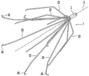
[0014] Figure 1 is a perspective view of an embodiment of a blood filter.
[0015] Figure 2 is a side perspective view of the filter of Figure 1 in a
folded
configuration.
[0016] Figure 3 is a perspective view of the blood filter of Figure 1
positioned within
a blood vessel.
[0017] Figure 4 is a perspective view of the blood filter of Figure 1
positioned at an
angle within a blood vessel.
[0018] Figure 5 is a perspective view of the blood filter of Figure 1 at a
stage of
delivery to the blood vessel.
[0019] Figure 6 is a side view of a push rod assembly for delivering a filter
of the
type illustrated in Figure 1 into a blood vessel.
[0020] Figure 7A is a detail view of the filter positioning assembly of the
push rod
assembly of Figure 6.
[0021] Figure 7B is an exploded detail view of the filter positioning assembly
illustrated in Figure 7A.
[0022] Figures 7C and 7D illustrate an alternative embodiment of the filter
positioning assembly of Figures 7A and 7B.
-7-
CA 02633855 2008-06-10
WO 2007/079410 PCT/US2006/062725
[0023] Figures 8A-8C and 9A-9E illustrate various embodiments of the filter
positioning assembly of the push rod assembly illustrated in Figure 6.
[0024] Figure 10 is a detail view of another embodiment of a filter
positioning
assembly for use with a push rod assembly such as that shown in Figure 6.
[0025] Figure 11 is a side view of a filter and the filter positioning
assembly of the
push rod assembly of Figure 6.
[0026] Figure 12 is a sectional view of a filter storage tube including a
filter and filter
positioning assembly.
100271 Figures 13A is a side view of an embodiment of the filter positioning
assembly including a spline near its base to contain the filter anchor
members.
[0028] Figure 13B is a cross sectional view of the filter positioning assembly
shown
in Figure 13A.
[0029] Figures 14A-14C are detail views of alternative embodiments of the
filter
positioning assembly and a portion of the push rod assembly of Figure 6.
[0030] Figures 15-20 illustrate steps in the delivery of a blood filter into a
blood
vessel.
[0031] Figure 21 is an assembly view of an example embodiment of a filter
delivery
system suitable for use with various embodiments of the present invention.
[0032] Figure 22 is a side sectional view of a delivery catheter suitable for
use with
various embodiments of the present invention.
[0033] Figure 23 is a detail sectional view of portions of the delivery
catheter of
Figure 22.
[0034] Figure 24 is a perspective view of portions of the delivery catheter of
Figure
22.
-8-
CA 02633855 2008-06-10
WO 2007/079410 PCT/US2006/062725
[0035] Figure 25 is a side sectional view of a catheter expander suitable for
use with
various embodiments of the present invention.
[0036] Figures 26-28 are detail views of portions of the catheter expander of
Figure
25.
[0037] Figure 29 is a detail view of an adapter portion of the filter delivery
system of
Figure 21.
[0038] Figure 30 is a perspective view of a filter storage tube portion of the
filter
delivery system of Figure 21.
Mode(s) For Carrying Out the Invention
[0039] The various embodiments will be described in detail with reference to
the
accompanying drawings. Wherever possible, the same reference numbers will be
used
throughout the drawings to refer to the same or like parts.
[0040] As used herein, the terms "about" or "approximately" for any numerical
values or ranges indicate a suitable dimensional tolerance that allows the
part or collection of
components to fun.ction for its intended purpose as described herein. Also, as
used herein, the
terms "patient", "host" and "subject" refer to any human or animal subject and
are not
intended to limit the systems or methods to human use, although use of the
subject invention
in a human patient represents a preferred embodiment.
[0041] The blood filter delivery apparatus and system in the various
embodiments
mechanically integrate components to safely and reliably deliver and emplace a
blood filter
within a patient's blood vessel, such as the vena cava. The apparatus and
system preferably
connects or is prepackaged with a filter in a filter storage tube and assists
in properly
positioning the filter in the vein in a reliable fashion.
-9-
CA 02633855 2008-06-10
WO 2007/079410 PCT/US2006/062725
[0042] The various embodiments are intended to be used with a variety of blood
filters, examples of which are described here to inform the discussion of the
embodiments of
the present invention. Referring to Figure 1, a blood filter typically will
include a hub 2 to
which are coupled a plurality of filter members 20, 30. The filter members 20,
30 both
position and anchor the filter 1 in the blood vessel and serve as the filter
mesh elements
which screen emboli from blood that passes through the filter. Common blood
filters include
anchor members 30 and locator members 20.
[0043] Anchor members 30 can include a hook 40 or hooks near their distal ends
(i.e.,
the ends opposite from the proximal ends, which are coupled to the hub 2). The
hooks 40 are
designed to penetrate and hook into the endothelial layer of the blood vessel
to prevent
longitudinal migration of the filter 1 within the vessel. Hooks 40 may have a
smaller cross
sectional area than the cross sectional area of the anchor members. Anchor
members 30 are
formed so they flex radially outward, as illustrated in Figure 1, so their
distal ends apply
sufficient pressure against the blood vessel wall to drive the hooks 40 into
the endothelial
tissue. In an example filter, the anchor members have a spread of
approximately 1.6 inches
(about 40 millimeters) in an unconstrained configuration (i.e., not installed
in the blood
vessel).
[0044] Locator members 20 also are formed so that when they are released from
the
delivery catheter they flex radially outward so their distal ends press
against the blood vessel
walls. Without hooks, the locator members' 20 distal ends apply generally
equal spring force
about the circumference of the vessel wall which, if all locators act
together, moves the filter
hub 2 toward the vessel's centerline. This centering movement positions the
filter for proper
expansion of the filter members 20, 30 and helps ensure forces are applied
equally to the
vessel walls.
-10-
CA 02633855 2008-06-10
WO 2007/079410 PCT/US2006/062725
[0045] Figure 3 illustrates a filter installed so its longitudinal axis A is
approximately
aligned with the centerline B of a blood vessel 6. Positioning the filter 1 so
it is aligned with
the vessel's centerline B helps to provide proper filter functioning and
reduce the potential for
injury to the blood vessel's wall due to application of excessive force by any
one or a few
filter members 20, 30.
[0046] Typically, filter locator members 20 are spaced equiangularly about the
filter
hub 2 so that even spacing exists between the locators 20 to provide an
effective filter basket.
For the same reason, anchor members 30 are typically positioned equiangularly
about the hub
2. To facilitate folding the filter into its narrowest possible profile
(illustrated in Figure 2) for
insertion in a delivery catheter, locator members 20 may be angularly offset
from the anchor
members 30 about the filter hub 2. In this manner, locator members 20 can fit
between
anchor members 30 in the folded, pre-delivery configuration.
[0047] To facilitate filter centering, common blood filters feature locator
members 20
that are shorter than the anchor members 30, so that when the filter is
ejected from the
delivery catheter hub-end first, the locator members 20 deploy before the
anchors, thereby
centering the filter 1 at a point of equal force on the locator members 20
(i.e., equidistant
from the locator member 20 ends) before the anchor members 30 deploy. If the
filter 1 is
aligned with the vessel centerline B, the filter hub 2 will be positioned at
or near the
centerline, as illustrated in Figure 3. This centering-before-anchoring
capability is important
because the hooks 40 will tend to lock the filter in place once they penetrate
the endothelial
layer.
[0048] While deploying the locator members 20 before the anchor members 30
tends
to center the filter hub 2 within the blood vessel at a point of equal force
on all locator
members, this action may not always align the filter with the vessel's
centerline. In some
?5 circumstances, the filter 1 may be misaligned within the blood vessel '6 so
tha
-11-
CA 02633855 2008-06-10
WO 2007/079410 PCT/US2006/062725
longitudinal axis A is at an angle to the vessel's centerline B. If the
misalignment is
significant enough, such as is illustrated in Figure 4, the filter 1 may have
less filtering
volume than a properly aligned filter.
[0049] Filter misalignment may occur when a filter 1 is delivered into a blood
vessel
6 with the catheter 16 positioned against the vessel wall, as illustrated in
Figure 5. Due to a
host's vessel anatomy, the end of the catheter 16 may be off center or even
resting on a side
of the blood vessel when the filter 1 is ejected. As shown in Figure 5, with
the catheter 16
misaligned within the blood vessel 6, the centering force provided by the
locator members 20
may contribute to misaligning the filter within the blood vessel. To avoid
this condition, a
clinician may use fluoroscopy to view the relative position of the catheter 16
in the blood
vessel 6. But this step involves additional procedure time and radiation
exposure to the
patient.
[0050] When the end of the catheter 16 is aligned with the vessel's centerline
B, the
anchor members 30 will tend to spring outward and engage the vessel wall 6 so
the entire
filter 1 is fixed in an aligned orientation with respect to the vessel's
centerline B. The various
embodiments provide a capability for centering the catheter's end, without
additional
clinician efforts, using structure which mechanically centers the catheter
within the vessel
prior to releasing the anchor members 30.
[0051] Referring to Figure 6, shown is a preferred elongated push rod assembly
60 for
advancing a filter 1 through a delivery catheter 16. The push rod assembly 60
may include a
handle 61 coupled to the proximal end, an extended portion 62 which may
feature a number
of elements of different lengths and cross sections 63-67, and a filter
positioning assembly 70
coupled to the distal end of the push rod 60. The push rod assembly 60 has a
longitudinal
length preferably in the range from about 12 inches (about 300 mm) to about 40
inches (about
1020 mm),. and is preferably about 36 inches (about 910 inm): .
-12-
CA 02633855 2008-06-10
WO 2007/079410 PCT/US2006/062725
[0052] The various elements of the push rod assembly 60 may be made from
different
materials. For example, the handle 61 may be formed of a number of metallic,
polymer or
plastic materials, and is preferably formed from PEBA, which is coupled to a
stainless steel
hollow section 63 having a diameter of about 0.041 inches. The hollow
stainless steel tube
63 may be connected to a suitable alloy material wire 64, including, for
example, a super-
elastic shape memory alloy (e.g., Nitinol), on which various elements can be
disposed, such
as a stop or boss portion 65. The shape memory alloy can further be defined as
preferably
having an austenite finish (Af) temperature below body temperature. The
stainless steel
hollow section 63 may be coupled to an extended wire 64, which may be made
from stainless
steel, the Cobalt-Chromium-Nickel alloy known as Elgiloy , or a super-elastic
shape
memory alloy, such as Nitinol. A terminal portion 67, positioned at the distal
end of the
extended portion 62, may have a smaller diameter than the wire 64, 66 and can
be made from
stainless steel or Elgiloy , and more preferably is made of Nitinol. In a
preferred
embodiment, the terminal portion 67 has a diameter of about 0.020 inches.
[0053] Coupled to the terminal portion 67 is a filter positioning assembly 70
which is
configured to push the filter 1 through a delivery catheter 16, position the
end of the catheter
near the center of the blood vessel after the filter's locator members 20
deploy and delay
release of the anchor members 30 until after the catheter end is centered.
Referring to Figure
7A, the filter positioning assembly 70 includes a hub 71 coupled on one end to
the terminal
portion 67 of the push rod 60. On the other end, the hub 71 is coupled to a
number of
positioner members 72 which function to: (1) position the end of the catheter
at or near the
centerline of the blood vessel; and/or (2) retain filter anchor members in a
collapsed
configuration until the catheter end has been centered.
[0054] Within the storage tube 15 and delivery catheter 16, the positioner
members
72 fold down over the filter anchor members 30 when the filter is in the
f.olde
-13-
CA 02633855 2008-06-10
WO 2007/079410 PCT/US2006/062725
illustrated in Figure 2 to present a narrow profile assembly as illustrated in
Figure 11. For
some embodiments, though not all, there is one positioner member 72 for each
anchor
member 30 in the filter 1, and the filter 1 and the positioner members 72 are
aligned so each
positioner lies on top of one anchor member 30. When folded over the filter so
the positioner
members 72 will fit within the storage tube 15 or a delivery catheter 16, as
illustrated in
Figures 11 and 12, the positioner members 72 can provide means to encircle the
anchor
members 30 thereby retaining the anchor members. In the folded configuration
within the
storage tube 15 or delivery catheter 16, the positioner members 72 lie
approximately parallel
to the longitudinal axis of the filter 1 and filter positioning assembly 70.
[0055] In an embodiment, positioner members 72 in the folded configuration
grip the
anchor members 30 sufficiently so that the pushing force required to push the
filter 1 through
the delivery catheter 16 by the push rod 60 is transferred from the positioner
members 72 to
the anchor members 30 and then to the filter hub 2. As illustrated in Figure
11, the positioner
members 72 may be sized to cover the anchor members 30 up to the point where
the locator
members 20 overlap the anchor members 30 in the folded configuration. Thus,
for example,
referring to filter 1 of Figure 2, the positioner members 72 of the assembly
70 are preferably
defined by a length equal to L2, which is the difference between the locator
member length Ll
and the anchor member length L3 in the folded configuration of the filter 1.
The locator
member length Ll and anchor member length L3 can be any length suitable for
use as an
implantable device. Preferably, length Ll can be from about 24 millimeters
(about 0.95
inches) to about 37 millimeters (about 1.5 inches) and length L3 can be from
about 36
millimeters (about 1.4 inches) to about 52 millimeters (about 2 inches).
[0056] In an embodiment, the plurality of positioner members 72 are joined at
their
proximal ends to the hub's periphery so that a pushing surface 73 is formed on
the hub 71, as
? 5 shown in Figure 7A. A central lumen 74 is provided through the hub 7.1 and
-14-
CA 02633855 2008-06-10
WO 2007/079410 PCT/US2006/062725
67 so that other implements (e.g., guidewire, borescope, saline, contrast
agents and so on) can
be transported from the proximal end of the catheter to the distal end of the
catheter.
[0057] In an embodiment, the hub 71 includes or is made of a radio-opaque
material,
which facilitates determining the location of the filter using fluoroscopy. As
used herein, a
radio-opaque marker is any material that is identifiable to machine or human
readable
radiographic equipment while the material is inside a patient's body, such as,
by way of
example, but not by way of limitation, gold, platinum, barium sulfate, or
tantalum.
[0058] The positioner members 72 preferably have a curved shape when
unconstrained, as illustrated in Figure 7A, with a length and curvature
configured so that
when the positioner members 72 are partially beyond the end of the delivery
catheter 16 their
distal ends are in contact with the walls of the blood vessel, as illustrated
in Figure 18.
Accordingly, as the positioning assembly 70 exits the end of the delivery
catheter 16, the tips
of the positioner members 72 bend radially outward and preferably away from
the central
lumen 74. The positioner members 72 are further configured so that they apply
a spring force
against the vessels walls when they contact the walls to position the end of
the catheter near
the blood vessel's centerline. The positioner members 72 are further
configured so that when
they are fully beyond the end of the catheter, the members bend away from the
longitudinal
axis (i.e., open) enough to release the plurality of anchor members 30 of the
filter 1. Thus,
the size and shape of the positioner members 72 will vary depending upon the
internal
diameter of the blood vessel into which a filter is to be delivered, just as
the sizes and
orientations of filter locator and anchor members 20, 30 depend upon the size
of the intended
blood vessel. Descriptions of various alternative positioner member 72 shape
and
configuration embodiments are provided below with reference to Figures 8A-C
and 9A-E.
[0059] When the ends of the positioner members 72 press against the vessel
wall, the
a 5 resulting spring force is transferred to .the hub 71, and thereby pr.ovide
a mean
-15-
CA 02633855 2008-06-10
WO 2007/079410 PCT/US2006/062725
hub 71 - and with it the end of the catheter 16 - toward the point of equal
force among the
various positioner members 72, which will normally be at or near the
centerline of the blood
vessel. In order to hold the positioner members 72 in their flexed
configuration and receive
the applied force, the positioner members 72 must be securely coupled to the
hub 71. The
positioner members 72 may be so coupled to the hub 71 by welding or brazing,
or the hub 71
and positioner members 72 may be machined from a single piece, such as, for
example, by
removing the center portion down to the surface 73 followed by cutting out
thin strips of
metal (e.g., Nitinol or Elgiloy ) to form the positioner members 72. As shown
in Figures 7C
and 7D, a unitary positioner assembly 70B can be provided by cutting a
generally tubular
stock 70A to provide for the positioner members 72 and a central lumen.
100601 An embodiment for assembling the filter positioning assembly 70 is
illustrated
in Figure 7B. In this embodiment, the hub 71 is made up of a sleeve 76 that
fits over an
internal plug 75. The internal plug 75 may have grooves 77 sized to
accommodate an
attachment portion 72D of the positioner members 72 so that the sleeve fits
close about the
plug 75 and positioner member 72. With the sleeve in place, the assembly may
be welded or
brazed together into a rigid assembly.
[0061] The positioner members 72 may be formed in a variety of shapes. Three
nonlimiting example embodiments are illustrated in Figures 8A-8C. In the
embodiment
illustrated in Figure 8A, the positioner members 72 may be a wire of
relatively constant
diameter, such as a circular, semicircular or elliptical cross section. While
this embodiment
features a constant cross section over its exposed length 72a, which is the
majority of the
member's length, it may also include a thicker portion 72d on its proximal end
for added
strength that is shaped to fit into a corresponding groove 77 on the plug 75
described above.
In another embodiment illustrated in Figure 8B, the positioner members 72 may
feature a
varied geometry along their expased length 72a, which isthe majority of the Y
-16-
CA 02633855 2008-06-10
WO 2007/079410 PCT/US2006/062725
length, such as narrow and cylindrical at the distal end, and wide and
rectangular or in the
form of an arch at the proximal end. This embodiment may also have a thicker
portion 72d
on its proximal end for added strength that is shaped to fit into a
corresponding groove 77 on
the plug 75 described above. This embodiment may have design advantages
because the tip
portion, which presses against the vessel wall, can be thin and thus more
flexible so as to
avoid damaging the endothelial layer, while the proximal end can span greater
width in order
to better retain the anchor members until full deployment. In a third
embodiment illustrated
in Figure 8C, the positioner members 72 may be thin strips, such as of a
rectangular cross
section over their exposed length 72a, which is the majority of the member's
length. Like the
other embodiments illustrated in Figures 8A and 8B, the positioner members 72
may have a
thicker portion 72d on their proximal ends for added strength that is shaped
to fit into a
corresponding groove 77 on the plug 75 described above.
[0062] The positioner members 72 may also have cross sectional features
provided to
retain the anchor members 30 until release and provide additional volume for
accommodating
the hooks 40 of filter 1. For example, Figure 8C illustrates a step portion
72b, which may be
included in any of the other embodiments of the positioner members 72. When
the positioner
members 72 are coupled to the hub 71 to form the filter positioning assembly
70, the step
portion 72b projects radially inward toward the centerline of the assembly.
This creates a
portion of the plurality of positioner members 72 having a narrower internal
diameter for
retaining anchor members 30 at that point. Such a narrow diameter on the
interior of the
positioner members 72 may tightly engage the anchor members so that
longitudinal force
necessary to push the filter 1 through the delivery catheter 16 can be
transferred from the
push rod assembly 60 to the filter anchor members 30 by the positioner members
72 at this
point. A thinner cross section portion 72c may be provided adjacent to and on
the proximal
~ S side of the narrower diameter formed by step portion 72b. This thinner
cross
-17-
CA 02633855 2008-06-10
WO 2007/079410 PCT/US2006/062725
72c can provide a larger volume in the assembly to accommodate the hooks 40 on
the ends of
the anchor members 30 in the assembled, pre-delivery configuration.
[0063] In addition to their cross sectional configuration, the positioner
members 72
are characterized by their unconstrained shape over their length. In order to
engage the blood
vessel wall during delivery of the filter 1, the positioner members 72 bend
radially away from
the longitudinal axis of the filter positioning assembly 70. Thus, the
positioner members 72
have a curved shaped and are fixed to the hub 71 so that their radiuses of
curvature are
outside the diameter of the hub 71. The curved shape may be formed before or
after the
positioner members 72 are coupled to the hub 71. When the positioner members
72 are made
from a shape memory alloy, such as Nitinol, the curved configuration is set as
the memory
shape by annealing the member in the shape at high temperature, a step which
may be
completed either before or after the positioner members 72 are coupled to the
hub 71. The
shape memory alloy can further be defined as preferably having an austenite
finish (Af)
temperature below body temperature. Additionally, the positioner member 72 can
include an
atraumatic tip such as, for example, a sphere, curved loop or a soft tip.
[0064] A number of positioner member 72 shape embodiments are possible
consistent
with the two functions performed by the structures. For example, Figure 7A
illustrates
positioner members 72 with different radiuses of curvature over their length
and which do not
bend through a full 180 degrees. This embodiment places the positioner members
72 under
less strain when they are collapsed around a filter 1 in the delivery
configuration, illustrated
in Figure 11, and thus may be well suited for positioner members 72 made from
spring
materials such as stainless steel or Elgiloy .
[0065] Another exemplary embodiment is illustrated in Figure 9A, in which the
positioner members 72 have a constant radius of curvature over their exposed
length. In the
embodiment illustrated in Figure 9A, the po.sitioner members 72 arch throtigh
-18-
CA 02633855 2008-06-10
WO 2007/079410 PCT/US2006/062725
semicircle (i.e., approximately 180 degrees), though they may curve through
more or less
than 180 degrees. In this embodiment, the positioner members 72 curve outward
as they exit
the delivery catheter 16, as illustrated in Figure 9B, until they contact the
vessel wall. This
embodiment of the positioner members 72 has a number of advantages over the
embodiment
illustrated in Figure 7A. For one, the wide angle between the positioner
members 72 adjacent
to the hub 71 may facilitate releasing the anchor members 30 by providing more
clearance for
the members to separate. For another, as the positioner members 72 advance out
of the
delivery catheter 16 (such as by the clinician pulling the catheter 16 in the
proximal direction
while holding the push rod 60 steady, as described more fully herein), the
positioner members
72 will tend to bow in the distal direction as their tips are radially
constrained by the blood
vessel wall. This flexure may provide more space for releasing the anchor
members 30 than
may be the case for positioner members 72 that do not further flex away from
the longitudinal
axis, such as the embodiment illustrated in Figure 7A.
[0066] Another example embodiment is illustrated in Figure 9C, in which the
positioner members 72 have an approximately constant radius of curvature over
their entire
length which is approximately equal to or longer than the circumference for
that radius (i.e.,
Lz 2*n*radius). In this embodiment, if the radius of curvature is set so that
four times that
radius plus the width of the hub 71 is approximately equal to the diameter of
the blood vessel,
the positioner members 72 will apply an approximately constant centering force
against the
vessel wall as the delivery catheter 16 is retreated. Referring to Figure 9E,
as the delivery
catheter 16 is pulled back, the positioner members 72 will arch radially
outward until the ends
contact the vessel wall, providing a centering force on the hub 71. Then, as
the delivery
catheter 16 is further pulled back, the distal end of the positioner members
72 will arch back
away from the vessel wall as illustrated in Figure 9D so that the radial
expansion of the
positioner members 72 does not increase as the delivery catheter 16 is withdr,
-19-
CA 02633855 2008-06-10
WO 2007/079410 PCT/US2006/062725
curling over motion of the positioner members 72 continues with further
retraction of the
delivery catheter 16, as illustrated in Figure 9C. Thus, in this embodiment,
the maximum
radial expansion of the positioner members 72 remains four times that radius
plus the width
of the hub 71. It should be noted that while the direction of curvature has
been shown as
counter-clockwise in Figures 9A-9E, it is also preferred that the direction of
curvature
originating from the catheter 16 is in a clockwise direction for Figures 9A-
9E. Moreover,
instead of a plurality of positioner members, a single helical coil can be
used where the coil
has an outside diameter at least as great as the blood vessel selected for
implantation of a
filter.
[0067] Since the embodiments illustrated in Figures 9A and 9C involve greater
strain
of the positioner members 72 to arrive at the pre-delivery, folded
configuration illustrated in
Figure 11, these embodiments are preferably made from a super-elastic shape
memory alloy,
such as Nitinol. Using Nitinol, the shapes illustrated in Figures 9A and 9C
can be set as the
memory shape by annealing the positioner members 72 in these shapes at high
temperature.
The shape memory alloy can further be defined as preferably having an
austenite finish (Af)
temperature below body temperature. After forming the members at high
temperature, the
members can be cooled below the martensitic-to-austenitic transition
temperature so they
become pliable for folding over the filter 1 into the configuration
illustrated in Figure 11.
This alloy and method of assembly helps ensure that the positioner members 72
are
elastically deformed during packaging.
[0068] The filter positioning assembly 70 may include other features to
facilitate
delivery of a filter into a blood vessel. For example, Figure 10 illustrates
an embodiment that
features an extension wire 79 projecting from the hub 71 along the
longitudinal axis of the
assembly for a length L3. Referring to Figure 2, length L3 is the length of
the anchor
members 30 from the filter hub 2. Thus, in this embodiment of the filter posi
-20-
CA 02633855 2008-06-10
WO 2007/079410 PCT/US2006/062725
assembly 70, the extension wire 79 will reach up to the base of the filter hub
2. Using a
relatively stiff material for the extension wire 79, the member can transfer
the pushing force
directly from the push rod 60 to the filter hub 2. This embodiment reduces the
longitudinal
force of pushing the filter 1 through the delivery catheter 16 that must be
resisted by the
anchor members 30. While the extension wire 79 is shown for pushing the filter
during
deployment, an alternative embodiment can be provided where the positioner
members 72
can be used to push on the filter hub 2 while the anchor members 30 are
restrained between a
splined hub and the catheter. Details of the splined hub and catheter to
restrain the anchor
members of the filter are shown and described in PCT International Application
No.
PCT/US06/17890, entitled "Embolus Blood Clot Filter and Delivery System,"
filed on May
9, 2006, which is hereby incorporated by reference in its entirety.
[0069] From the foregoing, it can be seen that the various embodiments of the
positioner members 72 described herein and illustrated in the figures provide
means for
aligning a blood filter with a blood vessel centerline and retaining anchor
members of the
filter until the alignment has been accomplished. The positioner members 72
also provide
means for releasing anchor members 30 only after the blood filter has been
aligned with the
blood vessel centerline. Further, the push rod assembly provides means for
pushing a blood
filter through a delivery catheter and deploying the filter in the blood
vessel so the filter is
aligned with the blood vessel's center.
[0070] Preferably, during manufacturing, the filter positioning assembly 70 is
fitted
over the filter 1 and positioned within a storage tube 15. In the assembled
configuration,
illustrated in Figures 11 and 12, positioner members 72 will be constrained by
the walls of
the storage tube 15 or catheter 16 so that they lie approximately parallel to
the longitudinal
axis, fitting tightly over the anchor members to present a narrow cross
section assembly that
will fit within a delivery catheter. The combined filter positioning assembly
'
-21-
CA 02633855 2008-06-10
WO 2007/079410 PCT/US2006/062725
may be kept in a storage tube 15 which preferably has approximately the same
internal
diameter as the delivery catheter and is configured to be coupled to the
catheter by a
clinician.
[0071] Figure 12 illustrates an example of a suitable storage tube 15
containing a
combined filter positioning assembly 70 and filter 1. The inner diameter of
the storage tube
resists radial expansion of the filter's locator members 20 and the positioner
members 72,
keeping them locked over the anchor members 30. In the embodiment illustrated
in Figure
12, the push rod terminal portion 67 is coupled to the hub 71 and included
within and/or
extends from an end of the storage tube 15. Alternative embodiments for
connecting the
10 terminal portion 67 to the hub 71 just before use are described herein with
respect to Figures
14A-C. In an embodiment, the terminal portion 67 is coupled to the push rod
assembly 60
during storage, so the filter 1, storage tube 15 and push rod assembly 60 are
packaged and
stored as complete unit. In another embodiment, the terminal portion 67
includes a coupling
mechanism that allows the clinician to connect the terminal portion 67 to the
rest of the push
15 rod assembly 60 at the time of use. Any number of well-known mechanisms for
connecting
rods together (e.g., threaded connections, bayonet fit, groove-and-detent fit,
etc.) may be used
for such a connection.
[0072] Referring to Figures 12 and 30, the storage tube 15 for various blood
filters
may be provided with a suitable fitting (e.g., threaded, snap or luer fitting)
at both ends for
connection to other elements of a delivery system, such as described in more
detail herein. In
an embodiment, the storage tube 15 has a threaded fitting 15b at one end to
connect with a
Touhy-Borst Adapter 10, such as is illustrated in Figure 29, and a snap
fitting 15a at the other
end to connect with the delivery catheter 16, as well as a taper section 15c
for insertion into
an elastomeric seal on the catheter. Alternatively, one end can be provided
with a snap-
-22-
CA 02633855 2008-06-10
WO 2007/079410 PCT/US2006/062725
fitting and the other end can be provided with a threaded fitting. The storage
tube 15 can be
formed from any of a number of suitable polymers and, preferably,
polycarbonate.
[0073] To shorten the as-assembled filter positioning assembly 70 and filter 1
combination so that it can be enclosed and sealed within a relatively short
storage tube 1 S, a
connectable fitting may be provided between the hub 71 and the push rod
terminal portion 67.
Such a connectable fitting may be any known mechanical joining connection.
Three
nonlimiting examples of known connections are illustrated in Figures 14A-C.
For example,
as illustrated in Figure 14A, the terminal portion 67 may be fitted with tabs
81 a that match
corresponding grooves within a bore 81b in the hub 71 to provide a bayonet
connection 81.
In another example, illustrated in Figure 14B, the end of the terminal portion
67 may be
threaded 82a to match corresponding threads in a bore 82b within the hub 71 to
provide a
threaded connection 82. In a third example, illustrated in Figure 14C, spring
tabs 83a may be
fixed to a nib end 83b of the terminal portion 67 which will slip into a bore
83c in the hub 71
and latch into an internal ridge 83d to provide a snap connection 83. By using
a connectable
fitting, the filter positioning assembly 70 and filter 1 may be stored in a
relatively short
sterilized and sealed storage tube 15 that can be maintained in a conventional
medicinal
refrigerator (e.g., in order to maintain Nitinol elements below the
martensitic-to-austenitic
transition temperature). Then, at the time of use, an end of the storage tube
15 may be
opened and the filter positioning assembly 70 connected to the terminal
portion 67 of the
push rod assembly 60 by the clinician.
[0074] Figures 15-20 illustrate the structure and functioning of the various
embodiments delivering a filter into a blood vessel. In operation and storage,
the positioner
members 72 remain tightly linked over the anchor members 30, held in place by
the walls of
the storage tube 15 or delivery catheter 16 until they are fully clear of the
catheter. To help
restrain the filter anchor members 30 prior to deployment, the filter
positionin
-23-
CA 02633855 2008-06-10
WO 2007/079410 PCT/US2006/062725
may include a spline 99 such as that illustrated in Figures 13A and 13B. The
spline 99,
which is preferably situated at or near and more preferably formed in the
positioning
assembly hub 71, secures the filter anchor members 30 generally about the hub
71 and
thereby prevents the filter hooks 40 from becoming entangled before
deployment. The
positioner members 72 may be integral with the spline 99 and the spline may
include a
central lumen 98, as illustrated in Figure 13B.
[0075] In use, a clinician will typically position the delivery catheter 16 to
a position
in a blood vessel 6 where filter placement is desired. To aid the clinician in
this step, the
delivery catheter 16 may have one or more radioWopaque markers 160 near its
distal end and
at various lengths, which can be imaged using fluoroscopy. With the catheter
so positioned,
the filter positioning assembly 70 and filter 1 may be loaded into the
proximal end of the
delivery catheter 16 and advanced by pushing on the handle 61 of the push rod
assembly 60
while holding the delivery catheter 16 in a fixed position. When the filter 1
is near or just
beyond the end of the catheter 16, as illustrated in Figure 15, the hub 2 is
at the position
where filter placement is desired. To help confirm the position of the filter
1, the hub 2 may
include a radio-opaque marker that can be imaged by fluoroscopy equipment. At
this point,
the filter locator members 20 remain retained by the catheter walls.
[0076] To deliver the filter, the clinician now extends the filter 1 and
positioner
members 72 beyond the end of the delivery catheter 16. This may be done by
either
advancing the push rod assembly in the distal direction while holding the
catheter in a fixed
position or, preferably, retracting the delivery catheter 16 in the proximal
(withdrawal)
direction while holding the push rod assembly 60 in a fixed position. The push
rod assembly
is advanced or the catheter 16 is retracted until the filter locator member
tips clear the end of
the catheter as illustrated in Figure 16. Once free of the catheter, the
locator members deploy
so that their ends press against the vessel wal16, thereby centering the
filter 1'-
-24-
CA 02633855 2008-06-10
WO 2007/079410 PCT/US2006/062725
vessel centerline. At this point, the positioner members 72 retain the anchor
members 30,
preventing them from deploying.
[0077] As the push rod assembly is further advanced or the delivery catheter
16 is
further retracted,, the positioner members 72 begin to be uncovered, which
allows the tips of
the positioner members 72 to flex radially outward seeking their memory shape,
as illustrated
in Figure 17. Once the push rod assembly has been sufficiently advanced or the
delivery
catheter 16 has been sufficiently retracted, the ends of the positioner
members 72 contact and
press against the blood vessel wall 6 as illustrated in Figure 18. The forces
applied by the
positioner members 72, being equal in all directions, center the distal end of
the delivery
catheter 16 within the blood vessel 6. The combination of the centering action
of the locator
members 20 acting on the filter hub 2 and the positioner members 72 acting on
the catheter
16 and hook-ends of the anchor members 30 aligns the filter 1 approximately
parallel with
the blood vessel's centerline. At this point, the positioner members 72 still
retain the anchor
members 30, preventing them from deploying until the aligning movements are
completed.
Then, when the delivery catheter 16 is retracted a little further, the
positioner members 72 are
fully released which by flexing away from the filter 1, release the anchor
members 30, as
illustrated in Figure 19. This allows the anchor members 30 to spring radially
outward
toward their memory shape, pressing the hooks 40 into the vessel wall 6,
thereby anchoring
the filter 1 in place. Finally, the clinician pulls the push rod assembly 60
in a proximal
direction while holding the delivery catheter 16 in a fixed position to pull
the positioner
members 72 back into the catheter 16, as illustrated in Figure 20. Once the
positioner
members 72 are securely within the delivery catheter 16, the assembly can be
removed from
the patient's body.
[0078] An example of a suitable method for joining filter 1 with the filter
positioning
assembly 70 and loading the combination in the storage tube 15 is now descri
-25-
CA 02633855 2008-06-10
WO 2007/079410 PCT/US2006/062725
method comprises several steps. First, the components are chilled below their
martensitic-to-
austenitic transition temperature so that the positioner members 72 and filter
members, which
are preferably made from a shape memory alloy like Nitinol, are flexible. At
this stage, the
positioner members 72 are compressed, such as by slipping a plastic tube over
the members,
and the filter positioning assembly 70 are passed through the storage tube so
they are
accessible on the other side, after which the plastic tube is removed.
[0079] Second, the filter is folded to its narrow profile configuration
illustrated in
Figure 2. This may be accomplished by slipping a plastic tube over the filter
hub 2 and
folding the locator members 20 and then the anchor members 30 toward the
longitudinal axis
while advancing the tube over the filter 1. The tube used for compression must
only be
advanced far enough to collapse both the locator members and the anchor
members. The
tube need not be advanced all the way to the hooks 40. In this stage, the
compressed filter
and surrounding plastic tube are positioned so the anchor hooks rest against
the hub 71 and
are centered among the positioner members 72. The anchor members 30 should be
aligned
with the positioner members 72.
[0080] Third, the storage tube 15 is moved slowly toward the filter and over
the
positioner members 72. The walls of the storage tube force the positioner
members 72 to
collapse toward the longitudinal access and close over the anchor members 30.
As the
positioner members 72 collapse upon the anchor members, the plastic tube used
to compress
the filter is retracted so the tube does not become bound between positioner
members 72 and
anchor members 30. This process continues until the positioner members 72 are
encompassed by the storage tube 15. At this stage, the positioner members 72
will lie
generally parallel to the longitudinal axis and fully encircle the anchor
members 30.
[0081] Finally, the storage tube 15 is advanced over the rest of the filter
with the
plastic ttibe in place to hold the locatar members in thei.r collapsed
configurat;
-26-
CA 02633855 2008-06-10
WO 2007/079410 PCT/US2006/062725
entire filter 1 is within the storage tube 15, the plastic tube is withdrawn
and the storage tube
is sealed. To facilitate this method of assembly, the plastic tube may be
clear and thin walled,
with an external diameter just smaller than the inside diameter of the storage
tube. This
assembly process is performed prior to shipment to the user or medical
practitioner. Other
assembly methods may be used, and assembly may be facilitated by using other
jigs or
assembly tools to assemble the filter members within positioner members 72 for
loading into
the storage tube 15.
[0082] To complete assembly, the storage tube 15 may be sealed on both ends to
prevent contamination from entering, and the entire assembly of the push rod
assembly 60,
filter 1 and storage tube 15 are sealed in sterile packaging. To avoid kinking
of the push rod
assembly 60 or lateral forces on the storage tube 15, the entire assembly may
be packed in a
linear manner within a foam form and hard outer package, such as cardboard or
plastic.
[0083] The various embodiments of the push rod assembly 60 will typically be
used
in combination with other filter delivery system components, particularly a
catheter 16 that
supports delivery of a filter into a blood vessel. A nonlimiting example
embodiment of a
filter delivery system suitable for use with the foregoing embodiments of the
push rod
assembly 60 follows with reference to Figures 21-30.
[0084] In overview, the blood filter delivery system 100 includes a storage
tube 15
containing the filter 1, a catheter introducer 16 ("catheter introducer" here
refers to a
particular embodiment of the delivery catheter 16 so the same designation
reference is used)
and the push rod assembly 60 for pushing the filter 1 from the storage tube
15, through the
catheter introducer 16 and then into the blood vessel, as well as supporting
adapters
illustrated in Figure 21. The blood filter delivery system 100 for a blood
filter device extends
along a longitudinal axis A-A. Components of the system include an adapter 10,
such as the
Touhy-E~orst Adapter shown in Figure 29, a filter storage tube l5 (Figures 12
-27-
CA 02633855 2008-06-10
WO 2007/079410 PCT/US2006/062725
be coupled to the Touhy-Borst Adapter 10 with a filter I stored in the storage
tube 15 along
with one of the various embodiments of the filter positioning assembly 70 that
can be used to
deploy the filter 1 in a blood vessel of a patient. Other components that may
be used with the
system include a catheter introducer 16, shown in Figure 22, and a catheter
dilator 18, shown
in Figure 25. Each system component is described in further detail below.
[0085] Referring to Figures 22, 23, and 24, a catheter introducer 16 includes
an
elongated generally tubular member, referred to herein as the introducer
sheath 16a coupled
to a coupling port 16b via an introducer body 16c, which may be provided with
a fluid valve
16d. The elongated introducer sheath member 16a is coupled to the introducer
body 16c by
suitable coupling techniques, such as, but not limited to, threading, bonding,
welding,
swaging or adhesives. The introducer body 16c can be provided with an internal
taper
portion 16f that allows for insertion of the external taper portion 15c of the
storage tube 15
(Figure 30) and to allow for insertion of the filter hub 2 without
interference by misalignment
of the storage tube 15 to the introducer sheath 16a during insertion of the
storage tube 15 into
the introducer 16. Each of the respective taper portions 16f and 15c is
preferably provided
with a taper angle of about 10 degrees to about 45 degrees with respect to the
longitudinal
axis A-A.
[0086] In various embodiments, the introducer sheath member 16a may be formed
from a range of biocompatible flexible materials, such as polyurethane,
polyethylene,
polyamide, polyether block amide (PEBA), nylon, and combinations thereof,
preferably from
a combination of PEBA 70D with PEBA 55D proximate the tip 16a1. The introducer
sheath
member 16a may be connected to the introducer body 16c by a bio-compatible
adhesive, e.g.,
cyanoacrylates. In an embodiment, the distal tip 16a1 of the introducer sheath
member 16a
can be provided with a suitable radio-opaque marker 160, or include radio-
opaque marker
-28-
CA 02633855 2008-06-10
WO 2007/079410 PCT/US2006/062725
substances within the material of the introducer tip 16a1. Preferably, a
tantalum radio-
opaque marker is formed on or near the tip 16a1 of the introducer sheath 16a.
[0087] In a preferred embodiment, the introducer sheath 16a has an outside
diameter
of less than about No. 10 French and an inside diameter of less than about No.
9 French and
more preferably, an outside diameter of about No. 9 French or less and an
inside diameter of
about No. 7 French or less, depending upon limits imposed by the diameter of
the blood filter
in the pre-deployed (i.e., folded) configuration.
[0088] The introducer body 16c may be provided with a coupling port 16b, which
may include a fluid seal 16e interposed between the port opening 16b1 coupled
to the
introducer sheath member 16a. The fluid sea116e may be any suitable seal, such
as but not
limited to, a membrane or a flexible arcuate sectioned seal disposed about a
central opening.
Preferably, the seal 16e is an elastic membrane made of a suitable
biocompatible elastomer,
e.g., silicone, with the arcuate sectioned seal disposed about a generally
central opening 16b1
for insertion of the dilator 18 or the filter storage tube 15. The introducer
body 16c may be
coupled to a fluid valve 16d via a polymeric (e.g., PVC) tubing 16g to allow
for a suitable
fluid (e.g., saline or a bio-active agent including drugs) to be introduced
into the introducer
sheath 16a or to drain fluid from the introducer sheath 16a. Preferably, the
valve 16d and
introducer body 16c are made of polycarbonate, polyethylene, polyurethane,
polyamide or
PEBA. The coupling port 16b may be provided with a circumferential edge that
may be.
configured to act in a snap-lock arrangement with a complementary boss portion
18f of the
dilator body 18a to attach and retain the dilator body 18a to the introducer
body 16c. That is,
the coupling port 16b includes the introducer body 16c that has the port
opening 16b1, which
has a seal 16e occluding the opening 16b1, and the introducer body 16c has an
edge 16b2
disposed about the opening 16b1 so as to allow the introducer body 16c to be
securable to a
'" projection 15a formed on one end of the storage tube 15 via a sudden sharp
c -29-
CA 02633855 2008-06-10
WO 2007/079410 PCT/US2006/062725
projection 15a of the storage tube 15 may include a curved surface disposed
circumferentially
about the longitudinal axis A-A.
[0089J The use of complementary snap-fittings for the storage tube 15 and
introducer
body 16c along with the internal and external tapers 16f and 15c are believed
to allow for
precise coupling of these two components without having to align the storage
tube with the
body 16c and threading the two components together, which under some
circumstances could
result in cross-threading or interference with the tip of the filter I into
the introducer sheath
16a.
[0090] Alternative embodiments of the introducer 16 may provide additional
capabilities for delivering and inspecting a blood filter. In one embodiment,
an ultrasound
imaging transducer is included in or near the distal end 16a1 positioned to be
capable of
imaging the deploying and deployed filter using ultrasound-imaging technology.
An example
of a suitable ultrasound transducer technology is disclosed in U.S. Patent No.
5,325,860,
which is incorporated by reference herein in its entirety. By incorporating an
ultrasound
imager within the introducer 16, a clinician can visually confirm the
placement, deployment
and emplacement of the filter 1 before removing the introducer 16 while
minimizing the use
of fluoroscopy. In an alternative embodiment, an ultrasound imaging transducer
may be
passed through the introducer 16 after the push rod assembly 60 has been
withdrawn
following delivery of the filter 1. This embodiment allows the clinician to
image the
implanted filter before removing the introducer 16 from the vicinity of the
filter.
[0091] In another alternative embodiment, a fiber optic imager or borescope is
included within the introducer sheath 16A to provide the clinician with a
visual image of the
deploying and deployed filter. A small lens on the distal end of an optical
fiber or bundle of
optical fibers conveys an image to a small video camera on the proximal end.
Illumination
may be provided to the point of inspection by another or the same optical fibe
-30-
CA 02633855 2008-06-10
WO 2007/079410 PCT/US2006/062725
optic imager may be built into the wall of the introducer sheath 16a or be
passed through the
introducer sheath 16a after the push rod 60 has been withdrawn following
delivery of the
filter. To aid in visualizing the implanted filter, saline solution may be
introduced in the
introducer to displace blood in the volume being imaged. Similar to an
ultrasound imager,
use of a fiber optic imager would permit a clinician to confirm the proper
placement,
deployment and emplacement of the filter before removing the introducer 16
while
minimizing the use of fluoroscopy.
[0092] The use of ultrasound or visual inspection of the deployed filter while
the
introducer 16 is in position near the filter may allow the clinician to remove
an improperly
deployed or located filter and replace it with another without requiring a
separate procedure
and before endothelial overgrowth of the filter hooks takes place. In this
manner, if the filter
is improperly deployed and requires removal, the introducer 16 is already
adjacent to the
filter and ready to be used in the removal procedure.
[00931 A catheter dilator 18 is preferably used in conjunction with the
introducer 16.
Referring to Figures 25-27, the dilator 18 includes a dilator body 18a coupled
to a dilator tube
18b. The dilator body 18a is provided with a threaded fitting 18f at the
proximal end to
connect to a suitable fluid valve, e.g., the Touhy-Borst Adapter 10 (Figure
29) so that fluids
can be injected into the dilator fluid passage 18g (Figure 28). A number of
fluids may be
injected during an operation, including dye marker for enabling fluoroscopic
imaging of the
introducer 16 within the patient, saline to flush body fluids from and provide
lubrication
within the introducer 16 and, in some embodiments, cooled saline to maintain
temperatures
of the push rod and/or the filter below their martensitic-to-austenitic
transition temperature.
The dilator body 18a is coupled to a dilator tube 18b that extends through and
provides a
longitudinal passage 18g of approximately 26 inches (approximately 661 mm)
from the
dilator body 18a to the distal dilator end 18c. At the distal- dilator end
18c; tht
-31-
CA 02633855 2008-06-10
WO 2007/079410 PCT/US2006/062725
18b may be provided with a generally truncated conic tip defined by the outer
surface of the
distal end 18c. The conic tip 18c1 can be defined by a conic outer surface
that extends at a
conic angle 0 of about 4 degrees with respect to the longitudinal axis with an
inside diameter
ID of about 0.041 inches and an outside diameter OD of about 0.084 inches.
[0094] A plurality of fluid communicating ports 18d may be provided through
the
wall of the dilator tube 18b in a generally spiral configuration to allow for
injection of
contrasting dye. Each fluid communicating port 18d can be of a suitable
configuration
including, but not limited to, circular, square, or diamond. Preferably, as
shown in Figure 27,
six circular communicating ports 18d1,18d2,18d3, 18d4, 18d5, and 18d6 are
provided with
an opening diameter of about 0.037 inches. Each port is preferably spaced
equidistantly from
the adjacent port over a distance d of about 0.16 inches along the
longitudinal axis A-A and
angularly disposed about the longitudinal axis A-A over an interval of 60
degrees with
respect to each adjacent port.
100951 One or more radio-opaque marker bands 18e may be coupled to the dilator
body 18a by a suitable technique, such as, but not limited to, forming a radio-
opaque material
integrally with the dilator tube 18b or mounting a separate radio-opaque
material onto or
inside the dilator tube 18b. Preferably, two radio-opaque markers 18e are
swaged onto the
dilator tube 18b near the distal end 18c, with a first marker 18e1 located
approximately 1.1
inches (approximately 28 mm) from the tip 18c and a second marker 18e2 located
at
approximately 1.1 inches (approximately 28 mm) from the first marker 18e1. In
an
embodiment, the ports 18d1-d6 are arranged in a spiral configuration between
two radio-
opaque marker bands.
[00961 The dilator tube 18d may be formed from a variety of biocompatible
flexible
materials, such as polyurethane, polyethylene, polyamide, polyether block
amide (PEBA),
nylon, and combinations thereof, preferably from a HDPE/LLDPE blend of pc
-32-
CA 02633855 2008-06-10
WO 2007/079410 PCT/US2006/062725
20% of barium sulfate by weight, with the barium sulfate providing the radio-
opaque
functionality.
[0097] When assembled, the dilator tube 18b slides inside the introducer
sheath 16a
such that the dilator tube tip 18c1 is close to the introducer tip 16a1. The
introducer 16 and
catheter dilator 18 may be packaged separately, such as in separate sterilized
packages, so
they can be unsealed and assembled by the clinician at the time of the
procedure.
Alternatively, the catheter dilator 18 may be inserted into the introducer 16
at the
manufacturer and sealed together in a sterile package, such that the clinician
can unpack and
use the two components as a unit.
[0098] Referring to Figure 29, the Touhy-Borst Adapter 10 may be provided with
at
least two passages. A first passage l0a allows for movements of the push rod
assembly 60.
A second passage l Ob allows for flow of saline into the introducer 16 to
increase lubricity
between the push rod assembly 60 and the introducer 16 as the elongated push
rod assembly
60 is moved along longitudinal axis A-A through the second passage 10b and the
passage of
the introducer 16. The saline solution also may be chilled before introduction
into the Touhy-
Borst Adapter 10 in order to maintain temperatures of the push rod and/or the
filter below
their martensitic-to-austenitic transition temperature.
[0099] Referring to Figure 30, the storage tube 15 is provided with a suitable
fitting
(e.g., threaded, snap or luer fitting) at both ends. In an embodiment, the
storage tube 15 has a
threaded fitting 15b at one end to connect with the Touhy-Borst Adapter 10 and
a snap fitting
15a at the other end to connect with the introducer 16, as well as a taper
section 15c for
insertion into the preferably triple arcuate sectioned elastomeric seal 16e.
[0100] The example filter delivery system may be used as follows for
implanting a
blood filter into a host. At the start, a suitable femoral venous vessel site
in the hostmay be
95 selected. Typically, this is the femoral vein on either the left or right
side, de]
-33-
CA 02633855 2008-06-10
WO 2007/079410 PCT/US2006/062725
the patient's size or anatomy, the clinician's preference and/or the location
of a venous
thrombosis. The site may be nicked with a blade and the vein punctured with a
suitable entry
needle, such as an 18-gauge needle, or trocar. A suitable guide wire, such as
a J-tipped guide
wire, is inserted into the needle and advanced into a distal vena cava or
iliac vessel where a
filter is to be delivered. Once the guide wire is in position, the entry
needle is removed from
the patient and slipped off the proximal end of the guide wire. Then the
proximal end of the
guide wire is inserted into the introducer distal tip 16a1 of the introducer
16. Saline or a
suitable bio-compatible fluid is provided to the introducer valve 16d to
remove air in the
introducer 16, and then introducer tip 16a1 (and preferably both the
introducer and dilator) is
inserted into the patient and advanced along the guide wire until it reaches
the desired
position in the vena cava or iliac vessel. Positioning of the introducer tip
16a1 within the
vein at the site for delivering the filter may be confirmed by fluoroscopy,
aided by the radio-
opaque markers on or within the introducer 16. The dilator tube 18b is then
inserted through
the introducer body 16c until the dilator hub 18a is snap-fitted onto the
coupling port 16b of
the introducer 16. Contrasting agent or dye may also be provided to the ports
18d of the
dilator tube 18b via the dilator body 18a to provide for visual imaging of the
introducer tip
16a1 via suitable fluoroscopic imaging equipment. The guide wire and the
dilator 18 can be
removed once the user or physician has determined that the introducer tip 16a1
is at the
desired location in the vein or vessel.
[0101] Saline infusion may be supplied to the Touhy-Borst Adapter 10. The
filter 14,
which is pre-stored in the storage tube 15, may be coupled to the coupling
port 16b via the
snap-fitting, and saline can be permitted to flow through the storage tube 15
to improve
lubricity between various components of the delivery system 100.
Alternatively, the saline
may be chilled during portions of the procedure in order to help maintain the
filter 1 and push
rod assembly 60 components below the super-elastic. shape memory alloy mar
-34-
CA 02633855 2008-06-10
WO 2007/079410 PCT/US2006/062725
austenitic transition temperature so that the positioning members, anchoring
members and
push wire portion 67 remain flexible. Similarly, the saline may be warmed
during portions of
the procedure, such as just prior to releasing the filter into the vein, to
help raise the filter 1
and filter positioning assembly 70 components above the Jnartensitic-to-
austenitic transition
temperature, causing the filter to seek its annealed shape. The introducer 16,
storage tube 15
and elongated push rod assembly 60 are preferably held in a linear
configuration to avoid
kinking and minimize friction. The filter I is physically advanced from the
storage tube 15
through the introducer 16 to a position near the distal tip 16a1 of the
introducer 16. The
advancement of the filter I can be accomplished by maintaining the introducer
16 stationary
while pushing on the handle 61 of the push rod assembly 60 in the distal
direction. The filter
I is maintained inside the introducer 16, i.e., undeployed at this point.
Markings on the push
rod assembly 60 may permit the clinician to know the position of the filter 1
with respect to
the end of the introducer 16. Additionally, fluoroscopy may be used to track
the position of
the filter 1 within the introducer 16 and with respect to the patient. When
the filter hub 2
approaches the distal end of the introducer 16, as is shown in Figure 15, the
filter is ready to
be deployed. At this point, the filter delivery steps described above with
reference to Figures
15-20 will be implemented.
[0102] While the present invention has been disclosed with reference to
certain
preferred embodiments, numerous modifications, alterations, and changes to the
described
embodiments are possible without departing from the sphere and scope of the
present
invention, which is described, by way of example, in the appended numbered
paragraphs
below. Accordingly, it is intended that the present invention not be limited
to the described
embodiments, but that it has the full scope defined by the language of the
following claims,
and equivalents thereof.
-35-