Note: Descriptions are shown in the official language in which they were submitted.
CA 02634351 2010-07-22
A BIOLOGICAL ARTIFICIAL NERVE GUIDE COMPRISING A TISSUE MEMBRANE
WITH A SPIRAL SUPPORT AND METHOD OF MAKING
BACKGROUND OF THE INVENTION
1. Field of the Invention
The present invention relates to a medical prosthesis for human implantation,
and in particular, to an
artificial device for repairing neurons, such as a biological nerve guide.
2. Description of the Prior Art
Nerve tissues have regenerating power, and even the central nervous system has
been discovered in recent
years to possess regenerating power. However, nerve tissues are fragile and
the regeneration speed is slow
so that when neurons are damaged, natural regeneration and repair often are
unable to reconnect the nerve
because of the slow growth rate. Also, the repair path is often blocked by the
faster growing surrounding
regenerated tissues or scar tissue.
To address these problems, some scientists have tried to utilize a guide to
connect the two ends of a
defective nerve to prevent the path from being blocked, and this guide is
called a nerve guide. Some
conventional nerve guides are prepared from non-degradable materials so that
irritation from foreign
matter was always present while regeneration of nerve tissues was also
adversely affected. Some of these
conventional nerve guides are prepared from degradable materials such as
polylactic acid or polyglycolic
acid, but their degraded products exhibit localized acidity, adversely
affecting the growth, the proliferation
and the migration of nerve cells.
Other conventional nerve guides are produced from natural materials such as
animal blood vessels, but
conventional glutaraldehyde is utilized in the treatment process, resulting
long-term residual toxicity and
rather potent cellular toxicity while also adversely affecting the growth and
proliferation of nerve cells.
One of the serious drawbacks of the current nerve guides is the thick guide
wall which does not allow the
penetration of nutrients and the passage of blood supply, and the nerve cells
inside the guide cannot obtain
enough nutrients for desirable differentiation and migration to repair damaged
tissue.
1
CA 02634351 2011-08-08
Another conventional nerve guide is produced from degradable natural materials
such as animal collagen,
but the mechanical properties such as flexibility, toughness, and kink
resistance are not desirable. A
noticeable drawback is that the degradation speed is difficult to match with
the speed of nerve tissue
regeneration, so that the treatment result is often uncertain.
SUMMARY OF THE DISCLOSURE
In one aspect of the present invention, there is provided a method for
preparing a biological nerve guide,
comprising: providing a natural animal tissue membrane comprising protein
molecules, the protein
molecules comprising spiral chains, the spiral chains comprising specific
hydrogen bonds; crosslinking and
fixing the membrane; minimizing antigens from the membrane comprising
utilizing a first active reagent to
block one or more antigen determinants of the membrane; and utilizing a second
reagent with hydrogen
bonding to replace the specific hydrogen bonds in the spiral chains of the
protein molecules in the
membrane and alter its specific conformation; tanning the membrane; coupling
an active layer to an inner
surface of the membrane; cutting the membrane into a desired shape and size;
positioning the cut
membrane onto a rod-shaped mould so that the cut membrane assumes a
cylindrical configuration having
an outer surface; and attaching a spiral support to the outer surface of the
cut membrane.
In another aspect of the present invention, there is provided a biological
nerve guide for implantation into a
human body, comprising: a cylindrical body made of a natural animal tissue
membrane that has been
crosslinked; the membrane comprising protein molecules; the protein molecules
comprising spiral chains,
the spiral chains comprising specific hydrogen bonds; the animal tissue
membrane comprising antigens,
the antigens having been minimized from the membrane by a first reagent that
blocks one or more
antigenic determinants of the membrane; and by a second reagent with hydrogen
bonding to replace the
specific hydrogen bonds in the spiral chains of the protein molecules in the
membrane and alter its specific
conformation; the cylindrical body having an inner surface to which an active
layer is coupled, and an
outer surface; and a spiral support that is attached to the outer surface of
the cylindrical body.
In a further aspect of the present invention, there is provided a biological
nerve guide for implantation into
a human body, the nerve guide made by a method comprising providing a natural
animal tissue membrane
comprising protein molecules, the protein molecules comprising spiral chains,
the spiral chains comprising
specific hydrogen bonds; crosslinking and fixing the membrane; minimizing
antigens from the membrane
comprising utilizing a first active reagent to block one or more antigen
determinants of the membrane; and
utilizing a second reagent with hydrogen bonding to replace specific hydrogen
bonds in the spiral chains of
the protein molecules in the membrane and alter its specific conformation;
tanning the membrane;
la
CA 02634351 2011-08-08
coupling an active layer to an inner surface of the membrane; cutting the
membrane into a desired shape
and size; positioning the cut membrane onto a rod-shaped mould so that the cut
membrane assumes a
cylindrical configuration having an outer surface; and attaching a spiral
support to the outer surface of the
cut membrane.
It is an object of the present invention to provide a biological nerve guide
having good biocompatibility.
lb
CA 02634351 2008-06-19
WO 2007/071167 PCT/CN2006/003442
It is another object of the present invention to provide a biological nerve
guide that
can be penetrated by nutrients and allows for effective flow of blood supply
while capable of
being absorbed.
It is another object of the present invention to provide a method of preparing
a
biological nerve guide that meets the objects set forth above while overcoming
the
disadvantages described above.
In order to accomplish the objects of the present invention, the present
invention
provides a biological nerve guide for implantation into a human body, the
nerve guide made
by the following method:
providing a natural animal tissue membrane;
crosslinking and fixing the membrane;
minimizing the antigens from the membrane;
tanning the membrane;
coupling an active layer to an inner surface of the membrane;
cutting the membrane into a desired shape and size;
positioning the cut membrane onto a rod-shaped mold so that the cut membrane
assumes a cylindrical configuration having an outer surface; and
attaching a spiral support to the outer surface of the cut membrane.
BRIEFDESCRIPTION OF THE DRAWINGS
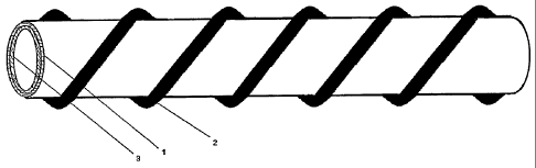
FIG. I is a perspective view of an artificial biological nerve guide according
to one
embodiment of the present invention.
FIG. 2 is a cross-sectional view of the artificial biological nerve guide of
FIG. 1.
FIGS. 3A-3C illustrate the surgical repair of a damaged nerve using the nerve
guide
of FIG. 1.
DETAILED DESCRIPTION OF THE PREFERRED EMBODIMENTS
The following detailed description is of the best presently contemplated modes
of
carrying out the invention. This description is not to be taken in a limiting
sense, but is made
merely for the purpose of illustrating general principles of embodiments of
the invention. The
scope of the invention is best defined by the appended claims.
The present invention provides a biological nerve guide having a thin guide
body
prepared from animal membrane materials treated by crosslinked fixation with a
non-aldehyde fixative, and having its antigens minimized with reagents having
strong
hydrogen bonding. A spiral support is formed by winding and immobilizing a
long strip of the
aforementioned membrane material around the guide wall.
Animal tissues are easily degraded or decomposed by microorganisms, so that
crosslinking and fixation with a fixative is required. Conventionally,
glutaraldehyde is utilized
as a fixative, but glutaraldehyde produces toxic radicals. Aldehydes undergo
crosslinking
with proteins through the acetal reaction and toxic aldehydes are released
when the
2
CA 02634351 2008-06-19
WO 2007/071167 PCT/CN2006/003442
crosslinked products are degraded, so that products fixed with an aldehyde
have long-term
residual toxicity. When non-aldehyde fixatives such as epoxides, diacyl
diamides,
diisocyanates, polyethylene glycol or carbodiimides are utilized as fixatives
in place of
aldehydes, this toxicity problem can be minimized or even eliminated. For
example, when
an epoxide is utilized to replace aldehyde-type fixatives, a ring-
opening/crosslinking reaction
occurs readily because epoxides are unstable, but the crosslinking product can
be made
very stable and not easily degraded by controlling the reaction condition. It
is slowly
degraded into polypeptides and amino acids and absorbed only when tissue
growth and
regeneration begin to devour it by secreting kallikrein, fibrinolysin and
glucocorticoid
hormone to help collagenase in the degradation. Such kind of passive
degradation and
tissue regeneration are occurring synchronously which is beneficial to tissue
regenerative
repair while having no residual toxicity of aldehydes. According to modern
immunological
theory, the antigenicity of animal tissues stems mainly from active groups
located at specific
sites and in specific conformations, and these active groups include -OH, -
NH2, -SH, etc.
The specific conformations result mainly from some specific hydrogen bonding
formed by
spiral protein chains. The specific sites and conformations are called antigen
determinants.
One or more active reagents (e.g., acid anhydrides, acyl chlorides, amides,
epoxides, etc.)
that react readily with these groups are utilized to bond with and block these
groups when
treating animal tissues so that the antigens can be effectively minimized or
eliminated.
Simultaneously, reagents with strong hydrogen bonding (e.g., guanidine
compounds) are
utilized to replace the hydrogen bonding that gives the specific
configurations so that the
configurations are altered and the antigenicity is effectively eliminated.
The wall of the nerve guide of the present invention is a thin permeable,
semi-transparent membrane for insuring easy penetration of nutrients and
microveins so that
the need for regeneration of the nerve tissues is provided. A spiral support
is provided on the
guide wall to provide sufficient supporting power for the body of the guide,
and to maintain
a space for the path required for regeneration of nerve tissues. In addition,
the winding,
stretching and mechanical compatibility of the spiral support facilitate nerve
repair at motor
parts. Both the guide body and the spiral support are prepared using animal
tissues as the
starting materials, and the main component is collagen with a small quantity
of glycoproteins,
and they can be degraded to amino acids and polypeptides which can be absorbed
by
human bodies.
Tanning
The present invention uses an additional cross-linking method and a protein
grafting
method as a tanning process to improve the mechanical strength and toughness
of the tissue.
In this regard, a piece of animal membrane tissue usually provides poor
mechanical
properties (after harvesting). As used herein, "mechanical properties" means
strength,
toughness, rigidity and modulus. Both cross-linking and protein grafting can
alter the
mechanical properties of the tissue collagen (protein) matrix. Although cross-
linking and
protein grafting are common methods that are used to improve the mechanical
properties of
3
CA 02634351 2008-06-19
WO 2007/071167 PCT/CN2006/003442
high polymers, it is still important to carefully determine the selection of
reagents as well as
the reaction conditions because protein can often be denatured. The length,
density and
distribution of cross-linkage are properly designed to ensure the stability of
the tissue
material and mechanical property.
For example, the molecular chain length of the crosslinking agent determines
the
cross-linking length. A longer chain results in better material flexibility.
However, larger
molecular chains are more difficult to penetrate into the collagen matrix. For
example, when
selecting an epoxy compound as the cross-linking agent, the molecular chain is
preferably
4-8 hydrocarbons. The cross-linking density determines the cross-linking
degree. Denser
cross-linking results in better material stability, but denser cross-linking
(especially when
combined with a shorter molecular chain) can introduce a higher local stress
in the material.
A relatively uniform distribution of the cross-linking is ideal, but is
usually difficult to obtain.
Utilizing a lower concentration of the cross-linking solution, under a lower
temperature,
longer reaction duration, and repeating a few more times with the same
reaction can often
yield better results. As an example, when using an epoxy compound as the cross-
linking
agent as described in U.S. Patent No. 6,106,555, good material stability, good
flexibility,
toughness and strength can be obtained by picking 4-8 hydrocarbon atom chain,
with a
concentration of 0.1 to 2%, under 4 to 24 degrees Celcius, reaction for 3-10
days, and
repeating 2 to 5 times.
The chemical reagents can be the same as those described herein for use with
tissue
fixation. The protein grafting process can further improve the tissue's
mechanical strength,
toughness, rigidity and modulus. Protein grafting requires a large amount of
polymer chains
so that the nature of the protein structure can be changed substantially. Some
high polymers
can be grafted into collagen molecules by means of polycondensative primers.
In order to
avoid introducing hazardous subject matter into the human body, it is
preferable to use
biodegradable high polymers as the grafting agents, such as polyglycolic acid
(PGA),
polylactic acid (PLA) and others. These biodegradable polymers can be
metabolized in the
host environment through a tracarboxylic acid cycle just like for
carbohydrates or fat
metabolism. After such an extensive protein modification, up to 25 kGy gamma
ray
sterilization can be applied without adversely affecting the mechanical
property of the tissue
material. The total amount of protein grafting can be controlled optimally.
Active Laver
The surface of the nerve guide can also include an active layer. This active
layer can
contain a polypeptide or glycosaminoglycan. One example of the polypeptides is
the
polypeptide obtained from the condensation of 16 lysines (K16), glycine (G),
arginine (R),
asparagic acid (D), serine (S), proline (P) and cysteine (C), and said
glycosaminoglycan is
hyaluronic acid, chondroitin sulfate, dermatan sulfate, heparin, acetylheparin
sulfate or
keratan sulfate. These polypeptides or glycosaminoglycans exhibit a broad-
spectrum
adherence and enriching effect for growth factors or activate undifferentiated
cells to perform
4
CA 02634351 2010-08-31
WO 2007/071167 PCT/CN2006/003442
oriented differentiation so that they are capable of exercising the function
of inducing
regenerative repair of organic tissues.
Materials
The body of the nerve guide, and the spiral support, can be made using animal
intestinal membrane, pericardium, pleura or omentum.
Method
A method of preparing the biological nerve guide according to the present
invention
comprises the following steps:
1. Selection and cleaning of materials: Fresh animal membrane tissues are
collected and sterilized with benzalkonium chloride or chlorhexidine, and
trimmed to remove
excessive impurities and irregular parts. The required membrane materials are
obtained by
taking and cleaning the neat and tough membrane materials.
2. Defatting: Fats and fat-soluble impurities in the membrane are extracted
with
an organic solvent.
3. Crosslinking fixation: The collagen molecules in the membrane are
crosslinked and fixed with a non-aldehyde fixative.
4. Minimize antigens: The specific active group, namely -OH or -NH2 or -SH, in
the proteins of the membrane is blocked with an active reagent and the
specific hydrogen
bonding in the spiral chains of the proteins in the membrane is replaced by
using a reagent
having strong hydrogen bonding.
5. Tanning process: First, the preformed polymers are produced from
monomers by synthesis. Second, the membrane is dehydrated with alcohol. Third,
the
preformed polymers are then grafted into collagen molecules by means of
polycondensative
primers. When using PGA as the grafting reagent, a small amount of glycolide
may be used
as the polycondensative primer. When using PLA as the grafting reagent, a
small amount
of lactide may be used as the polycondensative primer.
For example, using PLA as the protein grafting agent, the process could take
30-50
mg of lactide and dissolve it in 1000 ml of chloroform. 2-3 grams of
triisobutyl aluminum can
be added as the composite catalyst, and this solution can be stir-mixed for
one to two hours
under a temperature of 40-60 degrees Celcius. 100 ml of a 0.1N NaOH solution
is then
added and stir-mixed for 30-60 minutes to destroy the catalyst. Then take away
the
separated water layer (with catalyst) and have the preformed polymers ready.
Immerse the
dehydrated membrane into the preformed polymer solution. Add 0.1 to 2g of
lactide and 0.5
to 5g of proprionic anhydride as an initiation catalyst and then stir-mix for
2-4 hours under a
temperature of 34 to 40 degrees Celcius. Take out the membrane and put it into
chloroform
to clean away the residual preformed polymers. After rinsing with saline, the
membrane is
then immersed into saline for 12 to 24 hours to recover the water content. The
membrane is
now ready for the next processing step.
6. Coupling of active layer: An active surface layer is coupled to the surface
of
the guide body using a coupling agent. The active surface layer has an active
component
5
CA 02634351 2010-08-31
such as a polypeptide or glycosaminoglycan. Specifically, the surface of the
membrane material is coupled with
a polypeptide or glycosaminoglycan capable of adhering to growth factors to
form an active surface layer.
7. Preparation of the nerve guide: The membrane material is glued on a rod-
shaped mold with medical gel to
form a guide body. Separately, the same (or different) membrane material is
cut to a specific width and then
glued on the surface of the guide body by winding spirally at a given distance
in multiple layers to form a spiral
support having a particular supporting power. Next, the mold is removed to
yield the final product.
Fixative
The fixative applied in step 3 of the above method can be a reagent that
crosslinks easily with protein molecules
and is one or two reagents selected from epoxides, diacyl diamides,
diisocyanates, polyethylene glycol or
carbodiimides. This fixative may be an epoxy compound that has a hydrocarbon
backbone, that is water-soluble,
and which does not contain an ether or ester linkage in its backbone. This
fixative is described in U.S. Pat. No.
6,106,555. Examples include an epoxide, a diamide, a diisocyanate, a
polyethylene glycol, or a carbodiimide, in
that the epoxide may be a monocyclic epoxide, or a bicyclic epoxide, or it may
be a low poly(epoxide) (such as
low poly(ethylene oxide), poly(propylene oxide) or a glycidyl ether). The
epoxide may be a monocyclic epoxide
R-C~-9H2 or a dicyclic epoxide CHH-CH-(CH2)õC~O~H2 where R= H, CõH2,,+1-, and
n = 0-10, and
O
may also be a lower polyepoxide such as polypropylene oxide.
Active Reagents
The active reagents in step 4 of the above method may be low molecular weight
organic acid anhydrides, acyl
chlorides, acyl amides, monocyclic oxides or epoxide, and the reagents having
strong hydrogen bonding power
are guanidine compounds.
Coupling Agent for Active Layer
The coupling agent utilized for coupling the polypeptide in step 6 of the
above method may be a diacyl diamide,
diacid anhydride, diepoxide or other bifunctional reagents capable of carrying
out condensation with -NH2, -OH
and -COOH.
The present invention provides the following advantages. The final product is
prepared by using natural
biological materials such as animal tissues as the starting materials so that
there is no immunogenicity, and
minimal rejective reaction, while having excellent tissue compatibility and
being capable of inducing division,
proliferation and migration of nerve cells and promoting regeneration of nerve
tissues. Pathway space required
for the growth of nerve tissues is provided so that nutritional need for the
growth of nerve tissues is supplied
through penetration of nutrients and in-growth of blood vessels, thereby
creating an excellent microenvironment
for regenerative repair of the nerve tissues. After repair of the nerve
tissues is completed, the biological nerve
guide can be degraded and absorbed such that it is not present as a foreign
matter.
EXAMPLE 1
6
CA 02634351 2008-06-19
WO 2007/071167 PCT/CN2006/003442
Referring to FIGS. I and 2, fresh porcine membrane materials such as
pericardium,
omentum, pleura, diaphragm or small intestine membrane are excised, and the
fatty
materials and loose fibrous tissues are carefully removed, to trim the tough
membrane to be
as thin as possible. Then, the membrane is washed, cleaned and rinsed with
water, and then
the fats and fat-soluble impurities in the membrane materials are extracted
using an organic
solvent. The membrane will be used for the guide body 1 and the spiral support
2 shown in
FIGS. 1-2.
Next, the solvent is removed and crosslinking fixation is conducted using a
carbocyclic oxide.
After washing and freeze-drying, reaction with acetic anhydride or butyric
anhydride
is conducted to block the antigen groups, and the membrane is treated with
Tris buffer
solution of guanidine hydrochloride to alter the specific conformations of the
antigens.
Polyglycolic acid prepolymer is then grafted on the collagen molecules to
strengthen
the durability using an acid anhydride as a condensation agent.
A diacid intramolecular anhydride is then utilized as a bifunctional coupling
agent to
couple the polypeptide obtained by condensing 16 lysines (K16), glycine (G),
arginine (R),
asparagic acid (D), serine (S), proline (P) and cysteine (C) or a
glycosaminoglycan on the
surface of the membrane material to form the active surface layer 3 on what
would be the
inner surface of the cylindrical guide body 1.
At this point, the membrane material is cut according to the desired
specifications,
and then glued on a rod-shaped mold to form the guide body 1 using medical
gel. Separately
the membrane material is cut into a long strip (e.g., about 0.5-2.0 mm wide)
and rolled
around and glued on the outer surface of the guide body 1 in a spiral manner
to form the
spiral support 2. Multiple layers of the spiral support 2 can be provided (by
gluing) to
increase the diameter of the nerve guide or to add another piece of spiral
component until the
required supporting power is attained. The nerve guide is then removed from
the mold,
washed, sterilized and then packaged by sealing with physiological saline
solution being
used as a preservative solution.
FIGS. 3A-3C illustrate how the nerve guide of FIGS. 1-2 can be used in the
surgical
repair of a damaged nerve N. FIG. 3A shows a nerve N that has been damaged
(e.g.,
severed). As shown in FIG. 3B, the ends of the damaged nerve N can be inserted
into the
cylindrical bore of the nerve guide of FIG. 1, with the nerve guide serving as
a connector.
Sutures 4 can be applied to suture or attach the ends of the nerve guide to
the damaged
nerve.
While the description above refers to particular embodiments of the present
invention,
it will be understood that many modifications may be made without departing
from the spirit
thereof. The accompanying claims are intended to cover such modifications as
would fall
within the true scope and spirit of the present invention.
7