Note: Descriptions are shown in the official language in which they were submitted.
CA 02634667 2008-07-07
1
EVALUATING DISEASE PROGRESSION USING MAGNETIC RESONANCE
IMAGING
Field of the Invention
This invention relates to methods and apparatus for tracking disease
progression
using magnetic resonance imaging, including methods and apparatus for
efficiently and
precisely tracking the progression of rheumatic diseases affecting cartilage.
Background of the Invention
Osteoarthritis is a prevalent disease characterized mainly by cartilage
degradation
that is clinically reflected by a gradual development ofjoint pain, stiffness,
and loss of
motion. Osteoarthritis is extremely frequent in the general population, and it
is estimated
that its radiological prevalence is close to 50% overall. This figure is even
higher in the
elderly, with as much as 75% of the population between ages of 55 and 64
exhibiting
some degree of radiological osteoarthritis in one or more joints. Although
this disease is
offten benign, severe degenerative changes may cause serious disability.
Clinical osteoarthritis is now understood to be a complex interaction of
degradation and repair of the cartilage, bone, and synovium, with secondary
components
of inflammation. The biochemical changes of osteoarthritis affect several
cartilage
components, including major matrix constituents, proteoglycans, and collagens.
Decreased proteoglycan content in conjunction with damaged collagen structure
leads to
functional loss of normal matrix physiologic properties. Although the etiology
of
osteoarthritis is multiple and includes mechanical and biochemical factors, it
appears that
these culminate in an increased synthesis of proteolytic enzymes by the
chondrocytes,
which in turn leads to cartilage destruction.
There is no known cure for osteoarthritis, and current treatments are
essentially
limited to reliving the patient's symptoms. Research is under way, however, to
find a
therapeutic agent that will slow or stop the progression of the disease. One
current
approach to developing pharmacological treatments for osteoarthritis focuses
on
subchondral bone sclerosis, which is a well-recognized manifestation of
osteoarthritis that
could play a major role in the onset and/or progression of the disease.
CA 02634667 2008-07-07
2
Unfortunately, evaluating the efficacy of such agents is not an easy,
straightforward process. For many years, studies of drug interventions on
symptomatic
knee osteoarthritis focused only on clinical parameters like pain and joint
function,
without assessing the anatomical impact of the disease (i.e., cartilage
degradation and
bone sclerosis). Simple radiographs are now often used in clinical trials for
osteoarthritis
to establish inclusion criteria, but such trials have not employed them to
assess disease
progression. More complex radiographic methods have also been proposed for
measuring
joint space width, such as the Buckland-Wight method, which may be used in
clinical
trials. Arthroscopy appears reliable and sensitive to changes, but it only
allows for
evaluation of the cartilage surface. It also appears to be somewhat subjective
even when
independently trained evaluators review video recordings of the procedures,
and, above
all, it is invasive.
A number of academic researchers have evaluated the use of Magnetic Resonance
Imaging (MRI) for orthopedic investigations over the last ten years. Some
researchers
have proposed using MRI to reproducibly quantify articular dimensions to
follow disease
progression, and thereby assess whether proposed treatments may be responsible
for
changing the rate of cartilage loss. But the actual application of these
proposed systems
to the complex problem of making meaningful measurements on acutal diseased
joints
has not been shown to be entirely successful. This may be due to one or more
of a variety
of shortcomings, including extensive manual treatment and interpretation of
data,
excessive reliance on subjective human judgment, insufficient accuracy or
repeatability to
achieve meaningful results when used on actual diseased joints, inability to
distinguish
secondary symptoms, and/or excessively long scan times.
Summary of the Invention
Several aspects of the invention are presented in this application. These
relate to
methods and apparatus for tracking disease progression using magnetic
resonance
imaging, including methods and apparatus for efficiently and precisely
tracking the
progression of rheumatic diseases affecting cartilage.
In one general aspect, the invention features an orthopedic magnetic resonance
imaging system that includes a source of magnetic resonance imaging data sets
resulting
from successive magnetic resonance imaging acquisitions from a diseased joint
of a
CA 02634667 2008-07-07
3
patient. A segmentation module is responsive to the source of magnetic
resonance
imaging data sets and operative to segment surfaces in the joint based on
information
contained within at least one of the data sets. A registration module is
responsive to the
source of magnetic resonance imaging data sets and operative to spatially
register, in
three dimensions, information represented by a first of the data sets with
respect to
information represented by one or more further data sets for the same patient.
A
comparison module is responsive to the registration module and operative to
detect
differences between information represented by the data sets caused by
progression of the
disease in the joint of the patient between acquisitions.
In preferred embodiments, the comparison module can be operative to detect
changes in cartilage thickness within the joint. The comparison module can be
operative
to detect changes in cartilage volume within the joint. The comparison module
can be
operative to detect changes in characteristics of cartilage material within
the joint, which
can be reflected in changes in magnetic resonance signal from the cartilage
material. The
system can further include a cross-patient comparison module responsive to the
comparison module to compare detected differences for the patient with
detected
differences for at least one other patient. The system can further include a
multi-patient
database with the cross-patient comparison module including a statistical
analysis module
operative to derive statistical information about the progression of disease
in the joints of
a number of patients. The registration module can be operative to spatially
register the
data sets to within an average RMS value of about 50 microns, or even 10
microns. The
registration module can include an automatic registration module operative to
perform at
least a three-dimensional preliminary spatial registration independent of user
input. The
registration module can be operative to perform the registration based on
previously
acquired magnetic resonance imaging data for the same patient. The
segmentation
module can be an automatic segmentation module responsive to the source of
magnetic
resonance imaging data sets and operative to automatically segment anatomical
features
in the patient with substantially only supervisory and artifact-correcting
user input. The
source of magnetic resonance imaging data can be operative to provide data
sets
optimized for the detection of at least bone and cartilage. The source of
magnetic
resonance imaging data can include a magnetic resonance imaging system
operative to
acquire the data sets using a sequence which is less than about 30 minutes in
duration.
CA 02634667 2008-07-07
4
The source of magnetic resonance imaging data sets can include a magnetic
resonance
imaging system and a support assembly operative to immobilize the diseased
joint within
the magnetic resonance imaging system with the joint at a predetermined three-
dimensional position. The magnetic resonance imaging system can include a knee
coil
with the support assembly including a heel constraint and at least two
flexible wedges that
are each operative to interact with a leg of the patient and the knee coil.
The support
assembly can be operative to repeatedly immobilize the joint at predetermined
three-
dimensional positions that fall within a range of less than 17 or even 7
millimeters along
the longitudinal axis of the magnetic resonance imaging system. The system can
further
include a differential display module operative to generate a difference map
depicting
differences between the data sets detected by the comparison module. The joint
can be a
load-bearing joint, with the imaging data sets include imaging data for at
least the
majority of the load bearing surfaces of the joint. The segmentation module
can employ
an active contour algorithm. The active contour algorithm can be a subpixel
active
contour algorithm. The segmentation module can employ an active contour
algorithm
configured to segment open contours with minimal operator intervention. The
segmentation module can employ a three-dimensional gradient-driven active
contour
algorithm. The comparison module can be operative to detect differences
between
information represented by the data sets within one or more sub-regions of a
surface of
the joint caused by progression of the disease in the joint of the patient
between
acquisitions. The sub-regions can be based on polar coordinates or Cartesian
coordinates.
In another general aspect, the invention features a method of monitoring
disease
progression in a joint that includes obtaining successive images of a same
joint for each
of a plurality of patients, where at least some of the joints are diseased.
The method also
includes the steps of segmenting joint surfaces within at least one of the
images for each
patient, and, for each of the patients, spatially registering joint features
for one of the
successive images with another of the successive images. Differences are
detected
between the registered successive images for each of the individual patients,
and the
differences are compared for different ones of the patients.
In preferred embodiments, the method can further include the step of
administering a therapeutic agent to at least some of the patients before the
acquisition of
at least some of the successive images, and evaluating the differences between
the
CA 02634667 2008-07-07
registered successive images to obtain a measure of the efficacy of the
therapeutic agent.
The method can further include the step of evaluating the differences between
the
registered successive images to determine how to treat individual ones of the
patients.
The therapeutic agent can be designed to treat rheumatic diseases affecting
the cartilage.
The step of obtaining can include performing a magnetic resonance imaging
acquisition
and can further include the step of immobilizing the diseased joint with the
joint at a
predetermined flexion angle during the step of performing a magnetic resonance
imaging
acquisition. The step of obtaining can include performing a magnetic resonance
imaging
acquisition and further include the step of completely immobilizing the
diseased joint
with the joint at a predetermined three-dimensional position during the step
of performing
a magnetic resonance imaging acquisition. The step of immobilizing can be
operative to
repeatedly immobilize the joint at predetermined three-dimensional positions
that fall
within a range of less than 17 or even 7 millimeters along the longitudinal
axis of the
magnetic resonance imaging system used to perform the magnetic resonance
imaging
acquisition. The step of obtaining can include performing a magnetic resonance
imaging
acquisition, a step of positioning one or more markers proximate the joint
during the
magnetic resonance imaging, and a step of evaluating image distortion for the
joint based
on acquired image data for the markers. The step of obtaining can include
performing a
magnetic resonance imaging acquisition, a step of positioning one or more
markers
proximate the joint during the magnetic resonance imaging, and further
including a step
of evaluating patient movement artifact for the joint based on acquired image
data for the
marker. The step of positioning can position a pair of cylinders in orthogonal
locations
proximate the joint. The steps of detecting differences and comparing the
differences can
be operative to detect differences between information represented by the data
sets within
one or more sub-regions of a surface of the joint. The sub-regions can be
based on polar
coordinates or Cartesian coordinates.
In a further general aspect, the invention features an orthopedic magnetic
resonance imaging system that includes means for obtaining successive images
of a same
joint for each of a plurality of patients, wherein at least some of the joints
are diseased.
Also included are means for segmenting joint surfaces within at least one of
the images
for each patient, means for spatially registering joint features for one of
the successive
images with another of the successive images for each of the patients, means
for detecting
CA 02634667 2008-07-07
6
differences between the registered successive images for each of the
individual patients,
and means for comparing the differences obtained for different ones of the
patients.
In another general aspect, the invention features an orthopedic magnetic
resonance
imaging system that includes a source of magnetic resonance imaging data
resulting from
magnetic resonance imaging acquisitions from a diseased joint of a patient.
The system
also includes a segmentation module that is responsive to the source of
magnetic
resonance imaging data and to segmentation result storage, and that is
operative to detect
a boundary between two anatomical features of the joint in three dimensions
based on
both three-dimensional information from the diseased joint of the patient and
prior
segmentation results stored in the segmentation result storage.
In preferred embodiments, the system can further include a registration module
responsive to the source of magnetic resonance imaging data and operative to
spatially
register three-dimensional image data from a first acquisition for the patient
and three-
dimensional image data from a later acquisition for the same patient.
In a further general aspect, the invention features a method of monitoring
disease
progression in a joint that includes obtaining a first magnetic resonance
imaging data set
resulting from magnetic resonance imaging acquisition of a joint of a patient,
segmenting
a boundary between two anatomical features of the joint based on the first
magnetic
resonance imaging data set, and saving segmentation information derived during
the step
of segmenting. A second magnetic resonance imaging data set resulting from a
magnetic
resonance imaging acquisition of the same joint for the same patient is then
obtained, and
the boundary between the same two anatomical features of the same joint of the
same
patient is segmented based on both the second magnetic resonance imaging data
set and
the segmentation information saved in the step of saving.
In preferred embodiments, the method can further include the step of
administering a therapeutic agent for the disease to a plurality of patients,
with the steps
of obtaining, the steps of segmenting, and the step of saving being performed
for a
plurality of patients, and the method can further include the step of
evaluating the effect
of the therapeutic on the disease based on results of the steps of obtaining,
the steps of
segmenting, and the step of saving.
In another general aspect, the invention features an orthopedic magnetic
resonance
imaging system that includes means for obtaining a first magnetic resonance
imaging data
CA 02634667 2008-07-07
7
set resulting from magnetic resonance imaging acquisition of a joint of a
patient and for
obtaining a second magnetic resonance imaging data set resulting from a
magnetic
resonance imaging acquisition of the same joint for the same patient. Also
included are
means for segmenting a boundary between two anatomical features of the joint
based on
the first magnetic resonance imaging data set, means for saving segmentation
information
derived by the means for segmenting, and means for segmenting the boundary
between
the same two anatomical features of the same joint of the same patient based
on both the
second magnetic resonance imaging data set and the segmentation information
saved by
the means for saving.
In a further general aspect, the invention features an orthopedic magnetic
resonance imaging system that includes a source of magnetic resonance imaging
data
resulting from magnetic resonance imaging acquisitions from a diseased joint
of a patient,
and a segmentation module that is responsive to the source of magnetic
resonance
imaging data sets and is operative to detect a boundary between two anatomical
features
of the joint in three dimensions by detecting an outline in each of a
plurality of at least
generally parallel planes within the volume, wherein the outline in at least
some of the
planes is based on data from at least one other of the planes.
In another general aspect, the invention features a method of monitoring
disease
progression in a joint that includes obtaining a first magnetic resonance
imaging data set
resulting from magnetic resonance imaging acquisition of a joint of a patient,
and
segmenting an outline of a boundary between two anatomical features of the
joint of the
patient in three dimensions by detecting an outline in each of a plurality of
at least
generally parallel planes within the volume, wherein the outline in at least
some of the
planes is based on data from at least one other of the planes.
In a further general aspect, the invention features an orthopedic magnetic
resonance imaging system that includes means for obtaining a first magnetic
resonance
imaging data set resulting from magnetic resonance imaging acquisition of a
joint of a
patient, and means for segmenting an outline of a boundary between two
anatomical
features of the joint of the patient in three dimensions by detecting an
outline in each of a
plurality of at least generally parallel planes within the volume, wherein the
outline in at
least some of the planes is based on data from at least one other of the
planes.
CA 02634667 2008-07-07
8
In another general aspect, the invention features a magnetic resonance imaging
system that includes a source of magnetic resonance imaging data resulting
from
magnetic resonance imaging acquisition from an imaging volume for a patient, a
fitting
module operative to fit a biparametric surface to an anatomical feature
described by the
data for the patient, and a projection module responsive to the magnetic
resonance
imaging data source and operative to project at least a portion of the data
representing the
three-dimensional anatomical feature onto the biparametric surface.
In preferred embodiments, the surface can be a biparametric surface having a
three-dimensional topology. The system can further include a display module
responsive
to the projection module to display the two dimensional surface on a planar
display. The
anatomical feature can include at least the condyles of the femur with the
surface being a
cylinder. The anatomical feature can include at least the plateau regions of
the tibia and
wherein the surface is a plane. The anatomical feature can include at least
the posterior
surface of the patella and wherein the surface is a cylinder. The system can
further
include means for performing image manipulations on data representing the two
dimensional surface. The system can further include a repositioning module
operative to
user input to project the three-dimensional anatomical feature onto a further
biparametric
surface layers proximate the biparametric surface. The system can further
include an
inter-patient comparison module responsive to the projection module to compare
results
derived from the projections from the projection module for a plurality of
different
patients. The system can further include a display module responsive to the
inter-patient
comparison module to display comparison information for the projections.
In a further general aspect, the invention features a magnetic resonance
imaging
method that includes obtaining a magnetic resonance imaging data set resulting
from a
magnetic resonance imaging acquisition from an imaging volume for a patient,
fitting a
biparametric surface to an anatomical feature described by the data set for
the patient, and
projecting at least a portion of the data representing the three-dimensional
anatomical
feature onto the biparametric surface.
In preferred embodiments, the method can further include repeating the steps
of
obtaining, fitting, and projecting for a plurality of different patients, and
can further
include the steps of comparing resulting projections for the plurality of
different patients.
CA 02634667 2008-07-07
9
In another general aspect, the invention features a magnetic resonance imaging
system that includes means for obtaining a magnetic resonance imaging data set
resulting
from a magnetic resonance imaging acquisition from an imaging volume for a
patient,
means for fitting a biparametric surface to an anatomical feature described by
the data set
for the patient, and means for projecting at least a portion of the data
representing the
three-dimensional anatomical feature onto the biparametric surface.
In a further general aspect, the invention features a phantom for a magnetic
resonance imaging system that includes a body defining a first cavity for
holding a first
material that has at least one magnetic resonance property that is
substantially similar to
that of cartilage, and a second cavity for holding a second material that has
at least one
magnetic resonance property that is substantially similar to that of an
anatomical feature
that is adjacent to cartilage.
In preferred embodiments, the cavities can be on the order of the thickness of
joint
features to be imaged using magnetic resonance imaging. The cavities can be on
the
order of 0.125 inches thick. The body can define a first partition separating
the first and
second cavities. The partition can be on the order of less than 100 microns
thick. The
body can further define a third cavity for holding a third material, with the
body including
a second partition separating the second and third cavities.
In another general aspect, the invention features a magnetic resonance imaging
method that includes obtaining and processing a magnetic resonance image of a
phantom
of known geometry that simulates the contrast level between cartilage and at
least one
anatomical feature adjacent to cartilage, obtaining a magnetic resonance image
of a joint
of a patient, and processing results of the step of obtaining a magnetic
resonance image of
a joint of a patient based on results of the step of obtaining and processing
a magnetic
resonance image of a phantom.
In preferred embodiments, the step of processing can be a step of verifying
that
results of the step of obtaining a magnetic resonance image of a joint of a
patient fall
within a predetermined contrast range based on results of the step of
obtaining a magnetic
resonance image of a phantom. The step of processing can be a step of
correcting results
of the step of obtaining a magnetic resonance image of a joint based on
results of the step
of obtaining an image of a phantom. The step of obtaining a magnetic resonance
image
of a phantom and the step of obtaining a magnetic resonance image of a joint
can be
CA 02634667 2008-07-07
performed using a first magnetic resonance imaging configuration, and the
method can
further include a further step of obtaining a magnetic resonance image of a
phantom of
known geometry that simulates the contrast level between cartilage and at
least one
adjacent anatomical feature and a further step of obtaining a magnetic
resonance image of
ajoint of a patient. The step of obtaining a magnetic resonance image of a
phantom can
be performed for a first material that has at least one magnetic resonance
property that is
substantially similar to that of bone and a second material that has at least
one magnetic
resonance property that is substantially similar to that of cartilage. The
step of obtaining
a magnetic resonance image of a phantom can be performed for a phantom that
includes
volumes on the order of the volumes of joint features to be imaged using
magnetic
resonance imaging.
In a further general aspect, the invention features a phantom for a magnetic
resonance imaging system that includes first means having at least one
magnetic
resonance property that is substantially similar to that of cartilage, and
second means
having at least one magnetic resonance property that is substantially similar
to that of an
anatomical feature that is adjacent to cartilage.
In another general aspect, the invention features a magnetic resonance imaging
system that includes a source of three-dimensional magnetic resonance imaging
data sets
resulting from magnetic resonance imaging acquisition from a joint of a
patient, a
segmentation module that is responsive to the source of magnetic resonance
imaging data
sets and is operative to detect a boundary between two anatomical features of
the joint in
three dimensions based on three-dimensional information from a first of the
data sets, and
a comparison module responsive to the segmentation module and to a second of
the data
sets and operative to compare boundary surface data resulting from
segmentation by the
segmentation module for the first data set with volumetric data from the
second data set.
In preferred embodiments, the comparison module can be included in a second
segmentation module operative to segment the same boundary between the same
anatomical features in the second data set. The comparison module can be
included in a
registration module operative to spatially register the boundary between the
anatomical
features segmented in the first data set with the second data set.
In a further general aspect, the invention features a magnetic resonance
imaging
method that includes obtaining a first three-dimensional magnetic resonance
imaging data
CA 02634667 2008-07-07
11
set resulting from magnetic resonance imaging acquisition from a joint of a
patient,
segmenting a boundary between two anatomical features of the joint of the
patient based
on the first magnetic resonance imaging data set, obtaining a second three-
dimensional
magnetic resonance imaging data set resulting from a magnetic resonance
imaging
acquisition of an imaging volume for the same joint of the same patient, and
comparing
surface data resulting from the step of segmenting with volumetric data
resulting from the
second data set.
In preferred embodiments, the step of comparing can be part of a step of
segmenting the same boundary between two anatomical features of the patient
based on
the second magnetic resonance imaging data set. The step of comparing can be
part of a
second step of spatially registering the boundary between the anatomical
features
segmented in the first data set with the second data set.
In another general aspect, the invention features a magnetic resonance imaging
system that includes means for obtaining a first three-dimensional magnetic
resonance
imaging data set resulting from magnetic resonance imaging acquisition from a
joint of a
patient, means for segmenting a boundary between two anatomical features of
the joint of
the patient based on the first magnetic resonance imaging data set, means for
obtaining a
second three-dimensional magnetic resonance imaging data set resulting from a
magnetic
resonance imaging acquisition from the same joint of the same patient, and
means for
comparing surface data resulting from the step of segmenting with volumetric
data
resulting from the second data set.
Systems and methods according to the invention are advantageous in that they
can
allow precise quantitative tracking of the progression of diseases, such as
rheumatic
diseases affecting the cartilage. Such precise quantitative tracking can allow
for accurate
evaluation of the effects of pharmaceutical agents on these diseases in
clinical trials. It
may also allow physicians to accurately determine how and when to treat
individual
patients.
Systems according to the invention may also provide more insight into disease
progression. Because they allow physicians to view the effect of disease on
different
joint structures, systems according to the invention may permit physicians to
gain a more
detailed insight into the studied disease for a patient or group of patients.
This may result
CA 02634667 2008-07-07
12
in more finely targeted treatment research, or more effectively administered
treatment
delivery.
The benefits described above can be provided in a highly efficient manner.
Because many aspects of systems and methods according to the invention are
extensively
automated, little operator intervention is necessary. And because such systems
and
methods are highly sensitive, relatively short follow-up periods may be
achievable.
These efficiencies can have a significant impact on the cost of large-scale
clinical studies,
where many patients must be carefully evaluated. These cost savings may result
in the
evaluation of a larger number of potential treatments.
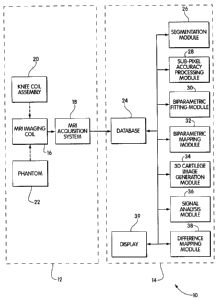
Brief Description of the Drawings
Fig. 1 is a block diagram of a disease progression monitoring system according
to
the invention configured for monitoring rheumatic diseases affecting
cartilage;
Fig. 2 is a flowchart illustrating the operation of the system of Fig. 1;
Fig. 3 is a schematic top view drawing illustrating general locations for the
constraints and markers used in positioning of a patient's right knee in the
system of
Fig. 2;
Fig. 4 is a flowchart illustrating the steps of a positioning protocol for the
system
of Fig. 2;
Fig. 5 is a waveform diagram illustrating the MRI sequence used by the system
of
Fig. 1 for one voxel in a slice;
Fig. 6 is a perspective drawing illustrating a phantom for use with the system
of
Fig. 1;
Fig. 7 is a copy of an image of a sagittal slice from a data set acquired
using the
system of claim 1;
Fig. 8 is a three-dimensional drawing illustrating the fitting by the system
of Fig. 1
of a biparametric surface of a three-dimensional geometrical primitive on bone
surfaces
for a femoral bone and a tibial bone;
Figs. 9(a) and 9(b) are images of a biparametric surface of the same bone
surface
shown in Fig. 8 before and after interpolation;
Fig. 10 is a perspective diagram illustrating the generation by the system of
Fig. 1
of new three-dimensional cartilage images;
CA 02634667 2008-07-07
13
Fig. 11 is a diagram illustrating the breakdown of a femoral cartilage image
into
sub-regions;
Fig. 12A is a diagram illustrating the breakdown of a medial tibial cartilage
image
into transversal sub-regions;
Fig. 12B is a diagram illustrating the breakdown of a lateral tibial cartilage
image
into transversal sub-regions;
Fig. 13A is a diagram illustrating the breakdown of a medial tibial cartilage
image
into sagital sub-regions;
Fig. 13B is a diagram illustrating the breakdown of a lateral tibial cartilage
image
into sagital sub-regions;
Fig. 14A is a diagram illustrating the breakdown of a medial tibial cartilage
image
into concentric sub-regions; and
Fig. 14B is a diagram illustrating the breakdown of a lateral tibial cartilage
image
into concentric sub-regions.
Detailed Description of an Illustrative Embodiment
Referring to Fig. 1, a disease progression monitoring system 10 according to
the
invention is configured for monitoring rheumatic diseases affecting cartilage
in the knee.
This system could also be configured to monitor disease progression in other
joints in the
body, such as the hip, or joints of the hands or spine. But the knee appears
to be an
appropriate choice for monitoring most rheumatic diseases affecting the
cartilage, such as
osteoarthritis. Because the knee usually bears a substantial load, it is
believed that it
tends to show arthritic symptoms at least as early as other joints, making it
a good
predictor of overall disease progression. And because of its relatively large
size and
accessibility, its internal surface can be more readily imaged and quantified
than other
j oints.
The disease progression monitoring system 10 includes an acquisition subsystem
12 and a processing subsystem 14. The acquisition subsystem includes an MRI
imaging
coil 16 operatively connected to an MRI acquisition system 18. A knee coil
assembly 20
that is compatible with the MRI imaging coil and a phantom 22 also form a part
of the
acquisition subsystem. The acquisition subsystem can include a commercially
available
CA 02634667 2008-07-07
14
1.5 Tesla MRI imaging system, such as are available from Siemens AG of Munich,
Germany. A suitable knee coil assembly is also available from Siemens.
The processing subsystem 14 includes a database 24 that is operatively
connected
to the MRI acquisition system. The operative connection between the MRI
acquisition
system and the database can take different forms, such as a network connection
or a
dedicated fiber-optic link. It may also take the form of an intermittent
connection, such
as an e-mail link, or a physically transported high-capacity storage medium,
such as an
optical disk. The database can range from a collection of files for smaller
research
systems to more powerful and feature-rich databases for systems configured to
process
data for larger numbers of patients. Also included in the processing system
are a
segmentation module 26, a sub-pixel processing module 28, a biparametric
fitting module
30, a biparametric mapping module 32, a three-dimensional cartilage image
gereration
module 34, a signal analysis module 36, a difference mapping module 38, and a
display
39. These can all be operatively connected to the database such that they can
access raw
data sets received from the acquisition subsystem 12, as well as different
processed
versions of these data sets. Each of these modules can be implemented using
special-
purpose hardware, software running on a general-purpose processor, or a
combination of
both. In addition, while the system can be broken into the series of modules
shown in
Fig. 1, one of ordinary skill in the art would recognize that it is also
possible to combine
them and/or split them to achieve a different breakdown. In one embodiment,
the
modules and database are part of a larger software system that runs on one or
more
workstation computers outfitted with an operating system such as Microsoft's
Windows
9X or Windows NT operating system.
In operation, referring to Figs. 1-3, an MRI system operator begins by
positioning
the patient in the MRI coil 12 (step 40). This involves lying the patient
generally in
parallel with a longitudinal axis of the imaging coil and precisely
positioning one of his or
her legs comfortably bent within the imaging coil according to a defined
positioning
protocol. This protocol reproducibly positions the knee at a particular three-
dimensional
position with a predetermined degree of flexion. Use of the positioning
protocol can be
important in currently available systems to achieve images that are of a
sufficient quality
to be effectively processed by the processing system 14.
CA 02634667 2008-07-07
Referring to Figs. 3 and 4, the positioning protocol includes first installing
two
three-dimensional markers 82 and 84 in generally orthogonal positions around
the
patient's knee (step 90). These markers are preferably cylindrical in shape
and highly
visible using the MRI protocol for imaging the knee. They provide reference
data that
can be used to detect any geometrical and signal drift in the data received
from the MRI
acquisition system, and to correct it if necessary. They also provide
reference data that
can be used to perform quality control analysis, such as if the patient moves
during image
acquisition. A first of the markers 82 is positioned next to the patient's
patella on the
inward side in a direction parallel to the longitudinal axis of the imaging
coil. A second
of the makers 84 is placed in the popliteal fossa in a generally horizontal
plane. The
markers can be implemented as hollow plastic tubes filled with NiSO4 solution
or
vitamin E.
The patient's knee is next centered within a horizontal plane parallel to the
longitudinal axis of the imaging coil (i.e., left-to-right-step 92). The rough
line formed
by the longitudinal axes of the femur and tibia is preferably centered as much
as possible
in this plane. The patient's patella is then centered along the longitudinal
axis of the coil
(step 94).
The positioned leg is constrained in place with a heel constraint and spacers.
This
process includes first constraining the heel with a commercially available
heel constraint
85 and a foam spacer 86 to adjust its height (step 96). One or more foam
spacers 87 are
also placed beneath the small of the knee. Two wedge-shaped spacers 88 and 89
are then
placed above the quadriceps to the left and right of the longitudinal axis of
the imaging
coil 16, and wedged in place within the knee coil 20 to hold the knee still
(step 98). Any
particular positioning issues are noted in the patient's record (step 100).
Once the patient's leg is positioned and constrained, the operator begins the
process of acquiring a three-dimensional image of the patient's knee (step
42). He or she
first instructs the MRI acquisition system 18 to acquire a scout scan of the
knee from the
MRI imaging coil 16 (step 102). The operator then instructs the MRI
acquisition system
to acquire a coronal scout scan based on the first scout scan (step 104). The
image plane
of this coronal scan is positioned at the center of the lower end of the
femur, and is then
backed up to the crossing point of the Bloomenstat line with the end of the
anterior
cruciate ligament. If necessary, the image plane is inclined to place it in
alignment with
CA 02634667 2008-07-07
16
the tibia, and this angle is noted in the patient's record. If this is the
patient's first
evaluation, the operator also acquires a short, standard SE T1 sagital scan
(step 106),
which will be used for anatomical evaluation. The final step in the protocol
is to acquire
a three-dimensional sagittal scan based on the coronal scout scan (step 108),
and centered
about the intercondylial notch (read-out along head-to-foot direction with
resolution in
the anterior-posterior axis reduced to 80% (NEX=0.8)).
Referring to Fig. 5, the acquisition follows a fat suppressed spoiled gradient
echo
sequence, which has been found to yield the best contrast for the interface
between
cartilage and the adjacent structures of the knee. It consists of 110 one mm
thick
partitions, obtained using a flip angle optimized for the Ernst angle of
cartilage, which is
about 20. The Repetition Time (TR) is set to 42 ms, and the Echo Time (TE) is
set to 7
ms. Each acquisition can cover a 308 x 512 or a 358 x 512 matrix over a
rectangular 6/8
field of view (FOV) of 160 mm, and the overall acquisition time ranges from 20
to 30
minutes. The resulting effective voxel size is of .31 x .39 x 1.0 mm3. The
imaging
protocol may require a 220 hertz manual adjustment for very obese individuals,
and the
field of view may need to be enlarged for individuals with very large knees.
The chosen methodology represents an optimized compromise between cartilage
contrast, 3D spatial resolution, maximization of signal/noise ratio, exposure
time for the
patient, and repeatability. The gradients are also optimised, with maximal
slew rate and
minimal gradient dwell time used throughout. Spoilers are minimised, as well.
It is
believed that the sequence should be transferable to other types of MRI
machines.
The three-dimensional data set obtained is in the form of a series of sagittal
image
planes through the volume that surrounds the joint. It is stored permanently
on a write-
only optical disk, which is to be transferred to the database 24 in the
processing
subsystem 14. The particulars of patient positioning and imaging parameters
are stored in
a paper file to be kept at the imaging site.
Referring to Fig. 6, the system operator can also obtain an image of a phantom
110 (step 44). This image provides important information about the acquisition
subsystem 12, which the processing subsystem can use to correct for variations
in
imaging parameters, such as may result from component drift or system repairs.
The use
of this phantom-based correction procedure can be particularly important in
following
rheumatic diseases, as successive scans of a same patient may be separated by
several
CA 02634667 2008-07-07
17
months, during which imaging conditions for a particular system may change.
The
phantom information may also be used to normalize data received from different
systems.
Note that phantom data may not need to be obtained each time a patient
acquisition is
performed, but can instead be obtained at regular intervals (e.g., weekly).
The phantom is designed to allow it to provide information about the MRI
system's acquisition of known materials configured in a known geometry. The
materials
are selected to correspond to the different materials to be imaged. In the
present
embodiment, these are bone, cartilage, and synovial fluid. The phantom
geometry is
designed to position these materials relative to each other in ways that are
comparable to
the configuration of the target structures in the patient. The total volume
and thickness of
at least some of the materials is also designed to be comparable to that of
the structures to
be imaged.
A suitable phantom 110 can be constructed using as a structure that defines
three
closely-spaced, refillable, cylindrical chambers 112, 114, and 116. These
chambers can
be defined by a stack of three hollowed-out plates 118, 120, and 122 separated
by thin
sheets 124 and 126, and screwed together by screws 128, 130, 132, and 134 at
its four
corners. In one embodiment, each plate is a square Lexan plate that defines a
cylindrical space measuring 0.125 inches in height by 1.5 inches in diameter.
The top and
bottom plates are partially hollowed out to act as caps, and the central plate
is bored
through. The first sheet 124, which is 50 microns thick, separates the top
plate 118 and
the middle plate 120. The second sheet 126, which is of the same thickness,
separates the
middle plate 120 and the bottom plate 122. Between each chamber and one of the
edges
of the plates is a fill hole measuring 0.063 inches in diameter.
Once a three-dimensional data set from the patient and phantom data for the
acquisition system have been obtained and transferred to the database 24,
segmentation of
the data can begin. Segmentation is the process of detecting edges of
anatomical surfaces
represented in the data contained in the data set for the patient.
Segmentation begins for
the bone surface (step 46) and then proceeds to the cartilage surface (step
48). This and
subsequent operations can be performed for the end of one or more of the bones
in the
joint, such as the femur, tibia, and/or patella of the knee.
Referring to Fig. 7, the segmentation module 26 processes the patient's first
data
set to determine the outline of the bone extremities and the outline of the
cartilage in each
CA 02634667 2008-07-07
18
of the MRI slices. The operator begins by manually delineating the bone-
cartilage
interface on a first of the slices, being careful to avoid obvious artifacts.
An active
contour algorithm is then applied to the manual contours, and this process
causes the
contours to more closely define the outline of the bone-cartilage interface.
In each
subsequent slice, the contours from the previous slice are used to initialize
the current
slice. The active contour algorithm is described in "Simplified Active Contour
Model
Applied To Bone Structure Segmentation In Digitral Radiographs," by C.
Kauffrnann, B.
Godbout, and J. A. de Guise, Medical Imaging 1998, Proceedings of SPIE, Image
Processing, 21-27, February 1998; "Simple 2D active contour model to segment
non-
convex objects in 3D images," by B. Godbout, C. Kauffinann, and J. A. de
Guise, Vision
Interface, '98, SFU Harbour Center, Vancouver, British Colombia, Canada, 18-
20, June,
1998; and "Segmentation d'Images Tridimensionelles a 1'Aide de Contours Actifs
Simplifies," by Benoit Godbout (Engineering Master's Thesis), Ecole Technique
Superieure, Montreal, December 1997.
The segmentation module then segments the cartilage-synovium interface
(step 48). This process proceeds in the same manner as it did with the bone-
cartilage
interface. A skilled professional, such as a radiologist generally reviews
results of the
segmentation processes to make sure that artifacts have not introduced errors
in the
images.
Referring to Fig. 8, once the data set has been segmented, the system fits
(step 50)
a simple geometrical primitive to the 3D active contour results from the bone-
cartilage
interface. The primitive is chosen to mimic the shape of the bone surface. A
cylirider is
used for the femur and planes are used for the tibia and patella.
The fitting algorithm performs an iterative search for the best transformation
in
order to minimize the squared distance between the transformed contour points
and a
normalized geometrical primitive centered at the origin.To fit a cylinder,
transformation
parameters are two rotations around orthogonal axis (principal axis), two
translations
(position) and a scaling factor (radius). To fit a plane, the transformation
parameters are
two rotations around orthogonal axis (normal) and one translation (position).
A grid is defined on the fitted biparametric primitive surface in order to
derive a
new representation for the contour points. All contour points are first
orthogonaly
projected on the grid surface. Each three-dimensional contour point (xi, yi,
zi) in the
CA 02634667 2008-07-07
19
imaging coordinate system is mapped to a corresponding coordinate on the grid
(column,
row, offset). The result can be seen as an offset map where the pixel
intensity is a
distance to the primitive.
The grid resolution is adjusted to match the MRI image slice resolution.
Because
of the uneven spacing between contour points projected on the grid, a-
Gaussian
interpolation technique is applied on the resulting offset image to fill the
gaps (see Figs.
9(a) and 9(b)). A similar offset map representation for the cartilage-synovium
interface is
obtained by projecting the contour points from the cartilage-synovium
interface on the
same biparametric surface grid used for the bone (step 54).
The new biparametric representation includes much of the information present
in
the three-dimensional representation, but has reduced processing requirements.
Because
it is two-dimensional, it can be efficiently displayed on conventional
monitors. The
biparametric view also represents a relatively standardized view of the joint,
and it is
contemplated that such views could be compared for different patients
qualitatively or
quantitatively to determine patterns of disease progression for patients or
groups of
patients.
Referring to Fig. 10, the system obtains new images of the cartilage based on
the
biparametric surface coordinate systems derived for the data (step 56). This
process
results in a layered representation of the cartilage that is akin to the
structure of an onion.
Each cartilage slice 150a, 150b ... 150n presents the intensity image obtained
by
extracting all pixels located at an isometric distance 152 from the bone
surface. The
operator can move through these slices, allowing him or her to see the effects
of the
disease on different levels of the bone and cartilage.
The sub-pixel accuracy processing module 28 uses these new three-dimensional
images and the offset image map of the bone surface to obtain a three-
dimensional sub-
pixel representation of the bone surface. This process improves the accuracy
of the first
image surfaces and subsequent operations performed on them.
The signal analysis module 36 also applies two signal processing methods (step
60) to the new three-dimensional images (from step 56). The first of these is
a textural
analysis of the cartilage pixel organization in the cartilage slices (from
step 56). The
second is local signal density analysis of the cartilage that can be displayed
as a "cartilage
radiograph" used to find local hypo-signal regions.
CA 02634667 2008-07-07
The system then generates a display mapping for the cartilage (step 62). For
comparison purposes, the cartilage is mainly represented by two maps . The
first is a
volume image map where each pixel represents a local volume localized on a 300
micron
x 300 micron surface, and the second is a thickness image map where each pixel
represents a local mean thickness localized on a 300 micron x 300 micron
surface. A
third map is used as a mask map that defines one or more topo-anatomical
regions. This
mask map is uses to obtain local thickness or volume.
Different structures within a joint can be quantified separately using the
mask
map. For example, the knee can be broken into anterior, central, and posterior
areas of
the tibial medial plateau, and medial, central, and lateral areas of the tibal
lateral plateau.
Posterior, central, and anterior areas of the femoral medial and lateral
condyles could also
be quantified, as could the patella. Different type of masks that have a
topological and
anatomical meaning can be easily tailored to the application to represent new
specific
region.An exemple to illustrates these process is the Bull-eyes mask used to
represent
four specific regions applied on the Tibial cartilage volume and thickness
maps (figure
11). By separating these regions, a physician may be able to glean a more
precise
understanding of the progression of the disease.
Other attributes of the three dimensional data can also be derived. Physical
characteristics of the cartilage that affect the quality of the MRI image
signal, such as
density or microstructural properties, can be mapped to colors. These
properties may
provide valuable diagnostic information about disease progression.
These three maps and the maps generated by the signal analysis module can be
evaluated in a number of ways. They can be displayed on a monitor of a
workstation
from a viewpoint defined by a skilled operator, such as a radiologist, who can
qualitatively evaluate them. They can also be transformed into other forms,
such as an
estimated thickness histogram.
After an appropriate interval, such as six months, a follow-up examination
takes
place. During this examination, an operator places the patient in the same
position that he
occupied during his initial examination (step 64) and obtains the same type of
imaging
data (step 66). Phantom data for the system may also be obtained (step 68).
The system then repositions the bone surface within the second image data set
to
match the position of the bone surface in the first data set (step 70). This
process begins
CA 02634667 2008-07-07
21
with a manual bone surface positioning in three planes (sagital, coronal,
axial) with
suitable interactive interface. This interface allows the user to move the
bone surface
with six degrees of freedom (three rotation controls and three translation
controls) to
obtain a first approximation of the surface position.
The rest of the procedure is performed automatically, and uses the manually
obtained approximate surface position as initialization parameter. During this
part of the
process, the bone surface is precisely fitted by least square distance
minimization
between surface points and corresponding three-dimensional image edges. The
repositioning operations for the bone also result in a repositioning of the
cartilage. The
bone surface is used as a reference for the repositioning because it is
expected that the
bone surface will normally not globally change the cartilage surface.
This process is performed by a robust least square minimization of the
difference
in combination with a surface filtering of the new image data to the sub-pixel
level. Once
the bone biparametric surface has been fitted in the new MR image sets of the
same
patient, the new Cartilage-synovium interface is segmented in a manner that is
similar to
the first cartilage segmentation step. A new biparametric surface can then be
derived for
the deformation of the cartilage (step 72). The data set resulting from this
step expresses
the difference between the two surfaces.
The system can then map the new data into one of the formats described above,
such as a volume or thickness map (step 74). These maps can be then be
combined with
their earlier counterparts to generate a difference mapping (step 76). The
difference
mapping can then be displayed (step 78).
Referring to Figs. 11-14, the system can also derive results for different
regions of
an anatomical feature. The contours of these regions can be based on
anatomical
principles or on the observation of symptoms from results for earlier
acquisitions.
Different regions may also be monitored for different conditions or different
patients, so
that the results obtained correlate as closely as possible with the
progression or state of
the condition being monitored.
The regions can be broken down based on Cartesian or polar coordinates. As
shown in Fig. 11, for example, the femoral cartilage 164 can be divided
according to
Cartesian coordinates into a medial condyle area 160, a lateral condyle area
162, and a
patelar area 164. The medial condyle and lateral condyle areas can be further
subdivided
CA 02634667 2008-07-07
22
into posterior areas (166, 172), central areas (168, 174) and anterior areas
(170, 176), and
the patelar area can be further subdivided into a medial area 178 and a
lateral area 180.
As shown in Figs. 12-13, the tibial cartilage can be represented as a
thickness map, as a
medial region 182 that is transversally divided into a number of sub regions
186, 188,
190, or as a lateral region 184 that is transversally divided into a number of
sub regions
192, 194, 196. The tibial cartilage can also be represented as a medial region
200 that is
sagitally divided into a number of sub regions 204, 206, 208, or a lateral
region 202 that is
sagitally divided into a number of sub regions 210, 212, 212.
As shown in Fig. 14, for example, the tibial cartilage can be divided
according to
polar coordinates into a "bull's-eye" representation. A medial slice 220 can
be divided
into one or more concentric rings 224 that surround a central area 226.
Similarly, a lateral
slice 222 can be divided into one or more concentric rings 228 that surround a
central area
230.
Example 1
Fifteen patients with knee osteoarthritis were recruited from outpatient
rheumatology clinics. These patients included male and female individuals
satisfying
American College of Rheumatologists (ACR) criteria for primary osteoarthritis.
They
were each symptomatic and required treatment.
In all cases there was radiological evidence of osteoarthritis in the affected
knee,
including an X-ray within six months. Each patient exhibited a minimal grade
two
severity on either space narrowing, osteophyte and/or sclerosis on the
Kellgren and
Lawrence scale. Absence of chondrocalcinosis was required, and patients with
end-stage
radiological disease (i.e., grade four) or isolated femoropateilar
osteoarthritis were not
included in the study.
Patients were ruled out on the basis of a number of possibly confounding
conditions, including secondary osteoarthrits, inflammatory arthritis, post-
traumatic
arthritis, metabolic arthritis, septic arthritis, crystal-induced disease,
Paget's disease of the
bone, avascular necrosis, or neurogenic arthritis. Previous corticoid
injections in the
study knee within the last three months or systemic corticoid use for any
other reason
were also grounds for exclusion. Ruled out as well were patients with severe
(i.e., class
CA 02634667 2008-07-07
23
IV) functional disability and candidates for imminent knee joint surgery, or
patients with
contralateral total joint replacement.
In the presence of bilateral symptomatic knees, the patient would choose the
most
symptomatic knee to be studied. In the case of similar symptoms for both
knees, the toss
of a coin would determine which one would be injected and studied. The
patient's
informed consent was required before admission into the study. A clinical
evaluation of
the patients, using validated measures, was also performed at baseline, six
months and
twelve months.
The patients were assessed at baseline, six months, and one year using an MRI
system generally comparable to that described above. As part of this
assessment, the
images obtained were systematically analyzed and quantified using a processing
system
generally comparable to that described above. Each MRI acquisition was
repeated by a
different technician on the same day.
The total cartilage volume was calculated for each of the fifteen patients.
The
resulting volume values computed for the same-day tests were correlated using
a
Sperman's Rank test. The significance of the overall cartilage volume changes
for the
fifteen patients was evaluated using a Wilcoxon-signed rank test at six months
and one
year.
The correlation coefficient for the same day acquisitions was consistently
found to
be close to 0.99 with a p value well in excess of 0.05. These results indicate
that the
technique exhibits a very high degree of repeatability in its measurements of
cartilage
volume. Preliminary 18 months results for global and topographical changes in
cartilage
volume and thickness are promising and further analysis of these results is in
progress.
Example 2
Thirty-five patients with knee osteoarthritis were recruited from outpatient
rheumatology
clinics using similar criteria to those used for the first Example. The
patients exhibited
the baseline demographics presented in table 1.
CA 02634667 2008-07-07
24
Table 1
Age (yrs.) 63.1 (9.1) Womac Pain 59.4 (3.93)
%female 74% Womac Stiff. 45.7 (4.77)
Weight (kg) 84.1(15.1) Womac Fnct. 60.3 (3.99)
% Analg. 82.6% Womac Total 56.9 (3.99)
% NSAIDs 77% Patient Global 54.5 (3.74)
SF-36 PCS 37.1 (1.65)
50 Walk (sec) 11.6 (3.6) VAS PAIN 48.2 (4.97)
ROM (deg.) 126.9 (12.2) MD Global 59.8 (3.12)
(VAS scores 100= worst)
The patients were assessed at baseline, six months, and one year using an MRI
system generally comparable to that described above. As part of this
assessment, the
images obtained were systematically analyzed and quantified using a processing
system
generally comparable to that described above. Imaging parameters were: Voxel
size: 0.3
x 0.4 X lmm, with a 512 X 410 grid; 3D FISP; TR=42, and TE=7.
The total cartilage volume was calculated for each of the thirty-five
patients.
Paired t-tests were computed for the 6-month data and an analysis of variance
(ANOVA)
for multiple measurments was performed for the 12-month data. The results are
presented in Table 2.
Table 2
MRI Location Mean (s.e.m.) Median t-value p-value *
At 6 months: n= 35
Medial Condyle -3.34 (0.96) -2.12 -3.48 0.001
Lateral Condyle -2.11 (0.48) -1.99 -4.35 0.0001
Medial Compart. -2.11 (0.65) -1.41 -3.27 0.002
Lateral Compart. -1.62 (0.39) -1.65 -4.09 0.0001
Global -1.81 (0.43) -1.49 -4.23 0.0001
At 12 months: n= 34
Medial Condyle -5.03 (1.33) -2.39 -3.79 0.001
Lateral Condyle -2.65 (0.76) -2.46 -3.49 0.001
Medial Compart. -3.91 (1.41) -1.84 -2.77 0.009
Lateral Compart. -1.78 (0.56) -1.36 -3.17 0.003
Global -2.38 (0.51) -1.50 -4.64 0.0001
* Paired t-test for 6-month data.
ANOVA for 12-month data.
CA 02634667 2008-07-07
Correlation coefficients for the cartilage volume losses against clinical
parameter
changes were computed, and are presented in Table 3.
Table 3
WOMAC Month 6 Month 12
Pain -0.025 -0.086
Stiffness -0.000 -0.070
Function +0.145 +0.030
VAS pain +0.189 -0.032
Pt Global +0.038 +0.071
MD Global +0.206 +0.290
SF36 Physical Funct. +0.110 +0.220
SF36 General Health +0.077 -0.058
p-values = all ns.
Treatment efficacy power calculations (alpha=0.05, beta=0.80) were performed,
and the
results are presented in Table 4.
Table 4
=Using the expected Internal-Compartment Volume loss:
-20 % Difference at 1 year: N= 216
-30 % Difference at 1 year: N= 97
-40 % Difference at 1 year: N= 55
=Using the expected Global-Cartilage Volume loss:
-20 % Difference at 1 year: N= 412
-30 % Difference at 1 year: N= 184
-40 % Difference at 1 year: N= 104
These results are quite promising. They indicate that cartilage volume losses
are
detectable and are statistically significant at 6 months and 1 year. Further
analyses are
needed, however, to establish the correlation of the cartilage losses with the
clinical
parameters. Nonetheless, the tool should be useful to evaluate the progression
of knee
osteoarthritis and the therapeutic efficacy of "chondroprotective" agents in
clinical trials.
The present invention has now been described in connection with a number of
specific embodiments thereof. However, numerous modifications which are
contemplated as falling within the scope of the present invention should now
be apparent
to those skilled in the art. For example, the techniques described may be used
in
CA 02634667 2008-07-07
26
veterinary applications or for the imaging of other types of structures in the
body.
Therefore, it is intended that the scope of the present invention be limited
only by the
scope of the claims appended hereto. In addition, the order of presentation of
the claims
should not be construed to limit the scope of any particular term in the
claims.