Note: Descriptions are shown in the official language in which they were submitted.
CA 02634900 2008-06-23
WO 2007/072484 PCT/IL2006/001463
URETHRAL BLOCKAGE DIAGNOSIS
FIELD OF THE INVENTION
This invention is generally in the field of medical devices, and relates to a
device and method for quantitative diagnosis of urethral blockage in patients.
BACKGROUND OF THE INVENTION
Prostate enlargement is a widespread phenomenon developed in more than
half men over age 50. By age 80, about 80% of men have enlarged prostates. The
prostate enlargement is thought to be related to hormonal disorders typical to
the
age, and is termed Benign Prostatic Hyperplasia or BPH. In a minority of the
lo cases, the prostate enlargement involves prostate cancer.
Whatsoever be the cause, enlarged prostate may lead to bladder control
problems. This is because the prostate gland encircles the urethra beneath the
bladder neck. An enlarged prostate exerts pressure on the urethra which may
defonn its shape and reduce its cross sectional area. In acute circumstances,
a
total blockage of the urethra might occur.
A quantitative diagnosis of the urethral blockage can help in early
detection of prostate problems, which in turn allows for anticipating
medication
or other appropriate treatment. In cases where bladder control problems exist
already, a quantitative diagnosis may help in determining severity of the case
and
in monitoring the effect of the treatinent procedures been taken.
CA 02634900 2008-06-23
WO 2007/072484 PCT/IL2006/001463
-2-
Froin a broader perspective, a quantitative diagnosis of urethral blockage
is only one of several coininon tests taken during the somewhat coinplicated
process of screening and diagnosing for Lower Urinary Tract Symptoms (LUTS).
Lower Urinary Tract Symptoins may involve several factors, including disorders
in the somatic nervous system, in the bladder/urethral autonoinic nervous
system,
in the detrusor and in the sphincter inuscles, and more. Said screening
process is
therefore a must for distinguishing between the plurality of medical
situations
that may cause a patient to experience urinary problems.
Facilitating and siinplifying the recognition and the quantitative diagnosis
lo of urethral blockage may therefore be essential not only in case a blockage
does
exist, but also in negating its existence in the opposite case thus leading
toward a
correct diagnosis.
The methods commonly used for quantitative detection of prostate
condition include the following techniques: a digital rectal exam to feel for
prostate enlargement; cystoscopy (under local anesthetic) consisting of
passing a
lens into the urethra and bladder to see if any abnormalities are present;
intravenous pyelogram consisting of X-ray irradiation of the urinary tract as
a
dye is injected into a vein that shows up tumors or blockages; ultrasound test
of
the prostate. The latter is generally implemented by using one of two methods:
2o Transrectal ultrasonography (TRUS) that uses a rectal probe for assessing
the
prostate, and can sometimes detect cancer; and Transabdominal ultrasonography
that uses a device placed over the abdomen. Comparing TRUS with
Transabdominal ultrasonography, TRUS is significantly more accurate for
deterinining prostate volume or the degree of urethral blockage, while
Transabdominal ultrasonography can give an accurate measure of postvoid
residual urine and is less invasive and expensive than TRUS.
Yet another known technique for quantitative detection of prostate
condition is based on the uroflowmetry test. This is aimed at detennining
whether the bladder is obstructed, by electronically measuring the speed of
urine
CA 02634900 2008-06-23
WO 2007/072484 PCT/IL2006/001463
-3-
flow. The test, however, cannot determine the cause of obstruction, which can
be
due not only to BPH, but possibly also to abnormalities in the urethra, weak
bladder inuscles, or other causes. According to this technique, the patient is
instructed not to urinate for several hours before the test and to drink
plenty of
fluids so he has a full bladder and a strong urge to urinate. To perforin this
test, a
patient urinates into a special toilet equipped with an uroflowineter. It is
important that the patient remains still while urinating to help ensure
accuracy,
and that he urinates normally and does not exert strain to einpty his bladder
or
attempt to retard his urine flow. Many factors can affect urine flow, such as
1o straining or holding back because of self-consciousness; so experts
recommend
then that the test be repeated at least twice. The rate of urine flow is
calculated as
milliliters of urine passed per second (mL/s). At its peak, the flow rate
measurement is recorded and referred to as the Q[max]. The higher the Q[max],
the better the patients flow rate. Men with a Q[max] of less than 12 mL/s have
four times the risk for urinary retention than men with a stronger urinary
flow.
The Q[max] measurement is sometimes used as the basis for determining the
severity of obstruction and for judging the success of treatinents. It is not
very
accurate, however, for a number of reasons: Urine flow varies widely among
individuals as well as from test to test. The patient's age must be
considered.
2o Flow rate normally decreases as men age, so the Q[max] typically ranges
from
more than 25 mL/s in young men to less than 10 mL/s in elderly men. The
Q[max] level does not necessarily coincide with a patient's perceptions of the
severity of his own symptoms.
It is appreciated that the currently used non invasive methods are
incapable of individually deterinining a urethral blockage or performing a
quantitative measurement thereof. For example, uroflowmetry may not
necessarily teach of a blockage and/or of its severity unless the internal
bladder
pressure is also known. This is because on the one hand a low flow rate may be
an indication of a detrusor problem rather than of a urethral blockage, while
on
the other hand a norinally detected flow rate should not necessarily indicate
of a
CA 02634900 2008-06-23
WO 2007/072484 PCT/IL2006/001463
-4-
normal urethra since it may result from extra abdominal/bladder pressures
compensating against certain flow resistance caused by urethral blockage.
Uroflowmetry combined with simultaneous measurement of internal bladder
pressure is thus required in order to allow for discrimination between the
different factors (i.e. the urethra flow resistance and the abdominal/bladder
pressure). Internal bladder pressure measurement involves however invasive
procedure - inserting a catheter into the bladder. The inconvenience and
infection risks accoinpanied to the procedure make its use rare and
appropriate
for special cases only.
U.S. Patent No. 6,063,043 discloses a passive acoustic method of
detecting the presence or absence of vesicoureteral reflux in a patient.
According
to this technique, sound from the abdomen of the patient from a time just
prior to
the onset of urination in the patient is amplified, and then the presence or
absence
of an audio signal characteristic of vesicureteral reflux in the amplified
sound is
detected. The presence of the signal indicates the presence of vesicoureteral
reflux in the patient.
U.S. Patent No. 6,428,479 discloses a technique of detecting prostate
abnormalities such as cancer. This technique utilizes ultrasonic determination
of
the in-flow kinetics of contrast agent-containing blood in the prostate and/or
observation of disease-related asymmetries in the spoke-like vascular pattern
of
the prostate.
WO 05/067392 discloses a rectal probe adapted for ultrasound and
magnetic resonance imaging of the prostate. This probe coinprises an
ultrasound
imaging probe; an MRI probe; and a link joining the ultrasound probe and the
MRI probe. The MRI probe comprises a first magnetic field source for creating
a
static magnetic field in an MRI imaging region outside the rectal probe, a
second
magnetic field source for creating a time-varying magnetic field which excites
nuclei in the MRI imaging region, and a receiver for receiving NMR signals
from
the excited nuclei and generating MRI imaging data indicative thereof.
CA 02634900 2008-06-23
WO 2007/072484 PCT/IL2006/001463
-5-
WO 05/004726 describes a method of analyzing a Doppler flow image of
a region containing a tuinor, wherein the region includes a pelvis, adnexa
uteri, a
uterus, an ovary, a breast, a prostate, a hepatic artery, a liver and the
like.
According to this technique, the Doppler flow image is represented as a three-
dimensional flow representation; and at least one paraineter characterizing a
velocity spectrum of the three-dimensional flow representation is calculated,
so
as to determine malignancy likelihood of the tumor; thereby analyzing the
Doppler flow image.
U.S. Patent No. 6,863,654 discloses a rriethod of identifying a patient's
lo urethral anatomic course in real time for the precise placement of a
treatment
element into the patient's prostate. This technique utilizes a catheter
containing
an external, inflatable imaging bladder. The catheter is introduced into a
urethra
of the patient until the image bladder is generally aligned with a treatment
site of
the prostate. An imaging probe of an imaging device is operatively positioned
relative to the treatinent site of the prostate and proximate portions of the
urethra.
The imaging device is activated so as to obtain a real time image of the
treatment
site of the prostate. The imaging bladder is filled when needed to essentially
turn
on and define an acoustic interface between the interior of the imaging
bladder
and the urethral wall. A boundary of the urethra is identified and viewed at
the
2o acoustic interface during placement of the treatment, element so as to
identify
proper positioning thereof relative to the urethra.
RU 2224464 discloses a method using ultrasonic Doppler echometric
examination of regional prostate blood circulation. Quantitative and
qualitative
indices are determined. According to this technique, chronic prostatitis is
diagnosed by detecting pulsation index greater than 1.1 and venous blood
circulation less than 4.5 cm/s relative to those of practically healthy
people.
CA 02634900 2008-06-23
WO 2007/072484 PCT/IL2006/001463
-6-
SUMMARY OF THE INVENTION
There is accordingly a need in the art for techniques for non-invasive
instant indication of urethral blockage, to thereby assist in shortening and
facilitating the process of screening and diagnosing for Lower Urinary Tract
Symptoms (LUTS), even before any physical syinptoms have actually been
experienced by the patient.
The present invention takes advantage of the fact that the urethral
blockage causes the urine flow through a channel of a variable cross-sectional
dimension, thereby resulting in a turbulence flow of the urine, which is of a
1o differing nature than that of urine flow in norinal urethras. The inventors
have
found that such a turbulence flow of the urine generates acoustic rustles of
unique frequencies in partially blocked urethras. Accordingly, the recognition
of
a rustle typical to a turbulent flow is indicative of the flow obstruction on
the
urine flow path through the urethra, the frequency and magnitude of which may
be indicative of the blockage percentage range and of the distance between the
transducer interface and the obstruction's location.
The present invention, according to its one broad aspect, provides a
system for the determination of urethral blockage, the system comprising a
transducer arrangement having at least one acoustic transducer capable of at
least
2o receiving acoustic waves, generated by the patient's urine flow, and
producing an
output signal indicative of the received acoustic waves; and a control unit in
cominunication with the transducer arrangement for receiving and processing
the
output signal and determining a change in the electrical output indicative of
the
urethral blockage.
Preferably, a specifically designed positioning unit is provided for
positioning the transducer arrangement in the vicinity of the patient's urine
flow
such that an acoustic interface of the transducer is in a position for
receiving
acoustic waves generated by the patient's urine flow.
CA 02634900 2008-06-23
WO 2007/072484 PCT/IL2006/001463
-7-
The control unit may include an amplifier for ainplifying the electrical
signal. The control unit may include a filtering unit for suppressing
background
noise, as well as discriminating between signal coinponents of different
frequencies. The filtering unit may be configured to separate from the
electrical
s. signal the wave coinponents of a predetermined frequency range for the
analysis
while repealing the wave components of other frequencies. Such filtering unit
may be configured either to repeal background noises or to direct wave
components of different frequencies for analysis through different algorithms.
According to some einbodiments of the present invention, the control unit
1o is preprograinmed with a certain physical model based on the information
(reference data) relating to frequency ranges associated with unique acoustic
rustles expected to be generated by the urine flow in partially blocked
urethras.
According to some embodiments the present invention, the model may utilize
information relating to the magnitudes of acoustic waves in said frequency
15 ranges. Preferably, the reference data includes different levels (at least
two such
levels) of acoustic waves' parameter(s) corresponding to different diseased
conditions, respectively. The analysis of the received acoustic waves allows
for
determining the dynamics in the patient's condition.
According to another broad aspect of the invention, there is provided a
20 system for the determination of urethral blockage, the system comprising
(i) a
transducer arrangement having at least one acoustic transducer capable of at
least
receiving acoustic waves and producing an output signal indicative of the
received acoustic waves; (ii) a positioning unit for positioning the
transducer
arrangement in the vicinity of the patient's urine flow such that an acoustic
25. interface of the transducer is in a position for receiving acoustic waves
generated
by the patient's urine flow; and (iii) a control unit in communication with
the
transducer arrangement for receiving and processing the output signal and
determining a change in the electrical output indicative of the urethral
blockage.
CA 02634900 2008-06-23
WO 2007/072484 PCT/IL2006/001463
-8-
According to yet another broad aspect of the invention, there is provided a
method for use in the determination of urethral blockage, the method
coinprising:
detecting acoustic signals originated by a urine flow during the patient's
urination
and generating output signals indicative thereof; processing and analyzing
said
output signals to deterinine a change in the output signals indicative of a
turbulence of the urine flow being thereby indicative of the urethral blockage
condition.
According to yet another aspect of the invention, there is provided a
transducer positioning unit for use in the above-defined system for the
lo detennination of the urethral bloclcage condition, the transducer
positioning unit
having adjustable fixation mechanism configured to fixate an interface of at
least
one transducer to a patient body so as to acquire acoustic waves from
patient's
urine flow.
Preferably, the transducer arrangement includes a plurality of transducers
1s (at least three transducers, each being at least an acoustic receiver)
operating
simultaneously during the examination, for acquiring acoustic waves from
different locations surrounding the focus of an expected problem, and or at
different locations along the penis. The control unit is associated with all
the
transducers and operates to produce a diagnosis based on the combined acoustic
2o data acquired by the transducers.
The system developed by the inventors provides for a continuous
analyzing of the urine flow acoustics during the urination. It should,
however, be
noted that it is not necessary to use the whole session information for the
diagnosis. Any piece of the acoustic information acquired during the urination
25 period may be selected and analyzed independently. Separate analysis of the
selected portions of acoustic information of the same session may be coinpared
before a fmal diagnosis is generated.
CA 02634900 2008-06-23
WO 2007/072484 PCT/IL2006/001463
-9-
Although the system of the present invention requires no timing reference,
other timing references may be of help. For example, the system of the present
invention may be configured for synchronizing with the operation of an
uroflowmetry system, such that the analysis of the electrical signal yielding
from
the acquired acoustic waves may concurrently involve data indicative of the
urine
flow rate measured by the uroflowmetry. Such combined analysis of both the
acoustic data and the urine flow rate data can be made on a continuous basis
along the entire urination cycle under examination.
The transducer positioning unit may be configured so as to enable
1o attachment of the transducer arrangement (its interface) to the patient
body, thus
receiving acoustic waves originated within the urethra after it has been
transmitted through the body tissues. This may for example be a piece of
patch,
or a ring-like arrangeinent to be mounted onto a penis.
Alternatively, the transducer positioning unit may be configured so as to
enable placing the transducer arrangement (its interface) free in the air for
receiving acoustic waves originated within the urethra through a free flow of
urine in the air (i.e. by using the urine flow as a medium for transmitting
the
acoustic wave from its location of origin to a location outside patient's
body).
This may for example be a ring-like element mountable onto a penis and
carrying
one or more transducers projecting from the ring so as to be in the urine flow
path outside the patient's body.
The transducer positioning unit may have an adjustable fixation
mechanism configured to attach the transducer(s) to a patient body so as to
acquire acoustic waves generated in response to patient's urine flow.
Also, according to some embodiments of the invention, the system for the
determination of the urethral blockage condition is configured to determine
various other parameters of the urine flow, e.g., the velocity profile. To
this end,
the transducer arrangement may include acoustic transceivers (which may or may
CA 02634900 2008-06-23
WO 2007/072484 PCT/IL2006/001463
-10-
not be the same used for the urethral blockage condition detennination)
operating
in the known Doppler-type measurement mode.
BRIEF DESCRIPTION OF THE DRAWINGS
In order to understand the invention and to see how it may be carried out
in practice, a preferred embodiment will now be described, by way of non-
limiting example only, with reference to the accompanying drawings, in which:
Fig. 1 is a block diagrain of the main components of a monitoring system
of the present invention for the determination of the urethra blockage
condition;
Fig. 2 exeinplifies the configuration of an acoustic transducer arrangement
suitable to be used in the system of the present invention;
Fig. 3 is a flow diagram of an exainple of a method of the present
invention for use in the determination of the urethra blockage condition; and
Figs. 4 to 10 show the experimental results.
DETAILED DESCRIPTION OF EXEMPLARY EMBODIMENTS
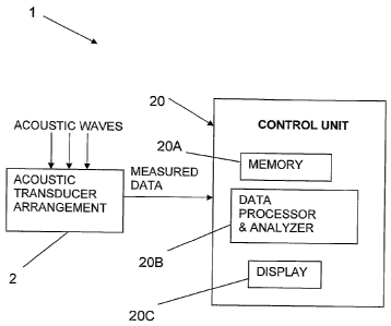
Referring to Fig. 1 there is schematically illustrated. an example of a
monitoring system 1 of the present invention for monitoring the urethra
blockage
condition. System 1 includes such main constructional parts as an acoustic
transducer arrangement 2 including one or more acoustic transducers capable of
at least receiving acoustic waves and generating an electrical output
indicative
thereof, and a control unit 20 connectable (via wires or wireless signal
transmission) to the output of the acoustic transducer arrangement. The latter
is
carried by a positioning unit (not shown here) to appropriately position the
acoustic transducer arrangement 2 with respect to a region of interest.
The acoustic transducer arrangement 2 may include one or more acoustic
transceiver. The acoustic transducer arrangement 2 may be a passive unit
(which
CA 02634900 2008-06-23
WO 2007/072484 PCT/IL2006/001463
-11-
is sufficient for the purposes of the present invention) thus including one or
more
acoustic receivers (microphones or accelerometers). Such an acoustic receiver
may be configured to provide an analog electrical output, or may be equipped
with an analog-to-digital converter thus providing digital output indicative
of the
received acoustic waves.
The system may be configured to deterinine various urine flow related
parameters other than the urethra blockage condition, for exainple the urine
flow
velocity profile. To this end, the acoustic transducer arrangement may be
configured and operable to implement Doppler-type measurements. The
io principles of this type of measurements are well known per se and do not
form
part of the present invention and therefore need not be specifically
described,
except to note that in this case the transducer arrangement is configured as
the
so-called "active" unit capable of transmitting acoustic signals towards a
region
of interest and receiving reflections of these signals from the region of
interest.
The control uilit 20 is a computer system having inter alia a memory
utility 20A (for storing certain reference data as will be described further
below),
a data processing and analyzing utility 20B (preprogrammed with a
predetermined algorithm for analyzing data indicative of the received acoustic
waves), and a control panel 20C with a display or any other data presentation
utility.
Reference is made to Fig. 2, showing an of the transducer arrangeinent
configuration suitable to be used in the above-described system 1. In the
present
example, transducer arrangement 2 is formed by four acoustic transducers,
generally at 17, arranged in a spaced-apart relationship to form a circular
array
around the region of interest, i.e., around the urine flow region. It should
be
understood that the invention is not limited to this specific example, and
generally at least one acoustic transducer can be used.
CA 02634900 2008-06-23
WO 2007/072484 PCT/IL2006/001463
-12-
A transducer positioning unit 10 is provided, which in the present exainple
includes a ring-like shaped fraine 11 and a plurality of radial shafts 12
(four such
shafts in the present exainple) each passing through a respective aperture 13
formed in the ring fra.ine. Each shaft has a first end 15 outside the frame 11
and a
second end 16 inside the frame 11. Each shaft is provided with a plate-like
member 14 facing the center of the ring shaped fraine. The shafts 12 are
preferably moveable through the apertures 13 such that the location of the
plates
14 relative to the center of the ring is adjustable by moving the shafts
through the
apertures 13.
The shafts may be held tight in the apertures due to a friction existing
between the inner face of the aperture and the outer surface of the shaft
contacting it. According to another embodiment, the shafts may be provided
with
threading matching that of the apertures, thus the adjustment of the shafts
through the apertures is by rotating the shafts like screws. According to yet
another embodiment, the shafts are spring biased so as to provide for
automatic
adaptation of the location of plates 14 to the dimensions of a body part to be
sandwiched between each pair of them.
It should be noted that generally, at least one of the plates may carry
transducer 17, and the other plates be used for the ring positioning around
the
2o body part. Preferably, however, each plate carries the transducer.
Transducer 17 is connectable to control unit 20. Considering wireless
connection, transducer 17 and control unit 20 are equipped with appropriate
coinmunication utilities based on IR; acoustic, or RF signal
transmission/reception. In the present specific but not limiting example,
transducer 17 is connected through a wiring 18, or through wires 19 passing
through the shaft, to control unit 20.
The transducer positioning unit 10 is used by placing it on a patient's penis
with the frame 11 circumferences the penis near penis's basis, and by
adjusting
CA 02634900 2008-06-23
WO 2007/072484 PCT/IL2006/001463
-13-
the shafts to bring the plates 14 into contact with the penis so as the unit
being
gripped on it. At least one shaft with transducer 17 will preferably be
contacting
the penis from bellow, closer to the urethra.
After the transducer is held in place accordingly, the patient is requested
to urinate, and so data indicative of the received acoustic waves produced by
the
urine flow is recorded and processed by the control unit 20. The related
infonnation indicative of the urine flow condition is displayed.
The urethra blockage condition or various such conditions are identified
as a corresponding change of the acoustic waves' parameter(s), such as
intensity
1o and/or frequency variation compared to reference data previously created
and
stored in the memory utility of the control unit. As indicated above, this
change
is caused by the turbulence nature of urine flow due to the urethra blockage.
Fig. 3 exemplifies the method of the present invention for the
determination of the urethra blockage condition.
As shown, reference data is provided (step I). The reference data is
indicative of the acoustic waves, generated by the urine flow, as a function
of
frequency and time, for healthy and various different diseased conditions.
Preferably, the reference data include such parameters for different groups of
patients, for example of different ages.
Measured data, from a specific patient, is collected (Step II). This
measured data is indicative of the acoustic waves received by an array of
acoustic
transducers from different locations with respect to the urine flow region
during
the patient's urination. The measured data is in the form of the acoustic
waves as
function of frequency and time.
The measured data is processed utilizing the reference data (Step III). The
processing of the measured data includes analogue processing (Step IV) aimed
at
noise reduction and normalization to the reference transmission, and digital
CA 02634900 2008-06-23
WO 2007/072484 PCT/IL2006/001463
-14-
processing (Step V) of the so-obtained normalized signal in the frequency and
time domain.
Generally, all acoustic signals are recorded by first pass through an analog
digitizer so to store analog acoustic signal as a digital sequence of
amplitude
versus time vector. In the present exainple, such a vector is subject to
further
signal processing treatment and in particular an FFT (Fast Fourier Transform)
filter can be employed in order to extract frequency and phase (compared to a
given reference signal) from each signal belonging to each active element
(each
transducer).
The processed data is compared to the reference data and the comparison
results, being indicative of the existence of physiological abnormalities and
the
degree of pathology, are displayed to the user, who may be a physician or the
patient himself (Step VI).
The following are the experimental results of using the technique of the
present invention for monitoring the urethral blockage condition. The
invention
has been exercised on two groups, the first included men of age above 55 who
reported of micturition problems (hereinafter "patient group"), and the second
included men of age below 30 reported no micturition difficulty (hereinafter
"reference group").
The acoustic equipment used for the experiment included a microphone or
accelerometer (constituting an acoustic transducer), amplifier and a digital
data
recorder. It should be noted that the term "accelerometer" is only used here
as an
example and any other suitable acoustic component may be used.
One possible example of using an accelerometer may be placing it
manually at the bottom of the penis as close as possible to the testicles. In
this
location, the urethra normally reaches its minimal distance from the penis
exterior where a transducer (accelerometer) can be placed.
CA 02634900 2008-06-23
WO 2007/072484 PCT/IL2006/001463
-15-
The accelerometer was connected to the input of the ainplifier, the output
of which was connected to the control unit (its data processor and analyzer
utility); it should be understood that ainplifier may alternatively be a
constructional part of the control unit. The amplifier has been adjusted to
30dB
amplification.
An exeinplary result of an examination taken on exaininee No. 1 of the
patient group in comparison with exemplary result of an examination taken on
exaininee No. 2 of the reference group will now be explained.
Fig. 4 illustrates a graph representing the amplitude versus time of an
io electrical signal generated by the accelerometer in response to a 32-
seconds
acoustic wave it has acquired before, during and after the micturition of
examinee No. 1.
Micturition starting and ending moments are indicated by vertical lines L2
and L3, respectively. The signals before line L2 and past line L3 are noise
signals
1s which include noises of placement and displacement of the accelerometer on
the
patient body. A section of the micturition interval between lines L2 and L3
marked by horizontal line L4 was picked for analysis. An expanded view of this
section is depicted in Fig. 5.
Fig. 5 illustrates an expanded view of a section from the graph illustrated
20 in Fig. 4. The illustrated graph section represents the ainplitude versus
time of an
electrical signal acquired by the accelerometer from the acoustic wave
generated
during the micturition of examinee No. 1, picked for analysis from the entire
graph of Fig. 4.
Fig. 6 illustrates a graph of the spectral power density in a dB scale versus
25 wave frequency for the frequency range 0-3KHz, of the signal section
illustrated
by Fig. 5. Fig. 7 illustrates an expanded view of the graph illustrated by
Fig. 6 for
the frequency range 0-1Khz. As can be noticed, there is a remarkable
CA 02634900 2008-06-23
WO 2007/072484 PCT/IL2006/001463
-16-
concentration of acoustic energy in the frequency range around 200Hz, showing
as a hill HS on the graph line of Figs. 6 and 7.
Figs. S- 10 illustrate mutatis mutandis graphs similar to those illustrated
by Figs. 5-7, talcen from the test of examinee No. 2 of the reference group
(healthy examinee).As can be observed, no remarlcable hill is recognizable in
the
graph taken from the test of the examinee of the reference group. A comparison
between the two graphs leads to the conclusion that the remarkable energy
concentration in the 200Hz frequency range in the graph of exaininee No. 1
corresponds to a urethral blockage pathology possibly resulting from BPH of
lo examinee No. 1.
This particular example only partially demonstrates the invention. It
should be understood that the invention could utilize various other aspects of
the
signal analysis, such as phase analysis; comparison between different signals
taken simultaneously from different components at different location, etc.
Those skilled in the art will readily appreciate that various modifications
and changes can be applied to the embodiments of the invention as herein
described without departing from its scope defmed in and by appended claims.