Note: Descriptions are shown in the official language in which they were submitted.
CA 02635534 2008-06-25
WO 2007/078922 PCT/US2006/048292
MICRONIZED DEVICE FOR THE DELIVERY OF BIOLOGICALLY ACTIVE
MOLECULES AND METHODS OF USE THEREOF
FIELD OF THE INVENTION
The present invention relates generally to the field of encapsulated cell
therapy.
BACKGROUND OF THE INVENTION
Many clinical conditions, deficiencies, and disease states can be remedied or
alleviated by supplying to the patient one or more biologically active
molecules produced by
living cells or by removing from the patient deleterious factors which are
metabolized by
living cells. In many cases, these molecules can restore or compensate for the
impairment or
loss of organ or tissue function. Accordingly, many investigators have
attempted to
reconstitute organ or tissue function by transplanting whole organs, organ
tissue, and/or cells,
which provide secreted products or affect metabolic functions. However, while
transplantation can provide dramatic benefits, it is limited in its
application by the relatively
small number of organs that are suitable and available for grafting. In
general,
transplantation patients must be immunosuppressed in order to avert
immunological rejection
of the transplant, which results in loss of transplant function and eventual
necrosis of the
transplanted tissue or cells. Likewise, in many cases, the transplant must
remain functional
for a long period of time, even for the remainder of the patient's lifetime.
It is both
undesirable and expensive to maintain a patient in an immunosuppressed state
for a
substantial period of time.
A number of vision-threatening disorders of the eye exist for which additional
good
therapies are still needed. One major problem in treatment of such diseases is
the inability to
deliver therapeutic agents into the eye and to maintain them there at
therapeutically effective
concentrations.
Many growth factors have shown promise in the treatment of ocular disease. For
example, BDNF and CNTF have been shown to slow degeneration of retinal
ganglion cells
and decrease degeneration of photoreceptors in various animal models. See,
e.g., Genetic
Technology News, vol. 13, no. 1 (Jan. 1993). Additionally, nerve growth factor
has been
CA 02635534 2013-11-01
shown to enhance retinal ganglion cell survival after optic nerve section and
has also been
shown to promote recovery of retinal neurons after ischemia. See, e.g.,
Siliprandi, et al.,
Invest. Ophthalmol. & Vis. Sci., 34, pp. 3232-3245 (1993). '
A desirable alternative to transplantation procedures is the implantation of
cells or
tissues within a physical barrier which will allow diffusion of nutrients,
metabolites, and
secreted products, but will block the cellular and molecular effectors of
immunological
rejection. A variety of macrocapsule devices which protect tissues or cells
producing a
selected product from the immune system have been explored. See, e.g., US
Patent No.
5,158,881; W092/03327; W091/00119; and W093/00128. These devices include, for
example, extravascular diffusion chambers, intravascular diffusion chambers,
intravascular
ultrafiltration chambers, and implantation of microencapsulated cells. See
Scharp, D. W., et
al., World J. Surg., 8, pp. 221-9 (1984). See, e.g., Lim et al., Science 210:
908-910 (1980);
Sun, A. M., Methods in Enzymology 137: 575-579 (1988); WO 93/03901; and U.S.
Pat. No.
5,002,661. Such devices would alleviate the need to maintain the patient in an
immunosuppressed state. However, none of these approaches have been
satisfactory for
providing long-term transplant function.
Thus, methods of delivering appropriate quantities of needed substances, such
as,
neurotrophic factors, anti-angiogenic factors, anti-inflammatory factors,
enzymes, hormones,
other factors or, of providing other needed metabolic functions, to the eye
for an extended
period of time are needed.
SUMMARY OF THE INVENTION
The invention provides micronized devices for the delivery of a biologically
active
molecule to the eye. Such micronized devices contain a capsule having a core
containing
between about 5x10² and 90x103 living cells that produce a biologically active
molecule and
a biocompatible jacket surrounding said core, wherein the jacket has a
molecular weight
cutoff permitting diffusion of the biologically active molecule into the eye.
Preferably, the
device is configured as a cylinder with an outer diameter of between 200 and
350 lam and a
length of between 0.5 and 6 mm. The dosage of the biologically active molecule
that diffuses
into the eye is between 0.1 pg and 1000 ng per eye per patient per day. In
various
embodiments, the BAM dosage may be between 0.1 pg and 500 ng per eye per
patient per
day; between 0.1 pg and 250 ng, between 0.1 pg and 100 ng, between 0.1 pg and
50 ng
2
CA 02635534 2008-06-25
WO 2007/078922 PCT/US2006/048292
between 0.1 pg and 25 ng, between 0.1 pg and 10 ng, or between 0.1 pg and 5 ng
per eye per
patient per day.
In some embodiments, the micronized device may optionally have a tether
adapted for
securing the capsule to an ocular structure. For example, the tether may be
selected from a
loop, a disk, and/or a suture. Such tethers can be made from a shape memory
material or any
other medical grade material known to those skilled in the art.
The jacket of the micronized devices of the invention may be a permselective,
irrununoisolatory membrane. Moreover, the biocompatible jacket can be made
from either an
ultrafiltration or a microporous membrane. Typically, the jacket is made from
a polymer
material. Suitable polymer materials include, but are not limited to,
polyacrylonitrile-
polyvinylchloride, polyacrylonitrile, polymethylmethacrylate,
polyvinyldifluoride,
polyolefins, polysulfones, polymide, and/or celluloses.
= The micronized devices of the invention can be implanted in the vitreous,
the Sub-
Tenon's capsule, the periocular space, and/or the anterior chamber.
Suitable biologically active molecules include, but are not limited to,
antiangiogenic
factors, anti-inflammatory factors, neurotrophic factors, growth factors,
trophic factors,
antibodies and antibody fragments, neurotransmitters, hormones, cytokines, and
lympholdnes. In some embodiments, the biologically active molecule is a
cytokine or a
lympholcine such as TGF13, GDNF, NGF, CN'TF, 13FGF, aFGF, IL-113,
IFN-a, .13DNF,
LIF, NT-4, NTN, NT4/5, CT-1, LEDGF, Neublastin, Axoldne, IL-23, RdCVF, IL-
10,
Alpha INF, IL-1Ra, and/or Remicade. In other embodiments, the biologically
active
molecule is an antiangiogenic factor such as vasculostatin, angiostatin,
endostatin, anti-
integrins, vascular endothelial growth factor inhibitors (VEGF-inhibitors),
platelet factor 4,
heparinase, bFGF-binding molecules, the VEGF receptor Flt, the VEGF receptor
Flk,
Lucentis, VEGF Trap, Tek A/Fc (angl/ang2 inhibitor), 2xCon4 (C), soluble VEGF
Receptors, and PEDF.
In further embodiments, at least one additional biologically active molecule
is
delivered from the capsule to the eye. The additional biologically active
molecule or
molecules may be from a cellular source or from a noncellular source. When the
at least one
additional biologically active molecule is from a noncellular source, it can
be encapsulated in,
dispersed within, or attached to one or more components of the micronized
device. By way
of nonlimiting example, the at least one additional biologically active
molecule from a
noncellular source can be selected from nucleic acids, nucleic acid fragments,
peptides,
3
CA 02635534 2008-06-25
WO 2007/078922 PCT/US2006/048292
polypeptides, peptidomimetics, carbohydrates, lipids, organic molecules,
inorganic
molecules, therapeutic agents, and various combinations thereof. Suitable
therapeutic agents
include, but are not limited to, anti-angiogenic drugs, steroidal and non-
steroidal anti-
inflammatory drugs, anti-mitotic drugs, anti-tumor drugs, anti-parasitic
drugs, IOP
reducers, peptide drugs, and other biologically active molecule drugs approved
for
ophthalmologic use.
The living cells contained within the core of the micronized devices of the
invention
may include insulin-producing cells, adrenal chromaffin cells, antibody-
secreting cells,
fibroblasts, astrocytes, Beta cell lines, Chinese hamster ovary cells, and/or
ARPE-19 cells.
These cells may be allogeneic and/or syngeneic.
The molecular weight cut off of the biocompatible jacket of the micronized
device of
the invention is between about 1 kJ) and about 150 IcD.
In some embodiments, the core of the micronized device has a volume of less
than 0.5
I. The core may also contain a substantially non-degradable filamentous cell-
supporting
matrix, wherein the matrix is made from a plurality of monofilaments, and
wherein the
monofilaments are either twisted into a yarn or woven into a mesh or twisted
into a yarn that
is in non-woven strands. The cells in the core can be distributed on the non-
degradable
filamentous cell-supporting matrix. Suitable filamentous cell-supporting
matrices include, but
are not limited to, biocompatible materials selected from acrylic, polyester,
polyethylene,
polypropylene polyacetonitrile, polyethylene terephthalate, nylon, polyamides,
polyurethanes, polybutester, silk, cotton, chitin, carbon, and biocompatible
metals.
The invention also provides methods for delivering a biologically active
molecule to
the eye by implanting at least one micronized device according to the
invention into the eye
or surrounding the eye and allowing said biologically active molecule to
diffuse from the
device into the vitreous, the aqueous humor, or the periocular space.
Optionally, the
implantation can be accomplished using a syringe.
Also provided are methods of treating ophthalmic disorders in patients
suffering
therefrom by implanting one or more of the micronized devices of the invention
into an eye
of the patient. For example, the ophthalmic disorder to be treated may be a
retinal
degeneration disease such as retinopathy of prematurity, glaucoma, cataract
formation,
retinoblastoma, retinal ischemia, uveitis, retinitis pigmentosa, forms of wet
and dry age-
related macular degeneration, diabetic retinopathy, and/or choroideremia.
4
CA 02635534 2013-11-01
The invention also provides for the use of the micronized devices of the
invention in
the manufacture of medicaments for treating ophthalmic disorders in patient
suffering there
from by implanting the device into an eye of the patient. For example, the
ophthalmic
disorder can be a retinal degeneration disease selected from retinopathy of
prematurity,
glaucoma, cataract formation, retinoblastoma, retinal ischemia, uveitis,
retinitis pigmentosa,
forms of wet and dry age-related macular degeneration, diabetic retinopathy,
and/or
choroideremia.
Unless otherwise defined, all technical and scientific terms used herein have
the same
meaning as commonly understood by one of ordinary skill in the art to which
this invention
belongs. Although methods and materials similar or equivalent to those
described herein can
be used in the practice or testing of the present invention, suitable methods
and materials are
described below. In the case of conflict, the present specification, including
definitions, will
control. In addition, the materials, methods, and examples are illustrative
only and are not
intended to be limiting.
Other features and advantages of the invention will be apparent from the
following
detailed description and claims.
BRIEF DESCRIPTION OF THE DRAWINGS
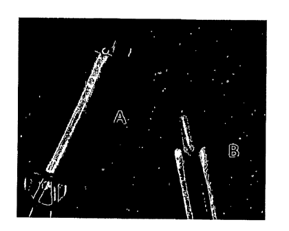
Figure 1 is a series of photographs of micronized ECT devices and the suture
attachment points used to facilitate surgical insertion and scleral attachment
in the eye of a
rodent. Figure lA shows the delivery of device through 30 gauge needle. Figure
113 shows
the delivery of the device using canulated titanium rod. Figure 1C shows an 11-
0 suture
embedded into the adhesive end of the micronized device.
Figure 2 is a graph showing the metabolic activity (CCK-8) of devices
comparing
several cell scaffolding materials. The results presented indicate that
polystyrene
microspheres and PET yarn were good candidates for further investigation.
Figure 3 is a series of photomicrographs demonstrating that both polysulfone
(hydrophilic) and polyimide (hydrophobic) materials were investigated as
encapsulation
membranes. Figure 3A shows that the attachment of cells to the inner wall of
the polyimide
5
CA 02635534 2008-06-25
WO 2007/078922 PCT/US2006/048292
membrane result in eventual cell death due to restricted diffusion. In
contrast, Figure 3B
shows that the polysulfone membranes create a 10 + micron separation between
the inner
wall and the encapsulated cell mass, thereby allowing effective diffusion to
maintain cell
viability.
Figure 4 is a graph showing the metabolic activity of microencapsulated
devices over
course of 2-weeks. These results indicate that scaffoldings coated with
fibronectin promoted
cell attachment to microspheres. Moreover, increasing yarn content may also
benefit cell
growth and activity.
Figure 5 is a histogram showing CNTF production and DNA assayed cell number of
cells contained within the micronized devices. These results confirmed the
advantages of
using fibronectin.to coat microspheres and demonstrated that an increase in
yarn density
benefits encapsulated cell performance.
Figure 6 is a series of photomicrographs showing the qualitative assessment of
microencapsulated cell viability using polysulfone membranes and either a
matrix of
microspheres coated with fibronectin or a matrix of PET scaffolding. Panel A
is a
micrograph of an extruded cell-microsphere tissue mass following 2-weeks of
encapsulation.
Panel B shows the extruded cell mass stained with calcein and ethidium to
visualize live and
dead cells. Panels C (microspheres) and D (PET yarn) are plastic embedded
sections stained
with DAPI and counter stained with fluorescein 12-dUPT to visualize apoptotic
cells. Few
apoptotic cells were observed in either device group. Panels E (microspheres)
and F (PET
yarn) show hemotoxylin and eosin stained plastic sections. Cell viability and
distribution
were good regardless of cell matrix investigated.
Figure 7 is a photograph comparing the size of a first generation ECT device
(1 mm x
6mm) and a micronized BM" device (0.2 mm x 1 mm).
Figure 8 is a graph Showing the pre-implant dose delivery of CNTF from the
first
generation and the micronized ECT devices.
6
CA 02635534 2008-06-25
WO 2007/078922 PCT/US2006/048292
Figure 9 is a graph showing the 18-month high dose CNTF release in rabbit
vitreous
(using the first generation ECT device).
Figure 10 is a graph showing the 18-month low dose CNTF release in rabbit
vitreous
(using the first generation ECT device).
Figure 11 is a series of micrographs showing histological (H&E) sections (10x
magnification) comparing encapsulated cells after 2 week (Figure 11A), 12
month (Figure
11B), and 18 month (Figure 11C) implantation periods.
Figure 12 is a histogram showing the dose delivery of interleukin-10 (IL-10)
from
micronized ECT devices.
= =
Figure 13 is a series of photomicrographs of histological sections showing
representative distribution (Figure 13A) and viability (Figure 13B) of
encapsulated IL-10
producing cells in the micronized device.
Figure 14 is a series of graphs demonstrating first generation ECT and
micronized
ECT
device performance. Figure 14A shows CNTF levels produced in vitro over course
of 8
weeks. Figure 14B shows the results of experiments where metabolic activity of
encapsulated cells over an 8 week period was quantified by a cell redox assay
(CCK-8).
Figure 14C shows in vivo explant device CNTF levels at 2 and 4 week time
points. Figure
14D shows explant vitreous CNTF levels at both 2 and 4 weeks.
Figure 15 A is a photograph demonstrating micronized device implantation using
a
23-gauge needle Figure 15B is a photograph that shows a 300 incision to the
surface of the
sclera through the pars plana. Figure 15C is a photograph that shows
withdrawal of the .
needle revealing inserted device with attached suture and needle. Figure 15D
is a photograph
that shows a single suture closure.
=
7
CA 02635534 2008-06-25
WO 2007/078922 PCT/US2006/048292
DETAILED DESCRIPTION OF THE INVENTION
The instant invention relates to micronized biocompatible, optionally
immunoisolatory, devices for the delivery of one or more biologically active
molecules
("BAMs") to the eye. More particularly, such micronized devices contain a core
containing
living cells that produce or secrete the BAM and a biocompatible jacket
surrounding the core,
wherein the jacket has a molecular weight cut off ("MWCO") that allows the
diffusion of the
BAM into the eye.
This invention further relates to delivery of the BAMs intraocularly (e.g., in
the
anterior chamber and the vitreous cavity) or periocularly (e.g., within or
beneath Tenon's
capsule), or both. The invention may also be used to provide controlled and
sustained release
of biologically active molecules effective in treating various ophthalmic
disorders,
ophthalmic diseases and/or diseases which have ocular effects.
The use of biologically compatible polymeric materials in the construction of
a
micronized encapsulation device is critical to the success of cell
encapsulation therapy
("ECT"). Important components of the encapsulation device include the
surrounding semi-
permeable membrane as well as the internal cell-supporting matrix or scaffold.
Micronized ECT devices were fabricated using 50 kDa molecular weight cut-off
dialysis membranes having a 200 micron diameters and an overall implant length
of 1
millimeter. Total displaced volume of such devices was less than about 0.5
microliters (for
example, about 0.3 g), which represents a volume reduction of more than 200
fold compared
to the current human clinical ECT devices (referred to herein as "the first
generation ECT
devices and/or "the first generation devices"). Implant device configurations
for the
micronized devices of the invention were developed to facilitate insertion and
attachment to
the sclera. (See Figure 1).
Those skilled in the art will recognize that the terms "micronized device(s)",
"micronized ECT device(s)", "microdevice(s)", and "micro-ECT device(s)" and
the like are
used interchangeably herein to refer to the encapsulated cell therapy devices
of the instant
invention.
Suitable device membranes were manufactured using either polysulfone/polyvinyl
pyrrolidone or polyimide. Various cell scaffolding matrices were investigated
for their
ability to induce cell attachment and cell growth and to sustain cell
viability. Scaffolding
matrices that were tested included, for example, alginate cross-linked with
CaC1, Matrigel,
Purapeptide, and non-degradable microspheres with and without fibronectin.
Moreover,
8
CA 02635534 2008-06-25
WO 2007/078922
PCT/US2006/048292
encapsulation using a PET monofilament yarn matrix was also investigated.
Various
combinations of membrane and scaffolding were evaluated and compared to
devices
encapsulated with engineered human retinal pigment epithelial cells (ARPE-19)
producing
either CNTF or IL-10. Protein secretion over the course of the in vitro
evaluation period was
quantified by ELISA. The viability of the encapsulated cells was evaluated
using a DNA
assay to determine total cell number, metabolic activity using a redox assay,
nuclear
fluorescent labeling of live cells, apoptotic cytohistochemistry and
histological examination
of sectioned devices. Additionally, various micronized devices were implanted
in the rodent
vitreous and clinically evaluated.
As shown in Figure 2, the use of hydrogel matrices did not allow adequate cell
viability following an initial screen of micronized devices. Additionally,
polyimide device
groups resulted in poor viability compared to polysulfone ("PS") groups. (See
Figure 3).
However, polysulfone/polyvinyl pyrrolidone membranes using either polystyrene.
microspheres ("PS-microsphere") coated with fibronectin or PET yarn as a cell
scaffold
resulted in sustained levels of protein production over the course of a one-
month evaluation
period. Cells encapsulated within both the PS-microsphere and PET groups of
micronized
devices remained healthy with no evidence of necrosis or apoptosis. (See
Figures 4-6). In
addition, a dose effect delivery of IL-10 and CNTF was achieved using
micronized devices
formulated with a PET yarn matrix. (See Table 1).
Table 1. Results of IL-10 and CNTF production in micronized
devices designed using PET yarn matrix.
Device Group IL-10
(pg/device/24 hrs) CNTF (pg/device/24 hrs)
High Dose 156 32 2.01-0.7
Low Dose 11 11 0.3 0.1
t-test P<0.001 P<0.001
(95% CI)
Clinical evaluation of micronized devices implanted into the vitreous of mice
showed
that these devices remained in a fixed position and avoided contact with the
large rat lens.
Moreover, no adverse findings were reported during the course of the one-month
follow-up
period. Based upon these initial experiments, it appears that manufacture and
maintenance of
9
CA 02635534 2008-06-25
WO 2007/078922 PCT/US2006/048292
micronized ECT devices capable of producing sustained levels of protein are
possible and
that these devices are well tolerated in the rodent vitreous.
As used herein, the term "individual" or "recipient" or "host" refers to a
human or an
animal subject.
A "biologically active molecule" ("BAM") is a substance that is capable of
exerting a
biologically useful effect upon the body of an individual in whom a micronized
device of the
present invention is implanted. As used herein a BAM is one which may exert
its biological
activity within the cell in which it is made or it may be expressed on the
cell surface and
effect the cell's interactions with other cells or biologically active
molecules (e.g., a
neurotransmitter receptor or cell adhesion molecule) or it may be released or
secreted from
the cell in which it is made and exert its effect on a separate target cell
(e.g., a
neurotransmitter, hormone, growth factor, soluble receptor, antibody, antibody
fragment,
anti-angiogenic factor, or cytokine). A BAM is any agent, such as a virus,
protein, peptide,
amino acid, lipid, carbohydrate, nucleic acid, nucleotide, drug, pro-drug or
other substance
that may have an effect on cells whether such effect is harmful, beneficial,
or otherwise.
BAMs that are beneficial to nervous system cells are "neurological agents", a
term which
encompasses any biologically or pharmaceutically active substance that may
prove
potentially useful for the proliferation, differentiation or functioning of
CNS or eye cells or
treatment of neurological or opthalmological disease or disorder. For example,
the term may
encompass certain neurotransmitters, neurotransmitter receptors, growth
factors, growth
factor receptors, soluble receptors, antibodies, antibody fragments, anti-
angiogenic factors
and the like, as well as enzymes used in the synthesis of these agents.
The terms "capsule'? and "device" and "vehicle" are used interchangeably
herein to
refer to the micronized ECT devices of the invention.
Unless otherwise specified, the term "cells" means cells in any form,
including but not
limited to cells retained in tissue, cell clusters, and individually isolated
cells.
As used herein a "biocompatible capsule" or "biocompatible device" or
"biocompatible vehicle" means that the capsule or device or vehicle, upon
implantation in an
individual, does not elicit a detrimental host response sufficient to result
in the rejection of
the capsule or to render it inoperable, for example through degradation.
As used herein an "immunoisolatory capsule" or "immunoisolatory device" or
"immunoisolatory vehicle" means that the capsule upon implantation into an
individual,
minimizes the deleterious effects of the host's immune system on the cells
within its core.
CA 02635534 2008-06-25
WO 2007/078922 PCT/US2006/048292
As used herein "long-term, stable expression of a biologically active
molecule" means
the continued production of a biologically active molecule at a level
sufficient to maintain its
useful biological activity for periods greater than one month, preferably
greater than three
months and most preferably greater than six months. Implants of the micronized
devices and
the contents thereof are able to retain functionality for greater than three
months in vivo and
in many cases for longer than a year. The first generation ECT devices have
been shown to
be able to retain functionality for at least 18 months. Accordingly, it is
believed that the
micronized devices of the instant invention will be able to maintain viability
and production
for equal or longer periods of time in vivo. In addition, the devices of the
current invention
may be prepared of sufficient size to deliver an entire therapeutic dose of a
substance from a
single or just a few (i.e., less than approximately 50) implanted and easily
retrievable devices.
The "semi-permeable" nature of the jacket membrane surrounding the core
permits
molecules produced by the cells (e.g., metabolites, nutrients and/or
therapeutic substances) to
diffuse from the device into the surrounding host eye tissue, but is
sufficiently impermeable
to protect the cells in the core from detrimental immunological attack by the
host.
The core of the immunoisolatory vehicle is constructed to provide a suitable
local
environment for the continued vitality and function of particular cells
isolated therein. The
core may contain a scaffold or a liquid medium sufficient to maintain the
cells.
The core of the micronized devices of the invention can function as a
reservoir for
growth factors (e.g., prolactin, or insulin-like growth factor 2), growth
regulatory substances
such as transforming growth factor f3 (T0F43) or the retinoblastoma gene
protein or nutrient-
transport enhancers (e.g., perfluorocarbons, which can enhance the
concentration of dissolved
oxygen in the core). Certain of these substances are also appropriate for
inclusion in liquid
media.
In addition, the instant devices can also be used as a reservoir for the
controlled
delivery of needed drugs or biotherapeutics. In such cases, the core contains
a high
concentration of the selected drug or biotherapeutic (alone or in combination
with cells or
tissues). In addition, satellite vehicles containing substances which prepare
or create a
hospitable environment in the area of the body in which a micronized device
according to the
invention is implanted can also be implanted into a recipient. In such
instances, the devices
containing immunoisolated cells are implanted in the region along with
satellite vehicles
releasing controlled amounts of, for example, a substance which down-modulates
or inhibits
11
CA 02635534 2013-11-01
an inflammatory response from the recipient (e.g., anti-inflammatory
steroids), or a substance
which stimulates the ingrowth of capillary beds (e.g., an angiogenic factor).
The surrounding or peripheral region (jacket) which surrounds the core of the
instant
micronized devices can be permselective, biocompatible, and/or
immunoisolatory. It is
produced in such a manner that it is free of isolated cells, and completely
surrounds (i.e.,
isolates) the core, thereby preventing contact between any cells in the core
and the recipient's
body. Biocompatible semi-permeable hollow fiber membranes, and methods of
making them
are disclosed in U.S. Pat. Nos. 5,284,761 and 5,158,881 (see also, WO
95/05452). For
example, the capsule jacket can be formed from a polyether sulfone hollow
fiber, such as
those described in U.S. Pat. Nos. 4,976,859 and 4,968,733, and 5,762,798.
To be permselective, the jacket is formed in such a manner that it has a
molecular
weight cut off ("MWCO") range appropriate both to the type and extent of
immunological
reaction anticipated to be encountered after the device is implanted and to
the molecular size
of the largest substance whose passage into and out of the device into the eye
is desirable.
The type and extent of immunological attacks which may be mounted by the
recipient
following implantation of the device depend in part upon the type(s) of moiety
isolated within
it and in part upon the identity of the recipient (i.e., how closely the
recipient is genetically
related to the source of the BAM). When the implanted tissue or cells are
allogeneic to the
recipient, immunological rejection may proceed largely through cell-mediated
attack by the
recipient's immune cells against the implanted cells. When the tissue or cells
are xenogeneic
to the recipient, molecular attack through assembly of the recipient's
cytolytic complement
attack complex may predominate, as well as the antibody interaction with
complement.
The jacket allows passage into the eye of substances up to a predetermined
size, but
prevents the passage of larger substances. More specifically, the surrounding
or peripheral
region is produced in such a manner that it has pores or voids of a
predetermined range of
sizes, and, as a result, the device is permselective. The MWCO of the
surrounding jacket
must be sufficiently low to prevent access of the substances required to carry
out
immunological attacks to the core, yet sufficiently high to allow delivery of
the BAM to the
recipient's eye. Preferably, the MWCO of the biocompatible jacket of the
micronized devices
of the instant invention is from about 1 kD to about 150 kD.
As used herein with respect to the jacket of the device, the term
"biocompatible"
refers collectively to both the device and its contents. Specifically, it
refers to the capability
12
CA 02635534 2008-06-25
WO 2007/078922 PCT/US2006/048292
of the implanted intact micronized device and its contents to avoid the
detrimental effects of
the body's various protective systems and to remain functional for a
significant period of
time. As used herein, the term "protective systems" refers to the types of
immunological
attack which can be mounted by the immune system of an individual in whom the
instant
vehicle is implanted, and to other rejection mechanisms, such as the fibrotic
response, foreign.
body response and other types of inflammatory response which can be induced by
the
presence of a foreign object in the individuals' body. In addition to the
avoidance of
protective responses from the immune system or foreign body fibrotic response,
the term
"biocompatible", as used herein, also implies that no specific undesirable
cytotoxic or
systemic effects are caused by the vehicle and its contents such as those that
would interfere
with the desired functioning of the vehicle or its contents.
The external surface of the micronized device can be selected or designed in
such a
manner that it is particularly suitable for implantation at a selected site.
For example, the
external surface can be smooth, stippled or rough, depending on whether
attachment by cells
of the surrounding tissue is desirable. The shape or configuration can also be
selected or
designed to be particularly appropriate for the implantation site chosen.
The biocompatibility of the surrounding or peripheral region (jacket) of the
micronized device is produced by a combination of factors. Important for
biocompatibility
and continued functionality are device morphology, hydrophobicity and the
absence of
undesirable substances either on the surface of, or leachable from, the device
itself. Thus,
brush surfaces, folds, interlayers or other shapes or structures eliciting a
foreign body
response are avoided. Moreover, the device-forming materials are sufficiently
pure to insure
that unwanted substances do not leach out from the device materials
themselves.
Additionally, following device preparation, the treatment of the external
surface of the device
with fluids or materials (e.g. serum) which may adhere to or be absorbed by
the device and
subsequently impair device biocompatibility is avoided.
First, the materials used to form the device jacket are substances selected
based upon
their ability to be compatible with, and accepted by, the tissues of the
recipient of the
implanted micronized device. Substances are used which are not harmful to the
recipient or to
the isolated cells. Preferred substances include polymer materials, i.e.,
thermoplastic
polymers. Particularly preferred thermoplastic polymer substances are those
which are
modestly hydrophobic, i.e. those having a solubility parameter as defined in
Brandrup J., et
at. Polymer Handbook 3rd Ed., John Wiley & Sons, NY (1989), between 8 and 15,
or more
13
CA 02635534 2013-11-01
preferably, between 9 and 14 (Joules/m3)1/2. The polymer substances are chosen
to have a
solubility parameter low enough so that they are soluble in organic solvents
and still high
enough so that they will partition to form a proper membrane. Such polymer
substances
should be substantially free of labile nucleophilic moieties and be highly
resistant to oxidants
and enzymes even in the absence of stabilizing agents. The period of residence
in vivo which
is contemplated for the particular vehicle must also be considered: substances
must be
chosen which are adequately stable when exposed to physiological conditions
and stresses.
Many thermoplastics are known which are sufficiently stable, even for extended
periods of
residence in vivo, such as periods in excess of one or two years. Examples of
stable materials
include, but are not limited to, polyacrilonitrile/polyvinylchloride
("PAN/PVC" or
"thermoplastic"), polyacrylonitrile, polymethylmethacrylate,
polyvinyldifluoride, polyolefins,
polysulfones, polymide, and/or celluloses.
The choice of materials used to construct the device is determined by a number
of
factors as described in detail in Dionne WO 92/19195. Briefly, various
polymers and
polymer blends can be used to manufacture the capsule jacket. Polymeric
membranes
forming the device and the growth surfaces therein may include polyacrylates
(including
acrylic copolymers), polyvinylidenes, polyvinyl chloride copolymers,
polyurethanes,
polystyrenes, polyamides, cellulose acetates, cellulose nitrates,
polysulfones,
polyphosphazenes, polyacrylonitriles, poly(acrylonitrile/covinyl chloride), as
well as
derivatives, copolymers and mixtures thereof.
A preferred membrane casting solution comprises a either a polysulfone
dissolved in
the water-miscible solvent dimethylacetamide (DMACSO) or polyethersulfone
dissolved in
the water-miscible solvent butyrolactone. This casting solution can optionally
comprise
hydrophilic or hydrophobic additives which affect the permeability
characteristics of the
finished membrane. A preferred hydrophilic additive for the polysulfone or
polyethersulfone
is polyvinylpyrrolidone (PVP). Other suitable polymers comprise
polyacrylonitrile (PAN),
polymethylmethacrylate (PMMA), polyvinyldifluoride (PVDF), polyethylene oxide,
polyolefins (e.g., polyisobutylene or polypropylene),
polyacrylonitrile/polyvinyl chloride
(PAN/PVC), and/or cellulose derivatives (e.g., cellulose acetate or cellulose
butyrate).
Compatible water-miscible solvents for these and other suitable polymers and
copolymers are
found in the teachings of U.S. Pat. No. 3,615,024.
Second, substances used in preparing the biocompatible jacket of the device
are either
free of leachable pyrogenic or otherwise harmful, irritating, or immunogenic
substances or
14
CA 02635534 2013-11-01
are exhaustively purified to remove such harmful substances. Thereafter, and
throughout the
manufacture and maintenance of the device prior to implantation, great care is
taken to
prevent the adulteration or contamination of the device or jacket with
substances, which
would adversely affect its biocompatibility.
Third, the exterior configuration of the device, including its texture, is
formed in such
a manner that it provides an optimal interface with the eye of the recipient
after implantation.
Certain device geometries have also been found to specifically elicit foreign
body fibrotic
responses and should be avoided. Thus, devices should not contain structures
having
interlayers such as brush surfaces or folds. In general, opposing vehicle
surfaces or edges
either from the same or adjacent vehicles should be at least 1 mm apart,
preferably greater
than 2 mm and most preferably greater than 5 mm. Preferred embodiments include
cylinders
having an outer diameter of between about 200 and 350 [tm and a length between
about 0.5
and 6 mm. Preferably, the cores of the micronized device of the invention have
a volume of
less than 0.5 111 (e.g., about 0.3 1).
The surrounding jacket of the biocompatible micronized devices can optionally
include substances which decrease or deter local inflammatory response to the
implanted
vehicle and/or generate or foster a suitable local environment for the
implanted cells or
tissues. For example antibodies to one or more mediators of the immune
response could be
included. Available potentially useful antibodies such as antibodies to the
lymphokines tumor
necrosis factor (TNF), and to interferons (IFN) can be included in the matrix
precursor
solution. Similarly, an anti-inflammatory steroid can be included. See
Christenson, L., et al.,
J. Biomed. Mat. Res., 23, pp. 705-718 (1989); Christenson, L., Ph.D. thesis,
Brown
University, 1989. Alternatively, a substance which stimulates angiogenesis
(ingrowth of
capillary beds) can be included.
In some embodiments, the jacket of the present micronized device is
immunoisolatory. That is, it protects cells in the core of the device from the
immune system
of the individual in whom the device is implanted. It does so (1) by
preventing harmful
substances of the individual's body from entering the core, (2) by minimizing
contact between
the individual and inflammatory, antigenic, or otherwise harmful materials
which may be
present in the core and (3) by providing a spatial and physical barrier
sufficient to prevent
immunological contact between the isolated moiety and detrimental portions of
the
individual's immune system.
CA 02635534 2008-06-25
WO 2007/078922 PCT/US2006/048292
The external jacket may be either an ultrafiltration membrane or a microporous
membrane. Those skilled in the art will recognize that ultrafiltration
membranes are those
having a pore size range of from about 1 to about 100 nanometers while a
microporous
membrane has a range of between about 0.05 to about 10 microns. The thickness
of this
physical barrier can vary, but it will always be sufficiently thick to prevent
direct contact
between the cells and/or substances on either side of the barrier. The
thickness of this region
generally ranges between 5 and 200 microns; thicknesses of 10 to 100 microns
are preferred,
and thickness of 20 to 50 or 20 to 75 microns are particularly preferred.
Types of
, immunological attack which can be prevented or minimized by the use of
the instant device
include attack by macrophages, neutrophils, cellular immune responses (e.g.
natural killer
cells and antibody-dependent T cell-mediated cytoloysis (ADCC)), and humoral
response
(e.g. antibody-dependent complement mediated cytolysis).
The type and extent of immunological response by the recipient to the
implanted
device will be influenced by the relationship of the recipient to the isolated
cells within the
core. For example, if core contains syngeneic cells, these will not cause a
vigorous
immunological reaction, unless the recipient suffers from an autoimmunity with
respect to the
particular cell or tissue type within the device. Syngeneic cells or tissue
are rarely available.
In many cases, allogeneic or xenogeneic cells or tissue (i.e., from donors of
the same species
as, or from a different species than, the prospective recipient) may be
available. The use of
immunoisolatory devices allows the implantation of allogeneic or xenogeneic
cells or tissue,
without a concomitant need to inununosuppress the recipient. Use of
immunoisolatory
capsules also allows the use of unmatched cells (allographs). Therefore, the
instant device
makes it possible to treat many more individuals than can be treated by
conventional
transplantation techniques.
The type and vigor of an immune response to xenografted tissue is expected to
differ
from the response encountered when syngeneic or allogeneic tissue is implanted
into a
recipient. This rejection may proceed primarily by cell-mediated, or by
complement-mediated
attack. The exclusion of IgG from the core of the vehicle is not the
touchstone of
inununoprotection, because in most cases IgG alone is insufficient to produce
cytolysis of the
target cells or tissues. Using immunoisolatory micronized devices, it is
possible to deliver
needed high molecular weight products or to provide metabolic functions
pertaining to high
molecular weight substances, provided that critical substances necessary to
the mediation of
immunological attack are excluded from the immunoisolatory capsule. These
substances may
16
CA 02635534 2008-06-25
WO 2007/078922 PCT/US2006/048292
comprise the complement attack complex component Clq, or they may comprise
phagocytic
or cytotoxic cells. Use of immunoisolatory capsules provides a protective
barrier between
these harmful substances and the isolated cells.
In previous devices, the core and jacket were linked via ionic bonds between
oppositely charged polymers in one of two ways. For example, the devices of
Rha (U.S. Pat.
No; 4,744, 933) were constructed of a charged inner core material and an outer
jacket
material of the opposite charge. Likewise, the devices of Lim and Sun (U.S.
Pat. Nos:
4,352,833 and 4,409,331) included an intermediate layer of poly-L-lysine
(PLL), which is
positively charged, to link the negatively charged core with the negatively
charged jacket
material. The elimination of a PLL layer is advantageous in that PLL is
believed to be
fibrogenic in the host. PLL may also have unwanted growth effects for some
cells. Also, the
jacket of the device of the invention can be controlled for permselectivity
better than those
made with PLL.
The micronized devices of the present invention are distinguished from the
microcapsules of Rha, Lim, and Sun (Rha, C. K. et al., U.S. Pat. No.
4,744,933; Sun, A. M.,
Methods in Enzymology 137, pp. 575-579 (1988)) by (1) the complete exclusion
of cells
from the outer layer of the device, and (2) the thickness of the outer layer
of the device. Both
qualities contribute to the immunoisolation of encapsulated cells in the
present invention.
The microcapsules of Rha were formed by ionic interaction of an ionic core
solution with an
ionic polymer of opposite charge. The microcapsules of Lim and Sun were formed
by
linking an external hydrogel jacket to the core through an intermediate layer
of poly-L-lysine
(PLL). In the microcapsules of Lim and Sun, the intermediate PLL layer was not
sufficiently
thick to guarantee that portions of the encapsulated cells would not penetrate
through and
beyond the layer. Cells penetrating the PLL layer are potential targets for an
immune
response.
Moreover, in the microcapsules of Rha, Lim, and Sun, because the chemical
identity
of the inner substance is either dictated by choice of outer layer, or PLL,
the ability to vary
growth conditions on the inside of these capsules is greatly limited. Since
there are often
specific growth conditions which need to be met in order to successfully
encapsulate specific
cell types, these capsules generally have a limited utility or require
considerable
experimentation to establish appropriate outer layers for a given internal
substance.
Thus, the microcapsules of Rha, Lim, and Sun have a greater potential for
bioincompatibility, fibrogenesis, and vehicle deterioration than the
micronized devices of the
17
CA 02635534 2013-11-01
present invention. A variety of biological systems are known to interact with
and break down
the ionic bonds required for the integrity of microcapsules. PLL evokes
unfavorable tissue
reactions to the capsule. Most notably, this is a fibrotic response. Thus, if
there is any break
in the external layer, if it is not of sufficient thickness, if the PLL layer
begins to degrade,
and/or if encapsulated cells are entrapped within the external layer
sufficiently close to its
outer surface, the microcapsule can trigger a fibrotic response. The term
"fibrogenic" is used
herein in reference to capsules or materials which elicit a fibrotic response
in the implantation
site.
In addition, the micronized devices of the present invention are also
distinguished
from microcapsules (see Sun, A. M., supra; Rha, U.S. Pat. No. 4,744,933) by
the capacity of
micronized devices to contain between 5x10² and 90x103 cells and maintain them
in viable
condition. In contrast, prior art microcapsules typically contain up to about
500 cells per
capsule.
The devices described herein must provide, in at least one dimension,
sufficiently
close proximity of any isolated cells in the core to the surrounding eye
tissues of the recipient
in order to maintain the viability and function of the isolated cells.
However, the diffusional
limitations of the materials used to form the device do not in all cases
solely prescribe its
configurational limits. Certain additives can be used which alter or enhance
the diffusional
properties, or nutrient or oxygen transport properties, of the basic vehicle.
For example, the
internal medium of the core can be supplemented with oxygen-saturated
perfluorocarbons,
thus reducing the needs for immediate contact with blood-borne oxygen. This
will allow
isolated cells or tissues to remain viable while, for instance, a gradient of
angiotensin is
released from the vehicle into the surrounding tissues, stimulating ingrowth
of capillaries.
References and methods for use of perfluorocarbons are given by Faithful, N.
S. Anaesthesia,
42, pp. 234-242 (1987) and NASA Tech Briefs MSC-21480, U.S. Govt. Printing
Office,
Washington, D.C. 20402. Alternatively for clonal cell lines such as PC12
cells, genetically
engineered hemoglobin sequences may be introduced into the cell lines to
produce superior
oxygen storage. See NPO-17517 NASA Tech Briefs, 15, p. 54.
The thickness of the device jacket should be sufficient to prevent an
immunoresponse
by the patient to the presence of the devices. For that purpose, the devices
preferably have a
minimum thickness of 1 lam or more and are free of the cells.
Additionally, reinforcing structural elements can be incorporated into the
micronized
devices. These structural elements can be made in such a fashion that they are
impermeable
18
CA 02635534 2013-11-01
and are appropriately configured to allow tethering or suturing of the device
to the eye tissues
of the recipient. In certain circumstances, these elements can act to securely
seal the jacket
(e.g., at the ends of the cylinder), thereby completing isolation of the core
materials (e.g., a
molded thermoplastic clip). In many embodiments, it is desirable that these
structural
elements should not occlude a significant area of the permselective jacket.
The scaffold defines the microenvironment for the encapsulated cells and keeps
the
cells well distributed within the core. The optimal internal scaffold for a
particular device is
highly dependent on the cell type to be used. In the absence of a scaffold,
adherent cells
aggregate to form clusters.
The filaments used to form a yarn or mesh internal scaffold are formed of any
suitable
biocompatible, substantially non-degradable material. (See United States
Patent Nos.
6,303,136 and 6,627,422). Materials useful in forming yarns or woven meshes
include any
biocompatible polymers that are able to be formed into fibers such as, for
example, acrylic,
polyester, polyethylene, polypropylene, polyacrylonitrile, polyethylene
terephthalate, nylon,
polyamides, polyurethanes, polybutester, or natural fibers such as cotton,
silk, chitin or
carbon. Any suitable thermoplastic polymer, thermoplastic elastomer, or other
synthetic or
natural material having fiber-forming properties may be inserted into a pre-
fabricated hollow
fiber membrane or a hollow cylinder formed from a flat membrane sheet. For
example, silk,
PET or nylon filaments used for suture materials or in the manufacture of
vascular grafts are
highly conducive to this type of application. In other embodiments, metal
ribbon or wire may
be used and woven. Each of these filament materials has well-controlled
surface and
geometric properties, may be mass produced, and has a long history of implant
use. In certain
embodiments, the filaments may be "texturized" to provide rough surfaces and
"hand-holds"
onto which cell projections may attach. The filaments may be coated with
extracellular
matrix molecules or surface-treated (e.g. plasma irradiation) to enhance
cellular adhesion to
the filaments.
In some embodiments, the filaments, preferably organized in a non-random
unidirectional orientation, are twisted in bundles to form yarns of varying
thickness and void
volume. Void volume is defined as the spaces existing between filaments. The
void volume
in the yarn should vary between 20-95%, but is preferably between 50-95%. The
preferred
void space between the filaments is between 20-200 [tm, sufficient to allow
the scaffold to be
seeded with cells along the length of the yarn, and to allow the cells to
attach to the filaments.
The preferred diameter of the filaments comprising the yarn is between 5-100
rn. These
19
CA 02635534 2008-06-25
WO 2007/078922 PCT/US2006/048292
filaments should have sufficient mechanical strength to allow twisting into a
bundle to
comprise a yarn. The filament cross-sectional shape can vary, with circular,
rectangular,
elliptical, triangular, and star-shaped cross-section being preferred.
Alternatively, the filaments or yarns can be woven into a mesh. The mesh can
be
produced on a braider using carriers, similar to bobbins, containing
monofilaments or
multifilaments, which serve to feed either the yarn or filaments into the mesh
during weaving.
The number of carriers is adjustable and may be wound with the same filaments
or a
combination of filaments with different compositions and structures. The angle
of the braid,
defined by the pick count, is controlled by the rotational speed of the
carriers and the
production speed. In one embodiment, a mandrel is used to produce a hollow
tube of mesh. In
certain embodiments, the braid is constructed as a single layer, in other
embodiments it is a
multi-layered structure. The tensile strength of the braid is the linear
summation of the tensile
strengths of the individual filaments.
In some embodiments, a tubular braid is constructed. The braid can be inserted
into a
hollow fiber membrane upon which the cells are seeded. Alternatively, the
cells can be
allowed to infiltrate the wall of the mesh tube to maximize the surface area
available for cell
attachment. When such cell infiltration occurs, the braid serves both as a
cell scaffold matrix
and as an inner support for the device. The increase in tensile strength for
the braid-supported
device is significantly higher than in alternative approaches.
The micronized device of the present invention is of a sufficient size and
durability
for complete retrieval after implantation. The preferred micronized devices of
the present
invention have a core of a preferable minimum volume of less than about 0.5 I
(e.g., about
0.3 1).
Preferably, the micronized device has a tether that aids in maintaining device
placement during implant, and aids in retrieval. Such a tether may have any
suitable shape
that is adapted to secure the capsule in place. For example, the suture may be
a loop, a disk,
or a suture. In some embodiments, the tether is shaped like an eyelet, so that
suture may be
used to secure the tether (and, thus, the device) to the sclera, or other
suitable ocular structure.
In other embodiments, the tether is continuous with the capsule at one end,
and forms a pre-
threaded suture needle at the other end. The tether may be constructed of a
shape memory
metal and/or any other suitable medical grade material known in the art.
Cells which are genetically engineered to secrete antibodies may also be
included in
the core. At least one additional BAM can be delivered from the micronized
device to the
CA 02635534 2013-11-01
eye. For example, the at least one additional BAM can be provided from a
cellular or a
noncellular source. When the at least one additional BAM is provided from a
noncellular
source, the additional BAM(s) may be encapsulated in, dispersed within, or
attached to one or
more components of the cell system. For example, the least one additional
biologically active
molecule can be a nucleic acid, a nucleic acid fragment, a peptide, a
polypeptide, a
peptidomimetic, a carbohydrate, a lipid, an organic molecule, an inorganic
molecule, a
therapeutic agent, or any combinations thereof. Specifically, the therapeutic
agents may be
an anti-angiogenic drug, a steroidal and non-steroidal anti-inflammatory drug,
an anti-mitotic
drug, an anti-tumor drug, an anti-parasitic drug, an TOP reducer, a peptide
drug, and any other
biologically active molecule drugs approved for ophthalmologic use.
The instant invention also relates to methods for making a micronized device.
Micronized devices may be formed by any suitable method known in the art.
(See, e.g.,
United States Patent Nos. 6,361,771; 5,639,275; 5,653,975; 4,892,538;
5,156,844; 5,283,138;
and 5,550,050).
Encapsulated cell therapy is based on the concept of isolating cells from the
recipient
host's immune system by surrounding the cells with a semipermeable
biocompatible material
before implantation within the host. The invention includes a micronized
device in which
ARPE-19 cells are encapsulated in an immunoisolatory capsule, which, upon
implantation
into a recipient host, minimizes the deleterious effects of the host's immune
system on the
ARPE-19 cells in the core of the device. ARPE-19 cells are immunoisolated from
the host by
enclosing them within implantable polymeric capsules formed by a microporous
membrane.
This approach prevents the cell-to-cell contact between the host and implanted
tissues,
thereby eliminating antigen recognition through direct presentation.
The membranes used can also be tailored to control the diffusion of molecules,
such
as antibody and complement, based on their molecular weight. (See Lysaght et
al., 56 J. Cell
Biochem. 196 (1996), Colton, 14 Trends Biotechnol. 158 (1996)). Using
encapsulation
techniques, cells can be transplanted into a host without immune rejection,
either with or
without use of immunosuppressive drugs. The capsule can be made from a
biocompatible
material that, after implantation in a host, does not elicit a detrimental
host response sufficient
to result in the rejection of the capsule or to render it inoperable, for
example through
degradation. The biocompatible material is relatively impermeable to large
molecules, such
as components of the host's immune system, but is permeable to small
molecules, such as
insulin, growth factors, and nutrients, while allowing metabolic waste to be
removed. A
21
CA 02635534 2013-11-01
variety of biocompatible materials are suitable for delivery of growth factors
by the
composition of the invention. Numerous biocompatible materials are known,
having various
outer surface morphologies and other mechanical and structural
characteristics.
Preferably, the capsule of this invention will be similar to those described
by PCT
International patent applications WO 92/19195 or WO 95/05452; or U.S. Pat.
Nos.
5,639,275; 5,653,975; 4,892,538; 5,156,844; 5,283,187; or 5,550,050.
Components of the
biocompatible material may include a surrounding semipermeable membrane and
the internal
cell-supporting scaffolding. The transformed cells are preferably seeded onto
the scaffolding,
which is encapsulated by the permselective membrane. The filamentous cell-
supporting
scaffold may be made from any biocompatible material selected from the group
consisting of
acrylic, polyester, polyethylene, polypropylene polyacetonitrile, polyethylene
teraphthalate,
nylon, polyamides, polyurethanes, polybutester, silk, cotton, chitin, carbon,
or biocompatible
metals. Also, bonded fiber structures can be used for cell implantation. (See
U.S. Pat. No.
5,512,600). Biodegradable polymers include those comprised of poly(lactic
acid) PLA,
poly(lactic-coglycolic acid) PLGA, and poly(glycolic acid) PGA and their
equivalents. Foam
scaffolds have been used to provide surfaces onto which transplanted cells may
adhere (PCT
International patent application Ser. No. 98/05304). Woven mesh tubes have
been used as
vascular grafts (PCT International patent application WO 99/52573).
Additionally, the core
can be composed of an immobilizing matrix formed from a hydrogel, which
stabilizes the
position of the cells. A hydrogel is a 3-dimensional network of cross-linked
hydrophilic
polymers in the form of a gel, substantially composed of water.
Various polymers and polymer blends can be used to manufacture the surrounding
semipermeable membrane, including polyacrylates (including acrylic
copolymers),
polyvinylidenes, polyvinyl chloride copolymers, polyurethanes, polystyrenes,
polyamides,
cellulose acetates, cellulose nitrates, polysulfones (including polyether
sulfones),
polyphosphazenes, polyacrylonitriles, poly(acrylonitrile/covinyl chloride), as
well as
derivatives, copolymers and mixtures thereof. Preferably, the surrounding
semipermeable
membrane is a biocompatible semipermeable hollow fiber membrane. Such
membranes, and
methods of making them are disclosed by U.S. Pat. Nos. 5,284,761 and
5,158,881. The
surrounding semipermeable membrane is formed from a polyether sulfone hollow
fiber, such
as those described by U.S. Pat. No. 4,976,859 or U.S.
22
CA 02635534 2013-11-01
Pat. No. 4,968,733. An alternate surrounding semipermeable membrane material
is
polysulfone.
The capsule can be any configuration appropriate for maintaining biological
activity
and providing access for delivery of the product or function, including for
example,
cylindrical, rectangular, disk-shaped, patch-shaped, ovoid, stellate, or
spherical. Moreover,
the capsule can be coiled or wrapped into a mesh-like or nested structure. If
the capsule is to
be retrieved after it is implanted, configurations which tend to lead to
migration of the
capsules from the site of implantation, such as spherical capsules small
enough to travel in
the recipient host's blood vessels, are not preferred. Certain shapes, such as
rectangles,
patches, disks, cylinders, and flat sheets offer greater structural integrity
and are preferable
where retrieval is desired.
If a device with a jacket of thermoplastic or polymer membrane is desired, the
pore
size range and distribution can be determined by varying the solids content of
the solution of
precursor material (the casting solution), the chemical composition of the
water-miscible
solvent, or optionally including a hydrophilic or hydrophobic additive to the
casting solution,
as taught by U.S. Pat. No. 3,615,024. The pore size may also be adjusted by
varying the
hydrophobicity of the coagulant and/or of the bath.
Typically, the casting solution will comprise a polar organic solvent
containing a
dissolved, water-insoluble polymer or copolymer. This polymer or copolymer
precipitates
upon contact with a solvent-miscible aqueous phase, forming a permselective
membrane at
the site of interface. The size of pores in the membrane depends upon the rate
of diffusion of
the aqueous phase into the solvent phase; the hydrophilic or hydrophobic
additives affect
pore size by altering this rate of diffusion. As the aqueous phase diffuses
farther into the
solvent, the remainder of the polymer or copolymer is precipitated to form a
trabecular
support which confers mechanical strength to the finished device.
The external surface of the device is similarly determined by the conditions
under
which the dissolved polymer or copolymer is precipitated (i.e., exposed to the
air, which
generates an open, trabecular or sponge-like outer skin, immersed in an
aqueous precipitation
bath, which results in a smooth permselective membrane bilayer, or exposed to
air saturated
with water vapor, which results in an intermediate structure).
The surface texture of the device is dependent in part on whether the
extrusion nozzle
is positioned above, or immersed in, the bath: if the nozzle is placed above
the surface of the
bath a roughened outer skin of PAN/PVC will be formed, whereas if the nozzle
is immersed
23
CA 02635534 2013-11-01
in the bath a smooth external surface is formed.
The surrounding or peripheral matrix or membrane can be preformed, filled with
the
materials which will form the core (for instance, using a syringe), and
subsequently sealed in
such a manner that the core materials are completely enclosed. The device can
then be
exposed to conditions which bring about the formation of a core matrix if a
matrix precursor
material is present in the core.
Any suitable method of sealing the device may be used, including the
employment of
polymer adhesives and/or crimping, knotting and heat sealing. These sealing
techniques are
known in the art. In addition, any suitable "dry" sealing method can also be
used. In such
methods, a substantially non-porous fitting is provided through which the cell-
containing
solution is introduced. Subsequent to filling, the device is sealed. Such
methods are described
in, e.g., United States Patent Nos. 5,653,688; 5,713,887; 5,738,673;
6,653,687; 5,932,460;
and 6,123,700.
The devices of the invention can provide for the implantation of diverse cell
or tissue
types, including fully-differentiated, anchorage-dependent, fetal or neonatal,
or transformed,
anchorage-independent cells or tissue. The cells to be isolated are prepared
either from a
donor (i.e., primary cells or tissues, including adult, neonatal, and fetal
cells or tissues) or
from cells which replicate in vitro (i.e., immortalized cells or cell lines,
including genetically
modified cells). In all cases, a sufficient quantity of cells to produce
effective levels of the
needed product or to supply an effective level of the needed metabolic
function is prepared,
generally under sterile conditions, and maintained appropriately (e.g. in a
balanced salt
solution such as Hank's salts, or in a nutrient medium, such as Ham's F12)
prior to isolation.
The micronized ECT devices of the invention are of a shape which tends to
reduce the
distance between the center of the device and the nearest portion of the
jacket for purposes of
permitting easy access of nutrients from the patient into the cell or of entry
of the patient's
proteins into the cell to be acted upon by the cell to provide a metabolic
function. In that
regard, a non-spherical shape, such as a cylinder, is preferred.
Four important factors that influence the number of cells or amount of tissue
to be
placed within the core of the device (i.e., loading density) of the instant
invention are: (1)
device size and geometry; (2) mitotic activity within the device; (3)
viscosity requirements
for core preparation and or loading; and (4) pre-implantation assay and
qualification
requirements.
24
CA 02635534 2008-06-25
WO 2007/078922 PCT/US2006/048292
With respect to the first of these factors, (device size and geometry), the
diffusion of
critical nutrients and metabolic requirements into the cells as well as
diffusion of metabolites
away from the cell are critical to the continued viability of the cells. In
the case of RPE cells
such as ARPE-19 cells, the neighboring cells are able to phagocytize the dying
cells and use
the debris as an energy source.
Among the metabolic requirements met by diffusion of substances into the
device is
the requirement for oxygen. The oxygen requirements of the specific cells must
be
determined for the cell of choice. See Methods and references for
determination of oxygen
metabolism are given in Wilson D. F. etal., J. Biol. Chem., 263, pp. 2712-
2718, (1988).
With respect to the second factor (cell division), if the cells selected are
expected to
be actively dividing while in the device, then they will continue to divide
until they fill the
available space, or until phenomena such as contact inhibition limit further
division. For
replicating cells, the geometry and size of the device will be chosen so that
complete filling
of the device core will not lead to deprivation of critical nutrients due to
diffusional
limitations.
With respect to the third factor (viscosity of core materials) cells in
densities
occupying up to 70% of the device volume can be viable, but cell solutions in
this
concentration range would have considerable viscosity. Introduction of cells
in a very
viscous solution into the device could be prohibitively difficult. In general,
for both two step
and coextrusion strategies, cell loading densities of higher than 30% will
seldom be useful,
and in general optimal loading densities will be 20% and below. For example,
for fragments
of tissues, it is important, in order to preserve the viability of interior
cells, to observe the
same general guidelines as above and tissue fragments should not exceed 250
microns in
diameter with the interior cells having less than 15, preferably less than 10
cells between
them and the nearest diffusional surface.
Finally, with respect to the fourth factor (preimplantation and assay
requirements), in
many cases, a certain amount of time will be required between device
preparation and
implantation. For instance, it may be important to qualify the device in terms
of its biological
activity. Thus, in the case of mitotically active cells, preferred loading
density will also
consider the number of cells which must be present in order to perform the
qualification
assay.
In most cases, prior to implantation in vivo, it will be important to use in
vitro assays
to establish the efficacy of the BAM within the device. Devices can be
constructed and
CA 02635534 2008-06-25
WO 2007/078922 PCT/US2006/048292
analyzed using model systems in order to allow the determination of the
efficacy of the
vehicle on a per cell or unit volume basis.
Following these guidelines for device loading and for determination of device
efficacy, the actual device size for implantation will then be determined by
the amount of
biological activity required for the particular application. The number of
devices and device
size should be sufficient to produce a therapeutic effect upon implantation is
determined by
the amount of biological activity required for the particular application. In
the case of
secretory cells releasing therapeutic substances, standard dosage
considerations and criteria
known to the art will be used to determine the amount of secretory substance
required.
Factors to be considered include; the size and weight of the recipient; the
productivity or
functional level of the cells; and, where appropriate, the normal productivity
or metabolic
activity of the organ or tissue whose function is being replaced or augmented.
It is also
important to consider that a fraction of the cells may not survive the
ilmnunoisolation and
implantation procedures. Moreover, whether the recipient has a preexisting
condition which
can interfere with the efficacy of the implant must also be considered.
Devices of the instant
invention can easily be manufactured which contain many thousands of cells
(e.g., between
=
about 5x10² and about 90x103 cells).
The treatment of many conditions according to the methods described herein
will
require only one or at most less than 50 implanted micronized devices per eye
to supply an
appropriate therapeutic dose. Therapeutic dosages may be between about 0.1 pg
and 1000 ng
per eye per patient per day (e.g., between 0.1 pg and 500 ng per eye per
patient per day;
between 0.1 pg and 250 ng, between 0.1 pg and 100 ng, between 0.1 pg and 50
ng, between
0.1 pg and 25 ng, between 0.1 pg and 10 ng, or between 0.1 pg and 5 ng per eye
per patient
per day). Each of the devices of the present invention is capable of storing
between about
1,000 and about 90,000 cells, in individual or cluster form, depending on
their type.
According to the methods of this invention, other molecules may be co-
delivered
from the micronized devices. For example, it may be preferable to deliver a
trophic factor(s)
with an anti-angiogenic factor(s).
Co-delivery can be accomplished in a number of ways. First, cells may be
transfected
with separate constructs containing the genes encoding the described
molecules. Second,
cells may be transfected with a single construct containing two or more genes
and the
necessary control elements. Third, two or more separately engineered cell
lines can be either
co-encapsulated or more than one device can be implanted at the site of
interest.
26
CA 02635534 2008-06-25
WO 2007/078922 PCT/US2006/048292
Multiple gene expression from a single transcript over expression from
multiple
transcription units can be employed. See, e.g., Macejak, Nature, 353, pp. 90-
94 (1991); WO
94/24870; Mountford and Smith, Trends Genet., 11, pp. 179-84 (1995); Dirks et
al., Gene,
128, pp. 24749 (1993); Martinez-Salas et al., J. Virology, 67, pp. 3748-55
(1993) and
Mountford et al., Proc. Natl. Acad. Sci. USA, 91, pp. 4303-07 (1994).
For some indications, it may be preferable to deliver BAMs to two different
sites in
the eye concurrently. For example, it may be desirable to deliver a
neurotrophic factor to the
vitreous to supply the neural retina (ganglion cells to the RPE) and to
deliver an anti-
angiogenic factor via the sub-Tenon's space to supply the choroidal
vasculature.
Additionally, another embodiment in this invention involves the co-delivery of
a
BAM from a noncellular source or mixture of a BAM from a noncellular source
and
excipient to a region of the eye wherein the BAM from a noncellular source is
encapsulated,
dispersed, or attached to device components including, but not limited to: (a)
sealant; (b)
scaffold; (c) jacket membrane; (d) tether anchor; and/or (e) core media. In
this embodiment,
co-delivery of the BAM from a noncellular source may occur from the same
device as the
BAM from the cellular source. Alternatively, two or more encapsulated cell
systems can be
used. The BAM from a noncellular source can include therapeutic agents such as
steroidal
and non-steroidal anti-inflammatory drugs, anti-mitotic drugs, anti-tumor
drugs, anti-parasitic
drugs, IOP reducers, peptide drugs, and other biologically active molecules
approved for
ophthalmologic use. Suitable excipients include, but are not limited to, any
non-degradable
or biodegradable polymers, hydrogels, solubility enhancers, hydrophobic
molecules, proteins,
salts, or other complexing agents approved for formulations.
Non-cellular dosages can be varied by any suitable method known in the art
such as
varying the concentration of the therapeutic agent, and/or the number of
devices per eye,
and/or modifying the composition of the encapsulating excipient. Cellular
dosage can be
varied by changing (1) the number of cells per device, (2) the number of
devices per eye, or
(3) the level of BAM production per cell. Cellular production can be varied by
changing, for =
example, the copy number of the gene for the BAM in the transduced cell, or
the efficiency
of the promoter driving expression of the BAM. Suitable dosages from non-
cellular sources
may range from about 1 pg to about 1000 ng per day.
This invention also contemplates use of different cell types during the course
of the
treatment regime. For example, a patient may be implanted with a capsule
device containing
a first cell type. If after time, the patient develops an immune response to
that cell type, the
27
CA 02635534 2008-06-25
WO 2007/078922 PCT/US2006/048292
capsule can be retrieved, or explanted, and a second capsule can be implanted
containing a
second cell type. In this manner, continuous provision of the therapeutic
molecule is possible,
even if the patient develops an immune response to one of the encapsulated
cell types.
The methods and devices of the instant invention are useful to deliver a wide
range of
cellular products, including high molecular weight products, to an individual
in need of them,
and/or to provide needed metabolic functions to an individual, such as the
removal of harmful
substances. Products which can be delivered using the instant devices include
a wide variety
of BAMs normally secreted by various organs or tissues. Alternatively, the
encapsulated
cells can be genetically engineered to secrete one or more BAMs.
Many cellular products which are difficult to provide using primary donor
tissues can
be provided using immortalized cells or cell lines. Immortalized cells are
those which are
capable of indefinite replication but which exhibit contact inhibition upon
confluence and are
not tumorigenic. An example of an immortalized cell line is the rat
pheochromocytoma cell
line PC12. Transformed cells or cell lines can be used in a similar manner.
Transformed cells
are unlike merely immortalized cells in that they do not exhibit contact
inhibition upon
confluence, and form tumors when implanted into an allogeneic host.
Immortalization can
allow the use of rare or notoriously fragile cell or tissue types for the long-
term delivery of a
chosen product or metabolic function. Suitable techniques for the
immortalization of cells are
described in Land H. et al., Nature 304, pp. 596-602 (1983) and Cepko, C. L.,
Neuron 1, pp.
345-353 (1988). Candidate cell lines include, for example, genetically
engineered beta-cell
lines which secrete insulin such as NIT' cells (see Hamaguchi, K., et al.,
Diabetes 40, p. 842
(1991)), RIN cells (see Chick, W. L., et al., Proc. Natl. Acad. Sci. USA, 74,
pp. 628-632
(1977)), ATT cells (see Hughes, S. D., et al, Proc. Natl. Acad. Sci. USA, 89,
pp. 688-692
(1992)), CHO cells (see Matsumoto, M., et al, 1990, Proc. Natl. Acad. Sci.
USA, 87, pp.
9133-9137 (1990)), beta-TC-3 cells (see Tal, M., et al, 1992, Mol. Cell Biol.,
12, pp. 422-432
(1992)), and ARPE-19 cells. Additionally, recombinant cells or cell lines can
be engineered
to provide novel products or functions and combinations thereof, using a wide
variety of
techniques well known to those of ordinary skill in the art.
The genes encoding numerous biologically active molecules have been cloned and
their nucleotide sequences published. Many of those genes are publicly
available from
depositories such as the American Type Culture Collection (ATCC) or various
commercial
sources. Genes encoding the biologically active molecules useful in this
invention that are
not publicly available may be obtained using standard recombinant DNA methods
such as
28
CA 02635534 2008-06-25
WO 2007/078922 PCT/US2006/048292
PCR amplification, genomic and cDNA library screening with oligonucleotide
probes. Any
of the known genes coding for biologically active molecules may be employed in
the
methods of this invention. See, e.g., U.S. Pat. No. 5,049,493; Gage et al.,
U.S. Pat. No.
5,082,670; and Genentech U.S. Pat. No. 5,167,762.
A gene of interest (i.e., a gene that encodes a suitable biologically active
molecule)
can be inserted into a cloning site of a suitable expression vector by using
standard
techniques. It will be appreciated that more than one gene may be inserted
into a suitable
expression vector. These techniques are well known to those skilled in the
art.
The expression vector containing the gene of interest may then be used to
transfect the
cell line to be used in the methods of this invention. Standard transfection
techniques such as
calcium phosphate co-precipitation, DEAE-dextran transfection or
electroporation may be
utilized. Commercially available mammalian transfection kits may be purchased
from e.g.,
Stratagene.
A wide variety of host/expression vector combinations may be used to express
the
gene encoding the biologically active molecule of interest. Long-term, stable
in vivo
expression is achieved using expression vectors (i.e., recombinant DNA
molecules) in which
the gene encoding the biologically active molecule is operatively linked to a
promoter that is
not subject to down regulation upon implantation in vivo in a mammalian host.
Accordingly,
such expression vectors would typically not contain a retroviral promoter.
Suitable
promoters include, for example, the early and late promoters of SV40 or
adenovirus and other
known non-retroviral promoters capable of controlling gene expression.
Useful expression vectors, for example, may consist of segments of
chromosomal,
non-chromosomal and synthetic DNA sequences, such as various known derivatives
of SV40
and known bacterial plasmids, e.g., pUC, pBlue Scriptrm plasmids from E. coli
including
pBR322, pCR1, pMB9, pUC, pBlue Scripirm and their derivatives. Expression
vectors
containing the geneticin (G418) or hygromycin drug selection genes (See
Southern, P. J.
(1981), In vitro, 18, p. 315, Southern, P. J. and Berg, P. (1982), J. Mol.
Appl. Genet., 1, p.
327) are also useful in practicing this invention. These vectors can employ a
variety of
different enhancer/promoter regions to drive the expression of both a biologic
gene of interest
(e.g., NGF) and/or a gene conferring resistance to selection with toxin such
as 0418 or
hygromycin B. The 0418 resistance gene codes for aminoglycoside
phosphotransferase
(APH) which enzymatically inactivates G418 (100-500 Ag/111) added to the
culture medium.
Only those cells expressing the APH gene will survive drug selection usually
resulting in the
29
CA 02635534 2008-06-25
WO 2007/078922 PCT/US2006/048292
expression of the second biologic gene as well. The hygromycin B
phosphotransferase (HPH)
gene codes for an enzyme which specifically modifies hygromycin toxin and
inactivates it.
Genes cotransfected with or contained on the same plasmid as the hygromycin B
phosphotransferase gene will be preferentially expressed in the presence of
hygromycin B at
A variety of different mammalian promoters can be employed to direct the
expression
of the genes for 0418 and hygromycin B and/or the BAM gene of interest. These
promoters
include, but are not limited to, the promoters of hDBH (human dopamine beta
hydoxylase)
(see Mercer et al., Neuron, 7, pp. 703-716, (1991)), hTH (human tyrosine
hydroxylase) (see
Examples of expression vectors that can be employed include, but are not
limited to,
the commercially available pRC/CMV, pRC/RSV, and pCDNA1NE0 (InVitrogen). The
viral
25 oligodendrocytes.
In some embodiments, the pNUT expression vector is used. In addition, the pNUT
expression vector can be modified such that the DHFR coding sequence is
replaced by the
coding sequence for G418 or hygromycin drug resistance. The SV40 promoter
within the
pNUT expression vector can also be replaced with any suitable constitutively
expressed
Suitable BA.Ms for use in the micronized devices of the invention include, but
are not
limited to, antiangiogenic factors, anti-inflammatory factors, neurotrophic
factors, growth
factors, trophic factors, antibodies and antibody fragments,
neurotransmitters, hormones,
CA 02635534 2008-06-25
WO 2007/078922 PCT/US2006/048292
cytokines, or lymphokines. Specifically, the BAMs may be TGF13, GDNF, NGF,
CNTF,
bFGF, aFGF, IL-113, IFN41,3FN¨a, BDNF, LIP, NT-4, NTN, NT4/5, CT-1, LEDGF,
Neublastin, Axolcine, IL-23, RdCVF, IL-10, Alpha INF, IL-1Ra, and Remicade.
Exemplary
antiangiogenic factors include, but are not limited to vasculostatin,
angiostatin, endostatin,
anti-integrins, vascular endothelial growth factor inhibitors (VEGF-
inhibitors), platelet factor
4, heparinase, bFGF-binding molecules, the VEGF receptor Flt, the VEGF
receptor Flk,
Lucentis, VEGF Trap, Tek A/Fc (angl/ang2 inhibitor), 2xCon4 (C), soluble VEGF
Receptors, and PEDF.
Other products which can be delivered through use of the instant micronized
device
include trophic factors such as erythropoietin, growth hormone, Substance P,
and
neurotensin. This invention is useful for treating individuals suffering from
acute and/or
chronic pain, by delivery of an analgesic or pain reduaing substance to the
individual. Such
pain reducing substances include enkephalins, catecholamines and other opioid
peptides.
Such compounds may be secreted by, e.g., adrenal chromaffin cells. Another
family of
products suited to delivery by the instant vehicle comprises biological
response modifiers,
including lymphokines and cytokines. Antibodies from antibody secreting cells
may also be
delivered. Specific antibodies which may be useful include those towards tumor
specific
antigens. The release of antibodies may also be useful in decreasing
circulating levels of
compounds such as hormones or growth factors. These products are useful in the
treatment of
a wide variety of diseases, inflammatory conditions or disorders, and
degenerative disorders
of the eye.
Modified, truncated and/or mutein forms of the above-mentioned molecules are
also
contemplated. Further, active fragments of these growth factors (i.e., those
fragments of
growth factors having biological activity sufficient to achieve a therapeutic
effect) are also
contemplated. Also contemplated are growth factor molecules modified by
attachment of one
or more polyethylene glycol (PEG) or other repeating polymeric moieties.
Combinations of
these proteins and polycistronic versions thereof are also contemplated.
- The choice of cells depends upon the intended application. The cells can be
chosen
for their secretion of hormones, cytokines, growth factors, trophic factors,
angiogenesis
factors, antibodies, blood coagulation factors, lymphokines, enzymes, and
other therapeutic
agents or agonists, precursors, active analogs, or active fragments thereof.
A wide variety of cells may be used in this invention. These include well
known,
publicly available immortalized cell lines as well as primary cell cultures.
Examples of
31
CA 02635534 2008-06-25
WO 2007/078922 PCT/US2006/048292
publicly available cell lines suitable for the practice of this invention
include, baby hamster
kidney (BHK), Chinese hamster ovary (CHO), mouse fibroblast (L-M), NIH Swiss
mouse
embryo (N111/3T3), African green monkey cell lines (including COS-a, COS-7,
BSC-1, BSC-
40, BMT-10 and Vero), rat adrenal pheochromocytoma (PC12), rat glial tumor
(C6), ARPE-
19 cells, and the like. Primary cells that may be used according to the
present invention
include, bFGF-responsive neural progenitor-stem cells derived from the CNS of
mammals
(Richards et at., Proc. Natl. Acad. Sci. USA 89, pp. 8591-8595 (1992); Ray et
al., Proc. Natl.
Acad. Sci. USA, 90, pp. 3602-3606 (1993)), primary fibroblasts, Schwann cells,
astrocytes,
0-TC cells, Hep-G2 cells, AT T20 cells, oligodencirocytes and their
precursors, myoblasts,
adrenal chromaffin cells, and the like.
The choice of cell depends upon the intended application. The encapsulated
cells may
be chosen for secretion of a particular BAM. Cells can also be employed which
synthesize
and secrete agonists, analogs, derivatives or fragments of BAMs, which are
active.
To be a platform cell line for an encapsulated cell based delivery system, the
cell line
should have as many of the following characteristics as possible; (1) the
cells should be hardy
under stringent conditions (the encapsulated cells should be functional in the
avascular tissue
cavities such as in the central nervous system or the eye, especially in the
intra-ocular
environment); (2) the cells should be able to be genetically modified (the
desired therapeutic
factors needed to be engineered into the cells); (3) the cells should have a
relatively long life
span (the cells should produce sufficient progenies to be banked,
characterized, engineered,
safety tested and clinical lot manufactured); (4) the cells should preferably
be of human
origin (which increases compatibility between the encapsulated cells and the
host); (5) the
cells should exhibit greater than 80% viability for a period of more than one
month in vivo in
device (which ensures long-term delivery); (6) the encapsulated cells should
deliver an
efficacious quantity of a useful biological product (which ensures
effectiveness of the
treatment); (7) the cells should have a low level of host immune reaction
(which ensures the
longevity of the graft); and (8) the cells should be nontumorigenic (to
provide added safety to
the host, in case of device leakage).
The ARPE-19 cell line (see Dunn et al., 62 Exp. Eye Res. 155-69 (1996), Dunn
et al.,
39 Invest. Ophthalmol. Vis. Sci. 2744-9 (1998), Finnemann et al., 94 Proc.
Natl. Acad. Sci.
USA 1293227 (1997), Handa et al., 66 Exp. Eye. 411-9 (1998), Holtkamp et at.,
112 Clin.
Exp. Immunol. 34-43 (1998), Maidji et al., 70 J. Virol. 8402-10 (1996); United
States Patent
No. 6,361,771) demonstrates all of the characteristics of a successful
platform cell for an
32
CA 02635534 2008-06-25
WO 2007/078922
PCT/US2006/048292
encapsulated cell-based delivery system. The ARPE-19 cell line is available
from the
American Type Culture Collection (ATCC Number CRL-2302). ARPE-19 cells are
normal
retinal pigmented epithelial (RPE) cells and express the retinal pigmentary
epithelial cell-
specific markers CRALBP and RPE-65. ARPE-19 cells form stable monolayers,
which
exhibit morphological and functional polarity.
When the micronized devices of the invention are used, preferably between 102
and
108 ARPE-19 cells, most preferably 5x10² to 90x103 ARPE-19 cells are
encapsulated in each
device. Dosage may be controlled by implanting a fewer or greater number of
capsules, .
preferably between 1 and 50 capsules per patient. The devices described herein
are capable
of delivering between about 0.1 pg and 1000 ng of the desired BAM per eye per
patient per
day. Both the first generation ECT devices as well as the micronized devices
of the instant
invention have been shown to provide the same level of protein to the eye.
Techniques and procedures for isolating cells or tissues which produce a
selected
product are known to those skilled in the art, or can be adapted from known
procedures with
no more than routine experimentation.
If the cells to be isolated are replicating cells or cell lines adapted to
growth in vitro, it
is particularly advantageous to generate a cell bank of these cells. A
particular advantage of a
cell bank is that it is a source of cells prepared from the same culture or
batch of cells. That
is, all cells originated from the same source of cells and have been exposed
to the same
conditions and stresses. Therefore, the vials can be treated as identical
clones. In the
transplantation context, this greatly facilitates the production of identical
or replacement
devices. It also allows simplified testing protocols, which assure that
implanted cells are free
of retroviruses and the like. It may also allow for parallel monitoring of
vehicles in vivo and
in vitro, thus allowing investigation of effects or factors unique to
residence in vivo.
In all cases, it is important that the cells or tissue contained in the device
are not
contaminated or adulterated.
The newly-formed micronized devices obtained by any of the methods described
herein can be maintained under sterile conditions in a non-pyrogenic, serum-
free defined
nutrient medium or balanced salt solution, at about 37 C, prior to
implantation. Lower
temperatures (20 C -37 C) may be optimal for certain cell types and/or
culturing
conditions. Other holding temperatures and medium compositions consistent with
good cell
viability may also be used. Alternatively, the device can be cryopreserved in
liquid nitrogen,
if a cryoprotective agent such as glycerin has been incorporated into the
matrix. (See Rajotte,
33
CA 02635534 2008-06-25
WO 2007/078922 PCT/US2006/048292
R. V. et al. Transplantation Proceedings, 21, pp. 2638-2640 (1989)). In such a
case, the
device is thawed before use and equilibrated under sterile conditions as
described above.
The methods and devices of this invention are intended for use in a mammalian
host,
recipient, subject or individual, preferably a primate, most preferably a
human. A number of
different implantation sites are contemplated for the devices and methods of
this invention.
Suitable implantation sites include, but are not limited to, the aqueous and
vitreous humors of
the eye, the periocular space, the anterior chamber, and/or the Subtenon's
capsule.
The invention provides methods of treating ophthalmic disorders by implanting
the
micronized devices of the invention into an eye of the patient. For example,
the ophthalmic
disorder may be a retinal degeneration disease. Exemplary retinal degeneration
diseases
include, but are not limited to, retinopathy of prematurity, glaucoma,
cataract formation,
retinoblastoma, retinal ischemia, uveitis, retinitis pigmentosa, forms of wet
and dry age-
related macular degeneration, diabetic retinopathy, and choroideremia. Other
ophthalmic
disorders that may be treated using the micronized devices of the present
invention include,
but are not limited to, proliferative retinopathies, retinal vascular
diseases, vascular
anomalies, age-related macular degeneration and other acquired disorders
(including but not
limited to dry age-related macular degeneration, exudative age-related macular
degeneration,
and myopic degeneration), endophthalmitis, infectious diseases, inflammatory
but non-
infectious diseases, AIDS-related disorders, ocular ischemia syndrome,
pregnancy-related
disorders, peripheral retinal degenerations, retinal degenerations, toxic
retinopathies, retinal
tumors, choroidal tumors, choroidal disorders, vitreous disorders, retinal
detachment and
proliferative vitreoretinopathy, non-penetrating trauma, penetrating trauma,
post-cataract
complications, and inflammatory optic neuropathies.
The devices of the present invention may also be useful for the treatment of
ocular
neovascularization, a condition associated with many ocular diseases and
disorders. For
example, retinal ischemia-associated ocular neovascularization is a major
cause of blindness
in diabetes and many other diseases. The present invention may also be used to
treat ocular
symptoms resulting from diseases or conditions that have both ocular and non-
ocular
symptoms. Some examples include cytomegalovirus retinitis in AIDS and other
conditions
and vitreous disorders, hypertensive changes in the retina as a result of
pregnancy, and ocular
effects of various infectious diseases such as tuberculosis, syphilis, Lyme
disease, parasitic
disease, toxocara canis, ophthalmonyiasis, cyst cercosis and fungal
infections. Likewise, the
invention may also be used to treat conditions relating to other intraocular
34
=
CA 02635534 2008-06-25
WO 2007/078922 PCT/US2006/048292
neovascularization-based diseases. Corneal neovascularization is a major
problem because it
interferes with vision and predisposes patients to corneal graft failure. A
majority of severe
visual loss is associated with disorders which result in ocular
neovascularization. For
example, neovascularization occurs in diseases such as diabetic retinopathy,
central retinal
vein occlusion and possibly age-related macular degeneration.
The micronized devices and techniques of this invention provide several
advantages
over other delivery routes. For example, BAMs can be delivered to the eye
directly, which
reduce or minimize unwanted peripheral side effects. Moreover, very small
doses of BAMs
(picogram or low nanogram quantities rather than milligrams) compared with
topical
applications can be delivered, thereby also potentially lessening side
effects. Likewise,
because viable cells continuously produce newly synthesized BAMs, these
techniques should
be superior to injection delivery of drugs, where the BAIVI dose fluctuates
greatly between
injections and the BAM is continuously degraded but not continuously
replenished.
Living cells can be encapsulated in the micronized device of the invention and
surgically inserted (under retrobulbar anesthesia) into the vitreous of the
eye. Preferably, the
micronized device is tethered to the sclera to aid in removal. The micronized
device can
remain in the vitreous as long as necessary to achieve the desired prophylaxis
or therapy.
Such therapies for example include promotion of neuron or photoreceptor
survival or repair,
or inhibition and/or reversal of retinal or choroidal neovascularization, as
well as inhibition of
uveal, retinal and optic nerve inflammation.
With vitreal placement, the biologically active molecule, preferably a trophic
factor,
may be delivered to the retina or the RPE. In addition, retinal
neovascularization may be best
treated by delivering an anti-angiogenic factor to the vitreous.
In other embodiments, cell-loaded devices are implanted periocularly, within
or
beneath the space known as Tenon's capsule. This embodiment is less invasive
than
implantation into the vitreous as complications such as vitreal hemorrhage
and/or retinal
detachment are potentially eliminated. This route of administration also
permits delivery of
BAMs (e.g., trophic factors and the like) to the RPE or the retina. This
embodiment is
especially preferred for treating choroidal neovascularization and
inflammation of the optic
nerve and uveal tract. In general, delivery from this implantation site will
permit circulation
of the desired biologically active molecule to the choroidal vasculature,
retinal vasculature,
and the optic nerve.
Preferred embodiments include, but are not limited to, periocular delivery
(implanting
CA 02635534 2008-06-25
WO 2007/078922 PCT/US2006/048292
beneath Tenon's capsule) of anti-angiogenic molecules, anti-inflammatory
molecules (such as
cytokines and hormones), and neurotrophic factors to the choroidal vasculature
to treat
macular degeneration (choroidal neovascularization). Delivery of anti-
angiogenic factors
directly to the choroidal vasculature (periocularly) or to the vitreous
(intraocularly) using the
devices and methods of this invention may reduce the above mentioned problems
and may
permit the treatment of poorly defined or occult choroidal neovascularization.
It may also
provide a way of reducing or preventing recurrent choroidal neovascularization
via
adjunctive or maintenance therapy.
Implantation of the biocompatible micronized device is performed under sterile
conditions. The micronized device can be implanted using a syringe or any
other method
known to those skilled in the art. Generally, the device is implanted at a
site in the recipient's
body which will allow appropriate delivery of the secreted product or function
to the recipient
and of nutrients to the implanted cells or tissue, and will also allow access
to the device for
retrieval and/or replacement. A number of different implantation sites are
contemplated.
These include, e.g., the aqueous humor, the vitreous humor, the sub-Tenon's
capsule, the
periocular space, and the anterior chamber. Preferably, for implant sites that
are not
immunologically privileged, such as periocular sites, and other areas outside
the anterior
chamber (aqueous) and the posterior chamber (vitreous), the capsules are
irmnunoisolatory.
It is preferable to verify that the cells immobilized within the micronized
device function
properly both before and after implantation; assays or diagnostic tests well
known in the art
can be used for these purposes. For example, an ELISA (enzyme-linked
immunosorbent
assay), chromatographic or enzymatic assay, or bioassay specific for the
secreted product can
be used. If desired, secretory function of an implant can be monitored over
time by collecting
appropriate samples (e.g., serum) from the recipient and assaying them.
The invention will be further described in the following examples, which do
not
limit the scope of the invention described in the claims.
EXAMPLES
Example 1: Comparison of "first generation" and micronized ECT devices
Materials and Methods
ECT devices were fabricated to allow intravitreal implantation into the eye.
The first
generation devices totaled 600 pi in displaced volume (1.1 mm diameter, 6 mm
length).
36
CA 02635534 2008-06-25
WO 2007/078922 PCT/US2006/048292
Micronized ECT devices were fabricated with a total displacement volume of
0.05 p.1 (200
mm diameter, 1 mm length). A comparison of the size differences between the
first
generation and micronized ECT devices is provided in Figure 7. Encapsulated
cell lines used
in these studies were genetically modified to secrete either ciliary
neurotrophic factor (CNTF)
or interleukin-10 (IL-10). Devices were designed to produce a high and a low
dose delivery
for both CNTF and I1.40. Implant periods ranged from 2 weeks to 18 months in
the rabbit
model and were 2 weeks in the rat model. Protein delivery levels were
quantified over the
course of the studies and clinical exams were conducted to assess ocular
irritation and
surgical wound healing.
Results
CNTF Delivery
Dose delivery was achieved using both first generation and micronized ECT
devices
in both the rabbit and rat animal models. Pre-implant dose separation is shown
for both size
devices in Figure 8.
Long-term implant dose delivery was achieved in the rabbit model resulting in
stable
delivery throughout the course of the 18 month implant. High dose CNTF
delivery
(ng/device/day) in the rabbit model ranged from 4.42 1.14 at 2-weeks to 2.20
1.08 at 18
months (Figure 9). Low dose delivery (ng/device/day) ranged from 1.14 0.24
at 2 weeks to
0.11 0.06 at 18 months (Figure 10). Vitreous CNTF levels for both high and
low dose
delivery were approximately 10 percent of device output and remained stable
throughout the
course of the study.
Histological examination of explanted devices (Figure 11) indicated stable
encapsulated cell viability throughout the course of the 18 month implant
period.
IL-10 Delivery
Micronized ECT devices were implanted into the rat vitreous as part of a study
to
investigate treatment of experimental autoimmune uveoretinitis (EAU) delivered
interleukin-
10 ("IL-10") at either a pre-implant high dose of 156 32 (pg/device/day) or a
low dose of
13 11 (pg/device/day) (Figure 12).
Histological evaluation of the cells in the micronized ECT devices revealed
robust
viability and a high degree of cell distribution within the devices (Figure
13). Preliminary
results in the EAU model showed a beneficial treatment effect using ECT to
deliver IL-10.
37
=
CA 02635534 2013-11-01
Throughout the course of these studies and regardless of animal models chosen
for
therapeutic delivery using ECT devices, no significant adverse post-operative
complications
were reported in any groups following periodic fundoscopy examinations.
Conclusions
Manufacture of ECT devices to deliver intravitreal levels of therapeutic
molecules in
both the rabbit (first generation devices) and rat (micronized devices) model
was
demonstrated. Both first generation and micronized implants were well
tolerated and the
delivery of therapeutics was continuous during the course of the entire
implant period of up
to 18 months.
Example 2: Delivery of Encapsulated Cell Technology (ECT) Micronized Device
Implants
Using a Small Gauge Needle
Implantation of first generation ECT devices, which are currently in Phase II
human
clinical trials for retinitis pigmentosa and age-related macular degeneration,
requires a 2.0
mm sclerotomy and three sutures to close the incision site. Thus, development
of an ECT
micronized device capable of producing comparable protein levels that could be
implanted
through a small gauge needle would improve the surgical procedure and minimize
surgical
risk.
Methods
Micronized devices were prepared that contained encapsulated cells transfected
to
produce either ciliary neurotrophic factor (CTNF), interleukin-10 (IL-10) or
pigment
epithelial derived factor (PEDF). Implantation of such micronized devices
delivering CNTF
was tested using a 23-gauge needle having a modified syringe. CNTF-producing
devices
were surgically implanted into New Zealand white rabbits and evaluated at 2
weeks and 1-
month using indirect opthalmoscopy and histological examination. Two methods
of closure
were investigated: (1) incision closure using a 10-0 suture and needle
attached to the device
and (2) sutureless closure using a modified procedure to that described by
Jaffe, et al, Arch
Ophthalmol 114:1273-75 (1996). Intravitreal CNTF levels, the production rate
of explanted
micronized devices and encapsulated cell viability were determined.
Results
Micronized devices produced 5.0 0.5, 3.0 0.7, and 39 4 ng/device/day of
CNTF,
IL-10 and PEDF, at the 2-week time point, respectively. Protein secretion from
the
38
CA 02635534 2008-06-25
WO 2007/078922 PCT/US2006/048292
micronized ECT devices was consistent and stable over the course of several
months. Protein
output to device volume levels were 10 times greater than the capacity of the
first generation
ECT devices.
Micronized device CNTF production was evaluated both in vitro and in vivo.
First
generation devices produced higher levels of CNTF during the in vitro
evaluation period.
(See Figure 14A). However, 2 and 4-week explanted micronized CNTF levels were
statistically equivalent (P = 0.5663 and P=0.6744) to the levels produced by
the first
generation ECT devices. (See Figure 14B). Moreover, vitreous CNTF levels were
also
statistically equivalent comparing the micronized device groups to the first
generation device
groups at both the 2 and 4-week in vivo evaluation time-points. (See Figure
14C, P=0.2344
and P=0.8665, respectively). Metabolic activity of the micronized devices
increased over
time as compared to the first generation devices, which decreased over the 8
week in vitro
hold period. (See Figure 14D).
CNTF-producing micronized devices were injected successfully via a 23-gauge
needle (see Figure 15) and post-surgical evaluations of anchoring methods
indicated
successful placement and constraint of the micronized devices. The injectable
sclerotomy
closure was considerably less invasive and reduced the surgical time by half
compared to
implantation of the first generation ECT device.
Conclusions
Encapsulation using micronized devices can provide therapeutic protein levels
comparable to the first generation device configuration currently used in ECT
clinical trial
evaluations. Likewise, surgical implantation of a micronized device using a 23-
gauge needle
to inject the devices appear to be feasible and may offer a simpler, less
invasive approach to
encapsulation cell therapy in the eye.
Example 3: Safety and Pharmacokinetics of an Injectable Micronized
Encapsulated Cell
Technology Device
Three concurrent studies of a first generation encapsulated cell technology
(ECT)
device delivering ciliary neurotrophic factor (CNTF) are in human clinical for
early and late-
stage retinitis pigmentosa and age-related macular degeneration. ECT devices
used in these
trials are implanted in patients eyes using conjunctival excision, sceral
incision, and inter-
ocular placement followed by securing the device followed by suture closure of
the sclera and
conjunctiva. The current study reports on the research efforts to develop the
long-term
pharmacolcinetics and safety profile of a smaller profile, micronized ECT
device capable of
39
CA 02635534 2008-06-25
WO 2007/078922 PCT/US2006/048292
delivering efficacious therapeutics, implanted using sutureless, 23-gauge
injection
procedures.
Methods
Human retinal pigment epithelial cells genetically modified to continuously
secrete
CNTF were encapsulated in 300 micron diameter hollow fiber semi-permeable
polymer
membranes and delivered by inter-ocular implantation into the rabbit vitreous
cavity.
Implantation of the devices was performed using a modified version of the 23-
gauge
sutureless sclerotomy technique. Safety and CNTF protein pharmacokinetics over
the course
of a 1-year period are the eventual outcome endpoints of this study. Vitreous
CNTF levels
and explanted micronized device CNTF output were evaluated by a commercial
ELISA.
Clinical and pathological evaluations were performed over the course of the
implantation
=
period in order to assess implant safety.
Results
At the three month time point, explanted micronized device production and
vitreous
levels of CNTF are 3.2 ng/day and 0.2 ng/ml, respectively. Preliminary fitted
kinetics and
half-life (tin) constants for the explanted devices are k = 0.0167 weeks' and
tin = 41 weeks,
respectively. Vitreous level k = 0.036 weeks' and tin= 19 weeks. No evidence
of ocular
toxicity was observed in eyes that were implanted using the sutureless
sclerotomy implant
method. Clinical and pathohistological evaluation of the implant site showed
normal wound
healing response that was consistent with expected tissue reaction following
surgical incision.
Additionally, no adverse pan-retinal, optic nerve or vascular toxicity was
observed in any of
the implanted eyes.
Conclusions
Thus, preliminary results of this study indicate that micronized ECT devices
are
capable of sustaining long term intra-ocular delivery of CNTF in the rabbit.
Additionally, the
safety profile of a trans-conjunctival implant procedure that mitigates the
necessity to suture
the micronized device also shows promise for the potential of an injectable
micronized ECT
device.
40