Note: Descriptions are shown in the official language in which they were submitted.
CA 02636450 2008-07-07
WO 2007/084762 PCT/US2007/001645
TITLE OF THE INVENTION
DEVICE FOR RAPID REPAIR OF BODY CONDUITS
CROSS REFERENCE TO RELATED APPLICATIONS
This application claims the benefit of United States Provisional Patent
Application No. 60/760,594, filed on January 20, 2006
FIELD OF THE INVENTION
The present invention relates to the field of medical devices useful in the
repair of
trauma to body conduits, particularly to an implantable device useful for such
repairs, and
more particularly to a self-expanding stent-graft useful for such repairs.
BACKGROUND OF THE INVENTION
Injuries to body conduits, particularly to the vascular system, are
commonplace.
These injuries are frequently life-threatening, exsanguination often occurring
as a result of
such injuries. Blood vessels may be lacerated or may be completely transected,
including
incidents involving amputations of limbs. The use of endoprostheses such as
stent grafts to
temporarily or permanently repair such injuries offers the potential to
considerably reduce
the loss of blood and risk of loss of life. These devices may be quickly
implanted under
direct visualization at the site of such injuries, halting or substantially
reducing loss of blood
and maintaining perfusion of an affected limb. This may be accomplished during
emergency
room procedures and may also be possible at the site of an accident by
qualified emergency
personnel.
Implantation of endoprostheses including stent-grafts under direct
visualization at the
site of surgically-created traumas is known. US Patent 3,657,744 to Ersek
describes the
implantation of a bifurcated vascular graft into a surgically-created
transection of the aorta
wherein the graft ends are secured within the blood vessel by individually
deployed balloon
expanded stents.
Similarly, US Patents 5,591,226 and 5,755,775 to Trerotola et al. teach the
use of
non-bifurcated stent-grafts for the repair of transected blood vessels under
direct
1
CA 02636450 2008-07-07
WO 2007/084762 PCT/US2007/001645
visualization wherein cannula devices ("vascular access means") are first
inserted into each
of the exposed, transected ends of the blood vessel. The two ends of the self-
expanding
stent-graft are retained in a compacted small diameter by individual,
longitudinally splittable
retaining *sheaths. The small compacted diameter of the stent-graft allows for
individual
insertion of the ends of the device into the cannula devices within the
exposed ends of the
transected vessel. After insertion into the ends of the blood vessel, each end
of the stent-
graft is separately deployed from its initial, compacted diameter to its
larger, final diameter
by longitudinal splitting of the cannula devices and the retaining sheaths;
these components
are simultaneously removed from the transected end of the blood vessel while
they are
being longitudinally split. The splitting of the retaining sheath is
accomplished beginning
from the end of the sheath closest to the middle of the length of the stent-
graft and
proceeding toward the end of the stent-graft, thereby allowing the stent-graft
to deploy to its
larger, full diameter in the same direction as the splitting of the retaining
sheath. Causing
the deployment of the stent-graft to occur from the middle toward the ends is
undesirable as
the ends of the graft may be pushed out of the ends of the blood vessel as the
diameter of
the stent-graft increased in that direction.
US Patent 6,019,788 to Butters et al. describes an arteriovenous shunt graft
having
y-shaped ends that are insertable under direct visualization into transected
blood vessels
and deployable from the smaller diameter at which they were inserted to a
larger diameter
that secures them with the transected ends of the blood vessel. US Patents
5,755,778 and
5,921,995 to Kleshinski teach tubular stent-grafts for use as anastomotic
devices that are
inserted into transected ends of blood vessels and deployed.
Percutaneously inserted stent-grafts have also been used for the repair of
traumatic
injuries. For example, a paper by Dr. Vinay Kumar ("Endovascular treatment of
penetrating
injury of axillary vein with Viabahn endoprosthesis," Journal of Vascular
Surgery, Dec. 2004,
pp. 1243-1244) describes repairing a knife wound of an axillary vein by
delivering the
endoprosthesis to the injured site via the basilic vein. Deployment of the
device at the injury
site resulted in immediate control of hemorrhage.
W099/65420 describes a restraining cover for retaining a self-expandable
endoprosthesis in its compacted, small diameter state prior to deployment. The
cover has
opposing ends that are separately releasable (allowing separate deployment of
the two
opposing ends of the contained endoprosthesis), with deployment of the
individual ends of
the contained endoprosthesis initiated by the application of tension to
separate rip cords that
release from the center of the length of the cover. W098/27894 teaches a stent-
graft that is
deployable beginning from the middle of the length of the device and
progressing
simultaneously toward both ends.
2
CA 02636450 2008-07-07
WO 2007/084762 PCT/US2007/001645
US Patent 3,221,746 to Noble teaches the use of an anastomotic connector
useful
for the repair of severed tubular canal members, regardless of whether the
severing is the
result of accident, illness or surgery. US Patent 4,721,109 to Healey
describes a temporary
anastomotic device for maintaining blood flow in damaged blood vessels.
Greenhalgh, in
US Patent Application Publication 2002/0087176 discusses a tubular support
intended as an
anastomosis device for veins and arteries, the device comprising a tubular
braided structure
of elastic filamentary fibers optionally including an elastomeric membrane
covering over the
tubular braided structure.
These various devices of the prior art have thus far been unsuccessful in the
field of
emergency repair of body conduits. There remains a need for a quickly-
effective device that
reduces the risk of loss of substantial amounts of blood and the associated
risk of loss of
limb or life.
SUMMARY OF THE INVENTION
The present invention relates to medical devices useful in the repair of
accidental or
intentional trauma to body conduits (e.g., blood vessels), particularly to
endoprostheses
useful for such repairs, and more particularly to self-expanding stent-grafts
useful for such
repairs. The stent-graft of the present invention is useful for the repair of
partially or entirely
transected body conduits such as blood vessels. The device serves as an
implantable self-
expanding shunt. It may be used to quickly stop or substantially reduce loss
of blood from
such damaged vessels and to quickly re-establish perfusion distal to the
trauma site. While
intended primarily for the repair of accident-induced trauma, these devices
may also be used
to accomplish surgical repairs that are not the result of accidents.
A stent-graft is considered herein to be a stent component typically
comprising a
metal frame having a generally tubular shape and provided with a covering of
biocompatible
graft material over surfaces of the stent component that covers spaces between
adjacent
elements of the stent component. The metal is preferably nitinol and may be
nitinol wire that
has preferably been electropolished. The graft covering may be provided over
the inner
surface of the stent component, or over the outer surface of the stent
component, or over
both the inner and outer surfaces of the stent component. While the stent
covering most
typically extends along the entire length of the stent component,
alternatively the stent
component may extend beyond the graft covering at either or both ends of the
device.
The term endoprosthesis is used herein to describe an implantable device that
has a
small compacted diameter for insertion into a body conduit and a subsequent
larger
diameter to which it is deployed when situated at the desired location in the
body conduit.
3
CA 02636450 2008-07-07
WO 2007/084762 PCT/US2007/001645
For many anticipated applications, only a portion of the length of the
endoprosthesis may be
inserted into and deployed within a portion of a body conduit while another
portion may
remain outside of the body conduit when used as described herein; i.e., it is
not required that
the entire length of the endoprosthesis is inserted into a body conduit.
While primarily self-expanding endoprostheses are described herein, it is
apparent
that such devices that are also balloon expandable may be useful. For example,
following
implantation of an endoprosthesis, it may be desirable to subsequently use a
catheter
balloon to slightly increase the diameter of the implanted device. Such self-
expanding,
balloon adjustable devices are known; see, for example, US Patent 6,336,937.
The device (or constrained endoprosthesis assembly) of the present invention
is
intended as a temporary repair or permanent (definitive) repair for situations
requiring
prompt intervention in order to reduce the risk of loss of life or limb. It
will typically be
manually implanted under direct visualization at an exposed site. Manual
implantation
involves the direct use of a practitioners hand and may include the use of
tools such as
hemostats, forceps, etc. The device may be used as a temporary repair, for
example, in use
for 96 hours or less, due to potential complications such as the risk of
infection at an
accidental trauma site. A subsequent permanent repair can be effected (by, for
example,
conventional vascular surgical techniques or by replacing the initially
implanted device with
another similar or equivalent device) at a later time when the patient is
stabilized and at
reduced risk of infection. However, it is appreciated that under suitable
circumstances the
device may preferably be left implanted as a definitive, permanent repair.
While it is anticipated that the device would be implanted under typical
emergency
room conditions, it might also be used in field situations by trained
paramedics or military
medics.
As implanted, the device creates effective sutureless anastomosis between the
endoprosthesis and the body conduit. Stay sutures may optionally be used,
however.
The constrained endoprosthesis assemblies may also be provided in bifurcated
form.
The device is created without requirement for any holes or punctures through
any
portion of the wall of the graft material covering the stent that could result
in loss of
contained liquid such as blood. The optional use of stay sutures may result in
temporary
bleeding through any resulting suture holes made through the wall of the
device. This type
of bleeding is typically quickly resolved through conventional vascular
surgery techniques.
For stent-grafts made with the stent elements provided on the exterior of the
stent-graft, the
device may also be sutured without creating holes through the wall of the
device. This is
accomplished by suturing under the wire elements of the stent without
puncturing the wall of
the graft material.
4
CA 02636450 2008-07-07
WO 2007/084762 PCT/US2007/001645
In a preferred embodiment, the two opposing ends of the device (each
preferably
extending to about the mid-length portion of the device) are individually
deployable from the
compacted, small diameter intended for insertion into a vessel, to the larger
diameter at
which they fit interferably into a portion of the vessel and provide an open
conduit for
passage of blood with little or no leakage. Also preferably, deployment
initiates from the
device end in a direction moving toward the middle of the length of the
device, with each end
of the device being individually and independently deployable. The opposing
ends may
optionally be deployed simultaneously if desired. The device is self-
expanding, being
contained within'one or more constraining sheaths to hold the device at its
compacted, small
diameter prior to deployment. Each constraining sheath is preferably formed
from a thin
sheet of strong, flexible and biocompatible material wrapped about the
compacted small
diameter of the self expanding device with two opposing edges of the sheet
secured
together temporarily to form a tubular constraint about the device. When two
constraining
sheaths are provided, they individually constrain opposing ends of the device
and each
preferably extends to about the middle of the length of the device, although
the two sheaths
may constrain portions of the graft that differ in length. In another
alternative, the two
sheaths together may constrain only a portion of the graft length leaving a
center portion
unconstrained. Further, in another embodiment, the two constrained erid
portions of the
assembly may be of different lengths.
While, as noted above, it is preferred that deployment occurs beginning from
the end
of the device and progressing toward the middle, it is possible to create
devices that deploy
in the opposite direction or that deploy simultaneously along the constrained
length.
The constraining sheath may take several forms. It may be a sheet of
biocompatible
material wrapped in cigarette-fashion (with longitudinally oriented adjacent
sheet edges)
about the exterior surface of the compacted endoprosthesis, with the adjacent
edges of the
wrapped sheet secured together in a quickly releasable manner. It may
alternatively take
the form of an unravelable tubular knit. Another form is an unravelable strand
structure
bound about the outside of the compacted endoprosthesis, an example of which
is taught by
US Patent 5,405,378 to Strecker. Additionally, the use of corrugations may be
provided on
any surface of the constraining sheath. For example, an everted portion may
not be
corrugated while an underlying portion may be corrugated. Of course, any
combination of
corrugated and non- corrugated portions may be used. Corrugations may be
uniform, non-
uniform, or combinations of the two throughout the length of the constraining
sheath.
When a sheet of material is used to make a constraining sheath that wraps in a
tubular fashion about the outer surface of the constrained endoprosthesis, it
may be secured
about the circumference of the compacted device by, for example, a coupling
member such
as a filament arranged so as to form a longitudinally oriented stitch that
holds the opposing,
5
CA 02636450 2008-07-07
WO 2007/084762 PCT/US2007/001645
longitudinally oriented edges of the constraining sheath together in adjacent
relationship.
The stitch is analogous to releasable stitches used, for example, as a closure
for feed bags
(e.g., an unravelable chain stitch arranged as a series of loops or slip
knots, such as a single
thread type 101 chain stitch). When tension is applied to one end of such a
stitch, the
securing stitch is released sequentially beginning from one end of the device
and
progressing toward the middle portion of the device, thereby progressively
releasing the
constraining sheath and allowing that end of the self-expanding device to
deploy to its larger
diameter. The constraining sheath may be implantable and remain in vivo as
long as the
device is left in place, or alternatively may be removable during or after
deployment of the
device. The implantable constraining sheath is optionally attached to the
endoprosthesis by
any suitable method such as one or more stitches on the side of the
endoprosthesis
diametrically opposite the joined sheath edges, these optional stitches
securing the sheath
to the stent component. A single constraining sheath may be used to constrain
the full
length of the device, with two different length portions of the constraining
sheath having
separate coupling members to allow release of the constraint thereby allowing
separate
deployment of the different length portions of the device. Thus the
application of tension to
only one of the two coupling members releases the constraint at one end of the
device when
the practitioner is ready to deploy that end of the device without affecting
the opposite end.
The edges of the constraining sheath may alternatively be configured in the
fashion
of a piano hinge whereby the coupling member is a filament or wire that,
analogous to a
hinge pin, secures the opposing edges of the constraining sheath together.
Device
deployment is initiated by applying tension to the coupling member to cause it
to slide axially
out of the piano-hinged edges of the constraining sheath, allowing these edges
to part and
release the constrained self-expanding device as will be further described.
In another preferred embodiment, the constrained endoprosthesis assembly is
provided with tapered tips (or end portions) serving as introducers that make
it easier to
introduce the ends of the device into a damaged vessel. The pointed tip
portion is preferably
created as the tip or end portion of the constraining sheath, with this tip
portion of the
constraining sheath extending beyond the end of the constrained
endoprosthesis. The
constraining sheath in this embodiment is preferably removable following
deployment of the
endoprosthesis. Removal of the constraining sheath following deployment may be
accomplished by gripping the exposed portion of the constraining sheath with
forceps and
applying axial tension, thereby causing the constraining sheath to slide
axially out of its
location between the outer surface of the deployed endoprosthesis and the
luminal surface
of the body conduit. Optionally, a portion of the constraining sheath near the
middle of the
device length may be provided with a handle to better enable removability.
6
CA 02636450 2008-07-07
WO 2007/084762 PCT/US2007/001645
The device may also be provided with an introducer component (i.e., an axial
stiffening component) that may optionally be incorporated into the
constraining sheath or
simply incorporated between the sheath and endoprosthesis to stiffen the
device for
introduction into one end of a damaged vessel and to also provide a relatively
pointed tip to
one end of the device. In another embodiment, an axial stiffening component
may be
incorporated within the lumen of the device. After the first end of the device
has been
successfully introduced into a trauma site, the stiffening component may be
withdrawn by
the application of tension to an exposed and accessible end of the stiffening
component, in a
direction away from the first end of the device.
These axial stiffening components may be provided with variable stiffness
along their
length if desired.
In still another alternative, two separate devices may be used to effect the
desired
repair, particularly in the case of a fully transected vessel. According to a
preferred method
of using two devices, one end of a first device is inserted and deployed into
the proximal end
of the transected vessel while one end of a second device is inserted and
deployed into the
distal end of the transected vessel. The opposing end of either device is
deployed
(preferably the distal device) and the opposing end of the other device is
inserted into that
deployed end for a suitable length (typically 2cm to 5cm) and deployed.
The deployed diameter of the device must fit interferably within the lumen of
the
vessel at the repair site in order to minimize any leakage between the two. It
is preferred
that the deployed diameter of the device should be about 5 to 100% larger than
the inside
diameter of the vessel into which the device is intended to be fitted. More
preferably, it
should be about 5 to 20% larger. It may be as much as 150% larger, however,
this much
interference risks damage to the vessel and creates a risk of folds,
particularly longitudinally
oriented folds, occurring in the device when it is deployed. Typically, about
1cm to about
5cm of the length of the device is inserted into the damaged vessel lumen
prior to
deployment to minimize risk of leakage, with about 3cm being preferred. For
fully transected
vessels, it is anticipated that an additional device length of approximately 3-
6cm may be
useful to compensate for typical retraction of the ends of the transected
vessel.
Preferred endoprostheses are Hemobahn Endoprosthesis and Viabahn
Endoprosthesis available from W.L. Gore & Associates, Flagstaff AZ. These
devices include
a self-expanding stent in the form of a helical winding of serpentine nitinol
wire provided with
a porous expanded polytetrafluoroethylene (hereinafter ePTFE) graft covering
within the
stent component. The stent design allows for the device to grip the luminal
surface of the
vessel, with minimal leakage. They may be secured to adjacent tissue
(temporarily or
permanently) by passing a suture between the stent component and the adjacent
graft
component without penetrating through the graft component. These devices may
also be
7
CA 02636450 2008-07-07
WO 2007/084762 PCT/US2007/001645
subsequently removed from the vessels in which they were previously deployed
by the
application of tension to the device. 5 to 20 cm long devices of this type may
be used, for
example, with 6 and 8mm deployed diameters being deemed to be suitable for
most
vascular applications. It is apparent that a wide range of lengths and
diameters may be
useful.
The constrained endoprosthesis may also be coated entirely or in part with any
desired therapeutic agent such as, for example, heparin. The use of an ePTFE
tubular graft
for that portion of the assembly is particularly effective in this regard due
to the microporous
nature of that material that may be used to advantage as a reservoir for
therapeutic agents.
More than one therapeutic agent may be used in combination. For example, the
outer
surface of the graft may be provided with a coating of an antimicrobial such
as silver
chlorhexidene while a heparin coating may be bonded to the luminai surface.
BRIEF DESCRIPTION OF THE DRAWINGS
Figures 1 A and 1 B show respectively a perspective view and an end view of a
self-
expanding endoprosthesis contained within a releasable constraining sheath,
according to the prior art.
Figures 1 C and 1 D show respectively a perspective view and an end view of
the self-
expanding endoprosthesis of Figures 1A and 1 B deployed following release from
within the constraining sheath, according to the prior art.
Figure 1 E shows a plan view of the constraining sheath of Figures 1 C and 1 D
as it appears
following release of the contained endoprosthesis.
Figures 1 F, 1 G and 1 H show details of an unravelable chain stitch that
allows release of the
constraining sheath and deployment of the endoprosthesis by the application of
tension to one end of a filament that makes up the unravelable chain stitch.
Figures 1 J, 1 K and 1 L show details of an unravelable chain stitch
incorporating an
alternative routing of the filament to which tension is applied to effect
unraveling of
the chain stitch.
Figure 2 shows a perspective view of an alternative constraining sheath made
from a knitted
tubular construction according to the prior art.
Figures 3A-3C show views of an alternative constraining sheath incorporating a
piano hinge
according to the prior art.
Figure 4A shows a perspective view of one end of a constrained endoprosthesis
of the
present invention, wherein at least one end of the constraining sheath extends
8
CA 02636450 2008-07-07
WO 2007/084762 PCT/US2007/001645
beyond the adjacent end of the constrained endoprosthesis with the extended
end of
the constraining sheath forming a pointed end of smaller diameter than the
constrained endoprosthesis to facilitate introduction of the end of the
assembly into a
traumatized vessel.
Figure 4B shows a longitudinal cutaway view of the entire length of the
assembly shown in
Figure 4A.
Figures 4C-4F are partial longitudinal cross sectional views of constraining
sheaths with
alternative tapered ends.
Figures 5A-5E show schematic representations of the assembly of the present
invention
being used to repair a transected artery.
Figures 6A-6D show schematic representations of the assembly of the present
invention
being used to repair a trauma to a blood vessel wherein the wound is only
partially
through the vessel.
Figure 7A shows a hybrid stent-graft and vascular graft of the present
invention, while Figure
7B shows an application of this hybrid device.
Figures 8A-8C are respectively a perspective view including a transverse cross
section, a
transverse cross sectional view and an application schematic showing the
optional
use of an axial stiffening component (a length of hypotube) with the
constrained
endoprosthesis assembly of the present invention.
Figures 9A-9C are side views of an embodiment incorporating an axial stiffener
that extends
for the full length of the device.
Figure 10 is a perspective view of an alternative axial stiffener in the form
of a guidewire.
Figure 11A is a perspective view of about one half of the length of a
constrained, compacted
endoprosthesis contained within an alternative constraining sheath having
everted
end portions.
Figure 11 B is a longitudinal cross sectional view of the device shown in
Figure 11 A.
Figure 12A is a schematic longitudinal cross sectional view of an alternative
embodiment
using a partially everted, corrugated constraining sheath.
Figure 12B is a schematic longitudinal cross section an alternative embodiment
to that of
Figure 12A, wherein the everted portion of the sheath is not corrugated while
the
underlying portion of the sheath is corrugated.
Figure 12C shows a perspective view of about one half of the length of the
embodiment of
the schematic longitudinal cross sectional view of Figure 12A.
Figure 12D shows a perspective view of initiation of deployment of the
embodiment shown in
Figure 12C by the application of tension to the end of the constraining sheath
via a
pull ring.
9
CA 02636450 2008-07-07
WO 2007/084762 PCT/US2007/001645
Figure 12E shows a longitudinal cross section of one end of the embodiment
described by
Figures 12A, 12C and 12D.
Figures 13A-13F show longitudinal cross sectional views of the manufacture of
the partially
everted, corrugated constraining sheath.
Figure 14 shows a perspective view of an alternative embodiment wherein a
guide is
provided at the middle of the length of the device for facilitating the
application of
tension to the sheath end to initiate deployment.
DETAILED DESCRIPTION OF THE DRAWINGS
Figure 1A shows a perspective view of a constrained endoprosthesis assembly
10,
generally as known in the prior art. Figure 1B shows an end view of the same
assembly 10.
The assembly 10 as shown is described in further detail by WO 98/27894. The
endoprosthesis 12 is typically a self-expanding stent-graft, i.e., a self-
expanding stent 13
provided with a tubular covering 15 of a prosthetic graft material (e.g.,
porous expanded
polytetrafluoroethylene, or ePTFE) that enables the endoprosthesis 12 to
convey and
contain a fluid such as blood between its ends without loss. The covering
graft material 15
may be provided on the inner surface of the stent 13, or the outer surface of
the stent 13, or
both the inner and outer surfaces of the stent 13 with the stent consequently
encapsulated
between inner and outer graft coverings 15.
The constrained endoprosthesis assembly 10 is shown compacted to a small
diameter to enable its practical insertion into a body conduit (e.g., the
vasculature). The self-
expanding endoprosthesis 12 is retained in the compacted, small diameter state
by
constraining sheath 14, typically a sheet of biocompatible material (e.g.,
ePTFE) wrapped
around the compacted endoprosthesis 12 to create a tubular form useful for
maintaining the
endoprosthesis 12 in its small diameter constrained state. The adjacent edges
of the
constraining sheath 14 are secured together with a coupling member such as a
filament 16,
arranged in an unravelable chain stitch sewn through a series of perforations
18 in the
adjacent edges of the constraining sheath 14, to allow for convenient release
of the
constrained endoprosthesis 12 in order to enable its deployment to a larger
diameter at a
desired location in vivo (e.g., in the vasculature). The edges of the
constraining sheath 14
may be optionally reinforced if desired, for example with an embedded filament
20 such as a
length of ePTFE suture material.
Figures 1C and 1D are respectively perspective and end views of the
endoprosthesis
assembly 10 following release of the constraining sheath 14 such as by the
application of
tension to the end of the coupling member (filament) 16. Endoprosthesis 12 is
shown at its
CA 02636450 2008-07-07
WO 2007/084762 PCT/US2007/001645
fully expanded diameter as it would appear in a deployed state at a desired in
vivo location.
Constraining sheath 14 is fully released from its previous tubular form and
remains adjacent
one side of deployed endoprosthesis 12. The constraining sheath 14 of the
present
invention may optionally be secured to one side of endoprosthesis 12 along the
line of
contact shown in Figures 1 C and 1 D by various methods such as sutures
through the
constraining sheath 14 attached to stent 13, preferably without penetrating
the graft covering
15, if it is desired to leave the constraining sheath 14 in vivo with
endoprosthesis 12.
Alternatively, sheath 14 may be left unsecured to endoprosthesis 12 if it is
intended that
sheath 14 be removable following deployment of endoprosthesis 12.
Figure 1 E shows a plan view of constraining sheath 14.
Figures 1 F, 1 G and 1 H show details of an unravelable chain stitch 17 useful
with
endoprostheses contained in a compacted state by the use of a constraining
sheath 14
(such as shown in Figures 1 A and 1 B) that allows release of the constraining
sheath 14 and
deployment of the endoprosthesis 12 by the application of tension to one end
19 of a
filament 16 that makes up the chain stitch. These figures describe one slip
knot
configuration for an unravelable chain stitch 17 that may be used in
conjunction with the
filamentary or thread-like coupling member 16. Constraining sheath 14 is shown
without an
implant -positioned therein for purposes of simplification. Figure 1 F
illustrates the slip knot in
a prerelease or predeployment state. The series of knots generally add very
li#tle profile
(thickness). Figure 1 G shows the assembly of Figure 1 F with the thread-like
coupling
member 16 loosened to illustrate how the chain knots 17A may be formed.
FigurelH
diagrammatically represents release of the assembly of Figures 1 F or 1G. The
illustrated
stitch 17 is releasable by pulling one end 19 of the coupling member 16 that
results in
releasing of the tubular constraining member 14 and then deployment of the
endoprosthesis
12 (not shown). This particular stitch is a type of unravelable chain stitch
17 and may be
created with a single needle and a single filament, resulting in a series of
loops or slip knots
17A that are looped through one another such that one slip knot prevents the
next slip knot
from releasing. When the filament 16 is pulled to release a slip knot 17A, the
following slip
knot is then released and that in turn releases the next slip knot. This
process continues
during pulling of the filament 16 until the entire filament is pulled out of
the constraining
member 14.
Referring to Figures 1 F-1 H, as the end portion 19 of the thread-like
coupling member
16 is pulled, such as in the direction shown by reference arrow 26, each
consecutive chain
knot 17 releases the next adjacent chain knot. Chain knots 17 of the coupling
member 16
are preferably arranged to progressively release the collapsed endoprosthesis
12 (not
shown) in a direction away from the end portion of the endoprosthesis 12
toward the middle
portion of the length of the endoprosthesis 12 by the use of a securing loop
21. Unlike the
11
CA 02636450 2008-07-07
WO 2007/084762 PCT/US2007/001645
chain stitch release orientation shown in Figures 1 A and 1 B, Figures 1 F-1 H
show how
filament 16 is routed back away from the end of the assembly 10 intended to be
initially
released, back to securing loop 21 located typically near the middle of the
length of the
assembly 10. Securing loop 21 also enables a ninety degree change in direction
of filament
16 in order that tension may be applied to end 19 of filament 16 in a
direction substantially
perpendicular to the length of assembly 10 as will be discussed in further
detail.
If assembly 10 is sufficiently flexible that the possibility of
"'bowstringing" of filament
16 during deployment may be a concern, one or more additional securing loops
may be
used between the end and middle portions of the assembly. Alternatively,
filament 16 may
be routed at intervals under one or more chain stitch loops as shown in
Figures 1 J-1 L.
Figure 2 shows a perspective view of an alternative constraining sheath 14
made
from a knitted tubular construction according to the prior art. In this
instance, the sheath 14
is unravelable, as described in detail in US Patent 6,224,627 to Armstrong et
al. The
embodiment shown is a four fiber 22, 24 warp knit (or knit-braid)
construction. The
application of tension as shown by arrow 26 to the four fibers at the end of
the constraining
sheath 14 causes the knitted tubular construction to unravel and thereby
expose an
underlying cylindrical device, shown as mandrel 28 for clarity although it is
apparent that the
cylindrical device may be a self-expanding endoprosthesis that is deployed as
a result of
releasing a constraining force by unraveling of sheath 14.
Figures 3A-3C show views of an alternative constraining sheath incorporating a
piano hinge according to the prior art; constraining sheaths of this type are
further described
by US Patent 6,827,731 to Armstrong et al. Figure 3A is a plan view of a
constrained
endoprosthesis assembly 10 incorporating a constraining sheath 14 utilizing a
piano hinge
closure 30 wherein the edges of the constraining sheath 14 are secured
together via a hinge
pin component 32 that is axially removable by the application of tension. This
release is
shown in progress in the plan vievtr of Figure 3C wherein tension is being
applied to hinge pin
component 32 in direction 26 causing progressive release of constraining
sheath 14 thereby
allowing deployment of the self-expanding endoprosthesis 12.
Hinge closure 30 may optionally incorporate a length of relatively small
diameter
polymeric tubing 34 shown in the transverse cross section of Figure 3B. The
incorporation
of a length of such tubing 34 is a convenient way to deal with the edges of
the material
comprising constraining sheath 14. Alternatively, the material of the
constraining sheath 14
may be sufficient without such a length of tubing if simply formed to create a
passageway for
the hinge pin component 32.
It is apparent that two separate hinge pin components 32 may be used whereby
each
one releases one end of the constrained endoprosthesis 12. These may be set up
so that
the exposed end to which tension is to be applied extends outwardly away from
the
12
CA 02636450 2008-07-07
WO 2007/084762 PCT/US2007/001645
constrained endoprosthesis assembly 10 near the middle of the length of the
assembly. In
this way, each end of the assembly may be separately and individually
deployable.
It is apparent that there are numerous ways that a suitable constraining
sheath 14
may be created to enable containment of a compacted endoprosthesis 12 and to
allow its
controlled release and deployment when desired. In particular, many (if not
all) of these
various constraining sheath constructions may be configured to allow for
separate and
individual deployment of the two opposing ends of the endoprosthesis as is
preferred for the
present invention. Methods of compacting self-expanding endoprostheses to
their smallest
practical diameter for delivery into a patient are known, as are various
methods of capturing
the compacted endoprosthesis within a suitable constraining sheath. One such
method of
compacting the endoprosthesis involves the use of a device such as described
in US Patent
6,702,845. The compacted endoprosthesis is then slid temporarily from the
compacting
device into a length of a relatively thinwall polymeric tubing that is of
greater length than the
length of the endoprosthesis. The constraining sheath of desired length (also
less than the
length of the temporary polymeric tubing) is then fitted tightly around the
polymeric tubing,
after which the polymeric tubing is slid out of the constraining sheath with
the endoprosthesis
blocked axially from moving from within the polymeric tubing by a length of
mandrel (of
smaller outside diameter than the outside diameter of the compacted
endoprosthesis),
thereby ensuring that the compacted endoprosthesis remains within the
constraining sheath
during and following removal of the temporary polymeric tubing.
Figure 4A shows a perspective view of one end of a constrained endoprosthesis
assembly 10 of the present invention, wherein at least one end of the
constraining sheath 14
extends beyond the adjacent end of the constrained endoprosthesis with the
extended end
of the constraining sheath forming a point 40 of smaller diameter than the
diameter of the
constrained endoprosthesis to facilitate introduction of the end of the
assembly into a
traumatized vessel. A pointed tip thus has a smallest measurable diameter at
its end that is
at least 85% or less than that of the outside diameter of the constrained
endoprosthesis
assembly. More preferably, the smallest measurable diameter at the pointed tip
is less than
80%, 75%, 70%, 65%, 60%, 55%, 50%, 45%, 40%, 35%, 30%, 25%, 20%, i 5 /p, or
even
10% of the outside diameter of the constrained endoprosthesis. Further, a line
drawn
parallel to the surface of the pointed portion will intersect the longitudinal
axis 41 of the
cylindrical assembly when the assembly is in a straight configuration, and
will intersect
another line through the surface of the constraining sheath portion that
covers the
endoprosthesis which latter line is parallel to the longitudinal axis 41 of
the assembly.
There are a variety of ways to provide the constraining sheath with a pointed
tip
portion 40 extending beyond the end of the endoprosthesis, including various
molding and
shaping techniques known in the art of forming polymeric shapes. For a
constraining sheath
13
CA 02636450 2008-07-07
WO 2007/084762 PCT/US2007/001645
14 made from porous expanded PTFE (ePTFE), this material may be densified to
reduce or
eliminate the porosity in the pointed tip portion 40'of the constraining
sheath 14. This
densification may be accomplished by the local application of heat to the
ePTFE material in
this tip region. The resulting substantial reduction or elimination of
porosity causes the
material to shrink, thereby reducing the dimensions of the material at the tip
portion 40 and
simultaneously increasing the stiffness of the material, also desirable for
creation of a
pointed introducer tip 40. The edges of this pointed tip portion 40 of the
constraining sheath
14 may be sewn together with the releasable chain stitch continuously with the
adjacent
portion of the sheath 14 that constrains the endoprosthesis, so that when
tension is applied
to filament 16 at the end of the chain stitch, the releasing of the joined
edges of the
constraining sheath commences beginning at the pointed tip 40 and continuing
away from
the tip toward the middle of the length of the constrained endoprosthesis.
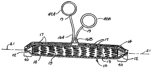
Figure 4B is a longitudinal cutaway view of the entire length of the
constrained
endoprosthesis assembly 10 shown in Figure 4A, wherein constraining sheath 14
is shown
cutaway to provide a view of the constrained endoprosthesis 12. Pointed ends
40 formed in
constraining sheath 14 extend beyond the ends of endoprosthesis 12. A pair of
separate
filaments 16A and 16B are arranged as unravelable chain stitches 17 to allow
separate
deployment of the two ends of assembly 10, with each filament 16 arranged to
initiate
deployment of the respective assembly end beginning from the end and
progressing back
toward the middle of the length of the assembly.
The ends 19 of each filament 16A and 16B are attached respectively to pull
rings
42A and 42B. The use of these preferred pull rings provides a convenient grip
for a
practitioner to use in the application of each individual filament 16. It is
further preferred that
the pull rings be differently colored, with, for example, pull ring 42A and
filament 16A colored
the same, and pull ring 42B and filament 16B colored the same, but different
from the color
used for pull ring 42A and filament 16A. For example, pull ring 42A and
filament 16A may
be made to be black, while pull ring 42B and filament 16B may be made to be
white. In this
fashion, it will be apparent to the practitioner which pull ring deploys which
end of the
assembly. For further clarity, it may be desired to color the pointed ends of
the constraining
sheath the same as the respective pull ring and filament (using different
colors for each of
the two pointed ends). In another alternative, each entire constraining sheath
end may be
colored with different colors used for the two ends, again with the respective
pull rings
colored the same as the ends that they are intended to release.
It is apparent that the filaments 16 and pull rings 42 allow for the
application of
tension (to initiate deployment) at an angle of about 90 degrees with respect
to the
longitudinal axis 41 of assembly 10. The filaments and pull rings 42 are
arranged so that
14
CA 02636450 2008-07-07
WO 2007/084762 PCT/US2007/001645
tension may be applied over a wide range of angles with respect to the
longitudinal axis 41,
ranging from virtually parallel to the longitudinal axis 41 to 90 degrees and
beyond.
Figures 4C-4F are partial longitudinal cross sectional views of constraining
sheaths
with alternative pointed ends. Figure 4C shows an embodiment wherein
constraining sheath
14 has a rounded point 40. As shown in Figure 4D, the constraining sheath 14
may simply
extend over and around the end of the endoprosthesis 12. Figure 4E describes
an
embodiment wherein pointed tip is asymmetrical with one side being
substantially parallel
with the longitudinal axis 41 and the other side possessing most of the taper.
It is apparent
that either side of point 40 may possess most of the taper (e.g., the side
including chain
stitch 17). This embodiment may be particularly useful with an axial
stiffening component as
will be further described. Figure 4F shows an embodiment wherein elements of
stent 13
extend beyond the end of graft component 15; these extended ends are
temporarily bent
inward toward longitudinal centerline 41 and secured with the end of chain
stitch 17 to create
point 40. Following insertion of this end of the assembly 10 into the body
conduit, the
application of tension to filament 16 begins deployment by initiating
unraveling of chain stitch
17 at the end of device 10, freeing the joined ends of stent 13 at the tip of
point 40 and
allowing the ends of the stent to open in alignment with the remainder of the
body of the
stent as it deploys.
Figures 5A-5E show schematic representations of the device of the present
invention
being used to repair a transected artery 50. Figure 5A describes the
transected blood
vessel, with arrows 52 indicating blood loss from the proximal side 50P of the
transection.
Figure 5B shows one end of assembly 10 being inserted into the proximal side
50P of
transected artery 50, preparatory to being deployed by the application of
tension (indicated
by arrow 26) to pull ring 42A and filament 16A. While this schematic shows
adequate
clearance between the lumen of the transected blood vessel and the outer
diameter of the
constrained endoprosthesis assembly, this may be a slip fit with a slight
interference. In the
case of transected blood vessels, the retraction of the vessel ends will often
require that the
vessel ends be gripped with forceps during insertion of one end of the
assembly 10. A
typical transection may require that 2 to (more preferably) 3cm of insertion
length of the end
of the assembly prior to deployment of the end. Often a 5cm length of
endoprosthesis will
be necessary between the retracted ends. Thus, a device length of about 11-
12cm may be
desired. A desirable amount of diametrical interference in the deployed device
would be
about 30-50%. For example, for a 6mm blood vessel, an endoprosthesis with a
nominal
deployed diameter of 8mm may be desirable.
Figure 5C shows the end of assembly 10 inserted into and deployed within the
proximal side 50P of transected artery 50, deployment having occurred from the
pointed tip
and proceeding toward the middle of the length of the assembly. As only this
end of the
CA 02636450 2008-07-07
WO 2007/084762 PCT/US2007/001645
assembly has been deployed, blood loss is substantially reduced or stopped
entirely, with
blood pressure largely re-established as shown by arrows 53. The opposing end
of
assembly 10, having not yet been deployed from its compacted state, serves to
block blood
flow. The released constraining sheath that had formerly constrained this end
of the device
is not shown, but is captured between the outer surface of a portion of the
deployed device
and the adjacent luminal surface of the proximal end of the artery.
Optionally, this portion of
the constraining sheath may be removed following deployment by the application
of axial
tension to the constraining sheath 14, if the constraining sheath 14 was not
physically
attached to the endoprosthesis. It is generally believed preferable to utilize
an implantable
constraining sheath and leave it in place between the deployed endoprosthesis
and the
vessel wall.
Figure 5D shows the opposing end of assembly 10 inserted into the distal end
50D of
artery 50, preparatory to deployment by the application of tension (indicated
by arrow 26) to
remaining pull ring 42B and filament 16B. Figure 5E shows the deployment of
the distal end
having been accomplished, with perfusion re-established distally as indicated
by arrows 54.
Figures 5A-5E describe one possible sequence of using the present invention to
repair transected vessels. It is apparent that there are other possible
sequences. For
example, the device may be inserted into both the proximal and distal vessel
ends and then
be deployed at both ends simultaneously.
Figure 6A is a schematic representation of a trauma to a blood vessel such as
artery
60 wherein the wound is only partially through the vessel (i.e., the vessel is
not fully
transected).
Figure 6B is a schematic representation of the same wound further showing a
constrained endoprosthesis assembly 10 of the present invention about to be
inserted into
the proximal side of the trauma site. Figure 6C shows the assembly 10 fully
inserted and
deployed-(by the application of tension to pull ring 42A of Figure 613) into
the proximal side of
the wound. As the distal end of the assembly 10 is as yet undeployed, the
compacted distal
portion of the endoprosthesis serves as a plug or occluder and prevents
further blood loss;
pressure is substantially restored (arrow 53). This distal portion of the
assembly 10 is now
bent appropriately to be directed into the distal portion 60D of the trauma
site, as indicated
by arrow 27.
Figure 6D shows the assembly 10 fully deployed with distal blood flow re-
established
(arrows 54) with little or no further loss of blood.
An alternative device is described in the plan view of Figure 7A which shows
that the
endoprosthesis can be a hybrid stent-graft and vascular graft 72, having a
stent component
13 adapted to the exterior surface, or alternatively to the interior surface,
of both ends of a
vascular graft 72 such as an ePTFE vascular graft. The same type of
constraining sheath
16
CA 02636450 2008-07-07
WO 2007/084762 PCT/US2007/001645
described above can be used independently at each end of the graft
(constraining sheath
not shown in this view). Such a hybrid device can be used to advantage to
perfuse a trauma
site from an entirely different location in the body, as shown by Figure 7B.
The assembly of the present invention may optionally be provided with various
components intended to add axial stiffness to the assembly to further
facilitate introduction
into an opening in a blood vessel. These axial stiffening components are
removable once
the introduction has been accomplished as desired. Such components include
hypotubes
and guidewires or rod components referred to herein as guide mandrels. They
may
optionally extend beyond the tip of the assembly.
Figure 8A is a perspective view terminating in a transverse cross section
showing a
length of a small tubular component such as a hypotube 82 fitted within the
constrained
endoprosthesis assembly 10. Figure 8B is an end cross sectional view of
assembly 10 fitted
with a hypotube as a stiffening component 82. The hypotube 82 resides between
the
endoprosthesis 12 and the constraining sheath 14. Its diameter, wall thickness
and material
are chosen for the appropriate degree of stiffness that is chosen to be added
to the
assembly 10. An appropriate nitinol hypotube is part no. SE508 from Nitinol
Devices and
Components, Fremont CA. The assembly 10 needs sufficient flexibility to be
adequately
conformable to the anatomy both during and following implantation_ However,
the additional
axial stiffness imparted by a stiffening component such as hypotube 82 can be
useful during
the process of inserting the tip 40 of the assembly into an opening in a
traumatized blood
vessel. It is possible to add an appropriate stiffening component without
excessively
compromising flexibility in the assembly.
The use of a hollow hypotube as an axial stiffening component 82 offers the
possibility of also allowing for a convenient access for local administration
of a therapeutic
agent. Likewise, the use of a hollow hypotube allows for the possible use of a
guidewire
device if desired to better enable access to the damaged vasculature. A
hypotube can also
serve as a channel for a deployment filament.
. Figure 8C is a side schematic view of a constrained endoprosthesis assembly
10
preparatory to implantation at a vascular trauma site. This view shows how the
axial
stiffening component, in this instance a length of hypotube 82, may extend
away from the
assembly near the middle portion of the length of the assembly, typically at
the same
location that the filament segments 16A and 16B extend away from the assembly
10 to join
their respective pull rings 42A and 42B. A suitable handle or optional luer
access fitting 84
may be fitted to this end of stiffening component 82. Once the assembly is
inserted into the
vasculature for the desired distance, stiffener 82 may be removed by pulling
it away from
assembly 10 while assembly 10 is firmly held in place by the practitioner.
Stiffener may be
17
CA 02636450 2008-07-07
WO 2007/084762 PCT/US2007/001645
removed prior to or following deployment of that end of endoprosthesis 12.
Alternatively, it
may be left in place for local drug delivery access or guidewire access.
Figure 8C also indicates how the opposite end of stiffener 82 may extend
beyond the
tip portion 40 of the assembly 10. This may be desirable to provide particular
stiffness at the
very end of the assembly 10 and as such may aid in locating and entering the
vascular
opening. The stiffener 82 is suitably formed to offset at tip portion 40 so
that it terminates at
the center of the pointed tip portion 40.
Figures 9A-9C are side views of an embodiment incorporating an axial stiffener
that
extends for the full length of the device. Figure 9A shows as a longitudinal
cross section
how device 10 can be provided with an axial stiffener 82 that extends for the
full length of the
device. As shown, this stiffener 82 is located within the device lumen. It may
alternatively
be located between constraining sheath 14 and endoprosthesis 12. Stiffener 82
may take
the form of a guidewire, a mandrel or rod, or a tube such as a hypotube. It
may be of
constant or variable stiffness along its length. Stiffener 82 may be provided
with handle 84
for convenience of removability if desired.
Figures 9B and 9C are side views showing this embodiment as typically
implanted into
a body conduit. Figure 9B describes how stiffener 82 may be used to aid in
introduction of
device 10 into the proximal end 50p of the body conduit 50. Figure 9C shows
how stiffener
82 may be removed from the distal end of device 10 following introduction of
device 10 into
the proximal end of the body conduit 50p. Stiffener 82 may be removed after
insertion into
proximal end of body conduit 50p, either prior to deployment or following
deployment of the
proximal end of device 10.
Figure 10 is a perspective view of an alternative axial stiffener in the form
of a
guidewire 86. In this embodiment, a moderately stiff guidewire 86 is contained
within the
constraining sheath 14 with the endoprosthesis 12, with the tip portion of the
guidewire 86
extending beyond the end of the constrained endoprosthesis 12 and bent into a
"J" form to
serve as an introducer. Additionally, guidewire 86 serves as an axial
stiffener. Both of these
functions better enable the device 10 to be introduced into a blood vessel
trauma site. If
desired, the guidewire can be removed by the application of tension (indicated
by arrow 26)
after the assembly 10 has been introduced into the vasculature, but prior to
deployment of
the endoprosthesis 12.
Figures 11A and 11 B show one end (e.g., the proximal end) of a constrained,
compacted endoprosthesis 12 contained within an alternative coristraining
sheath 140.
Figure 11 A is a perspective view and Figure 11 B is a longitudinal cross
sectional view. In
36 this embodiment, constraining sheath 140 is everted back over itself at the
end of
endoprosthesis 12. Edges of sheath 140 are again secured together by filament
16
arranged in a chain stitch 17 whereby the application of tension to the free
end of filament 16
18
CA 02636450 2008-07-07
WO 2007/084762 PCT/US2007/001645
causes chain stitch 17 to come unraveled. Deployment initiates from the end of
endoprosthesis 12 and progresses toward the middle of the length of
endoprosthesis 12 in
the similar manner as shown by Figures 5A-5E. Again, the opposing ends of the
endoprosthesis 12 are preferably individually and independently deployable.
Adjacent
edges of sheath 140, secured together by chain stitch 17, thus are freed to
separate and
allow deployment of the endoprosthesis 12 beginning at point 110 where sheath
140 everts
back over itself at the end of endoprosthesis 12. The advantage of this
embodiment is that
the end 142 of sheath 140 is located near the middle of the length of
endoprosthesis 12.
Following deployment of the proximal half of the full length of endoprosthesis
12, tension
may be applied to end 142 of everted sheath 140, allowing sheath 140 to be
pulled out from
between the body conduit and deployed endoprosthesis 12.
Figure 12 A is a schematic longitudinal cross sectional view of another
alternative
embodiment using a partially everted, corrugated constraining sheath 144.
Preferably, each
end of the device has its own constraining sheath 144, with the two sheaths
144 meeting at
about the middle of the length of the endoprosthesis 12. In this way, each end
of the
endoprosthesis can be separately and individually deployed. Similar to the
embodiment of
Figures 11 A and 11 B, a portion 144e, of constraining sheath 144 is everted
back over itself at
both ends of compacted and constrained endoprosthesis 12, with the result that
both ends
146 of constraining sheath 144 are located near the middle of the length of
endoprosthesis
12. Each end 146 is affixed to a gripping means such as pull rings 42. The use
of the
everted sheath 144 provides a means whereby sheath 144, during deployment of
endoprosthesis 12, may be removed from between the body conduit and the
deployed
endoprosthesis. In the embodiment shown in Figure 12A, everted constraining
sheath 144
is corrugated, with the direction of corrugations 145 running
circumferentially around
endoprosthesis 12. The use of a greater sheath length provided by the use of
corrugations
145 reduces the required tensile force necessary to cause removal of sheath
144 and
deployment of endoprosthesis 12 (due to the length of the corrugated sheath
144 being
greater than the length of a similar uncorrugated sheath 14). The use of
corrugations 145
also provides the sheath.with increased hoop strength.
In an alternative embodiment shown by the schematic longitudinal cross section
of
Figure 12B, the everted portion 144e is not corrugated while the underlying
portion of the
sheath 144 is corrugated.
Figure 12C shows a perspective view of one half of the length (e.g., the
proximal half)
of the embodiment of the schematic cross sectional view of Figure 12A.
Corrugated and
everted sheath 144 extends along the length of compacted and constrained
endoprosthesis
12, with the ends of sheath 144 everted back over the middle portion of the
length of sheath
144. One end 146 of sheath 144 is shown with the tubular form of the sheath
144.split
19
CA 02636450 2008-07-07
WO 2007/084762 PCT/US2007/001645
lengthwise and extending to pull ring 42. Sheath 144 is thus splittable along
its length by
various means such as perforations provided along a line 148. Other means may
also be
used, including the use of thin materials for sheath 144 that have anisotropic
strength
properties, offering good hoop strength to the sheath but being inherently
splittable along the
length of the sheath.
Figure 12D shows a perspective view of initiation of deployment of
endoprosthesis 12
by the application of tension (shown by arrow 26) to the end 146 of sheath 144
via ring 42.
This tension 26 causes end 146 to become progressively uncorrugated and causes
continuing splitting of sheath 144, for example by splitting of perforation
line 148. The outer,
everted portion 144e of sheath 144 has been split along perforation line 148
and withdrawn,
and the inner portion of sheath 144 is shown splitting as it also is
withdrawn, allowing
release and deployment of constrained endoprosthesis 12. Simultaneously,
tension 26
results in withdrawal of sheath 144 from between the deploying endoprosthesis
12 and the
adjacent wall of the body conduit into which it is being deployed.
Figure 12E shows a longitudinal cross section of one end (e.g., the proximal
end) of
device 10 according to the embodiment described by Figures 12A, 12C and 12D.
As shown,
corrugations 145 may be non-uniform, with the corrugations 145 of the outer
everted portion
of sheath 144 not necessarily corresponding exactly to (and consequently not
precisely
matching) the corrugations of the inner portion of sheath 144.
A preferred tubular material for the partially everted, corrugated
constraining sheath
144 is made from a laminated film that is a composite of fluorinated ethylene
propylene
(FEP) and ePTFE film wherein the FEP is applied to the ePTFE film as a
discontinuous
coating that allows the film to remain porous. These composite films are made
as taught by
US Patent 5,358,516 to Myers et al. A preferred ePTFE film for this laminate
is taught by US
Patent 5,814,405 to Branca.
To make a 1 ocm long, partially everted, corrugated sheath, a 130cm length of
this film
is paid off onto a slowly rotating stainless steel mandrel, with the 130 cm
length parallel to
the length of the mandrel. The mandrel is of the diameter desired for the
inside diameter of
the constraining sheath, with the film oriented with the FEP-coated side of
the film facing
away from the mandrel surface. The film has similar strength properties and
tear properties
in the length and width directions, so the microstructure of the ePTFE may be
oriented with
the length of the nodes oriented in a circumferential direction or oriented
parallel to the
length of the mandrel. Two layers of this film are applied, after which heat
from a source
such as a soldering iron, adequate to melt FEP, is applied along a line along
the length of
the resulting film tube. The direction of rotation of the mandrel is reversed,
and two
additional layers of the film are applied; the reversal of rotation results in
the FEP-coated
side of the film facing toward the mandrel surface. After the fourth layer is
complete, the film
CA 02636450 2008-07-07
WO 2007/084762 PCT/US2007/001645
is cut with a blade along the length of the mandrel. Finally, a temporary wrap
of a tape of
helically applied ePTFE film (without FEP-coating) is created over the initial
four layers to
hold them in place, and the film-covered mandrel is placed into a convection
oven set at
320 C for ten minutes. After this time, the mandrel is removed from the oven
and allowed to
cool to ambient temperature. Following cooling, the temporary overwrap of
helically applied
ePTFE tape is removed.
The resulting film tube had a wall thickness of about 0.020 to 0.025mm.
Next, the resulting film tube was slid toward one end of mandrel until one end
of the
film tube extended a short distance (approximately 1cm) beyond the end of the
mandrel. By
careful manual manipulation, the end of the tube was everted back over the
portion of the
tube remaining over the mandrel surface, until 10-12cm of the end of the tube
was everted
over the adjacent tube portion. This was repeated for the opposite end of the
film tube,
resulting in the tube having two layers in each everted region. The tube was
then fitted back
onto the same mandrel, or optionally, another mandrel of slightly larger
diameter to
compensate for any diameter increase that resulted from the everting process.
The tube
and mandrel assembly was then placed into a suitable programmable laser
cutting machine
(a suitable machine is, for example, a C02 Laser Marker, model ML-G9320F
available from
Keyence Corporation, Woodcliff Lake NJ). The machine had been previously
programmed
to cut a line of perforations for the full length of the film tube; each
individual perforation was
about 0.15mm wide and of about 0.45mm length, with adjacent perforations
separated by a
land of 0.2mm length.
Following the perforation process, the resulting film tube was cut in half
transversely
(at the mid-point of its length) using a sharp blade, so that separate sheaths
result for each
end of the endoprosthesis (thereby allowing separate deployment of each end of
the
endoprosthesis). Next, while still on the mandrel, the sheaths are uniformly
compressed in
an axial direction to create the corrugations. The sheath is axially
compressed until its
length is 10% of its original, uncompressed length. As shown by Figure 12E,
the everted
portion of the tube is corrugated simultaneously with the underlying tube
portion. This figure
also shows the relative non-uniformity of the corrugations.
Figure 13A shows a longitudinal cross sectional view of the manufacture= of
corrugated
and everted constraining sheath 144. The tubing from which the sheath 144 is
to be made
has its ends 146 everted back over the middle portion of the tube, creating an
everted
portion 144e of sheath 144. The resulted everted tube 144 is fitted over a
suitable mandrel
152, with the mandrel being a snug fit within the everted tube 144. The
opposing ends of the
everted tube 144 are then compressed axially toward each other, causing the
corrugations
145 to form along the length of the sheath144 as shown in Figure 13A.
21
CA 02636450 2008-07-07
WO 2007/084762 PCT/US2007/001645
Figure 13B shows a funnel device 132 useful for compacting a self-expanding
endoprosthesis and inserting it into a constraining sheath 144. Device 132
comprises a
funnel 134 of a type known in the art of manufacturing self-expandable
endoprostheses.
Other compaction methods may also be used, for example, iris-type compaction
devices
such as described by US Patent 6,629,350. Funnel 134 has a length of thin-wall
metal
tubing 136 affixed to the small end of funnel 134; the inside diameter of
tubing 136
corresponds to the inside diameter of the small end of funnel 134. A suitable
thin-wall tubing
is a stainless steel hypotube made by Microgroup, Inc., part no. 304H11XX
(Meadway MA).
A's shown by Figure 13C, corrugated and everted sheath 144 is next fitted over
the
outside of tube 136. Figure 13D shows an endoprosthesis 12 being pulled via
temporary
traction lines 138 into funnel 134 (nitinol stents may require simultaneous
chilling with a
refrigerant spray) and on into the lumen of tube 136 as endoprosthesis 12 is
compacted.
Figure 13E shows the full length of compacted endoprosthesis 12 contained
within the
lumeh of tube 136. As shown by Figure 13F, compacted endoprosthesis is pulled
out of the
end of tube 136 into the lumen of constraining sheath 144, until
endoprosthesis 12 is fully
contained within corrugated and everted constraining sheath 144.
Figure 14 shows a perspective view of an alternative embodiment wherein a
guide 162
is provided at the middle of the length of the device for sheath ends 146.
Guide 162 is
provided with a saddle 164 that holds a middle portion of endoprosthesis 12
between the two
constraining sheaths 14; saddle 164 grips endoprosthesis 12 by interference. A
pair of
cutting blades 166 are provided in the base of saddle 164 that progressively
splits each
sheath 14 when tension is applied to the respective sheath end 146. As each
sheath 14
splits, it is withdrawn, allowing deployment of endoprosthesis 12 beginning at
the end of the
endoprosthesis and progressing toward the middle, while the sheath is
simultaneously
withdrawn from between the endoprosthesis and the body conduit into which the
prosthesis
is being implanted.
Different assemblies according to the present invention were manufactured and
implanted into surgically created vascular wounds created in the iliac and
femoral arteries of
one juvenile pig and several adult greyhound dogs, as well as a femoral vein
in an adult
.30 greyhound dog. The procedures were performed under direct visualization
generally as
illustrated in Figures 5A-6D. These assemblies were based on Hemobahn
Endoprosthesis
devices available from W.L. Gore & Associates, Flagstaff AZ. These devices
were
compacted and constrained in ePTFE constraining sheaths having edges secured
with
ePTFE filaments arranged to form unravelable chain stitches. The stitch
arrangement was
such that each end of each assembly was individually deployable by application
of tension to
a pull ring fitted at one end of the ePTFE filament, resulting in deployment
beginning from
the end of the assembly and progressing toward the middle portion of the
length of the
22
CA 02636450 2008-07-07
WO 2007/084762 PCT/US2007/001645
assembly where the pull ring and filament end were located and accessible to
the
practitioner. Some of the constraining sheaths were provided with pointed tip
portions.
Some incorporated temporary axial stiffening components and some did not.
Devices of 6,
7, 8 and 10 mm nominal deployed diameter were used having compacted diameters
ranging
from 9 to 12 French. Both fully and partially transected wounds were created.
Using forceps
to grip the vessel adjacent the wound opening in the vessel, these devices
were inserted into
the openings and deployed without undue difficulty. Deployment resulted in
either complete
or very substantially complete halting of blood loss and re-establishment of
perfusion to the
anatomy distal to the trauma site. Following complete deployment, the
constraining sheaths
of some of these implants were removed from the space they occupied between
the
deployed end of the endoprosthesis and the adjacent vessel wall by gripping an
exposed
portion of the constraining sheath with forceps and applying tension.
These implants were generally quickly accomplished, usually in about five
minutes or
less. While the devices fitted with the axial stiffeners were deemed to
sometimes provide an
advantage, these stiffeners were generally deemed as unnecessary to the device
to enable
a successful and prompt outcome.
Two additional five Hemobahn Endoprosthesis devices were implanted in the
iliac
artery of two greyhound dogs. These devices had the partially everted,
corrugated sheath.
The sheath was deployed in one motion by pulling on its free end, one side at
a time starting
with the proximal side. The sheath was removed during deployment. There was
littte or no
bleeding, which stopped by itself within 2-3 minutes. Implantation of each
device lasted
required less than 3 minutes. The insertion depth was about 2.5 cm proximally
and 2.4 cm
distally.
While particular embodiments of the present invention have been illustrated
and
described herein, the present invention should not be limited to such
illustrations and
descriptions. It should be apparent that changes and modifications may be
incorporated and
embodied as part of the present invention within the scope of the following
claims.
23