Note: Descriptions are shown in the official language in which they were submitted.
CA 02637235 2008-07-15
WO 2007/084726 PCT/US2007/001541
TRANSLUMENAL APPARATUS, SYSTEM, AND METHOD
Field of'the Invention
The present invention relates generally to apparatus, systems, and
methods for use in a heart, more particularly to apparatus, systems, and
methods
for improving the function of a heart valve.
Background
The human heart is divided into four chambers. These four chambers
include the right atrium and the right ventricle, and the left atrium and the
left
ventricle. The heart contracts rhythmically under stimulation of electrical
currents to move blood through the chambers of the heart and the remainder of
the cardiovascular system.
Blood in the heart is kept flowing in a unidirectional manner through the
cardiovascular system by a system. of four one-way valves. As the heart cycles
the valves open and close to allow blood to move one-way through the heart
chambers.
The heart valves differ significantly in structure. For example, the
ventricles are separated from the atria by valves that, in addition to the
leaflets,
have thin but strong cords of fibrous tissue. Called chordae tendineae, these
cords tether the valve to the ventricular walls. When the ventricles contract,
small muscles in their walls, called papillary muscles, pull the cords which
act as
tethers, and control the closure of the valve leaflets, preventing them from
flapping too far backwards.
One such valve located between the left ventricle and the left atrium is
called the mitral valve. The mitral valve has two leaflets that form the
valve.
The leaflets are attached to papillary muscles by way of the chordae tendineae
and it allows blood to enter the left ventricle from the left atrium.
When operating properly, the mitral valve acts as a one-way valve.
There are, however, numerous conditions that can cause the mitral valve to not
act as a one-way valve. For example, deficiency or degeneration of one or more
of the mitral valve structures may result in dysfunction of the mitral valve
apparatus leading to mitral valve prolapse or regurgitation during a
contraction
1
CA 02637235 2008-07-15
WO 2007/084726 PCT/US2007/001541
of the heart. Prolapse or regurgitation of the mitral valve can eventually
lead to
severe cardiovascular problems, and even death.
Brief Description of the Drawings
Fig_ I provides a schematic cross-section of a heart, segments of which
have been removed to show detail.
Fig. 2 provides a schematic cross-section of a heart, segments of which
have been removed to show detail.
Fig. 3 illustrates one embodiment of an apparatus according to the
present invention.
Figs. 4A-4F illustrate one embodiment of an apparatus according to the
present invention.
Figs. 5A-5F illustrate one embodiment of an apparatus according to the
present invention located within the cardiovascular system.
Figs. 6A-6F illustrate one embodiment of an apparatus according to the
present invention located within the cardiovascular system.
Detailed Description
Embodiments of the present invention are directed to methods; apparatus,
and systems for helping to improve heart valve function. As discussed herein,
improving heart valve function can be accomplished by altering the
configuration of the heart valve according to various embodiments of the
invention. For example, altering the configuration of the heart valve can be
accomplished through the use of a cord delivered into the heart by a delivery
catheter. The cord can be positioned relative the heart valve in such a way
that
by manipulating aspects of the cord (e.g., its length) the configuration of
the
heart valve can be alter so as to improve the heart valve function. These and
other embodiments of the present invention are discussed herein.
The figures herein follow a numbering convention in which the first digit
or digits correspond to the drawing figure number and the remaining digits
identify an element or component in the drawing. Similar elements or
components between different figures may be identified by the use of similar
digits. For example, 110 may reference element "10" in Fig. 1, and a similar
2
CA 02637235 2008-07-15
WO 2007/084726 PCT/US2007/001541
element may be referenced as 210 in Fig. 2. As will be appreciated, elements
shown in the various embodiments herein can be added, exchanged, and/or
eliminated so as to provide a number of additional embodiments of the valve
according to the present invention.
Fig. 1 illustrates a schematic cross-section of a heartj 00. The heart 100
is divided into four chambers, which are referred to herein as a first chamber
102, a second chamber 104, a third chamber 106 and a fourth chamber 108.
With respect to the anatomy of the heart, the first chamber 102 can represent
the
left atrium, the second chamber 104 can represent the left ventricle, the
third
chamber 106 can represent the right atrium, and the fourth chamber 108 can
represent the right ventricle. Other representations for the chambers 102,
104,
106 and 108 are also possible.
Heart 100 further includes heart valves positioned at either an inlet or an
outlet of the four chambers of the heart 100. These heart valves include a
mitral
valve 114, an aortic valve 116, pulmonary valve 118, and tricuspid valve 120.
Generally, each heart valve includes valve leaflets. For example, the
structure of
the mitral valve 114, the one-way heart valve that divides the first chamber
102
(i.e., the left atrium) and the second chamber 104 (i.e., the left ventricle),
includes two leaflets. These two leaflets are referred to as the anterior
leaflet
122-1 and the posterior leaflet 122-2. The anterior and posterior leaflets 122-
1
and 122-2 move between an open position in which antegrade blood flow moves
from the first chamber 102 to the second chamber 104, to a closed position
that
prevents retrograde flow of the blood from the second chamber 104 to the first
chamber 102.
The anterior and posterior leaflets 122-1 and 122-2 are attached to a
variety of structures that help to maintain the function of the mitral valve
114.
For example, the mitral valve 114 includes a fibrous tissue ring structure,
referred to as the mitral annulus 124, which surrounds and supports the
anterior
and posterior leaflets 122-1 and 122-2. The mitral annulus 124 can be
conceptually divided into an anterior mitral annulus 126-1 and a posterior
mitral
annulus 126-2. The anterior leaflet 122-1 and posterior leaflet 122-2 are
supported by the mitral annulus 124 by their connection to the anterior mitral
annulus 126-1 and the posterior mitral annulus 126-2, respectively.
3
CA 02637235 2008-07-15
WO 2007/084726 PCT/US2007/001541
The mitral valve 114 further includes fibrous tissue called chordae
tendineae 110. The chordae tendineae 110 function to tether the leaflets 122-1
and 122-2 of the mitral valve 114 to the ventricular walls 115. In addition,
the
mitral valve 114 also includes papillary muscles 112 that extend from the
ventricular walls 115 to couple to the chordae tendineae 110. When the
ventricles contract, the papillary muscles 112 pull the chordae tendineae 110
which act as tethers, and control the closure or coaptation of the valve
leaflets
'122-1 and 122-2, preventing them from flapping too far backwards (prolapse).
When operating properly, the mitral valve 114 acts as a one-way valve.
There are, however, numerous conditions that can cause the mitral valve 114 to
not act as a one-way valve. For example, deficiency or degeneration of one or
more of the mitral valve 114 structures may result in dysfunction of the
mitral
valve apparatus leading to mitral valve prolapse or regurgitation during a
contraction of the heart. Mitral valve prolapse is a condition in which blood
leaks in the wrong direction (regurgitation of the blood) because one or more
of
the valve leaflets 122-1 and/or 122-2 close improperly. Reasons for why the
valve leaflets 122-1 and/or 122-2 close improperly can include, for example,
changes in the size and shape of the valves leaflets 122-1 and/or 122-2 and/or
the
mitral annulus 124 (e.g., an increase in the circumference of the mitral
annulus
124).
Fig. 2 illustrates a cross-section of the first chamber 202 and the second
chamber 204 of the heart 200. In the present example, the valve leaflets 222-1
and 222-2 in their native state do not close properly, leading to the
condition of
mitral valve prolapse for the heart 200.
Fig. 2 also illustrates the presence of a cord 230 within the heart 200
according to various embodiments of the invention. The cord 230 can be
delivered and positioned percutaneously within the heart 200, as discussed
herein. The cord 230 allows for the configuration of the native heart valve to
be
modified in such a way that there can be an improvement in the functioning of
the'heart valve. In other words, the cord 230 can be used to change the
physical
relationship of the different parts of the heart valve in such a way as to
help
restore a more normal operation of the heart valve (e.g., reduce regurgitation
of
the blood).
4
CA 02637235 2008-07-15
WO 2007/084726 PCT/US2007/001541
In the illustration provided in Fig. 2, the heart valve being modified by
the cord 230 is the mitral valve 214, as discussed herein. Other heart valves
can
be modified with the cord 230 so as to improve valve performance, where the
number and relationship of one or more cords used to accomplish this goal
depends upon the valve being modified. Fo'r exampl'e, a tri-leaflet heart
valve,
such as the aortic valve, might be modified using at least two cords having an
approximately equilateral relationship (i.e., the at least two cords cross
each
other at a point to form an angle of approximately ninety (90) degrees). In an
additional example, three cords could be used to modify the configuration of
the
valve, where the cords could have a predetermined relationship relative each
other (i.e., the at least two cords cross each other at a point to form an
angle of
approximately sixty (60) degrees).
As will be discussed more fully herein, the cord 230 can be delivered to
the heart valve (e.g., the mitral valve 214) by a delivery catheter. In one
example, the delivery catheter can be used to pass a first end and a second
end of
the cord between the first heart chamber 202 and the second heart chamber 204,
or visa versa. The first and second ends of the cord 230 can then be used in
forming a loop around the heart valve. The length of the loop formed with the
cord can then be manipulated (e.g., shortened) so as to modify the
configuration
of the heart valve.
In one embodiment, the configuration of the heart valve can be modified
so as to induce coaptation of the valve leaflets at the proper time in the
cardiac
cycle. For example, the closed circumference of the loop formed with the cord
230 can have a constraining effect on the valve leaflets perpendicularly to
the
plane of their coaptation. The cord can also change the shape of the mitral
annulus 224 in such a way that the valve leaflets 222-1 and 222-2 are drawn
more closely together to allow for major surfaces 228 of the leaflets to seal
when
the valve 214 is in its closed configuration. In its open configuration,
constraining the valve leaflets with the closed circumference of the loop
formed
with the cord 230 can further modify the heart valve to create a modified
orifice
(e.g., a double orifice) for the heart valve 214, where there had been an'
unmodified orifice (e.g., a single orifice) prior to the use of the cord 230.
As will be appreciated, a variety of apparatus and/or systems can be
utilized in delivering and manipulating the cord 230. For example, delivery
5
CA 02637235 2008-07-15
WO 2007/084726 PCT/US2007/001541
catheter based apparatus and/or systems can be utilized in delivering and
manipulating the cord 230 according to a variety of the embodiments of the
present invention. These apparatus and/or systems can both house the cord 230
and provide the structure through which the cord 230 can be delivered between
the first heart chamber 202 and the second heart chamber 204. The following
discussion provides various embodiments of the present invention.
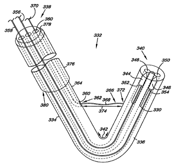
Fig. 3 provides a schematic illustration of an apparatus 332 according to
one embodiment of the present invention. The apparatus 332 includes a delivery
catheter 334 for positioning and passing at least a portion of the cord 330
between chambers of the heart, as discussed herein. The delivery catheter 334
has an elongate body 336 with a proximal end 338 and a distal end 340. The
delivery catheter 334 further includes at least one predetermined bend 342 in
the
elongate body 336 between the proximal end 338 and the distal end 340. As
discussed herein, the predetermined bend 342 allows the distal end 340 of the
catheter 334 to be positioned adjacent the heart valve, such as the mitral
valve,
as will be discussed herein.
The delivery catheter 334 of the present embodiment further includes a
first piercing member 344 and a second piercing member 346. The first and
second piercing members 344 and 346 are releasably positioned at least
partially
within delivery catheter 334 in such a way that they can extend and separate
from the delivery catheter 334. For example, as illustrated the first and
second
piercing members 344 and 346 are releasably positioned at least partially
within
a first lumen 348 and a second lumen 350, respectively, where the lumens 348
and 350 extend from the distal end 340 towards the proximal end 338 of the'
delivery catheter 334.
The first and second piercing members 344 and 346 can also be
associated with the cord 330. For example, the first piercing member 344 can
be
associated with a first end 352 of the cord 330 and the second piercing member
346 can be associated with a second end 354 of the cord 330. In one
embodiment, associating the cord 330 with the first and second piercing
members 344 and 346 can include coupling the structures together. Such
coupling can include, but is not limited to, chemically bonding the structures
together (e.g., gluing), and/or physically bonding the structures together
such as
6
CA 02637235 2008-07-15
WO 2007/084726 PCT/US2007/001541
by melting/fusing the structures together and/or through frictional
interactions
such as results from, for example, a crimping process.
Alternatively, the first and second piercing members 344 and 346 could
be formed from the cord 330 itself. For example, a portion of the cord 330 at
or
adjacent the first and second ends 352 and 354 of the cord 330 could be
modified
so as to form the first and second piercing members 344 and 346. Examples of
such modifications include, but are not limited to, melting and/or fusing the
cord
330 at and/or adjacent the first and second ends 352 and 354 to form at least
a
shaft having a sharp point to act as a piercing member.
Fig. 3 further illustrates that the cord 330 can be releasably positioned at
least partially within the first and second lumen 348 and 350. In the present
embodiment, the cord 330 can be released from the first and second lumen 348
and 350 as the first and second piercing members 344 and 346 are extended from
the delivery catheter 334. In one embodiment, the cord 330 could at least
partially reside in a groove that extends into the elongate body 336 from the
first
and second lumen 348 and 350. This allows the portion of the cord 330 adjacent
the first and second piercing members 344 and 346 to avoid contacting each
other as the piercing members are extended from the lumens 348 and 350. The
groove can also function to hold the cord 330 and the first and second
piercing
members 344 and 346 in place in the lumens 348 and 350 through frictional
interactions until the first and second piercing members 344 and 346 and the
cord 330 are deployed.
The delivery catheter 334 further includes a first deployment rod 356 and
a second deployment rod 358. In one embodiment, the first deployment rod 356
extends from the proximal end 338 through the first lumen 348 to abut the
first
piercing member 344. Similarly, the second deployment rod 358 extends from
the proximal end 338 through the second lumen 350 to abut the second piercing
member 346.
Both the first and second deployment rods 356 and 358 can be moved
longitudinally within their respective lumens. In addition, the first and
second
deployment rods 356 and 358 provide a column strength, or a"pushability," to
transfer force applied at their proximal end through to their distal end
sufficient
to extend the piercing members and=cord from the delivery catheter 334 and
into
the cardiac tissue as discussed herPi.n. As will be appreciated, the column
7
CA 02637235 2008-07-15
WO 2007/084726 PCT/US2007/001541
strength of the deployment rods 356 and 358 will also be dependent upon the
flexibility and'strength of the lumen 348 and 350 in which the rod travels.
The first and second deployment rods 356 and 358, and the cord 330 can
be formed from a number of different materials in a number of different
configurations. For example, the rods 356 and 358 and/or the cord 330 can be
formed of, by way of illustration and not by limitation, metals and/or metal
alloys. For example, suitable metals and/or metal alloys include, but are not
limited to, medical grade stainless steels (304, 306, 308, 316L, 318, etc.),
gold,
platinum, platinum alloys, palladium, rhodium, tungsten, tungsten alloys,
cobalt
chrome, titanium and titanium alloys, and other metai alloys such as those
composed of titanium/nickel and sold under the trade identifier "nitinol."
Other
materials, such as polymer materials, may also be used.
Heat treatment of the nitinol alloy may also be desirable. An example of
such a heat treatment includes, but is not limited to, placing the nitinol in
its
desired shape onto 'a mandrel. The nitinol is then heated to a temperature of
650 -750 F. for a predetermined time (e.g., two (2) to five (5) minutes),
possibly (but not necessarily) annealing the constituent nitinol. After heat
treatment, the flexible cord 330 retains its shape and the nitinol alloy
retains its
super-elastic properties.
By way of example, the cord 330 can be formed of a number of
polymeric materials. For example, the cord 330 can be formed of, by way of
illustration and not by limitation, thermoplastic and thermo-set polymers.
Examples of these polymers include polyolefins such as polyethylene and
polypropylene, polyesters such as Dacron, polyethylene terephthalate and
polybutylene terephthalate, vinyl halide polymers such as polyvinyl chloride
(PVC), polyvinylacetate such as ethyl vinyl acetate (EVA), polyurethanes,
polymethylmethacrylate, pellethane, polyamides such as nylon 4, nylon 6, nylon
66, nylon 610, nylon 11, nylon 12 and polycaprolactam, polyaramids (e.g.,
KEVLAR), polystyrene-polyisobutylene-polystyrene (SIBS), segmented
poly(carbonate-urethane), Rayon, fluoropolymers such as
polytetrafluoroethylene (PTFE or TFE) or expanded polytetrafluoroethylene
(ePTFE), ethylene-chlorofluoroethylene (ECTFE), fluorinated ethylene
propylene (FEP), polychlorotrifluoroethylene (PCTFE), polyvinylfluoride
8
CA 02637235 2008-07-15
WO 2007/084726 PCT/US2007/001541
(PVF), or polyvinylidenefluoride (PVDF), natural biopolymers such as
cellulose,
chitin, keratin, silk, and collagen, explanted veins, decellularized basement
membrane materials, such as small intestine submucosa (SIS) or umbilical vein,
or other naturally occurring extracellular matrix (ECM), and mixtures and
copolymers thereof. SIS and ECM materials can be autologous, allogeneic or
xenograft material derived from mammals, including sources, such as human,
cattle, sheep, and porcine.
Each of the polymers noted herein may be used in conjunction with
radiopaque filler materials such as barium sulfate, bismuth trioxide, bismuth
carbonate, powdered tungsten, powdered tantalum, or the like so that the
location of the cord 330'may be radiographically visualized within the human
body.
In another embodiment of the present invention, the polymers and blends
that are used to form the composite can be used as a drug delivery matrix. To
form this matrix, the polymer would be mixed with a therapeutic agent. The
variety of different therapeutic agents that can be used in conjunction with
the
polymers of the present invention is vast. In general, therapeutic agents
which
may be administered via the pharmaceutical compositions of the invention
include, without limitation: antiinfectives such as antibiotics and antiviral
agents;
analgesics and analgesic combinations; anti-inflammatory agents; hormones
such as steroids; and naturally derived or genetically engineered proteins,
polysaccharides, glycoproteins, or lipoproteins. Matrix formulations may be
formulated by mixing one or more therapeutic agents with the polymer. The
therapeutic agent may be present as a liquid, a finely divided solid, or any
other
appropriate physical form. Typically, but optionally, the matrix will include
one
or more additives, such as diluents, carriers, excipients, stabilizers or the
like.
Additionally, radiopaque markers may be added to the composite to allow
imaging of the composite after implantation.
The deployment rods 356 and 358, and/or the cord 330 can also include a
variety of cross-sectional configurations. For example, the deployment rods
356
and 358, and/or the cord 330 can have one or more of a round (e.g., circular,
oval, and/or elliptical), ribbon, semi-circular triangular, tubular, I-shaped,
T-
shaped, and trapezoidal. With respect to "braid," the term can include tubular
9
CA 02637235 2008-07-15
WO 2007/084726 PCT/US2007/001541
constructions in which the cord 330 making up the construction are woven
radially in an in-and-out fashion as they cross to form a tubular member
defining
a single lumen. The braid can also be constructed of flexible members of
different widths. The embodiments, however, are not limited to the examples as
other cross-sectional geometries are also possible.
In an additional embodiment; the cord 330 can include a number of forms
that contribute to both its mechanical and handling properties. Examples of
such
forms for the cord 330 include, but are not limited to, those selected from
the
group consisting of weaves, braids, meshes, knits, warped knitted (i.e., lace-
like), and non-woven structures, as the same will be known and understood by
one of ordinary skill in the art. In addition, mechanical properties of the
cord
330 can be altered by changing the density, form, and/or texture in one or
more
locations along the length of the cord 330. Examples of such changes include
alterations to the suitable structures used to create the cord 330 which can
include, for example, monofilaments, yams, threads, braids, or bundles of
fibers.
Regardless of its configuration, the structure of the cord 330 should possess
a
tensile strength adequate to withstand pressures (e.g., a stretching load)
imposed
by manipulating the cord 330, as discussed herein.
The first and second piercing members 344 and 346 can also be formed
from a number of different materials in a number of different configurations.
For example, the first and second piercing members 344 and 346 can be formed
of, by way of illustration and not by limitation, the materials discussed
herein in
conjunction with the cord 330. The first and second piercing members 344 and
346 can also include a variety of configurations. For example, the piercing
members 344 and 346 can have one or more of a round (e.g., circular, oval,
and/or elliptical), ribbon, semi-circular triangular, I-shaped, T-shaped, and
trapezoidal cross-sectional configuration. The embodiments, however, are not
limited to the present examples as other cross-sectional geometries are also
possible. In addition, the piercing members 344 and 346 further include a
leading edge or surface configured (e.g., sharp) to pe.netrate and pass
through
cardiac tissue under force applied by the respective deployment rod. In one
embodiment, the leading edge can have a conical configuration ending in a
point. Alternativefy, the leading edge of the piercing member can be defined
by
CA 02637235 2008-07-15
WO 2007/084726 PCT/US2007/001541
one or more surfaces of the piercing members that have an edge having an angle
(e.g., a 20 degree angle) to allow for the piercing members 344 and 346 to
pass
through the cardiac tissue.
The position of the distal end 340 can also be adjusted in a variety of
ways. For example, the elongate body 336 can have a variety of shapes and
curves that would be selected for appropriate use given the anatomy at hand.
In
this example, the elongate body 336 could have one or more predetermined
bends or curves to meet demands for the placement of the catheter 334.
An additional embodiment could use a single curve design that can be
modified by the operator during use of the catheter 334. For example, the
delivery catheter 334 can further include a third lumen 360 extending from the
proximal end 338 toward the distal end 340 of the delivery catheter 334. In
one
embodiment, the third lumen 360 has a surface defining an opening 362 through
a wall 364 of the delivery catheter 334. The opening 362 can be positioned
between the predetermined bend 342 and the proximal end 338 of the delivery
catheter 334.
The delivery catheter 334 can further include an adjustment member 366
extending from the third lumen 360. As illustrated in Fig. 3, the adjustment
member 366 has an elongate body 368 having a first end 370 and a second end
372. In one embodiment, the adjustment member 366 extends through the third
lumen 360 with the first end 370 extending from the delivery catheter 334 at
or
adjacent the proximal end 338 of the catheter 334. In an additional
embodiment,
the second end of the adjustment member 366 can be coupled to the delivery
catheter 334 at a point between the predetermined bend 342 and the distal end
340 of the catheter 334.
Applying tension (e.g., pulling and/or pushing) the adjustment member
366 from the first end 370 allows the second end 372 to move, thereby changing
the predetermined bend 342. In other words, the predetermined bend 342 can
flex under tension applied through the adjustment member to allow the distal
end
340 to be positioned at a second predetermined location (discussed herein)
adjacent the heart valve. When the tension on the adjustment member 366 is
released, the predetermined bend 342 returns towards its original
configuration
prior to being changed by the adjustment member 366.
11
CA 02637235 2008-07-15
WO 2007/084726 PCT/US2007/001541
The second end 372 of the adjustment member 366 can be anchored into
the elongate body 336 of the delivery catheter 334 in a number of ways. For
example, the second end 372 of the adjustment member 366 can be mechanically
anchored into the elongate body 336 of the catheter 334 with one or more barbs
that resist/prevent the second end 372 of the adjustment member 366 from
slipping or moving. Alternatively, the second end 372 of the adjustment
member 366 can be secured to a cleat embedded in the elongate body 336 of the
catheter 334. In an additional embodiment, the second end 372 of the
adjustment member 366 can be chemically fastened (e.g., glued) to the elongate
body 336 of the catheter 334. Combinations of these fastening methods, along
with other fastening methods, are also possible.
The adjustment member 366 can be moved longitudinally within the
third lumen 360 to change the position of the distal end 340 of the delivery
catheter 334. For example, the adjustment member 366 can be pulled to provide
tension at the second end 372 thereby reducing a distance 374 between the
second end 372 and the opening 362 of the delivery catheter 334. The
adjustment member 366 can also be used to push the elongate body 336 of the
delivery catheter 334, thereby increasing the distance 374 between the second
end 372 and the opening 362 of the delivery catheter 334. The pushing and/or
pulling of the adjustment member 366 allows for temporary changes in the
predetermined bend 342 and the relative position of the distal end 340 of the
delivery catheter 334. In an additional embodiment, upon changing the
predetermined bend 342 and the relative position of the distal end 340 of the
delivery catheter 334 the adjustment member 366 can be temporarily locked to
allow the position of the distal end 340 to be maintained.
The adjustment member 366 can also be formed from a number of
different materials in a number of different configurations. For example, the
adjustment member 366 can be formed of, by way of illustration and not by
limitation, metals and/or metal alloys, as recited herein. Other materials,
such as
various polymer recited herein, may also be used. As will be appreciated,
other
structural configurations that allow for altering the shape and/or the
position of
the predetermined bend 342 and the relative position of the distal end 340 of
the
delivery catheter 334 are also possible. For example, a push-pull and/or
torque
wire(s) could be used with one or more lumens (e.g., the third lumen) that
extend
12
CA 02637235 2008-07-15
WO 2007/084726 PCT/US2007/001541
to the distal end 340 of the delivery catheter 334. The push or pull or push-
pull
wire(s) could then be use to provide a steerable catheter having a deflectable
distal portion. As will be appreciated, a spring tube can also be provided at
the
distal portion of the delivery catheter 334 for improved torque transmission
and
kink-resistance. Example of suitable mechanisms for accomplishing delivery
catheter steering can also be found in U.S. Patent No. 5,318,525 to West et
al.,
herein incorporated by reference in its entirety. Other examples are also
possible.
The apparatus 332 can further include a sheath 376 having a lumen 378
large enough to receive and pass the delivery catheter 334. In one embodiment,
the sheath 376 can be used to introduce the delivery catheter 334 into the
heart,
as will be discussed herein. Briefly, the sheath 376 can be introduced into
and
passed through the vasculature to position a distal end 380 of the sheath 376
into
or adjacent a chamber of the heart. For example, the distal end 380 of the
sheath
376 could be positioned across or adjacent the aortic valve. The delivery
catheter 334 could then be extended from the sheath 376 to position the distal
end 340 of the delivery catheter 334 at or adjacent the mitral valve of the
heart
from within the left ventricle of the heart. As will be appreciated, there are
other
locations that the distal end 380 of the sheath 376 could be positioned to
allow
access and positioning of the distal end 340 of the delivery catheter 334. In
addition, it is appreciated that the sheath 376 can have one or more
predetermined bends (i.e., a predetermined shape) that would allow for access
and positioning of the distal end 340 of the delivery catheter 334 within the
heart.
The sheath 376 can also be used to house the delivery catheter 334 upon
its removal from the body. For example, the delivery catheter 334 can be
retracted back into the lumen 378. In one embodiment, the distal end 380 of
the
sheath 376 is configured to assist in allowing the predetermined bend 342 in
the
elongate body 336 to straighten out as the delivery catheter 334 is retracted
back
into the lumen 378. For example, the distal end 380 of the sheath 376 can have
a
funnel like flare to allow the elongate body 336 not to "catch" on the distal
end
380 of the sheath 376 as the predetermined bend 342 moves into the lumen 378.
13
CA 02637235 2008-07-15
WO 2007/084726 PCT/US2007/001541
In the various embodiments of the present invention, the elongate body
of the delivery catheter 334 and the sheath 376 can be formed from a variety
of
materials and in a variety of configurations. For example, the materials may
include, but are not limited to, polymer and polymer blends. Examples of such
materials include, but are not limited to, polyurethane (PU), polyvinyl
chloride
(PVC), polyethylene (PE), polyolefin copolymer (POC), polyethylene
terephthalate (PET), polyamid, mixtures, and block co-polymers thereof. As
will be appreciated, selection of the material can be based generally on a
broad
range of technical properties, including, but not limited to, modulus of
elasticity,
flexural modulus, and Shore A hardness required for the embodiments of the
present invention. Components of the present apparatus and/or system can also
be coated for lubrication, for abrasion resistance, or to deliver one or more
drugs
and/or therapeutic agents.
In an additional emliodiment, delivery catheter 334 can further include
radiopaque markers. For example, radiopaque markers (e.g., attached,
integrated, and/or coated), as discussed herein, can be used to mark the
location
of the first piercing member 344 and the second piercing member 346. In
addition, radiopaque markers can be used to mark the location of cord 330.
Other portions of delivery catheter 334 can also be marked with radiopaque
markers as necessary to allow for visualization of the-orientation and
positioning
of the delivery catheter 334.
Now referring to Figs. 4A-4F, there is provided an illustration of an
embodiment of an apparatus 482 according to the present invention: The
apparatus 482 includes a receiving catheter 484 having an elongate body 486
with a proximal end 488 and a distal end 490. The receiving catheter 484 also
includes a predetermined bend 492 positioned between the proximal and distal
ends 488 and 490 of the elongate body 486. As discussed herein, the distal end
490 can be adjusted in a variety of ways. For example, the elongate body 486
can have a variety of shapes and curves that would be selected for appropriate
use given the anatomy at hand. Alternatively, the position of the distal end
490
can be adjusted
by the operator during use of the catheter 484, as discussed herein.
14
CA 02637235 2008-07-15
WO 2007/084726 PCT/US2007/001541
The receiving catheter 484 is adapted, as discussed herein, to interact
with the cord 430, including the first and second piercing members 444, shown
in Fig. 4A, and 446, shown in Fig. 4D. The receiving catheter 484 includes a
first lumen 494, a second lumen 496; a third lumen 498, and a fourth lumen
401.
In one embodiment, the first, second, and third lumens 494, 496, and 498
extend
longitudinally within the elongate body 486 from the proximal end 488 to the
distal end 490 of the receiving catheter 484. The fourth lumen 401 extends
from
the proximal end 488 toward the distal end 490 of the receiving catheter 484.
In
one embodiment, the fourth lumen 401 has a surface defining an opening 403
through a wa11405 of the receiving catheter 484. The opening 403 can be
positioned between the predetermined bend 492 and the proximal end 488 of the
receiving catheter 484.
The receiving catheter 484 also includes an adjustment member 407
extending from the fourth lumen 401. As illustrated in Figs. 4A-4F, the
adjustment member 407 has an elongate body 409 having a first end 411 and a
second end 413. In one embodiment, the adjustment member 407 extends
through the fourth lumen 401 with the first end 411 extending from the
receiving
catheter 484 at or adjacent the proximal end 488 of the catheter 484. In an
additional embodiment, the second end of the adjustment member 407 can be
coupled to the receiving catheter 484 at a point between the predetermined
bend
492 and the distal end 490 of the catheter 484. In one embodiment, the
adjustment member 407 can be anchored/coupled to the receiving catheter 484
as discussed above for adjustment member 366 illustrated in Fig. 3. In
addition,
the adjustment member 407 can be used to apply tension (e.g., pulling and/or
pushing) from the first end 411 to allow the second end 413 to move, thereby
changing the predetermined bend 492 in a similar manner as discussed above for
adjustment member 366 illustrated in Fig. 3.
The adjustment member 407 can also be'formed from a number of
different materials in a number of different configurations. For example, the
adjustment member 407 can be formed of, by way of illustration and not by
limitation, metals and/or metal alloys, as recited herein. Other materials,
such as
various polymer recited herein, may also be used.
As will be appreciated, other structural configurations that allow for
altering the shape and/or the position of the predetermined bend 492 and the
CA 02637235 2008-07-15
WO 2007/084726 PCT/US2007/001541
relative position of the distal end 490 of the catheter 484 are also possible.
For
example, a push or pull or push-pull wire could be used with one or more
lumen(s) (e.g., the fourth lumen) that extend to the distal end 490 of the
catheter
484. As will be appreciated, other structural configurations that allow for
altering the shape and/or the position of the predetermined bend 492 and the
relative position of the distal end 490 of the receiving catheter 484 are also
possible, such as those discussed herein in connection with Fig. 3.
The receiving catheter 484 further includes a release member 417
extending through the third lumen 498. The release member 417 includes an
elongate body 419 having a first end 421 and a second end 423. In one
embodiment, the release member 417 encircles the wall 405 of the elongate body
486 between the distal end 490 and a coupling device 425. As will be discussed
herein, the coupling device 425 is used to join the cord 430 to form a loop.
The
coupling device 425 can then separate from the receiving catheter 484 through
the use of the release member 417.
As illustrated in Fig. 4A, the release member 417 extends from the distal
end 490 towards the wall 405 of the receiving catheter 484. In one embodiment,
the release member 417 loops around the perimeter of the wall 405
perpendicularly to the elongate body 486. The release member 417 crosses
itself
where it emerges at the wall 405,.returning towards the third lumen 498. In
this
way, the release member 417 completely encircles elongate body 486. The
second end 423 of the release member 417 then couples to the elongate body 486
at or adjacent the third lumen 498 of the receiving catheter 484. The coupling
device 425 can then be separated from the elongate body 486 of the receiving
catheter 484 by applying sufficient tension (e.g., pulling) to the release
member
417 so that it creates a cut between the through coupling device 425 and the
elongate body 486 of the receiving catheter 484. Fig. 4F illustrates an
embodiment in which the release member 417 has been used to separate the
coupling device 425 and the elongate body 486 of the receiving catheter 484.
In one embodiment, =to better ensure that the cut occurs between the
coupling device 425 and the elongate body 486 of the receiving catheter 484,
the
distal end 490 of the elongate body 486 can be formed of a material that is
harder (e.g., metal and/or polymer) than the material through which the
release
member 417 cuts. Similarly, the coupling device 425 can also be formed of a
16
CA 02637235 2008-07-15
WO 2007/084726 PCT/US2007/001541
harder material than the material through which the release member 417 cuts.
So, there can be a laminar structure in which both the distal end 490 of the
elongate body 486 and the coupling device 425 are constructed of a first
material
and a second sacrificial material, softer than the first material, is used to
connect
the coupling device 425 to the elongate body 486. The release member 417 can
then travel more easily through the second sacrificial material thereby better
ensuring a clean separation of the elongate body 486 and the coupling device
425.
Other release mechanisms for separating the release member 417 and the
coupling device 425 are also possible. For example, the second end 423 of the
release member 417 can include a threaded portion that releasably engages a
threaded socket in the coupling device 425. In this embodiment, the threaded
engagement of the release member 417 holds the coupling device 425 to the
receiving catheter 484 until torque is applied to the release member 417 to
disengage the threaded connection with the coupling device 425. Once the
threaded connection is disengaged, the coupling device 425 would be free of
the
receiving catheter 484. Other releasable coupling mechanisms are also
possible,
including the use of an electrolytic release mechanism.
The release member 417 can be constructed of a variety of materials and
in a variety of configurations. For example, the release member 417 can be
formed of, by way of illustration and not by limitation, inetals and/or metal
alloys, such as those discussed herein. Alternatively, the release member 417
can be formed from a number of polymeric materials, such as from many of
those discussed herein. The deployment release member 417 can also include a
variety of cross-sectional configurations. For example, the release member 417
can have one or more of a round (e.g., circular, oval, and/or elliptical),
ribbon,
semi-circular triangular, tubular, I-shaped, T-shaped, and trapezoidal. As
will be
appreciated, the release member 417 has a cross-sectional size that provides
both
sufficient strength and flexibility to perform its functions described herein.
The receiving catheter 484 further includes first retrieving members 427
and second retrieving members 429. Each of the first and second retrieving
members 427 and 429 includes multiple fingers 431 that can be extended
through first and second openings 433 and 435 of the coupling device 425. In
17
CA 02637235 2008-07-15
WO 2007/084726 PCT/US2007/001541
one embodiment, as the fingers 431 extend from the first and second openings
433 and 435 then spread open to form a receiving area between the fingers 431
for the piercing member. For example, as illustrated in Fig. 4B the fingers
431
have been extended from the first opening 433. The fingers 431 have flared
open to create the receiving area into which at least part of the piercing
member
444 can be positioned. In one embodiment, the ends of the fingers 431 can be
bent towards the center of the receiving area (e.g., hooked) to allow the
fingers
431 to better engage the piercing member 444.
As illustrated, the fingers 431 can be. extended from and retracted into the
elongate body 486 of the receiving catheter 484 through the use of the fingers
431 that extend through the first and second lumen 494 and 496 of the elongate
body 486. In one embodiment, the fingers 431 can be braided together to form a
shaft 437 that extends through the proximal end 488 of the receiving catheter
484 to a predetermined location along the shaft 437. At the predetermined
location, the fingers 431 transition from the braided structure into an
aligned
configuration in which the fingers 431 longitudinally extend in a radial
fashion
through the first and second lumens 494 and 496. Alternatively, the shaft 437
need not be formed from portions of the fingers 431. For example, the shaft
437
could be a member having one or more cross-sectional configurations described
herein onto which the fingers 431 are coupled (e.g., welded). As illustrated,
the
fingers 431 have predetermined bends so the receiving area can be formed upon
extending the fingers 431 from the receiving catheter 484.
Once captured, the shaft 437 can be pulled to draw the fingers 431, the
first piercing member 444 and the'cord 430 through the first opening 433 of
the
*25 coupling device 425 and into the first lumen 494. This process is
illustrated in
Figs. 4A-4C. The receiving catheter 484 can then be used to capture and retain
the second piercing member 446 and the cord 430 in a similar manner. For
example, Figs. 4D and 4E illustrate the fingers 431 being extended from the
second opening 433 in the coupling device 425 using the shaft 439. Once
captured, the second piercing member 446 and the cord 430 can be drawn
through the second opening 435 of the coupling device 425. The length of the
cord 430 can then be adjusted (e.g., shortened) depending upon how far one or
18
CA 02637235 2008-07-15
WO 2007/084726 PCT/US2007/001541
both of the first and/or second piercing members 444 and 446 and the cord 430
are drawn into the lumens 494 and 496 of the receiving catheter 484.
The fingers 431 and shafts 437 and 439 can also be formed from a
number bf different materials. For example, the fingers 431 and shafts 437 and
439 can be formed of, by way of illustration and not by limitation, metals
and/or
metal alloys, as recited herein. Other materials, such as various polymer
recited
herein, may also be used.
As discussed herein, the coupling device 425 includes the f rst opening
433 and the second opening 435. In one embodiment, the first opening 433 and
the second opening 435 are defined by tabs 441. For example, each of the
openings 433 and 435 can be defined by two or more tabs 441 that form a part
of
the coupling device 425. In one embodiment, as the fingers 431 having
retrieved
the piercing member 444 or 446 and cord 430 is drawn into the lumen 494 or
496, respectfully, the tabs 441 flex or bend to allow the structures to pass
through the opening. Once the motion stops, however, the tabs 441 return to
their un-flexed state to atleast partially engage a structure located in the
opening. For example, the openings 433 and 435 in their un-flexed state are
able
to physically engage the cord 430 thereby preventing the cord 430 from being
drawn out of the opening once it has entered. In other words, the tabs 441 can
function to form a one-way path for the cord 430 being drawn into either of
the
lumens 494 and 496.
In one embodiment, once the cord 430 has been draw into the openings
433 and 435, a loop is formed. The length of the loop formed by the cord 430
can then be adjusted by drawing the cord 430 though the "one-way" openings
433 and 435 of the coupling device. Once the length of the loop has been
adjusted, the release member 417 can be used to separate the cord 430 in its
looped configuration and the coupling device 425 from the elongate body 486 of
the receiving catheter 484. In other words, the release member 417 is able to
cut
the cord 430 along with the material connecting the coupling device 425 and
the
elongate body 486 of the receiving catheter 484. An illustration of this can
be
seen in Fig. 4F.
The elongate body 486 of receiving catheter 484 can have various lengths
between the proximal end 488 and the distal end 490. In one embodiment, the
19
CA 02637235 2008-07-15
WO 2007/084726 PCT/US2007/001541
length between the proximal end 488 and the distal end 490 can be sufficient
to
allow the receiving catheter 484 to be percutaneously implanted through a
patient's vasculature to position the -distal end 490 at a predetermined
location.
Examples of the predetermined locations include, bu.t. are not limited to,
cardiovascular locations such as on or adjacent to a cardiac valve of the
heart
(e.g., the mitral valve), including within a chamber of the patient's heart
(e.g., the
left atrium of the heart). As discussed above, the length between the proximal
end 488 and the distal end 490 will be dependent upon each patient's
physiological structure and the predetermined location within the patient.
Referring now to Figs: 5A-5F, there is shown an embodiment of the
apparatus 532 including both the sheath 576 and the delivery catheter 534. As
discussed herein, the delivery catheter 534 can be used to position and
deliver
the cord 530. The cord 530 can then be formed into a loop, the length of which
can be adjusted to modify the configuration of the heart valve in such as way
as
to induce coaptation of the valve leaflets.
In one embodiment, the cord 530 can be formed into a loop (illustrated in
Fig. 2) having a closed circumference around a cardiac valve and positioned
perpendicular to a plane of coaptation of the valve leaflets. The closed
circumference of the loop can then be adjusted to provide a constraining
effect
on the valve leaflets perpendicularly to the plane of coaptation. Constraining
the
valve leaflets in this way can create a valve having a double orifice, which
in
turn, can help to reduce regurgitation through the cardiac valve, such as
mitral
valve regurgitation as discussed herein.
Referring now to Fig. 5A there is illustrated one embodiment of the
apparatus 532 positioned within a cardiovascular system. In various
embodiments, methods for modifying a cardiac valve can include positioning the
delivery catheter 534 adjacent a heart valve. In one embodiment, positioning
the
delivery catheter 534 can include positioning the delivery catheter adjacent
the
fibrous ring surrounding the heart valve.
Specifically, the sheath 576 and the delivery oatheter 534 are pltLced in
such a way as to position the distal end 540 of the delivery catheter 534 at
or
adjacent a heart valve. In the present example, the heart valve is the mitral
valve
514 located between the left atrium chamber (first chamber 502) and the left
CA 02637235 2008-07-15
WO 2007/084726 PCT/US2007/001541
ventricle chamber (second chamber 504) of the heart 500. As will be
appreciated, the delivery catheter 534 could be positioned at or adjacent
another
heart valve. Orientation and visualization of the various components and
structures discussed herein may be accomplished through the use of any
combination of echogenic, angioscopic, ultrasound and fluoroscopic
visualization techniques.
Once in position, the delivery catheter 534 can then be used to pass the
first end 552 of the cord 530 between the second heart chamber 504 (the left
ventricle in this example) and the first heart chamber 502 (the left atrium in
this
example). To accomplish this, the sheath 576 can be introduced percutaneously
into the arterial portion of the vasculature. In the present embodiment, the
distal
end 580 of the sheath 576 can be positioned across the aortic valve 516. The
delivery catheter 534 can then be extended from the lumen 578 of the sheath
576, where upon emerging from the lumen 578 the predetermined bend 542 of
the delivery catheter 534 can be reestablished.
The distal end 540 can then be positioned at a first predetermined
location 543 by moving one or both the elongate body 536 of the delivery
catheter 534 and the adjustment member 566, as discussed herein. In one
embodiment, the distal end 540 of the delivery catheter 534 can be moved
between the chordae tendineae 510 of the mitral valve 514 in positioning the
distal end 540 of the delivery catheter 534 at or adjacent the first
predetermined
location 543 of the heart valve.
For example, the distal end 540 can be positioned at or adjacent a
posterior portion 545 of a fibrous ring 547 surround the heart valve, such as
the
posterior mitral annulus of the mitral valve 514. Once in position, the first
deployment rod can be used to extend the first piercing member 544 and the
cord
530 from the delivery catheter 534. In one embodiment, the first piercing
member 544 and a first portion of the cord 530 can be positioned within the
fibrous ring 547 using the first deployment rod so at least the first piercing
member 544 extends at least partially within the first heart chamber 502.
Fig. 5B illustrates a cross-sectional view of the mitral valve 514 taken
along the line 5B-5B in Fig. 5A. Fig. 5B illustrates a view of the mitral
valve
514 as seen from within from the left ventricle. As illustrated, the delivery
catheter 534 extends from the aortic valve 516 to position the distal end 540
of
21
CA 02637235 2008-07-15
WO 2007/084726 PCT/US2007/001541
the catheter 534 adjacent the fibrous ring 547 and the mitral annulus 524.
This
view of the catheter 534 also provides a further illustration of the implant
location for the first piercing member 544 that allows the cord 530 to be
positioned perpendicular to the plane of coaptation 551 of the mitral valve
514.
The distal end 540 can then be re-positioned, as illustrated in Fig. 5C, to
a second predetermined location 553. In one embodiment, re-positioning to the
second predetermined location 553 can be accomplished by moving one or both
the elongate body 536 of the delivery catheter 534 and the adjustment member
566, as discussed herein. In the present example, the second predetermined
location 553 can be an anterior portion 549 of the fibrous ring 547
surrounding
the heart valve. In re-positioning, the cord 530 can also be positioned
between at
least a portion of the chordae tendineae 510 attached to leaflets of the heart
valve
with the delivery catheter 534. Once in position, the second piercing member
546 and a second portion of the cord 530 can be positioned within the fibrous
ring 547 using the second deployment rod so at least the second piercing
member 546 extends at least partially within the second= heart chamber 504.
Fig. 5D illustrates a cross-sectional view of the mitral valve taken along
the line 5D-5D in Fig. 5C. Fig. 5D illustrates a view of the mitral valve 514
as
seen from within from the left ventricle. As illustrated, the delivery
catheter 534
extends from the aortic valve 516 to position the distal end 540 of the
catheter
534 adjacent the fibrous ring 545 and the mitral annulus 524. This view of the
catheter 534 also provides a further illustration of the implant location for
the
second piercing member 546 that allows the cord 530 to be positioned
perpendicular to the plane of coaptation 551 of the mitral valve 514.
Once the second piercing member 546 and tlie second portion of the cord
530 are positioned within the fibrous ring 547, the delivery catheter 534 can
be
retracted back into the sheath 576, and the apparatus 532 removed from the
vasculature. Figs. 5E and 5F provide an illustration of this embodiment.
Referring now to Figs. 6A-6F, there is shown an embodiment of the
apparatus 682 including both a sheath 655 and the receiving catheter 684. As
discussed herein, the receiving catheter 684 can be'used to interact with the
cord
630, including the first and second piercing members 644 and 646. The
receiving catheter 684 can also be used in forming the cord 630 into a loop
that
22
CA 02637235 2008-07-15
WO 2007/084726 PCT/US2007/001541
can be separated from the receiving catheter 684. In addition to forming the
loop,
the receiving catheter 684 can also be used in adjusting the length of the
cord
630 forming the loop to modify the configuration of the heart valve in such as
way as to induce coaptation of the valve leaflets.
Referring now to Fig. 6A there is illustrated one embodiment of the
apparatus 682 positioned within a cardiovascular system. In various
embodiments, methods for modifying a cardiac valve can include positioning the
receiving catheter 684 adjacent a heart valve. In one. embodiment, positioning
the receiving catheter 684 can include positioning the delivery catheter
adjacent
the fibrous ring surrounding the heart valve.
Specifically, the sheath 655 and the receiving catheter 684 are placed in
such a way as to position the distal end 690 of the receiving catheter 684 at
or
adjacent a heart valve in the first heart chamber 602, such as the mitral
valve
614. To accomplish this, the sheath 655 can be introduced percutaneousl'y into
the left atrium (the first heart chamber 602) by crossing the interatrial
septum
657. The receiving catheter 684 can then be extended from the sheath 655 to
position the distal end 690 adjacent the heart valve.
As will be appreciated, the receiving catheter 684 could be positioned at
or adjacent another heart valve. Orientation and visualization of the various
components and structures discussed herein may be accomplished through the
use of any combination of echogenic, angioscopic, ultrasound and fluoroscopic
visualization techniques.
The distal end 690 can then be positioned adjacent the first piercing
member 644 and/or the cord 630 by either selecting the appropriate shaped
elongate body and/or moving one or both the elongate body 686 of the receiving
catheter 684 and the adjustment member 607, as discussed herein. Once in
position, the receiving catheter 684 can then be used to capture the first
piercing
member 644 and/or at least a first portion of the cord 630. In one embodiment,
the fingers of the receiving catheter 684 can be used to capture and draw the
first
piercing member 644 and/or at least a first portion of the cord 630 into the
.first
opening of the coupling device, as discussed herein. Fig. 6A provides an
illustration of the first piercing member 644 being captured and drawn into
the
receiving catheter 684.
23
CA 02637235 2008-07-15
WO 2007/084726 PCT/US2007/001541
Fig. 6B illustrates a cross-sectional view of the mitral valve taken along
the line 6B-6B in Fig. 6A. Fig. 6B illustrates a view of the mitral valve 614
as
seen from within from the left atrium. As illustrated, the receiving catheter
684
extends from the sheath 655 to position the distal end 690 of the catheter 684
adjacent the fibrous ring 647 and the mitral annulus 624. This view of the
catheter 684 also provides a further illustration of the implant location for
the
first piercing member 644 that allows the cord 630 to be positioned
perpendicular to the plane of coaptation 651 of the mitral valve 614.
The distal end 690 can then be re-positioned, as illustrated in Fig. 6C,
adjacent the second piercing member 646 and/or the cord 630 by moving one or
both the elongate body 686 of the receiving catheter 684 and the adjustment
member 607, as discussed herein. Once in position, the receiving catheter 684
can then be used to capture the second piercing member 646 and/or at least a
second portion of the cord 630. In one embodiment, the fingers of the
receiving
catheter 684 can be used to capture and draw the second piercing member 646
and/or at least a second portion of the cord 630 into the second opening of
the
coupling device, as discussed herein. Fig. 6C provides an illustration of the
second piercing member 646 being captured and drawn into the receiving
catheter 684.
Fig. 6D illustrates a cross-sectional view of the mitral valve taken along
the line 6D-6D in Fig. 6C. Fig. 6D illustrates a view of the mitral valve 614
as
seen from within from the left atrium. As illustrated, the receiving catheter
684
has drawn the cord 630 perpendicularly across the plane of coaptation 651 of
the
mitral valve 614.
Once the second piercing member 646 and the second portion of the cord
630 are captured and drawn into the receiving catheter 684 a loop having a
closed circumference is formed. The length of the looped cord 630 can then be
manipulated (e.g., adjusting the length) so as to alter the configuration of
the
heart valve so as to induce the leaflets of the heart valve to coapt. In one
embodiment, adjusting the length of the cord 630 can be used to adjust the
tension of the cord 630 so as to apply force to the anterior and posterior
portion
of the fibrous ring of the heart valve.
Adjusting tension of the cord 630 through the receiving catheter 684, as
discussed herein, can be used to modify the configuration of the heart valve.
In
24
CA 02637235 2008-07-15
WO 2007/084726 PCT/US2007/001541
one embodiment, altering the configuration of the heart valve includes
constraining the elongate portion of the valve leaflets of the heart valve
perpendicularly to the plane of coaptation so as to create a double orifice
through
an opening of the mitral valve 614. The cord 630 in =its loop form, along with
the coupling device, can then be released from the receiving catheter 684
through the use of the release member, as discussed herein.
The present invention further includes a medical system. In one
embodiment, the medical system of the present invention includes both the
apparatus 332, as illustrated in Fig. 3, and the apparatus 482, as illustrated
in
Figs. 4A-4F.
While the present invention has been shown and described in detail
above, it will be clear to the person skilled in the art that changes and
modifications may be made without departing from the scope of the invention.
As such, that which is set forth in the foregoing description and accompanying
drawings is offered by way of illustration only and not as a limitation. The
actual
scope ofthe.invention is intended to be defined by the following claims, along
with the full range of equivalents to which such claims are entitled.
Iri addition, one of ordinary skill in the art will appreciate upon reading
and understanding this disclosure that other variations for the invention
described herein can be included within the scope of the present invention.
For
example, the delivery and receiving catheters can be coated with a non-
thrombogenic biocompatible material, as are known or will be known.
In the foregoing Detailed Description, various features are grouped
together in several embodiments for the purpose of streamlining the
disclosure.
This method of disclosure is not to be interpreted as reflecting an intention
that
the embodiments of the invention require more features than are expressly
recited in each claim. Rather, as the following claims reflect, inventive
subject
matter lies in less than all features of a single disclosed embodiment. 'Thus,
the
following claims are hereby incorporated into the Detailed Description, with
each claim standing on its own as a separate embodiment.