Note: Descriptions are shown in the official language in which they were submitted.
CA 02637265 2008-07-15
WO 2007/090155 PCT/US2007/061380
1
FISTULA GRAFT DEPLOYMENT SYSTEMS AND METHODS
REFERENCE TO RELATED APPLICATION
The present application claims the benefit of United States Provisional Patent
Application Serial No. 60/763,550 filed January 31, 2006 entitled FISTULA
GRAFT
DEPLOYMENT SYSTEMS AND METHODS which is hereby incorporated by
reference in its entirety.
BACKGROUND
The present invention relates generally to medical devices and in particular
aspects to systems and methods useful for deploying fistula grafts within
patients
to treat fistulae including those having a primary opening in the alimentary
canal.
As further background, a variety of fistulae can occur in humans. These
fistulae can occur for a variety of reasons, such as but not limited to, as a
congenital defect, as a result of inflammatory bowel disease, such as Chron's
disease, irradiation, trauma, such as childbirth, or as a side effect from a
surgical
procedure. Further, several different types of fistulae can occur, for
example,
urethro-vaginal fistulae, vesico-vaginal fistulae, tracheo-esophageal
fistulae,
gastro-cutaneous fistulae, and any number of anorectal fistulae, such as recto-
vaginal fistula, recto-vesical fistulae, recto-urethral fistulae, or recto-
prostatic
fistulae.
Anorectal fistulae can result from infection in the anal glands, which are
located around the circumference of the distal anal canal that forms the
anatomic
landmark known as the dentate line. Approximately 20-40 such glands are found
in humans. Infection in an anal gland can result in an abscess. This abscess
then
can track through soft tissues (e.g., through or around the sphincter muscles)
into
the perianal skin, where it drains either spontaneously or surgically. The
resulting
void through soft tissue is known as a fistula. The internal or inner opening
of the
CA 02637265 2008-07-15
WO 2007/090155 PCT/US2007/061380
2
fistula, usually located at or near the dentate line, is known as the primary
opening.
Any external or outer openings, which are usually located in the perianal
skin, are
known as secondary openings.
The path which these fistulae take, and their complexity, can vary. A fistula
may take a take a"straight line" path from the primary to the secondary
opening,
known as a simple fistula. Alternatively, the fistula may consist of multiple
tracts
ramifying from the primary opening and have multiple secondary openings. This
is known as a complex fistula.
CA 02637265 2008-07-15
WO 2007/090155 PCT/US2007/061380
3
The anatomic path which such fistulae take is classified according to its
relationship to the anal sphincter muscles. The anal sphincter consists of two
concentric bands of muscle, the inner or internal sphincter and the outer or
external
anal sphincter. Fistulae which pass between the two concentric anal sphincters
are
known as inter-sphincteric fistulae. Those which pass through both internal
and
external sphincters are known as trans-sphincteric fistulae, and those which
pass
above both sphincters are called supra-sphincteric fistula. Fistulae resulting
from
Crohn's disease usually "ignore" these anatomic planes, and are known a"extra-
anatomic" fistulae.
Many complex fistulae consist of multiple tracts, some blind-ending and
others leading to multiple secondary openings. One of the most common complex
fistulae is known as a horseshoe fistula. In this instance, the infection
starts in the
anal gland (primary opening) at or near the 12 o'clock location (with the
patient in
the prone position). From this primary opening, fistulae pass bilaterally
around the
anal canal, in a circumferential manner. Multiple secondary openings from a
horseshoe fistula may occur anywhere around the periphery of the anal canal,
resulting in a fistula tract with a characteristic horseshoe configuration.
One technique for treating a perianal fistulae is to make an incision
adjacent the anus until the incision contacts the fistula and then excise the
fistula
from the anal tissue. This surgical procedure tends to sever the fibers of the
anal
sphincter, and may cause incontinence.
Other surgical treatment of fistulae involve passing a fistula probe through
the tract of the fistula in a blind manner, using primarily only tactile
sensation and
experience to guide the probe. Having passed the probe through the fistula
tract,
the overlying tissue is surgically divided. This is known as a fistulotomy.
Since a
variable amount of sphincter muscle is divided during the procedure,
fistulotomy
also may result in impaired sphincter control, and even frank incontinence.
CA 02637265 2008-07-15
WO 2007/090155 PCT/US2007/061380
4
Still other methods involve injecting sclerosant or sealant (e.g., collagen or
fibrin glue) into the tract of the fistula to block the fistula. Closure of a
fistula
using a sealant is typically performed as a two-stage procedure, including a
first-
stage seton placement and injection of the fibrin glue several weeks later.
This
allows residual infection to resolve and to allow the fistula tract to
"mature" prior
to injecting a sealant. If sealant or sclerosant were injected as a one-stage
procedure, into an "unprepared" or infected fistula, this may cause a flare-up
of the
infection and even further abscess formation.
There remain needs for improved and/or alternative medical systems and
methods that are useful for deploying fistula grafts within patients. The
present
invention is addressed to those needs.
CA 02637265 2008-07-15
WO 2007/090155 PCT/US2007/061380
SUMMARY
The present invention provides, in certain aspects, unique systems and
methods for deploying fistula grafts within patients to treat fistulae, for
example,
5 fistulae having at least a primary opening in the alimentary canal, a
fistula tract,
and a secondary opening. Certain embodiments of the invention relate to
fistula
graft deployment systems that include a probing member exhibiting suitable
characteristics for translation through a fistula, the probing member being
associated with a mechanism for securing a material sufficiently thereto for
drawing the material into a primary fistula opening. For example, some
inventive
deployment systems include: (i) an elongate probing member having a lumen,
wherein the probing member includes an end configured to pass through at least
the secondary opening and a segment of the fistula tract; (ii) a fistula graft
device
retaining element extending through the probing member lumen; and (iii) a
fistula
graft device releasably retained by the retaining element, wherein the fistula
graft
device includes a biocompatible graft body configured to block at least the
primary
opening of the fistula. The graft body can include any suitable biocompatible
material, and preferably comprises a remodelable material, for example, a
remodelable extracellular matrix material such as submucosa.
In one particular embodiment, the present invention provides a method of
deploying a fistula graft within a patient to treat a fistula having at least
a primary
opening in the alimentary canal, a fistula tract, and a secondary opening.
This
method comprises providing a fistula graft deployment system. Included in the
system is an elongate probing member having a lumen, wherein a portion of the
probing member is positioned within the fistula tract during certain portions
of the
deployment method. Also included in the deployment system is a fistula graft
device retaining element, which extends through the lumen of the probing
member.
Further included in the deployment system is a fistula graft device releasably
retained by the retaining element. The fistula graft device includes a
biocompatible graft body. Once a suitable fistula graft deployment system has
been provided as described above, this method further comprises manipulating
the
CA 02637265 2008-07-15
WO 2007/090155 PCT/US2007/061380
6
deployment system so as to lodge the graft body within the primary opening,
and
releasing the fistula graft device fiom the retaining element.
Another embodiment of the present invention provides a medical product
useful to treat a fistula having at least a primary opening in the alimentary
canal, a
fistula tract, and a secondary opening. This medical product comprises (a) an
elongate probing member having a lumen, wherein the probing member includes
an end configured to pass through at least the secondary opening and a segment
of
the fistula tract; (b) a fistula graft device retaining element extending
through the
probing member lumen; (c) a fistula graft device releasably retainable by the
retaining element, wherein the fistula graft device includes a biocompatible
graft
body configured to block at least the primary opening of the fistula; and (d)
a
sealed package enclosing the elongate probing member, the fistula graft device
retaining element, and the fistula graft device. In certain aspects, the
sealed
package includes indicia identifying the contents of the package for use in
treating
a fistula.
Other objects, embodiments, forms, features, advantages, aspects, and
benefits of the present invention shall become apparent from the detailed
description and drawings included herein.
CA 02637265 2008-07-15
WO 2007/090155 PCT/US2007/061380
7
BRIEF DESCRIPTION OF THE DRAWINGS
Figure 1A is a side view of an illustrative fistula graft deployment system
of the invention at one stage of an illustrative deployment procedure.
Figure 1B is a side view of the fistula graft deployment system of Figure
1A at another stage of an illustrative deployment procedure.
Figure 1C is a side view of the fistula graft deployment system of Figure
1A at still another stage of an illustrative deployment procedure.
Figure 2 provides a side view of an illustrative fistula graft delivery device
of the invention.
Figure 3 provides a top view of a medical product of the invention.
CA 02637265 2008-07-15
WO 2007/090155 PCT/US2007/061380
8
DETAILED DESCRIPTION
While the present invention may be embodied in many different forms, for
the purpose of promoting an understanding of the principles of the present
invention, reference will now be made to the embodiments illustrated in the
drawings, and specific language will be used to describe the same. It will
nevertheless be understood that no limitation of the scope of the invention is
thereby intended. Any alterations and further modifications in the described
embodiments and any fiu-ther applications of the principles of the present
invention
as described herein are contemplated as would normally occur to one skilled in
the
art to which the invention relates.
As disclosed above, in certain aspects, the present invention provides
unique systems for deploying fistula grafts within patients to treat fistulae
having
at least a primary opening in the alimentary canal, a fistula tract, and a
secondary
opening. For example, some fistula graft deployment systems of the invention
include: (i) an elongate probing member having a lumen, wherein the probing
member includes an end configured to pass through at least the secondary
opening
and a segment of the fistula tract; (ii) a fistula graft device retaining
element
extending through the probing member lumen; and (iii) a fistula graft device
releasably retained by the retaining element, wherein the fistula graft device
includes a biocompatible graft body configured to block at least the primaty
opening of the fistula. The graft body can include any suitable biocompatible
material, and preferably comprises a remodelable material, for example, a
remodelable extracellular matrix material such as porcine small intestinal
submucosa. The invention also provides methods utilizing such fistula graft
deployment systems and medical products that include such systems enclosed
within sterile packaging.
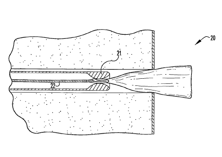
With reference now to Figures lA through 1C, shown are various stages of
an illustrative fistula graft deployment procedure utilizing a fistula graft
deployment system of the invention. The deployment system 20 includes an
CA 02637265 2008-07-15
WO 2007/090155 PCT/US2007/061380
9
elongate probing member 21, a fistula graft retaining element 22, and a
fistula graft
device 23.
The probing member 21 has a lumen 24, and includes a distal end 25
configured to pass through a secondary fistula opening and through at least a
segment of the remaining fistula, e.g., through at least a segment of the
fistula tract,
and potentially also through the primary opening. The lumen 24 generally
exhibits
a first diameter D1. However, a portion of the lumen 24 proximate the probing
member distal end 25 narrows to a second diameter D2 for reasons discussed
more
thoroughly below. In this particular embodiment, the probing member distal end
25 is initially passed through a secondary fistula opening (not shown), and
advanced through a fistula tract 40 to a point at or near the primaty opening
41 (as
shown in Figure 1A). The fistula graft device retaining element 22, which is
configured to extend through the probing member lumen 24, is similarly
advanced
through the fistula tract 40, and can be placed during or after placement of
the
probing member 21. In the current embodiment, the retaining element 22 is
advanced a distance beyond the associated probing member distal end 25 and
into
the alimentary cana142.
The retaining element 22 comprises a piece of wire including a deformable
wire loop 26 on one end. Such a wire loop 26 can be foimed in any suitable
manner including but not limited to bending one end of a length of wire in a
fashion that forms a loop and coupling this end to another portion of the wire
at a
point along the length of wire. Such coupling can include any suitable
coupling
means such as but not limited to welding or otherwise bonding, mechanically
fastening, and the like. Figure 1A shows the wire loop 26 in an "open"
configuration. When sufficiently positioned within the probing member lumen
24,
the wire loop 26 can be deformed to achieve a "closed" configuration to
releasably
retain the fistula graft device 23 therein. In the current embodiment,
deformation
of the wire loop 26 is facilitated by a segment of the probing member 21
proximate
its distal end 25, where, again, the inner lumen wall narrows from first
diameter D1
to second diameter D2. The second lumen diameter D2 is smaller than the width
CA 02637265 2008-07-15
WO 2007/090155 PCT/US2007/061380
of the deformable wire loop 26, such that when the loop 26 is forced into
second
lumen diameter D2, it is contacted by the inner lumen wall and forced to
deform to
a closed or collapsed configuration.
5 The fistula graft device 23 includes a biocompatible graft body 27
configured to block at least the primary fistula opening 41. The graft body
comprises a remodelable ECM material, for example, porcine SIS. As depicted in
Figure 1A, the graft body 27 can be presented in the alimentary canal 42 so
that a
tail end 28 of the graft body 27 approaches the wire loop 26. Thereafter, the
graft
10 body tail end 28 can be passed through the deformable wire loop 26, and the
loop
26 (with the tail end 28 received therethrough) can be passed through the
probing
member distal end 25 and into the second lumen diameter D2 to cause the wire
loop 26 to deform as described above. When sufficiently collapsed, the wire
loop
26 impinges the graft body tail end 28 that is received therethrough, and
thus, grips
the graft body tail end 28 to sufficiently releasably retain the same therein.
Then,
with the graft body 27 releasably retained by the retaining element 22, the
probing
member 21 and the retaining element 22 can be moved (in unison) back through
the fistula tract 40 and toward the secondary opening so as to lodge the graft
body
27 desirably within the primary opening 41. As depicted in Figure 1C, the wire
loop 26 can be "disengaged" from the probing member distal end 25 so that it,
again, attains an open configuration, releasing the graft body 27 therefrom.
This
can be accomplished by holding the wire loop 26 in a fixed position, while
forcibly
moving the probing member distal end 25 away from the primary opening 41, or
alternatively, by holding the probing member 21 in a fixed position, while
forcibly
moving the wire loop 26 toward the primary opening 41. The probing member 21
and fistula graft device retaining element 22 can then be withdrawn from the
fistula
through the secondary opening.
Turning now to a general discussion of fistula graft deployment systems
and methods of the invention useful for deploying fistula grafts within
patients.
Certain probing members of the invention have a lumen, and include a "leading"
distal end configured to pass through a secondary fistula opening and through
at
CA 02637265 2008-07-15
WO 2007/090155 PCT/US2007/061380
11
least a segment of the remaining fistula, Although not necessary to broader
aspects
of the invention, this distal end, or any portion thereof, may be tapered to
facilitate
passage through a secondary fistula opening and other segments of a fistula.
Illustratively, such probing members can be passed through a secondary fistula
opening, and advanced to any point within a fistula tract, for example, to a
point at
or near the primary opening as shown in Figure 1A, or alternatively, through
the
primary opening and a distance into the alimentary canal. Accordingly, probing
members of the invention can exhibit any suitable size and shape so as to be
able to
perform these functions while avoiding substantially cutting or tearing the
surrounding soft tissues. In certain embodiments, the length of a probing
member
is typically from about 2 inches to about 12 inches, more typically from about
3
inches to about 9 inches, and even more typically from about 4 to about 8
inches.
The outside diameter of a probing member is typically from about 0.3 mm to
about
3.2 mm, more typically from about 0.5 to about 3.0 mm, and even more typically
from about 1.0 mm to about 2.5 mm.
In some forms of the invention, the probing member is configured to be
generally straight in its relaxed condition. Such a probing member can be
used, in
certain aspects, to treat simple or straight fistulae. Alternatively, probing
members
of the invention can be configured to include one or more portions that are
curvilinear, bent, or otherwise suitably shaped. In certain aspects, the
distal end of
the probing member is curved to a degree to allow for easier passage of the
distal
end through a complex fistula, e.g., a horseshoe fistula, and/or through the
primary
fistula opening and into the alimentary canal.
Further in this regard, probing members of the invention can be formed
with any suitable material for facilitating deployment of a fistula graft in
accordance with the present invention. Such materials may be selected to take
advantage of one or more properties of the material such as but not limited to
its
weight, durability, flexibility, etc. For example, certain advantageous
probing
members of the invention are formed with materials exhibiting characteristics
to
enable the probing member to traverse a fistula, or a portion thereof, without
CA 02637265 2008-07-15
WO 2007/090155 PCT/US2007/061380
12
buckling or kinking or causing unacceptable damage to soft tissues defining
the
fistula. Illustratively, the probing member, or selected portions thereof
(e.g., the
tip of the distal end), can exhibit a degree of flexibility. In this regard, a
probing
member, or any portion thereof, may be rigid, malleable, semi-flexible, or
flexible.
For example, in certain embodiments, a fistula graft deployment system is
particularly adapted for treating fistulae that angulate sharply or curve
abruptly
such as in the case of certain horseshoe fistulae. In some of these
embodiments,
the probing member is configured to be directable or steerable through the
fistula
tract, and therefore, exhibits desirable characteristics, e.g., sufficient
stiffness, to
allow an operator to apply an adequate degree of ante-grade force to the
probing
member to allow it to traverse the fistula tract without substantially
buckling or
kinking.
In other embodiments, the probing member is rigid or substantially rigid,
and is configured to be generally straight, for example, for use in treating
certain
simple or straight fistulae. In other aspects of the invention, the probing
member is
composed of a malleable material such as but not limited to a woven or
spirally-
configured metal or alloy material, or a plastic (hydrocarbon-based) material,
which may be bent to the necessary angle or curvature, to allow passage
through
the fistula tract. The shape of such a probing member may be adjusted at
certain
intervals of the procedure so as to allow the probing member to pass further
and
further into the fistula tract, until the primary opening is identified. In
some forms,
the probing member is generally straight in a relaxed condition but can flex
to
adapt to contours during passage.
Suitable materials for forming probing members of the invention can
include but are not limited to metallic materials including stainless steel,
titanium,
cobalt, tantalum, gold, platinum, nickel, iron, copper and the like, as well
as alloys
of these metals (e.g., cobalt alloys, such as Elgiloy , a cobalt-chromium-
nickel
alloy, MP35N, a nickel-cobalt-chromium-molybdenum alloy, and Nitinol , a
nickel-titanium alloy). Additionally or alternatively, the probing member can
CA 02637265 2008-07-15
WO 2007/090155 PCT/US2007/061380
13
include material in the form of yarns, fibers, and/or resins, e.g.,
monofilament
yarns, high tenacity polyester, and the like. A probing member can also
include
other plastic, resin, polymer, woven, and fabric surgical materials, other
conventional synthetic surgical materials, sucli as a shape-memory plastic,
and/or
combinations of such materials. Further, appropriate ceramics can be used,
including, without limitation, hydroxyapatite, alumina and pyrolytic carbon.
The probing member lumen may or may not exhibit a constant diameter
along its length. In certain embodiments, for example as shown in Figures 1A
through 1 C, the probing member lumen includes a segment configured to aid or
facilitate releasable retention of the fistula graft device by the fistula
graft device
retaining element (although such probing member "segments" are certainly not
necessary to broader aspects of the invention). In this particular embodiment,
a
portion of the probing member lumen gradually narrows from a first diameter D1
to second diameter D2 in a curvilinear-like fashion. In other embodiments,
such
segments are configured to perform a similar function (i.e., aid or facilitate
releasable retention of the fistula graft device), yet are structured
differently than
the segment shown in Figures 1A through 1C. Illustratively, a portion of the
probing member lumen can narrow from a first diameter to second diameter in a
generally linear fashion or in another suitable fashion, or additionally or
alternatively, inner wall surfaces of the probing member can include
protuberances
or the like to help releasably grip the graft device. Such alternative probing
member segments can be configured in any suitable manner, with their size and
shape (including the size and shape of the corresponding lumen) potentially
depending on the size and shape of the fistula graft device retaining element
as
discussed in more detail below.
In some forms, the probing member lumen maintains generally the same
diameter along its length. For example, a probing member can be constructed
that
is similar in all respects to that shown in Figures 1A through 1C, except that
its
lumen has a constant diameter along its length that is equal to the smaller
diameter
D2. A device including such a probing member could be operated as discussed
CA 02637265 2008-07-15
WO 2007/090155 PCT/US2007/061380
14
above to move the deformable loop 26 in and out of the lumen to achieve closed
and open configurations, respectively. Alternatively, a probing member can be
constructed that is similar in all respects to that shown in Figures 1A
through 1C,
except that its lumen has a constant diameter along its length that is equal
to the
larger diameter D1 and the retaining element has additional or alternative
features
that enable it to releasably grip a fistula graft device.
In certain aspects, a fistula treatment method of the invention includes an
endoscopic visualization (fistuloscopy) step that is performed prior to
implanting a
fistula graft. Such endoscopic visualization can be used, for example, to
determine
the shape and size of a fistula, which in turn can be used to select an
appropriately
sized and shaped fistula graft device for treating the fistula.
Illustratively, a very
thin flexible endoscope can be inserted into a secondary opening of the
fistula and
advanced under direct vision through the fistula tract and out through the
primary
opening. By performing fistuloscopy of the fistula, the primary opening can be
accurately identified. Also, certain fistula treatment methods of the
invention
include a fistula cleaning step that is performed prior to implanting a
fistula graft.
For example, an irrigating fluid can be used to remove any inflammatory or
necrotic tissue located within the fistula prior to engrafting the graft
device. In
certain embodiments, one or more antibiotics are applied to the fistula graft
device
and/or the soft tissues surrounding the fistula as an extra precaution or
means of
treating any residual infection within the fistula.
In some modes of operation, means for visualizing and/or irrigating a
fistula can be received within the probing member lumen. Illustratively, such
means, as well as other desirable instruments and/or materials, can be passed
into
the proximal end of the probing member lumen (or alternatively, can be passed
into one or more openings in a sidewall of the probing member), and through at
least a portion of the probing member lumen. For example, in certain aspects,
a
probing member of the invention includes one or more ports in a sidewall
thereof,
wherein each port can be associated with a corresponding channel that extends
from the port toward the distal end of the probing member. In some forms, one
or
CA 02637265 2008-07-15
WO 2007/090155 PCT/US2007/061380
more port and channel combinations are each configured to receive one or more
instruments and/or materials therethrough. For example, a port can be
configured
to receive one or more optical fibers for visualization and/or illumination of
the
fistula and surrounding soft tissues, for example, fiber-optic bundles
including a
5 plurality of glass fibers comprised of silicone, silicone dioxide, and/or a
suitable
equivalent. When used in the invention, these optical fibers are provided
having
suitable characteristics for the particular application including but not
limited to
suitable lengths and diameters, as well as degrees of flexibility or
malleability.
Suitable probing member ports can also be configured to receive fluids for the
10 ante-grade irrigation of a fistula. Such fluids can be provided from an
external bag
of fluid that is connected to the port of the irrigation channel by means of
flexible
tubing. If necessary, the fluid can be infused under pressure using a pressure
bag
applied to the fluid source, to increase the pressure under which the fluid is
infused. Suitable probing member ports can further be configured to receive
15 guide-wires, drains, solutions such as sealants or sclerosants, high
intensity light
sources, a lever system to steer the probing member (e.g., wherein the probing
member and/or its distal tip is directable in one, two, or three planes),
and/or any
other suitable instruments and/or mateials. In some forms, a probing member
port
is configured to receive an optical viewing and lens system that may be
attached to
a video camera, a video monitor, and a video recorder for viewing at the
distal end
of the probing member.
Fistula graft device retaining elements of the invention can be configured to
extend through the lumen of the probing member, and in this regard, can
exhibit
any suitable size and shape to be able to do so. Further in this regard, any
suitable
material can be used in forming a retaining element of the invention including
any
of those previously described for the probing member. Illustratively, the
probing
member and the fistula graft device retaining element can include one or more
of
the same materials and/or one or more different materials. As one non-limiting
example, the probing member can include a first material, and the retaining
element can include a second material, wherein the second material is
relatively
more rigid than the first material. The relatively less rigid material may be
useful,
CA 02637265 2008-07-15
WO 2007/090155 PCT/US2007/061380
16
for example, to allow the probing member to be successfully directed or
steered
through the fistula tract, while the relatively more rigid material may be
useful, for
example, to allow adequate force to be applied to the retaining element
without
causing it to buckle or kink.
During a fistula graft deployment procedure, the retaining element (while
surrounded, at least in part, by the probing member) can be passed through a
secondary fistula opening, and advanced to any point within a fistula tract,
for
example, to a point at or near the primary opening, or alternatively, through
the
primary opening and into the alimentary canal as shown in Figure 1A.
Alternatively, the probing member can be suitably positioned within the
patient in
a first step, and the retaining element can be suitably positioned within the
probing
member in a second step.
When extending through the probing member lumen and at least a portion
of the fistula, the proximal end of the fistula graft device retaining element
can
protrude from the secondary opening. In this regard, the proximal end of the
retaining element can be manipulated by an operator, e.g., a surgeon, during
an
illustrative fistula graft deployment procedure of the invention. For example,
in
some embodiments, such a proximal end is forced toward the secondary opening,
causing the distal end of the retaining element to pass through the primary
opening
and into the alimentaty canal. After suitably manipulating the deployment
system
to releasably retain the fistula graft device with at least the retaining
element, the
proximal end is forced back away (or otheiwise caused to move back away) from
the secondary opening to cause the distal end to pass back through the primary
opening and into the fistula tract so as to sufficiently implant the graft
body within
the patient to block at least the primary opening of the fistula.
Further in this regard, the fistula graft device retaining element can be
configured in any suitable manner, can exhibit any suitable size and shape,
and can
include any suitable mechanism and/or material for releasably retaining a
fistula
graft device in accordance with the present invention. Illustratively, the
retaining
CA 02637265 2008-07-15
WO 2007/090155 PCT/US2007/061380
17
element can include surfaces, which are configured to contact the fistula
graft
device in a manner that releasably secures the fistula graft device to the
retaining
element. A retaining element of the invention can have any suitable number of
surfaces that are configured in this manner, and any of these surfaces may or
may
not be movable relative to another surface. Also, such releasable securement
may
or may not involve deformation and/or penetration of the fistula graft device,
or
any portion thereof. In certain aspects, frictional force is sufficient to
releasably
grip or otherwise secure the fistula graft device between surfaces of the
retaining
element.
In certain forms, the retaining element includes a first surface and a second
surface, wherein the first surface can attain at least two different spatial
orientations relative to the second surface for releasably retaining the
fistula graft
device. These differing orientations can be achieved by movement of the first
surface, the second surface, or both. Further, movement of either surface may
or
may not be facilitated by another object or device. For example, a retaining
element can include a spring or a hinged portion to enable movement of one or
more of such surfaces relative to another surface.
In some embodiments, the retaining element, or any portion thereof, is
translatable along the probing member. In these embodiments, such retaining
element translation can promote and/or facilitate movement of one or more
retaining element surfaces relative to another surface for releasably
retaining the
fistula graft device. For example, a retaining element can include two or more
arms or other suitable members, which are adapted to move between an "open"
configuration and a "closed" configuration when sufficiently moved along the
probing member, e.g., slid, twisted, or otherwise suitably moved within and/or
around the probing member. Such movement may or may not be facilitated by one
or more pivoting adaptations, which can join an arm to one or more other arms
and/or to the probing member. In such an open configuration, space is provided
between two or more of the surfaces into which the fistula graft device, or a
CA 02637265 2008-07-15
WO 2007/090155 PCT/US2007/061380
18
portion thereof, can be placed. In such a closed configuration, this space is
closed
or reduced to sufficiently contact the graft device to releasably retain the
device.
In certain modes of operation, translation of a fistula graft retaining
element
along (e.g., within the lumen of) a probing member is controlled or otherwise
influenced, at least in part, by a spring or spring-like member. For example
and
referring now to Figure 2, shown is an illustrative fistula graft delivery
device 60
of the invention that includes, inter alia, an elongated, generally
cylindrical
probing member 61 and a spring-loaded fistula graft retaining element 62
extending through the lumen 63 of the probing member 61.
The distal end of the retaining element 62 includes a deformable wire snare
64. The wire snare 64 is generally in the shape of a diamond, although the
snare
64 can exhibit other suitable shapes as well, for example, any of those
described
elsewhere herein such as that of the deformable wire loop of Figures lA-1C.
The
proximal end of the retaining element 62 is attached to the distal end of a
plunger
65, although in some forms, the plunger 65 is merely an extension of the
retaining
element 62. Also attached to or otherwise associated with the distal end of
the
plunger 65 is a spring 66, with a portion of the retaining element 62
including its
proximal end extending through the lumen of the spring 66. As depicted, the
spring 66 and a portion of the plunger 65 including its distal end are
positioned
within a generally cylindrical housing member 67. The distal end of the spring
66
is attached to the inside of the housing member 67 proximate its distal end,
although such attachment is not necessary to broader aspects of the invention,
i.e.,
the spring 66 need not be connected to any other component of the delivery
device
60. The distal end of the housing member 67, which includes an aperture
through
which portions of the retaining element 62 can pass, is attached to the
proximal end
of the probing member 61. Nonetheless, it should be noted that certain
components of the delivery device 60 could be combined into a single component
by one skilled in the art, for example, the probing member 61 and the housing
member 67 could form a single component.
CA 02637265 2008-07-15
WO 2007/090155 PCT/US2007/061380
19
Portions of the plunger 65 including its distal end are configured to move
back and forth a distance axially within the housing member 67. In this
regard, the
plunger 65 can be depressed against the resistive force of the spring 66,
thereby
forcing portions of the retaining element 62 to move distally within the
probing
member lumen 63. Figure 2 shows the plunger 65 in a partially depressed
position,
with the spring 66 in a corresponding partially compressed configuration
between
the distal end of the plunger 65 and the inside of the distal end of the
housing
member 67. With the plunger 65 in this position, the snare 64 extends a
distance
beyond the distal end of the probing member 61 to achieve a relaxed, "open"
configuration. Finger grips 68 are attached to the housing member 67 to give
the
operator leverage in depressing the plunger 65. When the plunger 65 is
released,
the spring 66 decompresses towards a relaxed configuration, thereby drawing
the
snare 64 at least partially back into the probing member lumen 63. When
sufficiently drawn into the probing member lumen 63, the snare 64 deforms to
achieve a "closed" configuration effective to releasably retain a fistula
graft device
therein. In the current embodiment, the diameter of the probing member lumen
63
is smaller than the width of the deformable wire snare 64, such that when the
snare
64 is drawn into the probing member lumen 63, it is contacted by the inner
lumen
wall and forced to deform to a closed or collapsed configuration.
In use, the distal end of the probing member 61 can be passed through a
secondary fistula opening and through at least a segment of the remaining
fistula,
e.g., through at least a segment of the fistula tract, and potentially also
through the
primary opening. During such passage, the spring 66 is preferably decompressed
so that the deformable wire snare 64, or a substantial portion thereof, is
positioned
within the probing member lumen 63.
During an illustrative procedure, the distal end of the probing member 61
can be advanced to a point within the fistula tract just shy of the primary
opening.
Thereafter, the plunger 65 can be depressed with the housing member 67 held in
a
generally stationary position, forcing the snare 64 out of the probing member
lumen 63 and into the alimentary canal. Then, with a portion of a fistula
graft
CA 02637265 2008-07-15
WO 2007/090155 PCT/US2007/061380
device suitably passed through the snare 64 opening, the plunger 65 can be
released to cause the snare 64 to pass back into the probing member lumen 63,
thereby releasably retaining the graft device in the snare 64. The force of
the
decompressing spring 66 may be sufficient to draw the graft device into the
5 primary opening to plug it, or alternatively, the delivery device 60 can be
retracted
a distance through the fistula tract (and potentially back out of the
secondary
opening depending on the size, shape, and configuration of the fistula graft)
to
suitably draw the fistula graft into the fistula tract to at least block the
primary
opening of the fistula or otherwise suitably seat the graft within the fistula
tract.
10 Thereafter, the fistula graft device can be suitably separated from the
snare 64,
preferably by again depressing the plunger 65 to open the snare.
In certain aspects, the retaining element is configured to releasably retain
the fistula graft device independent of the probing member. For example, the
15 retaining element can be configured so as to be manually actuatable by the
surgeon
or other suitable operator to grasp and ungrasp the fistula graft device
during a
deployment procedure. In other aspects, the retaining element, whether or not
coupled to or otherwise joined with the probing member, depends on the probing
member to releasably retain the fistula graft device, for example as depicted
in
20 Figures 1A through 1C.
Suitable fistula graft devices include a biocompatible graft body, which is
configured to block at least the primary opening of a fistula, i.e., the
primary
opening and potentially one or more other segments of a fistula, for example,
the
fistula tract and/or any secondary openings. In this context, the term
"fistula tract"
is meant to include, but is not limited to, a void in soft tissues extending
from a
primary fistula opening, whether blind-ending or leading to one or more
secondary
fistula openings, for example, to include what are generally described as
simple
and complex fistulae. As described in more detail below, in certain aspects,
fistula
graft devices suitable for deployment in accordance with the present invention
can
also include a suture or other similar adaptation in association with the
graft body,
which is useful, for example, for forcing the graft body into the primary
opening.
CA 02637265 2008-07-15
WO 2007/090155 PCT/US2007/061380
21
For example, in certain embodiments, a suture is coupled to the graft body,
and the
graft device is releasably retained by the retaining element at a point along
this
suture.
The fistula graft device can be releasably retained by the retaining element,
and while being retained, forced into the primary opening so as to lodge the
graft
body desirably within the primaiy opening. In certain aspects, forcing the
graft
body into the primary opening involves pushing the graft body into the primary
opening, while in other aspects, forcing the graft body into the primary
opening
involves pulling the graft body into the primary opening. Upon being suitably
lodged within the primary opening, the graft device can be released fiom the
retaining element (either before or after the retaining element, and
potentially also
the probing member are withdrawn from the fistula through the secondary
opening,
depending on the characteristics of the particular deployment system being
utilized).
The materials used to form the fistula graft devices useful in the invention
should generally be biocompatible, and in advantageous embodiments of the
devices, use a remodelable material. Particular advantage can be provided by
graft
devices including a remodelable collagenous material. Such remodelable
collagenous materials, whether reconstituted or non-reconstituted, can be
provided,
for example, by collagenous materials isolated from a warm-blooded vertebrate,
and especially a mammal. Such isolated collagenous material can be processed
so
as to have remodelable properties and promote cellular invasion and ingrowth.
Remodelable materials may be used in this context to promote cellular growth
on,
around, and/or within tissue in which a fistula graft device is implanted,
e.g., on,
around, and/or within tissue defining a fistula tract or an opening to a
fistula. In
some forms, the remodelable material can be broken down and replaced by new
tissue in such a way that the original fistula closure achieved by the
implanted
fistula graft is maintained throughout the remodeling process so as to
eventually
form a closure or substantial closure with the new tissue.
CA 02637265 2008-07-15
WO 2007/090155 PCT/US2007/061380
22
Suitable remodelable materials can be provided by collagenous
extracellular matrix (ECM) materials possessing biotropic properties. For
example, suitable collagenous materials include ECM materials such as
submucosa, renal capsule membrane, dermal collagen, dura mater, pericardium,
fascia lata, serosa, peritoneum or basement membrane layers, including liver
basement membrane. Suitable submucosa materials for these purposes include,
for
instance, intestinal submucosa including small intestinal submucosa, stomach
submucosa, urinary bladder submucosa, and uterine submucosa. Submucosa when
used in the present invention can be obtained by harvesting such tissue
sources and
delaminating the submucosa from smooth muscle layers, mucosal layers, and/or
other layers occurring in the tissue source. For additional information as to
submucosa that can be used in the present invention, and its isolation and
treatment, reference can be made, for example, to U.S. Patent Nos. 4,902,508,
5,554,389, 5,993,844, 6,206,931, and 6,099,567.
Submucosa or other ECM tissue when used in the invention is preferably
highly purified, for example, as described in U.S. Patent No. 6,206,931 to
Cook et
al. Thus, preferred ECM material will exhibit an endotoxin level of less than
about
12 endotoxin units (EU) per gram, more preferably less than about 5 EU per
gram,
and most preferably less than about 1 EU per gram. As additional preferences,
the
submucosa or other ECM material may have a bioburden of less than about 1
colony forming units (CFU) per gram, more preferably less than about 0.5 CFU
per
gram. Fungus levels are desirably similarly low, for example less than about 1
CFU per gram, more preferably less than about 0.5 CFU per gram. Nucleic acid
levels are preferably less than about 5 g/mg, more preferably less than about
2
g/mg, and virus levels are preferably less than about 50 plaque forming units
(PFU) per gram, more preferably less than about 5 PFU per gram. These and
additional properties of submucosa or other ECM tissue taught in U.S. Patent
No.
6,206,931 may be characteristic of any ECM tissue used in the present
invention.
A typical layer thickness for an as-isolated submucosa or other ECM tissue
layer useful in some forms of the invention ranges from about 50 to about 250
CA 02637265 2008-07-15
WO 2007/090155 PCT/US2007/061380
23
microns when fully hydrated, more typically from about 50 to about 200 microns
when fully hydrated, although isolated layers having other thicknesses may
also be
obtained and used. These layer thicknesses may vaiy with the type and age of
the
animal used as the tissue source. As well, these layer thicknesses may vary
with
the source of the tissue obtained from the animal source. Further, the
submucosa
and other ECM tissue materials useful in certain embodiments of the invention
can
be employed as xenografts (i.e., cross species, such as a non-human donor for
a
human recipient), allografts (i.e., intraspecies with a donor of the same
species as
the recipient) and/or autografts (i.e., the donor and the recipient being the
same
individual).
Suitable ECM materials may include one or more bioactive agents native to
the source tissue. For example, a submucosa or other remodelable ECM tissue
material used in some forms of the invention may retain one or more growth
factors such as but not limited to basic fibroblast growth factor (FGF-2),
transforming growth factor beta (TGF-beta), epidermal growth factor (EGF),
and/or platelet derived growth factor (PDGF). As well, submucosa or other ECM
materials when used in the invention may retain other native bioactive agents
such
as but not limited to proteins, glycoproteins, proteoglycans, and
glycosaminoglycans. For example, suitable graft materials may include heparin,
heparin sulfate, hyaluronic acid, fibronectin, cytokines, and the like. Thus,
generally speaking, a submucosa or other ECM material may retain one or more
bioactive components that induce, directly or indirectly, a cellular response
such as
a change in cell moiphology, proliferation, growth, protein or gene
expression.
Submucosa or other ECM materials of the present invention can be derived
from any suitable organ or other tissue source, usually sources containing
connective tissues. The ECM materials processed for use in the invention will
typically include abundant collagen, most commonly being constituted at least
about 80% by weight collagen on a dry weight basis. Such naturally-derived ECM
materials will for the most part include collagen fibers that are non-randomly
oriented, for instance occurring as generally uniaxial or multi-axial but
regularly
CA 02637265 2008-07-15
WO 2007/090155 PCT/US2007/061380
24
oriented fibers. When processed to retain native bioactive factors, the ECM
material can retain these factors interspersed as solids between, upon and/or
within
the collagen fibers. Particularly desirable naturally-derived ECM materials
for use
in the invention will include significant amounts of such interspersed, non-
collagenous solids that are readily ascertainable under light microscopic
examination with appropriate staining. Such non-collagenous solids can
constitute
a significant percentage of the dry weight of the ECM material in certain
inventive
embodiments, for example at least about 1%a, at least about 3%, and at least
about
5% by weight in various embodiments of the invention.
The submucosa or other ECM material used in the present invention may
also exhibit an angiogenic character and thus be effective to induce
angiogenesis in
a host engrafted with the material. In this regard, angiogenesis is the
process
through which the body makes new blood vessels to generate increased blood
supply to tissues. Thus, angiogenic materials, when contacted with host
tissues,
promote or encourage the formation of new blood vessels into the materials.
Methods for measuring in vivo angiogenesis in response to biomaterial
implantation have recently been developed. For example, one such method uses a
subcutaneous implant model to determine the angiogenic character of a
material.
See, C. Heeschen et al., Nature Medicine 7 (2001), No. 7, 833-839. When
combined with a fluorescence microangiography technique, this model can
provide
both quantitative and qualitative measures of angiogenesis into biomaterials.
C.
Johnson et al., Circulation Research 94 (2004), No. 2, 262-268.
Further, in addition or as an alternative to the inclusion of such native
bioactive components, non-native bioactive components such as those
synthetically
produced by recombinant technology or other methods (e.g., genetic material
such
as DNA), may be incorporated into an ECM material. These non-native bioactive
components may be naturally-derived or recombinantly produced proteins that
correspond to those natively occurring in an ECM tissue, but perhaps of a
different
species (e.g., human proteins applied to collagenous ECMs from other animals,
such as pigs). These non-native bioactive components may also be drug
CA 02637265 2008-07-15
WO 2007/090155 PCT/US2007/061380
substances. Illustrative drug substances that may be added to materials
include, for
example, anti-clotting agents, e.g. heparin, antibiotics, anti-inflammatory
agents,
thrombus-promoting substances such as blood clotting factors, e.g., thrombin,
fibrinogen, and the like, and anti-proliferative agents, e.g. taxol
derivatives such as
5 paclitaxel. Such non-native bioactive components can be incorporated into
and/or
onto a graft material in any suitable manner, for example, by surface
treatment
(e.g., spraying) and/or impregnation (e.g., soaking), just to name a few.
Also,
these substances may be applied to the ECM material in a premanufacturing
step,
immediately prior to the procedure (e.g., by soaking the material in a
solution
10 containing a suitable antibiotic such as cefazolin), or during or after
engraftment of
the material in the patient.
Graft bodies suitable for deployinent in accordance with the present
invention can be provided in any suitable state including hydrated, partially
15 hydrated, and dried states. Drying a graft body can be accomplished in any
suitable manner including but not limited to subjecting the graft body to
lyophilization, air dying, vacuum pressing, and other suitable drying
conditions
known in the art. However, when drying a graft body, it is advantageous to
perform drying operations under relatively mild temperature exposure
conditions
20 that minimize deleterious effects upon any ECM materials being used, for
example
native collagen structures and potentially bioactive substances present. Thus,
drying operations conducted with no or substantially no duration of exposure
to
temperatures above human body temperature or slightly higher, say, no higher
than
about 38 C, will preferably be used in preparing ECM materials useful in some
25 forms of the present invention. These include, for example, vacuum pressing
operations at less than about 38 C, forced air drying at less than about 38
C, or
either of these processes with no active heating - at about room temperature
(about
25 C) or with cooling. Relatively low temperature conditions also, of course,
include lyophilization conditions.
ECM materials when used in the invention may be free of additional, non-
native crosslinking, or may contain additional crosslinking. Such additional
CA 02637265 2008-07-15
WO 2007/090155 PCT/US2007/061380
26
crosslinking may be achieved by photo-crosslinking techniques, by chemical
crosslinkers, or by protein crosslinking induced by dehydration or other
means, and
may be used to enhance one or more plzysical, biological and/or other
characteristics of an ECM material, and/or to bond two or more pieces of ECM
material together. Nonetheless, because certain crosslinking techniques,
certain
crosslinking agents, and/or certain degrees of crosslinking can destroy the
remodelable properties of a remodelable material, where preservation of
remodelable properties is desired, any crosslinking of the remodelable ECM
material can be performed to an extent or in a fashion that allows the
material to
retain at least a portion of its remodelable properties.
A deployable fistula graft device, or any component thereof, can have a
level or degree of porosity. Remodelable ECM materials having a relatively
more
open matrix structure (i.e., higher porosity) are capable of exhibiting
different
material properties than those having a relatively more closed or collapsed
matrix
structure. For example, an ECM material having a relatively more open matrix
structure is generally softer and more readily compliant to an implant site
than one
having a relatively more closed matrix structure. Also, the rate and amount of
tissue growth in and/or around a remodelable material can be influenced by a
number of factors, including the amount of open space available in the
material's
matrix structure for the infusion and support of a patient's tissue-forming
components, such as fibroblasts. Therefore, a more open matrix structure can
provide for quicker, and potentially more, growth of patient tissue in and/or
around
the remodelable material, which in turn, can lead to quicker remodeling of the
material by patient tissue.
In certain aspects, a fistula graft device includes at least two regions
exhibiting differing properties, e.g., differing porosities. Such differing
regions
can be established in certain locations, for example, locations providing a
particular arrangement or pattern on and/or within the remodelable fistula
graft,
and in some forms, such differing regions are formed by subjecting the fistula
graft
to a suitable differential drying process. Illustratively, a graft body can be
CA 02637265 2008-07-15
WO 2007/090155 PCT/US2007/061380
27
configured so that portions of the graft body adapted to reside in and/or
around the
primary opening of the fistula occupy a more diminished porosity region, while
portions of the graft body adapted to reside within the fistula tract (and
potentially
one or more secondaiy openings) occupy a more open porosity region. In this
configuration, the diminished matrix region can help isolate the fistula tract
from
the alimentary canal, thus inhibiting bacteria and other undesirable
substances from
passing into the alimentary canal from the fistula, while the more open matrix
region serves to promote more rapid closure of the fistula with its desirable
remodeling properties.
Fistula graft devices for deployment in accordance with the present
invention may include biocompatible materials derived from a number of
biological polymers, which can be naturally occurring or the product of in
vitro
fermentation, recombinant genetic engineering, and the like. Purified
biological
polymers can be appropriately formed into a substrate by techniques such as
weaving, knitting, casting, molding, and extrusion. Suitable biological
polymers
include, without limitation, collagen, elastin, keratin, gelatin, polyamino
acids,
polysaccharides (e.g., cellulose and starch) and copolymers thereof.
Suitable biocompatible graft materials suitable for deployment in
accordance with the present invention can also include a variety of synthetic
polymeric materials including but not limited to bioresorbable and/or non-
bioresorbable plastics. Bioresorbable, or bioabsorbable polymers that may be
used
include, but are not limited to, poly(L-lactic acid), polycaprolactone,
poly(lactide-
co-glycolide), poly(hydroxybutyrate), poly(hydroxybutyrate-co-valerate),
polydioxanone, polyorthoester, polyanhydride, poly(glycolic acid), poly(D,L-
lactic
acid), poly(glycolic acid-co-trimethylene carbonate), polyhydroxyalkanaates,
polyphosphoester, polyphosphoester urethane, poly(amino acids),
cyanoacrylates,
poly(trimethylene carbonate), poly(iminocarbonate), copoly(ether-esters)
(e.g.,
PEQ/PLA), polyalkylene oxalates, and polyphosphazenes. These or other
bioresorbable materials may be used, for example, where only a temporary
blocking or closure function is desired, and/or in combination with non-
CA 02637265 2008-07-15
WO 2007/090155 PCT/US2007/061380
28
bioresorbable materials where only a temporary participation by the
bioresorable
material is desired.
Non-bioresorbable, or biostable polymers that may be used include, but are
not limited to, polytetrafluoroethylene (PTFE) (including expanded PTFE),
polyethylene terephthalate (PET), polyurethanes, silicones, and polyesters and
other polymers such as, but not limited to, polyolefins, polyisobutylene and
ethylene-alphaolefin copolymers; acrylic polymers and copolymers, vinyl halide
polymers and copolymers, such as polyvinyl chloride; polyvinyl ethers, such as
polyvinyl methyl ether; polyvinylidene halides, such as polyvinylidene
fluoride
and polyvinylidene chloride; polyacrylonitrile, polyvinyl ketones; polyvinyl
aromatics, such as polystyrene, polyvinyl esters, such as polyvinyl acetate;
copolymers of vinyl monomers with each other and olefins, such as ethylene-
methyl methacrylate copolymers, acrylonitrile-styrene copolymers, ABS resins,
and ethylene-vinyl acetate copolymers; polyamides, such as Nylon 66 and
polycaprolactam; alkyd resins, polycarbonates; polyoxymethylenes; polyimides;
polyethers; epoxy resins, polyurethanes; rayon; and rayon-triacetate.
In certain embodiments, the graft body is formed with material having a
suitable volumetric shape or space for promoting blockage of at least the
primary
fistula opening, such as an anorectal fistula opening. Suitable volumetric
graft
bodies for use in this aspect of the invention can be prepared, for example,
as
described in U.S. Patent Application Serial No. 11/415,403, titled
"VOLUMETRIC GRAFTS FOR TREATMENT OF FISTULAE AND RELATED
METHODS AND SYSTEMS" (Cook Biotech Incorporated) filed May 1, 2006,
which is hereby incorporated by reference in its entirety. Illustratively,
such graft
bodies can include a layered volumetric graft construct including, for
example, a
rolled remodelable material that occupies a substantially unitary volume. In
some
foims, suitable graft bodies are formed by folding or rolling, or otherwise
overlaying one or more portions of a biocompatible material, such as a
biocompatible sheet material. The overlaid biocompatible sheet material can be
compressed and dried or otherwise bonded into a volumetric shape such that a
CA 02637265 2008-07-15
WO 2007/090155 PCT/US2007/061380
29
substantially unitary construct is formed. Such a substantially unitary graft
body
can then be placed in a fistula in a manner such that it blocks at least the
primary
fistula opening, and potentially blocks at least a portion of the fistula
tract and/or
any secondary fistula openings. In some forms, a fistula graft device is
formed by
randomly or regularly packing one or more pieces of single or multilayer ECM
sheet material within a mold and thereafter processing the packed material.
Illustrative volumetric graft bodies will be of sufficient size and shape to
block at least the primary fistula opening and extend into at least a portion
of the
fistula tract. Such graft bodies will generally (but not necessarily) be of
sufficient
dimension to fill the fistula, or a segment thereof, e.g., the primary fistula
opening,
the fistula tract, and/or any secondary fistula openings, either alone or in
combination with other similar or differing devices. In certain embodiments,
such
graft bodies will have a length of at least about 0.20 cm, and in many
situations at
least about 1 cm to about 20 cm (approximately 1 to 8 inches). In illustrative
embodiments, the fistula graft device will have a length of from about 2 cm to
about 5 cm, or alternatively, from about 2 inches to about 4 inches.
Additionally,
in certain embodiments, graft bodies will have a diameter, which may or may
not
be constant along their length, of from about 0.1 mm to about 25 mm, or more
typically from about 5 mm to about 10 mm. In certain embodiments, a generally
conical plug device is tapered along its length so that the end of the plug
device
configured for placement in and/or around the primary opening has a diameter
of
about 5 mm to about 10 mm, and the opposite end of the plug device has a
diameter of about 0.5 mm to about 3 mm. Such a taper may or may not be
continuous along the length of the graft body.
A graft device, or any portion thereof, can include a suitable biocompatible
foam or sponge form material. Illustratively, a graft device may comprise a
porous, three-dimensionally stable body formed with one or more suitable
biocompatible matrix materials. Such biocompatible matrix materials can
include
naturally-occurring polymers and/or synthetic polymers. More preferred sponge
compositions will comprise collagen as a matrix-forming material, either alone
or
CA 02637265 2008-07-15
WO 2007/090155 PCT/US2007/061380
in combination with one or more other matrix forming materials, and
particularly
preferred sponge compositions will comprise an ECM material such as those
discussed elsewhere herein. In general, sponge matrices useful in certain
embodiments of the present invention can be formed by providing a liquid
solution
5 or suspension of a matrix-forming material, and causing the material to form
a
porous three-dimensionally stable structure; however, a sponge or foam
material
can be formed using any suitable formation method, as is known in the art. For
additional information concerning foam or sponge form materials that can be
useful in certain embodiments of the present invention, reference can be made,
for
10 example, to U.S. Pat. App. Pub. No. 2003/0013989.
In some forms, a compact, stabilized sponge construct is highly expansive
when wetted, which can desirably enhance the ability of the graft body to
block
(and to continue blocking) at least the primary opening of a fistula. These
15 compact, stabilized sponge constructs can be useful to allow the graft body
to
attain a more low-profile configuration for traversing a fistula. For example,
an
illustrative graft body can include a suitable sponge construct such that in a
stabilized, compressed first configuration, the graft body can fit within the
distal
end of a probing member exhibiting suitable characteristics so as to be able
to
20 traverse a fistula. Illustratively, this end of the probing member can be
passed into
a secondary opening, through a fistula tract, and out of a primary opening
into the
alimentary canal. Thereafter, the retaining element, which is at least
partially
received within the probing member lumen, can be used to force the graft body,
or
a portion thereof, out of the probing member and into the alimentary canal so
as to
25 allow the graft body, or a portion thereof, to attain an expanded second
configuration. In such an expanded configuration, the graft body, which was
previously able to easily traverse the primary fistula opening, is now sized
and
shaped so as to desirably contact soft tissues surrounding the fistula and
lodge
within the primary opening to block the primary opening, and potentially other
30 segments of the fistula, when pulled into the fistula by the retaining
element. In
illustrative procedures, a suitable hydrant, such as saline, may be applied or
delivered to the graft body after it is suitably located within a patient to
enhance
CA 02637265 2008-07-15
WO 2007/090155 PCT/US2007/061380
31
the expansion of the body within the fistula tract and/or a fistula opening.
Alternatively, or additionally, a bodily fluid of the patient can sufficiently
wet the
implanted graft body so as to promote the expansion of the body within the
fistula.
In certain aspects, an illustrative graft body includes a compliant sheet form
biocompatible material, e.g., multilaminate sheet material comprising one or
more
layers of material bonded together. Suitable compliant sheet form graft bodies
for
use in this aspect of the invention can be prepared, for example, as described
in
U.S. Patent Application Serial No. 11/414,682, titled "FISTULA GRAFT WITH
DEFORMABLE SHEET-FORM MATERIAL" (Cook Biotech Incorporated) filed
Apri128, 2006, which is hereby incorporated by reference in its entirety. Such
sheet form materials are deformable upon impingement by soft tissue
surrounding
a fistula (e.g., tissue surrounding the primary fistula opening, the fistula
tract,
and/or any secondary fistula openings). These deformable materials can include
any of the ECM or other biocompatible materials described herein, for example,
a
multilaminate sheet of remodelable SIS material. The bioactive nature of such
materials promotes desirable healing of the fistula, for example, by
overcoming the
effects of bacteria and other deleterious substances typical to the fistula
environment.
Suitable multilaminate materials, whether used in this or other aspects of
the invention, can include a plurality of ECM material layers bonded together,
a
plurality of non-ECM materials bonded together, or a combination of one or
more
ECM material layers and one or more non-ECM material layers bonded together.
Also, an adhesive, glue or other bonding agent may be used in achieving a bond
between two segments of ECM material, e.g., between two layers of ECM
material. Suitable bonding agents may include, for example, collagen gels or
pastes, gelatin, or other agents including reactive monomers or polymers, for
example cyanoacrylate adhesives. As well, bonding can be achieved or
facilitated
between ECM material layers using a suitable crosslinking technique, e.g.,
using
chemical cross-linking agents, such as glutaraldehyde, formaldehyde, epoxides,
genipin or derivatives thereof, carbodiimide compounds, polyepoxide compounds,
CA 02637265 2008-07-15
WO 2007/090155 PCT/US2007/061380
32
or other similar agents. A combination of one or more of these with
dehydration-
induced bonding may also be used to bond ECM material layers to one another.
Further, such sheet form graft bodies are sized and shaped so as to be
deformable to a three-dimensional volumetric body blocking at least the
primary
opening, and potentially filling at least a portion of the fistula tract
and/or any
secondary openings of the fistula. In so doing, advantageous implant materials
will also be sufficiently flaccid to avoid substantial cutting or tearing of
the
surrounding soft tissues. In certain aspects, such a three-dimensional
volumetric
body, when formed, includes a portion protruding through any secondary
openings
of the fistula. This extending portion can be used, in certain aspects, to
attach the
fistula graft device to soft tissues at or near a secondary opening of the
fistala as a
means of preventing the graft body from reverse migrating undesirably back
toward the alimentary canal.
In certain aspects, a sheet form graft body is shaped and sized such that the
diameter of the primary opening is less than the width of the sheet so that as
the
sheet of material is drawn into the fistula tract, it is forced to fold and/or
roll over
itself one or more times to conform to soft tissues surrounding the fistula,
and is
gradually "wedged" into the primary opening, and potentially at least a
portion of
the fistula tract and/or any secondary openings of the fistula, so as to block
these
spaces when sufficiently pulled therethrough. Such lodging in place may be
sufficient to obviate the need for otherwise securing the graft to the soft
tissues at
or near the primary opening, fistula tract, and/or any secondary openings.
Nonetheless, in certain aspects, the graft is further secured to such soft
tissues, for
example, by suturing. Also, any portion of the graft body can be trimmed, for
example, to prevent the engrafted graft body from protruding undesirably from
the
primary opening and/or any secondary openings of the fistula.
In addition to those described elsewhere herein, a variety of other suitable
fistula graft devices can be used in conjunction with the systems and methods
of
the present invention. Such graft devices can be prepared, for example, as
CA 02637265 2008-07-15
WO 2007/090155 PCT/US2007/061380
33
described in U.S. Provisional Application, entitled "FISTULA GRAFTS AND
RELATED METHODS AND SYSTEMS FOR TREATING FISTULAE" (Cook
Biotech Incorporated) filed on January 31, 2006, which is hereby incorporated
by
reference in its entirety.
With reference now to Figure 3, shown is a top view of an illustrative
medical product 80 of the present invention that includes a fistula graft
delivery
device 90 and fistula graft devices 91 sealed within sterile medical
packaging. In
particular, medical product 80 has packaging including a backing layer 81 and
a
front film layer 82 (shown partially drawn away from backing layer 81). The
fistula graft device is sealed between backing layer 81 and film 82 utilizing
a
boundary of pressure-adhesive 83 as is conventional in medical packaging. A
cut-
out 84 may be provided in the backing layer 81 to assist a user in separating
the
film layer 82 from the backing layer 81.
Sterilization of the medical product 80 may be achieved, for example, by
irradiation, ethylene oxide gas, or any other suitable sterilization
technique, and the
materials and other properties of the medical packaging will be selected
accordingly. Also, the fistula graft devices 91 can be contained in sterile
packaging in any suitable state. Suitable states include, for example, a
hydrated or
dehydrated state. The fistula graft devices 91 can be dehydrated by any means
known in the art (e.g., lyophilization or air dried). If a fistula graft
device is stored
in a dehydrated state, it is preferred that it retains all of its biological
and
mechanical properties (e.g., shape, density, flexibility, etc.) upon
rehydration.
The materials and other properties of the packaging will be selected
accordingly. For example, the package can include indicia to communicate the
contents of the package to a person and/or a machine, computer, or otller
electronic
device. Such indicia may include the dimensions of, the type of materials used
to
form, and/or the physical state of, the contents of the package. In certain
embodiments, a fistula graft device is packaged for sale with instructions for
use.
For example, in a particularly preferred embodiment, a medical product
includes at
CA 02637265 2008-07-15
WO 2007/090155 PCT/US2007/061380
34
least one fistula graft device sealed within a sterile package, wherein the
packaging
has visible indicia identifying the at least one fistula graft device as
having
physical characteristics as disclosed herein, and/or can contain or otherwise
be
associated with printed materials identifying the contents as having such
physical
characteristics and including information concerning its use as a fistula
graft device
for treating fistulae. The packaging can also include visible indicia relating
to the
dimension of the at least fistula graft device, and/or relating to the
treatment site(s)
for which the at least one fistula graft device is configured.
The present invention also provides a line of medical products, wherein a
medical product of the invention includes one or more fistula graft devices,
one or
more probing members, and/or one or more fistula graft device retaining
elements
such as those disclosed herein enclosed within a sealed package. When the
medical product includes more than one of any of these devices, for example, a
plurality of fistula graft devices, the devices can each be of substantially
the same
size and shape, or, alternatively, can vary with respect to size and shape.
A fistula graft device suitable for deployment in accordance with the
present invention can be modified before, during, and/or after deployment.
Illustratively, a graft body may be cut, trimmed, sterilized, and/or treated
(e.g.,
brought into contact, impregnated, coated, etc.) with one or more desirable
compositions, such as any of those previously disclosed herein, e.g.,
anticoagulants
(e.g., heparin), growth factors or other desirable property modifiers. In
certain
aspects, following deployment of a graft body in accordance witli the present
invention, one or more portions of the body are trimmed off or otherwise
removed,
for example, material protruding from the primary opening and/or any secondary
opening.
Suitable fistula graft devices can include an anchoring adaptation to
maintain the graft body in a desirable location following implantation. For
example, an adhesive can be applied to a fistula graft device before an
implantation
procedure, e.g., during manufacture of the graft device, or alternatively, can
be
CA 02637265 2008-07-15
WO 2007/090155 PCT/US2007/061380
applied to the fistula graft and/or to tissue at or near the primary opening
during
such an implantation procedure. Other suitable anchoring adaptations include
but
are not limited to barbs, hooks, sutures, protuberances, ribs, and the like.
Again,
such anchoring adaptations, while advantageous in certain situations, are not
a
5 necessary part of a graft device suitable for deployment in accordance with
the
present invention. Illustratively, certain fistula graft devices are
configured so that
the graft is able to maintain a desirable position within the fistula (e.g.,
bloclcing at
least the primary opening) following implantation without the need for such
anchoring adaptations. In other aspects, suitable anchoring adaptations aid or
10 facilitate the maintenance of such a position.
Additionally, in illustrative embodiments, one or more anchors, barbs, ribs,
protuberances, and/or other suitable surface modifications can be incorporated
on
and/or within an illustrative graft body to roughen, condition, or otherwise
de-
15 epithelialize at least a portion of the fistula, such as the fistula tract
and/or the
primary opening, during and/or after emplacement of the graft within the
patient.
The conditioning of the tissue surrounding the fistula can serve to initiate a
localized healing response in patient tissue that can be advantageous in
enhancing
the ingrowth of patient tissue into an illustrative plug construct, such as a
plug
20 comprising an ECM material. Further, in illustrative embodiments, where a
graft
device include a suture, leader, or string to assist with the emplacement of
an
illustrative graft construct within a patient, as is discussed elsewhere
herein, the
leader can comprise an abrasive material, or comprise one or more sections
and/or
surface features and/or adaptations, e.g. one or more bristles that can
directionally
25 emanate from the leader material and that can serve to roughen or otherwise
condition or de-epithelialize patient tissue upon travel through and/or
location
within a fistula.
In certain aspects, fistula graft devices incorporate an adhesive or, where
30 appropriate, a sclerosing agent to facilitate and/or promote blocking of at
least the
primary opening of the fistula. As well, fistula treatment methods of the
invention
can include steps where such substances or materials are applied to a fistula
graft
CA 02637265 2008-07-15
WO 2007/090155 PCT/US2007/061380
36
device being deployed and/or to the soft tissues surrounding the fistula. For
example, an adhesive, glue or other bonding agent may also be used in
achieving a
bond between a fistula graft device and the soft tissues defining a fistula
opening
or tract. Suitable bonding agents may include, for example, fibrin or collagen
gels
or pastes, gelatin, or other agents including reactive monomers or polymers,
e.g.,
cyanoacrylate adhesives. In some forms of the invention, a fistula treatment
method includes contacting soft tissue surfaces surrounding the fistula, e.g.,
soft
tissue surfaces at or near the primary opening and/or soft tissues lining the
fistula
tract, with a sclerosing agent prior to forcing the sheet from material into
the
fistula. Such use of a sclerosing agent can de-epithelialize or otherwise
damage or
disrupt these soft tissue surfaces, leading to the initiation of a healing
response.
As previously mentioned, a fistula graft device can include a suture or other
suitable device in association with the graft body. Such a device can be
coupled to
or otherwise associated with the graft body in any suitable manner, such as
but not
limited to utilizing a bonding agent or mechanical fastener. In certain
aspects, a
graft body includes an aperture that extends transversely through the graft,
or a
portion thereof, which can be used for the receipt of a suture or string. In
some
forms, the suture is releasably retained by the fistula graft device retaining
element,
and used to desirably locate the fistula graft within the patient. Thereafter,
the
suture can be removed from the graft, for example, using cutting shears. In
alternative embodiments, the string, suture, etc. can be made from a
remodelable or
otherwise resorbable material such that the string or suture can be left in
place
within the fistula tract. In these embodiments, the resorbable or remodelable
leader can be used to anchor or otherwise suitably secure the fistula graft
within
the implantation site. For example, the leader can be tied to patient tissue
at a
suitable location, for example, a location just inside or external to a
secondary
fistula opening. Further, in alternative embodiments, an illustrative fistula
graft
can be positioned so that it spans the entire length of a fistula tract, i.e.,
from the
primary opening to a location at or external to a secondary opening. In these
embodiments, the string or suture can be used to secure the tail of the graft
to
patient tissue at an external location.
CA 02637265 2008-07-15
WO 2007/090155 PCT/US2007/061380
37
Further, any exogenous bioactive substances incorporated into ECM
material may be from the same species of animal from which the ECM material
was derived (e.g. autologous or allogenic relative to the ECM material) or may
be
from a different species from the ECM material source (xenogenic relative to
the
ECM material). In certain embodiments, the ECM material will be xenogenic
relative to the patient receiving the graft, and any added exogenous
material(s) will
be from the same species (e.g. autologous or allogenic) as the patient
receiving the
graft. .Illustratively, human patients may be treated with xenogenic ECM
materials
(e.g. porcine-, bovine- or ovine-derived) that have been modified with
exogenous
human material(s) as described herein, those exogenous materials being
naturally
derived and/or recombinantly produced.
In some embodiments, a fistula is drained prior to receiving a fistula graft
device therein. Such draining can be accomplished by inserting a narrow
diameter
rubber drain known as a seton (Greek, "thread") through the fistula. The seton
is
passed through the fistula tract and tied as a loop around the contained
tissue and
left for several weeks or months, prior to definitive closure or sealing of
the fistula.
This procedure is usually performed to drain infection from the area, and to
mature
the fistula tract prior to a definitive closure procedure.
Additionally, fistula graft devices used in conjunction with some of the
systems and methods of the present invention include a radiopaque element such
as
but not limited to a radiopaque coating, attached radiopaque object, or
integrated
radiopaque substance. These radiopaque elements can allow the movement of the
device to be monitored during deployment so that the device may be placed at a
desirable location. Any suitable radiopaque substance, including but not
limited
to, tantalum such as tantalum powder, can be incorporated into a fistula graft
device used in the invention. Other radiopaque materials comprise bismuth,
iodine, and barium, as well as other suitable markers.
CA 02637265 2008-07-15
WO 2007/090155 PCT/US2007/061380
38
Further, the fistula treatment methods described herein can be used to close
one or more fistula during a given medical procedure. Also, the metllods of
the
invention can be used to treat complex fistula. For multiple fistula, multiple
fistula
graft devices can be engrafted until all the fistula have been addressed. In
cases of
complex fistula, for example a horse-shoe fistula, there may be one primary
opening and two or more fistula tracts extending from that opening. In such
instances, a fistula graft device may be configured with a graft body
including one
"head" and two "tails." Each tail can be drawn into the primary opening, and
thereafter into one of the fistula tracts extending therefrom. Each of the
tails can
be secured by sutures and/or an adhesive, if necessary, and any excess
material can
be trimmed.
In some forms, a fistula graft device incorporates an effective amount of
one or more antimicrobial agents or agents otherwise useful to inhibit the
population of the device or surrounding tissue with bacteria or other
deleterious
microorganisms. Illustrative such agents can include, for example, silver
compounds, such as silver salts (e.g. silver sulfate), dextran, chitosan,
chlorhexidine, and/or nitric oxide donor compounds. In illustrative
embodiments,
such agents can be incorporated throughout the graft devices and/or on
surfaces
and/or selected regions thereof. These or other similar therapeutic agents can
be
incorporated directly on or in the implant constructs of the invention, or
they can
be incorporated with a suitable binder or carrier material, including for
instance
hydrogel materials.
Additional embodiments of the invention provide methods for treating
fistulas that involve the use of flowable remodelable extracellular matrix
material.
In such embodiments, the flowable material can be used to fill openings and/or
tracts of fistulas, including anorectal or other alimentary fistulas, and
promote
tissue ingrowth to close the fistulas. In this regard, the flowable material
can be
delivered in any suitable fashion, including for example forcible ejection
from
cannulated members such as catheters, sheaths, or needles. Suitable flowable,
remodelable ECM materials for use in this aspect of the invention can be
prepared,
CA 02637265 2008-07-15
WO 2007/090155 PCT/US2007/061380
39
for example, as described in U.S. Patent Nos. 5,275,826 and 5,516,533 or in
International Publication No. W02005020847 (Cook Biotech Incorporated)
published March 10, 2005, which are each hereby incorporated by reference in
their entirety. Such flowable materials can include solubilized and/or
particulate
ECM components, and in preferred forms include ECM gels having suspended
therein ECM particles, for example having an average particle size of about 50
microns to about 500 microns, more preferably about 100 microns to about 400
microns. The ECM particulate can be added in any suitable amount relative to
the
solubilized ECM components, with preferred ECM particulate to ECM solubilized
component weight ratios (based on dry solids) being about 0.1:1 to about
200:1,
more preferably in the range of 1:1 to about 100:1. The inclusion of such ECM
particulates in the ultimate gel can serve to provide additional material that
can
function to provide bioactivity to the gel (e.g. itself including FGF-2 and/or
other
growth factors or bioactive substances as discussed herein) and/or serve as
scaffolding material for tissue ingrowth. Flowable ECM materials can also be
used
in conjunction with graft body devices as described herein, or implant bodies
having other constructions. Implanted bodies can, for example, be provided at
one
or more locations of the fistula, e.g. within the primary opening, and can act
as a
confining barrier to an amount of flowable ECM material introduced against the
barrier and filling the tract of the fistula to promote healing.
Further, in some aspects, the invention provides fistula deployment systems
useful for implanting fistula graft devices in openings anywhere on or within
the
body of a patient to block at least a portions of the openings, for example,
to block
at least the primary opening of a urethro-vaginal fistulae, vesico-vaginal
fistulae,
tracheo-esophageal fistulae, gastro-cutaneous fistulae, and any number of
anorectal
fistulae, such as recto-vaginal fistula, recto-vesical fistulae, recto-
urethral fistulae,
or recto-prostatic fistulae.
All publications and patent applications cited in this specification are
herein
incorporated by reference as if each individual publication or patent
application
were specifically and individually indicated to be incorporated by reference.
CA 02637265 2008-07-15
WO 2007/090155 PCT/US2007/061380
Further, any theory, mechanism of operation, proof, or finding stated herein
is
meant to further enhance understanding of the present invention, and is not
intended to limit the present invention in any way to such theory, mechanism
of
operation, proof, or finding. While the invention has been illustrated and
described
5 in detail in the drawings and foregoing description, the same is to be
considered as
illustrative and not restrictive in character, it being understood that only
selected
embodiments have been shown and described and that all equivalents, changes,
and modifications that come within the spirit of the inventions as defined
herein or
by the following claims are desired to be protected.