Note: Descriptions are shown in the official language in which they were submitted.
CA 02637512 2008-07-16
WO 2007/089700 PCT/US2007/002399
TITLE: METHOD AND SUBCUTANEOUS APPARATUS FOR FACILITATING THE
REPLACEMENT OF AN IMPLANTED CATHETER
INVENTOR: CHRISTOPHER H. PORTER, CLAUDE A. VIDAL, RUSS J. REDMOND,
BYRON L. MORAN, PAUL KALUZNIAK AND ABRAM D. JANIS
FIELD OF THE INVENTION
[0001] This invention relates generally to medical devices and more
particularly to the
use of percutaneously extending catheters for providing access to interior
body sites, e.g., the
central venous system for hemodialysis procedures.
BACKGROUND OF THE INVENTION
[0002] In a variety of medical procedures, catheters are implanted through a
patient's
skin to provide long term access to interior body sites; e.g., blood vessels
and organs.
Unless adequate precautions are taken, infections and inflammation can readily
occur at the
catheter entrysite. To mitigate such problems, a tissue integrating cuff is
sometimes
attached to the catheter and placed under the patient's skin to resist
infection. Although such
a cuff can reduce the likelihood of infection, its presence increases the
difficulty of removing
and/or repositioning an implanted catheter. More particularly, it is not
uncommon for an
implanted catheter to become damaged, e.g., clogged or kinked, over an
extended period of
use thus necessitating catheter removal and/or replacement. When this occurs,
the cuff must
be debrided thereby complicating and prolonging the surgical procedure.
[0003] The aforementioned Application 10/821,383 describes the use of a tissue
integrating structure on a percutaneously implanted medical device for
anchoring the device
and creating an infection resistant barrier around the device.
1
CA 02637512 2008-07-16
WO 2007/089700 PCT/US2007/002399
SUMMARY OF THE INVENTION
[0004] The present invention is directed to a medical apparatus and method of
use for
implanting a catheter in a patient's body so as to allow the catheter to be
easily positioned,
repositioned, and replaced.
[0005] A catheter assembly in accordance with the present invention ihcludes
an
elongate sleeve comprising a wall surrounding an interior elongate passageway.
The
passageway extends from a sleeve proximal end to a sleeve distal end. The
sleeve is
intended to be subcutaneously implanted through an incision in the patient's
skin so that the
sleeve proximal end resides just beneath the patient's outer skin layer. The
sleeve outer
peripheral surface carries a layer of porous material, e.g., a biocompatible
mesh, as
described in US Application 10/821,383, intended to be placed under the
patient's outer skin
layer in contact with the dermis layer to promote tissue ingrowth for
anchoring the sleeve and
forming an infection resistant barrier. The sleeve passageway includes an
interior peripheral
seal means for sealing against the outer surface of a catheter while
permitting the catheter to
slide in the passageway relative to the seal means. The seal means functions
to prevent
deleterious material from migrating into the patient's body along the catheter
outer surface.
[0006] In accordance with a preferred embodiment, the peripheral seal means
comprises a toroidal member defining a central bore and having one or more
annular nibs
extending into the bore for compliantly wiping against the catheter outer
surface. The
compliant nibs seal against the catheter outer surface for preventing
deleterious material from
migrating along the catheter outer surface into the patient's body while also
allowing the
catheter to slide through the bore for optimum positioning during
installation.
[0007] In typical use, a physician will make an incision proximate to the
patient's chest
or abdomen. A surgical tunneler tool is then typically inserted through the
incision to form a
2
CA 02637512 2008-07-16
WO 2007/089700 PCT/US2007/002399
subcutaneous tunnel to an interior site through which a catheter can be
inserted. In
accordance with the invention, a sleeve is mounted on the catheter as
previously described.
The distal end of the sleeve is then inserted through the incision to place
the sleeve proximal
end and porous layer subcutaneously in contact with the dermis beneath the
patient's outer
skin surface. The catheter extends outwardly through the sleeve proximal end
and
percutaneously through the patient's skin at the incision site. By applying
manual pressure
against the subcutaneous sleeve, the physician is able to slide and/or rotate
the catheter
within the sleeve for optimum catheter positioning. When the catheter is
properly positioned,
sutures and/or tape can be used to hold the catheter in place against the
patient's outer skin
surface. With the sleeve thus implanted, the patient's subcutaneous tissue
will, over time,
grow into the porous layer to anchor the sleeve and form an infection
resistant barrier. The
porous layer may be coated or impregnated with constituents having
antimicrobial and/or
anti-inflammatory properties to promote healing, e.g., silver containing
compounds or
antibiotic eluting coatings and/or steroids.
[00081 In one preferred embodiment of the invention, the porous layer on the
sleeve is
covered prior to use by a disposable protective sheath of thin flexible
material. The sheath
prevents abrasion damage as the sleeve porous layer is inserted through the
incision. The
sheath is preferably configured with a projecting tab which allows the
physician to readily peel
the sheath away, e.g., along a preformed score line, as the sleeve is inserted
through the
incision to place the porous layer adjacent to the patient's dermis. After the
sleeve and
catheter have been implanted, subcutaneous tissue will gradually grow into the
porous layer
to form an infection resistant barrier around the sleeve to prevent fluid
and/or other
deleterious material from migrating into the body along the sleeve outer
surface.
3
CA 02637512 2008-07-16
WO 2007/089700 PCT/US2007/002399
[0009] A catheter assembly implanted in accordance with the invention enables
the
physician at some later date (e.g., months) to replace the implanted catheter
while leaving
the sleeve in place. To do this, the physician can apply slight manual
pressure against the
patient's outer skin to hold the subcutaneous sleeve in place while pulling
the old catheter
from the sleeve proximal end outwardly through the incision. A new catheter
can then be
inserted through the incision and into the proximal end of the subcutaneous
sleeve for sliding
movement through the bore of the peripheral seal means. This procedure can be
facilitated
by running a guide wire through the old catheter before it is withdrawn. The
replacement
catheter can then be introduced over the guide wire. Once the replacement
catheter is
satisfactorily placed, the guide wire can be withdrawn. In order to avoid
insult to the patient's
incision site, the distal end of the replacement catheter is preferably
protected by a thin
disposable sheath which the physician peels away as he/she introduces the
catheter distal
end through the incision.
4
CA 02637512 2008-07-16
WO 2007/089700 PCT/US2007/002399
BRIEF DESCRIPTION OF THE FIGURES
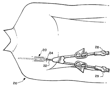
[0010] Figure 1 is a schematic representation depicting a medical device in
accordance with the invention for percutaneously implanting a catheter for an
exemplary
hemodialysis application;
[0011] Figure 2 is an isometric view of a preferred catheter assembly in
accordance
with the invention;
[0012] Figure 3 is an exploded view of the assembly of Figure 2 showing a
catheter in
phantom together with a sleeve intended for subcutaneous implantation, a
toroidal seal
member for mounting in the sleeve, a layer of porous material for mounting
around the sleeve
outer surface, and a disposable protective sheath temporarily mounted around
the porous
layer;
[0013] Figure 4 is a sectional view taken substantially along the plane 4-4 of
Figure 2;
[0014] Figure 5 is a plan view of the protective sheath;
[0015] Figure 6 is a sectional view taken substantially along the plane 6-6 of
Figure 5
particularly showing a performed score line;
[0016] Figures 7-9 show successive steps in an exemplary procedure for
implanting
and utilizing the cathetee assembly in accordance with the invention;
[0017] Figures 10-12 show successive steps in an exemplary procedure for
replacing
an implanted catheter; and
[0018] Figure 13 shows a cross-sectional view of the catheter assembly as
implanted
with the porous layer adjacent the patient's dermis.
CA 02637512 2008-07-16
WO 2007/089700 PCT/US2007/002399
DETAILED DESCRIPTION
[0019] Various medical regimens relating, for example, to hemodialysis drug
infusion,
plasmapheresis, etc., use a percutaneously implanted catheter for delivering
fluid to or
extracting fluid from an interior body site. The present invention is directed
to a method and
apparatus for facilitating the implantation and utilization of a percutaneous
catheter and for
facilitating the positioning, repositioning, and replacement, or exchange, of
the catheter.
[0020] Figure 1 schematically depicts an apparatus, or assembly, 20 in
accordance
with the invention which is subcutaneously implanted for allowing a catheter
22 to extend
percutaneously through an incision 24 in a patient 26 undergoing an exemplary
hemodialysis
procedure. In such a procedure, a dual lumen catheter 22 is typically used
with the two
lumen respectively coupled to separate exterior flow couplers 28 and 29.
[0021] Attention is now directed to Figures 2-4 which depict a preferred
catheter
assembly 20 in accordance with the present invention. The assembly 20 is
comprised of an
elongate sleeve 30 formed by a sleeve wall 32 having a peripheral outer
surface 34 and a
peripheral inner surface 36. The inner surface 36 surrounds a passageway 38
extending
from a first, or proximal, end 40 to a second, or distal, end 42.. The sleeve
30 is shown
mounted on a catheter 22 extending through the passageway 38. The catheter
outer surface
44 and passageway wall surface 36 are closely dimensioned but the gap
therebetween is
sufficient to enable the catheter to slide longitudinally in the passageway
28.
[0022] A layer 50 of porous material, e.g., titanium mesh, -as described in
said US
Application 10/821,383, is mounted on the sleeve outer surface 34 close to the
sleeve
proximal end 40. In use, it is intended that the sleeve distal end 42 be
inserted through the
incision 24 in the patient's skin sufficiently to position the sleeve proximal
end 40 and the
porous layer 50 just beneath the patient's epidermis skin layer 52 and in
contact with the
6
CA 02637512 2008-07-16
WO 2007/089700 PCT/US2007/002399
patient's dermis layer 54 (Figure 13). Note that the porous layer 50 is
preferably oriented
diagonally with respect to the axis of sleeve 30 to better conform to the
patient's skin contour
(Figure 13). This orientation optimizes contact between the porous layer 50
and the patient's
dermis 54 to promote, over time soft tissue ingrowth into the porous layer.
This tissue
ingrowth acts to form an infection resistant barrier around sleeve 30. This
barrier may be
enhanced by incorporating antimicrobial and/or anti-inflammatory constituents
into the porous
layer 50. For example, silver containing compounds and/or antibiotic eluting
coatings can be
used as antimicrobial agents and steroids can be used as anti-inflammatory
agents.
[0023] A protective sheath 60 (Figures 3-6) formed of thin flexible material
is
preferably mounted around sleeve 30 and porous layer 50 prior to use to avoid
tissue
abrasion damage when the sleeve distal end 42 is inserted through the
patient's incision. As
will be further discussed hereinafter, the sheath 60 is peeled away from the
sleeve 30 by the
physician as he/she inserts the sleeve through the incision. To facilitate
easily peeling, the
sheath 60 is preferably provided with, a pull tab 62 and a preformed score
line 64 along
which the sheath can readily separate.
[0024] As has previously been mentioned, in use, dermis tissue grows into the
porous
layer 50 to form a barrier preventing deleterious material from migrating into
the patient's
body along the sleeve outer surface 34. In order to prevent migration of
deleterious material
into the patient's body through the sleeve passageway 38 along the catheter
outer surface
44, a sealing means 70 is provided proximate to the sleeve inner surface 36. A
preferred
sealing means 70 is comprised of a toroidal member 72, preferably formed of a
polymeric
material such as silicone. The toroidal member 72 is comprised of a wall 73
having an outer
surface 74, and an inner surface 75 surrounding an interior bore 76
dimensioned to
accommodate the catheter 22_ At least one thin annular nib 80 is formed on the
toroidal
7
CA 02637512 2008-07-16
WO 2007/089700 PCT/US2007/002399
member inner surface 75 projecting radially into the bore 76 to bridge the gap
between the
sleeve inner surface 36 and the catheter outer surface 44. Each nib 80 is
configured to be
sufficiently axially compliant to wipe against the catheter outer surface 44
as the catheter 22
is slid through the bore 76. The nib 80 functions to prevent deleterious
material from
migrating along the catheter outer surface 44 into the patient's body while
allowing relative
sliding movement therebetween. For example only, an axial force on the
catheter of 1 pound
or less can be sufficient to slide the catheter relative to the nib 80.
[0025] Figures 7-9 schematically depict successive steps in an exemplary
procedure
for initially implanting the catheter assembly 20 shown in Figures 1-6; i.e.,
[0026] Figure 7 shows the use of a conventional tunneler tool 200 being
inserted through a patient's incision 202 to form a tunnel through which the
distal end of a
catheter 22 is pulled by the proximal end of tool 200;
[0027] Figure 8 shows the catheter assembly 20 with the sleeve distal end 42
and protective sheath 60 being inserted through the incision 202; and
[0028] Figure 9 shows the catheter assembly 20 inserted further into the
incision for subcutaneously positioning the sleeve proximal end 40 and porous
layer 50 just
beneath the patient's epidermal skin layer 52 (Figure 13) and also shows the
protective
sheath 60 being peeled away (as a consequence of the physician pulling tab 62)
from the
sleeve 30 so that exposed the porous layer 50 is e to the patient's dermis
layer 54.
[0029] Figures 10-12 schematically depict successive steps in an exemplary
procedure for removing an old implanted catheter 22 through the sleeve 30 and
replacing it
with a new catheter 22N; i.e.,
[0030] Figure 10 shows the old catheter 22 being withdrawn, along a guide
wire 210 which is inserted through the catheter prior to initiating the
procedure;
8
CA 02637512 2008-07-16
WO 2007/089700 PCT/US2007/002399
[0031] Figure 11 shows the new catheter 22N being inserted along the guide
wire 210 through the incision 202. Note that the distal end of the new
catheter 22N
preferably carries a disposable protective sheath 212, similar to
aforementioned sheath 60, to
minimize insult to the tissue adjacent to the incision; and
[0032] Figure 12 shows the subcutaneous sleeve 30 with the sheath 212 being
peeled away from the new catheter 22N. The guide wire 210 facilitates the
movement of the
catheter distal end through the steeve passageway 38 and past the annular
sealing nib 80 to
its intended destination site. Once the physician has satisfactorily
positioned and oriented
the catheter, the guide wire 210 can be withdrawn and the exterior catheter
portion can be
adhered to the patient's outer skin, e.g., by tape or sutures.
[0033] Figure 13 shows a cross-section of the subcutaneously installed
catheter
assembly 20 resulting from the steps represented in Figures 9 and 12 with the
porous layer
50 contacting the patient's dermis layer 54 to promote tissue ingrowth.
[0034] From the foregoing, it should now be understood that a catheter
assembly has
been provided intended for subcutaneous implantation and particularly
configured to facilitate
the positioning, repositioning, and/or replacement of a percutaneous catheter.
Although only
a limited number of structural embodiments have been described, it is
recognized that
various modifications and alterations will occur to persons skilled in the art
which fall within
the spirit and intended scope of the invention as defined by the appended
claims.
9