Note: Descriptions are shown in the official language in which they were submitted.
CA 02637525 2008-07-14
SURGICAL METHODS AND DEVICES WITH MOVEMENT ASSISTANCE
FIELD OF THE INVENTION
[0001] The present invention relates to surgical devices useful for moving
tissue and/or effecting
movement of device relative to tissue, and particularly through hollow organs
in a patient.
BACKGROUND OF THE INVENTION
[0002] Many surgical procedures require the movement or dissection of tissue,
or the movement
of a device relative to tissue. Space constraints as well as the relative
remoteness of a distal end
of a surgical tool from the surgeon can make it difficult to move tissue,
particularly in
endoscopic procedures that require surgical instruments to traverse a tortuous
pathway though a
tubular organ such as the colon. In some surgical procedures, particularly in
laparoscopic and
endoscopic procedures, movement of the surgical device can be challenging
because it is located
in a relatively constrained space that is remote from the surgeon. For
example, it can be difficult
for an endoscope to follow certain curves within the colon. Accordingly, there
is a need for
devices that conveniently and effectively enable the movement of tissue and/or
the movement of
surgical tools relative to tissue.
SUMMARY OF THE INVENTION
[0003] The present invention provides methods and devices to facilitate the
movement of
surgical devices through tortuous passageways (e.g., the colon) in the body.
In one aspect an
endoscopic device comprises an elongate insertion element adapted to be placed
within a
patient's body. A tissue engaging section is appended to at least a portion of
the insertion
element, and the tissue engaging section has an outer wall with a plurality of
openings formed
therein that communicate with a hollow chamber defined by the outer wall. In
one embodiment
the hollow chamber is configured to communicate with at least one of a vacuum
source and an
irrigation source. The device may also have a porous fabric extending over at
least a portion of
the tissue engaging section. The tissue engaging section can be configured to
move relative to
the insertion section or it can be configured to move only with the insertion
section. In one
embodiment the insertion section is a surgical tool for placement within the
body, such as an
endoscope.
1
CA 02637525 2008-07-14
[0004] In one embodiment the tissue engaging section is an accessory channel
that is appended
to an endoscope such that it can move independent of the endoscope. In another
embodiment the
tissue engaging section is a member that is appended to an endoscope, such as
in an interference
fit, that it is not movable relative to the endoscope.
[0005] In another aspect methods of moving a surgical device through a
passageway in the body
are provided. For example, a method of advancing a surgical instrument through
a body lumen
can include providing an elongate surgical instrument having an insertion
portion that has
appended thereto a tissue engaging section having an outer wall with a
plurality of openings
formed therein. The method further includes inserting the insertion portion
into a hollow body
lumen having a tortuous path; communicating a vacuum force to the tissue
engaging section such
that tissue of the body lumen is drawn against the tissue engaging section;
releasing the vacuum
force and moving the insertion portion within the body lumen; and repeating
the steps of
communicating the vacuum force, releasing the vacuum force and moving the
elongate insertion
portion to navigate the elongate insertion portion through the body lumen.
BRIEF DESCRIPTION OF THE DRAWINGS
[0006] The invention will be more fully understood from the following detailed
description
taken in conjunction with the accompanying drawings, in which:
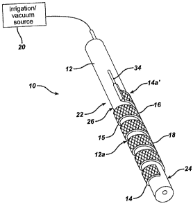
[0007] FIG. 1 is a perspective view of a portion of an endoscopic device
according to one
embodiment of the invention having a stationary tissue engaging section
appended thereto and
porous fabric covering part of the tissue engaging section;
[0008] FIG. 2 is a perspective view of a tissue engaging section useful with
the endoscopic
device of the type shown in FIG. 1;
[0009] FIG. 3 is a partial sectional view of a portion of a tissue engaging
section of the type
shown in FIG. 2;
[0010] FIG. 4 is a perspective view of a portion of an endoscopic device
according to another
embodiment of the invention having a movable tissue engaging section appended
thereto and
porous fabric covering part of the tissue engaging section;
2
CA 02637525 2008-07-14
[0011] FIG. 4A is a perspective view of the endoscopic device of FIG. 4 with
the tissue engaging
section in a more distal position;
[0012] FIG. 5 is an exploded view of the endoscopic device of FIG. 4;
[0013] FIG. 6A schematically illustrates an endoscopic device of the type
shown in FIG. 1 in a
first position in use during an endoscopic procedure;
[0014] FIG. 6B schematically illustrates an endoscopic device of the type
shown in FIG. 1 in a
second position in use during an endoscopic procedure;
[0015] FIG. 7A schematically illustrates an endoscopic device of the type
shown in FIG. 4 in a
first position in use during an endoscopic procedure; and
[0016] FIG. 7B schematically illustrates an endoscopic device of the type
shown in FIG. 4 in a
second position in use during an endoscopic procedure.
DETAILED DESCRIPTION OF THE INVENTION
[0017] Certain exemplary embodiments will now be described to provide an
overall
understanding of the principles of the structure, function, manufacture, and
use of the devices
and methods disclosed herein. One or more examples of these embodiments are
illustrated in the
accompanying drawings. Those skilled in the art will understand that the
devices and methods
specifically described herein and illustrated in the accompanying drawings are
non-limiting
exemplary embodiments and that the scope of the present invention is defined
solely by the
claims. The features illustrated or described in connection with one exemplary
embodiment may
be combined with the features of other embodiments. Such modifications and
variations are
intended to be included within the scope of the present invention.
[0018] The present invention generally provides devices and methods for moving
tissue and/or
moving the devices relative to the tissue during a surgical procedure. While
the devices and
methods disclosed herein can be used in conventional, open surgical
procedures, they are
particularly useful in minimally invasive surgical procedures, particularly
endoscopic
procedures. The principles described herein can be applicable to the
particular types of tools
described herein, and to a variety of other surgical tools having similar
functions. In addition,
3
CA 02637525 2008-07-14
the tools can be used alone in a surgical procedure, or they can be used in
conjunction with other
devices that facilitate minimally invasive surgical procedures. A particularly
useful aspect of the
systems and devices disclosed herein is that they enable movement and
manipulation of a
surgical instrument through a pathway in the body. That is, the invention
enables passage of a
device through a pathway in the body such that it is able to move relative to
the body tissue and
pass through regions of the body that can be difficult to traverse, such as
tortuous organs like the
colon.
[0019] The invention is described herein with reference to an endoscope that
is to be moved
through an organ in the body. However, a person skilled in the art will
understand that the
invention is applicable to a variety of other surgical tools that must be
passed through
passageway in the body, such as hollow organs, during a surgical procedure and
particularly
during minimally invasive surgical procedures such as endoscopic procedures.
[0020] A person skilled in the art will appreciate that the present invention
has application in
conventional endoscopic and open surgical instrumentation as well application
in robotic-
assisted surgery.
[0021] FIGS. 1-3 illustrate one embodiment of a surgical device 10 that is
configured to
facilitate movement of a surgical instrument relative to tissue. As shown, the
device 10 includes
an endoscope 12 (only a portion of which is shown) and a tissue engaging
section 14 that is
appended a distal portion 12a of the endoscope 12. At least a portion of the
tissue engaging
section 14 includes a porous fabric 16 that covers at least a tissue
contacting surface 18 of the
tissue engaging section 14. The tissue engaging section 14 can include at its
proximal end 14a a
conduit 34 that is in communication with an irrigation and/or a vacuum source
20 that can be
part of or separate from the endoscope system.
[0022] As noted above, the invention is applicable to virtually any surgical
tool. In the event
that the surgical tool used with the invention is an endoscope is used, it can
be any flexible,
elongate member that is capable of being inserted into the body, such as
through a natural orifice.
For example, FIG. 1 shows an insertion portion 22 of an endoscope 12 that is
to be inserted into
a patient's body, such as through a natural orifice. At least a portion of the
endoscope is flexible
and the endoscope may have a stearable portion 24 at a distal end thereof.
4
CA 02637525 2008-07-14
[0023] The tissue engaging section 14 can take the form of virtually any
member that can be
appended to an outer surface 26 of the endoscope 12. Generally, the tissue
engaging section 14
is secured to the endoscope 12 in such a manner that it does not move
independent of the
endoscope. The tissue engaging section 14 thus can take a variety of forms
that enable it to
securely fit over the endoscope 12. In an exemplary embodiment, the tissue
engaging section 14
is in the form of a helical, ribbon-like member 15 having an outer tissue-
contacting surface 18
and an inner tool-contacting surface 28. The helices of the helical member 15
define a central
lumen 30 within which the endoscope can seat and be engaged by the tool-
contacting surface 28.
The outer and inner surfaces 18, 28 of the helical member define a hollow
chamber 32 (FIG. 3)
that is in fluid communication with an irrigation/vacuum source 20 through
conduit 34 that
extends proximally from the helical member 15. A plurality of holes 36 can be
formed in the
tissue contacting surface 18 in fluid communication with the hollow chamber 32
and thus
conduit 34 and irrigation/vacuum source 20. Fluid can be passed through
conduit 34 and out of
holes 36, or a vacuum force can be drawn through the holes 36, as will be
explained below.
[0024] The helical member 15 can be applied to the endoscope by a variety of
techniques that
will enable it to remain secured to the endoscope and unable to move
independent of the
endoscope. In one example, the helical member 15 is appended to the endoscope
by an
interference fit. This can be effected by forming the helical member 15 from a
material that is at
least somewhat elastic (e.g., a superelastic alloy or a shape memory
material). Moreover, the
inner diameter of the lumen 30 when the helical member 15 is in a relaxed
condition can be
slightly less than the outer diameter of the endoscope 12. A force can be
applied to the helical
member 15, such as by axially compressing the helical member 15, to increase
the inner diameter
of the lumen 30. The helical member 15 can then be placed over the endoscope
12 in an
appropriate location and the force is removed, allowing the inner diameter of
the helical member
15 to decrease and engage the endoscope in an interference fit.
[0025] The tissue engaging section 14 can be applied to the endoscope 12 at
various appropriate
locations. Generally, however, the tissue engaging section 14 is applied at a
distal portion of the
endoscope 12. In one example, as shown in FIG. 1, the tissue engaging section
14 is applied
proximal to the distal most end 38. In one embodiment the tissue engaging
section 14 is applied
just a proximal to stearable portion 24.
CA 02637525 2008-07-14
[0026] As noted above, a porous fabric 16 extends over at least a portion of
the tissue contacting
surface 18 of the tissue engaging section 14. The material from which the
porous fabric 16 can
be made of virtually any material that is biocompatible, having properties
that enable an outer
surface of the fabric to contact tissue in such a way that there is
significant friction between the
contact tissue and the fabric and any device over which the fabric is applied.
In one
embodiment, the fabric material is a porous material such as a mesh material,
which can be
woven or non-woven. The material from which the mesh is formed can include a
variety of
synthetic and non-synthetic materials. Examples of synthetic materials include
polymers, such
as polypropylene, polyethylene, polyester, polytetrafluoroethylene, and nylon.
Examples of non-
synthetic mesh materials include, but are not limited to silk, cotton, and
stainless steel.
[0027] FIGS. 4-5 illustrate another embodiment of a surgical device 100 that
is configured to
facilitate movement of a surgical instrument relative to tissue. As shown, the
device 100
includes an endoscope 112 (only a portion of which is shown) and a tissue
engaging section 114
that is appended to a distal portion 112a of the endoscope. As explained
below, the tissue
engaging section 114 is of a type that is moveable with respect to the
endoscope 112. At least a
portion of the tissue engaging section 114 includes a porous fabric 116 that
covers at least a
tissue contacting surface 118 of the tissue engaging section 114. The tissue
engaging section 114
includes a mating element 140 that mates with a corresponding mating
receptacle 142 on the
endoscope to enable the tissue engaging section 114 to engage the endoscope
112 and to move
relative to the endoscope. The tissue engaging section 114 may include a
proximal end (not
shown) or a conduit (not shown) extending from the proximal end that is in
fluid communication
with an irrigation and/or vacuum source (not shown) that can be part of or
separate from the
endoscope system.
[0028] The endoscope 112 can be of the type described above with respect to
FIGS. 1-3.
However, as shown in FIGS. 4-5, the endoscope 112 includes a mating receptacle
142 that is
configured to mate with a corresponding mating element 140 on the tissue
engaging section 114
to enable the tissue engaging section 114 to be appended to the endoscope 112
in such a way that
the tissue engaging section and the endoscope are able to move independent of
one another.
Although illustrated as a female mating receptacle, one skilled in the art
will understand that the
mating receptacle 142 of the endoscope 112 can alternatively be a male-type
member. Similarly,
6
CA 02637525 2008-07-14
while the mating element of the tissue engaging section is illustrated as a
male element it can
alternatively be a female element. In the illustrated embodiment, mating
receptacle 142 is in the
form of a C-shaped channel or track 150 that is configured to receive a
complimentary mating
element 140 of the tissue engaging section 114, such as a T-shaped member 152.
[0029] The tissue engaging section 114 can be in the form of an accessory
member 154 that is
appended to the endoscope 112, such as an accessory channel of an endoscope,
in a manner such
that it is able to move relative to the endoscope. The accessory member 154
can take a variety of
forms. However, like the endoscope, the accessory member 154 can be a thin,
elongate and
flexible member that is capable of being inserted into a natural orifice of a
patient. In one
embodiment, as shown in FIGS. 4-5 the accessory member 154 can be a flexible,
elongate
tubular member having mating element 140 appended to a bottom portion thereof
An outer wall
156 of the accessory member 154 defines a lumen (not shown) that extends
within the accessory
member 154 and is in fluid communication with an irrigation/vacuum source (not
shown)
directly or through another conduit (not shown).
[0030] A distal end 154a of the accessory member 154 can include a plurality
of holes 158 in
fluid communication with the lumen (not shown) disposed within the accessory
member. In one
embodiment, the distal end 157 of the accessory member 154 is closed. The
holes 158 are
constructed such that fluid can be passed through the accessory member 154 and
out of holes 158
or a vacuum force can be drawn through the holes 158 as will be explained
below.
[0031] As described above with respect to FIGS. 1-3, a fabric 116 can cover a
tissue contacting
outer surface 156 of the accessory member 154. The 116 fabric can be a mesh
material of the
type described above with respect to FIGS. 1-3.
[0032] One skilled in the art will appreciate that the devices described
herein are applicable to a
variety of surgical procedures in which a surgical device must be advanced
through the body of a
patient along a relatively long and potentially tortuous pathway. Exemplary
techniques for using
the devices described herein will be described in the context of an endoscopic
procedure in
which an endoscope traverses a portion of the colon.
[0033] FIGS. 6A and 6B illustrate the use of a surgical device 10 of the type
shown in FIGS. 1-3
7
CA 02637525 2008-07-14
in a procedure in which the endoscope 12 enters a patient through the anus 200
and is passed into
the colon 202. As shown in FIG. 6A, the endoscope 12 is passed through the
rectum 204 which,
normally after slightly more than 90 right hand turn, leads to the sigmoid
colon 206. Because it
can be difficult to maneuver an endoscope through the tortuous pathway leading
to the sigmoid
colon 206, for example, the surgical device 10 of the invention can assist in
this passage. That is,
as a turn in the passageway is encountered, tissue tends to bunch up against
the distal end of the
endoscope, making further distal advancement of the endoscope difficult,
particularly when it is
necessary to make a turn.
[0034] Thus, as shown in FIG 6A, during passage of an endoscope through the
colon, the
endoscope encounters a portion of the passageway, e.g., leading to the sigmoid
colon 206, that
requires a difficult maneuver such as a sharp turn. At this point, using the
device 10, suction can
be applied to the device, drawing a vacuum through holes 36 in helical member
15 (FIGS. 1-3).
The results in tissue being drawn to the helical member 15. Thus, as shown in
FIG. 6A, as
suction is applied, the drawing of tissue against the helical member 15 causes
the normal
passageway of the colon in the vicinity of the sigmoid colon 206 to become
straighter than
normal. (Compare the straightened passageway shown in FIG. 6A in the vicinity
of the sigmoid
colon 206 to the normal anatomy of the same region shown in FIG. 6B.) The
device 10 can
assist in what is referred to as the "push-pull" technique, in which a surgeon
attempts to hook the
colon and then pulls the endoscope back in an attempt to straighten the lumen
of the colon. The
suction applied by the device 10 aids the surgeon in grasping and maintaining
control of the
colon rendering the push-pull technique more reliable. Once the passageway is
straightened as a
result of applying the vacuum force and pulling the endoscope back to
straighten the colon, the
vacuum can be withdrawn or reduced and, as shown in FIG. 6B, the endoscope 12
is further
advanced. Optionally, an irrigation fluid can be passed through holes 36 after
the vacuum is
withdrawn and during or after advancement of the endoscope 12 to reduce
friction between the
fabric that covers the helical member and the tissue. The process of applying
vacuum force,
removing or reducing the vacuum, applying irrigation to reduce friction, and
advancing the
endoscope can be repeated as necessary as the endoscope is advanced from the
descending colon
208 to the transverse colon 210.
[0035] FIGS. 7A and 7B illustrate the use of a surgical device 100 in a
similar surgical
8
CA 02637525 2008-07-14
procedure that requires passage of an endoscope 112 through the colon 202.
Similar to the
procedure described above, the endoscope 112 enters the patient through the
anus 200 and passes
through the rectum 204 into the sigmoid colon 206 as shown in FIG. 7A. At the
junction of the
sigmoid colon 206 and the descending colon 208, the passageway within the
colon becomes
tortuous as the colon makes a sharp (greater than 90 ) left turn into the
descending colon 208. At
a juncture such as this, where endoscope advancement becomes difficult as
tissue bunches up
against the distal end of the endoscope, suction can be applied to the device
100 by drawing a
vacuum through accessory member 154, thus anchoring the endoscope to the
passageway to
some extent. At this point, the endoscope 112 can be advanced distally beyond
the accessory
member 154 as shown in FIG. 7B. Optionally, an irrigation fluid can be passed
through holes
158 in the accessory member 154 after the vacuum is withdrawn and during or
after
advancement of the endoscope 112. Once the endoscope 112 is advanced beyond
the accessory
member 154, the accessory member can be advanced distally so that it is
approximately adjacent
the distal end of the endoscope (such as shown in FIG. 7A). This procedure can
be repeated as
necessary to advance the endoscope.
[0036] The devices disclosed herein can be designed to be disposed of after a
single use, or they
can be designed to be used multiple times. In either case, however, the device
can be
reconditioned for reuse after at least one use. Reconditioning can include any
combination of the
steps of disassembly of the device, followed by cleaning or replacement of
particular pieces, and
subsequent reassembly. In particular, the device can be disassembled, and any
number of the
particular pieces or parts of the device can be selectively replaced or
removed in any
combination. Upon cleaning and/or replacement of particular parts, the device
can be
reassembled for subsequent use either at a reconditioning facility, or by a
surgical team
immediately prior to a surgical procedure. Those skilled in the art will
appreciate that
reconditioning of a device can utilize a variety of techniques for
disassembly,
cleaning/replacement, and reassembly. Use of such techniques, and the
resulting reconditioned
device, are all within the scope of the present application.
[0037] Preferably, the invention described herein will be processed before
surgery. First, a new
or used instrument is obtained and if necessary cleaned. The instrument can
then be sterilized.
In one sterilization technique, the instrument is placed in a closed and
sealed container, such as a
9
CA 02637525 2015-06-12
plastic or TYVEX bag. The container and instrument are then placed in a field
of radiation that
can penetrate the container, such as gamma radiation, x-rays, or high-energy
electrons. The
radiation kills bacteria on the instrument and in the container. The
sterilized instrument can then
be stored in the sterile container. The sealed container keeps the instrument
sterile until it is
opened in the medical facility.
100381 It is preferred that device is sterilized. This can be done by any
number of ways known
to those skilled in the art including beta or gamma radiation, ethylene oxide,
steam.
100391 One skilled in the art will appreciate further features and advantages
of the invention
based on the above-described embodiments. Accordingly, the invention is not to
be limited by
what has been particularly shown and described, except as indicated by the
appended claims.